unifying theory of hypoxia tolerance: molecular/metabolic defense and rescue mechanisms for
TRANSCRIPT

Proc. Natl. Acad. Sci. USAVol. 93, pp. 9493-9498, September 1996Biochemistry
Unifying theory of hypoxia tolerance: Molecular/metabolicdefense and rescue mechanisms for surviving oxygen lack
(oxygen sensing/hypoxia defense/turtle hepatocytes/turtle brain)
P. W. HOCHACHKA, L. T. BUCK*, C. J. DOLLt, AND S. C. LANDtDepartment of Zoology and the Sports Medicine Division, University of British Columbia, Vancouver BC, Canada V6T 1Z4
Communicated by Louis Sokoloff, National Institute of Mental Health, Bethesda, MD, June 21, 1996 (received for review January 29, 1996)
ABSTRACT We develop a unifying theory of hypoxiatolerance based on information from two cell level models(brain cortical cells and isolated hepatocytes) from the highlyanoxia tolerant aquatic turtle and from other more hypoxiasensitive systems. We propose that the response of hypoxiatolerant systems to oxygen lack occurs in two phases (defenseand rescue). The first lines of defense against hypoxia includea balanced suppression of ATP-demand and ATP-supplypathways; this regulation stabilizes (adenylates) at newsteady-state levels even while ATP turnover rates greatlydecline. The ATP demands of ion pumping are down-regulatedby generalized "channel" arrest in hepatocytes and by "spike"arrest in neurons. Hypoxic ATP demands of protein synthesisare down-regulated probably by translational arrest. In hy-poxia sensitive cells this translational arrest seems irrevers-ible, but hypoxia-tolerant systems activate "rescue" mecha-nisms if the period of oxygen lack is extended by preferentiallyregulating the expression of several proteins. In these cells, acascade ofprocesses underpinning hypoxia rescue and defensebegins with an oxygen sensor (a heme protein) and a signal-transduction pathway, which leads to significant gene-basedmetabolic reprogramming-the rescue process-with main-tained down-regulation of energy-demand and energy-supplypathways in metabolism throughout the hypoxic period. Thisrecent work begins to clarify how normoxic maintenance ATPturnover rates can be drastically (10-fold) down-regulated toa new hypometabolic steady state, which is prerequisite forsurviving prolonged hypoxia or anoxia. The implications ofthese developments are extensive in biology and medicine.
energy turnover supplies the greatest protection against, andhence, advantage in, hypoxia. The immense advantage of thisdefense strategy is widely appreciated by many biologists (4, 5,11-13). In one of his last personal communications to one ofthe authors (P.W.H.), the great comparative physiologist KjellJohansen referred to this strategy as "turning down to the pilotlight" and he, like many earlier workers, was acutely aware ofits relative importance. Although recognized as a kind ofhallmark of reversible entry into and return from states ofsevere 02 deprivation, a number of unexplained problems haveremained. In particular, it has not been clear (i) how cells/tissues "know" when to turn on their hypoxia defense mech-anisms, (ii) which pathways of ATP demand and ATP supplyare down-regulated or by how much, (iii) how membraneelectrochemical gradients are stabilized, and (iv) what gene-expression and protein-expression level adjustments are in-volved in hypoxic reorganization of cell structure and function.Recent studies of a well-known vertebrate "facultative anaer-obe," the aquatic turtle, used brain cortical slices to probeelectrophysiological properties of neurons under anoxia (16-19) and isolated liver hepatocytes to probe cell level biochem-ical responses to anoxia (20-24). When integrated with inde-pendent lines of research in other laboratories, these studiessupply the raw material for developing a synthetic cell-levelmodel or general hypothesis of hypoxia tolerance, which goesa long way to answering the above unresolved questions.
How Cells Detect When Oxygen Becomes Limiting
Cell Level Model of Hypoxia Tolerance
Biologists have long known that certain vertebrate species haveevolved capabilities for surviving prolonged periods with lim-ited supplies of oxygen. Studies of such species [some so anoxiatolerant that they are referred to as "facultative" anaerobes(1)] have revealed several widely used strategies of hypoxiaadaptation. Two of the most significant of these are: (i) severedown-regulation of energy turnover (2-7) and (ii) up-regulation of energetic efficiency of ATP-producing pathways(8). The latter involve stoichiometric efficiencies. In hypoxiaadaptation, pathways that maximize the yield of ATP per molOf 02 are favored, whereas in anoxia adaptation, anaerobicpathways are favored that maximize the yield of ATP per molof HI formed in the fermentation (9, 10). In hypoxia adap-tation, the ratio of anaerobic/aerobic metabolic potentials maybe up-regulated, coincident with up-regulation of [glycogen](fermentable substrate) and of buffering capacities (2). Al-though all these mechanisms may be useful in survivinghypoxia, evaluation of such defense strategies naturallyevolved by hypoxia-tolerant animals shows that suppression of
Traditionally, biologists' views of cell level responses to 02limitation are formalized in the concept of the Pasteur effect;as ATP generation by oxidative phosphorylation begins to falloff due to 02 lack, the energetic deficit is made up by activationof anaerobic ATP supply pathways. In the case of turtle livercells, this kind of 02 sensing would mean activation of anaer-obic glycolysis (since this is the only known mechanism forATP generation without 02 in these cells). Two observationsargue persuasively against this as the means for sensinghypoxia in tolerant cells. First, hypoxia-tolerant cells/tissuesuse anaerobic metabolism not to make up energy deficits butto sustain a reduced energy turnover state instead (3, 4, 11, 14,15). Second, responses to falling [02] begin at concentrations(3) much higher than the Km(02) for mitochondria (termedoxygen conformity). In fact, it was the observation of oxygenconformity that first led workers in this area to postulatemechanisms more widespread than the erythropoietin (EPO)
Abbreviations: EPO, erythropoietin; EFla, elongation factor la;PGK, phosphoglycerate kinase; LDH, lactate dehydrogenase; HIFi,hypoxia-inducible factor 1.*Present address: Anesthesiology Department, University of Califor-nia, San Francisco, CA 94143-0542.
tPresent address: Department of Biology, University of California,Santa Barbara, CA 93106-9610.*Present address: Marine Biological Laboratory, Woods Hole, MA02543.
9493
The publication costs of this article were defrayed in part by page chargepayment. This article must therefore be hereby marked "advertisement" inaccordance with 18 U.S.C. §1734 solely to indicate this fact.
9494 Biochemistry: Hochachka et al.
system (25) for sensing 02 as the cell level means for detectinghypoxia (10, 11, 13, 26) (see below).
Balancing ATP-Demand and ATP-Supply Pathways DuringHypoxia
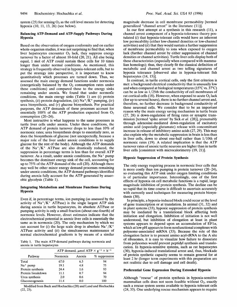
Based on the observation of oxygen conformity and on earlierwhole-organism studies, it was not surprising to find that, whenliver hepatocytes encounter 02 lack, they suppress energyturnover by a factor of almost 10-fold (20, 21). All else beingequal, 1 mol of ATP could sustain these cells for 10 timeslonger than under normal conditions. As mentioned, thisstrategy is frequently observed in hypoxia-tolerant cells, but toput the strategy into perspective, it is important to knowquantitatively which processes are turned down. Thus, weassessed the main energy demand functions under normoxia(energetically balanced by the 02 consumption rates underthese conditions) and compared these to the energy sinksremaining under anoxia. We found that under normoxicconditions, the main energy sinks (Table 1) are (i) proteinsynthesis, (ii) protein degradation, (iii) Na+/K+ pumping, (iv)urea biosynthesis, and (v) glucose biosynthesis. For practicalpurposes, the ATP demands of these processes account foressentially 100% of the ATP production expected from 02consumption (20-24).Most instructive is what happens to the same processes in
turtle liver cells under anoxia. Under these conditions, theATP demand of protein turnover drops to less than 10% ofnormoxic rates; urea biosynthesis drops to essentially zero, asdoes the biosynthesis of glucose (not unexpectedly, because amajor role of the liver under anoxic conditions is to supplyglucose for the rest of the body). Although the ATP demandsof the Na+/K+ ATPase are also drastically reduced, thesuppression in percentage terms is less than for overall ATPturnover. As a result, under anoxic conditions, the Na+ pumpbecomes the dominant energy sink of the cell, accounting forup to 75% of the ATP demand of the cell (20). Although theremay well be other minor energy demand processes remainingunder anoxic conditions, the ATP demand pathways identifiedduring anoxia fully account for the ATP generated by anaer-obic glycolysis (Table 1).
Integrating Metabolism and Membrane Functions DuringHypoxia
Even if, in percentage terms, ion pumping (as assessed by theactivity of Na+/K+ ATPase) is the single largest ATP sinkduring anoxia in turtle hepatocytes, its absolute ATPase orpumping activity is only a small fraction (about one-fourth) ofnormoxic levels. However, direct estimates indicate that theelectrochemical potential in anoxic liver cells is essentially thesame as in normoxia (20). The only mechanism by which wecan account for (i) the large scale drop in absolute Na+/K+ATPase activity and (ii) the simultaneous maintenance ofnormal electrochemical gradients is by means of a similar
Table 1. The main ATP-demand pathways during normoxia andanoxia in turtle hepatocytes
ATP demand, ,lmol ATP x g-1 x h-1
Pathway Normoxia Anoxia % suppressionTotal 67.0 6.3 94Na+ pump 19.1 4.8 75Protein synthesis 24.4 1.6 93Protein breakdown 11.1 0.7 94Urea synthesis 2.0 0.6 70
magnitude decrease in cell membrane permeability [termedgeneralized "channel arrest" in the literature (11)].
In our first attempt at a synthesis in this research (11), a
channel arrest component of a hypoxia-tolerance theory pos-
tulated (i) that hypoxia-tolerant cells would have an inherentlow permeability (either low-channel densities or low-channelactivities) and (ii) that they would sustain a further suppressionof membrane permeability to ions when exposed to oxygenlack (further channel arrest by either suppression of channeldensities or channel activities). Turtle liver cells display both ofthese characteristics (especially when compared with mamma-lian homologs); thus, they clearly fit the classical definition ofmetabolic and channel arrest as two telling signatures ofhypoxia tolerance [observed also in hypoxia-tolerant fishhepatocytes (14, 15)].
In contrast, in turtle cortical cells, only the first criterion ismet: a background electrical conductivity that is unusually lowand when compared at biological temperatures (15°C vs. 37°C)can be as low as 1/25th the conductivity of cell membranes ofrat cortical cells (18). However, when exposed to acute 02 lack(for up to several hours), there is no further channel arrest and,therefore, no further decrease in background conductivity ofthese neuronal cells. We consider that to be an importantreason why the main energy saving mechanism in turtle brain(27, 28) is down-regulation of firing rates or synaptic trans-mission [termed 'spike arrest' by Sick et al. (28)], presumablythrough adenosine-mediated down-regulation of excitatoryamino acid (especially glutamate) release with concomittantincrease in release of inhibitory amino acids (27, 29). This mayalso explain why the metabolic suppression in brain is less thanin liver cells, down to about one-half rather than 1/10th ofnormoxic rates (19). A related implication is that the ATPturnover rates of anoxic turtle neurons are higher than in turtleliver cells [consistent with microcalorimetry (19, 21)].
Hypoxic Suppression of Protein Synthesis
The only energy requiring process in normoxic liver cells thatis more costly than ion pumping is protein turnover (20-24),so evaluating this ATP sink under oxygen limiting conditionsis of particular importance. Interestingly, one of the firsteffects of hypoxia on cell metabolic functions is a rapid, largemagnitude inhibition of protein synthesis. The decline can beso rapid that its time course is difficult to ascertain accuratelywith currently used techniques for measuring protein biosyn-thesis (30).
In principle, a hypoxia-induced block could occur at the levelof gene transcription or at translation. In animal (31, 32) andin plant systems (33), hypoxic suppression of protein synthesismay be mediated by a translational block affecting bothinitiation and elongation. Inhibition of initiation is not wellunderstood, but inhibition of elongation at least in plantsystems appears to depend upon an accumulation of EFla,which at low pH appears to form nonfunctional complexes withpolysome-associated mRNA (33). Because the role of thiselongation factor is to present amino acyl-tRNA to the A siteof ribosomes, it is easy to visualize how failure to dissociatefrom polysomes would prevent peptidyl synthesis and translo-cation. In hypoxia-sensitive systems, such as rat hepatocytes(30), hypoxia-induced translational arrest and, thus, blockadeof protein synthetic capacity seems to remain general for atleast 2 hr (longer term experiments with this preparation are
not realistic due to cell damage and cell death).
Preferential Gene Expression During Extended Hypoxia
Although "rescue" of protein synthesis in hypoxia-sensitivemammalian cells does not seem feasible without 02, exactlysuch a rescue system seems available to hypoxia tolerant cells(24, 33). One underlying rescue mechanism appears to require
Gluconeogenesis 11.4 0.0 100
Modified from Buck and Hochachka (20) and Land and Hochachka(22, 23).
Proc. Natl. Acad. Sci. USA 93 (1996)
Proc. Natl. Acad. Sci. USA 93 (1996) 9495
the overproduction of key elongation factors such as EFla;with sustained hypoxia, the latter is overexpressed and accu-mulates throughout the stress period. Although the mecha-nism for selective translation (of only specific messages) is notwell understood, this rescue mechanism is known to prefer-entially favor expression of glycolytic enzymes (33) whoseactivity must be sustained to survive 02 lack.When we turned our attention to this issue in turtle hepa-
tocytes we found (23, 24) that the situation was more complex.Under conditions of prolonged 02 lack, the expression of fourproteins in turtle hepatocytes was preferentially up-regulated,whereas the expression of five proteins was preferentiallydown-regulated. The hypoxia dependence of this system ap-peared similar to 02 sensing and regulatory pathways in EPObiosynthesis (34-40). The field of EPO research developedfour experimental criteria for discriminating between 02 sens-ing and signal transduction pathways vs. simple 02-dependentmetabolic pathways. First, in the former, the response shouldbe negated by normoxia even in the presence of metabolicpoisons such as cyanide-as observed in the turtle hepatocytegene expression changes in anoxia. Second, in heme protein02-sensing pathways, Co2+ or Ni2+, which lock heme proteinsin their deoxy conformation, should mimic anoxia effects-asobserved in turtle hepatocyte gene expression changes. Third,in heme protein 02-sensing pathways, carbon monoxide, whichlocks heme proteins in oxy conformation, should reverseanoxia effects-also observed in the turtle hepatocyte studies.Finally, heme synthesis inhibitors should abrogate the effectsof Co2+ or Ni2+ in heme protein based 02-sensing pathways-again observed in the turtle hepatocyte protein expressionchanges in anoxia (23, 24).These studies, inspired by developments in EPO research,
led to the conclusion that a putative heme protein based02-sensing and signal transduction system in turtle hepato-cytes serves to activate the expression of a group of severalproteins during extended periods of 02 limitation, as well as tosimultaneously further slow down the expression of anothergroup of several proteins. The 02-sensing pathway in turtlehepatocytes is probably homologous to that found in rat livercells; in the latter (41, 42), hypoxia sensing and signal trans-duction seems to serve in negatively modulating the effects ofglucagon on gluconeogenic enzymes such as phosphoenolpyru-vate carboxykinase. Similar 02-sensing and signal transductioncontrol systems may be basic to the hypoxic up-regulation ofgenes for glycolytic enzymes (43), and to the hypoxic suppres-sion of Krebs cycle enzymes (44).So far, perhaps the most extensive analysis of the regulation
of a putative homologous 02-sensing system derives fromstudies of EPO regulation (34). In evaluating their postulate ofuniversal 02-sensing and response pathways, Firth et al. (35)recently confirmed that the genes for human phosphoglyceratekinase (PGK) 1 from human cancer cell lines and for mousemuscle type lactate dehydrogenase (LDH) A from mousefibroblasts are induced by hypoxia; expression is favored byCo2+ but not cyanide and blocked by protein synthesis inhi-bition. The cis-acting control sequences, necessary for thehypoxic PGK induction, are located in the 5' flanking regionof the PGK gene and are seemingly homologous to theso-called hypoxia-inducible factor 1 (HIF1)-binding site withinthe EPO enhancer (3' flanking region of the EPO gene), whichregulates EPO gene function (38-40). Similar studies of theLDH A 5' flanking (or promoter) region define three domainsrequired for hypoxia-regulated LDH expression. The first ofthese is an HIFl-binding site and is an absolute requirementfor hypoxia-sensitive function. Site two, 5' to the HIFi site, issimilar to a critical cooperative site in the EPO enhancer,whereas site three (3' to the HIF1 site) has the motif of acAMP response element (36). Interactions between all threesites in the LDH A promoter are required for the maximumhypoxic inducibility, but the physiological significance of in-
teractions between the HIF1 and cAMP response elementsites, between oxygen-initiated and cAMP-initiated signaltransduction pathways, remains to be clarified (36). Neverthe-less, the hypoxia-sensitive expression of each of these threeprotein systems (EPO, PGK, and LDH) and of glycolyticenzymes in other systems (37, 38) appears to be a classicaltwo-step or two-cycle system: the first cycle regulates theexpression of a key hypoxia-sensitive transcription factor(HIF1), whereas the second cycle of gene expression is me-diated by HIF1 and regulates the biosynthesis of EPO, PGK,LDH, and other glycolytic enzymes per se (37).
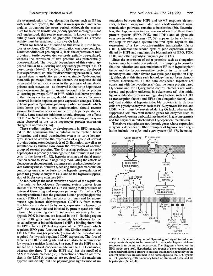
Since the expression of other proteins, such as elongationfactors, may be similarly regulated, it is tempting to considerthat the induction and accumulation of EFla in hypoxic planttissue and the hypoxia-sensitive proteins in turtle and rathepatocytes are under similar two-cycle gene regulation (Fig.1), although at this time such homology has not been demon-strated. Nevertheless, all the data considered together areconsistent with the hypotheses (i) that the heme protein based02 sensor and the 02-regulated control elements are wide-spread and possibly universal in eukaryotes; (ii) that initialhypoxia inducible proteins are regulatory factors, such as HIFM(a transcription factor) and EFla (an elongation factor); and(iii) that additional hypoxia inducible proteins in turtle livercells are glycolytic enzymes such as PGK, pyruvate kinase, andLDH, which must be sustained during 02 lack, whereas thesuppressed loci may well include genes for enzymes such asphosphoenolpyruvate carboxykinase involved in gluconeogenesisand for enzymes in mitochondrial 02-dependent metabolism.The above examples are not the only genes whose expression
is hypoxia dependent. Other examples of hypoxic gene regu-lation include the c-fos and c-jun system (45-47), hemeoxy-
Cobalt sensitivelOW[021
HYPOXICUP REGULATION
1°messengers
I.DNA3tend
PEPCK Krebs+ other cycle
glucogenic enzymesenzymes _
HYPOXICDOWN REGULATION
FIG. 1. Schematic diagram of 02-sensing and signal transductioncomponents thought to be involved in metabolic hypoxia defenseresponse in turtle and rat hepatocytes. The diagram is based on theEPO model (see text). Hypothetical two rounds of gene regulation areconnected with dotted arrows to indicate that in turtle hepatocytes thecontrol circuitries are assumed to be homologous to the EPO systemin EPO producing cells. Summary based on studies of turtle and rathepatocytes (24, 41, 42).
Biochemistry: Hochachka et al.
9496 Biochemistry: Hochachka et al.
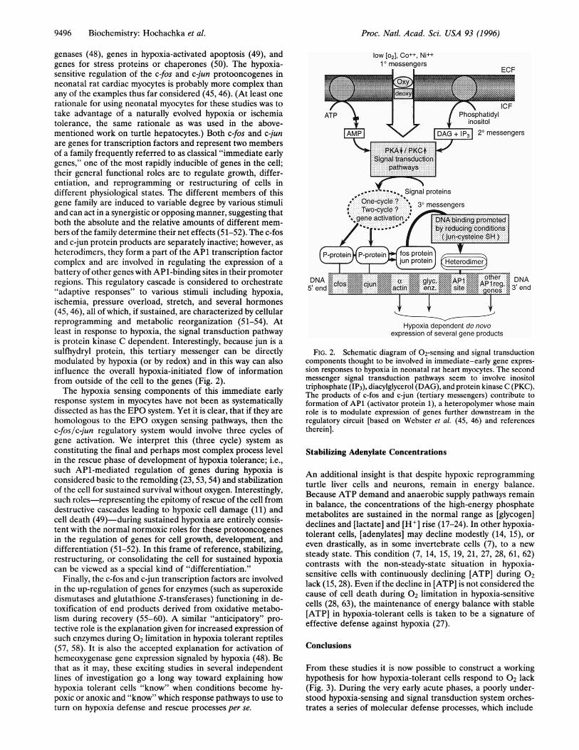
genases (48), genes in hypoxia-activated apoptosis (49), andgenes for stress proteins or chaperones (50). The hypoxia-sensitive regulation of the c-fos and c-jun protooncogenes inneonatal rat cardiac myocytes is probably more complex thanany of the examples thus far considered (45, 46). (At least onerationale for using neonatal myocytes for these studies was totake advantage of a naturally evolved hypoxia or ischemiatolerance, the same rationale as was used in the above-mentioned work on turtle hepatocytes.) Both c-fos and c-junare genes for transcription factors and represent two membersof a family frequently referred to as classical "immediate earlygenes," one of the most rapidly inducible of genes in the cell;their general functional roles are to regulate growth, differ-entiation, and reprogramming or restructuring of cells indifferent physiological states. The different members of thisgene family are induced to variable degree by various stimuliand can act in a synergistic or opposing manner, suggesting thatboth the absolute and the relative amounts of different mem-bers of the family determine their net effects (51-52). The c-fosand c-jun protein products are separately inactive; however, asheterodimers, they form a part of the AP1 transcription factorcomplex and are involved in regulating the expression of abattery of other genes with AP1-binding sites in their promoterregions. This regulatory cascade is considered to orchestrate"adaptive responses" to various stimuli including hypoxia,ischemia, pressure overload, stretch, and several hormones(45, 46), all of which, if sustained, are characterized by cellularreprogramming and metabolic reorganization (51-54). Atleast in response to hypoxia, the signal transduction pathwayis protein kinase C dependent. Interestingly, because jun is asulfhydryl protein, this tertiary messenger can be directlymodulated by hypoxia (or by redox) and in this way can alsoinfluence the overall hypoxia-initiated flow of informationfrom outside of the cell to the genes (Fig. 2).The hypoxia sensing components of this immediate early
response system in myocytes have not been as systematicallydissected as has the EPO system. Yet it is clear, that if they arehomologous to the EPO oxygen sensing pathways, then thec-fos/c-jun regulatory system would involve three cycles ofgene activation. We interpret this (three cycle) system asconstituting the final and perhaps most complex process levelin the rescue phase of development of hypoxia tolerance; i.e.,such APi-mediated regulation of genes during hypoxia isconsidered basic to the remolding (23, 53, 54) and stabilizationof the cell for sustained survival without oxygen. Interestingly,such roles-representing the epitomy of rescue of the cell fromdestructive cascades leading to hypoxic cell damage (11) andcell death (49)-during sustained hypoxia are entirely consis-tent with the normal normoxic roles for these protooncogenesin the regulation of genes for cell growth, development, anddifferentiation (51-52). In this frame of reference, stabilizing,restructuring, or consolidating the cell for sustained hypoxiacan be viewed as a special kind of "differentiation."
Finally, the c-fos and c-jun transcription factors are involvedin the up-regulation of genes for enzymes (such as superoxidedismutases and glutathione S-transferases) functioning in de-toxification of end products derived from oxidative metabo-lism during recovery (55-60). A similar "anticipatory" pro-tective role is the explanation given for increased expression ofsuch enzymes during 02 limitation in hypoxia tolerant reptiles(57, 58). It is also the accepted explanation for activation ofhemeoxygenase gene expression signaled by hypoxia (48). Bethat as it may, these exciting studies in several independentlines of investigation go a long way toward explaining howhypoxia tolerant cells "know" when conditions become hy-poxic or anoxic and "know" which response pathways to use toturn on hypoxia defense and rescue processes per se.
low [02], Co++, Ni++1° messengers
I I FCF
DNA3' end
Hypoxia dependent de novoexpression of several gene products
FIG. 2. Schematic diagram of 02-sensing and signal transductioncomponents thought to be involved in immediate-early gene expres-sion responses to hypoxia in neonatal rat heart myocytes. The secondmessenger signal transduction pathways seem to involve inositoltriphosphate (IP3), diacylglycerol (DAG), and protein kinase C (PKC).The products of c-fos and c-jun (tertiary messengers) contribute toformation of AP1 (activator protein 1), a heteropolymer whose mainrole is to modulate expression of genes further downstream in theregulatory circuit [based on Webster et al. (45, 46) and referencestherein].
Stabilizing Adenylate Concentrations
An additional insight is that despite hypoxic reprogrammingturtle liver cells and neurons, remain in energy balance.Because ATP demand and anaerobic supply pathways remainin balance, the concentrations of the high-energy phosphatemetabolites are sustained in the normal range as [glycogen]declines and [lactate] and [HI] rise (17-24). In other hypoxia-tolerant cells, [adenylates] may decline modestly (14, 15), oreven drastically, as in some invertebrate cells (7), to a newsteady state. This condition (7, 14, 15, 19, 21, 27, 28, 61, 62)contrasts with the non-steady-state situation in hypoxia-sensitive cells with continuously declining [ATP] during 02lack (15, 28). Even if the decline in [ATP] is not considered thecause of cell death during 02 limitation in hypoxia-sensitivecells (28, 63), the maintenance of energy balance with stable[ATP] in hypoxia-tolerant cells is taken to be a signature ofeffective defense against hypoxia (27).
Conclusions
From these studies it is now possible to construct a workinghypothesis for how hypoxia-tolerant cells respond to 02 lack(Fig. 3). During the very early acute phases, a poorly under-stood hypoxia-sensing and signal transduction system orches-trates a series of molecular defense processes, which include
Proc. Natl. Acad. Sci. USA 93 (1996)
Proc. Natl. Acad. Sci. USA 93 (1996) 9497
Defense Phase
Hypoxia sensing and signal transduction
Neurons: adenosinet glutamate releasel'spike' arrest; Na+K+ A4Pase 4J
eatcrest; Na+K+ ATPaselurea synthesis blockadeglucose synthesis blockadeproteolysis blockadeprotein synthesis blockade(translational arrest)
ATP demand = ATP supply
Suppression of ATPturnover rates
20 40
Rescue Phase
Round 3(?) production of tertiary messengers (fos & jun);stabilization(consolidation) for survival at 'pilot light' ATP turnover, ._
rates *
Round 2 HIF1 mediated activation of genes for sustained -survival at low ATP turnover (e.g. glycolyticenzymes); suppression of genes for lessrequired enzymes (e.g. Krebs cycle and
gluconeogenesis)
Accumulation of EFla: Rescue of specific -mRNA translational capacity *
Round 1 preferential activation of genes for *transcription factors (HIF) (& for *
EFl a + other elongation factors?) *
Hypoxia sensing and signal transduction *
mmM mm * a m - m
60 80 100
Time line (min) for Developing Hypoxia Tolerance
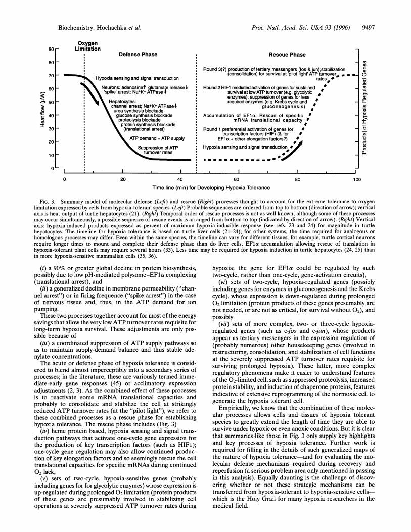
FIG. 3. Summary model of molecular defense (Left) and rescue (Right) processes thought to account for the extreme tolerance to oxygenlimitation expressed by cells from hypoxia-tolerant species. (Left) Probable sequences are ordered from top to bottom (direction of arrow); verticalaxis is heat output of turtle hepatocytes (21). (Right) Temporal order of rescue processes is not as well known; although some of these processesmay occur simultaneously, a possible sequence of rescue events is arranged from bottom to top (indicated by direction of arrow). (Right) Verticalaxis: hypoxia-induced products expressed as percent of maximum hypoxia-inducible response (see refs. 23 and 24) for magnitude in turtlehepatocytes. The timeline for hypoxia tolerance is based on turtle liver cells (21-24); for other systems, the time required for analogous or
homologous processes may differ. Even within the same species, the timeline can vary for different tissues; for example, turtle cortical neurons
require longer times to mount and complete their defense phase than do liver cells. EFla accumulation allowing rescue of translation inhypoxia-tolerant plant cells may require several hours (33). Less time may be required for hypoxia induction in turtle hepatocytes (24, 25) thanin more hypoxia-sensitive mammalian cells (35, 36).
(i) a 90% or greater global decline in protein biosynthesis,possibly due to low pH-mediated polysome-EFla complexing(translational arrest), and
(ii) a generalized decline in membrane permeability ("chan-nel arrest") or in firing frequence ("spike arrest") in the case
of nervous tissue and, thus, in the ATP demand for ionpumping.These two processes together account for most of the energy
savings that allow the very lowATP turnover rates requisite forlong-term hypoxia survival. These adjustments are only pos-sible because of
(iii) a coordinated suppression of ATP supply pathways soas to maintain supply-demand balance and thus stable ade-nylate concentrations.The acute or defense phase of hypoxia tolerance is consid-
ered to blend almost imperceptibly into a secondary series ofprocesses; in the literature, these are variously termed imme-diate-early gene responses (45) or acclimatory expressionadjustments (2, 3). As the combined effect of these processesis to reactivate some mRNA translational capacities andprobably to consolidate and stabilize the cell at strikinglyreduced ATP turnover rates (at the "pilot light"), we refer tothese combined processes as a rescue phase for establishinghypoxia tolerance. The rescue phase includes (Fig. 3)
(iv) heme protein based, hypoxia sensing and signal trans-duction pathways that activate one-cycle gene expression forthe production of key transcription factors (such as HIFM);one-cycle gene regulation may also allow continued produc-tion of key elongation factors and so seemingly rescue the celltranslational capacities for specific mRNAs during continued02 lack,
(v) sets of two-cycle, hypoxia-sensitive genes (probablyincluding genes for for glycolytic enzymes) whose expression isup-regulated during prolonged 02 limitation (protein productsof these genes are presumably involved in stabilizing celloperations at severely suppressed ATP turnover rates during
hypoxia; the gene for EFla could be regulated by suchtwo-cycle, rather than one-cycle, gene-activation circuits),
(vi) sets of two-cycle, hypoxia-regulated genes (possiblyincluding genes for enzymes in gluconeogenesis and the Krebscycle), whose expression is down-regulated during prolonged02 limitation (protein products of these genes presumably are
not needed, or are not as critical, for survival without 02), andpossibly
(vii) sets of more complex, two- or three-cycle hypoxia-regulated genes (such as c-fos and c-jun), whose productsappear as tertiary messengers in the expression regulation of(probably numerous) other housekeeping genes (involved inrestructuring, consolidation, and stabilization of cell functionsat the severely suppressed ATP turnover rates requisite forsurviving prolonged hypoxia). These latter, more complexregulatory phenomena make it easier to understand featuresof the 02-limited cell, such as suppressed proteolysis, increasedprotein stability, and induction of chaperone proteins, featuresindicative of extensive reprogramming of the normoxic cell togenerate the hypoxia tolerant cell.
Empirically, we know that the combination of these molec-ular processes allows cells and tissues of hypoxia tolerantspecies to greatly extend the length of time they are able tosurvive under hypoxic or even anoxic conditions. But it is clearthat summaries-like those in Fig. 3 only supply key highlightsand key processes of hypoxia tolerance. Further work isrequired for filling in the details of such generalized maps ofthe nature of hypoxia tolerance-and for evaluating the mo-
lecular defense mechanisms required during recovery andreperfusion (a serious problem area only mentioned in passingin this analysis). Equally daunting is the challenge of discov-ering whether or not these strategic mechanisms can betransferred from hypoxia-tolerant to hypoxia-sensitive cells-which is the Holy Grail for many hypoxia researchers in themedical field.
OxygenLimitation90 r
80t
701-
60
a
0
coI
50
401-
30
20
10 _
0
0
Cl)
a)CDa1)01)
0)cr.c
I0
C)200
0~
Biochemistry: Hochachka et al.
-ai
9498 Biochemistry: Hochachka et al.
We thank Dr. Michael Vayda for bringing his work on hypoxia-sensitive protein synthesis to our attention. This work was supportedby a National Sciences and Engineering Research Council ResearchGrant.
1. Storey, K. B. & Hochachka, P. W. (1974) J. Biol. Chem. 249,1423-1429.
2. Hochachka, P. W. & Somero, G. N. (1984) Biochemical Adapta-tion (Princeton Univ. Press, Princeton).
3. Hochachka, P. W. & Guppy, M. (1987) Metabolic Arrest and theControl of Biological Time (Harvard Univ. Press, Cambridge,MA).
4. Storey, K. B. & Storey, J. M. (1990) Q. Rev. Biol. 65, 145-193.5. Hand, S. C. (1993) in Surviving Hypoxia, eds. Hochachka, P. W.,
Lutz, P. L., Sick, T., Rosenthal, M. & van den Thillart, G. (CRC,Boca Raton, FL), pp. 171-185.
6. Guppy, M., Fuery, C. J. & Flanigan, J. E. (1994) Comp. Biochem.Physiol. B 109, 175-189.
7. Hand, S. C. (1996) Annu. Rev. Physiol. 58, 539-563.8. Hochachka, P. W. (1993) in Surviving Hypoxia, eds. Hochachka,
P. W., Lutz, P. L., Sick, T., Rosenthal, M. & van den Thillart, G.(CRC, Boca Raton, FL), pp. 127-135.
9. Hochachka, P. W. (1993) in Hypoxia and MolecularMedicine, eds.Sutton, J. R., Houston, C. S. & Coates, G. (Queen City Printers,Burlington, VT), pp. 146-155.
10. Hochachka, P. W. (1994) Muscles as Molecular and MetabolicMachines (CRC, Boca Raton, FL).
11. Hochachka, P. W. (1986) Science 231, 234-238.12. Hand, S. C. (1991) in Advances in Comparative and Environmen-
tal Physiology, ed. Gilles, R. (Springer, New York), Vol. 8, pp.1-47.
13. Hand, S. C. & Bernier, N. J. (1995) in Biochemistry and MolecularBiology of Fishes, eds. Hochachka, P. W. & Mommsen, T. P.(Elsevier, Amsterdam), Vol. 5, pp. 381-405.
14. Karumschnabel, G. P., Schwarzbaum, P. J. & Wieser, W. (1994)Physiol. Zool. 67, 438-448.
15. Karumschnabel, G. P., Biasi, C., Schwarzbaum, P. J. & Wieser,W. (1996) Am. J. Physiol. 270, R614-R620.
16. Doll, C. J. (1993) in Surviving Hypoxia, eds. Hochachka, P. W.,Lutz, P. L., Sick, T., Rosenthal, M. & van den Thillart, G. (CRC,Boca Raton, FL), pp. 389-400.
17. Doll, C. J., Hochachka, P. W. & Reiner, P. B. (1991) Am. J.Physiol. 261, R1321-R1324.
18. Doll, C. J., Hochachka, P. W. & Reiner, P. B. (1993) Am. J.Physiol. 265, R929-R933.
19. Doll, C. J., Hochachka, P. W. & Hand, S. C. (1994) J. Exp. Biol.191, 141-153.
20. Buck, L. T. & Hochachka, P. W. (1993) Am. J. Physiol. 265,R1020-R1025.
21. Buck, L. T., Hochachka, P. W., Schon, A. & Gnaiger, E. (1993)Am. J. Physiol. 265, R1014-R1019.
22. Land, S. C., Buck, L. T. & Hochachka, P. W. (1993) Am. J.Physiol. 265, R41-R48.
23. Land, S. C. & Hochachka, P. W. (1994) Am. J. Physiol. 266,C1028-C1036.
24. Land, S. C. & Hochachka, P. W. (1995) Proc. Natl. Acad. Sci.USA 92, 7505-7509.
25. Goldberg, M. A., Dunning, S. P. & Bunn, H. F. (1988) Science242, 1412-1415.
26. Thurman, R. G., Nakagawa, Y., Matsumura, T., Lemasters, J. J.,Misra, U. K. & Kauffman, F. C. (1993) in Surviving Hypoxia:Mechanisms of Control and Adaptation, eds. Hochachka, P. W.,Lutz, P. L., Sick, T., Rosenthal, M. & van den Thillart, G. (CRC,Boca Raton, FL), pp. 329-340.
27. Lutz, P. L. (1992) Annu. Rev. Physiol. 54, 619-637.28. Sick, T. J., Perez-Pinon, M., Lutz, P. L. & Rosenthal, M. (1993)
in Surviving Hypoxia, eds. Hochachka, P. W., Lutz, P. L., Sick, T.,Rosenthal, M. & van den Thillart, G. (CRC, Boca Raton, FL), pp.351-363.
29. Nilsson, G. (1993) in Surviving Hypoxia, eds. Hochachka, P. W.,Lutz, P. L., Sick, T., Rosenthal, M. & van den Thillart, G. (CRC,Boca Raton, FL), pp. 401-413.
30. Buc-Calderon, P., Lefebvre, V. & van Steenbrugge, M. (1993) inSurviving Hypoxia, eds. Hochachka, P. W., Lutz, P. L., Sick, T.,Rosenthal, M. & van den Thillart, G. (CRC, Boca Raton, FL), pp.271-280.
31. Hoffman, G. H. & Hand, S. C. (1994) Proc. Natl. Acad. Sci. USA91, 8492-8496.
32. Hoffman, G. H. & Hand, S. C. (1994) J. Exp. Biol. 164, 103-116.33. Vayda, M. E., Shewmaker, C. K. & Morelli, J. K. (1995) Plant
Mol. Biol. 28, 751-757.34. Eckardt, K. U. (1994) Nephron 67, 7-23.35. Firth, J. D., Ebert, B. L., Pugh, C. W. & Ratcliffe, P. J. (1994)
Proc. Natl. Acad. Sci. USA 91, 6496-6500.36. Firth, J. D., Ebert, B. L. & Ratcliffe, P. J. (1995) J. Biol. Chem.
270, 21021-21027.37. Wang, G. L. & Semenza, G. L. (1995) J. Biol. Chem. 270,
1230-1237.38. Semenza, G. L., Roth, P. H., Fang, F. M. & Wang, G. L. (1994)
J. Biol. Chem. 269, 23757-23763.39. Maxwell, P. H., Pugh, C. W. & Ratcliffe, P. J. (1993) Proc. Natl.
Acad. Sci. USA 90, 2423-2427.40. Wang, G. L. & Semenza, G. L. (1993) Proc. Natl. Acad. Sci. USA
90, 4304-4308.41. Keitzmann, T., Schmidt, H., Probst, I. & Jungermann, K. (1992)
FEBS Lett. 311, 251-255.42. Keitzmann, T., Schmidt, H., Unthan-Feschner, K., Probst, I. &
Jungermann, K. (1993) Biochem. Biophys. Res. Commun. 195,792-798.
43. Webster, K. A. & Murphy, B. J. (1988) Can. J. Zool. 66, 1046-1058.
44. Murphy, B. J., Robin, E. D., Tapper, D. P., Wong, R. J. &Clayton, D. A. (1984) Science 223, 707-709.
45. Webster, K. A., Discher, D. J. & Bishopric, N. H. (1994) Circ.Res. 74, 679-686.
46. Webster, K. A., Discher, D. J. & Bishopric, N. H. (1993) J. Biol.Chem. 268, 16852-16858.
47. Prabhakar, N. B., Chenoy, B. C. & Cherniak, N. S. (1995) FASEBJ. 9, 3769.
48. Murphy, B. J., Laderoute, K. R., Short, S. M. & Sutherland,R. M. (1991) Br. J. Cancer 63, 69-73.
49. Garaeber, T. G., Osmanian, C., Jacks, T., Housman, D. E., Koch,C. J., Lowe, S. W. & Glaccia, A. J. (1996) Nature (London) 379,88-91.
50. Benjamine, I. J., Horie, S., Greenberg, M. L., Alpern, R. J. &Williams, R. S. (1992) J. Clin. Invest. 89, 1685-1689.
51. Ransome, L. J. & Verma, I. M. (1990) Annu. Rev. Cell Biol. 6,539-557.
52. Schlingensiepen, K. H., Wollnik, F., Kunst, M., Schlingensiepen,R., Herdegen, T. & Brysch, W. (1994) Cell. Mol. Neurobiol. 14,487-505.
53. Anchordoguy, T. J. & Hand, S. C. (1994) Am. J. Physiol. 267,R895-R900.
54. Anchordoguy, T. J. & Hand, S. C. (1995) J. Exp. Biol. 198,1299-1305.
55. Bergelson, S., Pinkus, R. & Daniel, V. (1994) Cancer Res. 54,36-40.
56. Das, D. K., Engelman, R. M. & Kimura, Y. (1993) Cardiovasc.Res. 27, 578-584.
57. Hermes-Lima, M. & Storey, K. B. (1993) Am. J. Physiol. 265,R646-R652.
58. Moffat, G. J., McLaren, A. W. & Wolff, C. R. (1994) J. Biol.Chem. 269, 16397-16402.
59. Bedoya, F. J., Flodstrom, M. & Eizirik, D. L. (1995) Biochem.Biophys. Res. Commun. 210, 816-821.
60. Rice, M. E., Lee, E. J. K. & Choy, Y. (1995) J. Neurochem. 64,1790-1799.
61. Jackson, D. C. (1993) in Hypoxia and Molecular Medicine, eds.Sutton, J. R., Houston, C. S. & Coates, G. (Queen City Printers,Burlington, VT), pp. 119-126.
62. Chih, C. P., Rosenthal, M., Lutz, P. L. & Sick, T. J. (1989)Am. J.Physiol. 257, R854-R859.
63. Venkatachalam, M. A. & Winberg, J. M. (1993) in SurvivingHypoxia, eds. Hochachka, P. W., Lutz, P. L., Sick, T., Rosenthal,M. & van den Thillart, G. (CRC, Boca Raton, FL), pp. 473-494.
Proc. Natl. Acad. Sci. USA 93 (1996)