understanding the subacromial decompression and distal clavicle … · 2017-05-01 · our goal at...
TRANSCRIPT

William T. Pennington, M.D. [email protected]
The Orthopedic Institute of Wisconsin www.theorthoinstitute.com
(414) 643-8800
Our goal at The Orthopedic Institute of Wisconsin is to provide high quality care, both non-surgical and surgical. This approach allows our
patients to regain lost function and experience pain relief that will hopefully result in the improvement of their quality of life. If you have
any additional questions, please call: (414) 643-8800
Understanding The Subacromial
Decompression and Distal Clavicle
Excision
Understanding Shoulder Anatomy The shoulder is a large ball and socket joint. The bones of the shoulder joint include: your collarbone, your upper arm bone, and your shoulder blade. The rotator cuff keeps the humeral head (ball) inside the glenoid cavity (socket) during shoulder motion while also providing muscular force for movement of the shoulder. The outside end of the shoulder blade, to which the collarbone is attached, is called the acromion. The collarbone is a long bone that serves as a bridge between the scapula and sternum.
What is Subacromial Impingement? Some patients are born with a “hooked” acromion that predisposes them to impingement (pinching) of the rotator cuff. As individuals age, calcium deposits called bone spurs may also develop underneath the acromion. This decreases the space between the acromion and the rotator cuff. These bone spurs may lead to damage of the rotator cuff muscles, lessening their ability to stabilize the shoulder joint.
Who Needs ASD Surgery? The first form of treatment for
patients with subacromial impingement is a therapy program to strengthen the
shoulder and restore proper shoulder mechanics. A
subacromial corticosteroid injection may be given as well
to reduce pain and inflammation. For patients who do not respond to conservative
treatment, a subacromial decompression surgery can provide pain relief and help regain in range of motion. Surgery is referred to as an Arthroscopic Subacromial
Decompression (ASD).
Subacromial bone spur,
pinching the rotator cuff
muscles
2
William T. Pennington, M.D. [email protected]
Dana Hahn, Patient Care
Coord.
Brian Bartz, PA-C
Joann Pitton, PA-C
William Pennington,
MD
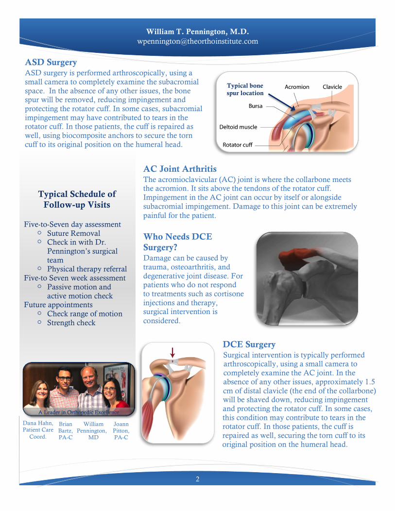
ASD Surgery ASD surgery is performed arthroscopically, using a small camera to completely examine the subacromial space. In the absence of any other issues, the bone spur will be removed, reducing impingement and protecting the rotator cuff. In some cases, subacromial impingement may have contributed to tears in the rotator cuff. In those patients, the cuff is repaired as well, using biocomposite anchors to secure the torn cuff to its original position on the humeral head.
Who Needs DCE Surgery? Damage can be caused by trauma, osteoarthritis, and degenerative joint disease. For patients who do not respond to treatments such as cortisone injections and therapy, surgical intervention is considered.
AC Joint Arthritis The acromioclavicular (AC) joint is where the collarbone meets the acromion. It sits above the tendons of the rotator cuff. Impingement in the AC joint can occur by itself or alongside subacromial impingement. Damage to this joint can be extremely painful for the patient.
DCE Surgery Surgical intervention is typically performed arthroscopically, using a small camera to completely examine the AC joint. In the absence of any other issues, approximately 1.5 cm of distal clavicle (the end of the collarbone) will be shaved down, reducing impingement and protecting the rotator cuff. In some cases, this condition may contribute to tears in the rotator cuff. In those patients, the cuff is repaired as well, securing the torn cuff to its original position on the humeral head.
Typical Schedule of Follow-up Visits
Five-to-Seven day assessment
! Suture Removal ! Check in with Dr.
Pennington’s surgical team
! Physical therapy referral Five-to Seven week assessment
! Passive motion and active motion check
Future appointments ! Check range of motion ! Strength check
Typical bone spur location