understanding the equine foot - ia802906.us.archive.org
TRANSCRIPT

Keeping the horse sound and at its athletic peak has
challenged humans for centuries. In Understanding The
Equine Foot, author Fran Jurga discusses how to provide
the horse with daily foot care, how to choose a farrier, and
how to prevent certain lameness problems.
An essential guide for every horse owner, Understanding
The Equine Foot covers various diseases which can afflict
this sensitive and complicated part of the horse’s anato-
my. Laminitis, navicular disease, and flexural deformities
are examined in detail. Emergencies such as lacerations
and hoof wall traumas also are discussed. The reader will
learn how to remove a loose shoe and when to remove a
foreign object from a wound. Jurga, an authority on the
equine foot, covers her subject matter in a highly read-
able, informative style. Most importantly, she encourages
the horse owner to become a conscientious caretaker.
The Blood-Horse, Inc. www. bloodhorse.com
www.thehorse.com800•582•5604
$14.95($19.95 in Canada)
the equine Foot
the equine Foot
U n d E r s T a n d i n G
the equine Foot
y o u r g u I d E T o h o r s E h E A L T h
c A r E A n d M A n A g E M E n T
y o u r g u I d E T o h o r s E
h E A L T h c A r E A n d M A n A g E M E n T
U N D E R S T A N D I N G
By Fran JurgaForeword by Hilary Clayton, DVM
By Fran JurgaForeword by Hilary Clayton, DVM
th
e e
qu
ine
Fo
ot
UNDERSTA
NDIN
G t
he
eq
uin
e F
oo
t
Jurga

u n d e r s t a n d i n G
The equine Foot
y o u r g u i d e t o h o r s e h e a l t h
c a r e a n d m a n a g e m e n t

Copyright © 1998 The Blood-horse, inc.
All Rights reserved. no part of this book may be repro-
duced in any form by any means, including photocopying,
audio recording, or any information storage or retrieval sys-
tem, without the permission in writing from the copyright
holder. inquiries should be addressed to Publisher, The
Blood-horse, inc., Box 4038, Lexington, KY 40544-4038.
iSBn 0-939049-96-1
Printed in the united States of America
First edition: May 1998
1 2 3 4 5 6 7 8 9 10

u n d e r s t a n d i n G
The equine
By Fran Jurga
The Blood-Horse, Inc. Lexington, KY
y o u r g u i d e t o h o r s e h e a l t h c a r e a n d m a n a g e m e n t

Other titles offered byThe Horse Health Care Library
EPM
Equine Lameness
Equine First Aid
Equine Nutrition
Laminitis

5
Foreword ................................................................6
Introduction ...........................................................8
Chapter 1 .............................................................12
Chapter 2 .................................................................26
Chapter 3 .............................................................40
Chapter 4 .............................................................60
Chapter 5 .............................................................88
CONTENTS

Understanding the Equine Foot is a book that every
horse owner should read from cover to cover to
gain a better understanding of how the horse’s hoof
works. A marvel of natural engineering, the hoof has
evolved as the interface between the horse and the
ground. As the first link in the weight-bearing chain of
the limb, the hoof is subject to high loading forces during
thousands of strides each day as it performs its functions
of dissipating concussion and supporting the horse’s
weight. These functions put the hoof at risk of a long list
of problems, including laminitis, navicular disease, punc-
ture wounds, quarter cracks, and white line disease. Our
knowledge of these diseases and their treatments contin-
ues to evolve, and it is the responsibility of each one of us
— owner, farrier and veterinarian — to keep abreast of
the latest developments. Herein lies the value of this
book.
The hoof is a dynamic structure that changes continu-
ously in response to its environment. Climate, terrain, nu-
trition, and workload play a role in the shape and consti-
tution of the hoof tissues. We take advantage of the hoof’s
malleability when we alter hoof balance or the type of
shoeing in an effort to enhance performance, improve
traction, or relieve stress in an injured area. Attempting to
6
FOREWORD

und e r s tand i nG THE EQUINE FOOT
7
improve one aspect of hoof function, however, may create
an imbalance elsewhere, which is a contributing factor in
the high incidence of lameness.
Since the hoof is the most common site of lameness,
almost every horse owner has first hand experience of a
hoof problem of one type or another. Understanding the
Equine Foot educates its readers about the structure, func-
tion, and care of the horse’s hoof. Through understanding
the inner workings of the hoof, the owner will be able to
communicate better with the farrier and veterinarian, and
to contribute to making an informed decision about the
use of hoof manipulations to improve performance, or to
prevent or treat an injury.
The author, Fran Jurga, is a journalist and publisher
with a passion for the horse’s hoof. She has devoted her
professional life to seeking out new information on the
hoof and farriery from all over the world. We are grateful
to her for sharing her knowledge and enthusiasm for this
important part of the horse’s anatomy.
Hilary M. Clayton
McPhail Dressage Chair in Equine Sports Medicine
Large Animal Clinical Sciences
College of Veterinary Medicine
Michigan State University
East Lansing, Michigan

Keeping the horse sound and at its athletic peak has
challenged humans for centuries. Even now, with all
that technology and new insights have to offer, equine
foot care remains an inexact science. We still find the
horse’s foot to be akin to a square peg that we try to force
into a round hole. Can the equine foot be analyzed me-
chanically, since it lacks true symmetry and the horse can
make individualized adaptations to gait and stance? Can
better foot care be managed biologically in order to help
our horses move faster, jump higher, or round a barrel
more efficiently?
For the past 20 years, I have devoted my professional
life to learning all I can about the horse’s foot. I have
carefully observed and recorded the efforts of dedicated,
caring professionals who work very hard to keep horses
sound and to care for lame horses. I’ve sat up all night
with anxious owners while horses underwent surgery,
or were treated for laminitis. I’ve held hoses on pasterns,
passed nails to farriers, soaked feet in buckets, and
washed bloody barn floors. Every time I think I have
seen the full spectrum of horse foot problems (and solu-
tions) ranging from the best of care given to the sound-
est equine athlete to special care needed for a foundered
8
INTRODUCTION

Und e r s tand i ng THE EQUINE FOOT
9
pony, someone calls with the challenge, “You MUST see
this one!” And they’re right. There is always something
to top the last one.
The more I observe, travel, and read about it, the more I
am convinced that all the old curses, whispers, chants,
and legends about horseshoes and horses’ feet have sur-
vived for as long as they have for a good reason: the
horse’s foot is one of the last great mysteries of animal
science.
Luckily for us, we live in a time when some of the
mysteries are unraveling, thanks in large part to diagnos-
tic techniques such as ultrasound imaging and scintigra-
phy. Sometimes it seems that horses are developing new
foot problems, including the increase of “white line
disease,” laminitis, and navicular-type lameness.
As our biomechanical tools give us a clearer vision of
how the horse’s foot works, the arguments grow. How
can we maximize the hoof’s best qualities of strength and
resiliency yet protect the foot’s weak points? What are the
best treatments for foot problems such as collapsed walls,
sore heels, new diseases, and lamenesses?
I was pleased when I was asked to write this book,
because it will be read by the people who need most to
understand the horse’s foot. It is the veterinarian’s job to
understand the medical and biological functions of the
foot. It is the farrier’s job to manage the foot mechanically.
It is the owner’s job to know when to ask for help. No
one person has all the answers; no doubt all three have
questions for the others. Only by working together can
the three-member team help the horse.
If there is one thing that can be learned from this book,
it is that the horse’s foot is a unique, complex, and
dynamic part of both the horse’s physiology and locomo-
tion systems. A lot can be done to a horse’s foot — just
look at any gaited horse or Standardbred. We need to
learn the wisdom to understand when to let nature take

I n t r o d u c t i o n
its course and the imagination to envision what that
horse’s foot might look like if nature had managed the
horse since birth.
An entire industry has been built on the horse’s foot —
shoes, pads, nails, oils, sealers, medications, boots, wraps,
frog supports, hospital plates, artificial walls, and all the
rest. The mission of these products is to help us keep the
horse sound.
The most important lessons for you to learn are which
battles you should choose to fight for your horse (with
the help of high-tech hoof care), and which ones your
horse should be allowed to fight on its own, with the help
of time off and “Doctor Green.” The long-term welfare of
your horse is always your goal, and there are tough deci-
sions to make in order to achieve that goal.
Instead of working from the outside of the hoof, by
building new walls, propping up collapsed heels, or in-
jecting exhausted coffin joints, in the near future it might
be possible to manage the hoof from the inside out. In
the future, we will be engineering the hoof with surgical
implants of collagen or cartilage, grafting coronary bands,
transplanting frog tissue, decalcifying sidebones, and
using arthroscopic tuneups in order to fix coffin and
pastern joints.
If the concept of a bionic hoof is repulsive to you, con-
sider the flip side: after a thousand years on the job, we
have failed to manage the horse’s feet in domesticity as
successfully as nature manages in the wild. Our breeding
programs have set a low value on hoof quality and high
value on developing speed. We have created horses pre-
programmed to break down, unable to stand on their
own four legs without the professional management of
veterinarians and farriers, shoes of steel, and boots of
Velcro and Neoprene.
When owners stop accepting horses with defective feet
and legs and demand better, more durable equine ath-
10

Und e r s tand i ng THE EQUINE FOOT
letes, then breeders will be forced to create them. It could
take generations, but the time is coming when horse
owners will have experienced enough heartbreak, enough
loss of performance, and most of all, will have had enough
of watching horses in pain.
The countdown to long-term soundness begins the next
time you pick up one of your horse’s feet. Look at each
one of them as if for the first time. The soundness of the
feet affects the potential of your horse as an athlete, and
is solely the result of your management. I plan to provide
you with sound advice so you will begin an equine foot-
care program to insure that you and your horse will go
forward from today, soundly.
Fran Jurga
Gloucester, Massachusetts
November, 1997
11

The best way to begin to learn about horses’ feet is to
find out what you already know. You might know
more than you think…or less.
On a plain piece of paper, draw a life-size image of your
horse’s right front foot (that’s the foot on its right). Think
about what shape the foot is, what shape the shoe is, how
wide the shoe is, how far back the shoe goes, and how
much of the foot the frog covers. Which way does the
frog point? Where is the widest part of the foot? How
many nails are in the shoe? Are the nails in the same
places on the inside and outside of the shoe? What’s the
shoe made of?
Now, on the other side of the paper, draw the foot as it
would look from the side. Is the coronary band level? Are
the heel bulbs both at the same angle as the toe? Do the
heels protrude behind the foot or sink beneath it? Is the
pastern dainty or thick, upright or sloping? How much of
the periople (the rough area at the top of the hoof where
it meets the hairline) is visible on the hoof wall? Is the
hoof cracked? Does the shoe have clips, a rolled toe, or
built-up heels?
If you are like most people, you will be hard put to draw
the parts of your horse’s foot realistically, regardless of
12
Chapter 1
How Well Do You Know Your Horse’s Feet?

Und e r s tand i ng THE EQUINE FOOT
13
your artistic ability. You probably won’t be able to answer
the questions about the foot and its shoe.
If you ran into your farrier
today at your local coffee
shop, you might say, “You
shoe my horse, you know, the
bay with the little snip on her
nose and one white stocking?” The farrier would look puzzled
for a minute, thinking to
himself, “There must be 50 bay
horses in that barn!” Then a
light goes on. He says, “Oh,
yeah, the one with the little
flare on the inside that we
wedged up last time. I bet
she’s standing under herself a
little better since then, huh?”
It is a farrier’s job to
know your horse’s feet,
not its face. If you learn to
look carefully at its feet,
you’ll soon gain insight
into your horse’s athletic
ability and learn to appre-
ciate a good shoeing job.
What do you see when
you look at your horse’s
feet?
Chances are, the first
thing you look for is a
nice, round foot, and a
sense of similarity between
the four feet. You are
aware if there is a discrep-
ancy in hoof angle (i.e.
at a glance• Become familiar with your horse’s
feet.
• Know the special features of its shoes.
• Ask your farrier for a “guided” tour of your horse’s feet.
• Keep photo records of your horse’s feet.
• Keep records of your horse’s foot care and of weather conditions.
How does your horse stand on his feet?

K n o w Yo u r H o r s e ’ s F e e t
club foot, flat foot, deformed foot) or if the horse is shod
only in front and left barefoot behind.
You might have noticed that the front feet are more
rounded than the hind feet, and that the hind feet might
tend to point out to the sides, giving your horse’s hind
legs a slightly splayed look, although the farrier has prob-
ably assured you that this is normal. You should notice if
there are cracks in the hoof wall, if the angle of the
trimmed foot is not the same as the angle of the pasterns,
or if the frog protruded. Look at the feet like a farrier.
Can you answer these questions without picking up one
of your horse’s feet?
1) What size shoes does your horse wear?
2) What are the special features of your horse’s
shoes? For example, do they have side or toe clips, built-
up heels, or toe pieces? Do the shoes have plates in the
toes? Are there trailers on the hind shoes? Is the shoe
creased? Is it a rim or polo shoe with different height on
the inside and outside?
3) Do you know how many nails are in your horse’s
right front shoe? If it is an uneven number, are there
more nails on the inside web or the outside?
4) What color is your farrier’s truck? What’s the
name of your farrier’s dog?
Time and again, horse owners can answer the ques-
tions in No. 4. They notice the truck, they remember the
dog. They can describe the truck and the dog in colorful
detail — much more detail than they can muster about all
four of their horse’s feet! They enjoy talking to their farriers
about what’s going on in the horse business, in the neigh-
borhood, or in the next barn aisle. But they “tune out”
while the farrier is working. After the farrier finishes, packs
up all of the tools, and heads down the driveway, the
average horse owner doesn’t even know how many nails
were driven into the horse’s foot.
Most farriers like it that way. Owners’ ignorance is bliss
14
Und e r s tand i ng THE EQUINE FOOT
to them. They would rather talk to you about politics or
how you did at an event than answer technical questions
for which there might be no concrete answers. However,
when something goes wrong, farriers are often incredu-
lous that an owner hasn’t noticed that a pad slipped, or
that a clinch has sprung, or that the outside quarter was
hot.
Farriers and veteri-
narians are profes-
sionals in whom you
place your trust. You
might think it’s OK
to “tune out” and
leave the technical
work to them, but
all too often you
later find yourself
scrambling for such
information when it
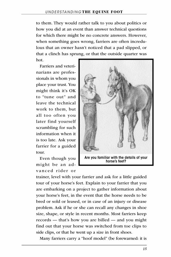
is too late. Ask your
farrier for a guided
tour.
Even though you
might be an ad -
vanced r i d e r o r
trainer, level with your farrier and ask for a little guided
tour of your horse’s feet. Explain to your farrier that you
are embarking on a project to gather information about
your horse’s feet, in the event that the horse needs to be
bred or sold or leased, or in case of an injury or disease
problem. Ask if he or she can recall any changes in shoe
size, shape, or style in recent months. Most farriers keep
records — that’s how you are billed — and you might
find out that your horse was switched from toe clips to
side clips, or that he went up a size in front shoes.
Many farriers carry a “hoof model” (be forewarned: it is
15
Are you familiar with the details of your horse’s feet?

K n o w Yo u r H o r s e ’ s F e e t
the preserved limb of a dead horse) or anatomy books in
their trucks, and will be happy to show you how your horse
compares to a “normal” foot. Warn the farrier that you’d
like to take notes for a little inventory of the horse’s feet,
for your own information, and that you plan to ask a lot
of questions. Ask the farrier to show you features in each
foot that makes it unique. Ask to be shown any warning
signs of weakness or cheering signs of a strong, healthy
hoof.
There are many factors that influence what your horse’s
feet look like, including the following: genetics, training
level, age, environment, medical history, level of farrier
care, and your management style.
Your f a r r i e r w i l l
warn you that “There’s
no such thing as a
pe r fe c t foo t ! ” and
might list off more de-
t r ac t ing comments
than you want to hear,
but it is the farrier’s
job to be acutely aware
of every feature of
every foot. You proba-
bly have the same at-
tention to detail in
your own profession.
PHoto records The best time to pho-
tograph your horse’s
feet is immediately
after they are trimmed,
before they are shod. As the last step in their shoeing job,
farriers often polish the finished hoof walls and apply
some type of sealant. Once shavings, straw, or dust hit the
16
take photographs of your horse’s feet and keep them on file.

Und e r s tand i ng THE EQUINE FOOT
hooves, they will never look as good for the photos!
If your horse’s feet do get dusty — dust clings to the
sealant — wipe the feet with a damp rag just before the
photo session.
Before the session, decide where the best place will be
to take photographs. Do not take photographs in direct
sunlight; light shade is best. Pick a spot under a tree or on
the shady side of a barn. Make sure that there is a level,
firm surface for the horse to stand on such as a roadway
or concrete walkway. Or lay down plywood or trailer mats
on the grass. Try to use a material that will show some
contrast between the color of the horse’s feet and the
flooring. Dark feet do not show up well on dark trailer
mats.
It’s a good idea to label the photographs. Make six in-
verted V-shaped “tents” of white paper. Label them: “Left
front,” “right front,” “left hind,” “right hind.” The other two
should be marked “inside” and “outside.” These refer to
the inner and outer sides of the hoof, when photographed
from the side.
Make sure someone assists you while you are taking
pictures, or have someone else take the picture while you
hold the horse. Always hold the horse firmly at the head,
with the neck set at a natural angle. Do your best to
square the horse up, so that its weight is equally distrib-
uted on all four feet. If you need to keep it calm, put a
hay bag within easy reach of the horse, so it doesn’t have
to stretch or turn its neck.
The photography session should only take about 10
minutes. Take several photographs of each foot and ex-
periment with different exposures to make sure that you
get good results.
Your goal is to get four views of each foot: from the front
(toe view), from the outside (outside wall and heel), from
the inside (inside wall and heel), and looking at the sole
and frog (ground surface view). Put the labels near the feet
17

K n o w Yo u r H o r s e ’ s F e e t
so that they will be included in the photographs for refer-
ence. This might sound unnecessary, but it can make a dif-
ference, particularly if the prints made it look like the horse
has four identical lower legs!
Once the photos of the feet are done, stand back and
take some general “posture” photographs of your horse at
rest, with its head and neck in a natural position. Photo-
graph the horse from the front so that the camera points
roughly at the horse’s chest (you might have to kneel
down to achieve this view.) Photograph the horse from
both sides, noticing where the legs are placed under the
body. Have an assistant lift its tail when you photograph
the horse from behind (aiming the camera just above the
hocks). The last photo should be attempted only if your
horse is trustworthy. Position yourself directly behind the
tail, which should hang naturally. Stand on a box and
photograph the horse down the midline of the back
facing toward the neck.
Have the film developed immediately, and carefully
attach permanent labels (marked with the horse’s name
and date taken) to the best shot of each view. There
should be 16 “foot prints” — four angles of each foot plus
four to six “whole horse” shots. Place the pictures in
plastic pages and store them in a safe, dry place with
other important records, such as vaccination records, reg-
istration papers, Coggins test, etc.
Imagine that your horse is injured at a horse show a
hundred miles away. The show’s veterinarian calls to de-
scribe the problem. While he is looking at your horse,
you are trying hard to remember what the problem area
looks like. You’ll be very glad to have the set of photo-
graphs to refer to!
Plan to repeat a photographic session every six months,
or more often if the horse is a foal or yearling, or if the
horse is new to you. Dressage horses are particularly
prone to changes in the shape of their front feet. As the
18

Und e r s tand i ng THE EQUINE FOOT
horses advance in collection, they put less weight on the
forehand; consequently their front feet might become
rounder.
This photographic exercise sounds tedious, but there is
no better way for you to learn about your horse’s feet.
Compare the photos with your horse’s feet on the day the
farrier is due to arrive. What differences do you see in the
feet?
Many horses’ feet change character between winter and
summer. Some change in condition over the course of a
rigorous show season. Sometimes, you will see “fever
rings” on the hoof wall, often a sign of past illness or a
dramatic change in nutrition. If you start training the
horse in support wraps or boots, you might see changes
in its feet or pasterns that will be visible when you
compare photographs, but were not obvious to you on
casual observation.
Photo records will also help if you are working with
more than one horse. You will be able to compare changes
that occurred in each horse and eliminate the problem of
confusing the foot problems of one horse with another.
You will be able to use the photos to discuss specific
details with your farrier.
Save these photographs! If you ever move, or change
farriers, or if the horse travels on a show circuit far from
his regular farrier, these photographs will be your best
evidence both to show to a new farrier, and to help
recover what might have been destroyed by changes in
shoeing in another farrier’s hands.
Obviously, the photo records will be helpful to you if
you decide to sell the horse or if the horse is stolen or lost.
Many horses have been recovered because of identifying
characteristics on horseshoes. Farriers who handmake
horseshoes often stamp their logos or initials into the steel;
most manufacturers will have an imprint and shoe size on
machine-made shoes. Even nail heads, such as the distinc-
19

K n o w Yo u r H o r s e ’ s F e e t
tive “checkerboard” heads of American “Capewell” nails
can be useful identifiers.
HoofcAre record-KeePing Secretarial duties might be the last thing you are think-
ing about doing when it comes to your horse, but you
need to keep a record of when your horse is shod, and by
whom, and whether new shoes were put on or the old
ones were reset. If the horse wears pads, make notes
when you stopped or started using full pads or rim pads,
leather pads or plastic pads, and if wedges were used,
what type and what angle. If the horse wears pads, make
note of what the farrier used for packing under the pads.
Sometimes the farrier recommends changing the time in-
terval between shoeing appointments. For example, if
winter is coming and you expect hoof growth to slow,
you might want more time between appointments.
Do your record keeping in a notebook with a page for
each horse (or on a large calendar if you have only one
horse). Make notes of the products you use on your
horse’s feet and legs. Add notes when you poultice, and
when you deworm. List the products by brand name. If
you add or change feed supplements or medications for
other problems the horse might have, be sure to write
down dates, quantities, and any details that might be per-
tinent if a problem arises.
If you notice any problems with your horse’s feet
between shoeings, write them in the notebook, such as
“thrush in right front seems completely gone” or “removed
hospital plate permanently today” or “started training with
grass studs today.” When your farrier comes by to shoe
the horse, check the notebook and see if there were any
details you wanted to discuss, such as the size of studs
relative to the shoe’s width, or if there were any negative
signs of stud use.
20

Und e r s tand i ng THE EQUINE FOOT
environmentAl records In the same notebook or on the same calendar, develop
a shorthand system for recording the weather. Very wet
or dry periods can affect hooves, so you might not need
to panic if you have a new horse and its feet seem mushy.
Recording this kind of information will be helpful to you
in the future. As the years go by, you will notice ways that
individual horses react to certain weather conditions, par-
ticularly in the winter if they are confined to their stalls a
good bit of the time. Some owners note the lengths of
time of turnout and training, along with feed quantities
and hay. Their calendars and notes cover each day and
list hours spent turned out or in training. A serious condi-
tioning program demands this meticulous type of record
keeping.
Conditions in which you train a horse can play a role in
foot problems or general condition of the hoof. Some
horses can become accustomed to certain arena surfaces,
particularly cushioned surfaces, and then will show signs
of soreness when worked on hard or frozen ground. Many
riders report problems with horses when they either are
sent to or return from a trainer’s facility. Working in sand
or any drastic change in surfaces will require time for a
horse to adjust. Keep notes of when you transport a horse
to a clinic or training session, or if you take a long hack
over frozen ground or a tarred road on a bright January
day after the horse has only been worked indoors for the
previous month. You might be surprised how helpful this
information will be if the horse shows subsequent signs
of bruising, stinging, or excess shoe wear.
Your Horse’s HistorYIs your horse’s health history shrouded in mystery, or is
it written on his legs and feet? Scars, old tendon injuries,
windpuffs, and ringbone or sidebone will tell you a lot
about where your horse has been and how he was used
21

K n o w Yo u r H o r s e ’ s F e e t
before he came to you. Racehorses are particularly prone
to a common set of leg injuries, as are former jumpers,
polo ponies, or Western performance horses.
A horse bought directly from its previous owners
shouldn’t require hard work to find out the horse’s pre-
vious lameness history, unless the owner is unwilling to
share the information in the belief that it may devalue
the horse. The owner has the right to privacy in this
matter, but you might find owners who are willing to
cooperate and who will turn over valuable radiographs
or refer you to a veterinarian or farrier who worked on
the horse during a problem period. Keep in mind that
veterinary records and radiographs are used fraudulent-
ly by some unscrupulous owners and agents.
If you know your horse’s pedigree, check other foals
from the same mare or by the same sire, or even check
horses from the same breeder. Problems often come in
groups. While the jury is still out on the decisive rule of
heredity in leg deformities and navicular disease, the old
saying “Forewarned is forearmed” will help you avoid
difficulty in the future. Making a trip to another state to
check the soundness of a dam or sire may give you a
picture of future problems you need to avoid.
tHe mAgic of motion: WAtcH Your Horse’s feetMany lameness or conformation evaluations will include
a simple walk/trot in a straight line on a hard surface,
along with longeing the horse in a tight circle in both di-
rections, also on a hard surface. You might hear a great
deal of discussion about whether the horse “lands flat” (as
opposed to toe-first or heel-first), “paddles” (feet don’t
swing forward in a straight line through the knee, fetlock,
and foot), “overtracks” (brings a hind foot forward of the
point where the front foot had been) and “displaces”
(slang for prematurely planting a hind foot in a trot or
canter).
22
Und e r s tand i ng THE EQUINE FOOT
Every horse has its own posture, its own gait and stance
adaptations, and its own compensations for weakness, old
injuries, pain, or the imbalance of a rider. Observations by
others of how your horse moves might be helpful to your
understanding, or just complicate the picture in your mind.
The best time to evaluate a horse’s gait characteristics is
when the horse is sound, so that you have something for
comparison in the event of future lameness or injury. A
video camera can be helpful, as can simply comparing
your horse to others of similar breed or sport type.
The basic tenets of doing your own movement evalua-
tion are to have an experienced handler lead and longe
the horse while you watch it. Make sure the horse moves
in a straight line at a steady gait, with the head in a
relaxed, normal position. Kneel on the ground and, if you
need to, shade your view so that the upper body of the
horse does not distract you. Video taken from the ground
level with a zoom lens can be a real help, but the camera
must be on a small tripod or sturdy box so that the lens is
steady, and the horse must be led along a line of refer-
ence, perhaps between a series of cones.
A special aid to this visual or video exam is to wrap the
legs in bright colored bandaging tape over the quilting so
there is no bulk. Racetrack bandages work well and are
thin. This might be a requirement if you have a dark
colored horse, since the legs are difficult to distinguish
and the hooves may blend in with the ground surface.
If you are interested in evaluating how a horse tracks or
“displaces” at the trot, try wrapping the legs in four differ-
ent colored bandages, and videotaping the horse from the
ground again.
Working a horse in this way in a raked, sandy arena or
on a sand beach, will give you hoofprints to evaluate.
From your previous awareness exercises, you should be
able to pick out the imprint of the hind foot from the
front foot and decide if the horse is overtracking. Many
23

K n o w Yo u r H o r s e ’ s F e e t
large, immature horses will place the hind foot in front of
and to the outside of the point where the front foot had
landed. Overtracking changes in horses as they advance
in training and collection.
Using a measuring tape, you can obtain a good mea-
surement of your horse’s stride, which might be useful in
training so that comparisons can be made.
evAluAtions:ProfessionAl And PersonAl recommendAtions
One of the best results of reading this book would be
for you to have developed an eye for your horse’ feet that
is objective and based on sound judgment. Most impor-
tant of all, it will be your personal evaluation of the state
of your horse’s feet, based on the past condition you have
seen, and a goal of what you want the horse to achieve
athletically…and what condition the feet must be in to
withstand the pressure of that level of training.
Many horse owners doggedly pursue veterinarians and
farriers and ask them to voice on-the-spot opinions of a
horse’s feet or the type of shoeing. Please be aware that
this type of questioning is a bit unfair to the professional,
since he or she has no idea of how long the horse has
been shod in a particular way, what the feet looked like a
year or more ago, and what the horse’s athletic career has
been like.
Trainers and other riders and owners are quick to eval-
uate foot conformation and shoeing, and make recom-
mendations, but you should accept their remarks with a
polite smile, and a sincere “thank you.” Only you have all
the information needed to evaluate your horse.
It might be possible for you to hire a veterinarian or
farrier to do a thorough soundness workup on your horse
and provide you with an evaluation, but their ethical obli-
gation is to remind you that their opinion is only valid for
the horse on the day it was examined, and in the way it
24
Und e r s tand i ng THE EQUINE FOOT
was shod. If you have a list of comments about your
horse’s feet passed along by a trainer, judge, veterinarian,
or farrier from a previous owner, you should realize that
time has passed, the horse could be shod completely dif-
ferently, or the horse might have matured or advanced in
training so that the information lacks validity.
Remember that what your horse ight lack in conforma-
tion or soundness may well be compensated for in heart,
intelligence, behavior, or insensitivity to pain. What looks
bad on a horse may not feel bad, and vice versa. Many
horses with “pretty feet” have been to a highly-skilled
farrier for creative management, so never allow your
horse to be compared with another. Every horse is an in-
dividual, and deserves a fair evaluation with all factors
taken into consideration.
PlAnning for soundnessFor some owners, the best course might be to say, “The
future starts today.” Instead of dwelling on what is wrong
with your horse’s feet, look for the strong points — thick
soles or walls, nicely concave sole, or a healthy frog —
and use that information as the foundation for improving
other foot attributes.
Don’t be discouraged if your horse has imperfect feet;
many horses have made great improvements in their hoof
quality when managed properly by their owners. Be
aware that some of the limiting factors you have identi-
fied may be cast in stone, such as an angular limb defor-
mity, and that you must not have overly high performance
or soundness expectations of the less-than-perfect horse.
Soundness is a blessing in a horse with a handicap to
overcome.
25

In the course of a normal week, how many people put
their hands on your horse or its tack? How many groom,
lead, blanket, unblanket, bandage, or exercise your horse?
Chances are good that most answers will fall into two
groups. People in the first group would answer, “Just one,
me!” In such cases, the horse is kept at home and the
owner or rider is the primary caregiver for the horse.
A second group of people would answer, “Wait a minute,
let me count…” Their horses are out for training or
boarded at a barn where the owner or rider is not always
present to give daily care.
Everyone who helps to take care of your horse has a
direct or indirect effect on your horse’s hooves. Think
about the 13-year-old girl you trust to wrap your horse’s
hind legs at night. Does she apply the wraps at the proper
pressure? Do the bandages ever slip? Does she always re-
member to pick your horse’s feet out?
Think about those roofers working on the barn — the
ones who let nails and tiny scraps of flashing fall to the
ground below. How about the trainer’s assistant who
longes your horse each morning? Do you think he or she
always remembers to put bell boots on your horse to
protect those expensive egg bar shoes? And how about
26
Chapter 2
The Professional Hoofcare Management Team

Und e r s tand i ng THE EQUINE FOOT
27
your friend, who generously offers to haul your horse to
a show…in a trailer without mats?
If you are a natural-born worrier, you probably shouldn’t
own horses because they seem to be the most accident-
prone of all animals. Chances are good, when they loaded
Noah’s Ark, one of the horses stumbled on the ramp and
cut its pastern (or wouldn’t load at all).
Boarding Barn recommendations The best professional farrier and veterinarian cannot
provide your horse with an appropriate level of care if
they are always patching up problems that require first
aid. It is your responsibility to make sure that, within
reason, your horse is protected from unnecessary hazards
and unedu-cated, careless handlers.
Unless your horse is in your direct care, a large amount
of responsibility has to be transferred to others. If your
horse is boarded out or kept by a trainer, you are still re-
sponsible for paying for medication and treatment when-
ever a mishap occurs.
Unfortunately, the quality of care given to a boarded
horse is rarely equated to the cost per month or the deluxe
appearance of a facility. Many horses receive great care at
the smallest barns, where the tiniest changes in their atti-
tude or the smallest limp will be noticed quickly. At huge
boarding barns, you might find that the barn helpers see
so many horses that they don’t notice immediately when
something looks different about one of their charges.
Before moving your horse to a boarding barn, introduce
yourself to everyone who will be caring for your horse.
Make sure that they know about any problems with your
horse’s feet or legs. Show them how you care for the
horse, explain its current care regimen, such as medicat-
ing for thrush or flushing out an abscess track. If your
horse is to be given feed supplements for hoof growth,
make sure that the barn staff knows the quantity to be
H o o f c a r e M a n a g e m e n t Te a m
given and mark all containers clearly with your name,
your horse’s name, and your phone number. Mark the
container “close tightly” or “do not expose to light,” ac-
cording to the manufacturer’s directions. State clearly that
your horse is not to be fed substitutes. Check containers
often to make sure that the supplement is being fed and
that the right size scoop is in the container.
Beware of barn help who recommend that you switch
to products they sell. Also beware of staff members who
recommend that you switch your horse to herbal prod-
ucts or racetrack remedies not familiar to you. They might
be excellent products, but talk to others before you switch
your horse over to the new product. Get the names of the
people who are in charge of the boarding facility at dif-
ferent times of day (and different days of the week). Make
sure that they are all aware of your horse’s special needs
and your “pet peeves” about care. Finally, mark all your
equipment — from your hoof pick to your bell boots to
medications and supplements — with your name, and
keep a record of the equipment you have left in the barn
to be used exclusively by your horse.
Why did you choose your farrier? A recent survey of horse owners asked why they em-
ployed their particular farriers instead of others. The
answers were surprising. One group of respondents
offered personal reasons unrelated to anything about the
horses or about shoeing. “He’s cute,” was a popular
answer, as was, “He tells great jokes.” Other respondents
employed farriers who were close friends or family
members. Several people answered the same way: that
their farrier had worked on their horses for a long time
and they didn’t want to switch.
Another group of respondents felt that they had little or
no choice in selecting a farrier because they were limited
by geography or by the fact that their horse was a special
28

Und e r s tand i ng THE EQUINE FOOT
breed, or used for a specialized sport that required a
certain kind of shoeing — one not widely available from
other farriers. This group in-
cluded owners of gaited
horses and reining horses.
Another group of owners
said they did not make the
decision of who shod their
horses. The decision was in
the hands of their trainers or
the managers of their board-
ing facility.
Still another group said they
had chosen their farriers
because of their businesslike
attitudes. The expertise of the farriers mattered less to them
than the farriers’ businesslike approach (whether they
showed up on time or accepted credit card payments).
A small group said they had developed a good working
relationship with farriers who gave the best possible care
to their horses.
The survey turned up even more surprising results.
First, almost all of the respondents thought that their far-
riers were “qualified” or even “the best around,” but those
surveyed could not list exactly what the farriers’ qualifica-
tions were. Owners were fiercely loyal to their farriers,
but often did not know much about the farriers’ back-
grounds.
Graduating from a horseshoeing school impressed a lot
of owners, but very few seemed to know the difference
between a diploma from a shoeing school and certifica-
tion from a national testing body such as the American
Farrier’s Association. Very few owners knew what experi-
ence their farriers had in horse handling.
What should matter when choosing a farrier? When you
choose a farrier, it is not unreasonable to ask for a resume
29
at a glance• Know your farrier’s qualifications.
• Get references.
• Find a farrier who fits your needs.
• If your horse becomes lame, call your veterinarian first.
• Consult with your veterinarian and farrier first before seeking a second opinion.

H o o f c a r e M a n a g e m e n t Te a m
— perhaps one delivered verbally instead of written —
but a resume nonetheless. Here are 10 good questions to
ask when you interview a prospective farrier.
1) Do you own a horse? This might seem inconsequential, but it will tell you im-
mediately whether or not the farrier cares about horses
and if he or she will be able to empathize with your
concern for your horse’s well-being.
2) How long have you been around horses? Believe it or not, some people graduate from farrier
school without learning how to lead a horse. Some have
never been around horses and wouldn’t know how to tie
one using a slip knot. However, they might be highly
skilled mechanics or blacksmiths. At the racetrack, farri-
ers are not usually required to handle horses much;
grooms do that. If your horse is not stabled at the track,
you’ll probably expect your farrier to be able to get your
horse out of a stall or pen. If your horse has any behavior
quirks at all, this is an important question.
3) How and when were you trained to be a farrier? Ask for the name(s) of horseshoeing schools and/or far-
riers with whom your prospective farrier claims to have
worked for as an apprentice. Many farriers claim to have
“apprenticed” with different top farriers, when what they
really mean is that they rode with that farrier for a few
days, or they might have attended a seminar given by that
farrier. An apprenticeship, or “working student” program,
is the most effective way for a new farrier to be trained
today. An ideal program would be to start as a “truck
helper” for a few months (holding horses, sorting shoes,
learning the routine); then attending farrier school (the
most reputable programs average two to three months);
then working as an assistant farrier, or apprentice, to a
qualified senior farrier for a minimum of one year.
4) What is your national certification level? At the time of this writing, there are three national orga-
30

Und e r s tand i ng THE EQUINE FOOT
nizations that certify farriers: the American Farrier’s
Association (AFA) , The Guild of Professional Farriers
(which is also called “The Guild”), and the Brotherhood
of Working Farriers (which is called “The Brotherhood” or
abbreviated with the initials BWFA). Each has different
levels of certification; many farriers who reach the jour-
neyman level are cross-certified in more than one organi-
zation. Most racetrack farriers belong to a union, or have
some other testing process.
Since these programs are new, recently trained farriers
are more likely to hold certification than older farriers.
Many older farriers feel that their years of experience are
a sufficient testimonial to prove their skill. Younger farri-
ers need certification to show their skills and prove that
they are serious about advancing in their profession. A
quick phone call to any of the national associations will
verify a farrier’s credentials. Beware of any farrier who
falsely claims to be certified or who bad-mouths the certi-
fication system.
5) What farrier organizations are you a member of? This question has nothing to do with a farrier’s ability
because a farrier could be a professional student of hoof
science and have no practical ability to get your horse
shod. But if he or she does not belong to any organiza-
tions, a red flag should go up as to whether or not he or
she is serious about keeping up with new products, tools,
or ideas on shoeing.
6) What type of horses do you generally shoe? Look for a farrier who shoes horses similar to your
own. Farriers who shoe reining horses might not have
the drill presses and taps needed to put stud holes in an
eventer’s shoes. A farrier unfamiliar with a particular
breed of horse might not understand their special needs. For example, the need for weight or toe-length limits for
Arabians or Morgans. Shoers of show horses are usually
aware of the changing preferences of the judges in their
31

H o o f c a r e M a n a g e m e n t Te a m
area. For example, the preference of judges regarding
the knee action of a show hunter.
7) What experience do you have with lameness cases (or foal work, or other criteria important to you)? What vet-erinary practice(s) do you work with?
As the farrier profession advances to the 21st Century,
farriers will become more specialized. Your “primary”
farrier might occasionally refer your horse to a specialist,
one who is a lameness specialist, for example. Currently,
there are some farriers who work as employees at veteri-
nary hospitals, or who own layup facilities to care for
injured or ailing horses, such as severe founder cases. If
your horse is sound and in the middle of a busy show
schedule, you might be happier with a farrier who shoes
performance horses rather than one who specializes in
working on lame horses.
If your horse has chronic lameness or a foot conforma-
tion problem, you might want to use a lameness farrier to
keep up an individualized shoeing program. Most farriers
will be honest about their lameness experience, particu-
larly if “fine tuning” a performance horse is their favorite
challenge. Many farriers will not work on foundered
horses, due to the time it takes to learn that specialized
type of shoeing. The failure rate for founder cases can be
discouraging for a farrier.
8) What is your billing/payment/record-keeping system? Many farriers expect payment at the time of service. You
need to know this in advance, so you can present them
with a check or credit card information. Other farriers
send bills. If you require a receipt, make sure you ask for
one, particularly if you are paying cash. If you want to
keep track of what is being done to your horse, ask for a
description on the bill — one that notes any special fea-
tures such as clips, trailers, calks, pads, etc. Keep the bills
for your long-term record keeping. If you have more than
one horse, you might want to make sure the work on
32

Und e r s tand i ng THE EQUINE FOOT
them is billed (or at least itemized) separately so you can
keep track of expenses.
9) How often are you in this area? Does this farrier make frequent trips to your area? If not,
you might find yourself in a predicament when a shoe is
lost or the horse needs special care. Most farriers will insist
on putting a horse on a schedule, usually from three to
eight weeks, depending on the complexity of the shoeing
it needs, the rate of hoof growth, or special lameness con-
siderations, such as navicular disease. If you hear a shoe
clanking and notice that it is loose, but it is still three
weeks until your next regular visit from the farrier, you
might feel frustrated if the farrier refuses to come back to
reset it and tells you to call someone else to fix the shoe. It
might be comforting for a farrier to tell you, “I’m in that
part of the county every Tuesday and Thursday.”
10) What options are available to communicate with
you in case of an emergency or a missed appoint-
ment?
Farrier trucks once were identifiable on the highway by
the smokestacks protruding from their roofs. Now anten-
nas are a sure sign of a farrier’s truck. Most farriers have
cellular phones and/or pagers (both of which are useless if
they aren’t turned on). Trying to explain a lameness
problem over the phone to a farrier’s 10-year-old child is
very frustrating, especially if you are standing at an outdoor
pay phone at a horse show making an emergency call.
Make sure that the communication links between you
and the farrier will be adequate for your needs. Ask for
names of some of the farrier’s customers and contact them
for a reference. Ask them for both good and bad experi-
ences they have had with the farrier. Ask about the farri-
er’s business attitude. Many owners will explain that they
use a farrier because he or she is either (1) so good with
their horses, or (2) so skilled at keeping a horse sound
despite the fact that “he never gets around to sending a
33

H o o f c a r e M a n a g e m e n t Te a m
bill until it’s past $1,000,” or (3) “she always loses the
checks I leave for her and I end up having to write
another one.” If business acumen (or lack of it) is an issue
with you, find a farrier who matches your needs.
Farriers like and appreciate professionally run barns,
no matter how small. Most farriers are animal-lovers, or
at least are so accustomed to working around them that
they are “handy” with most animals. On another level,
animals often sense a farrier’s innate animal-friendliness,
plus many are attracted by the smell of burning hoof or
the taste of hoof trimmings.
Beware of farriers who show any evidence of behavior
or demeanor that you would not tolerate in any other
professional you hire. This includes threatened or actual
rough treatment of horses, frequent verbal abuse of
horses or assistants, bursts of temper, and any sign of dis-
respect toward you or your animals and property.
In the unfortunate event that your horse dies or is euth-
anized (for whatever reason), you will want to write a
simple letter to your farrier thanking him or her for pro-
fessional service, perhaps including a photograph of the
horse, if it is appropriate.
In some cases, your farrier might ask for the feet of a
horse after it dies. This might sound repulsive to you, but
it is quite common for farriers to do this. A farrier will
either dissect the foot or have it sent to a professional dis-
section preparation company like “Horse Sense Educational
Models” for permanent preservation. In this way, the farrier
can see what was on the inside of a foot that he or she
had to work on from the outside. You have every right to
refuse the farrier’s request, if you feel uncomfortable about
this practice. However, it is an important part of a farrier’s
self-education. A necropsy, or post-mortem examination,
can often show the farrier more about the horse’s lame-
ness if there is evidence of damage inside the hoof capsule.
This might not help your horse, but it might help the next
34

Und e r s tand i ng THE EQUINE FOOT
several horses the farrier works on.
Veterinarians and farriers: Who does What? In this age of caring for “the whole horse,” both your
farrier and your vet share the responsibility for your
horse’s well-being. Your vet is responsible for care from
head to toe; your farrier needs to be aware of and knowl-
edgeable about conditions in the entire animal, but will
act only to the extent that it affects the foot. A farrier
should not administer treatment to your horse in any area
above the coronet. Even so, you will find that the farrier is
knowledgeable about therapy for the legs and how to use
boots, wraps, poultices, and liniments on the legs. Your
farrier also can tell you how your horse’s conformation is
affecting its gait, soundness, or hoof capsule shape.
Farriers and veterinarians often work together to care
for lame horses. If your horse goes lame, you should call
the veterinarian first. The vet will ask you a series of ques-
tions, trying to pinpoint whether the problem is shoeing
related or not. If the horse goes lame soon after shoeing,
the veteri-narian will probably suggest that you call the
farrier to check for a “hot” nail or to reset the shoe with a
pad under it.
With a potentially serious lameness, the veterinarian will
take charge of the situation as he or she is the responsible
party. The normal process has the following steps: The
veterinarian provides a lameness examination with flexion
tests, trotting out, nerve blocks, etc., as needed. If radio-
graphs are called for, the veterinarian might pull the shoe
before proceeding with the radiographs.
Alternately, the veterinarian will ask you to call your
farrier to get the shoe or shoes pulled. You also might be
asked to haul the horse over to the vet clinic for a radiog-
raphy appointment, and to set an appointment with the
farrier at the same time, so the farrier will be present, too.
After viewing the radiographs, examining the horse,
35

H o o f c a r e M a n a g e m e n t Te a m
and considering the horse’s medical history, the veterinar-
ian will provide a diagnosis (to identify the cause) and a
prognosis (to predict recovery). He or she probably will
confer with the farrier to see if the shoeing regimen has
been changed recently, or will ask if the farrier thinks that
special shoes might help the horse. You might have to
have any pulled shoes reset by the farrier or the horse
might need to go barefoot for a while.
The veterinarian will prescribe any medication and treat-
ment (such as joint injection, special feed, physical therapy,
etc.) and work up any shoeing treatment needed with the
farrier. This might be done directly between the two pro-
fessionals, either in person or by phone. You should not be
the conduit of information between the veterinarian and
farrier.
The farrier might have to make special shoes to help
the horse, and might do special trimming or open an
abscess location within the hoof wall. Technically, a farrier
usually limits his or her work to the exterior of the hoof
capsule. Most will not remove wall or sole without a vet-
erinarian’s written prescription. The farrier might ask that
the veterinarian be present when the work is done.
If surgery is needed, the farrier and veterinarian often
will work together so that special shoes can be applied
while the horse is under anesthesia. The shoes are made
and fitted in advance, then nailed on in a sterile environ-
ment. Such work is done during a ligament desmotomy
to help correct foot deformities in foals. As part of the
surgery, the farrier will nail on extension shoes or use
glue to apply them. In this kind of surgery, as well as
others, both professionals will provide you with follow-
up care instructions.
The farrier might teach you how to remove and replace
a hospital plate (the special shoe) or show you how to fit
bell boots over glue-on shoes so you can turn out the
patient in a ring or other grassy enclosure. You should be
36

Und e r s tand i ng THE EQUINE FOOT
sure to ask the farrier what to do in the event that a
special shoe falls off or gets twisted out of shape.
It is your responsibility to discern which professional to
call when there is a problem during the recovery period.
You might want to keep both apprised of how the horse
is doing and periodically call to assure them that you are
following their directions.
referral Veterinarians and farriers It doesn’t do much good to second-guess the veterinari-
an and farrier team you have chosen. There are times,
however, when calling an expert is warranted. Imagine
you’re the owner of a foundered horse. You are frustrated
because you don’t think it is recovering fast enough. You
read a magazine article about a new treatment for founder
prescribed by a farrier in Idaho, and decide to deplete
your credit card limit by flying in the expert to work on
your horse.
To your surprise, your farrier is offended. Your veteri-
narian balks when you ask to have the horse’s radio-
graphs shipped to the “expert.” What’s the problem? If
you have chosen the right farrier and veterinarian team,
you should not need to call in any outside experts. If
outside expertise is needed, your farrier and vet will ap-
proach you and suggest the name or names of experts
they recommend for a consultation.
To bring in a consultant, you might be willing to spend
more money in order to make your horse more comfort-
able. If so, approach your veterinarian and farrier to ask
their opinion on who to call. Most farriers and veterinarians
have been to national seminars like the Bluegrass Laminitis
Symposium and are familiar with the techniques and
talents of different laminitis (founder) consultants. The
American Association of Equine Practitioners and the
American Farrier’s Association have worked together to
compile a description of the suggested protocol between
37

H o o f c a r e M a n a g e m e n t Te a m
attending professionals (your regular veterinarian and
farrier) and consulting professionals (specialized veterinar-
ians and farriers brought in to work on special problems).
If you have brought in a referral farrier or veterinarian,
keep in mind that your attending professionals are still
the responsible parties for your horse. They have a lively
interest in its well being. They should be kept informed
of any changes in the horse’s condition and changes in
its medication and/or treatment. Put some extra work
into keeping the lines of communication open by sending
copies of letters and faxes to all parties.
adjunctiVe therapy care Most adjunctive therapy — laser work, massage, acu-
puncture, herbal or homeopathic medicine, or chiroprac-
tic work — does not involve the foot. However, occasion-
ally one of these types of treatments could have an effect
on factors contributing to lameness. Most aspects of ad-
junctive therapy are not scientifically “proven” but are
widely used and appreciated by owners of lame horses.
If your horse has a foot problem, a therapist might not
be your best investment, but you can look into how ad-
junctive therapies could stimulate circulation within the
leg and foot. Acupuncture meridian charts show the flow
of “chi” to the foot. Massage of the coronet and heel bulbs
might be helpful in stimulating blood flow within the
foot. Laser boots and magnetic boots and hoof pads are
designed to send a healing or stimulating force to the foot
either across the hoof wall or up from the sole.
Before starting adjunctive therapy on your horse,
consult your attending veterinarian and farrier. In some
states, a veterinarian’s referral might be needed for certain
treatments. Both your farrier and veterinarian will help
you to decide, by discussing other cases where they have
seen therapy work (or fail). They might be familiar with
the work of different therapists, and might know of one
38

Und e r s tand i ng THE EQUINE FOOT
who is well-suited to work on your horse’s foot problem.
When a therapist is working on your horse, he or she
should follow protocol, similar to the rules set down for
veterinarians and farriers acting on a referral basis. The
therapist should be briefed by the veterinarian as to the
cause of lameness and should contact the farrier in order
to understand any specialized shoeing that has been
done. If a therapist is not willing or able to communicate
with your veterinarian and your farrier, he or she might
be the wrong person to work on your horse.
Use the same criteria for selecting a therapist as you
would in selecting any other horse health professional. If
your horse is restricted to stall rest or pen turnout due to
a foot problem, consider working with a therapist to
massage the horse’s stiff muscles and keep its circulation
stimulated, on the surface at least. Most horses enjoy
being massaged. It gives them a welcome break from the
monotony of confinement.
If you hire a massage therapist, ask for help and sug-
gestions for stretching exercises you can do to keep your
horse supple. A simple example is coaxing the horse to
stretch its head back (flexing the neck) between its fore-
legs for a carrot or treat; ask him also to stretch to either
side for additional treats. Another example is simply ex-
tending the foreleg while the horse is standing squarely.
Beware of therapists who promise to “fix” your horse’s
feet. Also make sure that the therapist has extensive expe-
rience with horses and is well-versed in horse anatomy
and physiology. Sometimes human therapists offer their
services to animal owners, but the results may not be
what they — or you — expect. Remember, anyone who
works on a horse without proper knowledge of horse be-
havior is in grave danger of being hurt. A lame horse can
still kick!
39

It would be so easy if every horse came with an owner’s
manual to help the owner know its individual needs.
Lacking a manual, owners must apply deductive reason-
ing to figure out what the horse needs to maintain — or
achieve — optimum condition of its hooves.
Caring for the hooves is one of the oldest preoccupa-
tions that humans have had with horses. The Greeks
wrote volumes on how to avoid lameness, how to choose
horses with strong feet, and how to care for lame horses.
From the time of the Greek writer, Xenophon, to some
current writers, all have introduced a good dose of folk-
lore, misinformation, and a little bit of black magic into
hoofcare textbooks. We are now beginning to learn how
to care for the equine foot, applying biomechanical, sci-
entific research. Even so, some soothsayers predict that it
is too little, too late, since our breeding stock is composed
of a large percentage of unsound horses.
This abundance of lame or unsound horses has recently
created something called “the hoofcare industry,” which
had never existed before. A large chunk of the shelf space
in every tack and feed store is given over to feed supple-
ments, hoof oils and conditioners, hoof protection agents,
and medications designed to fix what ails your horse’s
40
Chapter 3
Team Work/Professional Care

Und e r s tand i ng THE EQUINE FOOT
41
feet. Every year, horse owners spend more on hoofcare
than they did the previous year. Farrier prices continue to
go up. There are new products and supplements to buy.
Hoofcare has even expanded into new areas including
creating artificial, presumably beneficial, types of bedding
for horses to stand in, and new artificial surfaces for
arenas and tracks.
Routine hoofcare requires no special purchases beyond
what you already have in your house and barn. If owners
paid more attention to simple daily care of their horses’
feet, it is quite possible that many “fix-it” remedies, fancy
hoofcare products, and supplements would sit on
shelves.
The Daily RouTine As with most health-care processes, the most important
aspect of hoofcare is to know
what you are doing; to be or-
ganized about it; and to do it
the same way, with the same
tools, on a regular schedule.
The care described in this
section would be typical for a
horse that is kept in a stall all
night and turned out all day.
Taking care of your horse
begins with taking care of
yourself. Make sure that you
are prepared to do the job.
Check your feet before you go
near the horse. If your horse
stepped on you, what would happen? Sturdy athletic
shoes, paddock boots, or at least thick, rubber “Bean”
boots should be worn. Never work around a horse in
sandals or high heels, even if you are on your way to
work!
at a glance• Clean your horse’s feet in a safe,
secure place.
• A hoof pick and a small, stiff brush are all you need for daily foot care.
• Notice any changes in the appearance of the feet, or any unusual smells.
• Keep feet and legs dry.
• Make sure you know how to use special equipment, such as studs and bell boots.

Te a m W o r k / P r o f e s s i o n a l C a r e
If you wear glasses, make sure you have them on before
you start, or that they are on a holder around your neck.
Pull your hair back so it doesn’t fall across your face while
you are holding up a heavy horse leg. In winter, it helps
to wear thin gloves under big mittens. You can whip off
the mittens when you are ready to start, and your hands
will stay warm for a little while with the thin gloves on.
Each hoof needs equal attention. Begin the daily routine
with the hind feet, which are more difficult to lift and to
care for. You then can move to the front feet as a reward!
If you have difficulty working on your horse’s feet, plan
a session with your farrier or with another experienced
horseperson around to help you. Watch the “expert” lift
and work on the horse’s foot, then try to imitate what you
have just seen. Ask the expert to watch you work on the
horse, and to tell you what you are doing wrong. If your
horse calmly lifts a foot for others, but won’t for you,
there must be a reason.
If you are intimidated by your horse, or if you feel that
your horse is in pain and will not lift a foot, have a more
experienced handler do your hoofcare chores for you.
Keep your safety and your horse’s in mind at all times.
Always work on level ground. Always use the same place
if possible. The first few times you do the daily foot-care
routine, have the horse held by someone who is paying
attention to you and to the horse. Make sure there are no
distractions. The holder should stand on the side of the
horse where you are working. The horse should not be
able to see its stablemates out in the field, because it will
start to fidget.
If you put the horse in crossties, make sure that all the
shanks, the clips, and the horse’s halter are in good con-
dition. Adjust the ties so that the horse cannot move back-
ward or forward more than a step, but he still should be
able to lower his head. The horse will naturally lower his
head when you lift a hind leg. Make sure no one ap-
42

Und e r s tand i ng THE EQUINE FOOT
proaches leading another horse while you are working
under yours. In a busy training facility or big boarding
barn, moving into the wash rack area will take you and
your horse out of the high traffic lane.
When cleaning the horse’s feet, stay close to the horse’s
side. If you feel the need to jump clear if the horse blows
up, do so, but jump fully clear of the horse’s range of
motion, particularly if the horse is cross-tied. Don’t pick
up the horse’s foot until you are ready to work on it. One
of the most dangerous things handlers can do is to hold
up a hoof while reaching for a hoof pick or brush an
arm’s reach away.
Most horses are easily trained to lift a foot when asked. You might need to tug or pinch gently on the back of the
fetlock for a second as you firmly but quietly command,
“Lift!” Soon, the horse will lift the foot on voice command
as soon as it feels your hand touching its fetlock.
Avoid rewarding the horse for lifting a foot in response
to a hand on its lower leg, because you will confuse the
horse. There will be times when you won’t want the horse
to lift its foot, when you are applying wraps or boots for
example.
Lifting the foot is not enough, though. The horse has to
be willing to hold the foot aloft so you can work on it,
and to support his weight on the other three legs, rather
than leaning on you. Spend the time to reinforce and
reward your horse for complying with the firm, clear “lift”
command. Your farrier and veterinarian will thank you for
it. If you ever need to medicate the foot, the process will
be much easier because your horse will hold its foot up
off the ground.
Make sure that you do not yank the horse’s leg to the
side; hold it close to the horse’s body and work quickly. Your horse should stand quietly and not fidget or try to
pull its foot away. Horses are creatures of habit, so always
start with the same foot, and finish with the same foot.
43

Te a m W o r k / P r o f e s s i o n a l C a r e
Don’t be discouraged if your horse seems to behave
beautifully for the farrier and then refuses to lift a foot for
you. Keep training the horse to respond to you. The horse
probably is fully aware of what the farrier is doing, and
“knows” that he is expected to lift each of its feet in turn.
It might associate standing on three legs with the sound
of the hammer and anvil, or the creak of a shoeing box
rolling across the floor.
While they do their chores, some horse owners wear
aprons or vests with plenty of pockets. They keep brushes
and hoof picks in their pockets so they are close at hand.
It’s very easy to lose a hoof pick. Hoof picks should be
bought by the dozen. Buy the one-piece kind, and paint a
bright, unnatural color on the handles so you can find
them when you loose them in the grass. The paint will
also help you identify your hoof picks from others at a
boarding barn (they are a much-borrowed item). Don’t
use a hoof pick as a tool to pry open everything in your
barn; the point will dull and be less effective on the
horse’s foot.
The only tools you should need for your horse’s daily
foot care are a good strong
hoof pick, a small but stiff
brush, and a towel. If the
horse has muddy feet, start
the care routine by wiping off
the mud with your towel.
Then lift the foot, draw the
hoof pick down either side of
the frog from the toe toward
the heel, and around the edge
of the foot near the inner
curve of the shoe. Always
draw the hoof pick down and
away from you as you work,
that way if the horse jerks its
44
Remove mud from the feet.

Und e r s tand i ng THE EQUINE FOOT
foot, you will not stab its sensitive heel bulbs — or your
own face — with the sharp point of the hoof pick.
When you use a hoof pick, it will loosen clods of dirt
and bedding stuck in the horse’s foot. You might notice
flakes of sole coming off, too. They look somewhat like
patches of sunburned skin. This is the sole’s normal shed-
ding mechanism. The flakes should be very thin and pa-
perlike. (Any sign of blood or a bulge in the sole is a
danger alert that warrants calling the veterinarian immedi-
ately.)
As soon as you finish using the hoof pick, pull out the
brush. Brush the
bottom of the foot
a nd che ck f o r
bruises, loose nail
clinches, or cuts.
Many people have
found nails em-
bedded in horses’
hooves this way,
o r f o u n d p u s
pockets that burst
through the heel
bulbs but were
not visible under
the hair.
Every horse’s hoof surface is like a human face: no two
are alike. Your farrier might leave the frog “natural” and
fleshy, or might trim part of the frog away. The frog
might protrude or be barely higher than the sole on a
flat-footed horse. If the frog protrudes, make sure that
the hook of the knife gets under the frog. Don’t use too
much pressure. Sniff for the smell and watch for the signs
of thrush in the center of the frog or for signs of sensi-
tivity and pain. Sometimes, small bits of frog, particularly
at the back of the foot, will loosen and fall off naturally.
45
Always draw the hoof pick down and away from you.

Te a m W o r k / P r o f e s s i o n a l C a r e
With the foot on the ground, brush the hoof wall in a
circular motion all the way up to the coronet. Pay special
attention to the juncture between shoe and hoof wall, and
notice the condition of the nail clinches.
Run both of your hands down the horse’s leg from the
knee or hock to the hoof. Make sure the horse doesn’t
flinch or show signs of heat or swelling. In the morning,
you might notice small signs of a “bandage bow” if stand-
ing wraps were left on overnight in a stall and the
bandage wasn’t wrapped with equal pressure all the way
around the lower leg.
If you practice this routine daily, you will grow accus-
tomed to the normal diameter of your horse’s lower legs,
the cool feel of them, and the normal curves of the fetlock.
Having established a norm, you then will be able to notice
any heat or other danger signs. Do not turn out the horse
if you suspect inflammation, swelling in the lower leg, or
pain in the foot.
If your horse’s hoof is not sanded for show use and the
hair has not been trimmed around the coronet band, pull
the hair back and brush the coronet in a circular motion
so that the periople, or rough skin, below the coronet is
visible. Feel the back of the pastern with your fingers,
and make sure that you run the brush vertically between
the heel bulbs, unless the horse is very sensitive there.
Occasionally, horseshoes will shift or twist, particularly
if the horse steps on its heels. Sometimes when a horse
paws, it catches a shoe in a dangerous wire stock fence. If
it manages to pull itself free, you can be sure the shoe
will be loosened from the hoof. If the shoe is falling off
— for instance, if all the clinches on one side have been
pulled — check for protruding nails and duct tape the
shoe onto the horse until the farrier comes to reset the
shoe. Put the horse in a stall and call your farrier immedi-
ately. Make sure that no nails can puncture your horse’s
sole.
46

Und e r s tand i ng THE EQUINE FOOT
When you are finished with the foot cleaning and in-
spection, take a few minutes to stand back and look at
your horse’s posture and attitude, and to let it relax. Don’t
forget a pat and a treat at the end of a successful work
session!
If you are leading your horse out to pasture, always lead
the horse along the same route, and pay attention to how
it walks. This path should be regularly inspected and
cleared of rocks, debris, nails, or paper that can fly up in
the wind and spook a horse.
Hoof CARe iN tHe SHow BARN If you own a show horse, you probably have little
hands-on contact with your horse’s feet. However, when
your horse’s show career is over, you might find yourself
trying to keep a broodmare shod who has had her feet
sanded for years!
The first thing that a show horse needs to know about
hoofcare is that it had better stand perfectly still. Clippers
and sanders are used directly on a horse’s hoofs, and the
danger of injury is great if a horse fidgets.
Clipping the coronet is the last step in a pastern clip.
The clipper is aimed upward, against the direction of hair
growth. On a light colored horse, the little hair that
remains will expose tender pink skin beneath.
Sanding the hoof is a controversial technique. Many
farriers feel it is detrimental to the condition of the hoof.
Owners may choose to have the horse’s hooves sanded
only when photographs are necessary or when larger
shows are at hand. Different grades of sandpaper, from
coarse to fine, are rubbed across each hoof in succession.
Once cleaned and sanded, the hoof is practically laminat-
ed with layers of hoof polish and finished with a product
found in paint stores or art supply stores. It’s called “clear
gloss enamel.”
Always apply hoof polish with the horse standing on
47
Te a m W o r k / P r o f e s s i o n a l C a r e
cardboard or on a sheet of plywood. Apply the polish
indoors, where doors opening and closing will not create
a draft that can send dirt or dust flying on to wet polish.
When you apply polish, start at the exposed coronet,
and apply the polish in a semicircular pattern on the an-
terior portion of the foot, stopping short of the heels. Do
not polish the heels or the bulbs. Dab the applicator to
rid it of excess polish, but know that any drips running
down the hoof will be covered by subsequent sweeps
lower down. Where the polish ends, be sure that a straight
vertical line is created to make the foot look tidy. Keep
the horse standing on the cardboard or plywood until all
four feet are perfectly dry.
In some breeds, it is a fashion statement to apply hoof
blacking, no matter what color the feet or lower legs are. To do this correctly, you must practice! Consider hiring an
expert groom to do this for you, or else you might have
uneven hairlines at the coronets, or get blacking all over
the shoes.
Removing show black or hoof polish might require a
paint thinner or other solvent, such as acetone. Make sure
you know in advance what will be needed to remove
polish. This is helpful information for you, too, when you
find that you have it on your clothes and skin! Some com-
mercial formulas have polish, shiner, and remover sold
under the same company name.
Remove the polish immediately after showing the horse,
and apply a hoof conditioner with lanolin. Do not let
anyone use spray paint on your horse’s feet to blacken
them.
Bell BootSSome horses tend to pull shoes often. In such cases, the
farrier might recommend turning the horses out wearing
bell boots. If your horse needs them, make sure that the
bell boots fit to the farrier’s specifications in order to
48
Und e r s tand i ng THE EQUINE FOOT
protect the heels of the shoes. Many people buy and fit
bell boots to protect the pasterns on the front legs from
an over-reaching hind foot, and put the boot high on the
pastern. This type of fit will not prevent the horse from
stepping on the heels of the shoes. Fully fitted bell boots
might not last as long, because the horse is likely to step
on them, but the bell boots are less expensive than new
shoes or calling the farrier out to re-nail a pulled shoe.
Bell boots come in three types: the pull-on type, the
“petal” type (with a buckle and strap), and the Velcro-
closure type. Pull-on boots usually last longer, but must
fit correctly.
Know what a bell boot is, and what the difference is
between a bell boot and an over-reach boot. A true bell
boot looks like the end of a toilet plunger and is the
same length all the way around its circumference. An
over-reach boot covers the heel bulbs but is open in
front, and the Velcro fasteners are up on the pastern.
Many products currently marketed as bell boots are
really fuller-circumferenced over-reach boots.
Run a flexible tape measure (the type used for sewing)
around your horse’s coronet to measure the size of boot
needed. There is some guess work involved because the
fit is variable. Most bell boots are approximately the same
height, but it might be helpful to measure from the
coronet to the ground to make sure that a boot will be
long enough, as that is the area you want to protect. The
formula is to find a boot that fits snugly on the pastern
and covers the heel bulbs.
To pull on a bell boot, turn it inside out and pull the
bubble rim over the foot. The boot will be upside down
on the pastern. Just invert it and pull it into position. The
boot should have a little play on the pastern and not be
too tight.
Remember that a bell boot can “invert” or pop up while
the horse is wearing it. If this happens often, you might
49

Te a m W o r k / P r o f e s s i o n a l C a r e
consider the petal type. The “leaves” move with the horse
and the boot can’t flip up. However, the horse can rip a
leaf right off the boot. For this reason, manufacturers
include extra leaves. They also are adjustable, since the
buckle can be fastened to different lengths. These kinds
of bell boots are popular with owners of event horses and
are frequently used in Great Britain.
If you buy the Velcro-closure boots, try to find ones that
have two strips of Velcro, or those that fasten in the front
with a long overlap. Some boots are sold as “no turn”
models. High-tech models can have as many as four
points of contact for the Velcro.
Bell boots can be an economical turnout aid for your
horse at around $10 per pair. If you pay as much as $50
per pair for bell boots, you are probably buying boots
that are designed for hoof protection during heavy train-
ing or competition, not for daily use in turnout.
Watch for signs of irritation from the boots riding up
and down on the tender back of the pastern. Some boots
are available with felt linings. The PVC or neoprene boots
can create excess heat on the pastern, or cause a slight
dermatitis to develop. Monitor the use of plastic bell boots
carefully and discontinue their use if they are bothering
your horse.
Clean the boots after each use. Rinse them under a faucet,
then scrub them with a plastic scrubber pad, and turn them
inside out to dry before putting them away. Do not use
commercial detergents or dishwashing liquid on any ban-
dages or boots. If your scrubber pad and rinsing in hot
water can’t clean your bell boots, try some Ivory liquid
soap. Do not soak boots.
Bell boots come in small, medium, and large sizes, and
manufacturers will generally equate the size with a horse’s
height. This is not very accurate; you might need to ex-
periment a little between sizes and manufacturers. Once
you find a company that makes a product that works on
50

Und e r s tand i ng THE EQUINE FOOT
your horse, stick with it. Some companies now make ex-
tra-large bell boots for draft horses and warmbloods.
Specialty catalogues often carry them.
Bell boots need to fit both the pastern and the heel bulbs.
It is better to buy a larger size that fits the pastern correctly,
and then use sharp industrial scissors to trim away parts of
the heel area, or “hem,” of the boots. Before you trim them,
put the boots on the horse and use a thick magic marker
to outline the area to be removed. Then take off the boots
and trim them. Don’t leave any jagged edges.
When cleaning bell boots, it is much more important to
clean the inside, which rubs against the heel bulbs and
coronet, than to worry about the outside. Always read a
manufacturer’s suggestions for care and cleaning. Some
high-tech Velcro bell boots might be machine washable.
Always use a mild detergent and dry them thoroughly
before you put them back on the horse (do not put them
in a dryer). Moisture problems seem to affect the feet of
most horses.
“Soggy feet”One of the most perplexing problems affecting many of
the world’s horses is that we humans do not really under-
stand how the horse’s hoof responds to moisture or alter-
nating changes between wet and dry conditions. In the
past ten years, many horses’ hooves have gone mushy,
and no one really knows why.
Some people blame the problem on the entire state of
Florida, saying, “My horses’ feet haven’t been the same
since the winter we spent in Florida.” Others blame it on
extreme weather conditions, lack of extreme weather con-
ditions, poor nutrition, over-rich nutrition, feed additives,
over-frequent washing and hosing, and heredity. But no
one really knows.
We do know that many of our horses are being sub-
jected to increasingly artificial environments, and often
51

Te a m W o r k / P r o f e s s i o n a l C a r e
are shifted from wet pastures and wash rack stalls to
super-dry bedding. The hoof seems to have a self-regu-
lating moisture system that works from the inside out;
either something is out of kilter in the mechanism, or
the horse is getting mixed signals from the environ-
ment, and is releasing too much moisture through the
hoof wall.
If your farrier comments that your horse has “soggy feet,”
ask him or her for help. Are there any new treatments for
it? Does the farrier have any idea how to remedy the situa-
tion? Soggy feet are weak feet, and the hoof wall is easily
deformed and could be more susceptible to invasion by
problems like “white line disease.” Nails won’t hold as well
in a soggy hoof wall, and shoe loss is increased.
Suggestions for curing soggy feet? Short of moving to
Arizona, there isn’t much you can do. Even in the arid
Southwest, farriers complain about soggy feet on show
horses who constantly are bathed.
If you do bathe your horse frequently, consider leaving
the feet and legs untouched. Use a bucket of water to rinse
the horse instead of generous hosing. Clean excess water
off the horse with a racehorse scraper immediately. Dry
the legs and feet with a clean, dry towel that has been
washed in a mild detergent. Never put a horse in a stall
with wet legs and feet.
What is your horse’s pasture like? If it is wet and boggy,
you won’t have much luck hardening up its feet. However,
if it is wet primarily along a fence line or near the gate
where horses congregate, consider doing some landscape
work to improve drainage. A simple load of washed stone
might do the trick. A temporary measure is a few bags of
mulch, wood chips, or even shavings. Do not lay sheets of
plywood or plastic down over mud holes, as this is far too
dangerous for horses.
Some horse owners insist of providing horses with a
“mud bath” spot around the water trough by letting the
52

Und e r s tand i ng THE EQUINE FOOT
trough overflow, thinking that they are helping the horse.
In reality, wood chips or an absorbent mulch would be
more beneficial. Mud simply dries temporarily on the
hoof and draws more moisture out, signaling the horse to
send more moisture to the foot.
Alternating pastures is good farm management, but you
might want to use as a criteria for pasture choice where
the sun hits first in the morning. Wait until dew has
burned off the grass before turning horses out, if they
have been kept in dry stalls all night.
Do you sand your horse’s hooves for showing? Sanding
removes the horse’s periople, or cuticle, below the
coronet. Sanding is another example of the artificial con-
ditions that we create for horses’ hooves which could
affect moisture balance.
How clean is your horse’s stall? Sure, the bedding is
changed regularly, but how often is the stall completely
cleaned and limed, so that ammonia from urine is com-
pletely neutralized? If you simply put clean bedding over
ammonia-soaked stall mats or wooden floor boards, your
horse will still feel the effects of the ammonia, which
could be drying out its feet and signaling the horse to
supply more moisture to the hoof wall, perpetuating the
cycle of over-moisturization.
Hoof-hardening agents were first popularized in Britain,
where wet feet are a way of life. Unfortunately, such hard-
ening agents contain formaldehyde or formalin, both of
which are highly toxic and give off noxious fumes. These
products are very expensive. Fortunately, they seem to
have a positive effect on improving hoof quality.
The decision to try to clear up the problem by using
products containing toxic agents is yours. Use caution if
you are not the person who will be applying it to the
horse. A spilled bottle around a child can be dangerous if
the child gets the products on the skin or in the eyes. Do
not put products containing formaldehyde in an un-
53

Te a m W o r k / P r o f e s s i o n a l C a r e
marked spray bottle. Always read the labels to learn about
the products’ hazards before you buy them. Do not apply
them to the coronary band or to any exposed live tissue.
Commercial products of this type include Keratex Hoof
Hardener, Crossapol Horn Stabilizer, and “Sole Freeze.”
Some farriers make their own formaldehyde or formalin
mixtures. Make sure that you know exactly what percent
active agent was used. If you need to go to the hospital,
you will be asked what was in the product.
Alternatives to the formaldehyde products include a con-
centrated paste made from Epsom salts and water (“magna
paste”), Venice turpentine, icthammol, or iodine. Any of
these substances can cause an adverse reaction in the horse
or to the person applying it. Commercial tougheners and
protecting agents include Tuff Stuff, which does not contain
formaldehyde, can be used on the hoof wall to help protect
nail holes and keep them dry. You or your horse could
have an adverse reaction or heightened sensitivity to any
of these products. If you apply them on a regular basis,
watch for any adverse reaction in the horse’s hooves.
USe of CoNditioNeRS ANd oilS The success of most oils and conditioners is based on
the personal experience of the owner. If you find a
product that is helpful to your horse and it is easy for you
to use, you will be happy. What works for one horse
might not work for its stablemate. Always make a record
listing brand names and when you stop and start using a
hoofcare product.
If you are using any external agent on your horse’s
hooves, make sure that none is applied the day your
farrier or vet is due to work on your horse.
feed SUppleMeNtS ANd “flex” SUppleMeNtS A large part of the horse-care manufacturing industry in
America is dedicated to producing powdered or pelleted
54

Und e r s tand i ng THE EQUINE FOOT
formulas designed to help improve hoof quality or rate of
growth (“hoof supplements”). A second group is designed
to improve joint mobility and supplement joint fluid (“flex
supplements”). While both products might have therapeu-
tic effects on lame horses and beneficial effects on sound
horses, they are two entirely different types of products.
Hoof supplements are like concentrated vitamins for
your horse. Different theories exist as to what a horse
needs to grow a strong hoof. Different horses respond to
different products, possibly because of variations in defi-
ciencies or palatability of the product, or quality of the
raw ingredients. Owner error is often a factor in hoof sup-
plement failure; especially if the horse receives the supple-
ment on an irregular basis, or is overfed or underfed.
A tremendous variety of hoof supplements are available.
They differ both in ingredients and price. Discuss your
goals and ask for recommendations from your veterinari-
an and farrier. Some professionals prefer natural “herbal”
products, while others recommend products they have
worked with for years.
Do not expect to see the effects of hoof supplements
overnight. Watch the area just below the coronet. You will
see the first signs of new growth there, but it will take six
months to a year for the new growth to reach the ground.
Supplementation requires a long-term commitment in
order to benefit the horse. Sometimes, a “fever ring” in
the hoof wall will appear when supplements are started
or stopped.
Flex supplements are relatively new to the horse scene.
Some include natural products like shark cartilage. Others
have high-tech formulas of glucosamine and chondroitin
sulfate. Again, there is a great range in prices, and little
accountability for packaging, shelf life, or quality of ingre-
dients.
Most flex and hoof supplement makers offer toll-free in-
formation numbers for owners to call. They will gladly
55

Te a m W o r k / P r o f e s s i o n a l C a r e
send you educational material and answer your questions.
Most manufacturers will refer you to a staff nutritionist or
chemist, plus veterinarians, farriers, and owners in your
area who are known to use their products. Follow up on
their recommendations.
Flex supplements cannot repair degenerated joint sur-
faces, dissolve OCD lesions, or make a horse exceed its
ability. No major studies have proven their effects, but
many owners are convinced that they have a positive
effect. Most scientists believe that these products work by
supplementing the joint fluid, perhaps by changing its
density or chemical makeup, which will help the horse
“flex” the coffin, fetlock, or hock joints with less resis-
tance than without the supplements.
The long-term effects of flex supplements are not
known. Many owners feed the supplements as preventa-
tive medicine, believing that they are boosting the joint’s
flexibility and preventing future injury. No one seems to
be willing to speculate about just how effective these
products will prove to be for horses that have been fed
supplements daily over a span of several years. Other
variables include the mixing of supplements and the pos-
sibly negative effects of over-supplementation.
Variations in the success of all supplements include the
quality of the horse’s basic feed, pasture, and hay, the
presence or absence of parasites (which could compro-
mise nutrient absorption), the continuity of feeding, and
the horse’s individual digestive health.
Still another factor is whether the supplement is pel-
leted or in powder form. Many horses dislike powders,
and simply blow out their nostrils into their feed pans,
sending expensive supplements flying into the air.
Manufacturers recommend diluting powdered formulas
and adding them to grain.
When shopping for supplements, always check the “use
before” expiration date. Don’t buy products whose quality
56

Und e r s tand i ng THE EQUINE FOOT
will be compromised before you are finished with that
bucket. Always close containers tightly, and store them in
a cool, dry place. Consider removing a week’s worth of
supplements at a time and storing them in a small plastic
container. This will prevent you from opening and closing
the main container more than necessary.
When you first bring a supplement home, check the
scoop provided by the manufacturer and make sure you
know the size of the scoop and if it matches the recom-
mended daily dose of supplement for your horse. Do not
take the manufacturer’s word for it; test it against measur-
ing cups you have at home. Make sure you know how
many scoops you need for each feeding.
If you are unhappy with or suspicious about your newly
purchased feed supplement for any reason, call the man-
ufacturer before feeding it to your horse.
tHe USe of StUdSWinter time riding and some forms of equine sport
require extra traction. Riders often ask their farriers to
prepare horse shoes for tungsten carbide studs. The re-
movable studs are attached to the shoes whenever extra
traction is needed.
Many jumpers, higher-level event horses, and driving
horses wear shoes with holes drilled in their heels.
Depending on the type of terrain and weather conditions,
the rider will select different sizes and shapes of studs, or
calks, to insert in the holes. The goal is to increase the
horse’s stability during takeoff and landing and to give the
horse confidence galloping across snow, mud, or wet grass.
Studs are controversial equipment for horses, but the
removable ones provide the best option for people who
want to be kind to their horses’ legs.
What to expect: Your farrier will drill and tap the shoes
for you. He or she will position the holes in the place
where optimum traction is needed and minimum damage
57

Te a m W o r k / P r o f e s s i o n a l C a r e
can be done. Often, this means drilling the hole on the
inner web of the shoe, to reduce danger of the horse step-
ping on himself and ripping the stud across tender flesh.
Studs should not be used on any narrow-webbed shoes.
Farriers need to be able to calculate the exact depth of
the stud hole, the thickness of the shoe, and the type of
studs to be used. Studs are a little more difficult to use in
aluminum shoes because the shoe material is softer and
could deform the holes more.
Some riders keep the stud
holes packed with cotton or
little plastic plugs made special-
ly for the job. Others allow the
holes to fill up with dirt. Either
method requires a sharp-point-
ed “stud hole cleaner” to get the
packing out before the studs
are installed. Each hole needs
to be cleaned, then sprayed
with lubricating oil. Then the
rider simply screws in each
stud, tightens it with a wrench, and is ready to compete.
The rider must be careful not to screw the stud into the
horse’s foot. If the threads on the hole are damaged, this
can happen with larger studs.
Riders need to understand that studs and boots go hand
in hand. Once you screw in a stud, your next step should
be to reach for boots that will protect the horse.
Many less-experienced riders equate using studs with
being ready to compete at more advanced levels. The
studs are a symbol, or a fad to them. Trainers should dis-
courage this, and help less-experienced riders feel more
confidence in their own ability to keep the horse balanced
and moving forward, particularly with less-experienced
horses. Young riders should assist advanced riders as
grooms in order to learn how to use studs before attempt-
58
An egg bar shoe with studs.
Und e r s tand i ng THE EQUINE FOOT
ing to insert and remove them by themselves.
Horses should be started in small studs and become ac-
customed to them before being fitted with larger studs.
Riders should go over their stud kits with their farriers.
Make sure that you do not buy studs whose shanks are
longer than the thickness of the shoe. Any shoe drilled
and tapped for studs will also have side clips to help sta-
bilize the shoe, since calks put so much torque on the
shoe. The farrier will show you how to screw the stud in,
and how to recognize what it will feel like when the
threads are damaged.
Another problem with studs is that the rider may have
removed all but one, and finds that one jammed in the
hole, or bent. With an event horse scheduled for the show
jumping phase after cross-country, this can create a
dilemma. Be prepared with several sizes of wrenches,
plenty of oil, and some elbow grease. Do not use adjust-
able wrenches to remove studs; use only box-style
wrenches or a socket wrench. Many people keep all their
stud supplies and wrenches in a fishing tackle box or
small plastic tool box.
If your horse is wearing studs, be very careful about
people crowding around it. Being kicked by a shod horse
is bad enough; being kicked by a horse wearing a tung-
sten carbide stud could be even more catastrophic. Do not
allow anyone to handle your horse’s feet until the studs
have been removed. Only allow people within your own
group to remove your horse’s studs, particularly on the
hind shoes. Do not load a horse into a trailer with studs
on; not only is it dangerous for the horse, but the studs
will rip through trailer and ramp mats.
When the studs have been removed, carefully clean
each stud and all the wrenches, taps, and hole cleaners.
Oil everything right away so you won’t open your kit one
day and find out the hard way that rust really doesn’t
sleep.
59
Nothing looks more helpless than a horse standing
on three legs — unless it’s a horse lying down.
Finding your horse in distress because of a foot-related
injury is an adrenaline-releasing experience, one that
hopefully you will never have to confront. Since no one
really can prevent a freak accident or medical emergency
from happening, the best steps to take are to prepare
yourself with first-aid information and medical supplies
before such an event happens to your horse.
Foot-related emergencies fall into two categories: trau-
matic injuries and medical emergencies. There are many
aspects of these emergencies that your veterinarian and
farrier must take care of for you, but there is no doubt
that the handling and treatment a horse receives in the
first few hours after an injury can make a big difference
in the horse’s recovery. During those critical hours, while
you are waiting for the veterinarian to arrive, you need to
know how to help your horse.
Planning ahead Let’s take a walk around your barn and your yard.
Where do you usually park your horse trailer? If an emer-
gency happened to your horse in the winter, would you
60
Chapter 4
Hoofcare Emergencies

Und e r s tand i ng THE EQUINE FOOT
61
be able to load it? Or would your trailer be buried in a
snowbank? Could the vet’s truck make it to your barn? Do
you have a gate that provides
the shortest possible route
from your pasture to your
barn? Does the gate have an
easy-opening mechanism and
does it swing easily while you
are holding onto a horse? Do
you keep a halter and a lead
line in a handy spot? If you
were in the barn coping with
an emergency, could you hear
the phone ring?
Training your horse to recognize voice commands is also
a way to prepare for an emergency. A well-trained horse
knows what to expect when it hears familiar commands.
If your horse can be trained to obey you in normal times,
and if you always reinforce his correct responses to your
voice commands, you will have a much easier time
keeping the horse’s attention, and keeping it quiet, in the
event of an emergency. A simple command like “stand”
can be a lifesaver for the handler and the horse — espe-
cially when they are coping with a grim emergency; for
example, when there is a one-gallon tin can sticking out
of the horse’s coronet. Training a horse such simple skills
as standing quietly in crossties and being tolerant of
people moving around in its stall are blessings at crisis
time. You need to gather a few farm supplies and keep
them on hand in case of an emergency.
As with most crises, the most important tools seem to
be duct tape, a few old burlap bags, and baling twine.
(Good luck finding real burlap and real twine. Both are
becoming hard to find.) Some handy tools for hoof emer-
gencies are tools found around any farm: a carpenter’s
claw hammer, electric clippers, and a turkey baster for
at a glance
• Be prepared for potential emergencies.
• Have first aid supplies and basic farrier tools on hand.
• Learn how to remove a shoe.
• Learn how to apply a pressure bandage.

H o o f c a r e E m e r g e n c i e s
flushing out wounds. I find
that old pairs of clean
panty hose come in handy,
along with (don’t laugh) a
package of disposable
diapers. When you assem-
ble the first-aid kit for
horses, you should put in
poultice powder, bandag-
es, gauze pads, roll cotton,
and vet wrap, along with
scissors, tape, gauze rolls,
and antiseptic ointment,
etc. Keep another roll of duct tape there, just in case. Keep
a cutting tool with each roll of duct tape; if you find a
knife that cuts the duct tape easily for you, tape it right to
the roll. I like to use a plain utility knife equipped with
replaceable blades (the kind sold in any art supply store
in the framing department).
Common hoofCare emergenCies Imagine yourself in the following situation: you’ve
always meant to get around to learning how to pull a
shoe, but there’s never been time — and now your horse
is dancing on three feet. The fourth foot has what looks
like a pretzel nailed to one side of it. You’re on a trail
miles from home and the only tool you have with you is
your wristwatch. Pinch yourself, and maybe you will wake
up from this rider’s nightmare. You did bring your cellular
phone, didn’t you? No matter where you are — on the
trail or at home — two of the best tools you can have are
a cellular phone (or long-range cordless phone, so you
can call for assistance) and your vet’s business card.
To turn the nightmare into a dream, a vet comes riding
over the hill responding to your emergency call and im-
mediately gives the horse an injection of anti-tetanus
62
Some basic first aid supplies.

Und e r s tand i ng THE EQUINE FOOT
toxoid, pulls the shoe, and gives the horse other needed
treatment. (In the nightmare, you also got scratched up
and should get an anti-tetanus toxiod shot yourself.)
Think of anti-tetanus toxoid as a stop-gap measure for
extra protection, but do make sure the horse has an
annual tetanus booster shot. If you work around the farm,
you’ll probably want to keep your tetanus innoculations
up to date, too.
If you are savvy about caring for your horse’s feet, it
should not pull a shoe on a trail ride. Remember, you are
checking all four feet twice a day. Included in that check
is an examination of all the clinches and making sure that
the shoe has not shifted. Still, a horse can and will pull a
shoe off at the most inconvenient times in the remotest
places. Usually, when the shoe is loose, you can hear it
clicking when the horse walks. When a horse pulls a shoe
by stepping on a front shoe with its hind foot, the shoe
will rip right off. That is a blessing for you, since you
won’t have to pull the shoe, but it’s not good for the
horse, since chunks of hoof wall will very likely stay with
that shoe! If you can, take a day-long course on emergen-
cy hoofcare.
Most of the day
will be spent with
you trying to get a
shoe off the foot.
Once you h ave
learned what hard
work pulling a shoe
can be, you will ap-
preciate your farrier
more! Most barns
wi l l have a few
rusty horseshoeing tools laying around somewhere; you
might have seen them and wondered what farrier left
them behind. Wrong! They are there because someone
63
Some basic farrier tools.

H o o f c a r e E m e r g e n c i e s
before you went out and bought a proper set of tools after
he or she experienced the frustration of having no tools
to use on a horse with a shoe half off. The tools aren’t
that expensive, when you consider how badly your horse
can be hurt. The basic tools you need include a clinch
cutter to undo the clinches, a pair of creased nail pullers
to extract the nails, and a pair of pull-offs, which are used
for exactly what their name suggests. Once the nails are
loose, the pull-offs grab onto the shoe, you yank and twist
in one motion. Voila, the shoe comes off! What a lovely
dream. However, it’s never that easy. Pulling a shoe is
never an easy task, but the alternative to making it easier
using these tools goes back to the nightmare scenario!
What if you have no tools? The most important thing is
to have the horse secured in crossties so you can concen-
trate on the foot instead of concentrating on controlling
the horse. Do not try to twist the shoe, thinking that you
can break off the twisted part; horseshoes (good ones,
anyway) don’t break. Horseshoe manufacturers pride
themselves on the unbreakability of their products.
Sometimes at a farrier trade show, you will see shoes de-
liberately made into pretzels to show the strength of the
steel. A horseshoe is one piece of steel. In a nightmare,
that strong shoe might have five or six nails sticking out
of it, each one aimed in a different direction! If any one of
them punctures the sole of the horse’s foot, your prob-
lems will get much worse.
Any loose shoe should be removed immediately. This
will prevent injury to the sole of the foot, in the event that
the shoe comes part way off. If the horse can put his foot
safely on the ground, let it do so. If you do not have any
shoeing tools, find a tool that is something like a chisel —
a putty knife used in painting might work if it is short
and stout. Place the chisel end under the clinch of each
nail head and tap the tool in an upward direction with a
blunt hammer. Remember that the farrier has wrung off
64

65
U n d e r s t a n d i n g the
equine
foot
Illustration courtesy of Chris P
ollitt, DV
M

U n d e r s t a n d i n g
66
Illustrations courtesy of C
hris Pollitt, D
VM

U n d e r s t a n d i n g the
equine
foot
67

U n d e r s t a n d i n g
68

U n d e r s t a n d i n g the
equine
foot
The foreleg of a young horse
(photo 1), showing how the
bones align; a hoof wall
removed (photo 2), to show
the coronary band and the
laminae; (photo 3) millions of
sensitive laminae attach
the hoof wall to
the inner foot.
69
P1
P2
P3

U n d e r s t a n d i n g
70
Egg bar shoes with wedge pads (photos 1-2); the shoe in photo 2 uses
leather, which forms to the sole of the foot and is more forgiving; an example
of a glue-on shoe (photo 3); a shoe with a pad (photo 4) in which the toe has
been ground down to facilitate breakover.

U n d e r s t a n d i n g the
equine
foot
71
A patten bar shoe (photo 5); a heart bar shoe (photo 6), drilled and tapped
for studs; examples of modified heart bars (photos 7-8), designed to prevent
the horse from bearing weight on the sole and
worsening the original injury.

U n d e r s t a n d i n g
72
Chronic toe crack and hoof wall deformity (photo 1); seedy toe
(photo 2), resulting from chronic laminitis; two different views of
hoof wall disease (photos 3-4). The foot in photo 3 is from a dead
horse and the affected areas have been enhanced. Notice the flat
foot, atrophied frog, and diseased toe in photo 4.
Und e r s tand i ng THE EQUINE FOOT
each nail end and secured it by clinching it (applying
pressure to bend the remaining end up tight against the
outside of the hoof).
Loose shoes often come unclinched easily. You might
have a problem with one or more nails, but as long as the
majority of them are loose, you can proceed with little
difficulty. Once the nails are loosened, the shoe should be
worked loose gently. Do not yank it. Some people put the
claw of a carpenter’s hammer under the shoe and pull...
and they often get more than they bargain for, namely a
big piece of the hoof wall! Start jimmying the shoe off
from first one heel, then the other, and work your way
around the foot, gradually, until you can pull the shoe
right off. This could take a while. Be patient. Let the horse
rest his foot, if it is safe to do so. Remember to take a
breather yourself!
Once you have the shoe off, toss it out of your way.
Look for any nails that remained behind, stuck in the foot.
Brush off the foot and clean around the frog, looking for
the signs of punctures. Clipped shoes can be a big
problem if they come loose, because the horse can step
on the clip and puncture the foot. Keep the horse in a
stall until the veterinarian can deal with the puncture and
the farrier can replace the shoe. If you think your horse
needs protection, put an Easy Boot on the foot, or wrap it
in vet wrap or duct tape with cotton or an old bandage
packed under the sole. Remember to ask your farrier
where you can buy a pair of pulloffs!
TraumaTiC injuries To The hoofHorses do the darnedest things. Tough as their feet are
designed to be, if there is a vulnerable spot, a horse will
stick something through it. Some horses seem to have
magnets in their feet that attract any sharp metal object
that has ever been in a field. Still others are able to loosen
a shoe (usually by relentless pawing) and get their feet
73

stuck in fencing wire, through hay mangers, under stall
doors. Sometimes, the shoe doesn’t even have to be loose;
the horse just hooks a heel on something and is trapped.
Most horses, when a foot is caught, will panic, making
matters worse. If you are running across a field and see a
horse struggling to take down a length of fence that is
hooked on his horseshoe and you can scream “Stand!” at
the top of your lungs, and the horse does stop and wait
for you...you have a wonderful horse.
Traumatic injuries to the foot can be bloody or blood-
less. Don’t be fooled into thinking that because there is
no blood, the injury is less serious. Some relatively innoc-
uous-looking puncture wounds create no blood loss, but
the horse ends up being put down. Some horse owners
are horrified at the amount of blood that a horse can lose
through its feet. Remember that there are two arterial
blood supplies to the foot. The entire hoof wall is lined
with a complex network of capillaries supplying “the
laminae,” which, if spread out, would go on for miles. As
a unit, the laminae attach the hoof wall to the solid bone
inside the foot, and it takes a lot of blood to keep the area
functioning properly. The blood inside the foot is under
great pressure, so any injury to the foot can create a
blowout of the pressurized blood supply. A simple equine
first aid class will teach you how to apply a simple pres-
sure bandage in the event of a major blood loss. It also
will teach you how to check your horse’s digital pulse (to
monitor the pumping of blood in the pastern area) and
what it feels like when the horse has a hot foot.
Injuries to the foot can be serious, no matter what part
of the foot is affected. Let’s say you head to the barn one
day after work and see your horse limping toward the
gate. It refuses to put its left hind foot down. As you
watch, it gingerly flexes its hock, starts to lower the foot,
then jerks it back up. As you get closer, you see that a big
nail is stuck in the bottom of your horse’s foot. What
74

Und e r s tand i ng THE EQUINE FOOT
should you do? If your immediate reaction is “yank it out”
first, then try to get your horse into the barn to flush out
the wound, think again. The exact opposite is your best
course of action. Leave the nail in there. Don’t even touch
it. You will be tempted to wiggle it or pull it part way out,
but don’t. The veterinarian will need to find out exactly
how deep the puncture goes. Since a metal nail (or most
other foreign objects) show up very well in radiographs,
leaving it in place is the best way to insure a clear set of
radiographs.
Call the veterinarian before you do anything else. Then,
get help from whoever you can. Slowly help the horse to
a barn or safe place where it can remain tied until the vet-
erinarian has examined it. The horse might have been
standing for a long time
on three legs, so let it
move slowly. Your veteri-
narian will probably ask
you if the horse’s digital
pulse is accelerated (you
probably know what it
normally feels like) or if
the foot feels hot.The vet-
erinarian will ask you
about the approximate
location of the entry
point of the wound.
Usually, puncture wounds
in the toe portion of the
foot are less serious than
those found in the heel
portion, but an object can
enter the foot at an angle
at any spot on the sole and go right into the bones of the
foot. The veterinarian will radiograph your horse’s foot
with the foreign object still in place, then remove the
75
Feeling for an elevated pulse.
object. The radiograph will show how deep the object
penetrated, and what sensitive structures might have been
injured. The veterinarian will flush out the injury, pack
the sole, and probably put on an Easy Boot or wrap the
foot in bandaging material. You will need to call your
farrier, who will repack the foot, add a pad on over the
whole sole, and nail on a shoe. Some wounds require
what is called a hospital plate, which is a removable steel
plate that is unbolted each time the wound is repacked or
medicated. The veterinarian will probably start the horse
on a course of penicillin and might leave more penicillin
for you to give the horse.
An alternative to this scenario is that you are cleaning
the feet at the end of the day and you notice a pus-filled
hole in the sole of your horse’s foot — an obvious sign
that it stepped on something, somewhere, and that the
foot is fighting the infection. In this case, you should
secure the horse, call the vet, and check the horse’s
tetanus record. Your veterinarian might take radiographs. After the veterinarian evaluates the radiographs, he or she
might insert a sterile probe in the wound to see what di-
rection the injury took. You will be responsible for fol-
low-up treatments, including administering antibiotics
and doing poultices for several days.
The worst scenario is a puncture that penetrates the
navicular area or coffin joint, or damage to the deep
digital flexor tendon’s sheath. Any of these injuries could
be life-threatening to a horse. Surgery might be required
to clean up the wound inside the foot (horsemen call it a
“street nail procedure”). When you see your horse favor-
ing one foot, examine it closely. A common site of punc-
ture wounds that is often missed are the bulbs of the
heels. This is a tender, exposed area. If the heel bulbs are
enlarged and round, a sharp object — even something as
simple as a thorn — can become lodged there or in the
sulci of the frog and cause an irritation that will mimic a
76

Und e r s tand i ng THE EQUINE FOOT
serious puncture wound. Never scrimp on having radio-
graphs taken when your horse shows signs of a puncture
wound. The horse could have stepped on something that
has broken off inside its foot. Some horses have serious
exterior wounds that are difficult to heal until a radio-
graph finally discloses a piece of glass or wood imbedded
deep within the foot.
Puncture wounds are one of the most under-rated
threats to a horse’s long-term soundness. They often go
unnoticed for days if the horses are not handled every
day. Infection in the foot can spread quickly. Puncture
wounds have to heal from the inside out. The original
problem is often complicated by infection. Horses with
puncture wounds require labor-intensive nursing, often
for several weeks. Complicate the picture with the follow-
ing facts: the horse might stand in manure all day, con-
taminating the wound over and over again, and the
injured foot must bear the horse’s weight as it flexes the
coffin joint thousands of times each day.
LacerationS oF tHe coronet and HeeL BuLBS Here’s another horseman’s nightmare: an old piece of
barbed wire wraps itself around your horse’s pastern or a
piece of flashing falls off your barn roof and your horse
steps on it, slashing its heel bulbs. Or your horse gets
loose, runs down the road, and gets its foot caught in a
culvert. Its hoof wall is flapping open. Here comes lots of
blood. Laceration is one of the television medical words.
People generally use it interchangeably with “cut.”
However, it makes a big difference to the veterinarian on
the other end of the phone whether your horse has a lac-
eration of the coronet or an abrasion.
The veterinarian might ask you if the accident resulted
in a hoof wall avulsion. This last case is the worst case. A
laceration is a cut that penetrates all the layers of skin. If
you pulled back the flaps of the cut, you would see whit-
77

ish-colored connective tissue. An abrasion is a less severe
cut or scrape that does not expose underlying tissue. A
hoof wall avulsion is a literal separation of the hoof wall
from the foot. In a severe accident, a horse can have its
hoof wall totally ripped off its foot. If only a portion of
the hoof has been injured, the vet will surgically remove
the loose section of wall, thereby deliberately creating an
avulsion. Trailer accidents often result in hoof avulsion
and laceration. If a floor board breaks in the trailer, the
horse’s foot might go straight down onto the roadway. If
you are traveling at highway speed, expect to find a
serious injury when you open the trailer to check on your
horse.
With a laceration wound, your first goal is to apply pres-
sure to the coronet or heel bulbs to stop the bleeding.
You should know how to apply a pressure bandage on
the lower leg. The severity of a laceration on the foot is
compounded by the constant stress of weightbearing and
movement; each time the horse shifts its weight, the edges
of the wound move. Remember my strong advice not to
remove a foreign object lodged in the bottom of the foot
in a puncture wound? The opposite is true in the lacera-
tion and avulsion cases! The sensitive structures exposed
need to be free of dirty foreign objects as soon as possi-
ble. However, the removal must ensure that no debris is
left behind. If you are unsure how to remove the object
causing the problem, leave it for the vet to remove. For
some reason, wood seems to be the hardest material to
get out of a wound. It leaves splinters behind; glass shards
are also difficult to remove completely. Do not get out the
electric clippers and try to trim hair off the pastern area
to expose a cut on the coronet or heel bulbs. Leave this
job for your veterinarian. If the horse has long feathers, as
many pony breeds, Friesians, and draft horses do, tie the
feathers up with that pair of panty hose you’ve been
saving. Your veterinarian will advise you over the phone
78

Und e r s tand i ng THE EQUINE FOOT
about how to proceed. You will be asked about your
horse’s tetanus status, and to look for signs of the horse
being in shock. You should have an equine first aid
manual on hand for these sorts of medical concerns. A
good videotape on horse health also will show you what
a horse in shock looks like.
Once you have stopped or slowed the bleeding and can
look at the wound, you can flush the wound with a
mixture of soap and water. (Use a mild soap like Ivory
Liquid.) The easiest way to flush the wound is to use a
clean turkey baster. Create a saline solution if you don’t
have some on hand (racetrack tack rooms usually have
some around) in a clean, new bucket or a big kitchen pot.
Just add a teaspoon of salt to a quart of warm water, and
squirt this around the wound to flush the soapy water out
of the wound. Do not apply any sort of medication to the
wound. Your first reaction might be to pour a bottle of
iodine in there; don’t. Do rig up something to keep flies
out of the wound. You can put the foot right into a clean
burlap feed bag and tape it to the leg until the vet arrives.
Don’t put it into a plastic bag in the heat of summer.
Many are the horseowners who have fainted watching
their veterinarians work the badly cut up feet of their
horses. After the veterinarian goes to work and you know
your horse is in safe hands, give yourself a break. Think
of your own health and safety. To relieve the tension
you’ve been under, go for a little walk breathing the fresh
air, or head back to the house long enough to wash up
and have a glass of fruit juice. Call your workplace and
explain why you’re late — and why you will need to take
off the following day. Plan your trip to the feedstore to
buy more first aid supplies. You’ll need plenty!
You should understand that a laceration or avulsion
injury might result in permanent disfigurement of the
hoof. A common result is a raised bump, like an arched
eyebrow, in the coronet. Sometimes, a hoof will be unable
79

to retain its normal shape, particularly if casting material
is applied. The new hoof wall might seem straighter than
the old wall, making the foot asymmetric. This will be a
long-term project for your farrier to work on. Using hoof
repair compounds, the farrier should be able to disguise
the injury cosmetically if it is detrimental to your use of
the horse. Many people do not truly appreciate the horse’s
foot until they see a horse recover from a serious lacera-
tion or avulsion.
Often an avulsion rips off the wall and takes the coronet
with it. Under the coronet is the coronary band, the main
blood supply to the hoof wall. While it might not be as
pretty as the original hoof, the foot will grow a new coro-
nary band and a new hoof. These remarkable structures
regenerate completely. If you watch this process over a
few months, you will be amazed. Another thing that will
amaze you is the range of therapeutic products available
to help your horse recover, including glue-on shoes for
hoof avulsion cases, special hospital plates, and antiseptic
hoof packings (a popular brand is called “Sole Pack”) that
go against the sole and frog and help prevent infection
and deterioration of the sole.
I have watched a farrier rig an ingenious drainage tube
made with narrow, plastic aquarium tubing that was laced
inside an artificial hoof wall, which made flushing out the
wound an easy procedure. Go to any major equine
surgery unit or to a veterinary teaching hospital and you
will see horses with very creative “rigs” to allow for drain-
age, medication, or protection.
With most major trauma cases involving a horse’s foot,
healing will require bandaging, special shoes, pads, and
lots of nursing. However, you need to be aware that the
horse might obviously or subtly adjust weightbearing to
avoid overstressing the injured foot. The injured horse
might put more weight on the bilateral leg. This often can
lead to a mechanical laminitis situation in the “good” leg,
80

Und e r s tand i ng THE EQUINE FOOT
a common complication of horses recovering from leg
fractures. Be sure to ask your vet and farrier if you need
to worry about laminitis in the opposite foot.
emergenCy PreParaTions A note about disasters: If you and your horse have ever
survived a major natural disaster, such as a hurricane,
flood, tornado, or fire, you know the importance of having
a barn well-stocked with first aid supplies. You must
become well-armed with knowledge in order to cope
with a natural disaster. The field of equine rescue is a
new, but important one. I recommend that you add to
your required reading list a little book called What Do I
Do with My Horse in Fire, Flood, and/or Earthquake?
by Stephanie Abronson and Joe Goodman. It is available
for $2.50 from Stephanie Abronson, 543 Cold Canyon
Road, Monte Nido, CA 91302. This little book is based on
the first-hand experiences of horse owners who helped
horses during recent earthquakes and fires in California. The book contains information about setting up your
barn, pasture, first aid kit, and contains emergency plans.
Reading through it and following the authors’ suggestions
might be the most important thing you ever do for your
animals.
Equine Rescue Techniques: Instruction for
Emergency Response Professionals by Kanee Haertel
and Craig Tisdale, published as a public service by Purina
Mills, is another excellent publication with advice on
emergencies. It was designed as a handbook for munici-
pal fire-and-rescue personnel to learn how to extricate
horses from dangerous situations, including trailer acci-
dents. A copy of this little publication should be in the
glove box of your truck any time you haul your horses
with a trailer. Although you may find reading about pos-
sible disasters on the road is enough to make you want to
stay home!
81

mediCal emergenCies affeCTing The fooT
Laminitis Any one of the diseases grouped
under the heading of laminitis is the
equivalent of a full-fledged disaster
for most horses. The subject is an
extremely complex medical issue
that has little to do with the foot
itself, other than that the foot bears
the brunt of the disease. Laminitis
can be a disease, or a secondary
ep i s ode o f ano the r med i c a l
problem, such as Potomac horse
fever, foaling problems, pituitary
tumors (“Cushing’s Syndrome”), or
almost any toxic condition in the
horse’s system that causes an aller-
gic reaction, such as the ingestion
of poisonous plants like the black
walnut.
The ways that the disease affects the feet are just as
varied as the causes of laminitis.When they are affected
by laminitis, some horses show no signs of lameness,
while others are unable to walk. Some horses never
recover from the initial onset of lameness. Others seem to
suffer recurring bouts and go lame, then recover with
amazing resiliency. If anyone tells you that they know all
about laminitis, or how to fix a laminitic horse, smile po-
litely and change the subject, quickly. It would be easy to
write a book just on laminitis and how it affects horses’
feet. What follows is a simplified view of laminitis. If you
have a horse that suffers from laminitis, you will need
first-rate, in-depth reference materials on laminitis plus
advice from your veterinarian relevant to your horse’s in-
dividual case and prognosis.
82
chronic founder in the right front foot.

Und e r s tand i ng THE EQUINE FOOT
What is laminitis? Laminitis is a major insult to the horse’s system; it can
be caused by a long list of
problems. The most common
cause is grain (carbohydrate)
overload. The next day, the
horse is in so much pain that
it can’t walk. That’s classic
laminitis. Most of the cases
veterinarians see now are not
the classic ones. They are the
disastrous ones. An example
of disastrous laminitis would
be a horse having a toxic re-
action to a medication, usually
from some type of steroid.
By whatever mechanism in
the horse’s blood, endocrine,
or nervous system (or combi-
nation of systems) causes the problem, the feet bear the
brunt of the toxic insult. The hoof wall is attached to the
coffin bone (P3) by a network of fibers called “laminae.” Think of your hands, wearing gloves. The glove on your
right hand is red, full of blood, and covers the coffin bone.
The glove on your left hand is black, insensitive, and
covers the inside of the hoof wall. Join your hands by in-
terweaving your fingers; this is the bone-to-hoof wall
bond. Imagine hundreds of smaller fingers coming out of
each of your main fingers; these would be “secondary
laminae” that create an even stronger bond. Each lamina is
surrounded by an envelope called a “basement mem-
brane.” For some reason, when laminitis strikes, the base-
ment membrane in the foot is destroyed. In some cases, it
is only partially destroyed and the tissues quickly regener-
ate. In other cases, the laminae might be destroyed only in
the toe area. If enough laminae let go, the coffin bone,
83
at a glance• Laminitis is an inflammation of
the sensitive tissue inside the foot.
• Symptoms can inc lude a bounding pulse, reluctance to move, and standing with both front feet far forward and hind feet under the belly.
• Laminitis often is secondary to other problems, such as a reaction to steroids or Potomac horse fever.
• if you suspect your horse is suffering from laminitis, call your veterinarian immediately.
which is part of the weightbearing system of the leg,
becomes unstable.
To oversimplify once more, the deep digital flexor
tendon runs down the back of the pastern and attaches to
the bottom of the coffin bone. It pulls the bone upwards
and backwards in the hoof capsule, but is usually offset by
the stabilizing force of the extensor tendon at the crown
of the coffin bone and the laminae network all around the
circumference of the coffin bone. When the laminae let
go, the bone responds to the pull of the tendon and is
pulled away from the hoof wall.
To evaluate the amount of damage done by the lamini-
tis insult, veterinarians take lateral (side view) radiographs
and check the angulation of the coffin bone inside the
foot. Normally, the coffin bone and the hoof wall are par-
allel. If the dorsal (front) edge of the bone seems tipped
downward, out of alignment with the toe of the hoof wall,
they say it has “rotated” out of its proper position. In some
laminitis cases, all of the basement membranes are de-
stroyed more or less at once, and the hoof capsule lets go
of the bones. The boney column literally “sinks” under
the horse’s weight (hence the term “a sinker case”). In
such a case, the horse’s hoof walls might just let go, which
is called “sloughing the hoof.”
Another grim problem with laminitis cases is that the
horses often develop laminitis without showing outward
signs of distress at first. It could be as much as 24 hours
before the horse shows signs of discomfort. By then, a lot
of the damage is irreversable. All the medical and mechani-
cal treatments can do is to prevent further damage. For too
many horses, such treatments are too little, too late.
Signs of laminitis A laminitic horse shows classic signs, usually in both
front feet. However, only one foot might be affected — or
all four. First, there will be a bounding pulse; you some-
84

Und e r s tand i ng THE EQUINE FOOT
times can see the pulse at the pastern without even feeling
for it. If you place your hand on the hoof wall, you can
feel how warm it is. The horse will resist your commands
to be lead.
By “pointing,” the horse might exhibit the classic stance
of a foundered horse. It will extend the hind feet as far
forward as possible under the body and simultaneously
extend its front feet, so that it rests most of its weight on
the hind end. The horse will pivot on its hind feet and
shuffle along stiffly, placing its weight on the heels of its
front feet. Watching a foundered horse painfully move
along can be a very unsettling and distressing experience
for the uninitiated.
If the horse has had
an extreme reaction,
and “sinking” has taken
place, the coronet will
seem to have dropped
inside the foot. If you
place your hand at the
top of the hoof capsule,
you can feel a shelf
where the coronary
band used to be. If you
see your horse exhibit
any of these signs, call
your veterinarian immediately.
Treatment of laminitis Many treatments for laminitis are used, all depending
on the case, the circumstances, and the preferences of the
medical professionals involved. You can expect your
horse to receive non-steroidal, anti-inflammatory medica-
tion immediately, and for the shoes to be pulled and ra-
diographs taken. This is the time when your old radio-
graphs should be dusted off and brought out for compari-
son to see how much, if any, damage has been done.
85
a re-sectioning of the hoof in a classic case of laminitis.

The immediate mechanical treatment of laminitis might
seem simple. A frog support device made from blocks of
thick styrofoam can be taped onto the foot using duct
tape or vet-wrap. The veterinarian might advise you to
prepare a hospital stall with a sand floor, or to add a few
more bales of shavings to an existing stall. He or she will
also ask you to put the horse on a “lite” diet, probably
consisting just of timothy or “grass” hay, to avoid any
further insult from the carbohydrates in feed, alfalfa, or
oats. The mechanical treatment of laminitis is also a per-
sonal preference, and usually will be decided by the vet-
erinarian and farrier.
You will need to consult with your farrier about how he
or she feels about working on a foundered horse. Many
farriers are not willing to spend the time finetuning the
hoofcare to keep the horse comfortable, and would rather
turn the horse over to a laminitis specialist who will be
able to spend the time with your horse. This is becoming
increasingly common around the United States. If you are
lucky, there will be such a farrier in your area. If not, you
might want to board your horse at a vet clinic with a staff
farrier (another increasingly common arrangement) where
the horse will be checked daily by the farrier.
What is founder? Laminitis is the technical name for the disease. Acute
laminitis marks the onset of the disease. This is the early
stage of the disease, when the damage is done. “Founder”
is a commonly overused word encompassing all aspects
of laminitis. Technically, it means the state of instability,
when the coffin bone is vulnerable. A horse that has had
a serious bout of laminitis might be susceptible to the
disease again days, weeks, or years later. Horses with re-
current laminitis are generally said to have “chronic
founder.” The recurrences can be mild or severe. Ponies
are especially susceptible to chronic founder episodes.
86
Und e r s tand i ng THE EQUINE FOOT
What to expect Always expect the worst when a horse has been hit hard
by laminitis, at least until our dedicated researchers learn
more about how to treat it successfully. Remember that it
was chronic founder that ended the life of the great race-
horse and stallion, Secretariat, along with thousands of
other performance horses and backyard “pasture pets.”
You should not let veterinarians and farriers give you false
hope for miracle cures. All you can do is get the best treat-
ment available for your horse, make sure it is on an excel-
lent quality feed supplement to stimulate healthy new
hoof growth, and wait and see how the horse recovers.
Recovery with a foundered horse means that the foot is
stabilized so that a new hoof can grow down. Different
techniques are sometimes used to remove part of the hoof
wall, or to relieve pressure on the wall. Special shoes, par-
ticularly heart-bar shoes, are sometimes successful in re-
directing stress from the toe of the hoof wall, but the
shoes must be applied by an expert farrier. While many
horses do fine without them, many are the horses of
whom are said, “We probably should have tried a heart-
bar…”
Aside from “sinkers,” the worst cases of laminitis are the
ones where the coffin bone rotates and the foot becomes
riddled with abscesses. The sole prolapses (drops). In
such cases, it is hard to know which problem to treat first.
It is equally hard to know when to give up; many vets
and farriers euphemistically say, “The horse told me to
give up.”
Horses seem to have varying thresholds of pain and
wills to live. Always do what you think is best for the
horse, and for your conscience. The miracle cases that you
hear about — horses winning at shows after recovering
from laminitis — are by far the exception to the rule.
Hopefully, it won’t always be that way.
87

The goal of this book has been to help you appreciate
the unique characteristics of your horse’s feet as you
provide routine but important care that will help prevent
lameness and injury. Now that you have developed stable
management skills and you have found a crack team of
professionals to work with you to care for your horse’s
feet, you need to appreciate how fragile and fantastic the
horse’s foot is. It is the structure responsible for keeping
the horse in motion, but it is also a portion of its anatomy
that must function properly, or it stops the horse dead in
its tracks. Don’t let that happen to your horse!
Each of the sections in this chapter covers an important
component of the equine foot and/or a specific foot problem
that can beset the horse. Each section includes a description
and tips on how to recognize the problem, some idea of the
treatment your veterinarian and farrier must do, and the
prognosis. They follow in alphabetical order.
Abscess An abscess, or pus pocket, is a reaction to a bacterial ir-
ritation in the foot. The body’s endocrine system sends
white blood cells to the point of irritation. They act like
commandos, defending the body against foreign agents.
88
chapter 5
Problems of the Equine Foot/Glossary

Und e r s tand i ng THE EQUINE FOOT
89
Pus and gas are by-products of the battle. When they are
trapped between the hoof wall and the coffin bone, there
is literally nowhere for the pus and gas to go except to
follow a path of least resistance out of the foot to day-
light. In some cases, this means the pus and gas pockets
migrate up the hoof wall and break out at the coronary
band (this is commonly called a “gravel”). In other cases,
the pus and gas erupt at the bulbs of the heels. If the pus
pocket is under the sole, it is called a subsolar abscess.
The vet or farrier will open a drainage tract by carving
through the sole.
Abscesses are one of the most common side-effects of
severe laminitis. Multiple and simultaneous abscesses are
common in such cases.
Warning signs: These include heat in the coronary band,
hoof wall, or sole and/or sudden severe lameness. A small
horizontal crack might become visible in the coronet.
Sometimes it only becomes visible after the pasterns are
trimmed in a show clip. If the horse has light-colored
hooves, telltale discoloration of the hoof or in the white
line is a warning sign as are distended veins in the pastern
and fetlock, and an increased digital pulse. Some horses
present no symptoms at all except lameness. Riders
should look for subtle signs when they ride, including a
shortened or uneven stride, reticence to change leads, and
refusal to jump.
Prevention tip: Keep the horse on a frequent shoeing
schedule. Avoid flares in the hoof wall and any over-
growth in the toe that causes stretching in the white line.
Inspect the feet daily for signs of heat, swelling, and
cracks. Don’t just look at them, feel them with your hands,
and learn what the normal temperature is and what areas
are normally hard or soft to touch.
Treatment: At the first sign of abscess, check the horse’s
tetanus status, and arrange for a booster shot if needed.
Your farrier or veterinarian will use hoof testers to find

P r o b l e m s o f t h e E q u i n e F o o t / G l o s s a r y
the exact source of the pain, which may or may not be
the inflamed area. Be sure that the abscess is completely
drained and treated before returning the horse to use,
since an abscess can cause permanent damage if allowed
to fester inside the foot. The drainage track will need to be
flushed and medicated. You may be asked to soak the foot
daily, usually in Epsom salts.
Your veterinarian might give you medications for the
horse designed to relieve pain, reduce the swelling, and
fight off the infection.
The foot will need to be wrapped with gauze pads and
vet wrap or kept in a clean medication boot with gauze
pads covering the drainage point. In the second phase of
treatment, the horse will probably need to be shod with a
full plastic pad under a regular shoe or be fitted with a
removable hospital plate.
AngulAr limb Deformities The equine foot is a wonderful mechanism. A hoof acts
as an adjustable stabilizer at the end of each leg. As the
weight and force moves downward through bones and
other structures of the leg, the foot adjusts by growing
more heel, or more hoof wall toward the inside or outside.
Each growth spurt is an adjustment designed to create a
support platform.
A well-conformed horse usually has well-conformed
feet. The rest of our horses are less than perfect. Some are
far from perfect. Their conformation creates leg and foot
problems such as toe-in, toe-out, knock-knees, bow-legs,
and cow hocks.
Abnormal development of the leg bones, abnormal low-
er-leg structures, and the foot’s attempts to compensate
for them are lumped together in a category called “angular
limb deformities.” The term falls under the broader topic
of developmental orthopedic diseases.
An angular limb deformity can be as common as the
90

Und e r s tand i ng THE EQUINE FOOT
“back at the knee” conformation found in many
Thoroughbreds.
Most people think that angular limb deformities are
congenital problems present in young foals at birth, but
mature horses can acquire limb deformities, too, particu-
larly as an after-effect of a traumatic injury.
Sometimes, foals are born with uneven or twisted limbs,
but if their upper body conformation is correct, the
normal weight-bearing process down the limb becomes a
self-adjusting mechanism. As the foal starts to exercise, its
feet and lower legs will grow and the unevenness often
goes away over time.
To learn more about angular limb problems, study the
anatomy of the equine knee, fetlock, and hock. Familiarize
yourself with the developmental time frame for the leg
bones of a horse. Problems can be categorized in two
groups: 1) uneven growth (one side of a bone grows
faster than the other side, which makes the “stacking” of
bones uneven); and 2) abnormal alignment of the bones
in a joint resulting in the rotation of the limb. Such angular
deformities don’t line up when you face the foal.
Rotational deformities might line up, but the limb is not
facing in the same direction as the head of the horse!
In a foal with good conformation, when you look at it
head on, you can imagine a straight, vertical line running
from its shoulder down through its knee, lower leg, and
center of the hoof.
If you are considering buying a horse with any type of
angular limb deformity, proceed with caution. These
horses are high maintenance and are prone to injury.
Often, their gaits will be choppy, inefficient, and uncom-
fortable for a rider. Such a horse might have limited po-
tential unless it is always ridden by a light-weight rider.
If you already have a horse with abnormal conforma-
tion, one that is toed-in or toed-out for example, pay at-
tention to how it is trimmed or shod so you’ll learn what
91

P r o b l e m s o f t h e E q u i n e F o o t / G l o s s a r y
makes it most comfortable. It might be helpful to examine
the horse’s worn shoes when the farrier pulls them. The
shoes will give you clues to how the horse is landing and
bearing its weight. It may be difficult to trim the feet of a
horse with an angular deformity so that it lands flat. Its
coronary band often will be distorted, too.
It is not unusual for horses with angular limb deformi-
ties to develop sidebone or low ringbone early in life.
Prevention: Avoid bloodlines with congenital abnormal
conformation. Treat all foal injuries as potentially life
threatening, and monitor their recovery carefully. Have
foals examined (not necessarily trimmed) regularly by a
veterinarian and a farrier. If the foal’s legs are developing
unevenly, seek treatment early. Learn all you can about
the horse’s leg.
Warning signs: Sometimes, the foal’s problem is not rec-
ognized by the owner until the hoof starts to look abnor-
mal or the coronary band appears to be distorted. The
foal can develop sheared heels, or the bottom of the foot
may show that the frog seems to point at an angle, instead
of straight ahead.
All of these are big red flags of warning to the owner,
indicating that the foal needs to be carefully examined by
an experienced farrier and veterinarian. Sometimes, both
of the foal’s front legs will be affected; at other times, it
may be just one leg.
Some amount of “toe out” conformation is normal in the
hind feet of adult horses.
Treatment: Treatment of angular limb deformities can
range from conservative approaches such as splinting or
bandaging legs to complex high-tech surgical interven-
tions. For some leg problems, surgery has become a
common solution.
Normal procedures for more foals include a physical
examination and taking radiographs of the leg. It might
be helpful if you have a clear profile, head on, and end
92

Und e r s tand i ng THE EQUINE FOOT
on photos of the dam and sire of the foal to show your
veterinarian. You might hear the words “valgus” and
“varus” in the diagnosis, which mean simply “out” and
“in” in respect to the leg below a specific joint. For
example, “fetlock varus” simply means toeing in. The
terms might sound complicated, but the veterinarian
needs them to identify and classify the angulation of the
different joints, since a deformity in one is usually com-
pensated for in the joint below it.
As a memory aid, I remember that valgus means “out” by
the simple means of it having more letters than varus; out
also has more letters than in. When someone says that a
horse has “carpus valgus” problems, I visualize a knee, with
the leg below it going “out”…and by thinking of the horse’s
two front legs, “knock-kneed” is the final picture I get.
“Carpus varus” would mean the cannon bone heads
inward, i.e. bow-legged stance. Likewise, “fetlock varus”
would send the pastern inward, giving a horse the “toe
in” stance. “Fetlock valgus” would send the pastern
outward, so the foot would toe out. Remember that the
source of the problem is not in the bone that is out of
alignment, but with the joint above it.
Conservative intervention could begin with splints ban-
daged onto the leg, or with glue-on shoes that have little
shelves on the inside or outside of the hoof wall. These
shelves, called medial (inside) or lateral (outside) exten-
sions create a loading surface that redirects weight and
force descending through the leg.
Minor surgery to equalize growth often gives excellent
results and many foals go on to successful athletic careers.
Other foals can be improved, but will always bear a char-
acteristic abnormal stance that can affect their perfor-
mance and their value. Still others cannot be helped by
surgery, but they still might be usable horses if given
proper hoofcare and therapy throughout their lives.
93
P r o b l e m s o f t h e E q u i n e F o o t / G l o s s a r y
AtrophieD frog An atrophied, or shrunken, frog is usually found in
horses with contracted heels. Which comes first? No one
seems to know. Often frog atrophy is seen in horses with
navicular lameness, since they deliberately avoid bearing
weight on the posterior part of their feet. Hence, circula-
tion is affected, the role of the frog is altered, and the frog
— possibly along with its digital cushion — loses mass,
and shrinks up into the foot. One of the most perplexing
instances of frog atrophy is in the flat-footed horse. One
would think that the frog of a flat-footed horse would grow
larger. Sometimes the frogs do, but in other horses, the at-
rophied frog loses its distinction from the rest of the foot.
Some horses have deviant hoof conformation all their
adult lives; others develop it as a compensatory reaction
to injury, pain, or uneven weight-bearing problems. If you
believe that your horse’s frog is shrinking, consult both
your farrier and veterinarian about the problem. It should
be noticeable if you’ve been cleaning your horse’s feet
regularly. If such a problem develops, consider leaving
your horse barefoot for a period of time and have it
trimmed frequently with nice short toes. If contracted
heels are a problem, treat both the heels and the frog.
Your veterinarian might take radiographs to show ossi-
fication in the collateral cartilages, which is often a pre-
disposing factor leading to atrophied frogs and contracted
heels. Both problems develop as the horse shifts its
weight, rocking back and forth, seeking a more comfort-
able place to land and stand on badly misshapen feet.
Upright pasterns may predispose a horse to any or all of
these problems. As the horse’s atrophied frogs and con-
tracted heels get worse, its pasterns may become more
upright as a result of its abnormal stance (see also the
section on contracted heels).
briDle lAmeness
94

Und e r s tand i ng THE EQUINE FOOT
Bridle lameness is not a specific type of lameness. Bridle
lameness means that a horse shows no signs of unsound-
ness when at liberty, or perhaps when it is exercised on a
long line. However, under tack with a rider, the horse
might be unable to perform certain movements. It might
resist changing leads, or refuse jumps. Sometimes, the
cause is simply ill-fitting tack, overbridling, inappropriate
tack, or poorly fitting boots. Sometimes a horse with
bridle lameness will go sound if a lighter-weight, or more
expert rider is substituted for its normal rider.
Riders often complain to farriers about their horses
stumbling or refusing leads. There is not much farriers
can do to correct cases of bridle lameness since shoeing
usually has no effect on the problem.
Treatment: If the horse has intermittent lameness prob-
lems that might be attributed to bridle lameness, its rider
should work with a trainer to make sure that the rider is
in balance and is not adding to the horse’s problem. All
of the horse’s tack should be carefully checked along with
examining how it fits both the horse and rider. Riders
should not expect farriers and veterinarians to evaluate
tack selection or fit.
If your horse has developed bridle lameness, arrange to
have your farrier and veterinarian watch as the horse is
first worked without tack, and then tack it up and ride it
while they watch. Sometimes a farrier can make minor
adjustments in the shoes’ breakover to decrease the effort
the horse puts into each stride.
bruising Many experts consider bruising to be the most common
lameness problem experienced by pleasure horses in
America today. The experts say there are thousands of
thin-walled, thin-soled, flat-footed horses with chronic
bruising problems. Other experts think that bruising is
over-diagnosed and that horses should not be lame from
95

P r o b l e m s o f t h e E q u i n e F o o t / G l o s s a r y
normal use. Horses with similar hoof conformation might
have completely different reactions to the same trauma
to their hooves. Horses have different thresholds of pain,
and some are simply tender-footed.
Bruising can occur at the coronet, inside the wall, under
the sole, at the white line, and at the heels. A bruise is
difficult to isolate as a cause for pain or loss of perfor-
mance in a horse, because a bruise is evidence of a past
trauma that may or may not be causing the horse pain
currently.
Coronet bruising Trauma to the coronet can cause a bruise to form in
that area. On a horse with white feet, you might see a
swollen area on the coronet, followed a few weeks later
by a red or purple area growing down the hoof wall. By
the time you see the bruise, it probably has healed com-
pletely at the source of the injury. Horses that forge and
interfere often show bruising; driving and draft horses or
horses shod with permanent studs who step on them-
selves also can have this problem. Bell boots or over-reach
boots are one way to protect the tender coronet area of
horses prone to interference.
Heel bruising (corns) Take a look at a shoe that has been pulled off a horse.
On the side that is nailed to the foot, notice the heel area.
Can you see what looks like scrape marks worn into the
steel? Those are heel wear marks, caused by the expan-
sion and contraction of the hoof capsule as it bears the
horse’s weight. Any sort of abnormal pressure, such as
unequal pressure between the two heels of the same foot,
a loose shoe, or nailing the shoe to an unlevel foot will
cause the bearing point of the heels (called “seat of corn”)
to be injured. Sometimes called a “corn,” the injury is a
bruise. It can become infected if left untreated and will
96
Und e r s tand i ng THE EQUINE FOOT
cause lameness. Heel bruises are one of the most common
causes of minor lameness in horses, and are almost always
caused by neglect.
The most common cause of a corn is shoes that are left
on too long. Other causes are shoes that are too short in
the heels or too small all over for the foot. A rivet, used to
hold a pad onto a shoe, could also dig into the heel area
and cause a corn. A completely avoidable cause of heel
bruises is screwing studs too deeply into their holes and
damaging the heel area. Corns are common in horses
with underrun heels or contracted heels.
Heel bruises will stop causing the horse pain once the
horseshoe has been removed, unless infection has set in.
In that case, the bruise is treated like an abscess.
Sole bruising (subsolar bruising) The horse’s sole has both an insensitive (outer) sole
and a sensitive inner sole. The sensitive sole has a
corium, a vast web of tiny blood vessels. If the outer
sole is thin, or the foot is flat, the sensitive sole is vul-
nerable to sharp objects or bruising from the constant
compression of the foot against a hard, unforgiving
surface. When the blood vessels are damaged, they leak,
then a bruise forms under the sole. The horse may or
may not go lame.
Ordinarily, an injury like this would become inflamed
and swell, but inside the unforgiving horn of the sole,
when a bruise forms between the coffin bone and the
horny sole, there is no room for the bruised area to swell
without displacing the coffin bone. When the farrier
trims the insensitive sole at the next shoeing, he or she
will see yellow or purple blotches on the fresh sole. On
a dark-footed horse, these bruises aren’t always visible.
A horse owner is often horrified looking over the farri-
er’s shoulder and seeing a bruise in the new sole. In most
cases, this bruising is evidence of an insult that has come
97

P r o b l e m s o f t h e E q u i n e F o o t / G l o s s a r y
and gone, and is no longer causing the horse pain.
An exception to this would be a semi-circular bruise
around the point of the frog. This might be an indication
of downward pressure of the coffin bone against the solar
corium. It is commonly seen in club-footed and laminitic
horses, especially road foundered horses that have been
ridden hard on paved roads without proper hoof protec-
tion. Chronic pawers also might show this type of a
bruise, or white line staining. Such horses should be kept
shod to protect the toes.
Some people use the term “stone bruise” in all cases
where there is sole bruising and pain in the sole, but that
is a misnomer. In most of those cases, the horse did not
accidentally step on a sharp stone. If your horse has a
sole bruising problem, it is probably because of his foot
condition and conformation, or the environment he is
worked on (soft, wet feet working on a hard surface are
damaged easily).
Always consult your veterinarian if the lameness per-
sists. Give the horse a few days off, with some extra time
after the pain subsides to make sure healing is complete.
If the lameness persists, the problem is probably not a
bruise in the sole.
Make sure that you clean out the concave area under
your horse’s shoe each day, so that there is no matter
caught there putting pressure on the sole. Consider
getting your farrier to put plastic or leather pads under
your horse’s shoes as a temporary treatment.
If the horse is lame, your veterinarian might prescribe
medication. Check the horse’s stall and make sure that it
is not standing on concrete. Iincrease the depth of its
bedding, and wall up the edges of the stall to keep the
bedding from dispersing. Hoof hardening products might
help toughen up the soles of your horse’s feet. Ask your
farrier about new ideas for treating flat feet or thin soles.
New products are introduced all the time. One might
98

Und e r s tand i ng THE EQUINE FOOT
help your horse.
Wall bruising (submural hoof tearing) This problem is difficult to see in a dark foot, difficult
to pinpoint as a source of pain if a bruise is seen, and
has no real treatment other than rest. It often is associat-
ed with horses that have thin hoof walls and weak feet,
and indicates that some sort of laminar insult has injured
the coffin bone-hoof wall tissues. Wall bruises are gener-
ally an indication of inferior foot condition rather than a
cause of lameness. Horses that have wall bruises often
have problems with solar bruises as well. Wall bruises
often are confused with abscesses in the hoof wall, and
what might appear to be a bruise under the wall, visible
on a thin-walled horse, is probably the evidence of a
former injury or a coronet bruise growing down the hoof.
It is not unusual for a light-colored hoof wall to show
bruising when a farrier rasps off a flare. The thinned wall
might have been stretched or damaged as a result of the
flare. Horses that are chronic stall kickers are thought to
suffer from lameness due to wall bruising, but it is difficult
to document. Hoof walls on flat-footed draft horses with
deliberately created lateral flares are excellent museums of
hoof wall bruises.
Another sign of laminar damage is blood in the white
zone, the light-colored area at the inner perimeter of the
foot where the hoof wall meets the sole. This is evidence
of a trauma to the laminae, but the insult is long past by
the time it becomes visible in the newly trimmed wall.
Blood staining at the white line should not be confused
with blood caused by nailing injuries during shoeing or
with the half-moon bruise of pressure from the coffin
bone, which would be between the white line and the
point of the frog. Horses that have dramatic correction
done to rebalance their feet often show blood at the white
line, probably caused by bending of the hoof wall from
99

P r o b l e m s o f t h e E q u i n e F o o t / G l o s s a r y
excess flares or toe length.
Prevention: The best way to avoid wall weakness is to
feed a good-quality hoof-growth supplement daily, main-
tain a frequent shoeing schedule, and avoid over-long
toes and flares. Protective bell boots could help horses
wearing toe grabs when they aren’t competing. Glue-on
shoes are an excellent option for horses prone to wall
problems.
cAnker Canker is a mess. It looks like someone poured acid on
the frog, and the tissue has exploded. Canker is often mis-
taken for a really bad case of thrush. The difference is
that canker is a type of hyperplasia, an excessive growth
of the cells. This abnormal frog tissue, or vegetative
growth, is characterized by an oozing, cottage cheese-like
substance and an extremely foul odor.
Prevention: Canker is associated with hot, humid cli-
mates, neglect of hoofcare, and unsanitary stabling. I
receive inquiries about new treatments for canker from
farriers in southeastern Asia and Africa, where it is still
common. In North America, the few cases seen are often
in draft breeds, and it is seen more often in hind limbs
than front limbs.
Treatment: Canker is a veterinary problem. Your horse
will require both debridement of hoof tissue (usually from
the sole and frog) and daily medication over a period of
time. The foot must be cleaned repeatedly. Before the
medication is put on topically, any loose horn is removed
and the affected area is scrubbed with Betadine. Then the
sole is packed with gauze soaked in a strong astringent
such as picric acid. (The astringent stings, so watch out
for your own safety as the horse starts dancing around.)
You will need to keep its foot cleaned and wrapped. After
you are done, use duct tape to cover the foot.
100

Und e r s tand i ng THE EQUINE FOOT
circulAtion (blooD flow) to the equine foot A complex blood supply constantly fills and drains the
horse’s foot. A major artery descends through the leg,
splits in two, and descends into the foot, where the two
branches of the main descending artery are rejoined in an
important juncture tucked inside the coffin bone called
“the terminal arch.” Branches of the terminal arch poke
through the coffin bone and emerge around its circumfer-
ence, forming a ring, providing a good blood supply
around the foot called “the circumflex artery.”
If you could see a diagram of the blood supply to the
foot, it would look like two rings arranged in a double-
decker affair. The circumflex artery travels around the
circumference of the coffin bone, creating a “ground
floor” blood supply, while a second orbital blood supply
travels through the coronary band, forming the “second
story” blood supply.
The coffin bone has many tiny holes in it. Each one
acts as a conduit for a tiny artery whose purpose is to
supply blood to the laminae, the Velcro-like fibers that
link the coffin bone to the hoof wall, which has no blood
supply at this level. The sole has a corium which holds a
bed of tiny blood vessels. This is where the blood usually
comes from if a horse is “pricked” during shoeing or
steps on a nail.
One of the most serious aspects of horse shoeing is
that a horse with a thin sole has inadequate protection
for the sensitive structures of the foot. This problem is
found in all breeds, but is particularly troublesome in
Thoroughbred racehorses. A classic example is a flat-
footed horse that is shod with a wide-web shoe. If the
shoe is wide enough and the sole thin enough, the cir-
cumflex artery can be pinched.
Major injuries to the blood supply in the foot are rare,
but sometimes freak accidents cut off circulation. For
example, when a horse gets its foot caught in wire, it can
101

P r o b l e m s o f t h e E q u i n e F o o t / G l o s s a r y
have a tourniquet effect on the horse’s lower leg, cutting
off circulation.
Laminitis is the most common disease associated with
the circulation of blood through the foot. How the
disease works is not known for certain. The bond
between inside and outside laminae is broken, perhaps
due to an interruption or because of the toxic agents in
the blood supply, and the coffin bone becomes unstable
in the foot. Laminae cannot reattach, so a new hoof wall
must grow down from the coronary band.
Healthy hooves require a healthy blood supply. Hoof
growth is dependent on a healthy coronary band.
Ordinary walking and pasture activity plus equal weight
bearing in the hoof’s structures are important for a horse
to keep the blood supply in good working order and
promote growth.
coffin Joint DegenerAtion (chronic hoof pAin, hoof cApsule DiseAse)
Many athletic horses are routinely treated for nonspe-
cific coffin joint lameness. Owners find the vagueness of
the diagnosis frustrating, but they usually are pleased
when their horses’ overall soundness improves following
treatment.
Coffin joint degeneration is a collective name for visible
and invisible “wear and tear” inside the coffin joint.
Veterinarians sometimes describe it as an “arthritis-like”
condition in the joint. The joint is made up of three bones:
coffin bone, short pastern bone, and navicular bone. The
coffin joint is a complex junction, where the constant
stress of weight bearing and flexion take their toll.
Veterinarians take radiographs to check for signs of de-
generation in the coffin joint. Treatment options are
limited. Since the joint is inside the hoof capsule, it can’t
be manipulated as easily as other joints. Surgery is diffi-
cult in this area.
102

Und e r s tand i ng THE EQUINE FOOT
Prevention: To prevent or slow down coffin bone de-
generation, use good sense in training and working your
horse. Avoid excessively hard surfaces and long work ses-
sions. Make sure the horse’s shoes fit, that the shoeing is
done right, and is done on a regular schedule. Cool your
horse off properly after work. With older or advanced-
level horses, work with your trainer, farrier, and vet to be
sensitive to any signs that might signal changes occurring
inside the foot.
Warning signs: Intermittent or continuing lameness has
several causes. Coffin-joint degeneration is one of them.
Symptoms of this hoof capsule disease include an
uneven gait, a shortened stride, stumbling, frequent
change of leads, refusal to change leads, and refusal to
jump. A common warning sign expressed by the rider
might be summed up in one sentence, “My horse isn’t
going right.”
Treatment: The most common treatment is a combina-
tion of “creative” shoeing to try to make the horse more
comfortable (pads, bar shoes, wider shoes, clips — or a
custom-made shoe) and medication for pain management.
Many veterinarians recommend a series of injections of
polysulfated glycosaminoglycans (PSAGs), which is basi-
cally like changing the oil in the joint. Corticosteroids are
sometimes used alone or in combination with PSAGs.
Neurectomy is a last resort treatment. The surgeon
severs one or more nerves in the horse’s foot so it can’t
feel pain. It also can’t feel anything else in the part of the
foot that has been desensitized. “Nerved” horses need
careful management and frequent foot examinations.
They can end up with a street nail, or other puncture
wound, and never even feel it.
contrActeD heels, sheAreD heelsContracted heels are usually seen in a foot with a shriv-
eled up frog. Nobody knows which problem starts first.
103

P r o b l e m s o f t h e E q u i n e F o o t / G l o s s a r y
Contracted heels mean that the foot is trying to change
its shape to relieve discomfort, either while weight
bearing or when landing in each stride. Structures inside
the foot begin collapsing or grating up against another
structure. Meanwhile, the hoof capsule adjusts by chang-
ing its shape.
Horses with sidebones often have contracted heels as
well. If the horse avoids putting weight on its heels, the
posterior part of the foot will cease to function properly
and will lose its normal circulation.
A foot might have one or both heels in a contracted
state. When only one heel is affected, the horse usually is
said to have “sheared heels.” This can be a minor blemish,
or a major cause of lameness, depending on severity and
duration of the condition.
Viewed from behind, a sheared heel foot looks uneven.
One heel bulb will be a high lump at the hairline, while
the other will look underrun, and have a hairline close to
the ground.
Sheared heels are difficult to correct. It’s a chronic
problem in many jumping horses. Often a deep sulcus
forms between the bulbs of the heels, and the foot literal-
ly “shears” with each step the horse takes. The heels move
independently of one another, instead of in unison. One
heel lands before the other at each stride.
Sheared heels can be caused by uneven landing, which
in turn can be caused by some source of pain in the foot
or leg, so that the horse compensates for the lameness
which ends up damaging the heels. Sheared heels also
can be inadvertently caused by poor shoeing. For
example, the farrier may try to correct faulty foot confor-
mation for a horse going into a halter class, or try to
correct gait faults by “jacking the foot” until it lands flat.
“Cranking on the hoof capsule” (lowering one side more
than another) usually creates a level-landing foot for only
a short time after the trimming. Then the foot, like water,
104

Und e r s tand i ng THE EQUINE FOOT
seeks its own level by growing hoof horn which puts the
foot almost instantly out of balance.
It is not unusual for sheared heels to become infected
with thrush and to bleed. Contracted heels also become
infected with thrush.
Prevention: Regular foot care and attention to hoof con-
formation will assure that the horse is bearing weight nor-
mally. Signs of the heels starting to contract should be taken
as a warning that something is going wrong in the foot.
Warning signs: The following problems may precede
sheared heels or contracted heels: a change in the angle
of the coronet, shrinking of the frog, foot soreness after
working on hard surfaces, exaggerated heel growth, heels
curling inward, and uneven heel bulbs (when viewed
from behind).
Treatment: Expert farrier care will be needed to get the
foot level and shod with a shoe that will protect and
support the foot with sheared or contracted heels. Egg bar
shoes are used on horses that must be kept in training.
If a horse can be laid up, simply pulling the shoes and
leveling the foot might be all that is needed to correct the
problem. Sometimes, the farrier will “float” affected parts
of the foot. If the horse’s bare foot can be wrapped in
moist towels, or put in a water boot, the floated section
soon will seem to sink to the ground. What is really hap-
pening is that the coronet is relaxing and returning to its
normal position. As the jammed portion comes down to
the ground, the horse should begin to bear weight nor-
mally again. The farrier should check the horse again in a
few days.
Many horses have permanent structural damage to their
feet from sheared heels. The deep crease between their
heels is a permanent fissure in the hoof capsule’s integri-
ty. Such horses require some sort of support bar shoe for
athletic use.
105

P r o b l e m s o f t h e E q u i n e F o o t / G l o s s a r y
coronAry bAnD AnD coronet“Coronet” and “coronary band” are two terms that are
often used interchangeably, although they are two separate
structures. I use a little word association to keep the two
separate. I think of the coronet as the “crown” of the hoof.
Underneath the coronet lies the coronary band, which con-
tains the blood supply necessary to “feed” all the new cells
growing in the hoof wall. I can associate “coronet” with
“crown” and “coronary” with “from the heart,” and keep
the two structures — and names — separate.
To continue with word association, think of the coro-
nary band as a bracelet carrying the blood supply. It sur-
rounds the top of the hoof wall (epidermis), which has
hair-like papilla, or plug-like units, that interlock with the
coronary groove’s own papilla. As long as they are meshed
together, they receive the nutrients and oxygen they need
from the blood supply in the coronary band and the hoof
wall can grow downward. On the inside, the dermis of
the coronet, a similar system nourishes the inner hoof
wall’s laminae, which interlock with the laminae attached
to the coffin bone.
Visualizing these interlocking parts attaching as Velcro
does is one of the best analogies I can use to discuss the
structures inside an equine foot. Velcro is a recently in-
vented product widely used as a fastener. Usually, one
Velcro strip carries tiny hook-like parts made to imitate
the burrs on a thistle, while the matching Velcro strip has
soft, springy fibers the “burrs” will readily stick to when
the two strips are pressed together.
While the horny hoof wall contains no bone and has no
direct blood supply, it can be said that it is attached like
Velcro to the coffin bone by the laminae and again to the
coronet by the papilla. Oddly enough, if you dissect a foot
and pull the inner and outer hoof apart, there is a little
sound much like the sound Velcro makes when its two
parts are separated.
106
Und e r s tand i ng THE EQUINE FOOT
Remember how you were advised to brush the coronet
each day? It might be valuable, particularly for a stall-
bound horse, to be massaged at the coronet or hosed off
with running water sprayed on the coronet to stimulate
blood supply in the upper region of the foot. Both tech-
niques help to ensure that nutrients are getting to the
hoof wall. If you are feeding your horse an expensive
hoof-growth supplement, you know how important it is
for the body to be able to absorb the nutrients. A coronet
that has been bruised or is recovering from a laceration
takes a long time to regenerate healthy tissue, and needs
all the help it can get.
coronAry bAnD JAmming The coronet should look like a nice, straight line of even
height. It should have a very slight hardness to the touch
under the skin and hair. It should curve down slightly at
the heels. If the horse is having trouble with imbalance in
its movement or uneven hoof wall growth, the coronet
might have been “jammed.” If so, there will be a raised,
hairless area at the coronet, usually at the quarters, where
the hoof wall curves. Farriers have different ways of de-
scribing what causes jamming: some will say that a section
of the foot grows faster than others, forcing the coronet
to be pushed upward (for more information, see the
section on flares in the hoof wall).
A more severe type of coronary band jamming is a side
effect of laminitis and chronic founder (see the descrip-
tion of laminitis in Chapter 4). Since the most common
area of damage to the laminae is in the toe region, the
heels often appear to grow more quickly than the toe.
The coronet becomes parallel to the ground and may
even curve upward at the heels. Once the corium of the
coronary band has been compressed, it compromises
the important blood supply to the toe area, compound-
ing the effect of the high heels.
107
P r o b l e m s o f t h e E q u i n e F o o t / G l o s s a r y
Warning signs: If the hairline over the coronet looks like
it has a bump in it, or if the hair stick outs and does not lie
flat on the hoof, you need to check for coronet jamming.
Here’s a prevention tip: Shorten the time between
shoeing appointments so that flares will not occur.
Treatment: Your farrier will automatically take care of
flares in the trimming process, or rasp off the flared area
after the shoe is in place. If you watch closely, you will
see the coronary band relax after the pressure is released.
To ease the pain of a horse with upright, high-heeled,
foundered feet, your farrier or veterinarian might suggest
a full or partial toe resection (i.e., the removal of the hoof
wall to relieve pressure on the coronary band) or a rela-
tively new technique in which a groove is cut in the hoof
wall parallel to the coronary band, just below the perio-
ple. This technique is called “dorsal wall grooving” or
“horizontal resectioning.”
coronAry bAnD swelling AnD lesions A puffy, swollen coronary band is a warning sign that a
major problem is developing. Localized swelling at one
point on the coronary band is an initial warning sign that
an infection inside the foot is looking for a way out.
Generalized swelling and lesions along the coronary band
are symptoms of vesicular stomatitis virus (VSV), a highly
contagious and deadly disease in horses. VSV is rarely
seen in the United States outside the southwestern states.
In some cases, the swelling can produce small blisters
that will burst and expose raw tissue.
If you suspect that the horse has VSV, do not touch the
infected area. Call your veterinarian immediately. Isolate
the horse from other animals and do not put a new horse
in the infected horse’s stall. Wear gloves whenever you
care for the horse. VSV can be transmitted to humans
from animals.
108

Und e r s tand i ng THE EQUINE FOOT
cushing’s DiseAse (Cushing’s Syndrome, Cushingoid horse,
Cushingoid laminitis, Cushingoid Disease, Pituitary Adenoma)
Horses suffering from Cushing’s disease really stand out
in the herd. They are usually older horses that have long,
shaggy coats no matter what the season. Such horses
might never shed out in warm weather. They crave drink-
ing water, sweat profusely, and usually become painfully
thin. Cushing sufferers are prone to frequent, and some-
times unpredictable, bouts of chronic laminitis and hoof
abscesses. The cause of all these symptoms is a tumor in
the pituitary gland in the brain.
Prevention: No one really knows how a tumor in the
brain can cause laminitis and abscesses in the feet. Your
veterinarian might take blood and urine tests to investi-
gate whether or not it is likely that Cushing’s disease is
the problem. Remember that many older ponies are prone
to chronic founder but do not have a pituitary tumor.
The best treatment for the laminitis part of Cushing’s
disease is to plan a preventive program of frequent hoof-
care. Keep the feet trimmed and level, assuring a good,
strong bond between the hoof wall and coffin bone. Your
veterinarian may also suggest dietary changes and pain
management through drug therapy. Keep the horse off
lush pasture and away from rich sweet feed.
Keeping the horse shod with frog-support shoes, such
as heart bars that are properly fit, may help keep the
horse more comfortable.
Warning signs: All the classic signs of laminitis — feet
“nailed to the ground,” hind legs camped far forward
beneath the body, strong digital pulse, heat in the feet —
will indicate the horse is foundering. Laminitis is an
almost inevitable result of Cushing’s disease. (Laminitis,
or chronic founder, also has many other causes.)
Treatment: Consult your veterinarian immediately when
signs of laminitis begin and do what you can to keep the
horse comfortable. Consider clipping the horse. You may
109

P r o b l e m s o f t h e E q u i n e F o o t / G l o s s a r y
be able to try different drugs or feed supplements to see
if there is an effect, but at this time there is no known
cure or treatment for this problem. Treat abscesses if they
develop. They are a secondary effect of the laminitis.
flAt feet Are horses born with flat feet or are such feet created?
Like many of the other problems described in this chapter,
flat feet are thought to be a man-made problem, due to
poor care (by improper shoeing and trimming the horse
at a young age or working it too hard on poor surfaces)
or due to the selection process on the part of breeders
(who ignore the value of good feet, soundness, and lon-
gevity of athleticism).
The foot is the great equalizer of the horse’s conforma-
tion. Nature tells the foot to grow longer, grow forward,
grow more heel, etc. to carry the load of the horse’s body
and head. Sometimes, the foot just isn’t in shape to carry
the load.
Flat feet are exposed to bruising with every step a horse
takes. Horses with this problem might be chronically foot-
sore and attempts to increase concavity in the sole could
make the horse even more sensitive.
Farriers and veterinarians often talk about “dropped”
soles, which is a change in the sole, and is very different
from (and much more serious than) “normal” flat feet
seen in many of the English-riding breeds.
The important consideration in a flat foot is how thick
the sole is (i.e,. how much tissue is between the ground
and the sensitive sole, and between it and the coffin bone).
Sometimes, horses can have a false sole, or double sole.
Flat-footed horses generally have a difficult time recov-
ering from laminitis. Also, many foundered horses
develop flat feet as a consequence of their ordeal. Some
farriers believe that overweight horses, particularly young
horses, develop flat feet.
110

Und e r s tand i ng THE EQUINE FOOT
To further complicate the flat foot, you should know that
the ground surface of the coffin bone is not normally par-
allel to the ground. It is normally suspended at an angle of
six to 10 degrees above parallel; you will notice that in a
typical lateral radiograph if you draw a line representing
the ground, then trace the ground surface of the coffin
bone. In many flat feet, the coffin bone will be parallel to
the ground, putting more strain on the deep digital flexor
tendon.
Prevention: This is a tough condition to prevent, partic-
ularly if the horse lives in a wet environment or has
Thoroughbred or northern European breeding. Horses in
wet, low countries (Ireland, England, France, The
Netherlands, etc.) often have extremely broad-toed, pan-
cake-like feet as part of their normal conformation.
Clydesdales and Shires are extreme examples. Their
terrain is often wet and they were bred to plow through
thick mud. The foot of a Clydesdale or Shire bears weight
differently than the foot of, say, an Arabian or Andalusian
who moves naturally above the ground, not through it.
Consider keeping flat-footed horses stabled in deep
bedding, particularly if concrete is at the bottom of the
stall. If you have any British or Irish trainers or barn man-
agers in your area, ask them to explain to you what a
“deep litter bed” is, and if they recommend that you try it.
If you have radiographs of your horse’s flat feet, ask a
veterinarian to look at them and show you how much (or
how little) sole cushioning is under the coffin bone. To
prevent lameness, pick the feet out often and don’t let
anything get trapped under the bevels of the shoes. If
your pasture is rocky or hard, consider turning the horse
out in an indoor arena or sand ring.
Warning signs: Lack of concavity, frequent bruising,
footsore, and going gimpy are all warning signs. A semi-
circular bruise between the toe and point of frog is a
serious cause for alarm because it indicates compression
111

P r o b l e m s o f t h e E q u i n e F o o t / G l o s s a r y
and bruising along the perimeter of the coffin bone (see
bruising section).
Treatment: Your farrier probably deals with flat-footed
horses every day. These are management-intensive
horses. Be careful where you work a flat-footed horse. It
will do better on soft going rather than going on a paved
road, or even a tar or gravel surface. Frog support shoes
are popular options to try on a flat-footed horse. The
shoes are thought to enhance circulation throughout the
foot. All shoes should be reverse-beveled at the inside
rim ground surface to minimize sole pressure. The farrier
will use a rasp to do this or will shape the shoe with a
grinder. If you see any sign of bruising or discomfort
soon after shoeing, notify your farrier so that the shoes
can be replaced or adjusted.
Pads might be helpful to some horses, but farriers some-
times advise against putting pads on flat-footed horses
because the horses seem to become “pad-dependent” and
will be footsore without the pads. Sometimes, the foot
will flatten even more, or the frog will change. Remember
that the pad traps moisture under it, which is not good
for even a healthy sole. It is difficult to get much packing
under a pad on a flat foot. Remember that a full pad hides
the sole from view, so if an abscess develops, you might
not be aware of it as quickly as you would with an un-
padded foot. You might prefer to shoe with pads when
the horse needs them for recovery from a bruise or
abscess, rather than as standard equipment, put on with
every shoeing.
Rim pads are a quick, temporary aid for flat feet, as they
increase the space between the foot and the shoe, lifting
the exposed sole a centimeter or so from the ground. They
usually flatten quickly, though. They can be used in con-
junction with full pads to create space where packing can
be used.
Egg bar shoes work on some flat-footed horses, but the
112

Und e r s tand i ng THE EQUINE FOOT
shoe width is a consideration, since you do not want any
pressure on the sole. Farriers hate to be called back to see
a flat-footed horse who has “fallen through” its new egg
bar shoes and has sole pressure bruises. What do you try
next? Going barefoot?
Artificially creating a concave foot with a knife is risky
on a flat foot, even though it is routinely done to many
racehorses before they race. Remember that most race-
horses are young, and their feet are strong. Perhaps many
of the problems we see with former racehorses is a result
of pre-race “doming,” which is thought to create a “cup”
effect and propel the horse forward. The resulting dome
effect usually won’t last long, and if the sole is thin, the
coffin bone will be even more susceptible to bruising.
Various home remedies and over-the-counter hoofcare
products are used on flat-footed horses. Ask racetrack
grooms in your area what they use for stinging feet. If
you live in a wet climate, consider “hoof hardener” prod-
ucts that you paint onto the bottom of the feet. A hoof
hardener is designed to dry out the sole and encourage
growth of a thicker sole.
flexurAl Deformities (“club foot”) Many problems in the upper limb are expressed through
the foot. One of the most obvious is a “club foot” on a
horse. A club foot is the visible outward effect of a flexur-
al deformity. “Flexural” structures in the horse are the
spring-like tendons that lift and bend the leg. As with
most problems, the tendon deformity may be present at
birth (congenital) or might appear later in the foal’s de-
velopment (acquired).
Three main tendons run down the horse’s leg. All three
ultimately attach in or near the foot. Disruption of normal
growth or function of one, two, or even all three of these
tendons will result in a foot being pulled in an unnatural
direction, both by the tendon as it works to lift and flex
113

P r o b l e m s o f t h e E q u i n e F o o t / G l o s s a r y
the leg, and in the way the foot travels through the air
and lands.
Flexural deformities are not very well-understood. No
one is really sure what causes them, although some sort
of response to pain seems to be at their root. In simple
terms: If a muscle hurts, it will not relax. It will be tense,
and the tendon running down the leg might be tense all
the way to the foot. In the tensed position, the tendon
might hold the coffin bone in an abnormal angle to the
ground, or the horse might bear weight differently. If the
condition persists, the adaptable horse once again tries to
compensate, by building up the hoof to ease the stress. If
the pain is up the back of the pastern and caused by the
deep digital flexor tendon, the horse’s foot might grow
exaggerated heels. At some point, the condition may be
irreversible.
Many myths and lots of unreliable information in the
horse industry have misinformed horse owners about
club feet. The problem is extremely common in some
breeds. Breeders are defensive about referring to club
feet as a deformity. Some people still believe that the
tendon is “contracted,” although it is physically impossi-
ble for a tendon to contract. Club feet are thought to be
an inherited characteristic, although cases can develop
through poor management and environmental factors.
Club feet are not caused by the bones growing faster
than the tendons.
In a foal, club foot is a manageable problem, especially
if it is treated soon after the onset of the problem. Existing
club feet in adult horses may or may not be manageable.
In an adult horse, club feet are rarely, if ever, reversible.
Complications of the club foot include chronic lame-
ness, collapse of the opposite foot, and even mechanical
laminitis, or rotation of the coffin bone so that the lami-
nae-hoof wall bond is ripped.
The opposite of a tight tendon is a “lax” tendon. It is a
114

Und e r s tand i ng THE EQUINE FOOT
problem found in newborn foals. The Clydesdale breed is
particularly susceptible to this problem. The pasterns
appear to be almost horizontal to the ground. Most foals
quickly recover. Tendon laxity may also be a temporary
problem after a leg cast or brace is removed.
Prevention: Notify your veterinarian and farrier of any
injury to foals. Keep photographic records of your horses’
feet to compare any suspect changes. If a horse is laid up,
find ways for the horse to get exercise, either by swim-
ming or regular massage and stretching exercises. Monitor
the diets of foals very carefully.
Warning signs: Change in the pastern angle (fetlock
seems to be moving up and forward), growth of excess
heel, abnormal growth rings in the hoof wall, leveling of
the coronary band, dish in the front of the hoof wall. The
horse might appear to be knuckling over at the pastern
or fetlock, or the pastern might drop and the horse
begins to walk on the bulbs of its heels, with the toe not
bearing any weight. In some cases, the horse might bear
weight only on the toe, popularly called “ballerina syn-
drome.” One or both front feet might be affected. Adult
horses might acquire a club foot following an injury.
Treatment: Conservative treatment begins with bandag-
ing and splints and toe or heel “extension” shoes that re-
direct stress or provide extra support for weak structures.
Medication therapy might be helpful as well.
More radical treatment involves surgery, probably fol-
lowed by corrective shoes.
Hoof trimming techniques are sometimes helpful in vi-
sually correcting mildly deformed feet, but the effect is
usually helpful for cosmetic purposes only and the foot
will soon return to its clubby appearance if left un-
trimmed.
Extension shoes and built-up heels may be helpful to
some horses. If you don’t think they look very pretty, your
farrier can disguise the shoes using a hoof-repair com-
115

P r o b l e m s o f t h e E q u i n e F o o t / G l o s s a r y
pound so that the horse will not pull them off and the
foot will appear normal.
gAit interference inJuries Does your horse always seem to have minor cuts and
scrapes on its heel bulbs or around the coronet? Do you
hear a clinking sound whenever you ride on a hard
surface? Does your horse pull shoes off frequently?
Horses with gait interference problems commonly injure
their front feet. Usually, a hind foot reaches forward and
grabs the heel of a shoe (“clink”). Sometimes it’s so bad
that the horse gets a bleeding sore on the outside of its
pastern or small cuts on the heel bulbs of its front feet.
Racehorses are prone to more serious injuries caused by
interference than pleasure horses. Racetrack farriers are
experts at “getting the front feet out of the way” because
“grabbing a quarter” is a serious injury for a racehorse.
Pleasure horses usually have gait interference problems
if their conformation is faulty. The opportunities for “brush-
ing” or intereference get worse when they are extremely
tired and when they are being ridden by inexperienced or
extra-heavy riders (who are heavy on the bit or cause their
horses to lose collection and go on the forehand).
Always examine your horse’s feet carefully after riding.
Keep minor cuts and sores medicated. If you are con-
cerned about interference, talk to your farrier about pos-
sible solutions and consider sending your horse out for
training with a professional.
hoof wAll crAcks Your horse’s feet might have small, superficial cracks in
them that do not cause lameness. The type of cracks to
worry about are called “stress fractures of the hoof wall.” They are the result of a physical imbalance in the hoof,
abnormal weight-bearing, or an inherent weakness in the
toe or hoof wall caused by injury to the coronet.
116

Und e r s tand i ng THE EQUINE FOOT
When your horse’s hoof wall fractures, seek immediate
medical and farrier care. This problem is out of your realm.
Most of the treatment will come from the farrier, but if the
crack is infected or causing lameness, medication might
be necessary.
In some cases, minor surgery is done to clean out the
crack and make sure that a new hoof wall can grow down
in one continuous piece.
Wall cracks, particularly medial quarter cracks, can
become a chronic problem. Usually, the horse needs to have
the foot reoriented, and it can take a year or so to grow a
proper new foot. Be patient with a horse recovering from
a serious toe or wall crack. Even though the horse appears
sound, it might not be ready for hard work.
hoof wAll sepArAtions Hoof wall disease can affect all breeds of horses, and
all ages, although cases in young horses are very rare.
Until a few years ago, this problem was rarely document-
ed in the United States outside of a few very wet south-
ern states, especially in Florida, where the problem was
often deemed a “yeast infection.”
In the study of hoof wall disease, no one really knows
whether an organism from the air, the earth, or water
invades the hoof wall or if conditions in the hoof wall
become imbalanced and an ordinary organism becomes
an opportunistic agent. Many problems can affect the in-
tegrity of the hoof-to-coffin bone bond. Think of the
bond as having three parts: the bone, the linkage materi-
al (laminae), and the hoof wall. A problem in any of the
three parts will make the foot unstable.
Tendon problems, particularly problems of the deep
digital flexor tendon, affect the bone and can be at the
root of a separation (see the previous section that de-
scribes flexural deformity). Laminitis is the most common
way for the bond itself to be compromised (see Chapter 4
117

P r o b l e m s o f t h e E q u i n e F o o t / G l o s s a r y
for more information about laminitis). Problems of the
hoof wall also cause separations.
Benign hoof wall separation occurs naturally while others
are created mechanically, by improper trimming, shoeing,
or neglect. An overlong toe creates an exaggerated lever
action in the foot whenever the coffin joint flexes. In such
a case, the horn tubules are constantly being bent and
thereby weakened or broken. Stress fractures (cracks) often
result, but separation can result as well.
In the worst cases, hoof walls are “eaten alive” by “kera-
tinolytic microorganisms,” aggressive organisms of
unknown origin, named by Professor Chris Pollitt, a re-
search scientist at the University of Queensland in
Australia. There seem to be no predictive parameters for
knowing when or if a horse will be affected by the dam-
aging keratinolytic microorganisms. Sometimes only one
horse in a barn will be infected, and sometimes the result-
ing hoof wall separation occurs in only one foot of that
horse. Curiously, the separation problems frequently
affect show horses that receive the best of care.
Untreated, hoof wall disease can spread quickly inside
the foot and destroy enough of the inner hoof wall so
that a condition develops that mimics mechanical lamini-
tis. If you tap gently on the hoof wall with a farrier’s
driving hammer, you will hear that it sounds hollow un-
derneath. The coffin bone might become unstable and
even rotate. Serious lameness can result.
Hoof wall disease is sometimes called “white line
disease.” The disease is a condition that affects the inner
hoof wall, which lies directly outside the white line, when
you look at the bottom of the foot. Other names for the
same disease include seedy toe (in the UK) and hollow
wall disease. Horsemen in Australia sometimes use the
term seedy toe if the case is a serious problem while they
call more benign cases white line fever. More research on
this disease should be done, to clinically recreate it in the
118

Und e r s tand i ng THE EQUINE FOOT
laboratory, so it can be classified — and cured. In the
meantime, this problem has stymied many a farrier and
veterinarian. Over the course of just a few weeks, the time
between shoeing appointments, a horse’s hoof wall can
be undermined so badly that the coffin bone rotates and
permanent lameness results.
Owners with horses exhibiting signs of white line
disease should press their farriers and veterinarians for
the most current information on prevention and treatment
available on this debilitating problem (see the entry on
seedy toe).
Prevention: Avoid soaking the horse’s feet. Avoid placing
the horse in pastures or stabling that subject it to extremes
of wet/dry conditions, especially if the horse has had
problems with separations before. Always ask your farrier
to let you know if any irregularities are present in the hoof
wall or the white line. Keep your horse on a frequent
shoeing schedule, even if the shoes are only to be reset.
Keep the feet dry and clean. Many farriers will fill in old
nail holes as a precaution, because they think the holes
allow moisture to get inside the hoof wall. Evaluate your
stall cleaning regimen to consider whether you need to
lime the stall more frequently. Make sure that all wet spots
in the stall are eliminated, right down to the flooring.
Remove soiled bedding every day so that ammonia does
not build up in the remaining bedding.
Warning signs: Farriers sometimes notice a widened
white line or separation inside the white line, usually in
the toe or quarters. The hoof wall might appear to be
bulged or flared in the separated area. With any signs of
hoof wall separation, watch for loose shoe clinches. The
sole might seem to be migrating outward toward the edge
of the hoof wall, often covering the defect. Through most
of these changes, the horse usually will not be lame.
Hoof wall disease signs include a liquid exudate (dis-
charge) that might look gray, white, or like black gunk; or
119

P r o b l e m s o f t h e E q u i n e F o o t / G l o s s a r y
diseased tissue sloughing off in the separation area. Liquid
or solid, the “gunk” smells terrible. Remember that more
than one foot can be infected. In early stages, there will be
no sign of lameness. Caution: On radiographs, hoof wall
problems often look identical to laminitis problems.
Treatment: Remember that the inner hoof wall can only
grow down from the coronary band. The damaged wall
can never “grow back” or reattach.
The body of knowledge about hoof wall separation
cases is growing every day. Many cases of what is called
“white line disease” are not that at all. Closer examination
of such cases show mechanical separations or minor
defects in the wall with a discharge. True hoof wall
disease smells bad!
Many folk remedies are practiced on horses with white
line disease including soaking the affected feet in the fol-
lowing secret potions: formaldehyde, iodine, bleach, gas-
oline, plant root killer, vinegar, volcanic ash, tea tree oil,
(and many combinations of these caustic liquids). None
of them has been proven to work. Some of these home
remedies are much more dangerous to the horse than its
hoof wall disease!
In field trials, applications of Merthiolate and 10%
benzyl peroxide have been documented to be the most
effective agents. Both can be applied topically to the
horse’s feet on a daily basis. When you hear tales of the
success of some folk remedy, you have to wonder whether
or not the separation was just a mechanical problem or a
minor hoof wall defect rather than true hoof wall disease.
In the early stages of hoof wall disease and in relatively
benign cases, trimming away the diseased tissue to clean
out the affected area then packing it with cotton soaked
in Merthiolate to prevent further spreading might be suf-
ficient treatment to prevent further hoof-wall damage. A
shoe is applied to hold the packing in place. In an aggres-
sive case, there is no way of knowing for sure if all the
120
Und e r s tand i ng THE EQUINE FOOT
affected tissue was removed. Left on its own inside a nice
moist wall, the keratinolytic microorganism can spread
quickly.
Most veterinarians and farriers agree that the only suc-
cessful treatment for hoof wall disease is removal of all
infected tissues, from the outside in. There are two ways
to get in: from the ground up or from the outer wall in.
From the outer wall in means that a special pair of “half
round” hoof nippers or a mechanical drill will be used
burrow through parts of the hoof wall, down to the inner
wall. Once the window has been made, the inner wall
will be “debrided” of all diseased tissue. Following the de-
bridement, a heart-bar shoe is usually applied to help sta-
bilize the foot and provide support. If a great deal of hoof
wall has been removed, nailing options are few. In such
cases, a glue-on shoe makes an excellent alternative.
Multiple clips also help keep the packing, pad, and shoe
in place.
In cases of true hoof wall disease, the debrided hoof
area should not be covered with hoof repair compound,
since that will create a warm environment conducive to
spreading the disease, if any infected tissue remains in
the foot.
Merthiolate will stain the affected tissues and show any
areas that need to be treated. Each stained area should be
cut away and removed.
A hoof-growth supplement will provide a range of nutri-
ents designed to speed growth of healthy new hoof wall.
Popular brands include Farriers Formula, Grand Hoof, and
Hoof Rite.
kerAtomA “Keratoma” sounds vaguely like the name of a famous
racehorse whose accomplishments you’ve forgotten.
When you hear the word bantered around in a diagnosis
of your horse’s lameness, you should pay attention.
121

P r o b l e m s o f t h e E q u i n e F o o t / G l o s s a r y
A keratoma is a mass of keratinous cells inside the hoof
wall. The fibrous proteins that make the cells of the hard,
horny part of the hoof are keratin. When such cells form
as they should, the horse continues to grow healthy
hooves. When such cells grow together inside the hoof
wall, they form a mass much like a cancer tumor that is
lodged between the horny outer wall and the white line.
It is not malignant, but it can cause serious lameness, par-
ticularly since the lameness mimics an abscess or coffin
bone fracture. If left inside the wall, it will continue to
grow causing pressure and it will eventually displace the
coffin bone.
Keratomas are not related to any other problem in the
foot, including white line disease. They are self-contained
and will not regrow after they are removed by surgery.
Most vets and farriers have favorite keratoma stories. It
is an unusual lameness problem, and the size of the tumor
inside the foot — and its location — makes its removal an
unusual task.
Treatment: A keratoma will be visible on a radiograph.
It must be removed to prevent damage inside the foot.
The horse might need to be anesthetized. Simple incisions
with an electric burr drill will be made in the wall on
either side of the tumor. Wall stripping is the normal pro-
cedure (a narrow but tall section of hoof wall is lifted off
the hoof capsule), but if the keratoma is close to the
ground surface, it might be possible to transect the toe at
the sole juncture and remove the mass. Most horses
recover quickly after surgery.
If your horse has just had a keratoma removed, it will
need special shoeing to support its foot until new hoof
wall can grow down. The foot must be kept stable, partic-
ularly if the coronet is weakened by the loss of a long
strip of wall.
nAviculAr type lAmeness
122

Und e r s tand i ng THE EQUINE FOOT
Most lamenesses in the foot have a simple cause and
effect: an injury or structural shortcoming creates a condi-
tion that eventually causes pain. If the source of the pain
is not immediately obvious, your veterinarian can use
nerve blocks, probes, and radiographs of the foot to de-
termine what area is painful.
One of the most common groups of lamenesses in the
foot are those centered in the back of the foot, from the
navicular bone area to the bulbs of the heels. Using hoof
testers and diagnostic nerve blocks, the veterinarian can
prove that the back part of the foot is the source of the
pain. Sometimes a more precise diagnosis is not possible.
At the root of this group of conditions are characteristic
foot structures, usually including a flat or dropped sole,
heels that have collapsed and look “low” on the foot com-
pared to the toe, swollen heel bulbs, or perhaps no
outward abnormality at all.
Navicular type lameness is frustrating for the owner, who
wants an answer to what is wrong with the horse and
when (and if) the horse will be sound again. It is frustrat-
ing for the veterinarian, who might see distinct outward
signs or radiographic signs, but not be able to say for
certain what is causing the pain. It is frustrating for the
farrier, who might have a limit in the range of trimming
changes that can be done to a foot, or a limit to the shoe
styles that can be applied to a certain shaped foot.
Navicular type lameness can be an acute problem that
responds to treatment and then disappears completely, or
it can be a permanent, low-grade unsoundness that will
always require complex shoeing and trimming, medica-
tion, and physical therapy to keep the horse comfortable.
Some horses never completely regain their previous ath-
letic potential. Unfortunately, this is a crippling condition
for some horses.
If your horse has or has had bouts of navicular type lame-
ness, it is important to rule out what did NOT cause the
123

P r o b l e m s o f t h e E q u i n e F o o t / G l o s s a r y
problem. Radiographs are expensive, but they do disclose
traumatic injuries to the foot like a fractured navicular
bone. One of the most frustrating aspects of navicular
lameness is that radiographs can show signs of arthritis or
degenerative changes at the back of the coffin joint where
the navicular bone sits, but those same irregularities are
sometimes seen in the radiographs of sound horses.
Most navicular horses respond, at least temporarily, to
changes in shoeing. However, structures within the foot,
particularly the digital cushion, cartilages, and blood
supply, might have been weakened and the horse has dif-
ficulty staying sound. A long layoff might be needed, but
there is no guarantee that the foot can be restored to its
former level of function.
Navicular type lameness is disastrous for a horse that is
being sold, since it can discourage prospective buyers and
wreak havoc at a pre-purchase exam. Veterinarians admit
frustration to “making the call” on a horse’s suitability for
purchase, and many horses that fail exams end up excel-
ling as athletes, while others, who seem only to have a
temporary lameness, are left permanently unsound.
Navicular syndrome is the largest cause of lameness in
performing horses, and the most difficult to manage.
Prepare yourself for a long-term education in foot
anatomy, shoeing, and physical therapy. Evaluate the
horse’s potential, your needs, and the horse’s well-being. Assemble the best team of professionals you can to help
you keep your horse sound.
Prevention: It is hard to think of a breed of horse that is
not prone to this type of lameness. The condition is often
made worse by infrequent shoeing and trimming, or
neglect of hoofcare, but it may also show up in horses
that have received top-level care all their lives.
Warning signs: Intermittent lameness, or low-grade
lameness that slowly compromises the horse’s willingness
to perform or exert itself. Feet seem to suddenly “col-
124

Und e r s tand i ng THE EQUINE FOOT
lapse” on the shoe, so that the heels are at a lower angle
than the toe. Heels actually can curl under the horse, or
to one side.
Treatment: Your veterinarian will evaluate the horse
using flexion tests, nerve blocks, and radiographs. If
nuclear scanning is available, that might be used, too.
Pain medication can be used to get the horse through an
initial bout of lameness, or be prescribed for long-term
use. Coffin joint injections are a common option, since
the navicular bone sits at the back of the joint, and since
research has shown that some of the coffin joint fluid will
“spill” into the navicular area. Injecting a contrast medium
into the bursa (sac) of the navicular bone and subsequent
radiographs will disclose imperfections in the sac that in-
dicate adhesions between the navicular bone and the
deep digital flexor tendon that runs beneath it. Your vet-
erinarian might be aware of new diagnostic media or
treatment options that might help your horse.
Farriers are put under a great deal of pressure to reverse
the signs of navicular lameness. They are asked to perform
miracles by changing the way they trim or shoe the horse.
It might take several shoeings to see any results from
shoeing changes. The farrier might need to “tweak” the
shoeing between regular visits. Egg bar shoes have always
been the first thing farriers try. Pads are the second thing.
Some horses benefit from simplifying the shoes, particu-
larly when horses have been wearing drive-in studs or
have trailers on their shoes. Instead of needing a miracle
shoeing job, your horse might need a simpler shoe!
Many gimmicks are available to change heel angles tem-
porarily or adjust weight bearing points on the horse’s
foot. Proceed with caution, and don’t be a victim of false
hope from a temporary aid. Always wait several weeks
before extolling the virtues of a new shoeing trick, since
many “cures” are soon left in the arena dust.
125

P r o b l e m s o f t h e E q u i n e F o o t / G l o s s a r y
osteomyelitis Osteomyelitis is one of the most debilitating lamenesses
a horse can develop. Almost any bone can be affected, but
in the foot, osteomyelitis generally is seen in the coffin
bone. It means, literally, “infection leading to loss (or death)
of bone tissue.” It occurs most often as an after-effect of a
severe puncture wound or damage to the coffin bone from
severe laminitis. Once destroyed, bone tissue cannot be re-
paired. The damage to the bone is usually visible on a ra-
diograph. Any diagnosis of this condition should come
from a veterinarian and is cause for serious concern.
peDAl osteitis Pedal osteitis literally means “inflammation of the
pedal (coffin) bone.” It is a hoof condition which, along
with navicular syndrome, is a diagnostic dance in the
dark. The symptoms are there. The radiographic evi-
dence might be visible, but pinpointing the exact source
of pain to the coffin bone is hard to do.
Technically, pedal osteitis is a chronic condition causing
intermittent lameness. It might make the horse habitually
tender in the feet. A simple way to think of how pedal
osteitis affects a horse is to compare its sore feet to bald
tires on a car. The wheels keep going around and as long
as the tires have air, everything is fine, but if you take that
car over a bumpy mountain road (or ride the horse over
rough terrain), you are increasing the chances of a
blowout, or in the horse’s case, a flareup of lameness.
Ride the car on smooth roads, and the tires may last
longer.
Prevention: There is no real way to prevent pedal osteitis,
as it is probably a condition secondary to other problems
in the foot, such as flat feet, laminitis, chronic bruising, and
abscesses.
Treat all lameness problems with diligence and speed.
Don’t wait to see if the problems will go away by them-
126

Und e r s tand i ng THE EQUINE FOOT
selves. If a horse needs a rest, give it plenty. If the horse is
prone to hoof injuries, keep an eye on the surfaces on
which it is worked. Make sure the farrier is aware of the
horse’s problems, particularly if you change farriers often.
Watch for news of any new products that might help your
footsore horse, and ask your farrier about them. He or
she may roll his eyes and think you have been brain-
washed by advertising claims, but you may hit on some-
thing that your farrier just learned about at a clinic or
lecture and you could hear a hopeful, “Well, yeah, we
could try that...”
If your horse shows repeated lameness and a footsore
attitude, your veterinarian finally might recommend that
you get radiographs that will show the perimeter of the
coffin bone. That way, the vet can check for demineraliza-
tion. Be aware that a sound horse’s radiograph might
show you even more irregularities in the coffin bone’s
solar margin than a lame horse’s radiographs. The differ-
ence is that the sound horse is not bothered by the
problem. Your horse might have a lower threshold of
pain, or the angle of its hoof might be different enough to
exert more pressure on the coffin bone. Also, the solar
margin of a coffin bone naturally deteriorates with a
horse’s age and use.
Don’t buy a horse that has radiographic evidence of
pedal osteitis and a history of intermittent lameness. If
you are told that the horse has been diagnosed with pedal
osteitis but that it is being managed, proceed with caution.
If your horse care budget is smaller than that of the
current owner, it may mean a difference in the kind of en-
vironment, nutrition, stabling, and farrier care given to the
horse.
Warning signs: The signs of pedal osteitis are no differ-
ent from any other solar lameness, such as subsolar
abscess, or bruising.
Treatment: Your veterinarian will use nerve blocks to
127

P r o b l e m s o f t h e E q u i n e F o o t / G l o s s a r y
isolate the source of pain and rule out other causes of
lameness. You might be asked to write up a lameness
history. It should include dates that injuries and periods
of lameness occurred, plus all of your records about
bruises, abscesses, and white line disease. It may be
helpful to compare the placement and condition of the
coffin bone shown in old radiographs against a newer
set of radiographs. You will need to develop a long-term
hoofcare management plan for the horse, and you must
be realistic about the horse’s future as an athlete. This
will be a high-maintenance horse to keep shod and
comfortable. Shoeing adjustments may make the horse
more comfortable. Pain medication will be helpful, too.
In extreme cases, neurectomy may be recommended.
You will need to discuss your options with your veteri-
narian.
quittor Abscesses that drain at the coronary band are a common
problem in horses. Quittor is not.
Quittor is a precise name for a precise infection in the
lateral cartilage, usually just on one side of the foot. A
puncture wound or laceration, or even repeated coronet
wounds from gait interference, can allow an infection to
take root in the cartilage area. Then pus from the infected
area seeks a way out of the foot, and breaks out at the
coronet. It looks exactly like an abscess. However, an
abscess heals quickly and goes away. Quittor is likely to
appear to heal, then breaks loose again.
Fortunately, quittor is a relatively rare problem in
horses, but it is an extremely serious one. A veterinarian
will have to open the coronet and remove infected tissue
from the cartilage. A long recovery, while the coronet
heals, is to be expected, and the horse’s long-term sound-
ness may be compromised. Always make sure that any
penetrating wound to the foot is properly treated by a
128

Und e r s tand i ng THE EQUINE FOOT
veterinarian and fully healed before the horse returns to
training.
seeDy toe In Britain, the term “seedy toe” means hoof wall disease.
In America, it means a stretched or irregular white line. If
someone brings up the subject of seedy toe, make sure
that you are both talking about the same condition! In
America, what we call seedy toe is usually an after-effect
of laminitis. The stretched white line is the separated area
of the hoof wall growing down. As the new hoof wall
grows down from above, the white line should improve
with each successive hoof trimming.
Seedy toe is a condition of the foot, but is usually not a
direct cause of lameness. It can make it difficult to nail a
shoe onto a foot, but the white line behind the quarters is
rarely affected.
Seedy toe often looks worse than it really is. As long as
the foot is properly trimmed, the problem will grow out. The area should be kept protected so that dirt and infec-
tion do not find their way into the hoof wall to cause ab-
scesses, and so that moisture will not create a welcome
environment for true hoof wall disease.
selenium poisoning Chronic selenium toxicity brings on a rare, but severe,
hoof problem. Selenium is a trace mineral found natu-
rally in most parts of the United States. Horses have a
dietary requirement for a certain amount of this mineral,
but an excess acts like poison, causing a toxic reaction.
Many brands of feed and supplements include enough
selenium to meet a horse’s daily requirements. This is
one very important reason why you should not double
up or combine feed supplements!
Outward signs of selenium toxicity are a dull coat and a
horizontal separation of the hoof wall. In severe cases,
129

P r o b l e m s o f t h e E q u i n e F o o t / G l o s s a r y
the hoof capsules might slough (fall off). In such cases,
selenium toxicity is sometimes mistaken with other dis-
eases that result in laminitis. In some cases, the foot looks
like it has two hoof walls, a new inner one and the old
outer one. Subsolar abscesses and chronic lameness are
frequent side effects.
Prevention: Read the contents labels on your horse’s
feed and supplements. Add the amounts of selenium up
then ask your vet if that amount meets, but does not
exceed, the horse’s needs.
superficiAl flexor tenDon inJury (boweD tenDon) There is no documentation that any sort of corrective
shoeing can speed recovery from bowed or ruptured
tendons, even when the horse is suffering from a low bow
and the shape of the foot seems to have contributed to the
injury.
Treatment: Most veterinarians will suggest that any sort
of special performance-related shoes be removed (i.e., toe
grabs, raised heels, sliding plates).Then the foot should
be trimmed to create as natural as possible hoof/pastern
alignment. Horses with foot problems should be shod
with the flattest, least manipulative shoe. Raised heel
shoes, cutting down the heels, and trying to increase or
decrease toe angles out of the normal range are not
helpful in most cases.
support limb lAmeness or breAkDown When a horse has been in an accident or is recovering
from surgery, it is natural for you to focus your attention
on the injured leg. After all, you are spending hours med-
icating and bandaging the leg, constantly monitoring its
swelling or the progress in wound healing. It is easy to
ignore the opposite leg. Don’t. That leg will be bearing
more of the horse’s weight. You may not notice when it
changes in appearance. After all, you cannot use the
130

Und e r s tand i ng THE EQUINE FOOT
injured leg as a basis for comparison, since the “healthy”
leg might not look swollen compared to a leg wrapped in
multiple layers of cotton! Also, lameness in the support
leg might not be discernible, since the horse is obviously
lame in the beginning from the injury!
Despite widespread education on the care of overload-
ed support (non-injured) legs, horses continually suffer.
Leg fractures, bowed tendons, and ligament injuries are
common problems that predispose a horse to support
limb lameness.
Prevention: Veterinarians include the care of an oppos-
ing support limb right along with their treatment plan for
the injured leg. Their goal is to prevent support limb
lameness and breakdown. They routinely will “prescribe”
that the farrier provide the support limb with a full
support shoe or some type of frog support. Your vet might
suggest that the injured horse be outfitted with a suspen-
sory support boot (“sports medicine boot”) during stall
rest. Many veterinary hospital clinics put injured horses in
belly slings to avoid overloading uninjured legs. Just
because the horse is laid up, do not scrimp on hoof moni-
toring and cleaning, and simple massage or stretching.
Learn how to do basic suppling, massage, and stretching
exercises. Consider using a “turbolator” boot occasionally
on the good leg.
Warning signs: Classic signs of lameness range from
heat in the leg or hoof wall to a bounding or rapid digital
pulse, swelling, constant shifting of weight, movement of
hind limbs forward under the body (if a front limb is
injured), changes in foot shape, stretching of the white
line, and collapsing of the sole. Noticeable changes in the
growth of the hoof (i.e., less growth in toe, more growth
in heel) might result as well.
Treatment: Farriers might apply a built-up shoe on the
support limb to make it equal height if the injured limb is
in a brace or cast that makes it longer. Support limb
131

P r o b l e m s o f t h e E q u i n e F o o t / G l o s s a r y
shoeing may be done at the time of treatment of the major
injury, to spare the horse increased pain; a common
example is fracture surgery, where a support shoe is
applied to the uninjured leg while the horse is under an-
esthesia. When resetting support shoes, it may be neces-
sary to sedate the horse, since weight often cannot be
borne on the opposite injured leg. Slings are also some-
times used during shoeing.
thrush Ask a group of horse owners what the most common
foot problem is, and most would surely answer “Thrush!”
in unison…and wrinkle their noses at the same time. As
you say the word “thrush,” you can almost smell it!
Thrush is often called “athlete’s foot in horses.” It is a
bacterial infection caused by poor management. An im-
portant part of the care regime to cure a horse of thrush
is to examine its environment in order to eliminate
excess moisture. It is found in horses kept in muddy
pens or dirty stalls. Paradoxically, thrush also shows up
in the best show barns. There, its prevalence can be
linked to the predominantly wet environment of barns,
paddocks, and show barn aisles (which have become
the show horses’equivalent of dirty locker rooms).
A horse can have thrush in one or all of its feet. The
problem is not contagious from one horse to another
and is not spread to humans.
Warning signs: Perhaps one of these days, you will be
cleaning out your horse’s feet and the point of the hoof
pick in the sulcus of the frog will draw blood. Or the horse
will start to flinch when you are cleaning around the frog.
Or, worse yet, you pick up the horse’s foot and are horri-
fied to smell what you are sure is rotten flesh. Could gan-
grene have set in? The frog is flaking and there is a runny
black serum around the frog, usually right in the cleft and
around the tip or in the back, at the base of the triangle.
132

Und e r s tand i ng THE EQUINE FOOT
Treatment: Many commercial medications are available
to treat thrush. A daily topical application to the sole and
frog will clear up the problem in short order.
Before you begin to medicate the frog, think about how
your horse’s feet are shod and trimmed. Some farriers,
particularly those who work at a racetrack, pare down
the frog routinely, using a curved blade hoof knife. Track
farriers claim that this prevents thrush. It might, but it
also can weaken the foot.
The so-called “natural foot” would have a full frog with
deep pockets around all the edges of the frog. Those
pockets need to be cleaned daily because they are natural
gardens in which thrush can grow. If your horse has a full
frog and smells of thrush, call you farrier and see if you
can schedule a frog-trim. This can be done without dis-
turbing the shoe. Your farrier will use the hooked end of
curved-blade and just trim away overhanging bits of frog,
making it easier for you to clean around the foot.
Thrush’s favorite target is a foot with contracted heels
and a shriveled up frog. Thrush can live happily ever after
inside the nooks and crannies of that kind of frog.
In recent years, farriers have reported an increase in
what they call “deep sulcus thrush.” This is a chronic
thrush that seems to be resistant to the over-the-counter
medications. Sometimes the deep sulcus thrush infection
spreads into deeper tissues making it nearly impossible
to cure simply by applying medication. Cutting away the
diseased tissue then packing the foot requires the com-
bined efforts of a veterinarian and farrier.
Even though thrush is common and quite curable, it can
become a chronic problem in some horses. An especially
bad place for thrush to grow is in the fissure between the
separated heels of a sheared-heel foot. Thrush can get out of
control and “eat” its way into the sensitive frog or under the
sole. Once this happens, the horse will go lame. You will
need to keep medicating the foot on a regular schedule.
133

New ideas in the study of the horse’s foot don’t come
along every day. The study of horses’ feet had its
heyday around the turn of the century. It wasn’t until the
1980s that a revival of interest in the foot encouraged bio-
mechanics and pathology professionals to apply new
technologies to the ancient art of hoofcare. Advances in
human biomechanics were transferable to the horse, and
the monetary value of horses was reaching new peaks, so
researchers found an eager audience.
No audience was more eager for the information than
the “new breed” of farriers, young professionals bent on
combining traditional skills with contemporary science. To
the scientific knowledge laid down by research at major
universities in America, Europe, Japan, and Australia, farri-
ers began publishing their own observations, experiences,
and theorems.
The best and the brightest of these early “hoof renais-
sance” theorems came from Great Britain. A farrier educa-
tor in formal training programs in England and Ireland,
and a champion competition farrier, David Duckett FWCF
pulled together the foot into a tidy package and opened it
134
Addendum
Three-Dimensional Appreciation of the Horse’s Foot

Und e r s tand i ng THE EQUINE FOOT
135
up for the world to see.
Duckett’s idea was that the foot, out there at the end of
the horse’s leg, had many more unique properties than
many people were willing to recognize. First, the foot
could regenerate its structures if they were injured or
even lopped off in an accident. A horse could grow a new
frog…but it couldn’t grow a new tongue or new ears.
Somehow, the new
frog ended up being
in the right place on
the foot and of the
right shape so that it
could function proper-
ly. Duckett suggested
that a larger “order of
things” was at work in
the hoof than the old
masters had touched
upon in their turn-of-
the-century tomes.
A foot had length
and width as qualities
by which to judge its
shape, but it also had
the third dimension, depth. Duckett started people think-
ing about the shape of the coffin bone inside the hoof
capsule, about the flexion of the coffin joint, about the in-
terconnectedness of the interior structures, and about the
uncanny vertical alignment of certain structures in the
foot.
Duckett’s seminars gained notoriety with a simple ex-
periment. He lectured on a position on the frog that he (at
first jokingly) called “Duckett’s Dot.” The name stuck! The
point was (and still is) about three-eighths of an inch
behind the tip of the trimmed frog on a normal-sized foot.
Using a leg taken from a cadaver, Duckett would drill a
After a balanced shoeing (left), and before.

T h r e e - D i m e n s i o n a l A p p r e c i a t i o n
hole right through the hoof capsule at the dot, until the tip
of the drill bit emerged in the coronet area.
He then dissected the foot by simply splitting it down
the centerline created by the tip of the frog.
The drill hole went through the frog, into the attach-
ment of the deep digital flexor tendon at the base of the
coffin bone. The drill hole bisected the coffin bone,
emerging at the extensor process, continuing up through
the coffin joint and attachment of the common digital ex-
tensor tendon, through the blood supply of the hoof wall
in the coronary band, and out through the coronet. Was it
a freak of nature that all these important structures line
up on a vertical axis…or is the foot truly a wonder of the
horse’s evolution, as so many paleontologists have always
suggested?
Coincidentally, the “dot” described by Duckett was also
the exact point where the tip of a heart bar shoe sits on
the frog.
No matter how much the perimeter of a foot changes in
shape — distortion from flares, long toes, underrun heels,
club feet, whatever the problem — the center point can
always be found, and a balanced foot created around it.
By finding “the dot,” farriers began using an exterior ref-
erence point on the foot to help them understand where
the interior structures were located. Guesswork was elim-
inated. The foot seemed much more logical.
Since 1986, when Duckett began teaching people about
the dot, hoof studies have exploded. The foot is approach-
able, understandable, and more manageable. Volumes
have been written, using Duckett’s work as a launching
pad. Volumes more need to be written. But we have
created a simple language to understand the foot, which
you should be able to assimilate into your own practical
management of your horse’s feet.
If you can remember only one thing, remember this:
the foot down there at the end of the leg is like a pendu-
136

Und e r s tand i ng THE EQUINE FOOT
lum. The pendulum swings smoothly if it is a uniform
shape, evenly balanced left to right and front to back. But,
in addition, the stem of the pendulum has to be inserted
in just the right point for a perpetual swing.
So it is with the horse’s foot. Look at it from side to side.
Look at it from front to back. But also look at it from
above, knowing that the leg needs a sturdy foot in just
the right place to make strides as effortless as possible
and avoid unnecessary jarring and injury.
137

abrasions ...............................77
abscess ....27, 36, 87-90, 97, 99 ................. 108, 111, 126-129
abscesses, laminitis ..............87
acupuncture ..........................38
adjunctive therapy ................38
American Farrier’s Association .............29, 31, 37
anesthesia ...........................131
angular limb deformities 90, 92
avulsion ............................77-80
“back at the knee” ...............90
ballerina syndrome ............114
basement membrane ...........83
bedding 41, 44, 51, 53, 98, 110
bell boots 28, 36, 48-51, 96, 99
bell boot, measuring for 49, 51
blood loss .............................74
boarding barn care ..27, 42, 44
boarding barn staff ..............27
boots, cleaning .....................50
boots, magnetic ....................38
boots, over-reach ............49, 96
boots, turbulator .................130
bowed tendon ....................129
bridle lameness ....................94
Brotherhood of
Working Farriers ...............31
bruising, coronet ............96, 99
bruising, foot ........................95
bruising, heel ........................96
bruising, sole ........................97
bruising, subsolar .................97
bruising, wall ........................98
canker ...................................99
cartilage, collateral ...............94
cartilage, lateral ..................127
chondroitin sulfates .............56
chronic founder 86, 106, 108-109
138
index

Und e r s tand i ng THE EQUINE FOOT
139
chronic hoof pain ..............102
circulation, foot 94, 100, 103, 111
circumflex artery ................100
club foot................13, 112, 114
coffin bone 82, 86, 89, 97-102, 105 108, 110-114, 117, 121, 125-127
coffin bone rotation .....84, 114
coffin joint ... 76, 102, 117, 123
coffin joint degeneration ...102
coffin joint injection ...........124
contracted tendon ..............113
corns .....................................96
coronary band 12, 79, 84, 89, 92 .101, 105-108, 114, 119, 127
coronary band jamming ....106
coronet 35, 38, 61, 77-79, 84, 89 96, 99, 104-107, 115, 122, 128
coronet laceration ..............106
coronet, appearance in laminitis .........................84
corticosteroids ....................103
crossties ................................64
Crossapol Horn Stabilizer ....54
Cushing’s disease .........81, 108
deep digital flexor tendon 76, 83 ............... 110, 113, 117, 124
dermis .................................105
developmental orthopedic
diseases (DOD) .................90
digital cushion ..............94, 123
disaster planning ..................80
draft breeds ..............50, 96, 99
Duckett’s Dot ......................135
Duckett, David....................134
Easy Boots ............................75
electric clippers ........47, 61, 78
epidermis ............................105
farrier, apprenticeships ........30
farrier, certified ................29-31
farrier, choosing ...................28
farrier, communication...33, 37
farrier, education ..................34
farrier, organizations ............30
farrier, role in treatment ......35
farrier, working conditions ..34
Farriers Formula .................121
feed supplements .54, 109, 129
feed supplements, use of 54, 129
fetlock knuckling ...............114
first aid kit ............................81
flares in hoof wall ..89, 99, 107
flat feet ......... 98, 109, 112, 126
flexural deformities ............112
founder 32, 37, 84-86, 97, 107, 109
frog 12, 14, 17, 25, 44, 73, 76, 80 .85, 92, 94, 97, 99, 103, 111, 131
frog support ...........85, 11, 130
frog, atrophied .....................94
gait, uneven ........................102
glucosamine ..........................56
grain overload (as cause of laminitis) .......................82

I n d e x
Grand Hoof ........................121
gravel.....................................89
Guild of Professional Farriers 31
heel bulb laceration .............77
heel bulbs 38, 44, 51, 76-78, 103 .................................115, 123
heel, collapsed ....................123
heel, contracted 94, 97, 103, 132
heel extension shoes .........115
heel lameness ...............96, 103
heel, sheared ......................103
heel, underrun ......................97
hoof blacking .......................48
hoof brushes ...................43-46
hoof capsule disease ..........102
hoof cleaning..................42, 46
hoof conditioner ..................48
hoof growth ............33, 86, 101
hoof hardeners .....................54
hoof models .........................15
hoof oil ...........................40, 54
hoof packing 80, 111, 120, 132
hoof pads ............38, 90, 98, 102 .........................................111, 125
hoof picks .............................28
hoof polish ...........................47
hoof, moisture (effects of) ...............51-53, 118, 128, 131
Hoof Rite ............................121
hoof sanding ..................47, 53
hoof tearing, submural ........98
hoof sloughing .....84, 119, 129
hoof supplements ....27, 55, 99 .................................106, 120
hoof wall separations 116, 119
hoof wall stress fracture ....116
hospital plate ..... 36, 75, 80, 90
interference ...........96, 115, 128
joint fluid ......................55, 124
Keratex Hoof Hardener .......54
keratinolytic microorganisms 117
keratoma .............................121
lacerations ....... 77-79, 106, 128
lameness examination ...21, 35
laminae .... 74, 83, 99, 101, 105 .................................114, 117
laminitis 37, 80-87, 89, 101, 106 .108-110, 117, 119, 125, 128
laminitis (from steroids) ......82
laminitis, mechanical .................... 80, 85, 114, 118
laminitis, stance ....................84
laminitis, treatment ......85, 108
laminitis, warning signs 84, 109
laser boots ............................38
lateral extension ...................93
lead change ........................102
massage ....... 38, 106, 114, 130
medial extensions ................93
mud bath ..............................52
navicular bone .....102, 122-124
navicular bone fractures ....123
navicular bursa ...................124
140

THE EQUINE FOOT
navicular syndrome ............124
navicular type lameness .......... ...........................94, 122, 125
nerving ....... 103, 122, 124, 127
neurectomy .................103, 127
nutrition ..................18, 51, 127
osteomyelitis .......................125
“pad-dependent” ................111
pads, full .............................111
pads, rim .............................112
papilla at coronary band ...105
pastern knuckling ..............114
pasture conditions ..........52, 81
pasture improvement ...........53
pedal osteitis ...............125-127
photography .........................16
pituitary adenoma ..............108
pituitary tumor .............81, 108
polysulfated glycosamino- glycans (PSAGs/Aqequan) 103
Potomac horse fever ............81
“pricked” by horse-shoe nail .......................35, 99, 101
puncture wound ....................... ............. 74-78, 103, 125, 127
quarter cracks .....................116
quittor .................................127
radiographs 21, 35, 37, 83, 85, 93 .102, 110, 119, 122-124, 126
record-keeping .....................19
referral farriers .....................36
referral veterinarians ............36
ringbone ...............................92
Secretariat .............................86
seedy toe.....................118, 128
selenium toxicity ................129
shoe, bar ...............86, 102, 105
shoe, clips 12, 14, 59, 102, 120
shoe, egg bar 26, 104, 112, 125
shoe, glue-on 36, 80, 93, 99, 120
shoe, heart-bar .............86, 120
shoe, loose ..............63, 96, 119
shoe, removal of ..................63
shoe, toe extension ......36, 115
shoe, worn ............................92
short stride .........................102
sidebone .......................92, 103
sinker ..............................84, 87
soggy feet .............................51
sole 17, 25, 36, 38, 45, 64, 75, 80 87, 89, 95-101, 109-112, 119 ..........................122, 131-133
sole concavity .............109, 111
sole doming ........................112
sole, dropped 84, 87, 109, 122
Sole Freeze ...........................54
Sole Pack ..............................80
sole, false ............................110
sole, sensitive ...............97, 110
sole, thin .................95, 98, 101
splints, for ALD ............92, 115
stall maintenance .................53
stone bruise ..........................98
141

I n d e x
street nail procedure ....76, 103
stretched white line 89, 128, 130
stud hole cleaner..................58
stud kits ................................59
stud oil ..................................58
stud wrench ..........................58
studs ........................31, 96, 125
stumbling ......................95, 102
supplements, flex ............54-56
supplements, hoof ..27, 55, 86
support limb lameness ......130
surgery 36, 76, 80, 92, 102, 115 .................................121, 130
suspensory ligament ..........130
suspensory support boot ..130
tendon laxity ......................114
tendon, superficial flexor ..129
terminal arch ......................100
therapist and veterinarian ...38
therapist, protocol ................38
thrush .......... 100, 104, 131-133
toed-in conformation ...........90
toed-out conformation .........90
traumatic injury ..............73, 90
Tuff Stuff ...............................54
valgus, definition ..................93
varus, definition ...................93
vesicular stomatitis virus ...108
veterinarian, role in treatment ..................34-36
wall stripping .....................122
white line disease ...52, 118, 127
white line fever ..................118
white zone, bleeding in .......99
wound, flushing . 61, 74, 78-80
yeast infection ....................117
142

Und e r s tand i ng THE EQUINE FOOT
143
acknowledgments
Information and advice provided in this text were collected
from the ongoing work of the consulting editors of Hoofcare &
Lameness: The Journal of Equine Foot Science.
FarrIErs
Alan Bailey, Doug Butler, Emil Carre, Simon Curtis, Bruce
Daniels, David Duckett, Bernard Duvernay, David Farley, Paul
Goodness, Alison Hayes, Alice Johnson, Edward Martin, Grant
Moon, Rob Sigafoos, Allen Smith, Michael Wildenstein.
VETErINarIaNs
Osamu Aoki, Olin Balch, Robert Bowker, Hilary Clayton, Jean-
Marie Denoix, Sue Dyson, William Moyer, Chris Pollitt, Tracy
Turner, Janice Young.
Special thanks to Beth Garner.
Hoofcare & Lameness is a subscription-based interdisciplinary
professional journal with advanced information on hoofcare
topics. For more information, write to H&L, PO Box 6600,
Gloucester, MA 01930 USA.

144
Clayton, H. Conditioning Sport Horses. Saskatoon: Sport Horse Publications, 1991.
Hertsch, B, Hoppner, S and Dallmer, H. The Hoof. Salzhausen-Putensen Helmuth Dallmer, 1996.
Hill, C and Klimesh, R. Maximum Hoof Power. New York: Howell Book House, 1994.
Hood, DM, Wagner, IP and Jocobson, AC. Proceedings of the Hoof Project 1997. College Station, Texas: Texas A&M University, 1997.
King, C and Mansmann, R. Equine Lameness. Grand Prairie Equine Research Inc., 1997.
Leach, D, Moyer, W, and Pollitt, CC. Equine Lameness and Foot Conditions, Refresher Course for Veterinarians. Post Graduate Committee in Veterinary Science, University of Sydney, 1990.
Mansmann, RA and Miller, PS. Instructions for Equine Clients. St. Louis: Mosby-Year Book, Inc., 1995.
Pollitt, CC. Color Atlas of the Horse’s Hoof. London: Mosby-Wolfe, 1995.
Pollitt, CC, Young, JM, Pascoe, R, and Swan, K. Equine Foot Science. St Lucia, Veterinary Science Postgraduate Committee of the School of Veterinary Science, University of Queensland, 1992.
Ramey, DW. Concise Guide To Navicular Syndrome in the Horse. New York: Howell Book House, 1997.
Rooney, JR. The Lame Horse rev, updated, and expanded. Neenah: Russell Meerdink Co., 1996.
Stashak, TS. Adams’ Lameness in Horses,4th ed. Philadelphia: Lea & Febiger, 1987.
Stashak, TS and Hill, C. Horseowner’s Guide To Lameness. Baltimore: Williams & Wilkins, 1995.
Yovich, JV. Ed. The Veterinary Clinics of North America: Equine Practice: The Equine Foot. Philadelphia, April 1989.
recommended readings

THE EQUINE FOOT
145
Equine Foot sites on the Internet
The Horse Interactive: http://www.thehorse.com
Hoofcare & Lameness: http://www.hoofcare.com
The Holistic Horse: http://www. holistichorse.com
The Haynet: http://www.haynet.net
American Farrier’s Association: http://www.amfarriers.com
American Association of Equine Practitioners Client Education articles: http://www.aaep.org/client.htm
The Equine Connection: The National AAEP Locator Service:
http://www.getadvm.com/equcon.html
Sport Horse Biomechanics and Effects of Shoeing studies at Michigan State University:
http://www.cvm.msu.edu/dressage
The Hoof Project at Texas A&M: http://www.cvm.tamu.edu/hoof

146
Picture Credits
Cover/Book Design — suzanne C. Depp
illustrations — roBin peterson
Cover photograph — Crowell haDDen, Jr.
Chapter 1Hoofcare & Lameness, 13; Anne M. Eberhardt, 22, 24.
Chapter 2Anne M. Eberhardt, 30, 38.
Chapter 3Anne M. Eberhardt, 43-45, 49, 52, 55;
John Blombach/Hoofcare & Lameness, 47, 58.
Chapter 4Anne M. Eberhardt, 62-64, 82; Alan Bailey, 69; Janet Douglas, 69; Chris
Pollitt, 69; John Blombach, 70; Emil Carre, 71; Bernard Duvernay, 71;
Alice Johnson, 72; Bruce Chase, 72; Burney Chapman, 72, 85; color
photos courtesy of Hoofcare & Lameness; Kendra Bond, 81.
Chapter 5Hoofcare & Lameness, 107, 114, 121, 122; Joe Painter, 113;
Anne M. Eberhardt, 116, 124.
aDDenDum
Chris Pollitt and John Arkley/Hoofcare & Lameness, 135.

Und e r s tand i ng THE EQUINE FOOT
147
Fran Jurga is the publisher of Hoofcare & Lameness, The
Journal of Equine Therapy, a quarterly publication devoted
to the dissemination of technical information about the care
of horses’ feet and legs and the treatment of lamenesses of all
kinds.
Jurga first became aware of the demand for
information about horses’ lameness problems
while she was working as a publicist for the
Tufts University College of Veterinary
Medicine. She has worked as the editor of the
American Farriers Journal and writes a
monthly column, “Step by Step,” for The Horse:
Your Guide to Equine Health, in addition to
compiling information for the popular Web
site, http://www.hoofcare.com.
Jurga’s pursuit of information for her readers has taken her
to work with horses in Europe, New Zealand, and Australia,
as well as North America. She conducts informal “hoof
awareness” seminars and arranges speaking engagements
for veterinarians and farriers for lecture organizers.
“I found that my role as a hoofcare information specialist
has enabled me to give something back to horses for all they
have done for me in my life,” Jurga says.
“I know that many of the people who buy Understanding
The Equine Foot want to help horses whose athletic poten-
tial has been compromised by prior injury or lack of good
footcare by previous owners,” she adds. “I want to help
these people help their horses. If I can, in some small way,
I’ll feel like I have done my job.”
Jurga lives in Gloucester, Massachusetts.
About the Author
Fran Jurga