understanding multiple myeloma and laboratory · pdf fileunderstanding multiple myeloma and...
TRANSCRIPT
UNDERSTANDING
MULTIPLE MYELOMA AND
LABORATORY VALUES Benjamin Parsons, DO
Gundersen Health System
Center for Cancer and Blood Disorders
La Crosse, WI

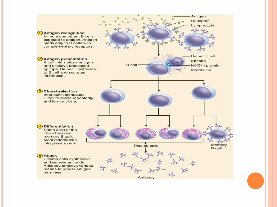
Multiple Myeloma is a cancer of the immune
system, in particular of the plasma cell a mature b
lymphocyte. Plasma cells produce antibodies(immunoglobulins)
that normally protect us from infection but in
multiple myeloma these
are nonfunctional and are called paraproteins.
All of the paraproteins from any one individual are
monoclonal (identical) because the myeloma cells
are identical clones of a single plasma cell.
UNDERSTANDING MULTIPLE MYELOMA
THE IMMUNE SYSTEM
Cellular Immune System
White Blood Cells: Granulocytes
Lymphocytes: T cells
Humoral Immune System
B cells and antibodies
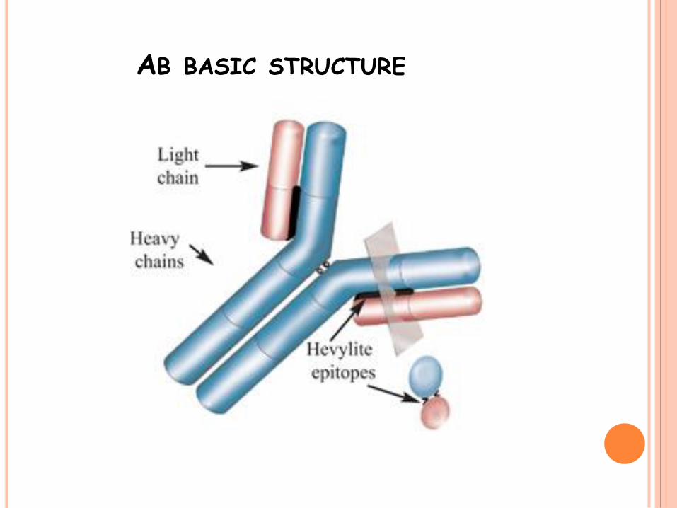
AB BASIC STRUCTURE
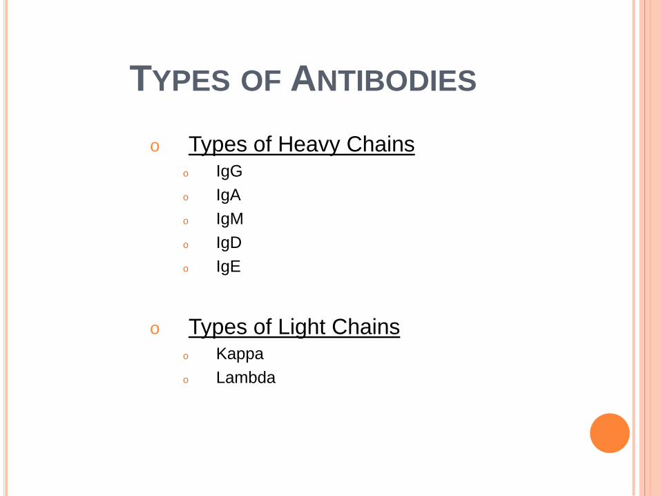
TYPES OF ANTIBODIES
o Types of Heavy Chains o IgG
o IgA
o IgM
o IgD
o IgE
o Types of Light Chains o Kappa
o Lambda
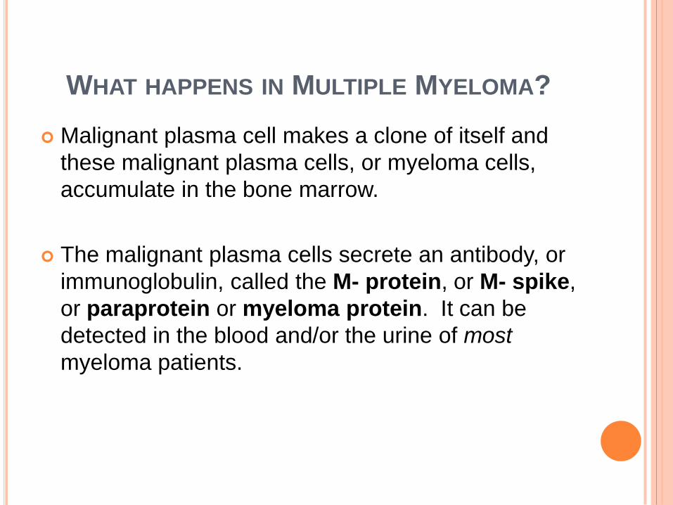
WHAT HAPPENS IN MULTIPLE MYELOMA?
Malignant plasma cell makes a clone of itself and
these malignant plasma cells, or myeloma cells,
accumulate in the bone marrow.
The malignant plasma cells secrete an antibody, or
immunoglobulin, called the M- protein, or M- spike,
or paraprotein or myeloma protein. It can be
detected in the blood and/or the urine of most
myeloma patients.
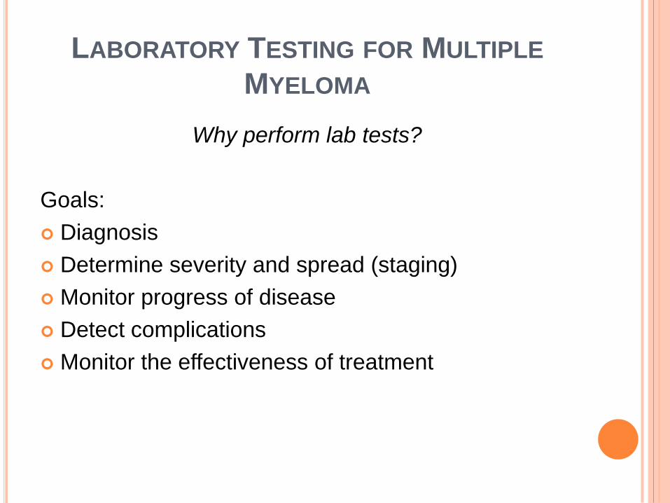
LABORATORY TESTING FOR MULTIPLE
MYELOMA
Why perform lab tests?
Goals:
Diagnosis
Determine severity and spread (staging)
Monitor progress of disease
Detect complications
Monitor the effectiveness of treatment
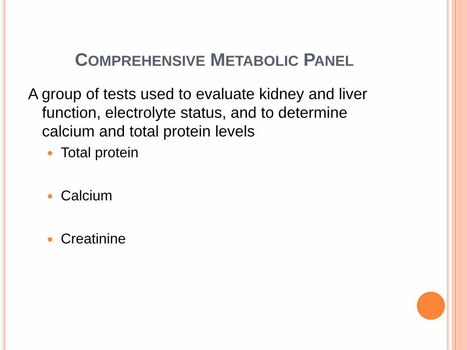
COMPREHENSIVE METABOLIC PANEL
A group of tests used to evaluate kidney and liver
function, electrolyte status, and to determine
calcium and total protein levels
Total protein
Calcium
Creatinine
OTHER CHEMISTRY TESTS IMPORTANT IN
MULTIPLE MYELOMA
Beta2-microglobulin
Serum albumin level
Serum Viscosity
COMPLETE BLOOD COUNT (CBC)
Hemoglobin / Hematocrit
- measure of red blood cells
- red blood cells carry oxygen
White Blood Cell Counts
-absolute neutrophil counts (ANC)
- fight off and protect against infection
Platelet Counts
-platelets are responsible for stopping bleeding and
blood clotting
QUANTITATIVE IMMUNOGLOBULINS
Measures amounts of the different
immunoglobulins
Myeloma protein will be an IgG or IgA, or
less commonly IgD or IgE
Levels will help monitor the course of the
disease
Important to be aware of the levels of the
normal (non-myeloma) immunoglobulins
PROTEIN ELECTROPHORESIS AND IMMUNOFIXATION
Protein electrophoresis separates the proteins in a blood or
urine sample into several groups based on their size and
electrical charge.
In most patients with MM, large amounts of an abnormal
immunoglobulin protein (M-spike) will appear as a large peak
on the graph.
Immunofixation is done to identify the specific type of protein
that is being produced by the malignant plasma cells. The
amount of protein produced may vary throughout the course
of the disease, but the type generally will remain the same.
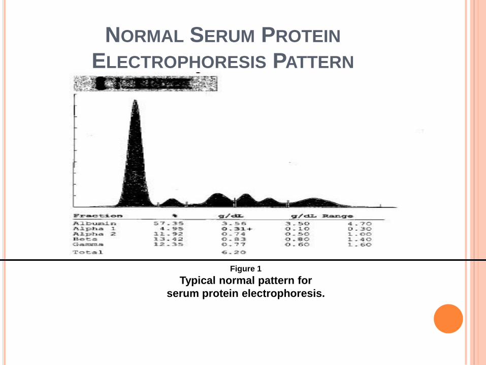
NORMAL SERUM PROTEIN
ELECTROPHORESIS PATTERN
Figure 1
Typical normal pattern for
serum protein electrophoresis.
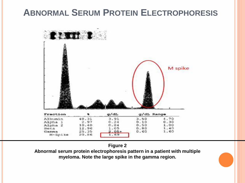
ABNORMAL SERUM PROTEIN ELECTROPHORESIS
PATTERN
Figure 2
Abnormal serum protein electrophoresis pattern in a patient with multiple
myeloma. Note the large spike in the gamma region.
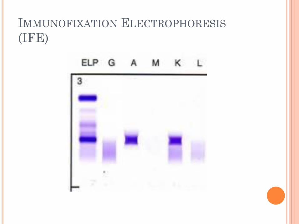
IMMUNOFIXATION ELECTROPHORESIS
(IFE)
SERUM FREE LIGHT CHAINS (FLC)
OR FREELITE ASSAY
Measures the amount of free light chains in the
serum (blood).
In normal circumstances, plasma cells produce an
excess of light chains compared to heavy chains. A
small amount of these light chains will not become
incorporated into intact immunoglobulins. These
are “free” light chains and are released into the
blood.
SERUM FREE LIGHT CHAINS (CONT’D)
Most patients with Multiple Myeloma produce increased amounts of either kappa or lambda free light chains, which can be measured in the blood.
Consequently, the ratio of kappa to lambda light chains is abnormal in most patients and is a sensitive indicator for this disease.
This test may be used to monitor progression and/or treatment.
BONE MARROW ASPIRATE AND BIOPSY
Most laboratory tests for Multiple Myeloma provide
indirect information about the amount of tumor
present, by measuring proteins that are secreted by
the tumor into the blood and/or the urine.
These tests do not provide the same information as
looking at the tumor itself. The myeloma cells are
usually only found inside the bone marrow.
BONE MARROW ASPIRATE AND BIOPSY (CONT’D)
Small fragments of bone marrow cells can be withdrawn with an aspirate needle, and a small core of bone and marrow is often removed at the same time with a different type of needle.
Both tests are done to determine how much of the normal bone marrow is replaced by myeloma cells.
Additional, specialized testing (cytogenetics and FISH) can be performed which can provide information about the biology of the tumor itself.
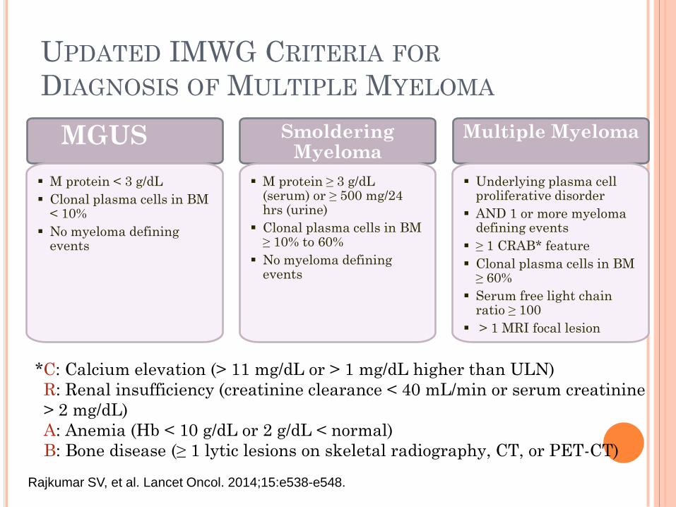
UPDATED IMWG CRITERIA FOR
DIAGNOSIS OF MULTIPLE MYELOMA
*C: Calcium elevation (> 11 mg/dL or > 1 mg/dL higher than ULN)
R: Renal insufficiency (creatinine clearance < 40 mL/min or serum creatinine
> 2 mg/dL)
A: Anemia (Hb < 10 g/dL or 2 g/dL < normal)
B: Bone disease (≥ 1 lytic lesions on skeletal radiography, CT, or PET-CT)
Rajkumar SV, et al. Lancet Oncol. 2014;15:e538-e548.
MGUS
M protein < 3 g/dL
Clonal plasma cells in BM < 10%
No myeloma defining events
Smoldering Myeloma
M protein ≥ 3 g/dL (serum) or ≥ 500 mg/24 hrs (urine)
Clonal plasma cells in BM ≥ 10% to 60%
No myeloma defining events
Multiple Myeloma
Underlying plasma cell proliferative disorder
AND 1 or more myeloma defining events
≥ 1 CRAB* feature
Clonal plasma cells in BM ≥ 60%
Serum free light chain ratio ≥ 100
> 1 MRI focal lesion
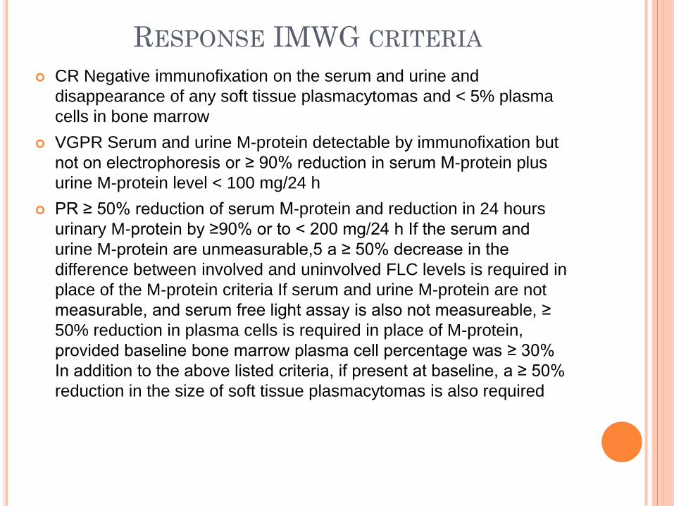
RESPONSE IMWG CRITERIA
CR Negative immunofixation on the serum and urine and
disappearance of any soft tissue plasmacytomas and < 5% plasma
cells in bone marrow
VGPR Serum and urine M-protein detectable by immunofixation but
not on electrophoresis or ≥ 90% reduction in serum M-protein plus
urine M-protein level < 100 mg/24 h
PR ≥ 50% reduction of serum M-protein and reduction in 24 hours
urinary M-protein by ≥90% or to < 200 mg/24 h If the serum and
urine M-protein are unmeasurable,5 a ≥ 50% decrease in the
difference between involved and uninvolved FLC levels is required in
place of the M-protein criteria If serum and urine M-protein are not
measurable, and serum free light assay is also not measureable, ≥
50% reduction in plasma cells is required in place of M-protein,
provided baseline bone marrow plasma cell percentage was ≥ 30%
In addition to the above listed criteria, if present at baseline, a ≥ 50%
reduction in the size of soft tissue plasmacytomas is also required
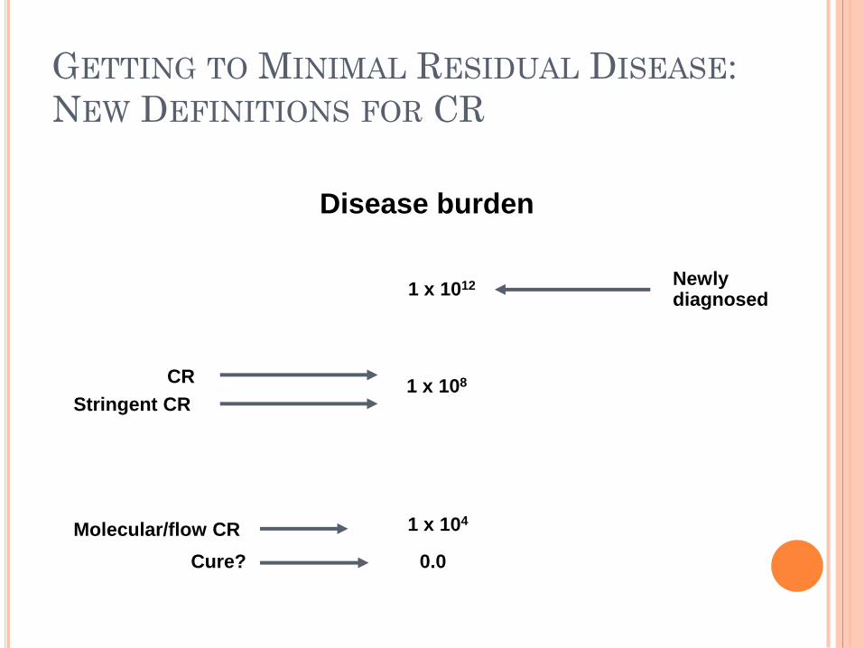
GETTING TO MINIMAL RESIDUAL DISEASE:
NEW DEFINITIONS FOR CR
1 x 10¹²
Stringent CR
Molecular/flow CR
Cure?
Disease burden
Newly diagnosed
1 x 10⁸
1 x 10⁴
0.0
CR
1.0
0.8
0.6
0.4
0.2
0 0 6 12 18 24 30 36 42
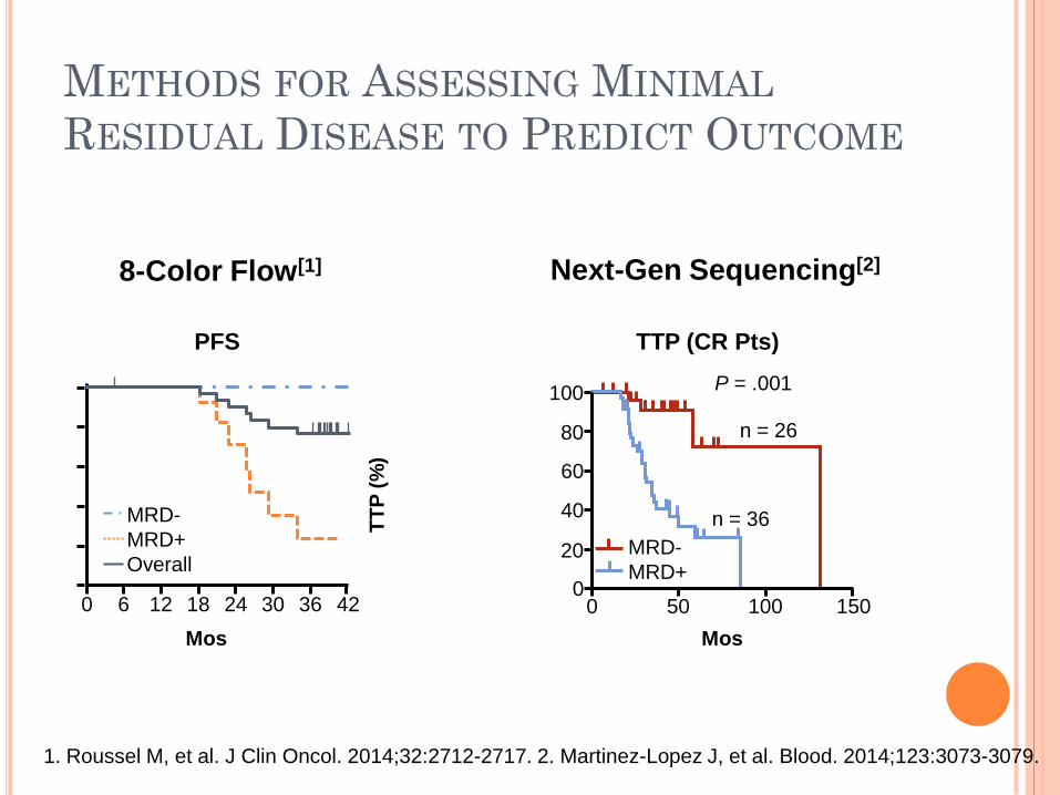
8-Color Flow[1] Next-Gen Sequencing[2]
TT
P (
%)
100
80
60
40
20
0 100 150 0 50
P = .001
n = 26
n = 36
TTP (CR Pts)
MRD-
MRD+
MRD-
MRD+
Overall
Mos
PF
S (
Pro
po
rtio
n)
Mos
PFS (CR Pts After
First-line Therapy) PFS
1. Roussel M, et al. J Clin Oncol. 2014;32:2712-2717. 2. Martinez-Lopez J, et al. Blood. 2014;123:3073-3079.
METHODS FOR ASSESSING MINIMAL
RESIDUAL DISEASE TO PREDICT OUTCOME
ROLE OF MRD ASSESSMENT
Remains a research tool, but indications are that
lower levels of MRD predict for better outcomes
Can contribute to better definition of response
Potential to monitor efficacy of therapy
Best, easily exportable method and optimal time
point is still under investigation
Even pts who achieve MRD- state can relapse, so
all may not be able to stop therapy
Unsure if changing therapy based on depth of
response alters survival outcomes, unsure of next
steps for MRD-