uncommon supramolecular phosphorescence‐capturing …
TRANSCRIPT

2104514 (1 of 9) © 2021 Wiley-VCH GmbH
www.small-journal.com
ReseaRch aRticle
Uncommon Supramolecular Phosphorescence-Capturing Assembly Based on Cucurbit[8]uril-Mediated Molecular Folding for Near-Infrared Lysosome Imaging
Man Huo, Xian-Yin Dai, and Yu Liu*
M. Huo, X.-Y. Dai, Y. LiuDepartment of ChemistryState Key Laboratory of Elemento-Organic ChemistryNankai UniversityTianjin 300071, P. R. ChinaE-mail: [email protected]
DOI: 10.1002/smll.202104514
hydrogen bonding,[19] π–π interaction,[20] host–guest interactions.[21,22] For example, Kim and co-workers reported the strategy to promote phosphorescence emission by using direct heavy atom effect (halogen and hydrogen bonding).[23] Zhao and co-workers presented a novel strategy to achieve color-tunable ultralong organic RTP by conjugating different phospho-rescence emitting agents onto a polymer backbone.[24] In particular, host–guest interactions have proven to be an acces-sible approach in achieving RTP emission both in solid- and solution-phase.[25,26] Host macrocyclic molecules with robust cavities, such as cyclodextrins[27] and cucurbiturils,[28] can provide a molecular vibration-limited microenvironment to inhibit the non-radiative relaxation decay of the phosphor and prevent excited tri-plet state from being quenched by oxygen or water. Our group previously reported an efficient solid-state RTP supermole-cule enhanced by cucurbit[6]uril, which
increased the phosphorescence quantum yield of 4-bromo-phenylpyridinium salt from 2.6% to 81.2%.[29] Notably, RTP materials in solution-phase are of particular interest for time-resolved biological imaging because of its unique character that it can be feasibly distinguished from the autofluorescence and background fluorescence in cellular biospecies.[30–32] However, the excited triplet state of phosphor tends to occur non-radiative relaxation decay caused by dissolved oxygen and water, so devel-oping purely organic RTP systems in aqueous solution is still limited, especially with NIR emissive performance.
Artificial light-harvesting systems (LHSs) are expectantly fabricated to mimic natural photosynthesis process for more efficient utilization of light energy, which are usually character-ized by high donor/acceptor ratio to ensure that large amounts of donors transfer energy to one acceptor.[33–35] Supramo-lecular self-assembly turns out to be a practicable approach to build LHSs through the encapsulation of the chromophores to enhance corresponding luminescence and avoid self-quenching effect.[36,37] Supramolecular LHSs can be conveniently obtained by mixing relevant building units driven by noncovalent inter-actions.[38] Cucurbit[8]uril (CB[8]), as a unique macrocyclic compound with stiff cavity, has been capable of accommo-dating charge-transfer complexes to form stable host–guest pairs, which can be utilized to design supramolecular-organic
The construction of highly effective phosphorescence energy transfer capturing system still remains great challenge in aqueous phase. Herein, a high-efficiency supramolecular purely organic room temperature phospho-rescence (RTP)-capturing system via a secondary assembly strategy by taking advantage of cucurbit[8]uril (CB[8]) and amphiphilic calixarene (SC4AH) is reported. Comparing with free bromonaphthalene-connected methoxyphenyl pyridinium salt (G), G⊂CB[8] exhibits an emerging RTP emission peak at 530 nm. Moreover, G⊂CB[8] further interacts with SC4AH to form the ternary assembly G⊂CB[8] @ SC4AH accompanied by remarkably enhanced RTP emission. Interestingly, RTP-capturing systems with delayed near-infrared (NIR) emissive performance (635, 675 nm) are feasibly acquired by intro-ducing Nile Red (NiR) or Nile Blue (NiB) as the acceptor into hydrophobic layer of G⊂CB[8] @ SC4AH, possessing ultrahigh antenna effects (352.9, 123.5) at a high donor/acceptor ratio (150:1, 300:1). More importantly, cell experiments indicate that G⊂CB[8] @ SC4AH/NiB not only hold low cytotox-icity but also can successfully realize NIR lysosome-targeted imaging of A549 cancer cells. This RTP-capturing system of delayed NIR emission via multi-stage assembly strategy offers a new approach for NIR imaging in living cells.
The ORCID identification number(s) for the author(s) of this article can be found under https://doi.org/10.1002/smll.202104514.
1. IntroductionPurely organic room temperature phosphorescence (RTP) mate-rials have aroused great research interests in the field of bio-imaging,[1–3] sensing,[4] information encryption,[5,6] and organic light emitting diodes[7] due to their long-lived photoemission and large Stokes shift.[8,9] Two vital factors contributing to RTP emission should be taken into consideration: 1) promoting intersystem crossing (ISC) from singlet to triplet state, 2) inhib-iting the process of non-radiative relaxation.[10] The common strategies to enhance RTP emission include introduction of heavy atom or other heteroatoms,[11,12] crystalline packing,[13–15] incorporation or covalent modification in polymers,[16–18]
Small 2021, 2104514

2104514 (2 of 9)
www.advancedsciencenews.com
© 2021 Wiley-VCH GmbH
www.small-journal.com
frameworks and supramolecular RTP materials.[39,40] Mean-while, amphiphilic p-sulfonatocalix[4]arene has been emerged as a significant building block for constructing supramolecular luminescent assemblies and biomedical nanosystems due to its special amphiphilicity.[41,42] Moreover, Förster resonance energy transfer (FRET) process is identified as an effective strategy to produce a large Stokes shift for luminescence to exhibit near-infrared (NIR) emission.[43,44] Especially, supramolecular systems with NIR emissive feature for biological imaging are highly desirable benefiting from their distinctive merits such as deep tissue penetration, moderate photo-damage, and min-imal interference distinguished from autofluorescence of bio-molecules.[45] Recently, George et al. developed tunable yellow and red afterglow fluorescence in solid state via the strategy of triplet-to-singlet FRET.[46] Consequently, inspired by the artifi-cial light-harvesting system based on FRET, utilization of phos-phorescence resonance energy transfer between RTP material and fluorescent dyes is hopeful to construct an artificial RTP-capturing system to achieve delayed NIR emission in aqueous phase.
Herein, we constructed a highly efficient phosphorescence energy transfer capturing system with delayed NIR emission via a secondary assembly strategy by using two kinds of mac-rocyclic molecules, CB[8] and amphiphilic calixarene p-sul-fonatocalix[4]arene tetrahexyl ether (SC4AH) Scheme 1. First, bromonaphthalene-connected methoxyphenyl pyridinium salt (G) was employed as the guest to associate with CB[8], leading to the appearance of a new RTP emission centered at 530 nm along with decrease of the original fluorescence at 425 nm. The resultant G⊂CB[8] subsequently secondarily assembled with SC4AH to form the ternary assembly G⊂CB[8] @ SC4AH, whose phosphorescence emission peak at 530 nm was dramatically enhanced as a result of the hydrophobic effect which efficiently avoided the triplet state non-radiative decay of phosphors. Fur-thermore, two hydrophobic organic dyes, NiR and NiB, as the acceptors were respectively introduced into G⊂CB[8] @ SC4AH assembly to fabricate RTP-capturing system due to the effective
overlap between their absorption band of each and the phos-phorescence emission band of the assembly. To our delight, the G⊂CB[8] @ SC4AH/NiR RTP-capturing system has an ultrahigh antenna effect (AE, as high as 352.9 at the donor/acceptor ratio of 150:1), while G⊂CB[8] @ SC4AH/NiB produced an excellent delayed NIR fluorescence emission (675 nm). Taking advantage of the delayed NIR emission of G⊂CB[8] @ SC4AH/NiB system in aqueous phase, it could be successfully applied for NIR lyso-some-targeted imaging of A549 cancer cells.
2. Results and Discussion
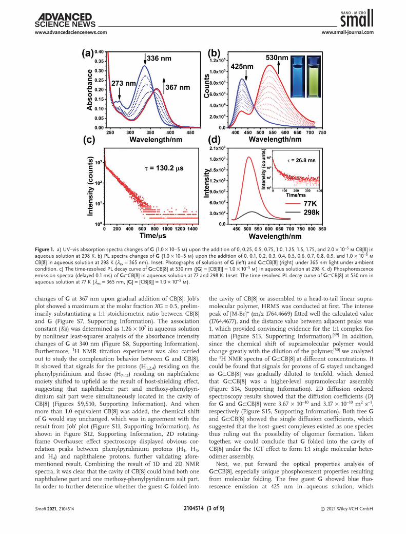
Compound G, 1-ethyl-4-(4-methoxyphenyl)pyridinium salt (G′) were synthesized according to the routes shown in Scheme S1, Supporting Information and fully characterized by 1H NMR, 13C NMR spectroscopies, and high-resolution mass spectrom-etry (HRMS) (Figures S1–S5, Supporting Information). It is well documented that CB[8], owing a large cavity, is compe-tent to hold an electron-deficient guest and an electron-rich guest through host-enhanced charge transfer interaction to form a 1:1:1 complex with high binding affinity.[47,48] In this study, the guest molecule G consisted of one electron-rich unit (bromonaphthalene) and one electron-deficient unit (methoxy-phenylpyridine salt), and it can be anticipated that they will be simultaneously included in the cavity of CB[8] owing to the intramolecular charge transfer (ICT). First, we investigated the host–guest recognition mode between CB[8] and G by UV–vis spectroscopy and NMR experiments. As shown in Figure 1a, upon stepwise addition of CB[8] into the solution of G, the maxima absorption of G was shifted bathochromically from 336 to 367 nm due to the enhancement of the ICT within the guest molecule by the inclusion of CB[8], demonstrating the forma-tion of host–guest inclusion complex G⊂CB[8]. The UV–vis absorption spectrum of CB[8] was also measured that showed very weak absorption at 260 nm (Figure S6, Supporting Infor-mation), which had negligible effect on the absorption spectra
Scheme 1. Schematic illustration of the construction of RTP-capturing system with delayed NIR emission in aqueous solution.
Small 2021, 2104514
2104514 (3 of 9)
www.advancedsciencenews.com
© 2021 Wiley-VCH GmbH
www.small-journal.com
changes of G at 367 nm upon gradual addition of CB[8]. Job’s plot showed a maximum at the molar fraction XG = 0.5, prelim-inarily substantiating a 1:1 stoichiometric ratio between CB[8] and G (Figure S7, Supporting Information). The association constant (Ks) was determined as 1.26 × 10⁷ in aqueous solution by nonlinear least-squares analysis of the absorbance intensity changes of G at 340 nm (Figure S8, Supporting Information). Furthermore, 1H NMR titration experiment was also carried out to study the complexation behavior between G and CB[8]. It showed that signals for the protons (H1,2,4) residing on the phenylpyridinium and those (H7–11) residing on naphthalene moiety shifted to upfield as the result of host-shielding effect, suggesting that naphthalene part and methoxy-phenylpyri-dinium salt part were simultaneously located in the cavity of CB[8] (Figures S9,S10, Supporting Information). And when more than 1.0 equivalent CB[8] was added, the chemical shift of G would stay unchanged, which was in agreement with the result from Job’ plot (Figure S11, Supporting Information). As shown in Figure S12, Supporting Information, 2D rotating-frame Overhauser effect spectroscopy displayed obvious cor-relation peaks between phenylpyridinium protons (H1, H3, and H4) and naphthalene protons, further validating afore-mentioned result. Combining the result of 1D and 2D NMR spectra, it was clear that the cavity of CB[8] could bind both one naphthalene part and one methoxy-phenylpyridinium salt part. In order to further determine whether the guest G folded into
the cavity of CB[8] or assembled to a head-to-tail linear supra-molecular polymer, HRMS was conducted at first. The intense peak of [M-Br]+ (m/z 1764.4669) fitted well the calculated value (1764.4677), and the distance value between adjacent peaks was 1, which provided convincing evidence for the 1:1 complex for-mation (Figure S13, Supporting Information).[49] In addition, since the chemical shift of supramolecular polymer would change greatly with the dilution of the polymer,[50] we analyzed the 1H NMR spectra of G⊂CB[8] at different concentrations. It could be found that signals for protons of G stayed unchanged as G⊂CB[8] was gradually diluted to tenfold, which denied that G⊂CB[8] was a higher-level supramolecular assembly (Figure S14, Supporting Information). 2D diffusion ordered spectroscopy results showed that the diffusion coefficients (D) for G and G⊂CB[8] were 3.67 × 10⁻¹⁰ and 3.17 × 10⁻¹⁰ m2 s−1, respectively (Figure S15, Supporting Information). Both free G and G⊂CB[8] showed the single diffusion coefficients, which suggested that the host–guest complexes existed as one species thus ruling out the possibility of oligomer formation. Taken together, we could conclude that G folded into the cavity of CB[8] under the ICT effect to form 1:1 single molecular heter-odimer assembly.
Next, we put forward the optical properties analysis of G⊂CB[8], especially unique phosphorescent properties resulting from molecular folding. The free guest G showed blue fluo-rescence emission at 425 nm in aqueous solution, which
Figure 1. a) UV–vis absorption spectra changes of G (1.0 × 10⁻⁵ m) upon the addition of 0, 0.25, 0.5, 0.75, 1.0, 1.25, 1.5, 1.75, and 2.0 × 10⁻⁵ m CB[8] in aqueous solution at 298 K. b) PL spectra changes of G (1.0 × 10⁻⁵ m) upon the addition of 0, 0.1, 0.2, 0.3, 0.4, 0.5, 0.6, 0.7, 0.8, 0.9, and 1.0 × 10⁻⁵ m CB[8] in aqueous solution at 298 K (λex = 365 nm). Inset: Photographs of solutions of G (left) and G⊂CB[8] (right) under 365 nm light under ambient condition. c) The time-resolved PL decay curve of G⊂CB[8] at 530 nm ([G] = [CB[8]] = 1.0 × 10⁻⁵ m) in aqueous solution at 298 K. d) Phosphorescence emission spectra (delayed 0.1 ms) of G⊂CB[8] in aqueous solution at 77 and 298 K. Inset: The time-resolved PL decay curve of G⊂CB[8] at 530 nm in aqueous solution at 77 K (λex = 365 nm, [G] = [CB[8]] = 1.0 × 10⁻⁵ m).
Small 2021, 2104514

2104514 (4 of 9)
www.advancedsciencenews.com
© 2021 Wiley-VCH GmbH
www.small-journal.com
was assigned to the emission of methoxyphenyl pyridinium (Figure S16, Supporting Information). The lifetime of free G at 425 nm was 1.1 ns, which was proved as the fluorescent emission (Figure S17, Supporting Information). As shown in Figure 1b, the photoluminescence (PL) spectra showed that a new emission peak centered at 530 nm emerged and increased accompanied by a decrease of the original fluorescence emission at 425 nm with the addition of CB[8] into the solution of G. Meanwhile, the solution color changed from blue to bright yellow under port-able UV lamp (365 nm) by the naked eye. The 1931 CIE chroma-ticity diagram also showed the trajectory of luminescence color changes upon stepwise addition of CB[8] (Figure S18, Supporting Information). Furthermore, the time-resolved PL decay curves were measured to determine the photophysical properties of the emerged emission peak at 530 nm, which indicated a lifetime of 130.2 µs for this emission peak (Figure 1c). And the lifetime on the order of microseconds suggested that there was an effective conversion of the singlet electrons to long-lived triplet state, indic-ative of enhanced ISC. In addition, the phosphorescence emis-sion spectra (delayed 0.1 ms) exhibited that the emission peak at 530 nm was increasing to maximum as CB[8] was gradually added into the solution under ambient conditions. After vacuum degassing of the solution, the emission intensity increased obvi-ously, and the lifetime of the emission peak at 530 nm extended to 219.4 µs (Figures S19,S20, Supporting Information), which was in accordance with the fact that oxygen could quench triplet-state electrons. More favorable evidence was that the long-lived emis-sion peak tremendously rose while the temperature went down to 77 K, with a longer lifetime of 26.8 ms because the non-radiative relaxation from triplet to ground state could be suppressed at low temperature (Figure 1d; Figure S21, Supporting Information). As a result, the emerged emission at 530 nm was determined to be phosphorescence emission in aqueous solution.
On the basis of charge recombination mechanism, the enhanced ISC efficiency of G could be attributed to CB[8]-enhanced charge transfer interaction.[25,51] The UV–vis absorp-tion spectrum of G showed two absorption peaks at around 260 and 340 nm (Figure 1a), respectively ascribed to naphtha-lene and methoxyphenyl pyridinium (Figure S16, Supporting Information). Then the original absorption peaks of G almost disappeared and a new absorption peak at 367 nm appeared after G binding with CB[8], suggesting the formation of CB[8]-mediated ICT assembly. Due to the strong binding between G and CB[8] in molecular folding mode, there was electronic cou-pling of naphthalene moiety and methoxyphenyl pyridinium salt moiety so that ICT effectively proceeded between them. Furthermore, upon addition of CB[8] into the aqueous solu-tion of G, the fluorescence emission quenching at 425 nm was observed, implying that the ISC process could be promoted as a result of host-enhanced ICT effect (Figure S22, Supporting Information). According to the time-resolved PL decay curve of G⊂CB[8], long-lived species was detected at 530 nm, which provided a critical evidence for the generation of triplet state (Figure 1c). The reasonable explanation was CB[8] provided an independent space to enhance ICT interaction between naph-thalene and methoxyphenyl pyridinium salt for improving ISC process to generate triplet. Besides, the robust rigid cavity of CB[8] could reduce the vibration of G and its triplet state was protected against quenching by solution and other molecules.
As a control experiment, cucurbit[7]uril (CB[7]) was utilized to bind with G for obtaining the G⊂CB[7] complex. According to 1H NMR titration experiment results, the binding stoichi-ometry was 1:1 between G and CB[7] (Figure S23, Supporting Information). It also showed that the signals for protons of methoxyphenyl pyridinium salt moiety exhibited upfield shifts, while protons of naphthalene showed down-field shifts. That meant only methoxyphenyl pyridinium moiety could be involved in the cavity of CB[7] excluding the molecular folding mode as G⊂CB[8]. Additionally, with stepwise addition of CB[7] (0.00–2.00 equiv.) into the aqueous solution of G, the emission at 425 nm was substantially increasing without new peak at 530 nm emerging (Figure S24, Supporting Information). Con-trol experiments illustrated that CB[7] will not engender phos-phorescence emission of G, while CB[8] plays an important role in inducing RTP of G in aqueous solution.
In order to further study the optical behaviors of this 1:1 single molecular heterodimer assembly G⊂CB[8], amphipathic SC4AH was introduced to secondarily assemble with G⊂CB[8] to form ternary assembly G⊂CB[8] @ SC4AH, allowing for the enhancement of phosphorescence emission. First, we exam-ined the transmission changes of G⊂CB[8] as the concentration of SC4AH from 0 to 0.07 mm (Figure S25, Supporting Informa-tion). The transmittance at 553 nm first slowly decreased and then dropped rapidly, and the inflection point was 0.04 mm for SC4AH. It should be noted that the molar ratio of SC4AH and G⊂CB[8] was determined as 4:3 which far exceeded that when SC4AH and G⊂CB[8] had equal negative and positive charges, suggesting that excess SC4AH could involve G⊂CB[8] into its hydrophobic layer to achieve secondary assembly under both hydrophobic interaction and electrostatic interaction.[52,53] Phos-phorescence emission spectra (delayed 0.1 ms) have also been performed to suggest the assembly-induced emission enhance-ment. When the concentration of SC4AH was twice as G⊂CB[8], the phosphorescence emission intensity of G⊂CB[8] @ SC4AH at 530 nm increased nearly 1.5-fold compared with the previous G⊂CB[8] due to the further suppression of non-radiative relaxa-tion after introduction of SC4AH (Figure 2a; Figure S26, Sup-porting Information). We carried out control experiment with p-sulfonatocalix[4]arene (SC4A) which lacked the alkyl chains to measure the phosphorescence changes of G⊂CB[8] upon the addition of SC4A. As shown in Figure S27, Supporting Infor-mation, it was found that SC4A could not enhance the phos-phorescence of G⊂CB[8]. This result suggested that the hydro-phobic layer of SC4AH played a key role in the co-assembly process. Therefore, to ensure that these ternary components were fully assembled, the concentration of SC4AH was chosen as 0.06 mm in the subsequent study of optical properties for this ternary assembly G⊂CB[8] @ SC4AH. Phosphorescence lifetime of G⊂CB[8] @ SC4AH was determined as t = 430.3 µs (Figure 2a; Figure S28, Supporting Information), which was about 3.3 times longer than before. It should be noted that the amphiphilic calixarene provided a hydrophobic environ-ment[37,53] for the phosphors to further prevent the excited tri-plet state from being quenched by water, so as to improve the efficiency of phosphorescence emission performance. Addition-ally, the total PL quantum yield of G⊂CB[8] was 4.14%, which substantially increased to 17.82% after co-assembly with SC4AH (Figure S29, Supporting Information). In contrast experiments,
Small 2021, 2104514

2104514 (5 of 9)
www.advancedsciencenews.com
© 2021 Wiley-VCH GmbH
www.small-journal.com
G/SC4AH mixture showed barely phosphorescence under same experimental set-up condition (Figure S30, Supporting Infor-mation). On the one hand, it was suggested that SC4AH could not induce phosphorescence emission of free G. On the other hand, it could also verify that there was no obvious competi-tive inclusion effect when SC4AH was added at twice the con-centration of G⊂CB[8]. Therefore, the introduction of SC4AH could indeed further strengthen phosphorescence emission of G⊂CB[8] instead of making G⊂CB[8] disassemble. Tyndall experiments, transmission electron microscopy (TEM), scan-ning electron microscopy (SEM), and atomic force microscopy (AFM) were also used to demonstrate the formation of ternary assembly. There were slight Tyndall effects of G and G⊂CB[8], while there was obvious Tyndall effects of G⊂CB[8] @ SC4AH (Figure S31, Supporting Information). TEM and SEM pro-vided visual information of G⊂CB[8] @ SC4AH assembly which exhibited several supramolecular nanoparticles with an average diameter of ≈200 nm (Figures S32a,S33a,b, Supporting Infor-mation). AFM also gave the similar morphology structure that a number of nanoparticles whose diameter was measured as 198 nm were found, and the height was determined to be about 38 nm, which were appreciably because of the shrinking of the sample upon air drying (Figure S33c,d, Supporting Information). A dynamic light scattering experiment was con-sistent with the above experimental results, which revealed that the average hydrodynamic diameter of G⊂CB[8] @ SC4AH assembly was 178.1 nm (Figure S32b, Supporting Information). The average zeta potential of the G⊂CB[8] @ SC4AH assembly was −43.19 mV, indicating that the surface of the assembly was negatively charged possessing biocompatibility and stability (Figure S34, Supporting Information).
Significantly, benefiting from the excellent phosphores-cence behaviors of this ternary assembly G⊂CB[8] @ SC4AH, we expected that the hydrophobic layer of the assembly can load substrates to construct a supramolecular light-capturing system. Considering that the organic dye NiR has little fluo-rescence in aqueous solution, while it has strong fluorescence emission in hydrophobic environment[36] and the absorption band of NiR possessed a good spectral overlap with emis-sion band of G⊂CB[8] @ SC4AH, thus it was chosen as the acceptor to build the phosphorescence energy transfer system (Figure 2b). When NiR was gradually added into the solution of G⊂CB[8] @ SC4AH assembly, the phosphorescence of donor at 530 nm kept decreasing while the emission of acceptor (635 nm) gradually increased (Figure 3a). Until the ratio of G⊂CB[8] @ SC4AH/NiR came to 150:1, the emission (delayed 0.1 ms) at 635 nm was nearly unchanging. To be specific, the emission peak (delayed 0.1 ms) at 635 nm ascribed to acceptors in the G⊂CB[8] @ SC4AH/NiR system was identical to the fluorescence emission of NiR, representing the characteristic delayed-fluorescence (DF) emission (Figure S35a, Supporting Information). According to time-resolved PL decay curve of G⊂CB[8] @ SC4AH/NiR, the lifetime for this DF emission at 635 nm was 276.7 µs, while the lifetime of free NiR was meas-ured as 1.0 ns (Figure 3f; Figure S36a, Supporting Information). Notably, the lifetime for ternary assembly G⊂CB[8] @ SC4AH at 530 nm was reduced to 201.6 µs at the donor/acceptor ratio of 150:1 (Figure 3e; Figure S28, Supporting Information). Such decrease also offered a strong case for the phosphorescence energy transfer process from triplet of donor to singlet of acceptor. In control experiments, the emission (delayed 0.1 ms) of NiR/SC4AH, NiR/G/SCA4AH, NiR/G⊂CB[8] at 635 nm was
Figure 2. a) Phosphorescence emission spectra (delayed 0.1 ms) of G⊂CB[8] (black) and G⊂CB[8] @ SC4AH ternary assembly (red) ([G] = [CB[8]] = 3.0 × 10⁻⁵ m, [SC4AH] = 6.0 × 10⁻⁵ m) in aqueous solution at 298 K. Inset: Time-resolved PL decay curve of G⊂CB[8] @ SC4AH ternary assembly at 530 nm at 298 K. b) Normalized emission spectrum of G⊂CB[8] @ SC4AH ternary assembly and absorption spectra of NiR and NiB. c) Simplified Jablonski dia-gram to explain the possible mechanism for RTP-capturing process in the present case. (Abs. = absorption, Fluo. = fluorescence, DF. = delayed fluores-cence, Non. rad. = non-radiation, ISC = intersystem crossing, Phos. = Phosphorescence, TS-FRET = triplet to singlet Förster resonance energy transfer).
Small 2021, 2104514

2104514 (6 of 9)
www.advancedsciencenews.com
© 2021 Wiley-VCH GmbH
www.small-journal.com
all silent, indicating that effective phosphorescence energy transfer between G⊂CB[8] and NiR was attributed to the co-assembly with amphiphilic calixarene (Figure S37, Supporting Information). Based on the above experimental results, we deduced that both G⊂CB[8] and NiR were located at the hydro-phobic layer of assembly, then energy transfer could effectively proceed from the triplet state of donor to the singlet state of acceptor and the possible mechanism for this RTP-capturing process is shown in Figure 2c. In control experiments, the usage of simple small-molecule surfactants such as sodium dodecyl benzene sulfonate and hexadecyl trimethyl ammo-nium bromide could not lead to effective energy transfer pro-cess, indicating that the pre-organized structure of amphiphilic SC4AH played a vital role in this phosphorescence-capturing system (Figures S38,S39, Supporting Information).
To confirm the phosphorescence-capturing adaptability of the G⊂CB[8] @ SC4AH ternary assembly, we selected another hydro-phobic dye, NiB, which had strong fluorescence emission in hydrophobic environment to investigate the behaviors of
phosphorescence energy transfer.[37] The absorption band of NiB could overlap with the emission band of G⊂CB[8] @ SC4AH assembly to a certain extent (Figure 2b). As shown in Figure 3c, with stepwise addition of NiB into the solution of G⊂CB[8] @ SC4AH assembly, the phosphorescence of donor at 530 nm decreased accompanied by the rising of acceptor emission at 675 nm. It was found that the fluorescence of NiB quenched at 675 nm when the ratio of donor/acceptor exceeded 300:1 which probably resulted from aggregation-caused quenching (ACQ) effect. Similarly, we could draw the analo-gous conclusion that the emerging emission peak at 675 nm in G⊂CB[8] @ SC4AH/NiB system was DF, by comparing the fluorescence of NiB with its emission peak (delayed 0.1 ms) in this phosphorescence-capturing system (Figure S35b, Sup-porting Information). There was the phosphorescence energy transfer process from the triplet state of donors to the singlet state of NiB molecules resulting from both of them being loaded into the hydrophobic layer of the assembly. As anticipated, the lifetime at 530 nm was declined to 145.8 µs in the case of the
Figure 3. a,c) Phosphorescence emission spectra (delayed 0.1 ms) and b,d) antenna effect/ФET of G⊂CB[8] @ SC4AH/NiR and G⊂CB[8] @ SC4AH/NiB at different donor/acceptor ratios in aqueous solution at 298 K ([G⊂CB[8]] = 3.0 × 10⁻⁵ m, [SC4AH] = 6.0 × 10⁻⁵ m, λex = 365 nm, 298 K). Time-resolved PL decay curves of G⊂CB[8] @ SC4AH, G⊂CB[8] @ SC4AH/NiR, and G⊂CB[8] @ SC4AH/NiB at e) 530 nm and at f) 635 or 675 nm in aqueous solution at 298 K ([G⊂CB[8]] = 3.0 × 10⁻⁵ m, [SC4AH] = 6.0 × 10⁻⁵ m, [NiR] = 2.0 × 10⁻⁷ m, and [NiB] = 2.4 × 10⁻⁷ m).
Small 2021, 2104514

2104514 (7 of 9)
www.advancedsciencenews.com
© 2021 Wiley-VCH GmbH
www.small-journal.com
G⊂CB[8] @ SC4AH/NiB system, further validating aforemen-tioned conclusion (Figure 3e; Figure S28, Supporting Informa-tion). The lifetime at 675 nm of NiB was 150.8 µs, while the life-time of free NiB was measured as 1.4 ns (Figure 3f; Figure S36b, Supporting Information). Notably, in contrast, phosphorescence emission (delayed 0.1 ms) of NiB/SC4AH, NiB/G/SCA4AH, and NiB/G⊂CB[8] at 675 nm were barely appreciable (Figure S37, Supporting Information). Furthermore, we performed control experiments of phosphorescence energy transfer taking advan-tage of SC4A to confirm that the hydrophobic layer of SC4AH was the necessary condition for phosphorescence energy transfer process. As shown in Figure S40, Supporting Information, it was observed that neither NiR nor NiB could induce efficient phos-phorescence energy transfer process in the G⊂CB[8] @ SC4A system because there is no hydrophobic environment. In com-parison with G⊂CB[8] @ SC4AH/NiR, G⊂CB[8] @ SC4AH/NiB exhibited an excellent NIR emission up to 675 nm, which would be more suitable for application in living cell imaging. We performed the phosphorescence emission (delayed 0.1 ms) of G⊂CB[8] @ SC4A/NiB at different time within 12 h in aqueous solution at 25 °C. As depicted in Figure S41, Supporting Infor-mation, there was no obvious change in the phosphorescence emission spectra of these assemblies within 12 h indicating the good stability of obtained nanoparticles in aqueous solution.
Subsequently, we investigated the energy transfer efficiency (ФET) and AE of the above two phosphorescence-capturing systems, which are important parameter for evaluating efficiency of light-capturing systems. In the case of G⊂CB[8] @ SC4AH/NiR, energy-transfer efficiency was calculated as 64.1% (Figure S42, Supporting Information), and the AE was 352.9 at a G⊂CB[8] @ SC4AH/NiR ratio of 150:1 (Figure 3b; Figure S45, Supporting Information). As for G⊂CB[8] @ SC4AH/NiB, the energy-transfer efficiency was 49.6% (Figure S43, Supporting Information), and the AE was calculated as 123.5 at a donor/acceptor ratio of 300:1 (Figure S46, Supporting Information). Moreover, the energy-transfer efficiency was 76.8% under the donor/acceptor ratio of 125:1 (Figure S44, Supporting Informa-tion). Such efficient AE was mainly because amphiphilic calix-arene not only created a hydrophobic environment to prevent the excited triplet state of phosphors from being quenched, but also accurately controlled the spatial distribution of donor and acceptor at isolated sites. Apparently, the uncommon value of AE of these ternary assemblies was extremely higher than the cor-responding values of recently reported LHSs both in solid state and aqueous environment (Table S1, Supporting Information).
By virtue of the excellent phosphorescence-capturing capa-bility of the G⊂CB[8] @ SC4AH/NiB system, we investigated its utility for NIR imaging in living cells. First, the cytotoxicity of these resultant assembly was evaluated by using human lung adenocarcinoma cells (A549 cancer cells) as a model followed by a standard cell counting kit-8 assay. It was observed that the G⊂CB[8] @ SC4AH/NiB assembly still showed negligible toxicity after incubation with A549 cells for 24 h even the con-centration up to 0.07 mm (Figure S47, Supporting Information). Subsequently, confocal laser scanning microscopy was utilized to examine the intracellular phosphorescence-capturing of the G⊂CB[8] @ SC4AH/NiB assembly. As shown in Figure S48, Supporting Information, cells incubated with G⊂CB[8] pre-sented relatively weak yellow luminescence, whereas strong
yellow luminescence was observed upon the cells were treated with G⊂CB[8] @ SC4AH for 12 h. These findings suggested that the G⊂CB[8] @ SC4AH assembly could display clear lumi-nescence signal after permeating the cells. Moreover, upon the cells were treated with G⊂CB[8] @ SC4AH/NiB assembly, bright NIR red luminescence was observed at 650–700 nm accompanied by the weakening of original yellow luminescence (Figure S49, Supporting Information), while the NIR red lumi-nescence of cells treated with free NiB was fairly weak when excited by 405 nm laser (Figure S50, Supporting Informa-tion). These results jointly demonstrated the phosphorescence energy transfer process from G⊂CB[8] @ SC4AH to NiB, which could be suitable for NIR imaging in living cells. More impor-tantly, colocalization assays were subsequently performed to visualize the subcellular distribution of G⊂CB[8] @ SC4AH/NiB assembly. A549 cells were first treated with G⊂CB[8] @ SC4AH/NiB assembly for 12 h and then co-stained with commercial staining reagents DAPI, MitoTracker Green, and LysoTracker Green, respectively. As shown in Figure 4c, the NIR red lumi-nescence of G⊂CB[8] @ SC4AH/NiB ranging from 650 to 700 nm overlapped well with LysoTracker Green accompanied by a high Pearson’s correlation coefficient of 0.90 (Figure S51a, Supporting Information), implying the specific accumulation of G⊂CB[8] @ SC4AH/NiB in lysosomes. In control groups, the nuclear dye DAPI (Figure 4a) and the mitochondrion dye MitoTraker Green (Figure 4b) all presented relatively poor correlation coefficients with G⊂CB[8] @ SC4AH/NiB, sug-gesting that the assembly neither localized in cell nucleus nor in mitochondria (Figure S51b,c, Supporting Informa-tion). Furthermore, when A549 cells were first incubated with G⊂CB[8] @ SC4AH/NiB for 12 h and then confocal microscopy images were respectively acquired after extra incubation time of 0 and 12 h, it could be observed that G⊂CB[8] @ SC4AH/NiB still mainly distributed in lysosomes accompanied by slightly decreased changes in emission intensity after 12 h extra incu-bation, proving that G⊂CB[8] @ SC4AH/NiB exhibited relatively good stability in living cells (Figure S52, Supporting Informa-tion). It was reported that lysosomes acted as waste disposal workstation in cells by digesting materials inside and outside cells.[54,55] Hence, G⊂CB[8] @ SC4AH/NiB might enter cells via the endocytosis process and then locate in lysosomes.
3. Conclusion
In conclusion, a ternary supramolecular assembly based on CB[8]-mediated single molecular folding was successfully constructed in two stages. CB[8] could first induce the single molecular het-erodimer assembly of bromonaphthalene-connected methoxy-phenyl pyridinium salt to awaken the RTP emission, which was further enhanced after secondary assembly with amphiphilic calixarene SC4AH. Moreover, two organic dyes, NiR and NiB, were respectively introduced as energy acceptors into the ter-nary assembly to build phosphorescence-capturing systems pos-sessing the ultrahigh AEs (352.9, 123.5) at a high donor/acceptor ratio (150:1, 300:1). In comparison with G⊂CB[8] @ SC4AH/NiR, G⊂CB[8] @ SC4AH/NiB displayed the markedly delayed NIR emission at 675 nm, which was successfully applied to NIR lys-osome-targeted imaging of A549 cancer cells. This uncommon
Small 2021, 2104514

2104514 (8 of 9)
www.advancedsciencenews.com
© 2021 Wiley-VCH GmbH
www.small-journal.com
supramolecular phosphorescence-capturing system with delayed NIR fluorescence performance not only sheds light on the con-struction of delayed NIR emissive assembly based on multistage assembly strategy, but also provides a convenient and feasible approach for targeted NIR imaging in living cells.
Supporting InformationSupporting Information is available from the Wiley Online Library or from the author.
AcknowledgementsThis work was financially supported by the National Natural Science Foundation of China (grant nos. 21772099 and 21861132001).
Conflict of InterestThe authors declare no conflict of interest.
Author ContributionsM.H. and X.-Y.D. contributed equally to this work. Y.L. acquired the funding, revised the manuscript, and supervised the project.
Data Availability StatementThe data that support the findings of this study are available from the corresponding author upon reasonable request.
Keywordsamphiphilic calixarene, cucurbit[8]uril, delayed near-infrared fluorescence, lysosome imaging, phosphorescence-capturing assembly
Received: July 30, 2021Revised: September 1, 2021
Published online:
[1] S. M. A. Fateminia, Z. Mao, S. Xu, Z. Yang, Z. Chi, B. Liu, Angew. Chem., Int. Ed. 2017, 56, 12160.
Figure 4. Confocal microscopy images of A549 cells co-stained with G⊂CB[8] @ SC4AH/NiB ([G⊂CB[8]] = 3.0 × 10⁻⁵ m, [SC4AH] = 6.0 × 10⁻⁵ m, and [NiB] = 1.0 × 10⁻⁷ m) and a) 4,6-diamidino-2-phenylindole (DAPI), b) MitoTracker Green, and c) LysoTracker Green, respectively. For DAPI, λex = 405 nm, λem = 420–470 nm. For MitoTracker Green, λex = 488 nm, λem = 500–550 nm. For LysoTracker Green, λex = 488 nm, λem = 500–550 nm. For G⊂CB[8] @ SC4AH/NiB (NIR emission), λex = 405 nm, λem = 650–700 nm.
Small 2021, 2104514

2104514 (9 of 9)
www.advancedsciencenews.com
© 2021 Wiley-VCH GmbH
www.small-journal.com
[2] J. Yang, X. Zhen, B. Wang, X. Gao, Z. Ren, J. Wang, Y. Xie, J. Li, Q. Peng, K. Pu, Z. Li, Nat. Commun. 2018, 9, 840.
[3] G. Zhang, G. M. Palmer, M. W. Dewhirst, C. L. Fraser, Nat. Mater. 2009, 8, 747.
[4] J.-J. Li, H.-Y. Zhang, Y. Zhang, W.-L. Zhou, Y. Liu, Adv. Opt. Mater. 2019, 7, 1900589.
[5] Z.-Y. Zhang, W.-W. Xu, W.-S. Xu, J. Niu, X.-H. Sun, Y. Liu, Angew. Chem., Int. Ed. 2020, 59, 18748.
[6] S. Cai, H. Shi, D. Tian, H. Ma, Z. Cheng, Q. Wu, M. Gu, L. Huang, Z. An, Q. Peng, W. Huang, Adv. Funct. Mater. 2018, 28, 1705045.
[7] R. Kabe, N. Notsuka, K. Yoshida, C. Adachi, Adv. Mater. 2016, 28, 655.
[8] Y. Yu, M. S. Kwon, J. Jung, Y. Zeng, M. Kim, K. Chung, J. Gierschner, J. H. Youk, S. M. Borisov, J. Kim, Angew. Chem., Int. Ed. 2017, 56, 16207.
[9] M. S. Kwon, Y. Yu, C. Coburn, A. W. Phillips, K. Chung, A. Shanker, J. Jung, G. Kim, K. Pipe, S. R. Forrest, J. H. Youk, J. Gierschner, J. Kim, Nat. Commun. 2015, 6, 8947.
[10] T. Zhang, X. Ma, H. Wu, H. Tian, Angew. Chem., Int. Ed. 2020, 59, 11206.
[11] D. R. Lee, K. H. Lee, W. Shao, C. L. Kim, J. Kim, J. Y. Lee, Chem. Mater. 2020, 32, 2583.
[12] S. Kuila, K. V. Rao, S. Garain, P. K. Samanta, S. Das, S. K. Pati, M. Eswaramoorthy, S. J. George, Angew. Chem., Int. Ed. 2018, 57, 17115.
[13] Z. He, W. Zhao, J. W. Y. Lam, Q. Peng, H. Ma, G. Liang, Z. Shuai, B. Z. Tang, Nat. Commun. 2017, 8, 416.
[14] X. F. Wang, W. J. Guo, H. Xiao, Q. Z. Yang, B. Chen, Y. Z. Chen, C. H. Tung, L. Z. Wu, Adv. Funct. Mater. 2020, 30, 1907282.
[15] X. F. Wang, H. Xiao, P. Z. Chen, Q. Z. Yang, B. Chen, C. H. Tung, Y. Z. Chen, L. Z. Wu, J. Am. Chem. Soc. 2019, 141, 5045
[16] X. Yao, J. Wang, D. Jiao, Z. Huang, O. Mhirsi, F. Lossada, L. Chen, B. Haehnle, A. J. C. Kuehne, X. Ma, H. Tian, A. Walther, Adv. Mater. 2021, 33, 2005973.
[17] Y. Su, Y. Zhang, Z. Wang, W. Gao, P. Jia, D. Zhang, C. Yang, Y. Li, Y. Zhao, Angew. Chem., Int. Ed. 2020, 59, 9967.
[18] X. Ma, C. Xu, J. Wang, H. Tian, Angew. Chem., Int. Ed. 2018, 57, 10854.
[19] X.-Q. Liu, K. Zhang, J.-F. Gao, Y.-Z. Chen, C.-H. Tung, L.-Z. Wu, Angew. Chem., Int. Ed. 2020, 59, 23456.
[20] W. Liu, J. Wang, Y. Gong, Q. Liao, Q. Dang, Z. Li, Z. Bo, Angew. Chem., Int. Ed. 2020, 59, 20161.
[21] Y. Lei, W. Dai, J. Guan, S. Guo, F. Ren, Y. Zhou, J. Shi, B. Tong, Z. Cai, J. Zheng, Y. Dong, Angew. Chem., Int. Ed. 2020, 59, 16054.
[22] P. Wei, X. Zhang, J. Liu, G.-G. Shan, H. Zhang, J. Qi, W. Zhao, H. H. Y. Sung, I. D. Williams, J. W. Y. Lam, B. Z. Tang, Angew. Chem., Int. Ed. 2020, 59, 9293.
[23] O. Bolton, K. Lee, H. J. Kim, K. Y. Lin, J. Kim, Nat. Chem. 2011, 3, 415.[24] L. Gu, H. Wu, H. Ma, W. Ye, W. Jia, H. Wang, H. Chen, N. Zhang,
D. Wang, C. Qian, Z. An, W. Huang, Y. Zhao, Nat. Commun. 2020, 11, 944.
[25] X. Ma, W. Zhang, Z. Liu, H. Zhang, B. Zhang, Y. Liu, Adv. Mater. 2021, 33, 2007476.
[26] W. Zhou, Y. Chen, Q. Yu, H. Zhang, Z. Liu, X. Dai, J. Li, Y. Liu, Nat. Commun. 2020, 11, 4655.
[27] D. Li, F. Lu, J. Wang, W. Hu, X. M. Cao, X. Ma, H. Tian, J. Am. Chem. Soc. 2018, 140, 1916.
[28] J. Wang, Z. Huang, X. Ma, H. Tian, Angew. Chem., Int. Ed. 2020, 59, 9928.
[29] Z.-Y. Zhang, Y. Chen, Y. Liu, Angew. Chem., Int. Ed. 2019, 58, 6028.[30] Y. Wang, H. Gao, J. Yang, M. Fang, D. Ding, B. Z. Tang, Z. Li, Adv.
Mater. 2021, 33, 2007811.[31] B. Zhao, H. Wang, M. Xie, C. Han, H. Yang, W. Zhao, Q. Zhao,
H. Xu, Adv. Photonics Res. 2020, 2, 2000096.[32] B. Zhao, G. Xie, H. Wang, C. Han, H. Xu, Chem. - Eur. J. 2019, 25,
1010.[33] Q. Song, S. Goia, J. Yang, S. C. L. Hall, M. Staniforth, V. G. Stavros,
S. Perrier, J. Am. Chem. Soc. 2021, 143, 382.[34] Y. Li, S. S. Rajasree, G. Y. Lee, J. Yu, J. H. Tang, R. Ni, G. Li,
K. N. Houk, P. Deria, P. J. Stang, J. Am. Chem. Soc. 2021, 143, 2908.[35] Z. Li, Y. Han, F. Wang, Nat. Commun. 2019, 10, 3735.[36] J.-J. Li, Y. Chen, J. Yu, N. Cheng, Y. Liu, Adv. Mater. 2017, 29,
1701905.[37] Z. Xu, S. Peng, Y. Y. Wang, J. K. Zhang, A. I. Lazar, D. S. Guo, Adv.
Mater. 2016, 28, 7666.[38] S. Guo, Y. Song, Y. He, X.-Y. Hu, L. Wang, Angew. Chem., Int. Ed.
2018, 57, 3163.[39] S. L. Cai, W. G. Zhang, R. N. Zuckermann, Z. T. Li, X. Zhao, Y. Liu,
Adv. Mater. 2015, 27, 5762.[40] X. Ma, J. Wang, H. Tian, Acc. Chem. Res. 2019, 52, 738.[41] P. Li, Y. Chen, Y. Liu, Chin. Chem. Lett. 2019, 30, 1190.[42] Y. C. Pan, X. Y. Hu, D. S. Guo, Angew. Chem., Int. Ed. 2021, 60,
2768.[43] X.-M. Chen, Q. Cao, H. K. Bisoyi, M. Wang, H. Yang, Q. Li, Angew.
Chem., Int. Ed. 2020, 59, 10493.[44] Y. Li, Y. Dong, L. Cheng, C. Qin, H. Nian, H. Zhang, Y. Yu, L. Cao,
J. Am. Chem. Soc. 2019, 141, 8412.[45] W. Xu, D. Wang, B. Z. Tang, Angew. Chem., Int. Ed. 2021, 60,
7476.[46] S. Kuila, S. J. George, Angew. Chem., Int. Ed. 2020, 59, 9393.[47] J. Liu, C. S. Y. Tan, O. A. Scherman, Angew. Chem., Int. Ed. 2018, 57,
8854.[48] H.-J. Kim, J. Heo, W. S. Jeon, E. Lee, J. Kim, S. Sakamoto,
K. Yamaguchi, K. Kim, Angew. Chem., Int. Ed. 2001, 40, 1526.[49] J. W. Lee, K. Kim, S. Choi, Y. H. Ko, S. Sakamoto, K. Yamaguchi,
K. Kim, Chem. Commun. 2002, 2692.[50] L. Chen, Y. Chen, H.-G. Fu, Y. Liu, Adv. Sci. 2020, 7, 2000803.[51] B. Tang, W. Xu, J.-F. Xu, X. Zhang, Angew. Chem., Int. Ed. 2021, 60,
9384.[52] Y. Wang, D. Guo, Y. Duan, Y. Wang, Y. Liu, Sci. Rep. 2015, 5,
9019.[53] X.-M. Chen, Y. Chen, Q. Yu, B.-H. Gu, Y. Liu, Angew. Chem., Int. Ed.
2018, 57, 12519.[54] H. Huang, B. Yu, P. Zhang, J. Huang, Y. Chen, G. Gasser, L. Ji,
H. Chao, Angew. Chem., Int. Ed. 2015, 54, 14049.[55] C. Settembre, A. Fraldi, D. L. Medina, A. Ballabio, Nat. Rev. Mol.
Cell Biol. 2013, 14, 283.
Small 2021, 2104514