ultrastructure of teliospores and promycelium and
TRANSCRIPT

Stephen F. Austin State University Stephen F. Austin State University
SFA ScholarWorks SFA ScholarWorks
Faculty Publications Biology
2007
Ultrastructure of Teliospores and Promycelium and Basidiospore Ultrastructure of Teliospores and Promycelium and Basidiospore
Formation in the Four-Spored Form of Gymnoconia Nitens, One of Formation in the Four-Spored Form of Gymnoconia Nitens, One of
the Causes of Orange Rust of Rubus the Causes of Orange Rust of Rubus
C. W. Mims
E. A. Richardson
Josephine Taylor Stephen F Austin State University, Department of Biology, [email protected]
Follow this and additional works at: https://scholarworks.sfasu.edu/biology
Part of the Biology Commons, and the Plant Sciences Commons
Tell us how this article helped you.
Repository Citation Repository Citation Mims, C. W.; Richardson, E. A.; and Taylor, Josephine, "Ultrastructure of Teliospores and Promycelium and Basidiospore Formation in the Four-Spored Form of Gymnoconia Nitens, One of the Causes of Orange Rust of Rubus" (2007). Faculty Publications. 93. https://scholarworks.sfasu.edu/biology/93
This Article is brought to you for free and open access by the Biology at SFA ScholarWorks. It has been accepted for inclusion in Faculty Publications by an authorized administrator of SFA ScholarWorks. For more information, please contact [email protected].

Ultrastructure of teliospores and promyceliumand basidiospore formation in the four-sporedform of Gymnoconia nitens, one of the causes oforange rust of Rubus
Charles W. Mims, Elizabeth A. Richardson, and Josephine Taylor
Abstract: Orange rust of Rubus is an interesting disease because of the fact that it can be caused by three different rustfungi that produce virtually identical symptoms. One is Gymnoconia peckiana (Howe in Peck) Trotter, which is a demicy-clic species, while the other two are endocyclic forms historically referred to as Gymnoconia nitens (Schwein.) Kern &H.W. Thurston. Although the spores produced on infected Rubus leaves by these latter two forms are morphologicallyidentical to the aeciospores of G. peckiana, they actually function as teliospores. However, the teliospores of one of theforms gives rise to two-celled promycelia that bear only two basidiospores, while teliospores of the other produce four-celled promycelia bearing four basidiospores. Here, we examined the teliospores of the four-spored form along with the se-quence of events that lead to basidiospore development. Developing and mature teliospores were binucleate, and we sawno evidence that karyogamy occurred in these spores. Upon germination, both spore nuclei migrated into the promyceliumand underwent mitosis to form a total of four nuclei. Four transverse septa then developed, creating four uninucleate cells.A tapered sterigma arose from each cell and gave rise to a basidiospore. These findings indicate that the basidiospores ofthe four-spored form of G. nitens were formed in an asexual fashion.
Key words: Uredinales, scanning electron microscopy, transmission electron microscopy.
Resume : La rouille orangee du Rubus constitue une maladie interessante, parce que trois champignons distincts de larouille peuvent la causer, en produisant des symptomes presque identiques. Le premier, une espece hemicyclique, appar-tient au Gymnoconia peckiana (Howe in Peck) Trotter, alors que l’on refere historiquement les deux autres, possedant desformes endocycliques, au Gymnoconia nitens (Schwein.) Kern & H.W. Thurston. Bien que les spores produites par cesdeux dernieres formes, sur les feuilles infectees du Rubus, possedent une morphologie identique a celle du G. peckiana,elles agissent plutot comme des teliospores. Cependant, alors que les teliospores d’une de ces formes produisent des pro-myceliums a deux cellules ne portant que deux basidiospores, les teliospores de l’autre forme produisent des promyceliumsa 4 cellules avec quatre basidiospores. Les auteurs ont examine les teliospores de la forme a quatre spores, ainsi que la se-quence des evenements qui conduisent au developpement de la basidiospore. Les teliospores en developpement et maturescomportent deux noyaux et ne montrent aucune evidence de caryogamie dans ces spores. Pendant la germination, les deuxnoyaux des deux spores migrent dans le promycelium et subissent la mitose pour former un total de quatre noyaux. Quatreseptations transverses se developpent, formant quatre cellules uninucleees. Un sterigmate effile apparaıt sur chaque cellulepour donner naissance a la basidiospore. Ces observations indiquent que les basidiospores de la forme a quatre spores duG. nitens se forment de facon asexuee.
Mots-cles : Uredinales, microscopie electronique par balayage, microscopie electronique en transmission.
[Traduit par la Redaction]
Introduction
Orange rust is an extremely common disease of both wildand cultivated blackberry (Rubus spp.) in the US, and hasbeen studied at the level of light-microscopy by numerous
early workers (Newcombe and Galloway 1890; Clinton1893; Olive 1908; Kunkel 1913, 1914, 1916; Dodge 1924).The disease is very important in the northeasternUS (Kleiner and Travis 1991), but is also widespread in thesouthern states. From a mycological standpoint, orange rustis particularly interesting because it can be caused by threedifferent pathogens that produce virtually identical symp-toms. One is Gymnoconia peckiana (Howe in Peck) Trotter,also known as Arthuriomyces peckianus (Howe in Peck)Cummins & Y. Hirats., which is demicyclic, while the othertwo are endocyclic forms historically referred to as Gymno-conia nitens (Schwein.) Kern & H.W. Thurston. However,according to Cummins and Hiratsuka (2003), neither ofthese the endocyclic forms has either a proper generic nameor a legitimate epithet. All three of the pathogens noted
Received 2 August 2007. Published on the NRC Research PressWeb site at canjbot.nrc.ca on 28 October 2007.
C.W. Mims.1 Department of Plant Pathology, University of GA,Athens, GA 30602, USA.E.A. Richardson. Department of Plant Biology, University ofGA, Athens, GA 30602, USA.J. Taylor. Department of Biology, Stephen F. Austin StateUniversity, Nacogdoches, TX 75962, USA.
1Corresponding author (e-mail: [email protected]).
926
Can. J. Bot. 85: 926–934 (2007) doi:10.1139/B07-097 # 2007 NRC Canada
Can
. J. B
ot. D
ownl
oade
d fr
om w
ww
.nrc
rese
arch
pres
s.co
m b
y D
IRE
CT
OR
AT
E O
F C
OL
DW
AT
ER
FIS
HE
RIE
S R
ES
on 1
0/12
/15
For
pers
onal
use
onl
y.

above are autoecious rusts that cause permanent, systemicinfections involving blackberry roots. Each spring, crownson infected roots give rise to weak, spindly shoots with typ-ically stunted and pale-green leaves whose undersides be-come bright orange as the result of the development oftremendous numbers of sori filled with spores (Kunkel1916; Dodge 1924; Kleiner and Travis 1991). In the case ofG. peckiana, these spores are aeciospores, but in the two en-docyclic forms they are actually caeomatoid teliospores (i.e.,spores that are formed in the same manner as the aecio-spores of the anamorphic rust genus Caeoma but whichfunction as teliospores). The study of orange-rust disease be-comes even more complicated because the two endocyclicforms, both referred to hereinafter as G. nitens, were likelyconfused with one another prior to 1924 (Dodge 1924). Oneof these forms possesses spermogonia and produces four-celled promycelia and a total of four basidiospores, whilethe other typically lacks spermogonia and produces two-celled promycelia with only two basidiospores. Whiletransmission electron microscopy (TEM) has been used toexamine aeciospores, aeciospore germination, and appresso-rium formation in G. peckiana (Swann and Mims 1991),there have been no detailed ultrastructural studies of eitherthe teliospores or the sequence of events leading to basidio-spore formation in the two endocyclic forms. In this paperwe provide ultrastructural data on the four-spored form. Theonly information currently available on the teliospores andthe events leading to basidiospore formation in this organ-ism comes from line drawings included in the early lightmicroscopic studies noted above.
Materials and methods
The specimens of G. nitens we examined came from in-fected southern-dewberry plants (Rubus trivialis Michx.)
collected near the city of Nacogdoches in central easternTexas. Infected shoots with leaves bearing sori were re-moved from plants and transported to the laboratory wheresome infected leaves were removed, and a razor blade wasused to excise small pieces bearing sori to be used in thestudy of developing and mature teliospores. Other shootswere left in water overnight before the leaves were removedand used as sources of spores for the germination study de-scribed below. For the study of developing teliospores, leafpieces were prepared for either scanning electron micro-scopy (SEM) or TEM using a standard glutaraldehyde–OsO4 fixation procedure (Taylor and Mims 1991). Samplesfor SEM were processed according to the procedures of En-kerli et al. (1997). Following critical-point drying, sampleswere mounted on specimen stubs using conductive tape,sputter-coated with gold, and examined using a JEOL 6400microscope operating at 15 kV. For TEM, thin sections ofresin-embedded samples were cut using a Reichert ultrami-crotome equipped with a diamond knife, collected on slotgrids, and allowed to dry on formvar-coated aluminum racks(Rowley and Moran 1975). Sections were post-stained withuranyl acetate followed by lead citrate, and examined usinga Zeiss 902A transmission electron microscope operating at80 kV. For light microscopy (LM) of resin-embeddedsamples, sections approximately 1 mm in thickness were cutusing a diamond histo-knife, collected on glass microscopeslides, stained with toluidine blue O, and examined andphotographed using bright-field microscopy.
To study the details of teliospore germination andbasidium/basidiospore formation, leaves bearing sori wereplaced in drops of water on glass microscope slides andtapped with the end of a wooden applicator stick to dis-lodge spores from sori. The leaves were discarded and theslides bearing the hydrating spores then were placed in amoist chamber on a laboratory bench at approximately
Figs. 1–3. Leaves of Rubus trivialis infected by Gymnoconia nitens. Fig. 1. Leaves whose undersides are covered with sori (near-actualsize). Figs. 2 and 3. Sori viewed using a dissecting microscope. Scale bars = 0.5 mm.
Mims et al. 927
# 2007 NRC Canada
Can
. J. B
ot. D
ownl
oade
d fr
om w
ww
.nrc
rese
arch
pres
s.co
m b
y D
IRE
CT
OR
AT
E O
F C
OL
DW
AT
ER
FIS
HE
RIE
S R
ES
on 1
0/12
/15
For
pers
onal
use
onl
y.

23 8C. At 1, 2, 4, 6, and 8 h time intervals after beingplaced in water, a gentle stream of water from a plasticsqueeze bottle was used to wash samples off the slidesand into the center of a Kimwipe1 tissue spread over andpushed into the mouth of a small beaker. A piece of threadwas used to tie off the depressed end of the tissue inwhich the samples accumulated, and the remainder of thetissue was cut off and discarded. The resulting small bagwas used to carry the samples through fixation, dehydra-tion, and resin embedment. Once samples had been com-pletely infiltrated with 100% resin, the thread wasremoved from the bag, its contents were teased into a small
amount of fresh 100% resin on a 25 mm � 75 mm plasticPermanox1 cell culture slide and an identical slide wasplaced on top of the resin. Following resin polymerization,the lower slide was removed by sliding a razor blade be-tween it and the thin layer of resin adhering to the upperslide. The samples in the resin layer were examined usingLM, and small rectangular pieces of resin-infiltrated sam-ples were excised using a pointed-tip blade, glued on blankresin blocks, and trimmed with a razor blade to produceblock faces for thin-sectioning (Mims et al. 1988). Thinsections were cut, collected, post-stained, and examined us-ing TEM, as described above.
Figs. 4–7. Scanning electron micrographs showing sori and teliospores of Gymnoconia nitens. Fig. 4. A young, circular sorus filled withspores. Scale bar = 100 mm. Fig. 5. A small portion of an older, elongated sorus. Scale bar = 40 mm. Fig. 6. Spores within a sorus (note theirangular nature). Scale bar = 20 mm. Fig. 7. A higher magnification view of the tiny warts present on spores. Scale bar = 10 mm.
928 Can. J. Bot. Vol. 85, 2007
# 2007 NRC Canada
Can
. J. B
ot. D
ownl
oade
d fr
om w
ww
.nrc
rese
arch
pres
s.co
m b
y D
IRE
CT
OR
AT
E O
F C
OL
DW
AT
ER
FIS
HE
RIE
S R
ES
on 1
0/12
/15
For
pers
onal
use
onl
y.

Results
Sori of G. nitens almost completely covered the under-sides of most infected leaves of R. trivialis (Figs. 1–3).
Although young sori were small with roughly sphericalmouths (Fig. 4), older sori were much larger and usuallyvery elongated structures (Figs. 2, 3, and 5). Each soruswas packed with caeomatoid teliospores that possessed dis-
Figs. 8–14. Light (Figs. 8, 9, 13, and 14) and transmission electron micrographs (Figs. 10–12) illustrating details of teliospore formationand promycelium formation in Gymnoconia nitens. Fig. 8. A low magnification view of a portion of a sectioned sorus showing chains ofdeveloping spores. Scale bar = 100 mm. Fig. 9. Slightly higher magnification view of sporogenous cells (asterisks) and chains of developingand mature teliospores. Developing and mature spores in which two nuclei are visible are shown at the arrowheads. Scale bar = 40 mm.Fig. 10. A section showing a binucleate sporogenous cell (SC) and a binucleate teliospore initial (TI) with nuclei shown at N. Scale bar =1.5 mm. Fig. 11. A section showing the two nuclei (N) of a young teliospore (T) with a disjunctor cell visible at DC. Scale bar = 1.5 mm.Fig. 12. A mature teliospore in which both nuclei (N) are visible. Scale bar = 1.5 mm. Figs. 13 and 14. Flat-embedded promycelia withnuclei visible at the arrows. Scale bars = 50 mm.
Mims et al. 929
# 2007 NRC Canada
Can
. J. B
ot. D
ownl
oade
d fr
om w
ww
.nrc
rese
arch
pres
s.co
m b
y D
IRE
CT
OR
AT
E O
F C
OL
DW
AT
ER
FIS
HE
RIE
S R
ES
on 1
0/12
/15
For
pers
onal
use
onl
y.

tinctly angular surface features when viewed using SEM(Fig. 6). Most spores measured 17–18 mm � 20–21 mm andwere covered with tiny, rounded warts (Fig. 7).
LM of sections of resin-embedded sori revealed chains ofteliospore initials and developing teliospores that arose froma basal layer of sporogenous cells (Figs. 8 and 9). Eachsporogenous cell was binucleate and gave rise to a series of
binucleate teliospore initials (Fig. 10), each of which subse-quently divided to form a larger apical teliospore and asmaller basal disjunctor cell (Fig. 11). Examination of serialsections confirmed that each young teliospore was binu-cleate. We found no evidence of karyogamy during eitherteliospore formation or maturation, and the mature sporeswe examined were binucleate (Fig. 12).
Figs. 15–18. Transmission electron micrographs of teliospores of Gymnoconia nitens. Fig. 15. A hydrated spore prior to the emergence ofa germ tube with the spore nuclei shown at N, lipid bodies at L, and germ pore regions at the arrowheads. Fig. 16. A hydrated spore withmore prominent germ-pore regions (arrowheads) than in Fig. 15. Only one of the two spore nuclei (N) is visible. Fig. 17. An early stage inteliospore germination where the germ tube (GT) or developing promycelium had just emerged from a germ-pore region. Fig. 18. A devel-oping promycelium (arrow) where the nuclei (N) are still in the spore. Scale bars = 1.5 mm.
930 Can. J. Bot. Vol. 85, 2007
# 2007 NRC Canada
Can
. J. B
ot. D
ownl
oade
d fr
om w
ww
.nrc
rese
arch
pres
s.co
m b
y D
IRE
CT
OR
AT
E O
F C
OL
DW
AT
ER
FIS
HE
RIE
S R
ES
on 1
0/12
/15
For
pers
onal
use
onl
y.
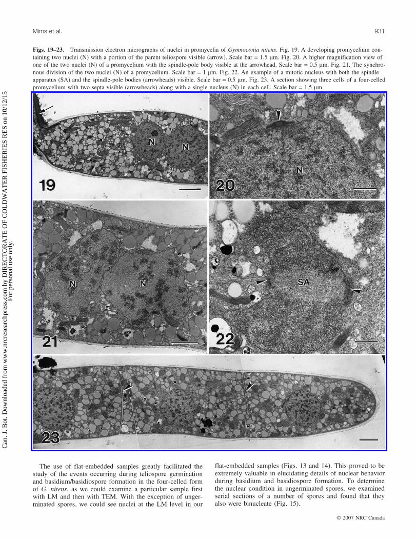
The use of flat-embedded samples greatly facilitated thestudy of the events occurring during teliospore germinationand basidium/basidiospore formation in the four-celled formof G. nitens, as we could examine a particular sample firstwith LM and then with TEM. With the exception of unger-minated spores, we could see nuclei at the LM level in our
flat-embedded samples (Figs. 13 and 14). This proved to beextremely valuable in elucidating details of nuclear behaviorduring basidium and basidiospore formation. To determinethe nuclear condition in ungerminated spores, we examinedserial sections of a number of spores and found that theyalso were binucleate (Fig. 15).
Figs. 19–23. Transmission electron micrographs of nuclei in promycelia of Gymnoconia nitens. Fig. 19. A developing promycelium con-taining two nuclei (N) with a portion of the parent teliospore visible (arrow). Scale bar = 1.5 mm. Fig. 20. A higher magnification view ofone of the two nuclei (N) of a promycelium with the spindle-pole body visible at the arrowhead. Scale bar = 0.5 mm. Fig. 21. The synchro-nous division of the two nuclei (N) of a promycelium. Scale bar = 1 mm. Fig. 22. An example of a mitotic nucleus with both the spindleapparatus (SA) and the spindle-pole bodies (arrowheads) visible. Scale bar = 0.5 mm. Fig. 23. A section showing three cells of a four-celledpromycelium with two septa visible (arrowheads) along with a single nucleus (N) in each cell. Scale bar = 1.5 mm.
Mims et al. 931
# 2007 NRC Canada
Can
. J. B
ot. D
ownl
oade
d fr
om w
ww
.nrc
rese
arch
pres
s.co
m b
y D
IRE
CT
OR
AT
E O
F C
OL
DW
AT
ER
FIS
HE
RIE
S R
ES
on 1
0/12
/15
For
pers
onal
use
onl
y.

Early stages of germ-tube emergence were common at the4 h sampling period. However, some spores did not begin togerminate until 6 or even 8 h after the onset of hydrationand many showed no evidence of germination even at 8 h.Each teliospore was packed with lipid bodies and possesseda number of germ pore regions in its wall (Figs. 15 and 16).Upon germination, a single germ tube emerged from eachspore (Figs. 17 and 18). Most of the spore cytoplasm aswell as both nuclei moved into the germ tube, which willbe referred to here as the promycelium. Fully elongatedpromycelia were somewhat variable in length but mostmeasured 80–105 mm. The two nuclei eventually becamesituated near the middle of a promycelium (Fig. 19). A
duplicated spindle-pole body was present on the surface ofeach nucleus (Fig. 21). The two nuclei divided synchro-nously (Figs. 21 and 22), and the four resulting daughternuclei became distributed more or less evenly along thelength of the promycelium. In most instances, four septathen developed to divide the promycelium into four uninu-cleate cells, three of which are visible in Fig. 23. Althoughsome four-celled promycelia were observed as early as 6 hafter the onset of hydration, they were much more commonat 8 h. By 8 h, basidiospores had also begun to develop onmany promycelia (Fig. 24). The first cell to give rise to asterigma was typically the apical cell (Fig. 25). The tip of amaturing sterigma became distinctly pointed (Fig. 26) prior
Fig. 24–28. Light (Fig. 24) and transmission electron micrographs (Figs. 25–28) of basidiospore development in Gymnoconia nitens. Fig.24. A maturing promycelium with four developing basidiospores (arrows). Scale bar = 50 mm. Fig. 25. The apical cell of a promyceliumwith a developing sterigma (S) with the nucleus is visible at N. Fig. 26. The tapered tip (arrowhead) of an elongating sterigma (S). Fig. 27.A developing basidiospore (BS) at the tip of a sterigma (S) where the nucleus has not yet entered the spore. Fig. 28. A maturing basidios-pore containing a mitotic nucleus (N). Scale bars = 1.5 mm for Figs. 25–28.
932 Can. J. Bot. Vol. 85, 2007
# 2007 NRC Canada
Can
. J. B
ot. D
ownl
oade
d fr
om w
ww
.nrc
rese
arch
pres
s.co
m b
y D
IRE
CT
OR
AT
E O
F C
OL
DW
AT
ER
FIS
HE
RIE
S R
ES
on 1
0/12
/15
For
pers
onal
use
onl
y.

to forming a basidiospore (Fig. 27). Most of the cytoplasmand the nucleus of the parent cell eventually migrated intothe basidiospore. Once in the spore the nucleus underwentmitosis (Fig. 28).
Discussion
The most significant finding in this study relates to thefact that while the four-celled form of G. nitens formspromycelia and basidiospores that are morphologicallyidentical to those produced by most other rust fungi, itapparently does so without undergoing sexual reproduction.The asexual production of promycelia and basidiospores inrust fungi has been reported previously in a few microcyclicspecies (Hiratsuka and Sato 1982), but this is the first timethe phenomenon has been demonstrated with TEM. TEMhas previously proven to be a valuable method for elucidat-ing details of the nuclear cycle in rust fungi, and has beenused to document the sites of nuclear fusion and meiosis ina number of rust fungi. Based upon reports of synaptonemalcomplexes in the nuclei of young rust teliospores (Mims1977, 1981; Boehm and Bushnell 1992; Boehm et al.1992;Mims et al. 1996; Mims and Richardson 2005), it appearsthat both karyogamy and prophase I of meiosis occur inyoung teliospores. Meiosis then appears to be arrested inlate prophase and not completed until after the nucleus hasmigrated into the promycelium (see O’Donnell andMcLaughlin 1981a, 1981b) that eventually arises from agerminating teliospore. In the four-celled form of G. nitensexamined here, we found no evidence that either karyogamyor meiosis occurred during the formation, maturation, orgermination of the caeomatoid teliospores. Both developingand mature teliospores were found to be binucleate and,following teliospore germination, both nuclei migrated intothe promycelium where they divided mitotically to yieldfour nuclei. This conforms to the type II behavior describedby Hiratsuka and Sato (1982). Overall, Hiratsuka and Sato(1982) described a total of eight types of nuclear behaviorin rust teliospores and basidia, which they referred to astypes I–VIII. They stated that the nuclei in speciesexhibiting type II behavior were haploid, but provided nodirect evidence to support this statement. Here we shouldnote that monokaryotic strains of certain holobasidiomycetes(see Stahl and Esser 1976) have also been reported to formbasidiospores without karyogamy and meiosis.
Although various workers have reported some minor sizeand possible color differences between the caeomatoidteliospores of G. nitens and the aeciospores of G. peckiana,the only reliable way to differentiate between these twotypes of spores is to germinate them. On inductive surfacessuch as pieces of dialysis membrane, an aeciospore ofG. peckiana gives rise to very short germ tube whose tipquickly differentiates into a swollen appressorium (Swannand Mims 1991), while as described in this study, the germtube or promycelium that arises from a caeomatoidteliospore of G. nitens eventually forms basidiospores.
The overall process of teliospore formation in G. nitensappears to be identical to that of aeciospore formation inother rust fungi. As described by various other workers (seeLittlefield and Heath 1979), the base of an aecium is linedwith binucleate mother cells that give rise to a succession
of binucleate sporogenous cells. Each sporogenous celldivides to form a small binucleate basal cell known as adisjunctor cells that eventually dies and a larger binucleatecell that develops into an aeciospore.
ReferencesBoehm, E.W.A., and Bushnell, W.R. 1992. An ultrastructural
pachytene karyotype for Melampsora lini. Phytopathology, 82:1212–1218.
Boehm, E.W.A., Wenstrom, J.C., McLaughlin, D.J., Szabo, L.J.,Roelfs, A.P., and Bushnell, W.R. 1992. An ultrastructural pachy-tene karyotype for Puccinia graminis f. sp. tritici. Can. J. Bot.70: 401–413. doi:10.1139/b92-054.
Clinton, G.P. 1893. Orange rust of raspberry and blackberry. Ill.Agr. Exp. Sta. Bull. 29: 273–300.
Cummins, G.B., and Hiratsuka, Y. 2003. Illustrated genera of rustfungi. 3rd ed. APS Press, St. Paul, Minn.
Dodge, B.O. 1924. Uninucleated aecidiospores in Caeoma nitens,and associated phenomena. J. Agric. Res. 28: 1045–1058.
Enkerli, K.M., Hahn, M.G., and Mims, C.W. 1997. Ultrastructureof compatible and incompatible interactions of soybean roots in-fected with the plant pathogenic oomycete Phytophthora sojae.Can. J. Bot. 75: 1493–1508. doi: 10.1139/b97-864.
Hiratsuka, Y., and Sato, S. 1982. Morphology and taxonomy of rustfungi. In The rust fungi. Edited by K.J. Scott andA.K. Chakravorty. Academic Press, New York. pp. 1–36.
Kleiner, W.C., and Travis, J.W. 1991. Orange rust. In Compendiumof raspberry and blackberry diseases and insects. Edited byM.A. Ellis, R.H. Converse, R.N. Williams and B. Williamson.APS Press, St. Paul, Minn. pp. 26–28.
Kunkel, O. 1913. The production of a promycelium by the aecios-pores of Caeoma nitens Burrill. Bull. Torrey Bot. Club, 40:361–366. doi:10.2307/2479880.
Kunkel, O. 1914. Nuclear behavior in the promycelia of Caeomanitens Burrill and Puccinia peckiana Howe. Am. J. Bot. 1: 37–46.doi:10.2307/2434959.
Kunkel, O. 1916. Further studies of the orange rusts of Rubus inthe United States. Bull. Torrey Bot. Club, 43: 559–567. doi:10.2307/2479608.
Littlefield, L.J., and Heath, M.C. 1979. Ultrastructure of rust fungi.Academic Press, New York.
Mims, C.W. 1977. Ultrastructure of teliospore germination in thecedar apple rust fungus Gymnosporangium juniperi-virginianae.Can. J. Bot. 55: 2319–2329. doi:10.1139/b77-263.
Mims, C.W. 1981. Ultrastructure of teliospore germination andbasidiospore formation in the rust fungus Gymnosporangiumclavipes. Can. J. Bot. 59: 1041–1049. doi:10.1139/b81-142.
Mims, C.W., and Richardson, E.A. 2005. Light and electron micro-scopy of teliospores and teliospore germination in the rust fun-gus Coleosporium ipomoeae. Can. J. Bot. 83: 451–458. doi:10.1139/b05-020.
Mims, C.W., Richardson, E.A., and Timberlake, W.E. 1988. Ultra-structural analysis of conidiophore development in the fungusAspergillis nidulans using freeze-substitution. Protoplasma, 144:132–141. doi:10.1007/BF01637246.
Mims, C.W., Liljebjelke, K.A., and Covert, S.F. 1996. Ultrastruc-ture of telia and teliospores of the rust fungus Cronartium quer-cuum f. sp. fusiforme. Mycologia, 88: 47–56. doi:10.2307/3760783.
Newcombe, F.C., and Galloway, B.T. 1890. Perennial mycelium ofthe fungus of the blackberry rust. J. Mycol. Plant Pathol 6:106–107.
O’Donnell, K.L., and McLaughlin, D.J. 1981a. Ultrastructure of
Mims et al. 933
# 2007 NRC Canada
Can
. J. B
ot. D
ownl
oade
d fr
om w
ww
.nrc
rese
arch
pres
s.co
m b
y D
IRE
CT
OR
AT
E O
F C
OL
DW
AT
ER
FIS
HE
RIE
S R
ES
on 1
0/12
/15
For
pers
onal
use
onl
y.

meiosis in the hollyhock rust fungus, Puccinia malvacearum.I. Prophase I – prometaphase I. Protoplasma, 108: 225–244.doi:10.1007/BF02224421.
O’Donnell, K.L., and McLaughlin, D.J. 1981b. Ultrastructure ofmeiosis in the hollyhock rust fungus, Puccinia malvacearum. II.Metaphase I – telophase I. Protoplasma, 108: 245–263. doi:10.1007/BF02224422.
Olive, E.W. 1908. Sexual cell fusions and vegetative nuclear divi-sions in the rusts. Ann. Bot. (Lond.), 22: 333–360.
Rowley, J.C., and Moran, D.T. 1975. A simple procedure formounting wrinkle-free sections on formvar coated slot grids.Ultramicroscopy, 1: 151–155. PMID:800684.
Stahl, U., and Esser, K. 1976. Genetics of fruit body production insome higher basidiomycetes. Mol. Gen. Genet. 148: 183–197.doi:10.1007/BF00268384.
Swann, E.C., and Mims, C.W. 1991. Ultrastructure of freeze-substituted appressoria produced by aeciospore germlings ofthe rust fungus Arthuriomyces peckianus. Can. J. Bot. 69:1655–1665. doi:10.1139/b91-210.
Taylor, J., and Mims, C.W. 1991. Fungal development and hostcell responses to the rust fungus Puccinia substriata var. indicain seedlings and mature leaves of susceptible and resistant pearlmillet. Can. J. Bot. 69: 1207–1219. doi:10.1139/b91-155.
934 Can. J. Bot. Vol. 85, 2007
# 2007 NRC Canada
Can
. J. B
ot. D
ownl
oade
d fr
om w
ww
.nrc
rese
arch
pres
s.co
m b
y D
IRE
CT
OR
AT
E O
F C
OL
DW
AT
ER
FIS
HE
RIE
S R
ES
on 1
0/12
/15
For
pers
onal
use
onl
y.

This article has been cited by:
1. Charles W.MimsC.W. Mims, Elizabeth A.RichardsonE.A. Richardson. 2008. Light and electron microscopy of the spermogonialstage of Gymnoconia peckiana, one of the causes of orange rust of Rubus. Botany 86:5, 533-538. [Abstract] [Full Text] [PDF][PDF Plus]
2. Charles W.MimsC.W. Mims, Elizabeth A.RichardsonE.A. Richardson. 2007. Ultrastructure of teliospores and promycelium andbasidiospore formation in the two-spored form of Gymnoconia nitens, one of the causes of orange rust of Rubus. CanadianJournal of Botany 85:10, 935-940. [Abstract] [Full Text] [PDF] [PDF Plus]
Can
. J. B
ot. D
ownl
oade
d fr
om w
ww
.nrc
rese
arch
pres
s.co
m b
y D
IRE
CT
OR
AT
E O
F C
OL
DW
AT
ER
FIS
HE
RIE
S R
ES
on 1
0/12
/15
For
pers
onal
use
onl
y.