ultrasound-guided pain interventions in shoulder region · letal shoulder pain, especially when...
TRANSCRIPT
Available online at www.sciencedirect.com
T E C H N I Q U E S I N R E G I O N A L A N E S T H E S I A A N D P A I N M A N A G E M E N T 1 7 ( 2 0 1 3 ) 8 1 – 9 5
1084-208X/$ - see frohttp://dx.doi.org/10.
nCorresponding auE-mail address:
www.elsevier.com/locate/trap
Ultrasound-guided pain interventions inshoulder region
Concepcion del-Olmo, MDa,n, Pilar de-Diego, MDa, Paloma Morillas, MDb,Miguel Garcia-Navlet, MDc
aDepartment of Anesthesia and Pain Medicine, Hospital ASEPEYO, Calle Joaquin de Cardenas 2, 28820 Coslada,Madrid, SpainbDepartment of Anesthesia and Intensive Care, Hospital General Universitario Gregorio Marañón, Madrid, SpaincDepartment of Traumatology, Hospital ASEPEYO, Coslada, Madrid, Spain
a r t i c l e i n f o
Keywords:
Shoulder injection
Rotator cuff
Glenohumeral joint
Axillary nerve
Suprascapular nerve
nt matter & 2014 Elsevie1053/j.trap.2014.01.012
a b s t r a c t
Shoulder pain is one of the common complaints to physicians in general practice. Among
therapeutic measures used to treat this pain, invasive techniques, such as joints and
periarticular injection, as well as suprascapular and axillary nerve block, play a crucial role.
Ultrasound guidance is a safe alternative to blind techniques, increasing the safety and
accuracy of the procedure and reducing complications. A good understanding of the anatomy
and sonoanatomy is of paramount importance in performing the ultrasound-guided injections.
& 2014 Elsevier Inc. All rights reserved.
Introduction
Pain in shoulder region can originate from various structures,including the subacromial-subdeltoid bursa, the glenohumeraland acromioclavicular joint, the long head of biceps, and therotator cuff. Interventional pain procedures are an importantmodality in multidisciplinary care of patients with musculoske-letal shoulder pain, especially when these patients do notrespond to conservative measures. The shoulder is one of themost common regions where ultrasound-guidedmusculoskeletalinjection is applied. Shoulder blocks can be used as a diagnostictool or as a therapeutic modality for short-term shoulder painand long-term pain syndromes. This revision article's objective isto describe the anatomy and sonoanatomy of both the shoulderand the surrounding structures and also summarize differentinfiltration techniques and peripheral nerve blocks.
Shoulder anatomy
Anatomically, the shoulder girdle consists of 3 bones (thescapula, clavicle, and humerus), 3 synovial joints (the
r Inc. All rights reserved
. del-Olmo).
glenohumeral, acromioclavicular, and sternoclavicular), and2 gliding mechanisms (the scapulothoracic and subacromial)all acting as a single biomechanical unit.The glenohumeral joint (GHJ) is the joint with the greatest
range of mobility. Its articular surfaces are the humeral headand the glenoid fossa. The shallowness and laxity of the fossasurrounding the GHJ and the fact that only a portion of thehumeral head is covered by the glenoid fossa makes this ahighly mobile but very unstable joint.1
The muscles comprising the shoulder girdle have 2 planes:a superficial plane that consists of the deltoid muscle and aprofound plane comprising the supraspinatus, infraspinatus,subscapularis, and teres minor muscles. The tendons of themuscles in the deep plane are called the rotator cuff, and thefunction of these tendons is to reinforce the joint fossa andimprove its stability. These tendons are the subscapularis inthe anterior aspect, supraspinatus in the superior aspect, andinfraspinatus and teres minor in the posterior aspect.2
The subscapularis muscle is filling the subscapularis fossaand it is attached to the lesser tubercle of the humerus bymeans of a strong tendon. The supraspinatus muscleattaches to the medial two-thirds of the supraspinatus fossa
.

T E C H N I Q U E S I N R E G I O N A L A N E S T H E S I A A N D P A I N M A N A G E M E N T 1 7 ( 2 0 1 3 ) 8 1 – 9 582
located in the superior aspect of the scapula, the tendoninserts into the anterior most part of the greater tubercle ofthe humerus.The infraspinatus attaches to the medial two-thirds of the
infraspinatus fossa and its tendon inserts into the middleportion of the greater tubercle, together with the teres minorit is situated in the posterior aspect of the shoulder joint.The cuff muscles' combined main action is to stabilize the
humeral head into the glenoid fossa's concavity and assistshoulder rotation.Other periarticular structures include the tendon of the long
head of the biceps brachii muscle and the subacromial-subdeltoid bursa. The brachial biceps muscle tendon passesthrough the bicipital groove of the humerus between thegreater and lesser tuberosities, and a synovial sheath surroundsit. It enters the GHJ and inserts on the highest part of theglenoid labrum and the bony edge of the glenoid fossa ofthe scapula.3 The subacromial-subdeltoid bursa, located underthe acromion, the coracoacromial ligament, and the deltoidmuscle allows the gliding of the rotator cuff under the deltoidmuscle and the acromion during arm movements, and it is inclose contact with the supraspinatus tendon (SST) underneath.4
Infiltration of shoulder joint and periarticularstructures
Infiltration may apply for diseases that do not respond toconservative treatments as in the case of osteoarthritis,synovitis, etc or soft tissue (extra-articular) injections as in
Fig. 1 – Biceps tendon. The inset shows the position of the patiensuperior aspect of bicipital groove. Arm adducted, hand supinatLandmark: 1—deltoid muscle; 2—lesser tuberosity; 3—greater tuligament; 7—subscapularis tendon; 8—anterior circumflex arter
the case of tenosynovitis, entrapment neuropathies, synovialcysts, bursitis, fasciitis, and tender and trigger points.Generally, most cases of shoulder pain are due to injury of
the periarticular structures, with rotator cuff conditions(degeneration-tears) being the most common cause, whereassevere joint disease itself is less common.The shoulder structures are usually infiltrated including the
glenohumeral and acromioclavicular joints, tendons of rota-tor cuff muscles, biceps brachii tendon and subacromial-subdeltoid bursa.Rotator cuff tendinitis is the most common shoulder path-
ology treated by local injection. The SST is the most affected,followed by the infraspinatus tendon and less frequently thesubscapularis. The subacromial-subdeltoid bursa is involvedin most SST injuries, as well as crystalline diseases.Infiltrations are conducted for diagnostic purposes, in cases
where the pain origin is unknown, for pain relief as ananalgesic, and as a support measure in rehabilitation of thesepatients (shoulder stiffness).
Ultrasound-guided techniques for shoulder injection
Ultrasound has proved to be a useful tool guiding the needleand increasing safety and accuracy of the procedure.5,6 Theultrasound approach to these joints and the periarticularstructures surrounding them is usually performed withpatient in a sitting position using a high-frequency linearprobe (7.5-13 MHz).The following anatomical structures of the shoulder can be
visualized via ultrasound: bone structures, such as the
t and the linear ultrasound probe: probe transverse acrossed. Note that the biceps tendon appears hyperechoic.berosity; 4—biceps tendon; 5—floor of groove; 6—transversey. (Color version of figure is available online.)
T E C H N I Q U E S I N R E G I O N A L A N E S T H E S I A A N D P A I N M A N A G E M E N T 1 7 ( 2 0 1 3 ) 8 1 – 9 5 83
humeral head, coracoid, acromion, and clavicle; musclestructures, such as the deltoid muscle, the muscles of therotator cuff, and the subdeltoid bursa; and tendon structures,such as the brachial biceps tendon and rotator cuff (supra-spinatus, infraspinatus, and subscapularis tendons).
Long head of biceps tendonThe main indication for long head of biceps tendon (LHBT)injections is biceps tendinopathy (from inflammatory tendi-nitis to degenerative tendinosis).7
The biceps tendon should be examined with the forearm insupination and resting on the thigh or with the arm in slightexternal rotation (Figure 1). This position produces an externalrotation of the GHJ, positioning the bicipital groove in theanterior position. The probe is placed transversely (short-axisview) over the anterior aspect of the patient's shoulderbetween the clavicle and the anterior axillary fold (Figure 1).At this level, the bicipital groove is observed as a depressionbetween the greater and lesser tubercles of the humerus. Theultrasound image of the greater tubercle is larger and morerounded than the lesser tubercle, which is smaller and moretapered. The intertubercular groove runs between thetubercles and houses LHBT (oval hyperechoic structure), andit is covered by the intertubercular ligament (hyperechogenicline) (Figure 1). With tendon pathology or GHJ effusion, thetendon sheath will be filled with synovial fluid. Tilting of theprobe is essential at this level because the echogenicity ofthe tendon of the biceps in short-axis view depends on theangle of the probe position (anisotropy).8 A color Doppler scanis used to locate the anterior circumflex artery as small vessel
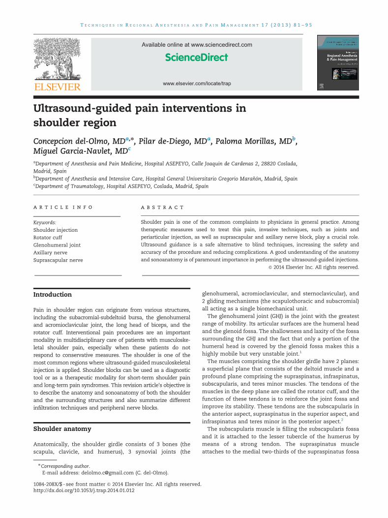
Fig. 2 – Biceps tendon: (A) the intra-articular portion of the bicepslocated between the subscapularis medially and supraspinatus llesser tuberosities (bicipital groove); and (C) the biceps tendon atendon; 2—subscapularis tendon; 3—supraspinatous tendon; 4—online.)
lateral to the tendon biceps tendon (Figure 1). The tendon isexamined in a transverse plane (short axis), where it emergesfrom under the acromion, to the musculotendinous junctiondistally. Moving the probe proximally we will able to observethe intra-articular portion as an oval-shaped hyperechoicstructure situated between the subscapularis medially andsupraspinatus laterally (Figure 2A). Moving the probe distallyalong the arm, we can follow the course of the tendon to themyotendinous junction (Figure 2C).A 901 rotation of the probe permits a longitudinal view of
the tendon, showing a fibrillar pattern of hyperechoic andhypoechoic interlaced lines9 (Figure 3).Both the bicipital groove and the LHBT are used as ultra-
sound landmarks to identify other rotator cuff components.The LHBT ultrasound approach can be performed either in
plane or out of plane. The target is the small space between thetendon and the lesser tubercle of the humerus, just medial tothe tendon. The needle should be directed through the trans-verse humeral ligament and reach the bicipital groove (Figure 1).Injection on the lateral aspect of the groove is equally effectiveas the medial side, but caution should be taken to avoid theascending branch of the circumflex humeral artery. The anes-thetic solution is injected into the bicipital groove surroundingthe tendon, injecting directly over or against the tendon shouldbe avoided. Low volumes (4 mL) are sufficient and injection ofsteroid directly into the tendon may lead to rupture.
Subscapularis tendonThe patient is seated with elbow flexed to 901, the arm inexternal rotation, fixing the elbow on the iliac crest and slight
tendon appears as an oval-shaped hyperechoic structureaterally; (B) the biceps tendon lies in between the greater andt its distal myotendinous junction. Landmark: 1—bicepspectoralis major muscle. (Color version of figure is available

Fig. 3 – Biceps tendon. Probe longitudinal to long head of biceps tendon. Arm adducted, hand supinated. Landmark: 1—deltoidmuscle; 2—biceps tendon; 3—humerus. (Color version of figure is available online.)
T E C H N I Q U E S I N R E G I O N A L A N E S T H E S I A A N D P A I N M A N A G E M E N T 1 7 ( 2 0 1 3 ) 8 1 – 9 584
supination of the hand (Figure 4). This maneuver stretchesthe subscapularis tendon (internal rotator) and takes it outfrom under the coracoid. The coracoid process medially andthe lesser tubercle of the humerus laterally are the 2 bonyreferences used for ultrasound identification of the subsca-pularis tendon. We use a linear probe placed transversally onthe anterior aspect of the shoulder at the level of the coracoidprocess. The ultrasound image provides a longitudinal viewof the subscapularis tendon, which has a “beak shape” with aconvex surface ending in an acute angle at its insertion intothe lesser tubercle10 (Figure 4). The superficial margin of thetendon is delimited by an echogenic layer, which correspondsto the subdeltoid fat.To obtain a transversal view, we rotate the probe to 901 and
place it along the longitudinal axis of the body (Figure 5).With this view, the tendon has a heterogeneous echotexturewith well-defined hyperechoic and slightly hypoechoic areas,representing its wide myotendinous junction11 (Figure 5).
SST and subacromial-subdeltoid bursaThe SST is lateral and posterior to the bicipital groove. It isexamined with patient's arm in internal rotation, resting thehand upon the ipsilateral iliac crest or touching the contrala-teral scapula (Figure 6). This maneuver allows the tendon tobe almost completely withdrawn from under the acromion.The SST should be evaluated along its long and short axis. Inthe short axis (Figure 6), we use the LHBT as the reference asits intra-articular portion is located between the SST laterallyand the subscapular tendon medially (Figure 2A). The SST hasa convex or “wheellike” shape, with a homogeneous echostructure, more echogenic than the deltoid muscle, which islocated above it11 (Figure 6).
In the long axis, the tendon is seen as a convex fibrillarstructure located below the deltoid muscle and subacromialbursa, has bird-beak shape attached to the greater tubercle(Figure 7).The ultrasound is unable to differentiate between the
supraspinatus and the infraspinatus tendons, as they havea common insertion into the greater tubercle.10
Between the supraspinatus and the deltoid, the normalsubacromial-subdeltoid bursa appears as a thin hypoechoicband. It overlies the superior aspect of the SST. The main roleof this largest bursa is to minimize attrition of the cuffagainst the coracoacromial arch (acromion and coracoacro-mial ligament) and the deltoid muscle during movements ofthe arm.Ultrasound image of the subacromial-subdeltoid bursa
looks like a thin hyperechoic structure of approximately2 mm touching the SST, greater tubercle, and humeral artic-ular cartilage (anechoic line) (Figure 7). With the probe trans-verse, the bursa is located between the deltoid muscle andthe SST on the lateral aspect, and the coracoid processmedially (Figure 6). The bursa is surrounded by a thin layerof peribursal fat, and under pathologic conditions it is easilydetectable on ultrasound examination.10 This bursa isinvolved in most lesions of the SST; its inflammation rendersshoulder movements painful. Dynamic assessment of sub-acromial impingement can be assessed, with the patientabducting the arm while in internal rotation. With thismaneuver, the SST can be seen passing deep to the coracoa-cromial arch. The ultrasound approach to the subacromial-deltoid bursa is usually in plane, from lateral to medial.Solution infiltration (5-10 mL) inside the bursa allows itsdistension between these muscle masses.

Fig. 4 – Subscapularis tendon. Long axis view. Probe longitudinal to the subscapularis muscle (transverse to anteriorshoulder). Shoulder adducted and externally rotated with elbow kept against chest wall. Landmark: 1—lesser tuberosity;2—subscapularis tendon; 3—deltoid muscle; 4—coracoid process. (Color version of figure is available online.)
T E C H N I Q U E S I N R E G I O N A L A N E S T H E S I A A N D P A I N M A N A G E M E N T 1 7 ( 2 0 1 3 ) 8 1 – 9 5 85
Infraspinatus tendonThe infraspinatus tendon is posterior, and it is explored frombehind the patient with arm in neutral position or with thepatient's hand touching the opposite shoulder. The probe is
Fig. 5 – Subscapularis tendon. Short-axis view. Landmark: 1—sub(Color version of figure is available online.)
placed in the posterior aspect of the scapulohumeral joint.The infraspinatus muscle is seen as an individual structurefilling the infraspinous fossa deep to the deltoid muscle12
(Figure 8).
scapularis tendon; 2—deltoid muscle; 3—lesser tuberosity.

Fig. 6 – Supraspinatus tendon. Probe transverse to supraspinatus tendon, with shoulder extended and internally rotated.The tendon passes over the superior aspect of the shoulder joint to insert into the uppermost facet of the greater tuberosity ofthe humerus. The normal tendon shows a smooth convex superior surface. Landmark: 1—supraspinatus tendon; 2—bicepstendon; 3—humeral head; 4—coracoid process; 5—deltoid muscle; 6—subacromial-subdeltoid bursa. (Color version of figure isavailable online.)
Fig. 7 – Supraspinatus tendon. Probe longitudinal to supraspinatus tendon, with shoulder extended and internally rotated.Landmark: 1—humerus; 2—supraspinatus tendon (bird-beak shape); 3—deltoid muscle; 4—greater tuberosity; 5—articularcartilage; 6—subacromial-subdeltoid bursa. (Color version of figure is available online.)
T E C H N I Q U E S I N R E G I O N A L A N E S T H E S I A A N D P A I N M A N A G E M E N T 1 7 ( 2 0 1 3 ) 8 1 – 9 586

Fig. 8 – Infraspinatus muscle or tendon. Landmark: 1—deltoid muscle; 2—infraspinatous muscle; 3—greater tuberosity. (Colorversion of figure is available online.)
T E C H N I Q U E S I N R E G I O N A L A N E S T H E S I A A N D P A I N M A N A G E M E N T 1 7 ( 2 0 1 3 ) 8 1 – 9 5 87
Acromioclavicular joint injectionThe acromioclavicular joint is a small synovial joint betweenthe acromion and the clavicle with its inferior aspect incontact with the subacromial bursa and the rotator cuff.The patient is seated with arm in neutral position. In this
position, the depth of the joint space is wider13 (Figure 9). Weuse a high-frequency linear probe positioned on the superioraspect of the shoulder, along the coronal plane (Figure 9). Theprobe is moved from the lateral edge of the acromion mediallyuntil the acromioclavicular joint is visualized. The ultrasoundimage over this joint show 2 hyperechoic lines, followed by anacoustic shadow that corresponds to the acromion and theclavicle joined together in the superior aspect with a hyper-echoic line corresponding to the joint socket, which is usuallyin close contact with the periarticular surface of the clavicle8
(Figure 9). The cortical surface of the clavicle is usually slightlyhigher than the acromion. The approach can be performed inplane or out of plane, but as it is a very superficial structure,the out-of-plane approach may be more comfortable for thepatient (Figure 9). The joint is often completely distended by asmall volume of anesthetic solution (2 mL).8 There are nosignificant vascular or neural structures to consider in thisinjection, but the skin is often thin and friable over theacromioclavicular joint, so care should be taken not to depositsteroids superficially above the joint.
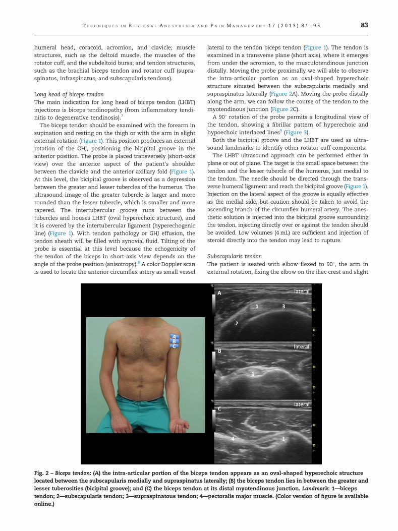
GHJ injectionThe principal indications for GHJ injection are pain relief andhelping rehabilitation in patients with osteoarthritis andadhesive capsulitis (frozen shoulder).Ultrasound GHJ injection can be performed through an
anterior or posterior approach or via the rotator cuff interval.14
The easiest access to the shoulder joint is from the posterioraspect, owing to the lack of vessels, nerves, and majorligament and intra-articular structures along the needle path-way, and there is also lower extravasation of the solution.8,15
Posterior approach to GHJ. The patient is well positioned inlateral decubitus or sitting, with the humerus adductedacross the thorax, thus opening the posterior joint space. Ahigh-frequency linear probe is usually used, but for very largeshoulders, a convex probe may be necessary.The probe is placed under the spine of the scapula parallel
to its lateral end (Figure 10). At this level, we can see thefollowing structures: the infraspinatous muscle, humeralhead, posterior glenoid border, and posterior labrum(Figure 10). The needle is inserted in plane, from lateral tomedial, positioning the needle in the space between theglenoid labrum, hyperechoic triangular shape, and humeralhead8 (Figure 10). It is often necessary to increase the depth ofthe field of view to avoid loss of view of this area. Theintroduction of an anesthetic solution displaces the posteriorcapsule (hyperechoic line) surrounding these structures. Ifany resistance to the injection is noted, rotate the needlebevel or withdraw the needle a little, as it may have reachedthe cartilage of the humeral head or the labrum.
Anterior approach to GHJ. This is conducted with the patientin sitting or supine position with arm in external rotation. Ahigh-frequency linear probe is used although, like with theposterior approach, if the patient is obese or has very largeshoulders, a convex probe may be necessary. The probe ispositioned under and parallel to the acromion, on the medialaspect of the acromion above the coracoid (Figure 11). In the

Fig. 9 – Acromioclavicular joint. The inset shows the position of the ultrasound probe (coronal plane adjacent to superior aspectof joint) and the needle with out-of-plane technique. The ultrasound image shows the pathway of the needle (arrow).Landmark: 1—clavicle; 2—acromion; 3—capsule joint or superior acromioclavicular ligament; 4—acromioclavicular joint. (Colorversion of figure is available online.)
Fig. 10 – Posterior approach to the GHJ. The inset shows the position of the ultrasound probe and the needle with in-planetechnique. The ultrasound probe is placed just caudal and parallel to the lateral end of scapular spine. The needle is insertedin plane from the lateral aspect of the shoulder. The ultrasound image is shown with the line representing the needle path,between the free edge of the labrum5 and the hypoechoic articular cartilage of the humeral head.6 Landmark: 1—humeralhead; 2—posterior glenoid rim; 3—infraspinatus muscle; 4—deltoid muscle; 5—labrum. (Color version of figure is availableonline.)
T E C H N I Q U E S I N R E G I O N A L A N E S T H E S I A A N D P A I N M A N A G E M E N T 1 7 ( 2 0 1 3 ) 8 1 – 9 588
T E C H N I Q U E S I N R E G I O N A L A N E S T H E S I A A N D P A I N M A N A G E M E N T 1 7 ( 2 0 1 3 ) 8 1 – 9 5 89
ultrasound image, we can see the head of the humerus,subscapularis tendon, and coracoid process (Figure 11). Thisapproach is conducted in plane from lateral to medial. Theneedle passes over the humeral head toward the spacebetween the subscapularis tendon superiorly, the head ofthe humerus laterally, and the coracoid medially (Figure 11).
Shoulder nerve block
Innervation of the shoulder is complex with involvement ofseveral nerves: the suprascapular, axillary, subscapular, mus-culocutaneous, and lateral pectoral nerves.About 70% of shoulder innervation arises from the supra-
scapular nerve (SSN),16 and the rest from the axillary nervewith a relatively small contribution from the lateral pectoral,musculocutaneous, and subscapularis nerves.17
The blocking of the brachial plexus at the interscalene levelis considered the most effective analgesic technique forshoulder surgery18 because it ensures blockage of everybranch. However, the interescalene nerve block (ISB) isassociated with well-documented adverse effects, includingunwelcome arm paralysis and intolerance of hemidiaphragmparalysis.19 ISB is also associated with more frequent long-term neurologic deficits compared with other nerve blocks20
and must be performed in proximity to important anatomicalstructures (eg, vertebral artery). An alternative to ISB may beadvantageous and has been described, whereby terminalnerves supplying the shoulder joint are blocked rather thanthe entire brachial plexus.21,22
Fig. 11 – Anterior approach to the GHJ. The inset shows the positioThe probe is placed caudal and parallel to the acromion, with thimage is shown with the line representing the needle patch. Theat the medial border of the humeral head. Landmark: 1—deltoid4—humeral head. (Color version of figure is available online.)
Combined suprascapular and axillary nerve block (shoulderblock) appears to be an effective alternative to ISB for painrelief following shoulder surgery with advantages of minimalside effects,23 reduced potential for serious complications,and less reported pain during block resolution.21 The“shoulder block” attains better results than with single SSNblock.24,25
Axillary nerve block
This nerve is blocked together with the SSN as an analgesicalternative to brachial plexus interscalene block.26 However,the clinical role of a specific axillary nerve block remainsundetermined.
AnatomyThe axillary nerve is one of the terminal branches of theposterior cord of the brachial plexus, together with the radialnerve. It innervates the deltoid muscle, teres minor muscle, andlong head of the triceps brachii muscle. It also provides sensoryinnervation to the shoulder joint as well as the skin coveringthe area inferior to the deltoid muscle in the arm.27 The axillarynerve originates at the lateral border of the subscapularismuscle and then advances posteriorly under the shoulder jointtoward the surgical neck of the humerus in close proximity tothe posterior circumflex humeral artery (PCHA), a branch of theaxillary artery. It subsequently divides into 2 branches, oneanterior and another posterior.28 The anterior branch inner-vates the medial and anterior aspect of the deltoid muscle and
n of the ultrasound probe and the needle in-plane technique.e medial part covering the coracoid process. The ultrasoundneedle was inserted from the lateral side of the probe aimingmuscle; 2—coracoid process; 3—subscapularis tendon;
T E C H N I Q U E S I N R E G I O N A L A N E S T H E S I A A N D P A I N M A N A G E M E N T 1 7 ( 2 0 1 3 ) 8 1 – 9 590
branches into the anterior aspect of the joint capsule. Theposterior branch innervates the teres minor muscle, posterioraspect of the deltoid muscle and terminates as the superiorlateral cutaneous nerve of the arm that runs along the medialborder of the deltoid muscle and provides sensory innervationof skin of the inferior aspect of this muscle.29
Sonoanatomy and block techniqueThe patient is in lateral decubitus position with the arm to beblocked facing up and elbow flexed at about 901 or sitting withthe shoulder in neutral position, slight internal rotation of thearm, elbow flexed at 901, and the hand on the knee.A high-frequency linear probe is used, which is positioned
in the posterior aspect of the arm.The probe is placed parallel to the long axis of the humerus,
about 2-3 cm below the posterolateral aspect of the acromion,on the dorsal side of the arm27 (Figure 12). First, we locate thehead of the humerus, and we then move the probe along thesurgical neck of the humerus so that the ultrasound image ofthe cortex of the head continues with that of the surgical neckand the shaft of the humerus. At the neck of the humerus, it isessential to identify the PCHA in the transverse view using aDoppler, as the axillary nerve is localized cranial to this and itis in close contact with it.27 Both structures, the nerve and theartery, are located in a neurovascular space between the teresminor cranially, long head of triceps brachii caudally, thedeltoid muscle in the posterior aspect, and the shaft of thehumerus in the anterior aspect (Figure 12).The nerve is visualized as a hyperechoic structure located
above the artery, but sometimes it is difficult to identify. Theinjection of the anesthetic often helps to identify it. The nerveapproach has been described in plane from cranial to caudal.The needle tip must be located, below the fascia caudal to theteres minor muscle, and just cranial to the PCHA (Figure 12).
SSN block
The SSN is the main sensory nerve in the shoulder, primarilyinnervating its posterior and superior aspect.30
This nerve block is indicated for the treatment of short-term and long-term shoulder pain. In short-term shoulderpain, it has been used primarily in the management ofpostoperative pain following shoulder surgery in cases wherethe brachial plexus block was contraindicated. In long-termshoulder pain, SSN block has been used as a diagnosticprocedure in patients with suspected neuropathy of thisnerve and as a therapeutic procedure in cases of oncologicpain, arthritis of the shoulder joint, adhesive capsulitis, andlong-term lesions of the rotator cuff.31
AnatomyThe SSN is a mixed sensory and motor nerve that originatesin the anterior rami of the C5 and C6 spinal nerves.32 It leavesfrom the lateral aspect of the superior trunk of the brachialplexus, travels through the posterior triangle of the neck, andcourses deep to the trapezius muscle and the omohyoidmuscle. It enters the supraspinous fossa through the supra-scapular notch under the superior transverse ligament of thescapula. The suprascapular artery (subclavian artery branch)and vein pass above this ligament.33 On its path from the
supraspinous fossa, the nerve together with the suprascapu-lar artery are in direct contact with the bony plane and comeout of it through the greater scapular (spinoglenoid) notchand continues in the direction of the infraspinous fossa.34
Shortly after passing through the suprascapular notch, theSSN sends out 2 branches, one is the motor nerve for thesupraspinatus muscle35 and the other is known as thesuperior articular branch. The articular branch is sensoryand supplies the coracoclavicular, coracohumeral ligaments,acromioclavicular joint, GHJ (posterior and superior aspects),and subacromial bursa.36
The main trunk of the nerve exits the supraspinous fossathrough the lateral border of the spine of the scapula, througha fibro-osseous tunnel (greater scapular or spinoglenoidnotch), terminating in motor branches to the infraspinatusmuscle.37 This fibro-osseous tunnel is formed by the spine ofthe scapula and the inferior transverse ligament of thescapula (spinoglenoid ligament).38 The number of motorbranches for the infraspinatus muscle varies from 2-4.35 Itis important to point out that although sensory branches areissued, there is usually no cutaneous innervation.
Ultrasound-guided SSN block techniquesVarious techniques have been described for SSN block: blindtechniques with or without neural stimulation, or imagingtechniques (fluoroscopy, computed tomography, and ultra-sound). In most of these, the nerve is located near the supra-scapular notch or in the supraspinous fossa, between thesuprascapular notch and spinoglenoid notch39 (Figure 13). Mostof the landmark-based and ultrasound-guided approaches areclassified as posterior approach when the SSN is targeted alongits course between the suprascapular notch and spinoglenoidnotch. A new anterior approach is recently described in whichthe SSN is approached soon after it branches out from theupper trunk.40
We describe ultrasound-guided approaches to SSN block,together with some of the advantages and disadvantages.
A suprascapular notch approach. This is an attractive site forSSN blocking, as at this level the nerve has not yet dividedinto its terminal branches.39
The block is usually performed with patient in supineposition, but it can also be performed with the patient inthe lateral position with the limb to be blocked facing up. Ahigh-frequency linear probe is used. The basic reference pointis the spine of the scapula. Placing the probe above this,toward the middle, and then we let it “fall” toward thesupraspinous fossa. The probe should be placed at a slightangle toward the anterior (Figure 14) to visualize the supra-scapular notch. Typically, the ultrasound image of the notchis that of a hyperechoic line with slight superior concavityfollowed by acoustic shadowing, defined in the superioraspect by a hyperechoic line that corresponds to the super-ficial transverse ligament of the scapula (Figure 14). Thenerve, which is often difficult to see, is located at the levelof the notch, below the superior transverse ligament of thescapula. Above this ligament and with the help of Doppler,we can visualize the suprascapular artery. The muscle masswe identify at this level is the supraspinatus muscle, and thetrapezius muscle at the more superficial level (Figure 14).
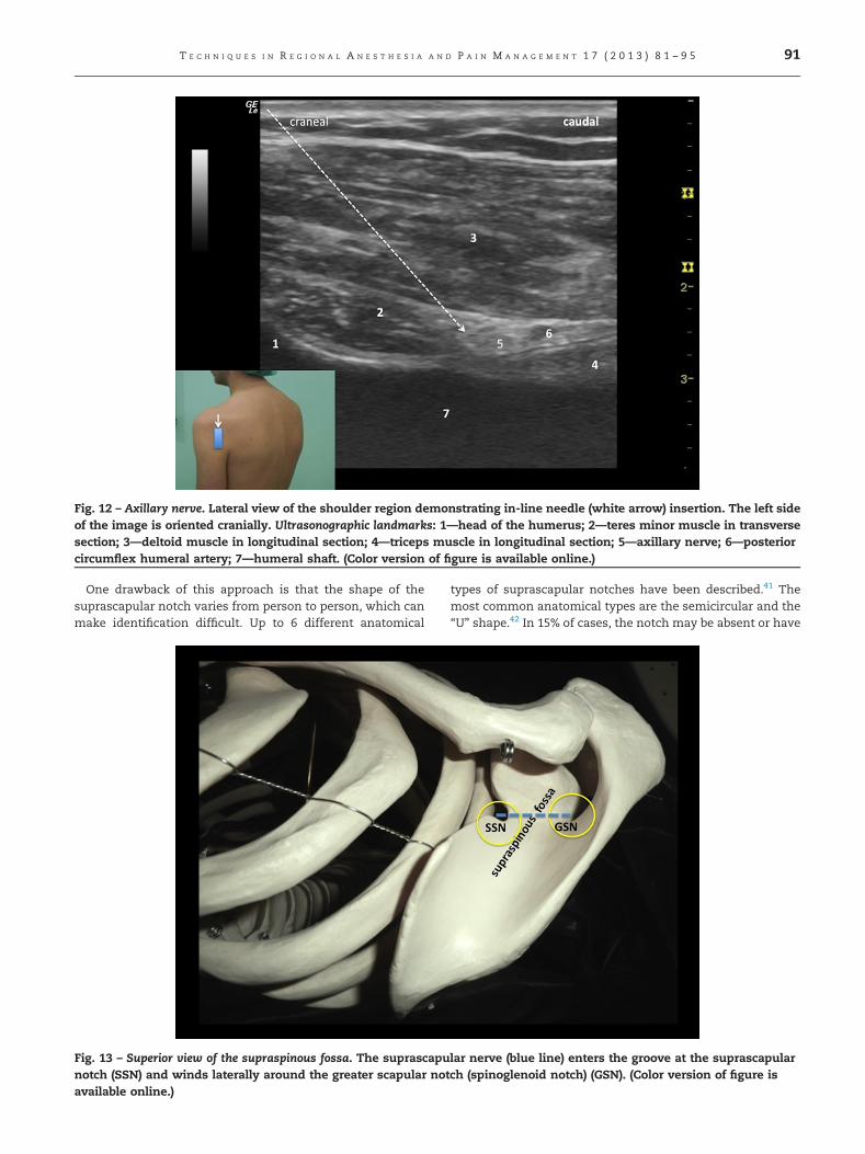
Fig. 12 – Axillary nerve. Lateral view of the shoulder region demonstrating in-line needle (white arrow) insertion. The left sideof the image is oriented cranially. Ultrasonographic landmarks: 1—head of the humerus; 2—teres minor muscle in transversesection; 3—deltoid muscle in longitudinal section; 4—triceps muscle in longitudinal section; 5—axillary nerve; 6—posteriorcircumflex humeral artery; 7—humeral shaft. (Color version of figure is available online.)
T E C H N I Q U E S I N R E G I O N A L A N E S T H E S I A A N D P A I N M A N A G E M E N T 1 7 ( 2 0 1 3 ) 8 1 – 9 5 91
One drawback of this approach is that the shape of thesuprascapular notch varies from person to person, which canmake identification difficult. Up to 6 different anatomical
Fig. 13 – Superior view of the supraspinous fossa. The suprascapunotch (SSN) and winds laterally around the greater scapular notavailable online.)
types of suprascapular notches have been described.41 Themost common anatomical types are the semicircular and the“U” shape.42 In 15% of cases, the notch may be absent or have
lar nerve (blue line) enters the groove at the suprascapularch (spinoglenoid notch) (GSN). (Color version of figure is

Fig. 14 – Blockade of suprascapular nerve in the suprascapular notch. The arrow represents the pathway of the needle.Landmarks: 1—trapezius muscle; 2—supraspinatus muscle; 3—suprascapular nerve; 4—superior transverse suprascapularligament; 5—suprascapular artery. (Color version of figure is available online.)
T E C H N I Q U E S I N R E G I O N A L A N E S T H E S I A A N D P A I N M A N A G E M E N T 1 7 ( 2 0 1 3 ) 8 1 – 9 592
been converted into a hole owing to the ossification of thesuperior transverse scapular ligament.41
The main complication with this approach, and basicallywith blind techniques, is pneumothorax, in cases where theneedle pathway is directed toward the chest cavity.39
A supraspinous fossa nerve block approach. Some authorsconsider this as the ideal site for ultrasound-guided SSNblock.43,44
The supraspinous fossa is concave with a smooth surfaceand broader and less profound medially than laterally,located above the spine of the scapula between the supra-scapular notch and spinoglenoid notch (Figure 13). Its medialthree-quarters give origin to the supraspinatus muscle. TheSSN together with the suprascapular artery passes deeplyalong the floor of the fossa below the supraspinatus musclesand covered by the inferior fascia of this muscle, in a naturalcompartment (Figure 15).Nerve block is performed with patient in lateral decubitus
or sitting position (Figure 15). A high-frequency linear probe isused but sometimes a profound block is needed, so use of alow-frequency probe may be necessary.The reference image is a hyperechoic line followed by an
acoustic shadow, which corresponds to the floor of the supra-spinous fossa. Identifying the suprascapular vessels helpswith the identification. The needle is inserted in plane withthe probe, from medial to lateral (Figure15). If the neuro-vascular bundle is not visible, which can occur in 5%-10% ofcases,45 we direct the needle toward the lateral aspect of thesupraspinous fossa. Injecting 10-15 mL of local anestheticunder the supraspinatus muscle will produce SSN block. The
supraspinatus fossa is a strictly bordered compartment, occu-pied (filled) by the supraspinatus muscle. The fibers of thismuscle originate from the bony wall of this fossa (medialthree-quarters) and from the supraspinatus fascia, which isthick and strong medially, and thin laterally, or may even beabsent. At this level, the SSN runs along a natural compart-ment that would contain the diffusion of the local anestheticinjected, would also favor the extension of the injectiontoward the lateral of the fossa, and prevent its diffusionmedially owing to its higher resistance, so that small volumes(5 mL) would be sufficient to ensure the successful blocking.46
The supraspinous fossa could be confused with spinogle-noid notch, particularly in cases where the probe is locatedvery perpendicular to the supraspinatus fossa and close tothe lateral portion of the spine of the scapula (Figure 13).The advantages of this approach include ease of access,
reference of suprascapular notch not required, and theextremely low risk of pneumothorax.47
Supraclavicular approach to the SSN. SSN crosses supracla-vicular region behind the omohyoid muscle, before leavingthe posterior cervical triangle and passing toward the supra-scapular notch of the scapula.40 Patient in supine positionwith head slightly rotated toward the side opposite to thatbeing blocked. A high-frequency linear probe is used that isplaced along the short axis of the neck (Figure 16). Thereference points are the omohyoid muscle and the root ofC5. To find the root of C5, we identify the transverse processof C7 that is easily recognized as it lacks the anterior tubercle,and we ascend to identify the transverse processes of thecervical vertebrae C6 and C5. Between these vertebrae, we

Fig. 15 – Blockade of suprascapular nerve in the supraspinous fossa. The arrow represents the pathway of the needle.Landmarks: 1—subcutaneous cellular tissue; 2—trapezius muscle; 3—supraspinatus muscle; 4—suprascapular nerve;5—suprascapular artery; 6—inferior fascia of the supraspinatus muscle. (Color version of figure is available online.)
T E C H N I Q U E S I N R E G I O N A L A N E S T H E S I A A N D P A I N M A N A G E M E N T 1 7 ( 2 0 1 3 ) 8 1 – 9 5 93
visualize the root of C5 as a rounded anechoic structure, andfollowing this, we can identify the superior trunk of thebrachial plexus (Figure 16).
Fig. 16 – Blockade of the suprascapular nerve in the supraclavic3—first rib; 4—omohyoid muscle; 5—the upper trunk of brachiaversion of figure is available online.)
The SSN comes out of the lateral wall of the superior trunkand its ultrasound image is that of a small, rounded,anechoic-hypoechoic structure that follows a lateral and
ular region. Landmarks: 1—subclavian artery; 2—pleura;l plexus; 6—suprascapular nerve (white solid arrow). (Color

T E C H N I Q U E S I N R E G I O N A L A N E S T H E S I A A N D P A I N M A N A G E M E N T 1 7 ( 2 0 1 3 ) 8 1 – 9 594
posterior direction and crosses behind and below the omo-hyoid muscle (Figure 16).Comparing this approach with classic approaches (supra-
spinous fossa), the nerve is more superficial, 8 vs 35 mm, andit is easily identified in a greater percentage of patients.40
Another advantage is that the patient is in supine position,which increases patient comfort as well as being moreconvenient for the anesthesiologist.The disadvantages arise mainly from the proximity of the
SSN to the brachial plexus and to the pleura. The distancebetween the SSN and brachial plexus is very small, about6 mm. Therefore, the local anesthetic, even with low vol-umes, can spread to other compartments of the plexus,causing unwanted blockage of these. This proximity shouldalso be taken into account when conducting neurodestructiveprocedures (radiofrequency or cryotherapy) of the SSN.Other complications include neurologic injury and intra-
vascular injection as the nerve runs alongside the supra-scapular vessels, piercing these while inserting the needlemay produce systemic toxicity following the administrationof a local anesthetic. Careful aspiration before injection oflocal anesthetic may help prevent this complication.The ideal ultrasound technique used to locate the SSN will
depend on the indication.44 If postoperative analgesia ordiagnostic block with local anesthetic is required, then locat-ing and blocking the SSN in the lateral supraspinous fossa isefficient, safe, and effective. If radiofrequency ablation isrequired, then perhaps locating the SSN more proximally willallow isolation of the nerve in a more superficial location.
r e f e r e n c e s
1. Iannotti JP, Gabriel JP, Schneck SL, Evans BG, Misra S, et al.The normal glenohumeral relationships. An anatomical studyof one hundred and forty shoulders. J Bone Joint Surg Am.1992;74:491–500.
2. DeFranco MJ, Cole BJ. Current perspectives on rotator cuffanatomy. Arthroscopy. 2009;25:305–320.
3. Rouviere H, Delmas A. Anatomia Humana. Vol III. Musculosdel tronco, 11end Ed, 2005 Elsevier; Barcelona, 90–102.
4. Berquist TH, Peterson JJ. Shoulder and arm. In: Berquist TH, ed,MRI of the Musculoskeletal System. Philadelphia, PA: LippincottWilliams and Wilkins; 2006;569–585.
5. Hsieh LF, Hsu WC, Lin YJ, Wu SH, Chang KC, Chang HL.Is ultrasound-guided injection more effective in chronicsubacromial bursitis? Med Sci Sports Exerc. 2013;45(12):2205–2213.
6. Uncunu F, Capkin E, Karkucak M, Ozden G, Cakirbay H, TosunM. A comparison of the effectiveness of landmark guidedinjection and ultrasonography guided injection for shoulderpain. Clin J Pain. 2009;25:786–789.
7. Ostor A, Richards CA, Prevost AT, Seed CA, Hazleman BL.Diagnosis and relation to general health of shoulder disorderspresenting to primary care. Rheumatology. 2005;44:800–805.
8. Peng PWH, Cheng P. Ultrasound-guided interventional proce-dures in pain medicine. A review of anatomy, sonoanatomy,and procedures. Part III: shoulder. Reg Anesth Pain Med. 2011;366:592–605.
9. Van Holsbeeck I. Ecografía musculoesquelética. Cap 15. Ecografíadel hombro, 2nd ed. Detroit: Marban; 2002;463–516.
10. Bianchi S, Martinoli C. Ultrasound of the Musculoskeletal System:Shoulder. Springer-Verlag, Berlin; 2007;190–331.
11. Precerutti M, Garioni E, Madonia L, Draghi F. US anatomy ofthe shoulder. Pictorial essay. J Ultrasound. 2010;13:179–187.
12. Curtis AS, Burbank KM, Tierney JJ, Scheller AD, Curran AR.The insertional footprints of the rotator cuff: an anatomicstudy. Arthroscopy. 2006;22:609.
13. Park GY, Park JH, Bae JH. Structural changes in the acromio-clavicular joint measured by ultrasonography during provo-cative tests. Clin Anat. 2009;19:292–295.
14. Lim JB, Kim SW, Sung KW, Jung I, Lee C. Ultrasound-guidedshoulder joint injection through rotator cuff interval. Korean JPain. 2008;21:57–61.
15. Gokalp G, Dusak A, Yazici Z. Efficacy of ultrasonography-guided shoulder MR arthrography using a posterior approach.Skeletal Radiol. 2010;39:575–579.
16. Ritchie ED, Tong D, Chung F, et al. Suprascapular nerve blockfor postoperative pain relief in arthroscopic shoulder surgery:a new modality? Reg Anesth Pain Med. 1997;84:1306–1312.
17. Halata Z, Bauman K. Mechanoreceptors of the shoulder joint:structure and function. In: Di Giacomo G, Pouliart N, CostantiniA, Vita A, et al., eds. Atlas of Functional Shoulder Anatomy. Milan,Italy: Springer; 2008;206–208.
18. Singelyn FJ, Lhotel L, Fabre B. Pain relief after arthroscopicshoulder surgery: a comparison of intraarticular analgesia,suprascapular nerve block, and interscalene brachial plexusblock. Anesth Analg. 2004;99:589–592.
19. Urmey W, Gloeggler M. Pulmonary function changesduring interscalene brachial plexus block: effects of decreas-ing local anaesthetic injection volume. Reg Anesth. 1993;18:244–249.
20. Borgeat A, Ekatodramis G, Kalbere F, Benz C. Acute andnonacute complications associated with interscalene blockand shoulder surgery: a prospective study. Anesthesiology. 2001;95:875–880.
21. Price DJ. The shoulder block: a new alternative to interscalenebrachial plexus blockade for the control of postoperativeshoulder pain. Anaesth Intensive Care. 2007;35:575–581.
22. Price DJ, Abeysekera A, Chaddock MA. A randomised compar-ison of combined suprascapular and axillary (circumflex)nerve block with interscalene block for postoperative analge-sia following arthroscopic shoulder surgery. Anaesth IntensiveCare. 2012;40:183–184.
23. Verelst P, Van Zundert A. Respiratory impact of analgesicstrategies for shoulder surgery. Reg Anesth Pain Med. 2013;38:50–53.
24. Nam YS, Jeong JJ, Han SH, et al. An anatomic and clinicalstudy of the suprascapular and axillary nerve blocks forshoulder arthroscopy. J Shoulder Elbow Surg. 2011;20:1061–1068.
25. Lee SM, Park SE, Nam YS, et al. Analgesic effectiveness ofnerve block in shoulder arthroscopy: comparison betweeninterscalene, suprascapular and axillary nerve blocks. KneeSurg Sports Traumatol Arthrosc. 2012;20:2573–2578.
26. Price DJ. Axillary (circumplex) nerve block used in associationwith suprascapular nerve block for the control of painfollowing total shoulder joint replacement. Reg Anesth PainMed. 2008;33:280–281.
27. Rothe C, Asghar S, Andersen HL, Christensen JK, Lange KHW.Ultrasound-guided block of the axillary nerve: a volunteerstudy of a new method. Acta Anaesthesiol Scand. 2011;55:565–570.
28. Loukas M, Grabska J, Tubbs RS, Apaydin N, Jordan R. Mappingthe axillary nerve within the deltoid muscle. Surg Radiol Anat.2009;31:43–47.
29. Ball CM, Steger T, Galatz LM, Yamaguchi K. The posteriorbranch of the axillary nerve: an anatomic study. J Bone JointSurg Am. 2003;85-A:1497–1501.
30. Aszmann OC, Dellon AL, Bireley BT, McFarland EG. Innerva-tion of the human shoulder joint and its implications forsurgery. Clin Orthop Relat Res. 1996;330:202–207.

T E C H N I Q U E S I N R E G I O N A L A N E S T H E S I A A N D P A I N M A N A G E M E N T 1 7 ( 2 0 1 3 ) 8 1 – 9 5 95
31. Emery P, Bowman S, Wedderburn L, et al. Suprascapularnerve block for chronic shoulder pain in rheumatoid arthritis.Br Med J. 1989;299:1079–1080.
32. Voster W, Lange CPE, Briet RJP, et al. The sensory branchdistribution of the suprascapular nerve: an anatomic study.J Shoulder Elbow Surg. 2008;17:500–502.
33. Anderson JE. Grant's Atlas of Anatomy 8th ed. Baltimore, MD:Williams and Wilkins; 1983;6–31.
34. Rouviere H, Delmas A. Anatomia Humana. Vol III. Anatomiatopografica del miembro superior, 11th Ed, 2005, Elsevier,Barcelona, 233–234.
35. Bigliani LU, Dalsey RM, McCann PD, April EW. An anatomicalstudy of the suprascapular nerve. Arthroscopy. 1990;6:301–305.
36. Ajmani ML. The cutaneous branch of the human supra-scapular nerve. J Anat. 1994;185:439–442.
37. Mestdagh H, Drizenko A, Ghestem P. Anatomical basis ofsuprascapular nerve syndrome. Anat Clin. 1981;3:67–71.
38. Cummins CA, Messer TM, Nuber GW. Suprascapular nerveentrapment. J Bone Joint Surg Am. 2000;82:415–424.
39. Chan C, Peng PWH. Suprascapular nerve block. A narrativereview. Reg Anesth Pain Med. 2011;36:358–373.
40. Siegenthaler A, Morriggl B, Mlekusch S, et al. Ultrasound-guided suprascapular nerve block, description of a novelsupraclavicular approach. Reg Anesth Pain Med. 2012;37:325–327.
41. Natsis K, Totlis T, Tsikaras P, Appell HJ, Skandalakis P,Koebke J. Proposal for classification of the suprascapularnotch: a study on 423 dried scapulas. Clin Anat. 2007;20:135–139.
42. Rengacharry SS, Neff JP, Singer PA, Brackett CE. Suprascapularentrapment neuropathy: a clinical, anatomical and compara-tive study. Part 1: clinical study. Neurosurgery. 1979;5:441–446.
43. Peng PWH, Wiley MJ, Liang J, Bellingham GA. Ultrasound-guided suprascapular nerve block: a correlation with fluoro-scopic and cadaveric findings. Can J Anesth. 2010;57:143–148.
44. Prize D. Novel ultrasound-guided suprascapular nerve block(letter). Reg Anesth Pain Med. 2012;37:676–677.
45. Prize D, Kershaw N, Clark B, Fletcher T. An overwiew of theanatomy of the suprascapular and axillary nerves usingultrasound and magnetic resonance imaging. Anesth IntensiveCare. 2012;40(1):183.
46. Feigl GC, Dorn C, Likar R. What local anesthetic volumeshould be used for suprascapular nerve block? Reg AnesthPain Med. 2008;33:571–573.
47. Di Lorenzo L, Pappagallo M, Gimigliano R, et al. Pain relief inearly rehabilitation of rotator cuff tendinitis: any role forindirect suprascapular nerve block? Eura Medicophys. 2006;42:195–204.