ultrasound-guided infraorbital nerve pulsed radiofrequency
TRANSCRIPT
Korean J Pain 2013 January; Vol. 26, No. 1: 84-88pISSN 2005-9159 eISSN 2093-0569http://dx.doi.org/10.3344/kjp.2013.26.1.84
| Case Report |
Ultrasound-Guided Infraorbital Nerve Pulsed Radiofrequency Treatment for Intractable Postherpetic Neuralgia
- A Case Report -
Departments of Anesthesiology and Pain Medicine, *Surgery, College of Medicine, Chung-Ang University, Seoul, Korea
Seung Mo Lim, MD, Hae Lang Park, MD, Hyong Yong Moon, MD, Kyung Ho Kang, MD*, Hyun Kang, MD, Chong Hwa Baek, MD, Yong Hun Jung, MD,
Jin Yun Kim, MD, Gill Hoi Koo, MD, and Hwa Yong Shin, MD
A 60-year-old man presented with pain on the left cheek and lateral nose. The patient had been diagnosed with facial herpes zoster in the left V2 area 6 months previously. Medical treatment was prescribed for 6 months but it had little effect. We blocked the left infraorbital nerve under ultrasound guidance, but pain relief was short term. Therefore, we performed pulsed radiofrequency treatment on the left infraorbital nerve under ultrasound guidance. Six months after the procedure, the reduction of pain was still maintained, and there was no need for further management. (Korean J Pain 2013; 26: 84-88)
Key Words:
infraorbital nerve, radiofrequency, ultrasound.
Received September 18, 2012. Revised November 13, 2012. Accepted November 17, 2012.Correspondence to: Hwa Yong Shin, MDDepartment of Anesthesiology and Pain Medicine, College of Medicine, Chung-Ang University, 224-1 Heukseok-dong, Dongjak-gu, Seoul 156-755, KoreaTel: +82-2-6299-3164, Fax: +82-2-6299-2585, E-mail: [email protected]
This is an open-access article distributed under the terms of the Creative Commons Attribution Non-Commercial License (http:// creativecommons.org/licenses/by-nc/3.0/), which permits unrestricted non-commercial use, distribution, and reproduction in any medium, provided the original work is properly cited.Copyright ⓒ The Korean Pain Society, 2013
One of the most difficult pain syndromes to treat is
postherpetic neuralgia (PHN), which occurs in about 10%
of patients with acute herpes zoster. The reason why this
painful condition occurs in some patients but not in others
is unknown, but it occurs more frequently in older aged
patients or those with acute herpes zoster of the trigeminal
nerve (CN V) compared with that involving the thoracic
dermatomes [1].
PHN of the trigeminal nerve is a neuropathic disorder
characterized by constant dysesthetic facial pain, origi-
nating from the trigeminal nerve with superimposed sud-
den, sharp and shooting neuritic pain. Some patients may
complain of burning pain [2].
Several treatments have been suggested for PHN, in-
cluding medication, botulinum toxin injections, nerve blocks,
peripheral nerve stimulation, surgical intervention, pulsed
radiofrequency treatment (PRFT) [3], and radiofrequency
ablation [4]. Infraorbital nerve block is widely used to treat
postherpetic V2 (maxillary nerve) neuralgia. But, the land-
mark based approach can be challenging because identify-
ing the foramen by palpation is difficult due to anatomical
variations in the infraorbital foramen [5]. Thus, it is easier
Lim, et al / US-Guided Infraorbital Nerve PRFT 85
www.epain.org
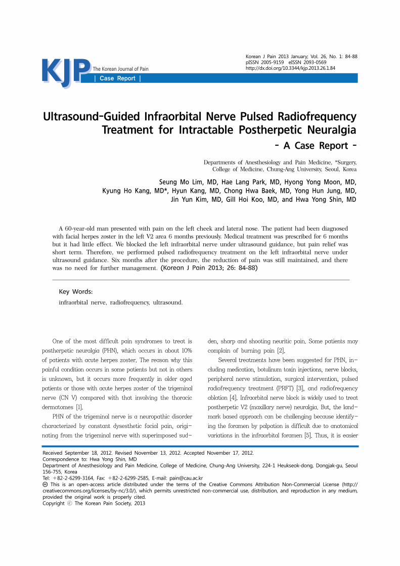
Fig. 1. A photograph of the radiofrequency needle placement under ultrasound guidance (10−12 MHz linear transducer).(A) In the actual patient. (B) Anterior view in a human skull model, which is empirically depicted. (C) Lateral view in ahuman skull model, which is empirically depicted. The ultrasound transducer was initially applied longitudinally at the lateralside of the nose and rotated slightly clockwise. Then, the transducer was moved laterally until the left infraorbital foramen was identified.
and safer if the infraorbital nerve block and PRFT are per-
formed under ultrasound (US) guidance [5]. Here, we pres-
ent a case of successful treatment of infraorbital PHN us-
ing US-guided (USG) PRFT.
CASE REPORT
A 60-year-old man visited our pain clinic with the
chief complaint of left facial pain. The patient had been
diagnosed with facial herpes zoster in the left V2 area 6
months previously. The left facial pain had continued for
6 months despite medical treatment. During this period,
150 mg pregabalin, 10 mg amitriptyline, 37.5 mg trama-
dol/375 mg acetaminophen combination tablet (Paramacet
tab; Donga, Seoul, Korea) were administrated per os three
times daily but had little effect. He had paroxysmal, sharp
and shooting pain on his left cheek and lateral nose. The
pain was aggravated by touching the left facial area, and
palpation over the left infraorbital foramen reproduced the
pain. Pain severity was 9-10/10 on the visual analogue
scale (VAS).
Hence, we started pain intervention for trigeminal PHN
in the left V2 area. We initially blocked the left infraorbital
nerve under USG with a 10-12 MHz linear transducer (Vivid
E, General Electronics, Fairfield, CT, USA) using a mixture
of 2 ml 2% mepivacaine and 20 mg triamcinolone. This
procedure decreased the pain immediately. When he re-
turned to our pain clinic 1 month after the infraorbital
nerve block, the patient stated that the VAS score had de-
creased from 9 to 4 for about 2 weeks following the in-
fraorbital nerve block but then his symptoms returned to
a previous state. Therefore, we tried the same additional
USG left infraorbital nerve treatment, but the outcome was
the same.
The USG infraorbital nerve blocks were effective but
the effects were maintained for only 2 weeks, so we decided
to perform PRFT. After explaining the procedure, efficacy,
and possible side effects of PRFT, the patient was placed
in a supine position. The skin was aseptically draped with
betadine. The ultrasound was prepared with a sterile
transparent sheath and aseptic ultrasound gel. The trans-
ducer was applied longitudinally to the lateral side of the
nose and then rotated slightly clockwise to obtain an ideal
longitudinal (long-axis) view of the infraorbital canal to
find the left infraorbital foramen. We scanned his face
from medial to lateral in the sagittal plane along the left
lower orbital margin (Fig. 1). We easily identified the infra-
orbital foramen with a hyperechoic protuberance (Fig. 2).
Radiofrequency needle (10 cm) insulated with a 5-mm
active tip (22 G, SMK-C10; Radionics Inc, Burlington, MA,
USA) was advanced slightly via the infraorbital foramen
under USG (Fig. 2). Confirmation of the needle position in-
side the infraorbital foramen was achieved under fluoro-
scopy. Following negative aspiration, 0.5 ml of radio-con-
86 Korean J Pain Vol. 26, No. 1, 2013
www.epain.org
Fig. 2. An ultrasound image of the radiofrequency needle in the infraorbital foramen (10−12 MHz linear transducer,long-axis in-plane technique). The protuberance within the hyperechoic line indicates the infraorbital foramen. The radiofrequency needle through the infraorbital foramen is indicated with arrow heads.
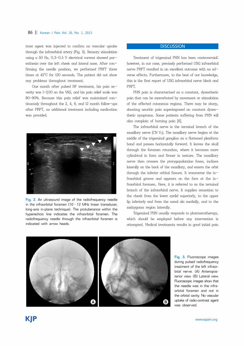
Fig. 3. Fluoroscope images during pulsed radiofrequencytreatment of the left infraor-bital nerve. (A) Anteropos-terior view. (B) Lateral view. Fluoroscopic images show thatthe needle was in the infra-orbital foramen and not in the orbital cavity. No vascularuptake of radio-contrast agentwas observed.
trast agent was injected to confirm no vascular uptake
through the infraorbital artery (Fig. 3). Sensory stimulation
using a 50 Hz, 0.3-0.5 V electrical current showed par-
esthesia over the left cheek and lateral nose. After con-
firming the needle position, we performed PRFT three
times at 42oC for 120 seconds. The patient did not show
any problems throughout treatment.
One month after pulsed RF treatment, his pain se-
verity was 1-2/10 on the VAS, and his pain relief scale was
80-90%. Because this pain relief was maintained con-
tinuously throughout the 2, 4, 6, and 12 month follow-ups
after PRFT, no additional treatment including medication
was provided.
DISCUSSION
Treatment of trigeminal PHN has been controversial;
however, in our case, precisely performed USG infraorbital
nerve PRFT resulted in an excellent outcome with no ad-
verse effects. Furthermore, to the best of our knowledge,
this is the first report of USG infraorbital nerve block and
PRFT.
PHN pain is characterized as a constant, dysesthetic
pain that can be exacerbated by movement or stimulation
of the affected cutaneous regions. There may be sharp,
shooting neuritic pain superimposed on constant dyses-
thetic symptoms. Some patients suffering from PHN will
also complain of burning pain [6].
The infraorbital nerve is the terminal branch of the
maxillary nerve (CN V2). The maxillary nerve begins at the
middle of the trigeminal ganglion as a flattened plexiform
band and passes horizontally forward. It leaves the skull
through the foramen rotundum, where it becomes more
cylindrical in form and firmer in texture. The maxillary
nerve then crosses the pterygopalantine fossa, inclines
laterally on the back of the maxillary, and enters the orbit
through the inferior orbital fissure. It transverse the in-
fraorbital groove and appears on the face at the in-
fraorbital foramen. Here, it is referred to as the terminal
branch of the infraorbital nerve. It supplies sensation to
the cheek from the lower eyelid superiorly, to the upper
lip inferiorly and from the nasal ala medially, and to the
midzygoma region laterally.
Trigeminal PHN usually responds to pharmacotherapy,
which should be employed before any intervention is
attempted. Medical treatments results in good initial pain
Lim, et al / US-Guided Infraorbital Nerve PRFT 87
www.epain.org
relief, but relief rates fall off dramatically over the
long-term [7]. Cases refractory to medical management
can be treated with minimally invasive procedures such as
a nerve block. Nerve blocks with local anesthetics and ste-
roids serving the painful area are a reasonable next step
if pharmacological modalities fail to control the pain. The
exact mechanism of pain relief from neural blockade during
treatment of PHN is unknown, but it may be related to
modulation of pain transmission [8].
Radiofrequency treatment for trigeminal PHN can also
be considered. Heat radiofrequency lesioning (HRFL) has
been effective for idiopathic trigeminal neuralgia, sympto-
matic facial pain, and herpes zoster of the fifth cranial
nerve. HRFL is the recommended option for treating trige-
minal pain in the elderly [4] and results in pain relief with-
out medication in the relatively long term. But, it necessa-
rily accompanies hypoesthesia or topoanesthesia. PRFT
has gained interest because it seems to be a non-neu-
ro-destructive method unlike conventional HRFL. The pulse
output during PRFT is interrupted; thus, allowing sufficient
time for the generated heat to be washed out. No reports
have identified sensory or motor loss or complications re-
lated to PRFT [3].
The infraorbital nerve has a close anatomical relation-
ship with the infraorbital foramen. Usually, we perform an
infraorbital nerve block using the landmark-based ap-
proach and palpate the foramen. The infraorbital foramen
can be palpated at 1-1.5 cm under the inferior edge of the
orbit at the midpupillary line. It can occasionally be difficult
to identify the infraorbital foramen by relying on palpation
only, particularly in cases of anatomical variations in the
infraorbital foramen [5]. Higher efficacy and a reduced risk
of complications are realized using advanced imaging mo-
dalities including computed tomography or virtual reality
techniques such as US or fluoroscopy [9]. Hence, US or
fluoroscopy-guided infraorbital nerve PRFT can be a val-
uable treatment for trigeminal PHN.
US imaging is a safe, simple, and non-invasive modal-
ity through which soft tissues and nerve structures can be
visualized and identified when combined with a thorough
knowledge of regional anatomy. According to Koscielniak-
Nielsen et al. [10], a USG peripheral nerve block sig-
nificantly shortens performance time and reduces the
number of needle passages to the target. The occurrence
of paresthesia during block is also reduced but not the in-
cidence of short-lasting post-operative neuropraxia.
However, limited information is available on the use of US
for identifying bony structures. Definitive identification of
osseous landmarks may be important when the target
nerve of the block is unidentifiable with US due to its small
size and imaging artifacts. When we perform USG in-
fraorbital nerve block, bone appears as a hyperechoic line-
ar structure. Any disruption or protuberance within the
hyperechoic line may indicate the infraorbital foramen (Fig.
2) [5]. Additionally, we confirmed arterial pulsation with
Doppler. After we advanced the needle slightly into the in-
fraorbital canal, we checked the needle position with
fluoroscopy. Then we confirmed again that there was no
vascular uptake through the infraorbital artery with ra-
dio-contrast agent (Fig. 3), which could help perform the
procedure.
In summary, our patient had been treated medically
for PHN. However, medication alone did not provide suffi-
cient pain relief. USG infraorbital nerve block seemed to
be effective for reducing the pain. But, its effect was
insufficient. For these reasons, we finally decided to per-
form USG PRFT of the infraorbital nerve and gained ex-
cellent results. As with USG in our case, PRFT could be
performed more precisely and efficiently. US provides in-
sight into deeper structures under the skin and avoid the
dangerous vessels with Doppler compared with the land-
mark approach. We could apply additional pulsed radio-
frequency output to the infraorbital nerve compared to that
of other nerves, because the infraorbital nerve is tight
within infraorbital canal compared to other free-moving
nerves during PRFT.
We have presented our experience of USG infraorbital
nerve PRFT, which is safer and more effective than that
of the landmark method. But, further randomized con-
trolled studies will be needed to demonstrate the safety
and efficacy of this treatment.
REFERENCES
1. Hashizume K. Herpes zoster and post-herpetic neuralgia. Nihon Rinsho 2001; 59: 1738-42.
2. Fashner J, Bell AL. Herpes zoster and postherpetic neuralgia: prevention and management. Am Fam Physician 2011; 83: 1432-7.
3. Nguyen M, Wilkes D. Pulsed radiofrequency V2 treatment and intranasal sphenopalatine ganglion block: a combination therapy for atypical trigeminal neuralgia. Pain Pract 2010; 10: 370-4.
88 Korean J Pain Vol. 26, No. 1, 2013
www.epain.org
4. Rahman M, Richter EO, Osawa S, Rhoton AL Jr. Anatomic study of the infraorbital foramen for radiofrequency neurotomy of the infraorbital nerve. Neurosurgery 2009; 64: 423-7.
5. Tsui BC. Ultrasound imaging to localize foramina for superficial trigeminal nerve block. Can J Anaesth 2009; 56: 704-6.
6. Schmid T, Pautex S, Lang PO. Acute and postherpetic neuralgia in the elderly: analysis of evidence for therapeutic options. Rev Med Suisse 2012; 8: 1374-8, 1380-2.
7. Haridas A, Mathewson C, Eljamel S. Long-term results of 405 refractory trigeminal neuralgia surgeries in 256 patients.
Zentralbl Neurochir 2008; 69: 170-4. 8. Hall GC, Carroll D, McQuay HJ. Primary care incidence and
treatment of four neuropathic pain conditions: a descriptive study, 2002-2005. BMC Fam Pract 2008; 9: 26.
9. Bhaskar AK. Interventional management of cancer pain. Curr Opin Support Palliat Care 2012; 6: 1-9.
10. Koscielniak-Nielsen ZJ. Ultrasound-guided peripheral nerve blocks: what are the benefits? Acta Anaesthesiol Scand 2008; 52: 727-37.