ultrasonography-guided common musculoskeletal
TRANSCRIPT

1Copyright © 2021 The Korean Society of Radiology
INTRODUCTION
The confidence of a general radiologist in performing a given image-guided musculoskeletal procedure varies with clinical exposure as a trainee, and the duration after the last performance of a particular procedure. This didactic pictorial essay is aimed at general radiologists and trainees with a methodological and clinically oriented approach to enhancing user understanding and confidence. This pictorial essay is based on the experience of the authors with musculoskeletal procedures, coupled with information on evidence-based practice from the literature. This pictorial essay is categorized by anatomical location, with procedures presented from rostral to caudal end. Individual procedures
Ultrasonography-Guided Common Musculoskeletal Interventions from Head to Toe: Procedural Tips for General RadiologistsRoland White1, 2, Michael Croft1, Stephen Bird3, Matthew Sampson3, 4
1Department of Radiology, Royal Adelaide Hospital, Adelaide, Australia; 2Department of Radiology, The University of Adelaide, School of Medicine, Adelaide, Australia; 3Benson Radiology, Wayville, Australia; 4Department of Radiology, Flinders University, School of Medicine Adelaide, Adelaide, Australia
The expanding scope of interventional musculoskeletal procedures has resulted in increased pressure on general radiologists. The confidence of general radiologists in performing ultrasound-guided musculoskeletal procedures varies with their clinical exposure. This didactic review provides a methodologically and clinically oriented approach to enhancing user understanding and confidence in performing ultrasound-guided musculoskeletal procedures. The body of the text is accompanied by figures depicting the procedural approach, injection site, and labeled ultrasonography images. This paper aims to provide a teaching and bedside aid for education on and the execution of musculoskeletal procedures to ensure the provision of quality health care.Keywords: Diagnostic imaging; Education; Musculoskeletal intervention; Interventional radiology; Ultrasound; Ultrasonography
Received: January 20, 2021 Revised: April 28, 2021 Accepted: May 22, 2021Corresponding author: Roland White, BMedSci, MD, Department of Radiology, Royal Adelaide Hospital, Port Rd, Adelaide SA 5000, Australia.• E-mail: [email protected] is an Open Access article distributed under the terms of the Creative Commons Attribution Non-Commercial License (https://creativecommons.org/licenses/by-nc/4.0) which permits unrestricted non-commercial use, distribution, and reproduction in any medium, provided the original work is properly cited.
are preluded by a brief revision of procedural definitions, indications, clinical pearls, and complications. The body of the text is accompanied by figures depicting the procedural approach, injection site, and labeled ultrasound anatomy. This pictorial essay is aimed at becoming a teaching and bedside aid in the education and execution of ultrasound-guided musculoskeletal procedures to ensure the provision of quality health care.
The following procedures are discussed in this article: - Greater occipital nerve block- Barbotage- Shoulder hydrodilatation- Suprascapular nerve block - Subacromial bursa injection- Tendon injection (elbow) - common flexor and extensor
origin- Wrist joint injection and arthrogram- Ganglion cyst aspiration- Pulley release- Hip joint injection- Mechanical hydro release of sciatic nerve- Knee joint injection
Korean J Radiol 2021
eISSN 2005-8330https://doi.org/10.3348/kjr.2021.0022
Pictorial Review | Intervention

2
White et al.
https://doi.org/10.3348/kjr.2021.0022 kjronline.org
- Paratenon stripping (brisement) of non-insertional achilles tendinosis
- Subtalar joint injection- Intermetatarsal bursa injection- Plantar fascia injection with nerve block
Greater Occipital Nerve Block
The greater occipital nerve (GON) is derived from the posterior ramus of C2 and supplies the skin of the posterior scalp. Its sensory distribution is attributed to its involvement in occipital neuralgia, cervicogenic headaches, and migraines. It can be found superficially at the inferolateral aspect of the occipital protuberance between the inferior obliquus capitis and semispinalis capitis/splenius capitis muscles. It is thought that occipital neuralgia can cause migraine symptoms as the GON converges with the ophthalmic branch of the trigeminal nerve at the trigeminal nucleus caudalis [1-3].
1) Best performed at the inferior obliquus capitis level. The alternative location at the external occipital protuberance is difficult because of the hair of the patient (Fig. 1A) [2,3].
2) Use dexamethasone/non-particulate steroid for injecting adjacent to the cervical spine.
3) To locate the nerve sonographically, position the probe transversely at the base of the occiput to find the posterior arch of C1, identified by a single spinous process.
4) Move inferior until the bifid C2 spinous process is in view, and move the probe laterally and obliquely to locate
the obliquus capitis muscle (Fig. 1A). 5) The location of the GON is between the inferior
obliquus capitis and semispinalis capitis/splenius capitis muscles (Fig. 1B) [2,3].
Complications include pain, bleeding, dizziness, weakness, and numbness.
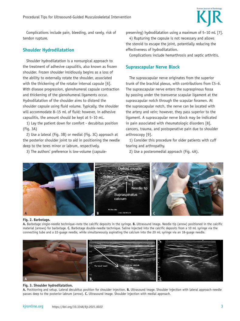
Barbotage
Barbotage refers to the repetitive injection and withdrawal of fluids. Barbotage is used to aspirate and fragment calcium hydroxyapatite crystals in the shoulder. Calcium hydroxyapatite deposition in the shoulder causes calcific tendinitis [4,5], most commonly affecting the rotator cuff tendons (supraspinatus, 80%; infraspinatus, 15%; subscapularis, 5%).
1) To break up adhesions and provide analgesia simultaneously, the subacromial bursa is liberally injected with 10–15 mL of 1% Xylocaine.
2) Flush calcific deposits with saline using a single 18-gauge needle and 20-mL syringe (Fig. 2A, B) or a 10-mL syringe via a connecting tube and 22-gauge needle, while simultaneously aspirating the calcium into the 20 mL syringe via the 18-gauge needle (Fig. 2C).
3) Inject steroid into the subacromial bursa at the end of the procedure to prevent associated bursitis.
4) Avoid mistaking linear dystrophic calcification at the enthesis insertion as calcific tendonitis. Calcific tendonitis is often round, oval, or dumbbell-shaped, although it can have various appearances according to the stage.
Fig. 1. GON block. A. Positioning and setup. IOC level for GON block. B. Ultrasound image. Note the location of the GON (arrow) between the IOC and the SSPC/SPC. GON = greater occipital nerve, IOC = inferior obliquus capitis, SPC = splenius capitis, SSPC = semispinalis capitis
A B
3
Procedural Tips for Ultrasound-Guided Musculoskeletal Intervention
https://doi.org/10.3348/kjr.2021.0022kjronline.org
Complications include pain, bleeding, and rarely, risk of tendon rupture.
Shoulder Hydrodilatation
Shoulder hydrodilatation is a nonsurgical approach to the treatment of adhesive capsulitis, also known as frozen shoulder. Frozen shoulder insidiously begins as a loss of the ability to externally rotate the shoulder, associated with the thickening of the rotator interval capsule [6]. With disease progression, glenohumeral capsule contraction and thickening of the glenohumeral ligaments occur. Hydrodilatation of the shoulder aims to distend the shoulder capsule using fluid volume. Typically, the shoulder will accommodate 8–15 mL of fluid; however, in adhesive capsulitis, the amount should be kept at 5–10 mL.
1) Lay the patient down for comfort - decubitus position (Fig. 3A)
2) Use a lateral (Fig. 3B) or medial (Fig. 3C) approach at the posterior shoulder joint to aid in positioning the needle deep to the teres minor or labrum, respectively.
3) The authors’ preference is low-volume (capsule-
preserving) hydrodilatation using a maximum of 5–10 mL [7].4) Rupturing the capsule is not necessary and allows
the steroid to escape the joint, potentially reducing the effectiveness of hydrodilatation.
Complications include hemarthrosis and septic arthritis.
Suprascapular Nerve Block
The suprascapular nerve originates from the superior trunk of the brachial plexus, with contributions from C5–6. The suprascapular nerve enters the supraspinous fossa by passing under the transverse scapular ligament at the suprascapular notch through the scapular foramen. At the suprascapular notch, the nerve can be located with the artery and vein; however, they pass superior to the ligament. A suprascapular nerve block may be indicated in pain associated with rheumatologic disorders [8], cancers, trauma, and postoperative pain due to shoulder arthroscopy [9].
1) Consider this procedure for older patients with cuff tearing and arthropathy.
2) Use a posteromedial approach (Fig. 4A).
Fig. 2. Barbotage. A. Barbotage single-needle technique–note the calcific deposits in the syringe. B. Ultrasound image. Needle tip (arrow) positioned in the calcific material (arrows) for barbotage. C. Barbotage double-needle technique. Saline injected into the calcific deposits from a 10 mL syringe via the connecting tube and a 22-guage needle, while simultaneously aspirating the calcium into the 20 mL syringe via an 18-guage needle.
A B C
Fig. 3. Shoulder hydrodilatation. A. Positioning and setup. Lateral decubitus position for shoulder injection. B. Ultrasound image. Shoulder injection with lateral approach-needle passes deep to the posterior labrum (arrow). C. Ultrasound image. Shoulder injection with medial approach.
A B C

4
White et al.
https://doi.org/10.3348/kjr.2021.0022 kjronline.org
3) Inject into the suprascapular notch (Fig. 4B).4) Injecting into the suprascapular notch is associated
with the risk of pleural puncture if excessive accidental anterior needle advancement occurs.
Complications include pneumothorax, nerve injury, infection, and bleeding.
Subacromial Bursa Injection
The subacromial bursa is a fluid-filled space located inferior to the acromion process and superior to the supraspinatus. Bursitis leads to anterolateral pain exacerbated by overhead activities as the inflamed bursa rubs against the humeral head and supraspinatus inferiorly and the acromion and deltoid superiorly.
1) Asking the patient to watch the ultrasound screen can create a distraction.
2) Use a posterior approach to avoid anterior supraspinatus tears (the most common site).
3) Relaxed-arm positioning allows relaxation of the
deltoid muscle, reducing pain and discomfort (Fig. 5).Complications include infection, bleeding, and recurrence
of pain.
Tendon Injection (Elbow)-Common Flexor and Extensor Origin
Common flexor and extensor tendinosis are overuse injuries that correspond to their respective common flexor and extensor tendon origins. Common extensor tendinosis is more common than flexor tendinosis. Tendinosis results from repetitive microtrauma through excessive contraction of the wrist extensors or flexors. The extensor carpi radialis brevis is the most affected extensor tendon [10]. The most involved tendons in common flexor tendinosis include the pronator teres and flexor carpi radialis [10]. Common flexor tendon injection can be performed as follows:
1) Laying the patient in the supine position with the arm abducted and resting above the head (flexed abducted, supinated) allows easy access to the common flexor origin
Fig. 4. SSN block. A. Positioning and setup. Positioning for SSN block. B. Ultrasound image. Injection into the suprascapular notch (arrows). SSN = suprascapular nerve
A B
Fig. 5. SA bursa injection. A. Positioning and setup. Ergonomic alignment for the posterior approach to SA bursa injection. Note the relaxed position. B. Ultrasound image. Steroid injection into the SA bursa (arrow). SA = subacromial
A B

5
Procedural Tips for Ultrasound-Guided Musculoskeletal Intervention
https://doi.org/10.3348/kjr.2021.0022kjronline.org
(Fig. 6).2) Injecting steroids and anesthetics along the tendon
surface can help strip neovessels and reduce pain related to tendinopathy.
Complications include infection, bleeding, and tendon damage/rupture.
Wrist Joint Injection and Arthrogram
Arthrography is a procedure designed to assist radiologists in the assessment of articular structures or visualization of the distribution of a therapeutic injection. Wrist arthrography is most commonly used to assess the triangular fibrocartilage complex and intrinsic ligaments [11]. The commonly used wrist joint for arthrography is the articulation of the scaphoid and radius.
1) Lay the patient prone in a neutral wrist position with volar flexion over a pillow (Fig. 7A).
2) Use a dorsal/distal approach to the scaphoid (Fig. 7B).3) The radial approach with ulnar deviation is an
alternative option. Complications include joint pain, infection, hand
swelling, and bleeding.
Ganglion Cyst Aspiration
A ganglion cyst is the coalescence of extra-articular mucin connected to a synovial joint via a pedicle [12], which clinically presents as a soft tissue swelling proximal to this joint. It is the most common soft tissue swelling in the hand and wrist, with 70% occurring on the dorsal wrist and 20% on the volar. These are generally asymptomatic; however, they can cause cosmetic concerns, paresthesia, pain, and weakness. It has been reported that 58% of the cysts resolve spontaneously with time; however, aspiration is a management option for those requiring symptomatic relief or cosmetic respite. It should be communicated to the patients that cyst aspiration has a high incidence of recurrence [13].
1) Loosen cyst contents with 3–5 mL of 1% Xylocaine and
Fig. 6. Common flexor tendon (elbow) injection. A, B. Positioning and setup. Positioning for common flexor tendon injection (screen at head of bed).
A B
Fig. 7. Wrist joint injection. A. Positioning and setup. Position for wrist joint injection. B. Ultrasound image. Needle inserted from distal approach (arrow) to pass below the dorsal radius.
A B

6
White et al.
https://doi.org/10.3348/kjr.2021.0022 kjronline.org
a 25-gauge needle (Fig. 8). 2) Inject 1% Xylocaine under the skin with the same
needle. Allow the anesthetic time to work.3) Use an 18-gauge needle and a 10 mL syringe to
perforate and aspirate the ganglion.4) Inject 1 mL Celestone into the ganglion at the end of
the procedure to reduce inflammation after the procedure.Complications include infection, bleeding, nerve and
tendon injury, scarring, and vascular injury.
Pulley Release
Percutaneous pulley release is a procedure used to treat stenosing tenosynovitis, otherwise known as the trigger finger. The trigger finger is an imbalance of the flexor tendons and sheath, with thickening of the A1 pulley causing snapping and locking of the finger. Ultrasound features of the trigger finger include hypoechoic thickening of the A1 pulley, with increased edema of the surrounding tissue [14].
1) Best reserved for fingers because of the challenging anatomy of the thumb for a radiologist without experience in this procedure.
2) Completely sterile technique.3) Mark the tendon path as a guide of the neurovascular
bundle location. 4) First, inject local anesthetic with a 25-gauge needle.
5) A rolled-up towel under the patient’s hand creates metacarpophalangeal joint hyperextension, which allows the needle to enter the target area unimpeded.
6) Bend an 18-gauge needle, using its cap, at the needle hub and mid-point to use as a cutting device [15]. The bevel should be placed in the sagittal plane. Attach a 3 mL syringe as a handle.
7) Position the needle tip deep into the pulley and apply a downward and backward force (Fig. 9A, B).
8) Concurrent steroid injection at the time of release will treat any coexisting tenosynovitis.
9) The opinion of a hand therapist or physical therapist is recommended post-release to assess short- and long-term functionality.
Common complications include pain, infection, bleeding, and nerve injury.
Hip Joint Injection
The diagnosis of hip pain is a difficult, with poor localization of symptoms and pain arising from either a primary hip structure or referred from an adjacent area. Hip pain can be intra-articular; extraarticular; from the lumbar spine or pelvic floor; referred from the bowel, bladder, or reproductive organs; or a mixture of the above [16]. Intra-articular ultrasound-guided corticosteroid injection of the hip joint can assist in the management of osteoarthritis/
Fig. 8. Ganglion aspiration. A. Positioning and setup. Positioning for dorsal wrist ganglion injection. B. Ultrasound image. Loosening contents of the ganglion with local anesthetic prior to aspiration.
A B

7
Procedural Tips for Ultrasound-Guided Musculoskeletal Intervention
https://doi.org/10.3348/kjr.2021.0022kjronline.org
femoroacetabular impingement. Other injectable therapies, such as platelet-rich plasma, can also be injected under ultrasound guidance.
1) Use a 9-cm 22-gauge or longer spinal needle to ensure adequate needle length to reach the hip joint.
2) Internally rotating the foot moves the femoral vessels medially out of the path of the needle. The inferolateral approach also helps to avoid the femoral nerve (Fig. 10A).
3) In contrast with fluoroscopic-guided injection, it does not aim for the femoral neck, as this puts the needle more perpendicular to the probe, reducing needle visualization. On ultrasound, the position is confirmed by the needle tip beneath the labrum and microbubbles in the joint (Fig. 10B).
4) The inferior capsule is thicker and harder to penetrate
as opposed to a thinner superior capsule, another benefit of aiming deep to the labrum.
5) Turning the needle 180° clockwise and anticlockwise will help the bevel bore through a tented capsule.
Complications include infection, bleeding, neurovascular injury, and tendon injury.
Mechanical Hydro Release of the Sciatic Nerve
The sciatic nerve arises from the L4–S3 lumbosacral nerve roots. After exiting the pelvis, the sciatic nerve courses anteroinferior to the piriformis, and posterior to the gemelli, obturator internus, and quadratus femoris. Sciatic nerve irritation can cause lumbar, gluteal, and thigh
Fig. 9. A1 pulley release. A. Positioning and setup. Hyperextension of the metacarpophalangeal joint is created with a small rolled up towel, and gentle pressure is applied to the patients’ fingers. B. Schematic representation of syringe movement. Withdraw needle (blue arrow) whilst applying downward force on the syringe (orange arrow), which forces the needle tip in a palmar direction to cut the A1 pulley. C. Insert an 18-gauge needle (arrow) deep into the A1 pulley (arrow) prior to release.
A B C
Fig. 10. Hip joint injection. A. Positioning and setup. Probe positioning for anterior hip joint injection. Allowing for inferolateral approach. B. Ultrasound image. Anterior hip joint injection, needle tip should be directed deep to the labrum (arrow).
A B

8
White et al.
https://doi.org/10.3348/kjr.2021.0022 kjronline.org
neuropathic pain, with a significant impact on quality of life. Adhesions can form on the sciatic nerve in response to muscle injury or chronic muscle stretching associated with gait disturbances [17].
1) Under ultrasound guidance, use a 22-gauge needle with local anesthetic and cold saline to strip away adhesions from the sciatic nerve, which form secondary to chronic hamstring or piriformis injury (Fig. 11).
2) Use liquid pressure ahead of the needle to mechanically open tissue planes.
Complications include bleeding, infection, and nerve or tendon damage.
Knee Joint Injection
Different approaches can be used for aspiration or injection for diagnostic and therapeutic procedures for the knee. The most common intra-articular knee injection
includes corticosteroids, which can relieve osteoarthritis, juvenile idiopathic arthritis, gout, pseudogout, and rheumatoid arthritis.
1) In the absence of suprapatellar pouch fluid, the aim is for the trochlea with a lateral approach with the knee in mid-flexion (Fig. 12A).
2) In a small number of patients, the suprapatellar pouch does not freely communicate with the remainder of the joint; therefore, this technique is preferable in all cases (Fig. 12B).
Common complications include hemarthrosis and septic arthritis.
Paratenon Stripping (Brisement) of Non-Insertional Achilles Tendinosis
Achilles tendinosis is a degenerative condition that may be related to overuse and repetitive microtrauma exhibiting tendon thickening and neovascularization on ultrasound [18]. Insertional tendinosis involves tendinopathy occurring along the 2-cm portion between the tendon and its insertion at the calcaneum. Non-insertional tendinosis relates to the 2–6-cm segment proximal to the calcaneum. Adhesions form between the paratenon and the Achilles tendon through chronic inflammation. Paratenon stripping is the process of injecting saline into the tendon/paratenon interspace to break up adhesions.
1) Medial or lateral approach, placing a 22-gauge needle deep to the paratenon but not into the tendon substance (Fig. 13A).
2) High-volume injection was started with 10 mL of 1% Xylocaine, followed by 1 mL Celestone and cold saline to a
Fig. 11. Sciatic nerve hydro dissection. Ultrasound image. Injecting with a 22-gauge needle around the sciatic nerve (arrows).
Fig. 12. Knee joint injection. A. Positioning and setup. Lateral positioning for knee joint injection. B. Ultrasound image. Knee injection at the lateral trochlear level (arrows).
A B

9
Procedural Tips for Ultrasound-Guided Musculoskeletal Intervention
https://doi.org/10.3348/kjr.2021.0022kjronline.org
total volume of 40–60 mL (Fig. 13B, C).3) Celestone is used because of its anti-inflammatory
properties.Common complications include bleeding, infection, nerve
injury, and chronic pain.
Intermetatarsal Bursa Injection
The intermetatarsal bursa is found between the metatarsal phalangeal joints. It is dorsal to the digital nerves, and this anatomical association accounts for symptomatology in intermetatarsal bursitis. Bursitis causes pain that can
Fig. 13. Achilles paratenon stripping. A. Positioning and setup. Approach for paratenon stripping for non-insertional Achilles tendonitis. B. Ultrasound image. Needle positioned at the deep paratenon for stripping (arrows). C. Ultrasound image. Needle positioned at the superficial paratenon (arrows).
A B C
Fig. 14. Intermetatarsal injection. A. Positioning and setup. Positioning for dorsal approach to intermetatarsal injection. B. Ultrasound image. Intermetatarsal bursa (arrow) in the 3rd and 4th intermetatarsal space.
A B
Fig. 15. Plantar fascia injection. A. Positioning and setup. Tibial nerve block prior to plantar fascia injection. B. Ultrasound image. Inject steroid deep into the plantar fascia (arrow) to prevent fat pad necrosis.
A B
10
White et al.
https://doi.org/10.3348/kjr.2021.0022 kjronline.org
radiate into the toes of the affected space, known as metatarsalgia, exacerbated by weight bearing, high heels, and narrow shoes. If the intermetatarsal bursitis is left untreated, the bursa can calcify and fibrose the surrounding interdigital space.
1) A dorsal needle approach with the ultrasound probe on the plantar aspect of the foot is beneficial as it is less painful, and there is potentially less skin flora on the dorsal aspect of the foot compared to that on the plantar aspect (Fig. 14).
2) Massaging jelly into the skin at the plantar aspect of the foot improves contact and visualization, particularly in patients with thick, dry skin [19].
Common complications include bleeding, infection, pain, and pain recurrence.
Plantar Fascia Injection with Nerve Block
Plantar fasciitis is a self-limiting inferior heel condition characterized by pain at the calcaneal origin of the plantar fascia. It is the most common cause of heel pain, exacerbated by prolonged standing and running. Histologically, there is a predominance of collagen fiber deterioration, fibroblast proliferation, and increased vascularity at the plantar fascia enthesis [20]. When combined with tibial nerve block, discomfort of the procedure can be alleviated by reducing the signals of the afferent medial and lateral plantar nerves [21].
1) Blocking the tibial nerve at the level just above the medial malleolus with 5–10 mL of 1% Xylocaine makes this procedure much more tolerable (Fig. 15A).
2) If the injection is too inferior, the medial calcaneal and lateral plantar nerves can be missed.
3) Inject steroid deep into the plantar fascia (not superficial) to prevent fat pad necrosis (Fig. 15B).
4) Dry needling can be performed at the same time.Common complications include infection, bleeding, fat
pad necrosis, and lateral plantar nerve injury.
CONCLUSION
This essay provides methodical, easy-to-understand steps in conjunction with pictorial explanations, which will serve as a valuable and easy-to-use reference for general and trainee radiologists in teaching and performing ultrasound-guided musculoskeletal procedures.
Conflicts of InterestThe authors have no potential conflicts of interest to disclose.
Author ContributionsConceptualization: Roland White, Michael Croft,
Matthew Sampson. Data curation: Stephen Bird, Matthew Sampson. Investigation: Roland White, Michael Croft, Matthew Sampson. Project administration: Roland White, Michael Croft, Matthew Sampson. Resources: all authors. Supervision: Matthew Sampson. Validation: all authors. Visualization: all authors. Writing—original draft: Roland White, Michael Croft. Writing—review & editing: Roland White.
ORCID iDsRoland White
https://orcid.org/0000-0002-4455-609XMichael Croft
https://orcid.org/0000-0003-0367-2729
REFERENCES
1. Allen SM, Mookadam F, Cha SS, Freeman JA, Starling AJ, Mookadam M. Greater occipital nerve block for acute treatment of migraine headache: a large retrospective cohort study. J Am Board Fam Med 2018;31:211-218
2. Greher M, Moriggl B, Curatolo M, Kirchmair L, Eichenberger U. Sonographic visualization and ultrasound-guided blockade of the greater occipital nerve: a comparison of two selective techniques confirmed by anatomical dissection. Br J Anaesth 2010;104:637-642
3. Pingree MJ, Sole JS, O’ Brien TG, Eldrige JS, Moeschler SM. Clinical efficacy of an ultrasound-guided greater occipital nerve block at the level of C2. Reg Anesth Pain Med 2017;42:99-104
4. Hayes CW, Conway WF. Calcium hydroxyapatite deposition disease. Radiographics 1990;10:1031-1048
5. Chianca V, Albano D, Messina C, Midiri F, Mauri G, Aliprandi A, et al. Rotator cuff calcific tendinopathy: from diagnosis to treatment. Acta Biomed 2018;89:186-196
6. Rymaruk S, Peach C. Indications for hydrodilatation for frozen shoulder. EFORT Open Rev 2017;2:462-468
7. Lee DH, Yoon SH, Lee MY, Kwack KS, Rah UW. Capsule-preserving hydrodilatation with corticosteroid versus corticosteroid injection alone in refractory adhesive capsulitis of shoulder: a randomized controlled trial. Arch Phys Med Rehabil 2017;98:815-821
8. Shanahan EM, Shanahan KR, Hill CL, Ahern MJ, Smith MD. Safety and acceptability of suprascapular nerve block in

11
Procedural Tips for Ultrasound-Guided Musculoskeletal Intervention
https://doi.org/10.3348/kjr.2021.0022kjronline.org
rheumatology patients. Clin Rheumatol 2012;31:145-1499. Chan CW, Peng PW. Suprascapular nerve block: a narrative
review. Reg Anesth Pain Med 2011;36:358-373 10. Taylor SA, Hannafin JA. Evaluation and management of elbow
tendinopathy. Sports Health 2012;4:384-39311. Shelat N, Shah R, O’Rourke H, Bennett DL. Common
musculoskeletal procedures: how we do it. J Am Osteopath Coll Radiol 2016;5:12-19
12. Gude W, Morelli V. Ganglion cysts of the wrist: pathophysiology, clinical picture, and management. Curr Rev Musculoskelet Med 2008;1:205-211
13. Suen M, Fung B, Lung CP. Treatment of ganglion cysts. ISRN Orthop 2013;2013:940615
14. Pan M, Sheng S, Fan Z, Lu H, Yang H, Yan F, et al. Ultrasound-guided percutaneous release of A1 pulley by using a needle knife: a prospective study of 41 cases. Front Pharmacol 2019;10:267
15. Rajeswaran G, Lee JC, Eckersley R, Katsarma E, Healy JC. Ultrasound-guided percutaneous release of the annular pulley
in trigger digit. Eur Radiol 2009;19:2232-223716. Lynch TS, Oshlag BL, Bottiglieri TS, Desai NN.
Ultrasound-guided hip injections. J Am Acad Orthop Surg 2019;27:e451-e461
17. Burke CJ, Walter WR, Adler RS. Targeted ultrasound-guided perineural hydrodissection of the sciatic nerve for the treatment of piriformis syndrome. Ultrasound Q 2019;35:125-129
18. Singh A, Calafi A, Diefenbach C, Kreulen C, Giza E. Noninsertional tendinopathy of the achilles. Foot Ankle Clin 2017;22:745-760
19. Linklater JM, Bird SJ. Imaging of lesser metatarsophalangeal joint plantar plate degeneration, tear, and repair. Semin Musculoskelet Radiol 2016;20:192-204
20. Nair AS, Sahoo RK. Ultrasound-guided injection for plantar fasciitis: a brief review. Saudi J Anaesth 2016;10:440-443
21. Mulherin D, Price M. Efficacy of tibial nerve block, local steroid injection or both in the treatment of plantar heel pain syndrome. Foot (Edinb) 2009;19:98-100