ucsf&quest!diagnostics!partnership! …...ucsf/quest!mrimaging! quick!reference!guide!...
TRANSCRIPT

1
UCSF-‐QUEST DIAGNOSTICS PARTNERSHIP
DEMENTIA CARE PATHWAY
MR PROTOCOL FOR DEMENTIA PATIENTS

2
1. MR PROTOCOL OVERVIEW Page 3 1.1 Overall Protocol Goals Page 3 1.2 Components of MR Protocol Page 3 2. SITE SET-‐UP Page 4 2.1 Site Requirements Page 4 2.2 Protocol Installation Page 4 3. MRI SUBJECT PRE-‐SCAN PROCEDURES Page 5 3.1 Subject Pre-‐Screening Page 5 3.2 Subject Safety and Monitoring Page 5 4. QUEST MRI SCAN PROTOCOL Page 6-‐9 4.1 Protocol Overview Page 6 4.2 RPD Trouble Shooting Protocol Page 6-‐7 4.3 Scanning Sequences Page 7-‐10 4.4 Quality Assurance Page 10 5. APPENDICES Page 11-‐94 5.1 MR Protocol Quick Reference Guide Page 12 5.2 Uploading MR protocols Page 13 5.3 Protocol Parameter Listing 5.3.1 GE 1.5T Parameters Page 14-‐29 5.3.2 GE 3T Parameters Page 30-‐40 5.3.3 Philips 1.5T Parameters Page 41-‐53 5.3.4 Philips 3T Parameters Page 54-‐67 5.3.5 Siemens 1.5T Parameters Page 68-‐82 5.3.6 Siemens 3T Parameters Page 83-‐96

3
1. NEUROIMAGING OVERVIEW This section provides a high level overview of the imaging protocol and analysis that has been established for imaging patients that are being assessed for dementia. The critical components to this effort are summarized below. 1.1 Background and Goals of Quest Neuroimaging Initiative The UCSF/Quest MR protocol is designed to provide consistent and relevant imaging information that will be used in the diagnosis of dementia. A range of image contrasts are prescribed that are suitable for this application and the established imaging protocol is designed for use on a wide range of MR systems operating at field strengths of both 1.5 and 3 Tesla. Improved diagnostic power is expected by utilizing a consistent and standardized MR imaging protocol that is additionally amenable to quantitative analysis. More consistent clinical interpretations, with improved diagnostic sensitivity and specificity, are anticipated through this initiative. 1.2 Components of MR Protocol The MR protocol is designed to be run in a 30-‐minute time slot and actual scan time is 20-‐25 minutes. The protocol includes a volumetric T1-‐weighted protocol that is based on the Alzheimer ’s Disease Neuroimaging Initiative (ADNI) protocol. This well established and standardized imaging sequence is well suited to quantitative analysis and has been utilized in a wide range of scientific studies. The protocol further contains multi-‐slice sequences with T2, T2* and T2-‐FLAIR contrasts. Each of these multi-‐slice imaging sequences has matching spatial coverage and can be acquired in a time efficient fashion. The final element of the protocol is diffusion weighted imaging (DWI), which is acquired in both axial and coronal scan planes. DWI is acquired with diffusion sensitization in three orthogonal directions and with a b-‐value of 1000s/mm2.

4
2. SITE SET-‐UP This section reviews the steps that are necessary for an imaging center to participate in this neuroimaging initiative. 2.1 Site Requirements Participant sites must have an MR system for which a standardized imaging protocol has been established and tested. To date, standardized imaging protocols have been established for General Electric (GE), Philips, and Siemens scanners operating at field strengths of 1.5T and 3T. The imaging protocol is designed to run on all relatively recent software releases and minimal specialized software packages are required. Once established, the UCSF/Quest imaging protocol must be “locked” and left unaltered. Study coordinators should be notified if a software upgrade or system change result in changes in any protocol parameters. 2.2 Protocol Installation Protocols can be installed on scanners via one of two different mechanisms. Whenever possible, the protocol should be loaded via the protocol exchange (GE), examcard (Philips), or EDX (Siemens) protocol sharing systems. This assures that all parameter settings are consistent with the Quest imaging protocol. Details on how to upload via this mechanism are provided in Appendix 5.2 and may require the assistance of a service engineer. In some instances software incompatibilities cause this installation approach to fail. In those instances the protocol can be manually entered and parameter file specifications are provided for each manufacturer and field strength in Appendix 5.3. Regardless of installation method, sites are required to scan a phantom and submit the resulting data set for certification that the protocol is properly installed on the system. Once this review has been completed the protocol should be locked and archived so that later modifications at the site do not occur.

5
3. MRI SUBJECT PRE-‐SCAN PROCEDURES 3.1 Subject Pre-‐Screening All subjects must be screened for MRI contraindications immediately before the MRI scan using your local standard protocol. Contraindications may include, but are not limited to:
• The presence of non-‐removable ferrous metal objects • Aneurysm clips • Metal fragments in the eyes • Pacemakers • Other contraindications such as defibrillators, etc.
Subjects must also not be severely claustrophobic and must be capable of remaining still for the 30-‐minute examination period. Patient fixation devices, straps etc. can be used to minimize patient motion. 3.2 Subject Safety and Monitoring MR is generally a very safe imaging modality and does not involve ionizing radiation. For best results the scan procedure should be explained to the subject so that they know what to expect during the MRI. Make sure that all loose metal objects are removed from the patient prior to entering the magnet room. If available, ask the subject to change into hospital robes or scrubs to ensure no ferrous objects or materials enter the scan room. Use standard local practice for monitoring the subject during the scan. These may include devices to monitor pulse and O2 levels.

6
4. QUEST MRI SCAN PROTOCOL The scan protocol consists of 20-‐25 minutes of MR scanning and can be completed within a 30-‐minute time slot. Patients should be placed in a multi-‐element head coil such as the 8-‐channel In Vivo head coil (below). Other variants including manufacturer head and head/neck coils are also acceptable.
4.1 Protocol Overview The protocol consists of the following scans: UCSF/Quest MRI Scan Protocol: 1) Localizer 1a) Calibration/Reference Scan (if necessary) 2) Sagittal 3D T1 MPRAGE/IR-‐SPGR (angled to midline) 3) Axial 2D T2* GRE (angled to AC/PC) 4) Axial 2D T2 turbo-‐FLAIR (angled to AC/PC; matches scan 3) 5) Axial 2D T2 turbo/fast spin echo (angled to AC/PC; matches scan 3) 6) Axial 2D Diffusion Weighted Imaging (angled to AC/PC) 7) Coronal 2D Diffusion Weighted Imaging (angled to brain stem) These scans should be run exactly as prescribed in the originally installed protocol and in the indicated order. 4.2 Troubleshooting Protocol: Additional Scans for RPD cases not diagnosed by General Dementia Protocol (e.g., need for contrast or question of prion disease remains)
The standardized dementia protocol is recommended for both chronic and rapidly progressive dementia. Sites should detect many if not most causes for RPD on the initial standardized dementia protocol scans. The additional scans listed below would be used
In Vivo’s Hi-‐Res Head Coil is available for GE, Philips or Siemens systems.

7
only for troubleshooting purposes if a diagnosis is unclear or more imaging information is required.
RPD Trouble Shooting Protocol: General dementia protocol plus the following: 8) Coronal 2D T2 turbo/fast spin echo (coronal orientation along hippocampal axis 3 skip 0 mm) 9) Coronal 2D T2 turbo-‐FLAIR (coronal orientation along hippocampal axis 3 skip 0 mm) 10) Sagittal 3D T1 MPRAGE/IR-‐SPGR (angled to midline) post contrast 11) Axial spin echo T1 post contrast Recommended adjustments to General Dementia Protocol scans: 6) Axial 2D B2000 Diffusion Weighted Imaging (angled to AC/PC) *¥ 7) Coronal 2D B2000 Diffusion Weighted Imaging (angled to brainstem) *¥ * Should be B2000 DWI either for 1.5 and 3.T ¥ On Siemens 3T B2000 should be “Resolve” for coronal. 4.3 Scanning Sequences 4.3.1 – Localizer + Calibration/Reference Scans
A conventional 3-‐plane localizer should be used to position subsequent scans. Parallel imaging (ASSET/SENSE/GRAPPA) will be used on the DWI scans so any calibration /reference scans required by your MR system should also be acquired at this time.

8
4.3.2 -‐ Sagittal 3D T1 MPRAGE/IR-‐SPGR Scanning Notes: This sagittal T1 volume should be angled on the axial scout such that the middle slice runs along the mid-‐line of the brain and encompasses the full width of the brain. Additional slices can be added to achieve this but slices MUST NOT be removed. Make sure that the scan volume extends SLIGHTLY BEYOND the top of the scalp.
4.3.3 -‐ Axial 2D T2* GRE Scanning Notes: This axial stack of 2D slices should be angulated on a midline sagittal scan such that it runs parallel to the Anterior Commissure/Posterior Commissure (AC/PC) of the brain (see below). The volume should extend from the top of the brain and continue as low as possible WITHOUT changing the number of slices, slice thickness or slice gap.
Scanning Notes: Angulation of the axial scan plane to AC/PC is demonstrated. The previously acquired Sagittal T1 volume can be used to identify AC/PC (dotted area) if it is not clearly seen on the localizer images.

9
4.3.4 -‐ Axial 2D T2 turbo FLAIR Scanning Notes: The field of view (FOV) and coverage of this scan exactly match that of Scan (3) -‐ Axial 2D T2* GRE. The scan volume for this acquisition should EXACTLY MATCH that of the prior scan.
4.3.5 -‐ Axial 2D T2 turbo/fast Spin Echo Scanning Notes: The field of view (FOV) and coverage of this scan exactly match that of Scan (3) -‐ Axial 2D T2* GRE. The scan volume for this acquisition should EXACTLY MATCH that of the prior scan.
4.3.6 -‐ Axial Diffusion Weighted Imaging
Scanning Notes: The axial DWI volume should also be angulated to AC/PC, but its coverage is not identical to scans (3) – (5). The axial scan plane should similarly be positioned such that it reaches the top of the brain and extends inferiorly as far as the prescribed slices allow.

10
4.3.7 -‐ Coronal Diffusion Weighted Imaging
Scanning Notes: The coronal plane should be angulated to be parallel to the brain stem and coverage should extend from the anterior to posterior extremes of the brain. If necessary slices can be added to achieve this coverage (this may prolong the TR, which is acceptable).
4.4 Quality Assurance It is important for the MR technologist to inspect scans to assure that they are of high quality and have not been degraded by patient motion. This is particularly important for the 3D T1 MPRAGE/IR-‐SPGR (scan 2), which will be used for quantitative analysis. Scans that are degraded by artifact or patient motion should be repeated.

11
5. APPENDICES The following pages contain a series of appendices that contain information on protocol content and uploading. Please refer to the page numbers for the content. 5.1 MR Protocol Quick Reference Guide Page 11 5.2 Uploading MR protocols Page 12 5.2 Protocol Parameter Listings
5.2.1: 1.5T GE Pages 13-‐28 5.2.2: 1.5T Philips Pages 29-‐39 5.2.3: 1.5T Siemens Pages 40-‐52 5.2.4: 3T GE Pages 53-‐66 5.2.5: 3T Philips Pages 67-‐81 5.2.6: 3T Siemens Pages 82-‐95

12
UCSF/Quest MR Imaging
QUICK REFERENCE GUIDE
MR manufacturers have all established methods for sharing scanning protocols between systems. The process typically involves loading the necessary protocol files onto and USB drive, connecting to USB to the MR system, and following a series of commands. It may be necessary to have your service engineer perform this installation if you don’t have the necessary privileges on you MR system.
GE ProtocolExchange: Begin by placing the protocol exchange files on a USB drive and plug the drive into the MR console computer. Under the “Image Management” tab there is a feature called “Protocol Exchange”, which you should click on. A window will pop up and you should then select “Import Mode”. Another window will appear
Philips Examcard: Begin by placing the examcard file on a USB drive and plug the drive into the MR console computer. Select the windows button on the keyboard and select “
Siemens “.edx” file: .

13
UCSF/Quest MR Imaging
QUICK REFERENCE GUIDE The UCSF/Quest Neuroimaging protocol is a standardized set of MR imaging sequences that have been established for a range of imaging platforms. It is important to run the correct protocol and assure that patient motion has not corrupted a scan before proceeding to the next acquisition.1*
Step 1: Follow your institutions standard MR pre-‐screening practices. Step 2: Position the subject in a head coil in a head first supine orientation. Use straps
and/or pads to help the subject remain stationary during scanning Step 3: Acquire a standard 3-‐plane localizer and any necessary calibration scans.
Step 4: Prescribe the ADNI MP-‐RAGE/IR-‐FSPGR scan in an oblique sagittal plane that is oriented to midline.
Step 5: Use the mid-‐sagittal plane of the ADNI MP-‐RAGE/IR-‐FSPGR scan to orient oblique axials scans to the anterior and posterior commissures (AC/PC).
Step 6: Acquire the axial T2* gradient echo, T2-‐FLAIR and T2 TSE scans in this orientation and with matching coverage. Cover from the top of the brain down with the available slice coverage (do not add or subtract).
Step 7: Acquire the oblique axial DWI with the same angulations as the prior axial scans (coverage won’t match). Again, Cover from the top of the brain down with the available slice coverage (do not add or subtract)
Step 8: Acquire the oblique coronal DWI such that the scan plane is oriented to the brain stem. Additional slices may be added to achieve full anterior/posterior coverage of the brain.
*This material

LOC
ALI
ZER
LOC
ALI
ZER
GE 1.5T UCSF/Quest Protocol - Survey
PATIENT POSITION IMAGING PARAMETERS
Patient Entry Head First Gradient Mode WHOLE
Patient Position Supine Imaging Mode 2D
Coil Configuration 8 Ch High Res Brain Array by
MRI Devices
Pulse Sequence Spin Echo
Plane 3-PLANE Imaging Options Seq, EDR, Fast, SS, ARC
Series Description LOCALIZER n/a 12
SCAN TIMING SCAN RANGE
TE Minimum FOV 35.0
TR Minimum Slice Thickness 10.0
Receiver Bandwidth 62.50 Slice Spacing 5.0
Center Location 1 0.0
Center Location 2 A25.0
Center Location 3 0.0
Slice for 3 Plane 0 Localizaion
Slice for 3 Plane 10 Localizaion
Slice for 3 Plane 6 Localizaion
Slice for 3 Plane 6 Localizaion
Space per Plane 1 5.0
Space per Plane 2 4.0
Table Delta 0.00
IMAGE ENHANCE ACQ TIMING
Filter Choice None Freq 256
Phase 128
Freq DIR Unswap
# of Acq. Before Pause 0
Phase FOV 1.00
Auto Shim Auto
Phase Correction No
GATING/TRIGGER USER CVS
Heart Beat per Minute Mode 0 User CV2 240.00
Auto Trigger Type Off User CV13 1.00
Auto Trigger Window Off User CV Mask 8262 FMRI
MULTI-PHASE PSD Trigger
Internal
# of Acquisition
0
Slice Order Interleaved Seperate Series 0
View Order Bottom/Up Mask Phase 0
# of Repetitions REST 0 Mask Pause 0
# of Repetitions ACTIVE 0

SAT
DIFFUSION
Tag Type None Recon All Images On
TRICKS CONTRAST
Pause On/Off On Contrast Yes/No No
Auto Subtract 0 Contrast Amount 6
Auto SCIC Off

Cal
ibra
tion
Cal
ibra
tion
UCSF/Quest Protocol – ASSET Calibration
PATIENT POSITION IMAGING PARAMETERS
Patient Entry Head First Gradient Mode WHOLE
Patient Position Supine Imaging Mode 2D
Coil Configuration 8 Ch High Res Brain Array by MRI Devices
Pulse Sequence Gradient Echo
Plane AXIAL Imaging Options Fast, Calib
Series Description Calibration n/a 5
IDEAL 3
SCAN TIMING SCAN RANGE
Number of Echoes 1 FOV 30.0
Slice Thickness 7.0
Slice Spacing 0.0
GRXOPT 0
Start Location 1 I135.9
End Location 1 S130.1
End Location 2 L1.7
End Location 3 A40.2
Center of Location Start L1.7
Center of Location End A40.2
Number of Slices 39
Slice for 3 Plane 0 Localizaion
Slice for 3 Plane 0 Localizaion
Slice for 3 Plane 0 Localizaion
Slice for 3 Plane 0 Localizaion
Space per Plane 1 0.0
Space per Plane 2 0.0
Table Delta 0.00
IMAGE ENHANCE ACQ TIMING
Filter Choice None Freq DIR R/L
Auto Shim Auto
Phase Correction No
GATING/TRIGGER USER CVS
Heart Beat per Minute Mode 0 User CV Mask 0
Auto Trigger Type Auto Trigger Window
Off
Off
FMRI
MULTI-PHASE PSD Trigger
Internal
# of Acquisition
0
Slice Order Interleaved Seperate Series 0
View Order Bottom/Up Mask Phase 0

# of Repetitions REST 0 Mask Pause 0
# of Repetitions ACTIVE 0 SAT
DIFFUSION Tag Type
None
Recon All Images
On TRICKS
CONTRAST
Pause On/Off
Auto Subtract
Auto SCIC
On
0
Off
Contrast Yes/No
Contrast Amount
No
7

IR-F
SP
GR
IR-F
SP
GR
UCSF/Quest Protocol – IR-FSPGR
PATIENT POSITION IMAGING PARAMETERS
Patient Entry Head First Gradient Mode ZOOM
Patient Position Supine Imaging Mode 3D
Coil Configuration 0 Pulse Sequence SPGR
Plane OBLIQUE Imaging Options EDR, Fast, IrP, Asset
Series Description IR-FSPGR n/a 6
SCAN TIMING SCAN RANGE
Flip Angle 8 FOV 24.0
TE Min Full Slice Thickness 1.2
TI 1000 Location per Slab 170
Receiver Bandwidth 15.63 Overlap Locations 0
Start Location 1 0.0
Start Location 2 0.0
Start Location 3 0.0
End Location 1 R25.0
End Location 2 0.0
End Location 3 0.0
Number of Slices 1
Slice for 3 Plane 0 Localizaion
Slice for 3 Plane 0 Localizaion
Slice for 3 Plane 0 Localizaion
Slice for 3 Plane 0 Localizaion
Space per Plane 1 0.0
Space per Plane 2 0.0
Table Delta 0.00
IMAGE ENHANCE ACQ TIMING
Filter Choice None Freq 192
Phase 192
Freq DIR S/I
NEX 1.00
Phase FOV 1.00
Auto Shim Auto
Phase Correction No
GATING/TRIGGER USER CVS
Heart Beat per Minute Mode 0 User CV6 1.00
Auto Trigger Type Off User CV23 100.00
Auto Trigger Window Off User CV Mask 8388688 FMRI
MULTI-PHASE PSD Trigger
Internal
# of Acquisition
0

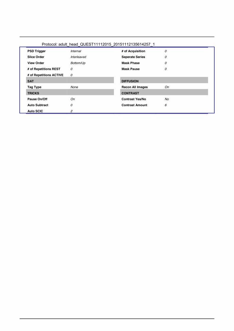
Protocol: adult_head_QUEST11112015_20151112135614257_1
Slice Order Interleaved Seperate Series 0
View Order Bottom/Up Mask Phase 0
# of Repetitions REST 0 Mask Pause 0
# of Repetitions ACTIVE 0 SAT
DIFFUSION Tag Type
None
Recon All Images
On TRICKS
CONTRAST Pause On/Off
Auto Subtract
On
0
Contrast Yes/No
Contrast Amount
No
6
Auto SCIC 2

Ax
T2*
GR
E
Ax
T2*
GR
E
UCSF/Quest Protocol – Axial T2*
PATIENT POSITION IMAGING PARAMETERS
Patient Entry Head First Gradient Mode ZOOM
Patient Position Supine Imaging Mode 2D
Coil Configuration HD 8Ch High Res Brain Array by Invivo
Pulse Sequence Gradient Echo
Plane OBLIQUE Imaging Options FC, EDR, ZIP512
Series Description Ax T2* GRE n/a 0
SCAN TIMING SCAN RANGE
Flip Angle 20 FOV 25.6
TE 20.0 Slice Thickness 4.0
Number of Echoes 1 Slice Spacing 0.0
TR 650.0 Start Location 1 L5.0
Receiver Bandwidth 25.00 Start Location 2 A6.4
Start Location 3 I72.7
End Location 1 L5.0
End Location 2 P0.5
End Location 3 S99.0
Number of Slices 44
Slice for 3 Plane 0 Localizaion
Slice for 3 Plane 0 Localizaion
Slice for 3 Plane 0 Localizaion
Slice for 3 Plane 0 Localizaion
Space per Plane 1 0.0
Space per Plane 2 0.0
Table Delta 0.00
IMAGE ENHANCE ACQ TIMING
Filter Choice None Freq 192
Phase 192
Freq DIR A/P
NEX 1.00
# of Acq. Before Pause 0
Phase FOV 1.00
Auto Shim Auto
Phase Correction No
GATING/TRIGGER USER CVS
Heart Beat per Minute Mode 0 User CV Mask 0
Auto Trigger Type Off
Auto Trigger Window Off
FMRI MULTI-PHASE
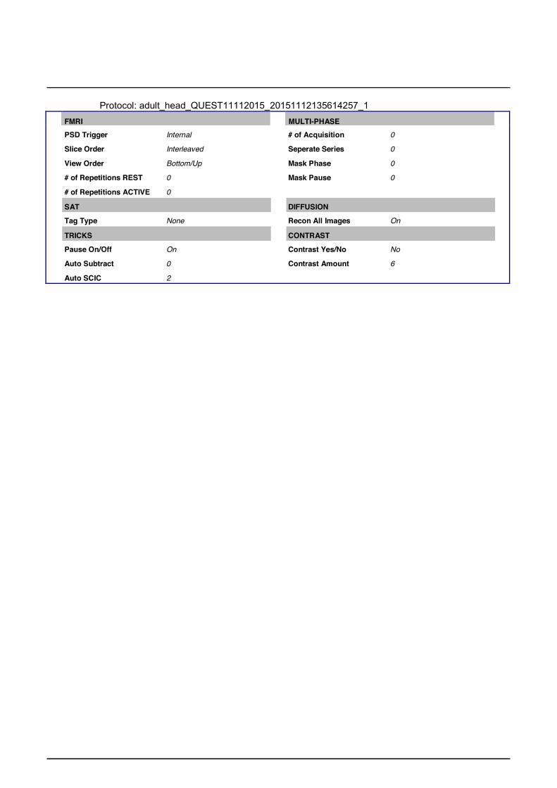
Protocol: adult_head_QUEST11112015_20151112135614257_1
PSD Trigger Internal # of Acquisition 0
Slice Order Interleaved Seperate Series 0
View Order Bottom/Up Mask Phase 0
# of Repetitions REST 0 Mask Pause 0
# of Repetitions ACTIVE 0 SAT
DIFFUSION Tag Type
None
Recon All Images
On TRICKS
CONTRAST Pause On/Off
Auto Subtract
On
0
Contrast Yes/No
Contrast Amount
No
6
Auto SCIC 2

AX
T2
FLA
IR
AX
T2
FLA
IR
UCSF/Quest Protocol – Axial T2 FLAIR
PATIENT POSITION IMAGING PARAMETERS
Patient Entry Head First Gradient Mode ZOOM
Patient Position Supine Imaging Mode 2D
Coil Configuration HD 8Ch High Res Brain Array by Invivo
Pulse Sequence T2flair
Plane OBLIQUE Imaging Options EDR, Fast
Series Description AX T2 FLAIR PSD Name enhflair
n/a 25
SCAN TIMING SCAN RANGE
TE 90.0 FOV 25.6
TR 9000.0 Slice Thickness 4.0
TI 2200 Slice Spacing 0.0
Echo Train Length 16 Start Location 1 L5.0
Receiver Bandwidth 20.83 Start Location 2 A6.4
Start Location 3 I72.7
End Location 1 L5.0
End Location 2 P0.5
End Location 3 S53.6
Number of Slices 44
Slice for 3 Plane 0 Localizaion
Slice for 3 Plane 0 Localizaion
Slice for 3 Plane 0 Localizaion
Slice for 3 Plane 0 Localizaion
Space per Plane 1 0.0
Space per Plane 2 0.0
Table Delta 0.00
IMAGE ENHANCE ACQ TIMING
Filter Choice None Freq 256
Phase 256
Freq DIR A/P
NEX 1.00
# of Acq. Before Pause 0
Phase FOV 0.88
Auto Shim Auto
Phase Correction No
GATING/TRIGGER USER CVS
Heart Beat per Minute Mode 0 User CV12 1.00
Auto Trigger Type Off User CV17 1.00
Auto Trigger Window Off User CV Mask 10639616
Protocol: adult_head_QUEST11112015_20151112135614257_1
FMRI
MULTI-PHASE
PSD Trigger Internal # of Acquisition 0
Slice Order Interleaved Seperate Series 0
View Order Bottom/Up Mask Phase 0
# of Repetitions REST 0 Mask Pause 0
# of Repetitions ACTIVE 0
SAT DIFFUSION
Tag Type None Recon All Images On
TRICKS CONTRAST
Pause On/Off On Contrast Yes/No No
Auto Subtract 0 Contrast Amount 6
Auto SCIC 2

AX
T2
4MM
AX
T2
4MM
UCSF/Quest Protocol – Axial T2
PATIENT POSITION IMAGING PARAMETERS
Patient Entry Head First Gradient Mode ZOOM
Patient Position Supine Imaging Mode 2D
Coil Configuration HD 8Ch High Res Brain Array by Invivo
Pulse Sequence FRFSE-XL
Plane OBLIQUE Imaging Options FC, EDR, TRF, Fast, ZIP512,
FR, ARC
Series Description AX T2 4MM n/a 23
SCAN TIMING SCAN RANGE
TE 90.0 FOV 25.6
Number of Echoes 1 Slice Thickness 4.0
TR 3000.0 Slice Spacing 0.0
Echo Train Length 16 Start Location 1 0.0
Receiver Bandwidth 19.23 Start Location 2 0.0
Start Location 3 I60.0
End Location 1 0.0
End Location 2 0.0
End Location 3 S120.0
Number of Slices 44
Slice for 3 Plane 0 Localizaion
Slice for 3 Plane 0 Localizaion
Slice for 3 Plane 0 Localizaion
Slice for 3 Plane 0 Localizaion
Space per Plane 1 0.0
Space per Plane 2 0.0
Table Delta 0.00
IMAGE ENHANCE ACQ TIMING
Filter Choice None Freq 256
Phase 256
Freq DIR A/P
NEX 1.00
# of Acq. Before Pause 0
Phase FOV 0.88
Auto Shim Auto
Phase Correction No
Flow Direction Compensation
GATING/TRIGGER USER CVS
Slice
Heart Beat per Minute Mode 0 User CV7 1.00

Protocol: adult_head_QUEST11112015_20151112135614257_1
Auto Trigger Type Off User CV17 1.00
Auto Trigger Window Off User CV19 1.00
User CV Mask 2769344 FMRI
MULTI-PHASE PSD Trigger
Internal
# of Acquisition
0
Slice Order Interleaved Seperate Series 0
View Order Bottom/Up Mask Phase 0
# of Repetitions REST 0 Mask Pause 0
# of Repetitions ACTIVE 0 SAT
DIFFUSION Tag Type
Fat/Water Saturation
None
Fat
Recon All Images
On
TRICKS
CONTRAST Pause On/Off
Auto Subtract
On
0
Contrast Yes/No
Contrast Amount
No
6
Auto SCIC 2

Ax
DW
I 4M
M B
=100
0
Ax
DW
I 4M
M B
=100
0
UCSF/Quest Protocol – Axial DWI
PATIENT POSITION IMAGING PARAMETERS
Patient Entry Head First Gradient Mode ZOOM
Patient Position Supine Imaging Mode 2D
Coil Configuration 8 Ch High Res Brain Array by MRI Devices
Pulse Sequence Spin Echo
Plane OBLIQUE Imaging Options EPI, DIFF, Asset
Series Description Ax DWI 4MM B=1000 n/a 8
SCAN TIMING SCAN RANGE
TE Minimum FOV 25.6
TR 8000.0 Slice Thickness 4.0
Number of Shots 1 Slice Spacing 0.8
Start Location 1 0.0
Start Location 2 0.0
Start Location 3 0.0
End Location 1 0.0
End Location 2 0.0
End Location 3 S148.0
Number of Slices 32
Slice for 3 Plane 0 Localizaion
Slice for 3 Plane 0 Localizaion
Slice for 3 Plane 0 Localizaion
Slice for 3 Plane 0 Localizaion
Space per Plane 1 0.0
Space per Plane 2 0.0
Table Delta 0.00
IMAGE ENHANCE ACQ TIMING
Filter Choice None Freq 160
Phase 160
Freq DIR R/L
Phase FOV 1.00
Auto Shim Auto
Phase Correction Yes
GATING/TRIGGER USER CVS
Heart Beat per Minute Mode 0 User CV0 1.00
Auto Trigger Type Off User CV5 1.00
Auto Trigger Window Off User CV Mask 262689 FMRI
MULTI-PHASE PSD Trigger
Internal
# of Acquisition
0
Slice Order Interleaved Seperate Series 0
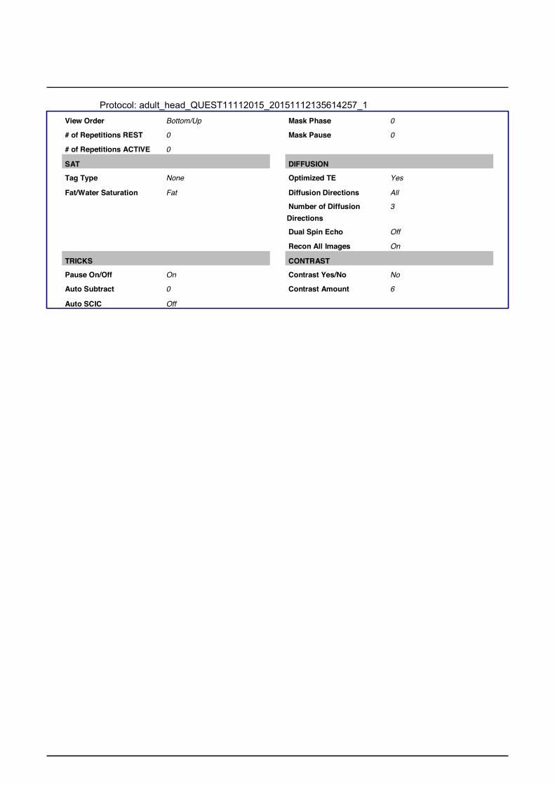
Protocol: adult_head_QUEST11112015_20151112135614257_1
View Order Bottom/Up Mask Phase 0
# of Repetitions REST 0 Mask Pause 0
# of Repetitions ACTIVE 0
SAT DIFFUSION
Tag Type None Optimized TE Yes
Fat/Water Saturation Fat Diffusion Directions All
Number of Diffusion 3 Directions
Dual Spin Echo Off
Recon All Images On
TRICKS CONTRAST
Pause On/Off On Contrast Yes/No No
Auto Subtract 0 Contrast Amount 6
Auto SCIC Off

Cor
DW
I 4M
M B
=100
0
Cor
DW
I 4M
M B
=100
0
UCSF/Quest Protocol – Coronal DWI
PATIENT POSITION IMAGING PARAMETERS
Patient Entry Head First Gradient Mode ZOOM
Patient Position Supine Imaging Mode 2D
Coil Configuration 8 Ch High Res Brain Array by MRI Devices
Pulse Sequence Spin Echo
Plane OBLIQUE Imaging Options EPI, DIFF, Asset
Series Description Cor DWI 4MM B=1000 n/a 8
SCAN TIMING SCAN RANGE
TE Minimum FOV 25.6
TR 9000.0 Slice Thickness 4.0
Number of Shots 1 Slice Spacing 0.8
Start Location 1 0.0
Start Location 2 0.0
Start Location 3 0.0
End Location 1 0.0
End Location 2 A168.0
End Location 3 0.0
Number of Slices 36
Slice for 3 Plane 0 Localizaion
Slice for 3 Plane 0 Localizaion
Slice for 3 Plane 0 Localizaion
Slice for 3 Plane 0 Localizaion
Space per Plane 1 0.0
Space per Plane 2 0.0
Table Delta 0.00
IMAGE ENHANCE ACQ TIMING
Filter Choice None Freq 160
Phase 160
Freq DIR S/I
Phase FOV 1.00
Auto Shim Auto
Phase Correction Yes
GATING/TRIGGER USER CVS
Heart Beat per Minute Mode 0 User CV0 1.00
Auto Trigger Type Off User CV5 1.00
Auto Trigger Window Off User CV Mask 262689 FMRI
MULTI-PHASE PSD Trigger
Internal
# of Acquisition
0
Slice Order Interleaved Seperate Series 0

Protocol: adult_head_QUEST11112015_20151112135614257_1
View Order Bottom/Up Mask Phase 0
# of Repetitions REST 0 Mask Pause 0
# of Repetitions ACTIVE 0
SAT DIFFUSION
Tag Type None Optimized TE Yes
Fat/Water Saturation Fat Diffusion Directions All
Number of Diffusion 3 Directions
Dual Spin Echo Off
Recon All Images On
TRICKS CONTRAST
Pause On/Off On Contrast Yes/No No
Auto Subtract 0 Contrast Amount 6
Auto SCIC Off

LOC
ALI
ZER
LOC
ALI
ZER
GE 3T UCSF/Quest Protocol - Survey
PATIENT POSITION Patient Entry Head First Patient Position Supine Coil Configuration -1 Plane 3-PLANE Series Description LOCALIZER
SCAN TIMING TE Minimum TR Minimum Receiver Bandwidth 62.50
IMAGE ENHANCE Filter Choice None
GATING/TRIGGER Heart Beat per Minute 0 Mode
IMAGING PARAMETERS Imaging Mode 2D Pulse Sequence Spin Echo Imaging Options Seq, EDR, Fast, SS, ARC n/a 12
SCAN RANGE FOV 34.0 Slice Thickness 10.0 Slice Spacing 0.0 Center Location 1 0.0 Center Location 2 A30.0 Center Location 3 0.0 Slice for 3 Plane 0 Localizaion Slice for 3 Plane 9 Localizaion
Auto Trigger Type Off Auto Trigger Window Off
FMRI PSD Trigger Internal Slice Order Interleaved View Order Bottom/Up # of Repetitions REST 0 # of Repetitions ACTIVE 0
SAT Tag Type None
TRICKS Pause On/Off On Auto Subtract 0 Auto SCIC Off
OTHERS Protocol Notes S/I
fat sat on asset
Slice for 3 Plane 9 Localizaion Slice for 3 Plane 9 Localizaion Space per Plane 1 0.0 Space per Plane 2 0.0 Table Delta 0.00
ACQ TIMING Freq 256 Phase 192 Freq DIR Unswap # of Acq. Before Pause 0 Phase FOV 1.00 Auto Shim Auto Phase Correction No
USER CVS User CV2 184.00 User CV13 1.00 In-range AutoTR Control 0 User CV Mask 57414
MULTI-PHASE Seperate Series 0 Mask Phase 0 Mask Pause 0
DIFFUSION Recon All Images On
CONTRAST Contrast Yes/No No Contrast Amount Yes

AS
SE
T ca
libra
tion
AS
SE
T ca
libra
tion
UCSF/Quest Protocol – ASSET Calibration
PATIENT POSITION Patient Entry Head First Patient Position Supine Coil Configuration -1 Plane AXIAL Series Description ASSET calibration
SCAN TIMING Number of Echoes 1
IMAGE ENHANCE Filter Choice None
GATING/TRIGGER Heart Beat per Minute 0 Mode Auto Trigger Type Off Auto Trigger Window Off
FMRI PSD Trigger Internal Slice Order Interleaved View Order Bottom/Up # of Repetitions REST 0 # of Repetitions ACTIVE 0
SAT Tag Type None
IMAGING PARAMETERS Imaging Mode 3D Pulse Sequence SPGR Imaging Options EDR, Fast, ZIP2, Calib n/a 6
SCAN RANGE FOV 30.0 Slice Thickness 9.4 GRXOPT 0 Start Location 1 I133.7 End Location 1 S157.7 End Location 2 R8.0 End Location 3 A38.6 Center of Location Start R8.0 Center of Location End A38.6 Scan Locations 32 Number of Slices 1 Slice for 3 Plane 0 Localizaion Slice for 3 Plane 0 Localizaion Slice for 3 Plane 0 Localizaion Slice for 3 Plane 0 Localizaion
TRICKS Pause On/Off On Auto Subtract 0 Auto SCIC Off
Space per Plane 1 0.0 Space per Plane 2 0.0 Table Delta 0.00
ACQ TIMING Freq DIR R/L Auto Shim Auto Phase Correction No
USER CVS In-range AutoTR Control 0 User CV Mask 8388672
MULTI-PHASE Seperate Series 0 Mask Phase 0 Mask Pause 0
DIFFUSION Recon All Images On
CONTRAST Contrast Yes/No No Contrast Amount Yes

3D S
AG
AD
NI I
R-S
PG
R
3D S
AG
AD
NI I
R-S
PG
R
UCSF/Quest Protocol – IR-FSPGR
PATIENT POSITION Patient Entry Head First Patient Position Supine Coil Configuration -1 Plane SAGITTAL Series Description 3D SAG ADNI IR-SPGR
IMAGING PARAMETERS Imaging Mode 3D Pulse Sequence BRAVO Imaging Options EDR, Fast, IrP n/a 41
SCAN RANGE
SCAN TIMING FOV 25.6 Flip Angle 11 Slice Thickness 1.2 Number of Echoes 1 Location per Slab 200 TI 400 Overlap Locations 0 Receiver Bandwidth 31.25 GRXOPT 0
IMAGE ENHANCE Filter Choice None
GATING/TRIGGER Heart Beat per Minute 0 Mode Auto Trigger Type Off Auto Trigger Window Off
FMRI PSD Trigger Internal Slice Order Interleaved View Order Bottom/Up # of Repetitions REST 0 # of Repetitions ACTIVE 0
Start Location 1 L110.8 End Location 1 R128.0 End Location 2 A37.2 End Location 3 S7.3 Center of Location Start A37.2 Center of Location End S7.3 Number of Slices 1 Slice for 3 Plane 0 Localizaion Slice for 3 Plane 0 Localizaion Slice for 3 Plane 0 Localizaion Slice for 3 Plane 0 Localizaion
SAT Tag Type None
TRICKS Pause On/Off On Auto Subtract 0 Auto SCIC 2
Space per Plane 1 0.0 Space per Plane 2 0.0 Table Delta 0.00
ACQ TIMING Freq 256 Phase 256 Freq DIR S/I NEX 1.00 Phase FOV 1.00 Auto Shim Auto Phase Correction No
USER CVS In-range AutoTR Control 0 User CV Mask 16
MULTI-PHASE Seperate Series 0 Mask Phase 0 Mask Pause 0
DIFFUSION Recon All Images On
CONTRAST Contrast Yes/No No Contrast Amount Yes

Ax
T2*
Ax
T2*
UCSF/Quest Protocol – Axial T2*
PATIENT POSITION Patient Entry Head First Patient Position Supine Coil Configuration HD 8Ch High Res Brain
Array by Invivo Plane OBLIQUE Series Description Ax T2*
SCAN TIMING Flip Angle 20 TE 20.0 Number of Echoes 1 TR 650.0 Receiver Bandwidth 25.00
IMAGE ENHANCE Filter Choice None
GATING/TRIGGER Heart Beat per Minute 0 Mode
IMAGING PARAMETERS Imaging Mode 2D Pulse Sequence Gradient Echo Imaging Options FC, EDR, SqP, ZIP512 n/a 0
SCAN RANGE FOV 25.6 Slice Thickness 4.0 Slice Spacing 0.0 Start Location 1 R8.0 Start Location 2 A26.4 Start Location 3 I61.2 End Location 1 R8.0 End Location 2 A50.9 End Location 3 S109.0 Number of Slices 44 Slice for 3 Plane 0 Localizaion
Auto Trigger Type Off Auto Trigger Window Off
FMRI PSD Trigger Internal Slice Order Interleaved View Order Bottom/Up # of Repetitions REST 0 # of Repetitions ACTIVE 0
SAT Tag Type None
TRICKS Pause On/Off On Auto Subtract 0 Auto SCIC 2
Slice for 3 Plane 0 Localizaion Slice for 3 Plane 0 Localizaion Slice for 3 Plane 0 Localizaion Space per Plane 1 0.0 Space per Plane 2 0.0 Table Delta 0.00
ACQ TIMING Freq 256 Phase 224 Freq DIR A/P NEX 1.00 # of Acq. Before Pause 0 Auto Shim Auto Phase Correction No
USER CVS In-range AutoTR Control 0 TR Min 200.0 TR Max 11000.0 User CV Mask 0
MULTI-PHASE Seperate Series 0 Mask Phase 0 Mask Pause 0
DIFFUSION Recon All Images On
CONTRAST Contrast Yes/No No Contrast Amount Yes

T2 F
LAIR
AR
C
T2 F
LAIR
AR
C
UCSF/Quest Protocol – Axial T2 FLAIR
PATIENT POSITION Patient Entry Head First Patient Position Supine Coil Configuration HD 8Ch High Res Brain
Array by Invivo Plane OBLIQUE Series Description T2 FLAIR ARC
SCAN TIMING Flip Angle 160 TE 90.0 Number of Echoes 1 TR 9000.0 TI 2474 Echo Train Length 16 Receiver Bandwidth 31.25
IMAGE ENHANCE Filter Choice B
GATING/TRIGGER Auto Trigger Type Off Auto Trigger Window Off
FMRI PSD Trigger Internal Slice Order Interleaved View Order Bottom/Up # of Repetitions REST 0 # of Repetitions ACTIVE 0
SAT SAT Location I SAT Thickness 40.0 Tag Type None
TRICKS Pause On/Off On Auto Subtract 0 Auto SCIC 2
IMAGING PARAMETERS Imaging Mode 2D Pulse Sequence T2flair Imaging Options EDR, TRF, Fast, ARC n/a 25
SCAN RANGE FOV 25.6 Slice Thickness 4.0 Slice Spacing 0.0 GRXOPT 0 Start Location 1 R8.0 Start Location 2 A26.4 Start Location 3 I61.2 End Location 1 R8.0 End Location 2 A50.9 End Location 3 S109.0 Number of Slices 44 Slice for 3 Plane 0 Localizaion Slice for 3 Plane 0 Localizaion Slice for 3 Plane 0 Localizaion Slice for 3 Plane 0 Localizaion Space per Plane 1 0.0 Space per Plane 2 0.0 Table Delta 0.00
ACQ TIMING Freq 256 Phase 256 Freq DIR A/P NEX 1.00 Phase FOV 0.88 Auto Shim Auto Phase Correction No
USER CVS User CV5 1.00 User CV22 1.00 In-range AutoTR Control 0 User CV Mask 6325024
MULTI-PHASE Seperate Series 0 Mask Phase 0 Mask Pause 0
DIFFUSION Recon All Images On
CONTRAST Contrast Yes/No No Contrast Amount Yes

Ax
T2 F
SE
Ax
T2 F
SE
UCSF/Quest Protocol – Axial T2
PATIENT POSITION Patient Entry Head First Patient Position Supine Coil Configuration HD 8Ch High Res Brain
Array by Invivo Plane OBLIQUE Series Description Ax T2 FSE
SCAN TIMING Flip Angle 142 TE 80.0 Number of Echoes 1 TR 3000.0 Echo Train Length 16 Receiver Bandwidth 31.25
IMAGE ENHANCE Filter Choice None
GATING/TRIGGER Heart Beat per Minute 0 Mode Auto Trigger Type Off Auto Trigger Window Off
FMRI PSD Trigger Internal Slice Order Interleaved View Order Bottom/Up # of Repetitions REST 0 # of Repetitions ACTIVE 0
SAT Tag Type None Fat/Water Saturation Fat
TRICKS Pause On/Off On Auto Subtract 0 Auto SCIC 2
IMAGING PARAMETERS Imaging Mode 2D Pulse Sequence FRFSE-XL Imaging Options FC, EDR, TRF, Fast,
ZIP512, FR n/a 23
SCAN RANGE FOV 25.6 Slice Thickness 4.0 Slice Spacing 0.0 Start Location 1 R8.0 Start Location 2 A26.4 Start Location 3 I61.2 End Location 1 R8.0 End Location 2 A50.9 End Location 3 S109.0 Number of Slices 44 Slice for 3 Plane 0 Localizaion Slice for 3 Plane 0 Localizaion Slice for 3 Plane 0 Localizaion Slice for 3 Plane 0 Localizaion Space per Plane 1 0.0 Space per Plane 2 0.0 Table Delta 0.00
ACQ TIMING Freq 256 Phase 256 Freq DIR A/P NEX 1.00 # of Acq. Before Pause 0 Phase FOV 0.88 Auto Shim Auto Phase Correction Yes Flow Direction Compensation
Freq
USER CVS User CV19 1.00 User CV22 1.00 In-range AutoTR Control 0 TR Min 2500.0 TR Max 11000.0 User CV Mask 6848960
MULTI-PHASE Seperate Series 0 Mask Phase 0 Mask Pause 0
DIFFUSION Recon All Images On

CONTRAST Contrast Yes/No No Contrast Amount Yes

Ax
DW
I
Ax
DW
I
UCSF/Quest Protocol – Axial DWI
PATIENT POSITION Patient Entry Head First Patient Position Supine Coil Configuration HD 8Ch High Res Brain
Array by Invivo Plane AXIAL Series Description Ax DWI
SCAN TIMING TE Minimum Number of Echoes 1 TR 6000.0 Number of Shots 1
IMAGE ENHANCE Filter Choice None
GATING/TRIGGER Heart Beat per Minute 0 Mode Auto Trigger Type Off Auto Trigger Window Off
FMRI PSD Trigger Internal Slice Order Interleaved View Order Bottom/Up # of Repetitions REST 0 # of Repetitions ACTIVE 0
SAT Tag Type None Fat/Water Saturation Fat
TRICKS Pause On/Off On Auto Subtract 0 Auto SCIC 2
IMAGING PARAMETERS Imaging Mode 2D Pulse Sequence Spin Echo Imaging Options EDR, EPI, DIFF, Asset n/a 8
SCAN RANGE FOV 25.6 Slice Thickness 4.0 Slice Spacing 0.8 GRXOPT 0 Start Location 1 I41.6 End Location 1 S107.2 End Location 2 R8.6 End Location 3 A40.6 Center of Location Start R8.6 Center of Location End A40.6 Number of Slices 32 Slice for 3 Plane 0 Localizaion Slice for 3 Plane 0 Localizaion Slice for 3 Plane 0 Localizaion Slice for 3 Plane 0 Localizaion Space per Plane 1 0.0 Space per Plane 2 0.0 Table Delta 0.00
ACQ TIMING Freq 192 Phase 192 Freq DIR R/L Phase FOV 1.00 Auto Shim Auto Phase Correction Yes
USER CVS User CV0 1.00 User CV5 1.00 User CV17 1.00 In-range AutoTR Control 0 User CV Mask 131745
MULTI-PHASE Seperate Series 0 Mask Phase 0 Mask Pause 0
DIFFUSION Optimized TE Yes Diffusion Directions All Number of Diffusion 3 Directions Number of T2 Images 1 Dual Spin Echo Off Recon All Images On

CONTRAST Contrast Yes/No No Contrast Amount Yes

Cor
DW
I
Cor
DW
I
UCSF/Quest Protocol – Coronal DWI
PATIENT POSITION Patient Entry Head First Patient Position Supine Coil Configuration HD 8Ch High Res Brain
Array by Invivo Plane CORONAL Series Description Cor DWI
SCAN TIMING TE Minimum TR 7500.0 Number of Shots 1
IMAGE ENHANCE Filter Choice None
GATING/TRIGGER Heart Beat per Minute 0 Mode Auto Trigger Type Off Auto Trigger Window Off
FMRI PSD Trigger Internal Slice Order Interleaved View Order Bottom/Up # of Repetitions REST 0 # of Repetitions ACTIVE 0
SAT Tag Type None Fat/Water Saturation Fat
TRICKS Pause On/Off On Auto Subtract 0 Auto SCIC 2
IMAGING PARAMETERS Imaging Mode 2D Pulse Sequence Spin Echo Imaging Options EDR, EPI, DIFF, Asset n/a 8
SCAN RANGE FOV 25.6 Slice Thickness 4.0 Slice Spacing 0.8 Start Location 1 P50.7 End Location 1 A117.3 End Location 2 S21.3 End Location 3 R9.3 Center of Location Start S21.3 Center of Location End R9.3 Number of Slices 36 Slice for 3 Plane 0 Localizaion Slice for 3 Plane 0 Localizaion Slice for 3 Plane 0 Localizaion Slice for 3 Plane 0 Localizaion Space per Plane 1 0.0 Space per Plane 2 0.0 Table Delta 0.00
ACQ TIMING Freq 192 Phase 192 Freq DIR S/I Phase FOV 1.00 Auto Shim Auto Phase Correction Yes
USER CVS User CV0 1.00 User CV5 1.00 User CV17 1.00 In-range AutoTR Control 0 User CV Mask 393889
MULTI-PHASE Seperate Series 0 Mask Phase 0 Mask Pause 0
DIFFUSION Optimized TE Yes Diffusion Directions All Number of Diffusion 3 Directions Number of T2 Images 1 Dual Spin Echo Off Recon All Images On

CONTRAST Contrast Yes/No No Contrast Amount Yes

Philips 1.5T UCSF/Quest Survey Coil selection = "Head"; element selection = "123456"; connection = "d"; Dual coil = "no"; Multi coil = "no"; CLEAR = "no"; FOV FH (mm) = 250; AP (mm) = 250; FOV (mm) = (3) 50, (17) 250; Voxel size FH (mm) = 0.9765625; AP (mm) = 1.953125; Slice thickness (mm) = 10; Bar orientation = "RL"; VOI orientation = "transverse"; VOI size AP (mm) = 30; RL (mm) = 30; FH (mm) = 30; Recon voxel size (mm) = 0.9765625; Fold-over suppression = "no"; Reconstruction matrix = 256; SENSE = "no"; k-t BLAST = "no"; Stacks = 3; type = (20) "parallel"; Slices = (3) 3, (17) 1; slice gap = "user defined"; gap (mm) = (20) 10; Slice orientation = "sagittal", "coronal", (18) "transverse"; Fold-over direction = "AP", (2) "RL", (17) "AP"; Fat shift direction = (2) "F", "P", (17) "L"; radial axis = "AP", "RL", "AP", (17) "RL"; radial angle (deg) = (20) 0; Slice Offc. AP (P=+mm) = 0, (2) 20, (17) 0; RL (L=+mm) = (20) 0; FH (H=+mm) = (2) 0, -80, (17) 0; Ang. AP (deg) = (20) 0; RL (deg) = (20) 0; FH (deg) = (20) 0; VOI offc. AP (P=+mm) = 0; RL (L=+mm) = 0; FH (H=+mm) = 0; VOI ang. AP (deg) = 0; RL (deg) = 0; FH (deg) = 0; 2nd VOI lat. offc. (mm) = 80; 2nd VOI offc. axis = "AP"; Slice scan order = "default"; Stack scan order = "ascend"; Move table per stack = "no"; Large table movement = "no";
Stack alignment = " no"; Stack display order = " no"; PlanAlign = "no"; REST slabs = 0; Catheter tracking = "no"; Interactive positioning = "no"; Patient position = "head first"; orientation = "supine"; Scan type = "Imaging"; Scan mode = "M2D"; technique = "FFE"; Contrast enhancement = "T1"; Acquisition mode = "cartesian"; Fast Imaging mode = "TFE"; shot mode = "multishot"; TFE factor = 42; startup echoes = "default"; shot interval = "shortest"; profile order = "linear"; Echoes = 1; partial echo = "no"; shifted echo = "no"; TE = "shortest"; Flip angle (deg) = 20; TR = "user defined"; (ms) = 15; Halfscan = "no"; Water-fat shift = "maximum"; Shim = "default"; Fat suppression = "no"; Water suppression = "no"; TFE prepulse = "invert"; slice selection = "no"; shared = "no"; delay = "shortest"; PSIR = "no"; MTC = "no"; T2prep = "no"; Diffusion mode = "no"; SAR mode = "high"; B1 mode = "default"; PNS mode = "moderate"; Gradient mode = "regular"; SofTone mode = "no"; Cardiac synchronization = "no"; Heart rate > 250 bpm = "no"; Respiratory compensation = "no"; Navigator respiratory comp = "no"; Flow compensation = "no"; fMRI echo stabilisation = "no"; Motion smoothing = "no"; NSA = 1; Angio / Contrast enh. = "no"; Quantitative flow = "no"; Manual start = "no"; Dynamic study = "no"; Arterial Spin labeling = "no"; Preparation phases = "auto"; Interactive F0 = "no"; B1 field map = "no"; MIP/MPR = "no"; Images = " M", (3) " no";

Autoview image = " M"; Calculated images = (4) " no"; Reference tissue = "White matter"; Preset window contrast = "soft"; Reconstruction mode = "immediate"; Save raw data = "no"; Hardcopy protocol = "no"; Ringing filtering = "default"; Geometry correction = "default"; IF_info_seperator = 0; Total scan duration = "00:31.4"; Rel. signal level (%) = 100; Act. TR/TE (ms) = "15 / 5.2"; ACQ matrix M x P = "256 x 126"; ACQ voxel MPS (mm) = "0.98 / 1.98 / 10.0"; REC voxel MPS (mm) = "0.98 / 0.98 / 10.0"; Scan percentage (%) = 49.21875; TFE shots = 3; TFE dur. shot / acq (ms) = "697.7 / 630.0"; TFE shot interval (ms) = 697.661499; Min. TI delay = 373.136902; Act. WFS (pix) / BW (Hz) = "1.162 / 186.9"; Min. WFS (pix) / Max. BW (Hz) ="0.522 / 415.8"; Min. TR/TE (ms) = "10 / 5.2"; SAR / whole body = "< 6 %"; Whole body / level = "< 0.2 W/kg / normal"; B1 rms = "1.12 uT"; PNS / level = "16 % / normal"; Sound Pressure Level (dB) = -3.4767971;

UCSF/Quest MP-RAGE Coil selection = "Head"; element selection = "123456"; connection = "d"; Dual coil = "no"; Homogeneity correction = "none"; CLEAR = "yes"; body tuned = "no"; FOV FH (mm) = 240; AP (mm) = 240; RL (mm) = 204.000015; Voxel size FH (mm) = 1.25; AP (mm) = 1.25; RL (mm) = 1.20000005; Recon voxel size (mm) = 1; Fold-over suppression = "no"; Slice oversampling = "user defined"; oversample factor = 1.10000002; RF select. FOS = "no"; Reconstruction matrix = 240; SENSE = "no"; k-t BLAST = "no"; Overcontiguous slices = "no"; Stacks = 1; slices = 170; slice orientation = "sagittal"; fold-over direction = "AP"; fat shift direction = "F"; Stack Offc. AP (P=+mm) = -0.496321857; RL (L=+mm) = 1.41061175; FH (H=+mm) = -66.9705124; Ang. AP (deg) = 2.82272959; RL (deg) = 9.18244553; FH (deg) = -0.52383393; Chunks = 1; Large table movement = "no"; PlanAlign = "no"; REST slabs = 0; Catheter tracking = "no"; Interactive positioning = "no"; Allow table movement = "no"; Patient position = "head first"; orientation = "supine"; Scan type = "Imaging"; Scan mode = "3D"; technique = "FFE"; Contrast enhancement = "T1"; Acquisition mode = "cartesian"; Fast Imaging mode = "TFE"; 3D non-selective = "no"; shot mode = "multishot"; TFE factor = 192; 3D free factor = "no"; startup echoes = "default"; shot interval = "user defined"; (ms) = 2300; profile order = "linear"; turbo direction = "Y"; Echoes = 1;
partial echo = "no"; shifted echo = "no"; TE = "user defined"; (ms) = 4; Flip angle (deg) = 8; TR = "shortest"; Halfscan = "no"; Water-fat shift = "maximum"; Shim = "auto"; Fat suppression = "no"; Water suppression = "no"; TFE prepulse = "invert"; slice selection = "no"; delay = "user defined"; (ms) = 1000; PSIR = "no"; MTC = "no"; T2prep = "no"; Research prepulse = "no"; Diffusion mode = "no"; SAR mode = "high"; B1 mode = "default"; PNS mode = "moderate"; Gradient mode = "default"; SofTone mode = "no"; Cardiac synchronization = "no"; Heart rate > 250 bpm = "no"; Respiratory compensation = "no"; Navigator respiratory comp = "no"; Flow compensation = "no"; fMRI echo stabilisation = "no"; Motion smoothing = "no"; NSA = 1; Angio / Contrast enh. = "no"; Quantitative flow = "no"; CENTRA = "no"; Manual start = "no"; Dynamic study = "no"; Arterial Spin labeling = "no"; Preparation phases = "auto"; Interactive F0 = "no"; B0 field map = "no"; B1 field map = "no"; MIP/MPR = "no"; Images = " M", (3) " no"; Autoview image = " M"; Calculated images = (4) " no"; Reference tissue = "Grey matter"; Preset window contrast = "soft"; Reconstruction mode = "immediate"; Save raw data = "no"; Hardcopy protocol = "no"; Ringing filtering = "default"; Geometry correction = "default"; Elliptical k-space shutter = "default"; IF_info_seperator = 0; Total scan duration = "07:11.9"; Rel. signal level (%) = 100; Act. TR/TE (ms) = "8.5 / 4.0"; ACQ matrix M x P = "192 x 192"; ACQ voxel MPS (mm) = "1.25 / 1.25 / 1.20"; REC voxel MPS (mm) = "1.00 / 1.00 / 1.20";

Scan percentage (%) = 100; TFE shots = 187; TFE dur. shot / acq (ms) = "1826.1 / 1638.6"; Min. TI delay = 856.297791; Act. WFS (pix) / BW (Hz) = "1.270 / 171.0"; Min. WFS (pix) / Max. BW (Hz) ="0.209 / 1041.7"; Min. TR/TE (ms) = "8.5 / 3.2"; SAR / whole body = "< 3 %"; Whole body / level = "< 0.1 W/kg / normal"; B1 rms = "0.75 uT"; PNS / level = "54 % / normal"; Sound Pressure Level (dB) = 11.7563028;

UCSF/Quest T2* Coil selection = "Head"; element selection = "123456"; connection = "d"; Dual coil = "no"; Homogeneity correction = "none"; CLEAR = "yes"; body tuned = "no"; FOV AP (mm) = 256; RL (mm) = 224; FH (mm) = 176; Voxel size AP (mm) = 1.33000004; RL (mm) = 1.33333337; Slice thickness (mm) = 4; Recon voxel size (mm) = 0.939999998; Fold-over suppression = "no"; Reconstruction matrix = 288; SENSE = "no"; k-t BLAST = "no"; Stacks = 1; type = "parallel"; slices = 44; slice gap = "user defined"; gap (mm) = 0; slice orientation = "transverse"; fold-over direction = "RL"; fat shift direction = "P"; Stack Offc. AP (P=+mm) = -0.496321857; RL (L=+mm) = 1.41061175; FH (H=+mm) = -66.9705124; Ang. AP (deg) = 2.82272959; RL (deg) = 9.18244553; FH (deg) = -0.52383393; Minimum number of packages = 1; Slice scan order = "default"; Large table movement = "no"; PlanAlign = "no"; REST slabs = 0; Catheter tracking = "no"; Interactive positioning = "no"; Allow table movement = "no"; Patient position = "head first"; orientation = "supine"; Scan type = "Imaging"; Scan mode = "MS"; technique = "FFE"; Contrast enhancement = "no"; Acquisition mode = "cartesian"; Fast Imaging mode = "none"; Echoes = 1; partial echo = "no"; shifted echo = "no"; TE = "user defined"; (ms) = 30; Flip angle (deg) = 20; TR = "user defined"; (ms) = 650; Halfscan = "no"; Water-fat shift = "user defined";
(pixels) = 2.70000005; Shim = "default"; Fat suppression = "no"; Water suppression = "no"; MTC = "no"; Research prepulse = "no"; Diffusion mode = "no"; SAR mode = "low"; B1 mode = "default"; PNS mode = "low"; Gradient mode = "default"; SofTone mode = "no"; Cardiac synchronization = "no"; Heart rate > 250 bpm = "no"; Respiratory compensation = "no"; Navigator respiratory comp = "no"; Flow compensation = "yes"; Temporal slice spacing = "default"; fMRI echo stabilisation = "no"; NSA = 1; Angio / Contrast enh. = "no"; Quantitative flow = "no"; Manual start = "no"; Dynamic study = "no"; Arterial Spin labeling = "no"; Preparation phases = "auto"; Interactive F0 = "no"; B0 field map = "no"; B1 field map = "no"; MIP/MPR = "no"; Images = " M", (3) " no"; Autoview image = " M"; Calculated images = (4) " no"; Reference tissue = "White matter"; Preset window contrast = "soft"; Reconstruction mode = "immediate"; Save raw data = "no"; Hardcopy protocol = "no"; Ringing filtering = "default"; Geometry correction = "default"; IF_info_seperator = 0; Total scan duration = "05:31.5"; Rel. signal level (%) = 100; Act. TR/TE (ms) = "650 / 30"; ACQ matrix M x P = "192 x 168"; ACQ voxel MPS (mm) = "1.33 / 1.33 / 4.00"; REC voxel MPS (mm) = "0.89 / 0.89 / 4.00"; Scan percentage (%) = 100; Packages = 3; Min. slice gap (mm) = 0; Optimal slices = 16; Max. slices = 48; Act. WFS (pix) / BW (Hz) = "2.695 / 80.6"; Min. WFS (pix) / Max. BW (Hz) ="0.319 / 681.2"; Min. TR/TE (ms) = "580 / 6.7"; SAR / whole body = " 0 %"; Whole body / level = "0.0 W/kg / normal"; B1 rms = "0.23 uT"; PNS / level = "9 % / normal"; Sound Pressure Level (dB) = -9.25968838;

UCSF/Quest T2 FLAIR Coil selection = "Head"; element selection = "123456"; connection = "d"; Dual coil = "no"; CLEAR = "yes"; body tuned = "no"; FOV AP (mm) = 256; RL (mm) = 224; FH (mm) = 176; Voxel size AP (mm) = 1; RL (mm) = 1; Slice thickness (mm) = 4; Recon voxel size (mm) = 0.9375; Fold-over suppression = "no"; Reconstruction matrix = 288; SENSE = "yes"; P reduction (RL) = 2; P os factor = 1; k-t BLAST = "no"; Stacks = 1; type = "parallel"; slices = 44; slice gap = "user defined"; gap (mm) = 0; slice orientation = "transverse"; fold-over direction = "RL"; fat shift direction = "P"; Stack Offc. AP (P=+mm) = -0.496321857; RL (L=+mm) = 1.41061175; FH (H=+mm) = -66.9705124; Ang. AP (deg) = 2.82272959; RL (deg) = 9.18244553; FH (deg) = -0.52383393; Minimum number of packages = 3; Slice scan order = "default"; Large table movement = "no"; PlanAlign = "no"; REST slabs = 0; Catheter tracking = "no"; Interactive positioning = "no"; Allow table movement = "no"; Patient position = "head first"; orientation = "supine"; Scan type = "Imaging"; Scan mode = "MS"; technique = "IR"; Acquisition mode = "cartesian"; Fast Imaging mode = "TSE"; shot mode = "multishot"; TSE factor = 16; startup echoes = 0; profile order = "linear"; DRIVE = "no"; ultrashort = "no"; strong FID crushing = "no"; Echoes = 1; partial echo = "no"; TE = "user defined";
(ms) = 90; Refocusing control = "yes"; angle (deg) = 90; TR = "user defined"; (ms) = 9000; Halfscan = "no"; Water-fat shift = "maximum"; IR delay (ms) = 2200; acquire during delay = "yes"; dual = "no"; power = "1"; Shim = "default"; Fat suppression = "no"; Water suppression = "no"; MTC = "no"; Silicone Only Sequence = "no"; T2prep = "no"; Research prepulse = "no"; Zoom imaging = "no"; Diffusion mode = "no"; SAR mode = "moderate"; B1 mode = "default"; PNS mode = "low"; Gradient mode = "default"; SofTone mode = "no"; Cardiac synchronization = "no"; Heart rate > 250 bpm = "no"; Respiratory compensation = "no"; Navigator respiratory comp = "no"; Flow compensation = "no"; Motion smoothing = "no"; NSA = 1; Manual start = "no"; Dynamic study = "no"; Arterial Spin labeling = "no"; Preparation phases = "auto"; Interactive F0 = "no"; B0 field map = "no"; B1 field map = "no"; MIP/MPR = "no"; Images = " M", (3) " no"; Autoview image = " M"; Reference tissue = "White matter"; Preset window contrast = "soft"; Reconstruction mode = "real time"; Save raw data = "no"; Hardcopy protocol = "no"; Ringing filtering = "default"; Geometry correction = "default"; IF_info_seperator = 0; Total scan duration = "03:36.0"; Rel. signal level (%) = 100; Act. TR/TI (ms) = "9000 / 2200"; Act. TE (ms) = "90"; ACQ matrix M x P = "256 x 218"; ACQ voxel MPS (mm) = "1.00 / 1.03 / 4.00"; REC voxel MPS (mm) = "0.89 / 0.89 / 4.00"; Scan percentage (%) = 97.391304; Packages = 3; Min. slice gap (mm) = 0.800000012; Optimal slices = 30; Max. slices = 45;

WFS (pix) / BW (Hz) = "1.157 / 187.8"; TSE es / shot (ms) = "10.6 / 169"; TEeff / TEequiv (ms) = "90 / 69"; Min. TR/TI (ms) = "5162 / 50"; SAR / whole body = "< 13 %"; Whole body / level = "< 0.5 W/kg / normal"; B1 rms = "1.66 uT"; PNS / level = "32 % / normal"; Sound Pressure Level (dB) = 9.06399822;

UCSF/Quest T2 TSE Coil selection = "Head"; element selection = "123456"; connection = "d"; Dual coil = "no"; Homogeneity correction = "none"; CLEAR = "yes"; body tuned = "no"; FOV AP (mm) = 256; RL (mm) = 224; FH (mm) = 176; Voxel size AP (mm) = 1; RL (mm) = 1; Slice thickness (mm) = 4; Recon voxel size (mm) = 1; Small FOV imaging = "no"; Fold-over suppression = "no"; Reconstruction matrix = 256; SENSE = "no"; k-t BLAST = "no"; Stacks = 1; type = "parallel"; slices = 44; slice gap = "user defined"; gap (mm) = 0; slice orientation = "transverse"; fold-over direction = "RL"; fat shift direction = "P"; Stack Offc. AP (P=+mm) = -0.496321857; RL (L=+mm) = 1.41061175; FH (H=+mm) = -66.9705124; Ang. AP (deg) = 2.82272959; RL (deg) = 9.18244553; FH (deg) = -0.52383393; Minimum number of packages = 2; Slice scan order = "default"; Large table movement = "no"; PlanAlign = "no"; REST slabs = 0; Catheter tracking = "no"; Interactive positioning = "no"; Allow table movement = "no"; Patient position = "head first"; orientation = "supine"; Scan type = "Imaging"; Scan mode = "MS"; technique = "SE"; Modified SE = "no"; Acquisition mode = "cartesian"; Fast Imaging mode = "TSE"; shot mode = "multishot"; TSE factor = 16; startup echoes = 0; profile order = "linear"; DRIVE = "no"; ultrashort = "no"; strong FID crushing = "no"; Echoes = 1; partial echo = "no";
TE = "user defined"; (ms) = 90; Flip angle (deg) = 90; Refocusing control = "yes"; angle (deg) = 100; TR = "user defined"; (ms) = 3000; Halfscan = "no"; Water-fat shift = "maximum"; Shim = "default"; Fat suppression = "SPIR"; strength = "strong"; frequency offset = "default"; Water suppression = "no"; BB pulse = "no"; MTC = "no"; T2prep = "no"; Research prepulse = "no"; Zoom imaging = "no"; Diffusion mode = "no"; SAR mode = "high"; B1 mode = "default"; PNS mode = "low"; Gradient mode = "default"; SofTone mode = "no"; Cardiac synchronization = "no"; Heart rate > 250 bpm = "no"; Respiratory compensation = "no"; Navigator respiratory comp = "no"; Flow compensation = "no"; Temporal slice spacing = "default"; Motion smoothing = "no"; NSA = 1; Manual start = "no"; Dynamic study = "no"; Arterial Spin labeling = "no"; Preparation phases = "auto"; Interactive F0 = "no"; B0 field map = "no"; B1 field map = "no"; MIP/MPR = "no"; Images = " M", (3) " no"; Autoview image = " M"; Calculated images = (4) " no"; Reference tissue = "White matter"; Preset window contrast = "soft"; Reconstruction mode = "immediate"; Save raw data = "no"; Hardcopy protocol = "no"; Ringing filtering = "default"; Geometry correction = "default"; IF_info_seperator = 1634755923; Total scan duration = "02:15.0"; Rel. signal level (%) = 100; Act. TR (ms) = "3000"; Act. TE (ms) = "90"; ACQ matrix M x P = "256 x 224"; ACQ voxel MPS (mm) = "1.00 / 1.00 / 4.00"; REC voxel MPS (mm) = "1.00 / 1.00 / 4.00"; Scan percentage (%) = 100; Packages = 3; Min. slice gap (mm) = 0;

Optimal slices = 15; Max. slices = 45; WFS (pix) / BW (Hz) = "1.193 / 182.1"; TSE es / shot (ms) = "10.6 / 169"; TEeff / TEequiv (ms) = "90 / 73"; Min. TR (ms) = "2966"; SAR / whole body = "< 38 %"; Whole body / level = "< 1.5 W/kg / normal"; B1 rms = "2.81 uT"; PNS / level = "46 % / normal"; Sound Pressure Level (dB) = 9.9265461;

UCSF/Quest Axial DWI Coil selection = "Head"; element selection = "123456"; connection = "d"; Dual coil = "no"; CLEAR = "yes"; body tuned = "no"; FOV RL (mm) = 256; AP (mm) = 256; FH (mm) = 152.800003; Voxel size RL (mm) = 1.60000002; AP (mm) = 1.60000002; Slice thickness (mm) = 4; Recon voxel size (mm) = 1; Small FOV imaging = "no"; Fold-over suppression = "no"; Reconstruction matrix = 256; SENSE = "yes"; P reduction (AP) = 2; P os factor = 1; k-t BLAST = "no"; Stacks = 1; type = "parallel"; slices = 32; slice gap = "user defined"; gap (mm) = 0.800000191; slice orientation = "transverse"; fold-over direction = "AP"; fat shift direction = "P"; Stack Offc. AP (P=+mm) = -0.496321857; RL (L=+mm) = 1.41061175; FH (H=+mm) = -66.9705124; Ang. AP (deg) = 2.82272959; RL (deg) = 9.18244553; FH (deg) = -0.52383393; Minimum number of packages = 1; Slice scan order = "default"; Large table movement = "no"; PlanAlign = "no"; REST slabs = 0; Catheter tracking = "no"; Interactive positioning = "no"; Allow table movement = "no"; Patient position = "head first"; orientation = "supine"; Scan type = "Imaging"; Scan mode = "MS"; technique = "SE"; Modified SE = "no"; Acquisition mode = "cartesian"; Fast Imaging mode = "EPI"; shot mode = "single-shot"; Echoes = 1; partial echo = "no"; TE = "shortest"; Flip angle (deg) = 90; TR = "shortest"; Halfscan = "yes"; factor = 0.698795199;
Water-fat shift = "minimum"; Shim = "auto"; Fat suppression = "SPIR"; strength = "strong"; frequency offset = "default"; Water suppression = "no"; BB pulse = "no"; MTC = "no"; Research prepulse = "no"; Diffusion mode = "DWI"; sequence = "SE"; gradient duration = "maximum"; gradient overplus = "no"; direction = "M", "P", "S"; nr of b-factors = 2; b-factor order = "ascending"; max b-factor = 1000; average high b = "no"; SAR mode = "high"; B1 mode = "default"; PNS mode = "high"; Gradient mode = "default"; SofTone mode = "no"; Cardiac synchronization = "no"; Heart rate > 250 bpm = "no"; Respiratory compensation = "no"; Navigator respiratory comp = "no"; Flow compensation = "no"; Temporal slice spacing = "default"; NSA = 3; SMART = "no"; Manual start = "no"; Dynamic study = "no"; Arterial Spin labeling = "no"; Preparation phases = "auto"; Interactive F0 = "no"; B0 field map = "no"; B1 field map = "no"; MIP/MPR = "no"; Images = " M", (3) " no"; Autoview image = " M"; Calculated images = (4) " no"; Reference tissue = "White matter"; EPI 2D phase correction = "no"; Preset window contrast = "soft"; Reconstruction mode = "immediate"; Save raw data = "no"; Hardcopy protocol = "no"; Ringing filtering = "default"; Geometry correction = "default"; IF_info_seperator = 1634755923; Total scan duration = "01:31.2"; Rel. signal level (%) = 100; Act. TR (ms) = "6513"; Act. TE (ms) = "100"; ACQ matrix M x P = "160 x 160"; ACQ voxel MPS (mm) = "1.60 / 1.60 / 4.00"; REC voxel MPS (mm) = "1.00 / 1.00 / 4.00"; Scan percentage (%) = 100; Packages = 1; Min. slice gap (mm) = 0; EPI factor = 83;

WFS (pix) / BW (Hz) = "18.596 / 11.7"; BW in EPI freq. dir. (Hz) = "1249.1"; SAR / whole body = "< 5 %"; Whole body / level = "< 0.2 W/kg / normal"; B1 rms = "0.96 uT"; PNS / level = "66 % / normal"; Sound Pressure Level (dB) = 10.3647041;

UCSF/Quest Coronal DWI Coil selection = "Head"; element selection = "123456"; connection = "d"; Dual coil = "no"; CLEAR = "yes"; body tuned = "no"; FOV FH (mm) = 256; RL (mm) = 256; AP (mm) = 172; Voxel size FH (mm) = 1.60000002; RL (mm) = 1.60000002; Slice thickness (mm) = 4; Recon voxel size (mm) = 1; Small FOV imaging = "no"; Fold-over suppression = "no"; Reconstruction matrix = 256; SENSE = "yes"; P reduction (RL) = 2; P os factor = 1; k-t BLAST = "no"; Stacks = 1; type = "parallel"; slices = 36; slice gap = "user defined"; gap (mm) = 0.800000191; slice orientation = "coronal"; fold-over direction = "RL"; fat shift direction = "L"; Stack Offc. AP (P=+mm) = -0.496321857; RL (L=+mm) = 1.41061175; FH (H=+mm) = -66.9705124; Ang. AP (deg) = 2.82272959; RL (deg) = 9.18244553; FH (deg) = -0.52383393; Minimum number of packages = 1; Slice scan order = "default"; Large table movement = "no"; PlanAlign = "no"; REST slabs = 0; Catheter tracking = "no"; Interactive positioning = "no"; Allow table movement = "no"; Patient position = "head first"; orientation = "supine"; Scan type = "Imaging"; Scan mode = "MS"; technique = "SE"; Modified SE = "no"; Acquisition mode = "cartesian"; Fast Imaging mode = "EPI"; shot mode = "single-shot"; Echoes = 1; partial echo = "no"; TE = "shortest"; Flip angle (deg) = 90; TR = "shortest"; Halfscan = "yes"; factor = 0.698795199;
Water-fat shift = "minimum"; Shim = "auto"; Fat suppression = "SPIR"; strength = "strong"; frequency offset = "default"; Water suppression = "no"; BB pulse = "no"; MTC = "no"; Research prepulse = "no"; Diffusion mode = "DWI"; sequence = "SE"; gradient duration = "maximum"; gradient overplus = "no"; direction = "M", "P", "S"; nr of b-factors = 2; b-factor order = "ascending"; max b-factor = 1000; average high b = "no"; SAR mode = "high"; B1 mode = "default"; PNS mode = "high"; Gradient mode = "default"; SofTone mode = "no"; Cardiac synchronization = "no"; Heart rate > 250 bpm = "no"; Respiratory compensation = "no"; Navigator respiratory comp = "no"; Flow compensation = "no"; Temporal slice spacing = "default"; NSA = 3; SMART = "no"; Manual start = "no"; Dynamic study = "no"; Arterial Spin labeling = "no"; Preparation phases = "auto"; Interactive F0 = "no"; B0 field map = "no"; B1 field map = "no"; MIP/MPR = "no"; Images = " M", (3) " no"; Autoview image = " M"; Calculated images = (4) " no"; Reference tissue = "White matter"; EPI 2D phase correction = "no"; Preset window contrast = "soft"; Reconstruction mode = "immediate"; Save raw data = "no"; Hardcopy protocol = "no"; Ringing filtering = "default"; Geometry correction = "default"; IF_info_seperator = 1634755923; Total scan duration = "01:39.5"; Rel. signal level (%) = 100; Act. TR (ms) = "7104"; Act. TE (ms) = "101"; ACQ matrix M x P = "160 x 160"; ACQ voxel MPS (mm) = "1.60 / 1.60 / 4.00"; REC voxel MPS (mm) = "1.00 / 1.00 / 4.00"; Scan percentage (%) = 100; Packages = 1; Min. slice gap (mm) = 0; Diffusion gradient timing DELTA / delta (ms) =

"51.0 / 27.5"; EPI factor = 83; WFS (pix) / BW (Hz) = "18.596 / 11.7"; BW in EPI freq. dir. (Hz) = "1249.1"; SAR / whole body = "< 5 %"; Whole body / level = "< 0.2 W/kg / normal"; B1 rms = "0.98 uT"; PNS / level = "50 % / normal"; Sound Pressure Level (dB) = 9.92135143;

Philips 3T UCSF/Quest Survey Uniformity = "Classic"; FOV FH (mm) = 300; AP (mm) = 300; FOV (mm) = (20) 50; Voxel size FH (mm) = 0.976562977; AP (mm) = 1.953125; Slice thickness (mm) = 10; Bar orientation = "RL"; VOI orientation = "transverse"; VOI size AP (mm) = 30; RL (mm) = 30; FH (mm) = 30; Recon voxel size (mm) = 0.9375; Fold-over suppression = "no"; P (mm) = 50; A (mm) = 50; Reconstruction matrix = 320; SENSE = "no"; k-t BLAST = "no"; Stacks = 3; type = (20) "parallel"; Slices = (20) 3; slice gap = "user defined"; gap (mm) = (20) 10; Slice orientation = "sagittal",
"coronal", (18) "transverse";
Fold-over direction = "AP", (19) "RL"; Fat shift direction = (2) "F", (18) "P"; radial axis = "AP", "RL", "AP", (17) "RL"; radial angle (deg) = (20) 0; Slice Offc. AP (P=+mm) = (20) 0; RL (L=+mm) = (20) 0; FH (H=+mm) = (20) 0; Ang. AP (deg) = (20) 0; RL (deg) = (20) -0; FH (deg) = (20) -0; Slice scan order = "default"; Stack scan order = "ascend"; Move table per stack = "no"; Large table movement = "no"; Stack alignment = "no"; Stack display order = "no"; PlanAlign = "no"; REST slabs = 0; Interactive positioning = "no"; Patient position = "head first"; orientation = "supine"; Scan type = "Imaging"; Scan mode = "M2D"; technique = "FFE"; Contrast enhancement = "T1"; Acquisition mode = "cartesian"; Fast Imaging mode = "TFE";
shot mode = "multishot"; TFE factor = 64; startup echoes = "default"; shot interval = "shortest"; profile order = "linear"; Echoes = 1; partial echo = "no"; shifted echo = "no"; TE = "in-phase"; (ms) = 4.60383224; Flip angle (deg) = 15; TR = "user defined"; (ms) = 11; Halfscan = "no"; Water-fat shift = "user defined"; (pixels) = 3.5; RF Shims = "fixed"; Shim = "default"; mDIXON = "no"; Fat suppression = "no"; Water suppression = "no"; TFE prepulse = "invert"; slice selection = "no"; shared = "no"; delay = "user defined"; (ms) = 800; PSIR = "no"; MTC = "no"; T2prep = "no"; Diffusion mode = "no"; SAR mode = "high"; B1 mode = "default"; PNS mode = "low"; Gradient mode = "default"; SofTone mode = "no"; Cardiac synchronization = "no"; Respiratory compensation = "no"; Navigator respiratory comp = "no"; Flow compensation = "no"; fMRI echo stabilisation = "no"; NSA = 1; Angio / Contrast enh. = "no"; Quantitative flow = "no"; Manual start = "no"; Dynamic study = "no"; Arterial Spin labeling = "no"; Preparation phases = "auto"; Interactive F0 = "no"; B0 field map = "no"; MIP/MPR = "no"; SWIp = "no"; Images = "M", (3) "no"; Autoview image = "M"; Calculated images = (4) "no"; Reference tissue = "White matter"; Recon compression = "No"; Preset window contrast = "soft"; Reconstruction mode = "immediate"; Save raw data = "no"; Hardcopy protocol = "no"; Image filter = "system default"; Geometry correction = "default";

IF_info_seperator = 1634755923; Total scan duration = "00:31.5"; Rel. SNR = 1; Act. TR/TE (ms) = "11 / 4.6"; ACQ matrix M x P = "308 x 128"; ACQ voxel MPS (mm) = "0.97 / 2.34 / 10.0"; REC voxel MPS (mm) = "0.94 / 0.94 / 10.0"; Scan percentage (%) = 41.5584412; TFE shots = 2; TFE dur. shot / acq (ms) = "1165.5 / 704.0"; TFE shot interval (ms) = 1165.52527; Min. TI delay = 400.447815; Act. WFS (pix) / BW (Hz) = "3.099 / 140.2"; Min. WFS (pix) / Max. BW (Hz) ="0.674 / 644.2"; Min. TR/TE (ms) = "9.4 / 3.7"; SAR / local torso = "< 16 %"; Whole body / level = "< 0.5 W/kg / normal"; SED = " 0.0 kJ/kg"; B1+rms / Coil Power = "0.92 uT / 16 %"; Max B1+rms = "0.92 uT"; PNS / level = "52 % / normal"; dB/dt = "43.9 T/s"; Sound Pressure Level (dB) = 17.918438;

UCSF/Quest MP-RAGE SmartSelect = "yes"; Coil 1 (exclude) = "None"; Uniformity = "CLEAR"; FOV FH (mm) = 256; AP (mm) = 240; RL (mm) = 204.000015; Voxel size FH (mm) = 1; AP (mm) = 1; RL (mm) = 1.20000005; Recon voxel size (mm) = 1; Fold-over suppression = "no"; Slice oversampling = "default"; Reconstruction matrix = 256; SENSE = "no"; k-t BLAST = "no"; Overcontiguous slices = "no"; Stacks = 1; slices = 170; slice orientation = "sagittal"; fold-over direction = "AP"; fat shift direction = "F"; Stack Offc. AP (P=+mm) = -20.4408798; RL (L=+mm) = 3.60722065; FH (H=+mm) = 4.80962276; Ang. AP (deg) = 0; RL (deg) = -0; FH (deg) = -0; Free rotatable = "no"; Multi-chunk = "no"; Large table movement = "no"; PlanAlign = "no"; REST slabs = 0; Interactive positioning = "no"; Patient position = "head first"; orientation = "supine"; Scan type = "Imaging"; Scan mode = "3D"; technique = "FFE"; Contrast enhancement = "T1"; Acquisition mode = "cartesian"; Fast Imaging mode = "TFE"; shot mode = "multishot"; TFE factor = 240; startup echoes = "default"; shot interval = "user defined"; (ms) = 2500; profile order = "linear"; turbo direction = "Y"; Echoes = 1; partial echo = "no"; shifted echo = "no"; TE = "shortest"; Flip angle (deg) = 9; TR = "shortest"; Halfscan = "no"; Water-fat shift = "user defined"; (pixels) = 1.79999995; RF Shims = "fixed"; Shim = "auto";
mDIXON = "no"; Fat suppression = "no"; Water suppression = "no"; TFE prepulse = "invert"; slice selection = "no"; delay = "user defined"; (ms) = 900; PSIR = "no"; MTC = "no"; T2prep = "no"; Diffusion mode = "no"; SAR mode = "high"; B1 mode = "default"; PNS mode = "moderate"; Gradient mode = "default"; SofTone mode = "no"; Cardiac synchronization = "no"; Respiratory compensation = "no"; Navigator respiratory comp = "no"; Flow compensation = "no"; fMRI echo stabilisation = "no"; NSA = 1; Angio / Contrast enh. = "no"; Quantitative flow = "no"; CENTRA = "no"; Manual start = "no"; Dynamic study = "no"; Arterial Spin labeling = "no"; Preparation phases = "auto"; Interactive F0 = "no"; B0 field map = "no"; MIP/MPR = "no"; SWIp = "no"; Images = "M", (3) "no"; Autoview image = "M"; Calculated images = (4) "no"; Reference tissue = "White matter"; Recon compression = "No"; Preset window contrast = "soft"; Reconstruction mode = "immediate"; Save raw data = "no"; Hardcopy protocol = "no"; Image filter = "system default"; Geometry correction = "default"; Elliptical k-space shutter = "default"; IF_info_seperator = 1634755923; Total scan duration = "09:06.7"; Rel. SNR = 1; Act. TR/TE (ms) = "6.8 / 3.2"; ACQ matrix M x P = "256 x 240"; ACQ voxel MPS (mm) = "1.00 / 1.00 / 1.20"; REC voxel MPS (mm) = "1.00 / 1.00 / 1.20"; Scan percentage (%) = 100; TFE shots = 218; TFE dur. shot / acq (ms) = "1725.2 / 1633.8"; Min. TI delay = 850.217285; Act. WFS (pix) / BW (Hz) = "1.802 / 241.1"; Min. WFS (pix) / Max. BW (Hz) = "0.561 / 775.0"; SAR / local torso = "< 10 %"; Whole body / level = "< 0.3 W/kg / normal"; SED = "< 0.2 kJ/kg"; B1+rms / Coil Power = "0.73 uT / 10 %";

Max B1+rms = "0.73 uT"; PNS / level = "59 % / normal"; dB/dt = "56.4 T/s"; Sound Pressure Level (dB) = 13.5690823;

UCSF/Quest T2* SmartSelect = "yes"; Coil 1 (exclude) = "None"; Uniformity = "CLEAR"; FOV AP (mm) = 256; RL (mm) = 224; FH (mm) = 176; Voxel size AP (mm) = 1; RL (mm) = 1; Slice thickness (mm) = 4; Recon voxel size (mm) = 1; Fold-over suppression = "no"; Reconstruction matrix = 256; SENSE = "no"; k-t BLAST = "no"; Stacks = 1; type = "parallel"; slices = 44; slice gap = "user defined"; gap (mm) = 0; slice orientation = "transverse"; fold-over direction = "RL"; fat shift direction = "P"; Stack Offc. AP (P=+mm) = -20.4408798; RL (L=+mm) = 3.60722065; FH (H=+mm) = 32.4649353; Ang. AP (deg) = 0; RL (deg) = -0; FH (deg) = -0; Free rotatable = "no"; Minimum number of packages = 3; Slice scan order = "default"; Large table movement = "no"; PlanAlign = "no"; REST slabs = 0; Interactive positioning = "no"; Patient position = "head first"; orientation = "supine"; Scan type = "Imaging"; Scan mode = "MS"; technique = "SE"; Modified SE = "no"; Acquisition mode = "cartesian"; Fast Imaging mode = "TSE"; shot mode = "multishot"; TSE factor = 16; startup echoes = 0; profile order = "linear"; DRIVE = "no"; ultrashort = "no"; fid reduction = "default"; Echoes = 1; partial echo = "no"; TE = "user defined"; (ms) = 80; Flip angle (deg) = 90; Refocusing control = "yes"; angle (deg) = 120; TR = "user defined"; (ms) = 3000;
Halfscan = "no"; Water-fat shift = "maximum"; RF Shims = "fixed"; Shim = "default"; mDIXON = "no"; Fat suppression = "SPIR"; strength = "strong"; frequency offset = "default"; Water suppression = "no"; BB pulse = "no"; MTC = "no"; Zoom imaging = "no"; Diffusion mode = "no"; SAR mode = "high"; B1 mode = "user defined"; amplitude (uT) = 12; PNS mode = "moderate"; Gradient mode = "default"; SofTone mode = "no"; Cardiac synchronization = "no"; Respiratory compensation = "no"; Navigator respiratory comp = "no"; Flow compensation = "no"; Temporal slice spacing = "default"; Motion smoothing = "no"; NSA = 1; Manual start = "no"; Dynamic study = "no"; Arterial Spin labeling = "no"; Preparation phases = "auto"; Interactive F0 = "no"; B0 field map = "no"; MIP/MPR = "no"; Images = "M", (3) "no"; Autoview image = "M"; Calculated images = (4) "no"; Reference tissue = "Grey matter"; Recon compression = "No"; Preset window contrast = "soft"; Reconstruction mode = "real time"; Save raw data = "no"; Hardcopy protocol = "no"; Image filter = "medium"; Geometry correction = "default"; IF_info_seperator = 1634755923; Total scan duration = "02:15.0"; Rel. SNR = 1; Act. TR (ms) = "3000"; Act. TE (ms) = "80"; ACQ matrix M x P = "256 x 224"; ACQ voxel MPS (mm) = "1.00 / 1.00 / 4.00"; REC voxel MPS (mm) = "1.00 / 1.00 / 4.00"; Scan percentage (%) = 100; Packages = 3; Min. slice gap (mm) = 4; Optimal slices = 30; Max. slices = 45; WFS (pix) / BW (Hz) = "2.553 / 170.1"; TSE es / shot (ms) = "9.4 / 151"; Min. TR (ms) = "2578"; SAR / local torso = "< 85 %"; Whole body / level = "< 2.7 W/kg / 1st level";

SED = "< 0.4 kJ/kg"; B1+rms / Coil Power = "2.10 uT / 81 %"; Max B1+rms = "2.10 uT"; PNS / level = "47 % / normal"; dB/dt = "38.8 T/s"; Sound Pressure Level (dB) = 14.8630791;

UCSF/Quest T2 FLAIR SmartSelect = "yes"; Coil 1 (exclude) = "None"; Uniformity = "CLEAR"; FOV AP (mm) = 256; RL (mm) = 224; FH (mm) = 176; Voxel size AP (mm) = 1; RL (mm) = 1; Slice thickness (mm) = 4; Recon voxel size (mm) = 1; Fold-over suppression = "no"; Reconstruction matrix = 256; SENSE = "yes"; P reduction (RL) = 2; k-t BLAST = "no"; Stacks = 1; type = "parallel"; slices = 44; slice gap = "user defined"; gap (mm) = 0; slice orientation = "transverse"; fold-over direction = "RL"; fat shift direction = "P"; Stack Offc. AP (P=+mm) = -20.4408798; RL (L=+mm) = 3.60722065; FH (H=+mm) = 32.4649353; Ang. AP (deg) = 0; RL (deg) = -0; FH (deg) = -0; Free rotatable = "no"; Minimum number of packages = 3; Slice scan order = "default"; Large table movement = "no"; PlanAlign = "no"; REST slabs = 1; type = "parallel"; thickness (mm) = 60; position = "feet"; gap = "default"; power = "1"; Interactive positioning = "no"; Patient position = "head first"; orientation = "supine"; Scan type = "Imaging"; Scan mode = "MS"; technique = "IR"; Acquisition mode = "cartesian"; Fast Imaging mode = "TSE"; shot mode = "multishot"; TSE factor = 16; startup echoes = 0; profile order = "linear"; DRIVE = "no"; ultrashort = "yes"; fid reduction = "default"; Echoes = 1; partial echo = "no"; TE = "user defined";
(ms) = 90; Refocusing control = "yes"; angle (deg) = 120; TR = "user defined"; (ms) = 9000; Halfscan = "no"; Water-fat shift = "user defined"; (pixels) = 2; IR delay (ms) = 2500; acquire during delay = "yes"; dual = "no"; power = "1"; RF Shims = "fixed"; Shim = "default"; mDIXON = "no"; Fat suppression = "no"; Water suppression = "no"; MTC = "no"; Zoom imaging = "no"; Diffusion mode = "no"; SAR mode = "high"; B1 mode = "default"; PNS mode = "high"; Gradient mode = "default"; SofTone mode = "no"; Cardiac synchronization = "no"; Respiratory compensation = "no"; Navigator respiratory comp = "no"; Flow compensation = "no"; Motion smoothing = "no"; NSA = 1; Manual start = "no"; Dynamic study = "no"; Arterial Spin labeling = "no"; Preparation phases = "auto"; Interactive F0 = "no"; B0 field map = "no"; MIP/MPR = "no"; Images = "M", (3) "no"; Autoview image = "M"; Reference tissue = "Grey matter"; Recon compression = "No"; Preset window contrast = "soft"; Reconstruction mode = "real time"; Save raw data = "no"; Hardcopy protocol = "no"; Image filter = "system default"; Geometry correction = "default"; IF_info_seperator = 1634755923; Total scan duration = "03:36.0"; Rel. SNR = 1; Act. TR/TI (ms) = "9000 / 2500"; Act. TE (ms) = "90"; ACQ matrix M x P = "256 x 224"; ACQ voxel MPS (mm) = "1.00 / 1.00 / 4.00"; REC voxel MPS (mm) = "1.00 / 1.00 / 4.00"; Scan percentage (%) = 100; Packages = 3; Min. slice gap (mm) = 4; Optimal slices = 30; Max. slices = 45; WFS (pix) / BW (Hz) = "1.993 / 218.0";

TSE es / shot (ms) = "10.6 / 169"; Min. TR/TI (ms) = "7075 / 50"; SAR / local torso = "< 54 %"; Whole body / level = "< 1.7 W/kg / normal"; SED = "< 0.4 kJ/kg"; B1+rms / Coil Power = "1.68 uT / 52 %"; Max B1+rms = "1.70 uT"; PNS / level = "49 % / normal"; dB/dt = "47.1 T/s"; Sound Pressure Level (dB) = 11.7457676;

UCSF/Quest T2 TSE SmartSelect = "yes"; Coil 1 (exclude) = "None"; Uniformity = "CLEAR"; FOV AP (mm) = 256; RL (mm) = 224; FH (mm) = 176; Voxel size AP (mm) = 1; RL (mm) = 1; Slice thickness (mm) = 4; Recon voxel size (mm) = 1; Fold-over suppression = "no"; Reconstruction matrix = 256; SENSE = "no"; k-t BLAST = "no"; Stacks = 1; type = "parallel"; slices = 44; slice gap = "user defined"; gap (mm) = 0; slice orientation = "transverse"; fold-over direction = "RL"; fat shift direction = "P"; Stack Offc. AP (P=+mm) = -20.4408798; RL (L=+mm) = 3.60722065; FH (H=+mm) = 32.4649353; Ang. AP (deg) = 0; RL (deg) = -0; FH (deg) = -0; Free rotatable = "no"; Minimum number of packages = 3; Slice scan order = "default"; Large table movement = "no"; PlanAlign = "no"; REST slabs = 0; Interactive positioning = "no"; Patient position = "head first"; orientation = "supine"; Scan type = "Imaging"; Scan mode = "MS"; technique = "SE"; Modified SE = "no"; Acquisition mode = "cartesian"; Fast Imaging mode = "TSE"; shot mode = "multishot"; TSE factor = 16; startup echoes = 0; profile order = "linear"; DRIVE = "no"; ultrashort = "no"; fid reduction = "default"; Echoes = 1; partial echo = "no"; TE = "user defined"; (ms) = 80; Flip angle (deg) = 90; Refocusing control = "yes"; angle (deg) = 120; TR = "user defined"; (ms) = 3000;
Halfscan = "no"; Water-fat shift = "maximum"; RF Shims = "fixed"; Shim = "default"; mDIXON = "no"; Fat suppression = "SPIR"; strength = "strong"; frequency offset = "default"; Water suppression = "no"; BB pulse = "no"; MTC = "no"; Zoom imaging = "no"; Diffusion mode = "no"; SAR mode = "high"; B1 mode = "user defined"; amplitude (uT) = 12; PNS mode = "moderate"; Gradient mode = "default"; SofTone mode = "no"; Cardiac synchronization = "no"; Respiratory compensation = "no"; Navigator respiratory comp = "no"; Flow compensation = "no"; Temporal slice spacing = "default"; Motion smoothing = "no"; NSA = 1; Manual start = "no"; Dynamic study = "no"; Arterial Spin labeling = "no"; Preparation phases = "auto"; Interactive F0 = "no"; B0 field map = "no"; MIP/MPR = "no"; Images = "M", (3) "no"; Autoview image = "M"; Calculated images = (4) "no"; Reference tissue = "Grey matter"; Recon compression = "No"; Preset window contrast = "soft"; Reconstruction mode = "real time"; Save raw data = "no"; Hardcopy protocol = "no"; Image filter = "medium"; Geometry correction = "default"; IF_info_seperator = 1634755923; Total scan duration = "02:15.0"; Rel. SNR = 1; Act. TR (ms) = "3000"; Act. TE (ms) = "80"; ACQ matrix M x P = "256 x 224"; ACQ voxel MPS (mm) = "1.00 / 1.00 / 4.00"; REC voxel MPS (mm) = "1.00 / 1.00 / 4.00"; Scan percentage (%) = 100; Packages = 3; Min. slice gap (mm) = 4; Optimal slices = 30; Max. slices = 45; WFS (pix) / BW (Hz) = "2.553 / 170.1"; TSE es / shot (ms) = "9.4 / 151"; Min. TR (ms) = "2578"; SAR / local torso = "< 85 %"; Whole body / level = "< 2.7 W/kg / 1st level";

SED = "< 0.4 kJ/kg"; B1+rms / Coil Power = "2.10 uT / 81 %"; Max B1+rms = "2.10 uT"; PNS / level = "47 % / normal"; dB/dt = "38.8 T/s"; Sound Pressure Level (dB) = 14.8630791;

UCSF/Quest Axial DWI SmartSelect = "yes"; Coil 1 (exclude) = "None"; Uniformity = "CLEAR"; FOV RL (mm) = 256; AP (mm) = 256; FH (mm) = 152.800003; Voxel size RL (mm) = 1.33000004; AP (mm) = 1.33333337; Slice thickness (mm) = 4; Recon voxel size (mm) = 1.33333302; Fold-over suppression = "no"; Reconstruction matrix = 192; SENSE = "yes"; P reduction (AP) = 2; k-t BLAST = "no"; Stacks = 1; type = "parallel"; slices = 32; slice gap = "user defined"; gap (mm) = 0.800000191; slice orientation = "transverse"; fold-over direction = "AP"; fat shift direction = "P"; Stack Offc. AP (P=+mm) = 4.63397026; RL (L=+mm) = -18.1911373; FH (H=+mm) = 30.1373558; Ang. AP (deg) = 0; RL (deg) = -0; FH (deg) = -0; Free rotatable = "no"; Minimum number of packages = 1; Slice scan order = "default"; Large table movement = "no"; PlanAlign = "no"; REST slabs = 0; Interactive positioning = "no"; Patient position = "head first"; orientation = "supine"; Scan type = "Imaging"; Scan mode = "MS"; technique = "SE"; Modified SE = "no"; Acquisition mode = "cartesian"; Fast Imaging mode = "EPI"; shot mode = "single-shot"; Echoes = 1; partial echo = "no"; TE = "shortest"; Flip angle (deg) = 90; TR = "user defined"; (ms) = 6000; Halfscan = "yes"; factor = 0.74683547; Water-fat shift = "minimum"; RF Shims = "fixed"; Shim = "auto"; mDIXON = "no"; Fat suppression = "SPIR"; strength = "strong";
frequency offset = "user defined"; offset (Hz) = 220; Grad Rev Fat suppression = "yes"; Water suppression = "no"; BB pulse = "no"; MTC = "no"; Diffusion mode = "DWI"; gradient overplus = "no"; direction = "M", "P", "S"; nr of b-factors = 2; b-factor order = "ascending"; max b-factor = 1000; average high b = "no"; SAR mode = "high"; B1 mode = "default"; PNS mode = "high"; Gradient mode = "maximum"; SofTone mode = "no"; Cardiac synchronization = "no"; Respiratory compensation = "no"; Navigator respiratory comp = "no"; Flow compensation = "no"; Temporal slice spacing = "default"; NSA = 3; SMART = "no"; Manual start = "no"; Dynamic study = "no"; Arterial Spin labeling = "no"; Preparation phases = "auto"; Interactive F0 = "no"; B0 field map = "no"; MIP/MPR = "no"; Images = "M", (3) "no"; Autoview image = "M"; Calculated images = (4) "no"; Reference tissue = "Grey matter"; Recon compression = "No"; Preset window contrast = "soft"; Reconstruction mode = "real time"; Save raw data = "no"; Hardcopy protocol = "no"; Image filter = "system default"; Geometry correction = "default"; IF_info_seperator = 1634755923; Total scan duration = "01:24.0"; Rel. SNR = 1; Act. TR (ms) = "6000"; Act. TE (ms) = "110"; ACQ matrix M x P = "192 x 190"; ACQ voxel MPS (mm) = "1.33 / 1.35 / 4.00"; REC voxel MPS (mm) = "1.33 / 1.33 / 4.00"; Scan percentage (%) = 98.9583359; Packages = 1; Min. slice gap (mm) = -0; EPI factor = 95; WFS (pix) / BW (Hz) = "41.446 / 10.5"; BW in EPI freq. dir. (Hz) = "1141.8"; Min. TR (ms) = "5494"; SPIR offset act./default (Hz) = "220 [220]"; SAR / local torso = "< 21 %"; Whole body / level = "< 0.7 W/kg / normal"; SED = "< 0.1 kJ/kg";

B1+rms / Coil Power = "1.06 uT / 20 %"; Max B1+rms = "1.06 uT"; PNS / level = "82 % / 1st level"; dB/dt = "124.8 T/s"; Sound Pressure Level (dB) = 20.3896351;
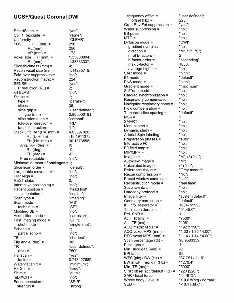
UCSF/Quest Coronal DWI SmartSelect = "yes"; Coil 1 (exclude) = "None"; Uniformity = "CLEAR"; FOV FH (mm) = 256; RL (mm) = 256; AP (mm) = 172; Voxel size FH (mm) = 1.33000004; RL (mm) = 1.33333337; Slice thickness (mm) = 4; Recon voxel size (mm) = 1.14285719; Fold-over suppression = "no"; Reconstruction matrix = 224; SENSE = "yes"; P reduction (RL) = 2; k-t BLAST = "no"; Stacks = 1; type = "parallel"; slices = 36; slice gap = "user defined"; gap (mm) = 0.800000191; slice orientation = "coronal"; fold-over direction = "RL"; fat shift direction = "L"; Stack Offc. AP (P=+mm) = 4.63397026; RL (L=+mm) = -18.1911373; FH (H=+mm) = 30.1373558; Ang. AP (deg) = 0; RL (deg) = -0; FH (deg) = -0; Free rotatable = "no"; Minimum number of packages = 1; Slice scan order = "default"; Large table movement = "no"; PlanAlign = "no"; REST slabs = 0; Interactive positioning = "no"; Patient position = "head first"; orientation = "supine"; Scan type = "Imaging"; Scan mode = "MS"; technique = "SE"; Modified SE = "no"; Acquisition mode = "cartesian"; Fast Imaging mode = "EPI"; shot mode = "single-shot"; Echoes = 1; partial echo = "no"; TE = "shortest"; Flip angle (deg) = 90; TR = "user defined"; (ms) = 7500; Halfscan = "yes"; factor = 0.748427689; Water-fat shift = "minimum"; RF Shims = "fixed"; Shim = "auto"; mDIXON = "no"; Fat suppression = "SPIR"; strength = "strong";
frequency offset = "user defined"; offset (Hz) = 220; Grad Rev Fat suppression = "yes"; Water suppression = "no"; BB pulse = "no"; MTC = "no"; Diffusion mode = "DWI"; gradient overplus = "no"; direction = "M", "P", "S"; nr of b-factors = 2; b-factor order = "ascending"; max b-factor = 1000; average high b = "no"; SAR mode = "high"; B1 mode = "default"; PNS mode = "high"; Gradient mode = "maximum"; SofTone mode = "no"; Cardiac synchronization = "no"; Respiratory compensation = "no"; Navigator respiratory comp = "no"; Flow compensation = "no"; Temporal slice spacing = "default"; NSA = 3; SMART = "no"; Manual start = "no"; Dynamic study = "no"; Arterial Spin labeling = "no"; Preparation phases = "auto"; Interactive F0 = "no"; B0 field map = "no"; MIP/MPR = "no"; Images = "M", (3) "no"; Autoview image = "M"; Calculated images = (4) "no"; Reference tissue = "Grey matter"; Recon compression = "No"; Preset window contrast = "soft"; Reconstruction mode = "real time"; Save raw data = "no"; Hardcopy protocol = "no"; Image filter = "system default"; Geometry correction = "default"; IF_info_seperator = 1634755923; Total scan duration = "01:45.0"; Rel. SNR = 1; Act. TR (ms) = "7500"; Act. TE (ms) = "106"; ACQ matrix M x P = "192 x 190"; ACQ voxel MPS (mm) = "1.33 / 1.35 / 4.00"; REC voxel MPS (mm) = "1.14 / 1.14 / 4.00"; Scan percentage (%) = 98.9583359; Packages = 1; Min. slice gap (mm) = -0; EPI factor = 95; WFS (pix) / BW (Hz) = "37.751 / 11.5"; BW in EPI freq. dir. (Hz) = "1270.4"; Min. TR (ms) = "5900"; SPIR offset act./default (Hz) = "220 [220]"; SAR / local torso = "< 19 %"; Whole body / level = "< 0.6 W/kg / normal"; SED = "< 0.1 kJ/kg";

B1+rms / Coil Power = "1.00 uT / 18 %"; Max B1+rms = "1.00 uT"; PNS / level = "82 % / 1st level"; dB/dt = "124.8 T/s"; Sound Pressure Level (dB) = 24.0876598;

SIEMENS MAGNETOM Avanto syngo MR B17
\\USER\VIRC_RESEARCH\Quest-v1\Quest\localizerTA: 9.2 s PAT: Off Voxel size: 1.6×0.8×10.0 mm Rel. SNR: 1.00 SIEMENS: gre
PropertiesPrio Recon OffBefore measurementAfter measurementLoad to viewer OnInline movie OffAuto store images OnLoad to stamp segments OnLoad images to graphicsegments
Off
Auto open inline display OffStart measurement withoutfurther preparation
On
Wait for user to start OffStart measurements single
RoutineSlice group 1 Slices 1 Dist. factor 20 % Position Isocenter Orientation Sagittal Phase enc. dir. A >> P Rotation 0.00 degSlice group 2 Slices 1 Dist. factor 20 % Position L0.0 A27.1 H7.7 Orientation Transversal Phase enc. dir. A >> P Rotation 0.00 degSlice group 3 Slices 1 Dist. factor 20 % Position L0.0 A50.4 H5.3 Orientation Coronal Phase enc. dir. R >> L Rotation 0.00 degPhase oversampling 0 %FoV read 200 mmFoV phase 100.0 %Slice thickness 10.0 mmTR 20.0 msTE 5.00 msAverages 1Concatenations 3Filter Distortion Corr.(2D), Elliptical
filterCoil elements D11-18
ContrastTD 0 msMTC OffMagn. preparation NoneFlip angle 40 degFat suppr. NoneWater suppr. None
- - - - - - - - - - - - - - - - - - - - - - - - - - - - - - - - - - - - - - - - - - - - - - - - - - - - - - - - - - - - - - - - - - - - - - - - - - - - - - - - - - - -
Averaging mode Short termReconstruction MagnitudeMeasurements 1Multiple series Each measurement
ResolutionBase resolution 256
Phase resolution 50 %Phase partial Fourier OffInterpolation On
- - - - - - - - - - - - - - - - - - - - - - - - - - - - - - - - - - - - - - - - - - - - - - - - - - - - - - - - - - - - - - - - - - - - - - - - - - - - - - - - - - - -
PAT mode NoneMatrix Coil Mode Auto (CP)
- - - - - - - - - - - - - - - - - - - - - - - - - - - - - - - - - - - - - - - - - - - - - - - - - - - - - - - - - - - - - - - - - - - - - - - - - - - - - - - - - - - -
Image Filter OffDistortion Corr. OnMode 2DUnfiltered images OffPrescan Normalize OffNormalize OffB1 filter OffRaw filter OffElliptical filter OnMode Inplane
GeometryMulti-slice mode SequentialSeries Interleaved
- - - - - - - - - - - - - - - - - - - - - - - - - - - - - - - - - - - - - - - - - - - - - - - - - - - - - - - - - - - - - - - - - - - - - - - - - - - - - - - - - - - -
Saturation mode StandardSpecial sat. None
- - - - - - - - - - - - - - - - - - - - - - - - - - - - - - - - - - - - - - - - - - - - - - - - - - - - - - - - - - - - - - - - - - - - - - - - - - - - - - - - - - - -- - - - - - - - - - - - - - - - - - - - - - - - - - - - - - - - - - - - - - - - - - - - - - - - - - - - - - - - - - - - - - - - - - - - - - - - - - - - - - - - - - - -
Tim CT mode OffSystem
Body OffD11 OnD12 OnD13 OnD14 OnD15 OnD16 OnD17 OnD18 On
- - - - - - - - - - - - - - - - - - - - - - - - - - - - - - - - - - - - - - - - - - - - - - - - - - - - - - - - - - - - - - - - - - - - - - - - - - - - - - - - - - - -
Positioning mode REFTable position HTable position 0 mmMSMA S - C - TSagittal R >> LCoronal P >> ATransversal H >> FSave uncombined OffCoil Combine Mode Adaptive CombineAuto Coil Select Default
- - - - - - - - - - - - - - - - - - - - - - - - - - - - - - - - - - - - - - - - - - - - - - - - - - - - - - - - - - - - - - - - - - - - - - - - - - - - - - - - - - - -
Shim mode Tune upAdjust with body coil OffConfirm freq. adjustment OffAssume Silicone Off? Ref. amplitude 1H 0.000 VAdjustment Tolerance AutoAdjust volume Position Isocenter Orientation Transversal Rotation 0.00 deg R >> L 350 mm A >> P 263 mm F >> H 350 mm
Physio1st Signal/Mode NoneSegments 1
- - - - - - - - - - - - - - - - - - - - - - - - - - - - - - - - - - - - - - - - - - - - - - - - - - - - - - - - - - - - - - - - - - - - - - - - - - - - - - - - - - - -
Tagging None1/+
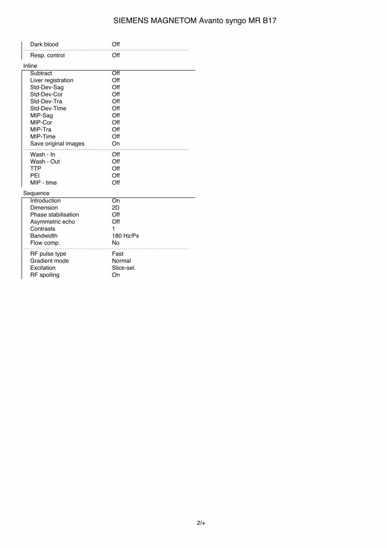
SIEMENS MAGNETOM Avanto syngo MR B17
Dark blood Off- - - - - - - - - - - - - - - - - - - - - - - - - - - - - - - - - - - - - - - - - - - - - - - - - - - - - - - - - - - - - - - - - - - - - - - - - - - - - - - - - - - -
Resp. control OffInline
Subtract OffLiver registration OffStd-Dev-Sag OffStd-Dev-Cor OffStd-Dev-Tra OffStd-Dev-Time OffMIP-Sag OffMIP-Cor OffMIP-Tra OffMIP-Time OffSave original images On
- - - - - - - - - - - - - - - - - - - - - - - - - - - - - - - - - - - - - - - - - - - - - - - - - - - - - - - - - - - - - - - - - - - - - - - - - - - - - - - - - - - -
Wash - In OffWash - Out OffTTP OffPEI OffMIP - time Off
SequenceIntroduction OnDimension 2DPhase stabilisation OffAsymmetric echo OffContrasts 1Bandwidth 180 Hz/PxFlow comp. No
- - - - - - - - - - - - - - - - - - - - - - - - - - - - - - - - - - - - - - - - - - - - - - - - - - - - - - - - - - - - - - - - - - - - - - - - - - - - - - - - - - - -
RF pulse type FastGradient mode NormalExcitation Slice-sel.RF spoiling On
2/+

SIEMENS MAGNETOM Avanto syngo MR B17
\\USER\VIRC_RESEARCH\Quest-v1\Quest\Sag MPRAGETA: 7:42 PAT: Off Voxel size: 1.3×1.3×1.2 mm Rel. SNR: 1.00 SIEMENS: tfl
PropertiesPrio Recon OffBefore measurementAfter measurementLoad to viewer OnInline movie OffAuto store images OnLoad to stamp segments OffLoad images to graphicsegments
Off
Auto open inline display OffStart measurement withoutfurther preparation
On
Wait for user to start OffStart measurements single
RoutineSlab group 1 Slabs 1 Dist. factor 50 % Position L0.8 A22.5 H9.3 Orientation Sagittal Phase enc. dir. A >> P Rotation 0.00 degPhase oversampling 0 %Slice oversampling 20.0 %Slices per slab 160FoV read 240 mmFoV phase 100.0 %Slice thickness 1.20 mmTR 2400 msTE 3.5 msAverages 1Concatenations 1Filter NoneCoil elements HE1-4
ContrastMagn. preparation Non-sel. IRTI 1000 msFlip angle 8 degFat suppr. NoneWater suppr. None
- - - - - - - - - - - - - - - - - - - - - - - - - - - - - - - - - - - - - - - - - - - - - - - - - - - - - - - - - - - - - - - - - - - - - - - - - - - - - - - - - - - -
Averaging mode Long termReconstruction MagnitudeMeasurements 1Multiple series Off
ResolutionBase resolution 192Phase resolution 100 %Slice resolution 100 %Phase partial Fourier OffSlice partial Fourier OffInterpolation Off
- - - - - - - - - - - - - - - - - - - - - - - - - - - - - - - - - - - - - - - - - - - - - - - - - - - - - - - - - - - - - - - - - - - - - - - - - - - - - - - - - - - -
PAT mode NoneMatrix Coil Mode Auto (CP)
- - - - - - - - - - - - - - - - - - - - - - - - - - - - - - - - - - - - - - - - - - - - - - - - - - - - - - - - - - - - - - - - - - - - - - - - - - - - - - - - - - - -
Image Filter OffDistortion Corr. OffPrescan Normalize OffNormalize OffB1 filter OffRaw filter Off
Elliptical filter OffGeometry
Multi-slice mode Single shotSeries Interleaved
- - - - - - - - - - - - - - - - - - - - - - - - - - - - - - - - - - - - - - - - - - - - - - - - - - - - - - - - - - - - - - - - - - - - - - - - - - - - - - - - - - - -
SystemBody OffHE2 OnHE4 OnHE1 OnHE3 OnSP4 OffSP2 OffSP8 OffSP6 OffSP3 OffSP1 OffSP7 OffSP5 Off
- - - - - - - - - - - - - - - - - - - - - - - - - - - - - - - - - - - - - - - - - - - - - - - - - - - - - - - - - - - - - - - - - - - - - - - - - - - - - - - - - - - -
Positioning mode REFTable position HTable position 0 mmMSMA S - C - TSagittal R >> LCoronal A >> PTransversal F >> HSave uncombined OffCoil Combine Mode Sum of SquaresAuto Coil Select Default
- - - - - - - - - - - - - - - - - - - - - - - - - - - - - - - - - - - - - - - - - - - - - - - - - - - - - - - - - - - - - - - - - - - - - - - - - - - - - - - - - - - -
Shim mode StandardAdjust with body coil OffConfirm freq. adjustment OffAssume Silicone Off? Ref. amplitude 1H 0.000 VAdjustment Tolerance AutoAdjust volume Position L0.8 A22.5 H9.3 Orientation Sagittal Rotation 0.00 deg F >> H 240 mm A >> P 240 mm R >> L 192 mm
Physio1st Signal/Mode None
- - - - - - - - - - - - - - - - - - - - - - - - - - - - - - - - - - - - - - - - - - - - - - - - - - - - - - - - - - - - - - - - - - - - - - - - - - - - - - - - - - - -
Dark blood Off- - - - - - - - - - - - - - - - - - - - - - - - - - - - - - - - - - - - - - - - - - - - - - - - - - - - - - - - - - - - - - - - - - - - - - - - - - - - - - - - - - - -
Resp. control OffInline
Subtract OffStd-Dev-Sag OffStd-Dev-Cor OffStd-Dev-Tra OffStd-Dev-Time OffMIP-Sag OffMIP-Cor OffMIP-Tra OffMIP-Time OffSave original images On
SequenceIntroduction OnDimension 3D
3/+
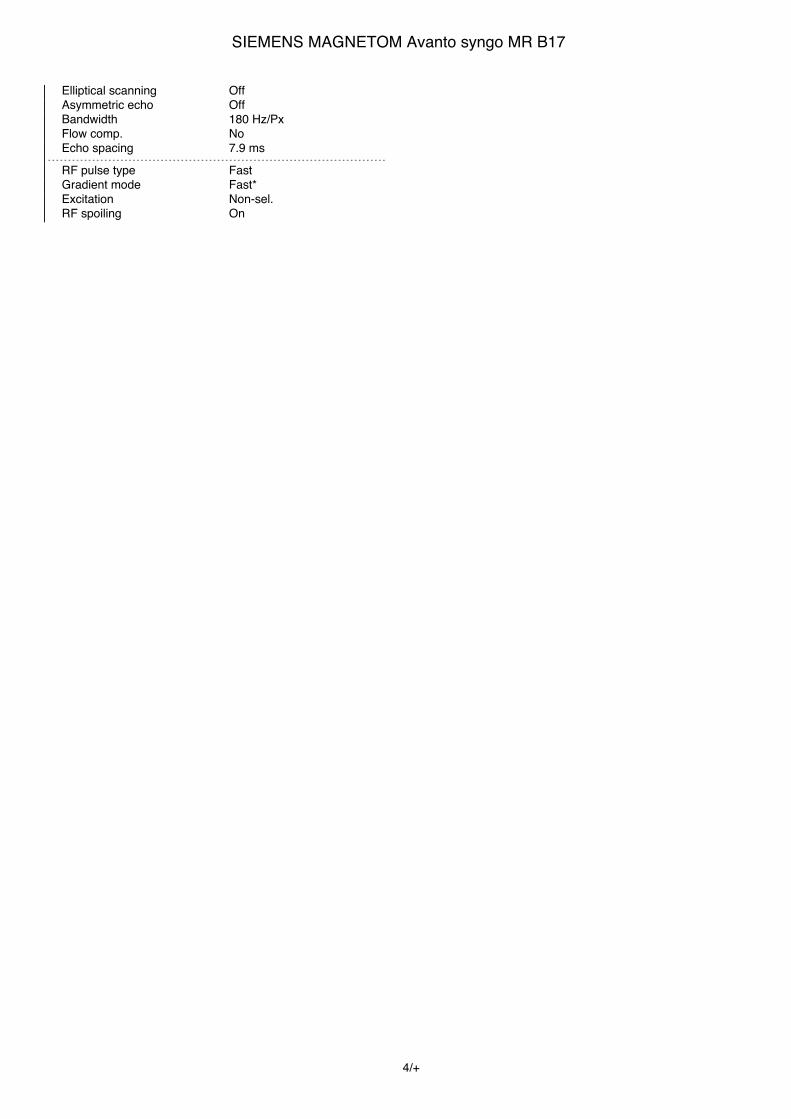
SIEMENS MAGNETOM Avanto syngo MR B17
Elliptical scanning OffAsymmetric echo OffBandwidth 180 Hz/PxFlow comp. NoEcho spacing 7.9 ms
- - - - - - - - - - - - - - - - - - - - - - - - - - - - - - - - - - - - - - - - - - - - - - - - - - - - - - - - - - - - - - - - - - - - - - - - - - - - - - - - - - - -
RF pulse type FastGradient mode Fast*Excitation Non-sel.RF spoiling On
4/+

SIEMENS MAGNETOM Avanto syngo MR B17
\\USER\VIRC_RESEARCH\Quest-v1\Quest\Ax T2starTA: 5:29 PAT: Off Voxel size: 1.3×1.3×4.0 mm Rel. SNR: 1.00 SIEMENS: gre
PropertiesPrio Recon OffBefore measurementAfter measurementLoad to viewer OnInline movie OffAuto store images OnLoad to stamp segments OnLoad images to graphicsegments
Off
Auto open inline display OffStart measurement withoutfurther preparation
On
Wait for user to start OffStart measurements single
RoutineSlice group 1 Slices 44 Dist. factor 0 % Position Isocenter Orientation Transversal Phase enc. dir. R >> L Rotation 90.00 degPhase oversampling 0 %FoV read 256 mmFoV phase 87.5 %Slice thickness 4.0 mmTR 650 msTE 30.0 msAverages 1Concatenations 3Filter Prescan Normalize, Elliptical
filterCoil elements HE
ContrastTD 0 msMTC OffMagn. preparation NoneFlip angle 20 degFat suppr. NoneWater suppr. None
- - - - - - - - - - - - - - - - - - - - - - - - - - - - - - - - - - - - - - - - - - - - - - - - - - - - - - - - - - - - - - - - - - - - - - - - - - - - - - - - - - - -
Averaging mode Short termReconstruction MagnitudeMeasurements 1Multiple series Each measurement
ResolutionBase resolution 192Phase resolution 100 %Phase partial Fourier OffInterpolation Off
- - - - - - - - - - - - - - - - - - - - - - - - - - - - - - - - - - - - - - - - - - - - - - - - - - - - - - - - - - - - - - - - - - - - - - - - - - - - - - - - - - - -
PAT mode NoneMatrix Coil Mode Auto (CP)
- - - - - - - - - - - - - - - - - - - - - - - - - - - - - - - - - - - - - - - - - - - - - - - - - - - - - - - - - - - - - - - - - - - - - - - - - - - - - - - - - - - -
Image Filter OffDistortion Corr. OffUnfiltered images OffPrescan Normalize OnNormalize OffB1 filter OffRaw filter OffElliptical filter On
Mode InplaneGeometry
Multi-slice mode InterleavedSeries Interleaved
- - - - - - - - - - - - - - - - - - - - - - - - - - - - - - - - - - - - - - - - - - - - - - - - - - - - - - - - - - - - - - - - - - - - - - - - - - - - - - - - - - - -
Saturation mode StandardSpecial sat. None
- - - - - - - - - - - - - - - - - - - - - - - - - - - - - - - - - - - - - - - - - - - - - - - - - - - - - - - - - - - - - - - - - - - - - - - - - - - - - - - - - - - -- - - - - - - - - - - - - - - - - - - - - - - - - - - - - - - - - - - - - - - - - - - - - - - - - - - - - - - - - - - - - - - - - - - - - - - - - - - - - - - - - - - -
Tim CT mode OffSystem
Body OffHE OnDLL Off
- - - - - - - - - - - - - - - - - - - - - - - - - - - - - - - - - - - - - - - - - - - - - - - - - - - - - - - - - - - - - - - - - - - - - - - - - - - - - - - - - - - -
Positioning mode REFTable position HTable position 0 mmMSMA T - C - SSagittal R >> LCoronal P >> ATransversal F >> HSave uncombined OffCoil Combine Mode Adaptive CombineAuto Coil Select Default
- - - - - - - - - - - - - - - - - - - - - - - - - - - - - - - - - - - - - - - - - - - - - - - - - - - - - - - - - - - - - - - - - - - - - - - - - - - - - - - - - - - -
Shim mode Tune upAdjust with body coil OffConfirm freq. adjustment OffAssume Silicone Off? Ref. amplitude 1H 0.000 VAdjustment Tolerance AutoAdjust volume Position Isocenter Orientation Transversal Rotation 0.00 deg R >> L 350 mm A >> P 263 mm F >> H 350 mm
Physio1st Signal/Mode NoneSegments 1
- - - - - - - - - - - - - - - - - - - - - - - - - - - - - - - - - - - - - - - - - - - - - - - - - - - - - - - - - - - - - - - - - - - - - - - - - - - - - - - - - - - -
Tagging NoneDark blood Off
- - - - - - - - - - - - - - - - - - - - - - - - - - - - - - - - - - - - - - - - - - - - - - - - - - - - - - - - - - - - - - - - - - - - - - - - - - - - - - - - - - - -
Resp. control OffInline
Subtract OffLiver registration OffStd-Dev-Sag OffStd-Dev-Cor OffStd-Dev-Tra OffStd-Dev-Time OffMIP-Sag OffMIP-Cor OffMIP-Tra OffMIP-Time OffSave original images On
- - - - - - - - - - - - - - - - - - - - - - - - - - - - - - - - - - - - - - - - - - - - - - - - - - - - - - - - - - - - - - - - - - - - - - - - - - - - - - - - - - - -
Wash - In OffWash - Out OffTTP OffPEI OffMIP - time Off
5/+

SIEMENS MAGNETOM Avanto syngo MR B17
SequenceIntroduction OnDimension 2DPhase stabilisation OffAsymmetric echo OffContrasts 1Bandwidth 50 Hz/PxFlow comp. Slice/Read
- - - - - - - - - - - - - - - - - - - - - - - - - - - - - - - - - - - - - - - - - - - - - - - - - - - - - - - - - - - - - - - - - - - - - - - - - - - - - - - - - - - -
RF pulse type NormalGradient mode FastExcitation Slice-sel.RF spoiling On
6/+

SIEMENS MAGNETOM Avanto syngo MR B17
\\USER\VIRC_RESEARCH\Quest-v1\Quest\Ax FLAIRTA: 4:30 PAT: Off Voxel size: 1.0×1.0×4.0 mm Rel. SNR: 1.00 SIEMENS: tse
PropertiesPrio Recon OffBefore measurementAfter measurementLoad to viewer OnInline movie OffAuto store images OnLoad to stamp segments OnLoad images to graphicsegments
Off
Auto open inline display OffStart measurement withoutfurther preparation
On
Wait for user to start OffStart measurements single
RoutineSlice group 1 Slices 44 Dist. factor 0 % Position Isocenter Orientation Transversal Phase enc. dir. A >> P Rotation 0.00 degPhase oversampling 0 %FoV read 256 mmFoV phase 87.5 %Slice thickness 4.0 mmTR 9000 msTE 91 msAverages 1Concatenations 2Filter Prescan Normalize, Elliptical
filterCoil elements HE
ContrastTD 0.0 msMTC OffMagn. preparation Slice-sel. IRTI 2250 msFreeze suppressed tissue OffFlip angle 180 degFat suppr. NoneWater suppr. NoneRestore magn. Off
- - - - - - - - - - - - - - - - - - - - - - - - - - - - - - - - - - - - - - - - - - - - - - - - - - - - - - - - - - - - - - - - - - - - - - - - - - - - - - - - - - - -
Averaging mode Short termReconstruction MagnitudeMeasurements 1Multiple series Each measurement
ResolutionBase resolution 256Phase resolution 100 %Phase partial Fourier OffTrajectory CartesianInterpolation Off
- - - - - - - - - - - - - - - - - - - - - - - - - - - - - - - - - - - - - - - - - - - - - - - - - - - - - - - - - - - - - - - - - - - - - - - - - - - - - - - - - - - -
PAT mode NoneMatrix Coil Mode Auto (CP)
- - - - - - - - - - - - - - - - - - - - - - - - - - - - - - - - - - - - - - - - - - - - - - - - - - - - - - - - - - - - - - - - - - - - - - - - - - - - - - - - - - - -
Image Filter OffDistortion Corr. OffUnfiltered images OffPrescan Normalize On
Normalize OffB1 filter OffRaw filter OffElliptical filter OnMode Inplane
GeometryMulti-slice mode InterleavedSeries Interleaved
- - - - - - - - - - - - - - - - - - - - - - - - - - - - - - - - - - - - - - - - - - - - - - - - - - - - - - - - - - - - - - - - - - - - - - - - - - - - - - - - - - - -
Special sat. Parallel FGap 10 mmThickness 50 mm
- - - - - - - - - - - - - - - - - - - - - - - - - - - - - - - - - - - - - - - - - - - - - - - - - - - - - - - - - - - - - - - - - - - - - - - - - - - - - - - - - - - -- - - - - - - - - - - - - - - - - - - - - - - - - - - - - - - - - - - - - - - - - - - - - - - - - - - - - - - - - - - - - - - - - - - - - - - - - - - - - - - - - - - -
Tim CT mode OffSystem
Body OffHE OnDLL Off
- - - - - - - - - - - - - - - - - - - - - - - - - - - - - - - - - - - - - - - - - - - - - - - - - - - - - - - - - - - - - - - - - - - - - - - - - - - - - - - - - - - -
Positioning mode FIXTable position HTable position 0 mmMSMA T - C - SSagittal R >> LCoronal P >> ATransversal F >> HSave uncombined OffCoil Combine Mode Adaptive CombineAuto Coil Select Default
- - - - - - - - - - - - - - - - - - - - - - - - - - - - - - - - - - - - - - - - - - - - - - - - - - - - - - - - - - - - - - - - - - - - - - - - - - - - - - - - - - - -
Shim mode StandardAdjust with body coil OffConfirm freq. adjustment OffAssume Silicone Off? Ref. amplitude 1H 0.000 VAdjustment Tolerance AutoAdjust volume Position Isocenter Orientation Transversal Rotation 0.00 deg R >> L 256 mm A >> P 224 mm F >> H 176 mm
Physio1st Signal/Mode None
- - - - - - - - - - - - - - - - - - - - - - - - - - - - - - - - - - - - - - - - - - - - - - - - - - - - - - - - - - - - - - - - - - - - - - - - - - - - - - - - - - - -
Dark blood Off- - - - - - - - - - - - - - - - - - - - - - - - - - - - - - - - - - - - - - - - - - - - - - - - - - - - - - - - - - - - - - - - - - - - - - - - - - - - - - - - - - - -
Resp. control OffInline
Subtract OffStd-Dev-Sag OffStd-Dev-Cor OffStd-Dev-Tra OffStd-Dev-Time OffMIP-Sag OffMIP-Cor OffMIP-Tra OffMIP-Time OffSave original images On
SequenceIntroduction OffDimension 2D
7/+

SIEMENS MAGNETOM Avanto syngo MR B17
Compensate T2 decay OffReduce Motion Sens. OffContrasts 1Bandwidth 120 Hz/PxFlow comp. NoAllowed delay 30 sEcho spacing 13 ms
- - - - - - - - - - - - - - - - - - - - - - - - - - - - - - - - - - - - - - - - - - - - - - - - - - - - - - - - - - - - - - - - - - - - - - - - - - - - - - - - - - - -
Define Turbo factorTurbo factor 16Echo trains per slice 14RF pulse type NormalGradient mode Normal
8/+

SIEMENS MAGNETOM Avanto syngo MR B17
\\USER\VIRC_RESEARCH\Quest-v1\Quest\Ax T2 TSETA: 2:26 PAT: Off Voxel size: 1.0×1.0×4.0 mm Rel. SNR: 1.00 SIEMENS: tse
PropertiesPrio Recon OffBefore measurementAfter measurementLoad to viewer OffInline movie OffAuto store images OnLoad to stamp segments OnLoad images to graphicsegments
On
Auto open inline display OffStart measurement withoutfurther preparation
On
Wait for user to start OffStart measurements single
RoutineSlice group 1 Slices 44 Dist. factor 0 % Position Isocenter Orientation Transversal Phase enc. dir. R >> L Rotation 90.00 degPhase oversampling 0 %FoV read 256 mmFoV phase 87.5 %Slice thickness 4.0 mmTR 3000 msTE 92 msAverages 1Concatenations 3Filter Distortion Corr.(2D), Elliptical
filterCoil elements HE
ContrastTD 0.0 msMTC OffMagn. preparation NoneFlip angle 178 degFat suppr. Fat sat.Fat sat. mode StrongWater suppr. NoneRestore magn. Off
- - - - - - - - - - - - - - - - - - - - - - - - - - - - - - - - - - - - - - - - - - - - - - - - - - - - - - - - - - - - - - - - - - - - - - - - - - - - - - - - - - - -
Averaging mode Short termReconstruction MagnitudeMeasurements 1Multiple series Each measurement
ResolutionBase resolution 256Phase resolution 100 %Phase partial Fourier OffTrajectory CartesianInterpolation On
- - - - - - - - - - - - - - - - - - - - - - - - - - - - - - - - - - - - - - - - - - - - - - - - - - - - - - - - - - - - - - - - - - - - - - - - - - - - - - - - - - - -
PAT mode NoneMatrix Coil Mode Auto (CP)
- - - - - - - - - - - - - - - - - - - - - - - - - - - - - - - - - - - - - - - - - - - - - - - - - - - - - - - - - - - - - - - - - - - - - - - - - - - - - - - - - - - -
Image Filter OffDistortion Corr. OnMode 2DUnfiltered images OffPrescan Normalize Off
Normalize OffB1 filter OffRaw filter OffElliptical filter OnMode Inplane
GeometryMulti-slice mode InterleavedSeries Interleaved
- - - - - - - - - - - - - - - - - - - - - - - - - - - - - - - - - - - - - - - - - - - - - - - - - - - - - - - - - - - - - - - - - - - - - - - - - - - - - - - - - - - -
Special sat. None- - - - - - - - - - - - - - - - - - - - - - - - - - - - - - - - - - - - - - - - - - - - - - - - - - - - - - - - - - - - - - - - - - - - - - - - - - - - - - - - - - - -- - - - - - - - - - - - - - - - - - - - - - - - - - - - - - - - - - - - - - - - - - - - - - - - - - - - - - - - - - - - - - - - - - - - - - - - - - - - - - - - - - - -
Tim CT mode OffSystem
Body OffHE OnDLL Off
- - - - - - - - - - - - - - - - - - - - - - - - - - - - - - - - - - - - - - - - - - - - - - - - - - - - - - - - - - - - - - - - - - - - - - - - - - - - - - - - - - - -
Positioning mode ISOTable position HTable position 0 mmMSMA T - C - SSagittal R >> LCoronal P >> ATransversal F >> HSave uncombined OffCoil Combine Mode Adaptive CombineAuto Coil Select Default
- - - - - - - - - - - - - - - - - - - - - - - - - - - - - - - - - - - - - - - - - - - - - - - - - - - - - - - - - - - - - - - - - - - - - - - - - - - - - - - - - - - -
Shim mode StandardAdjust with body coil OffConfirm freq. adjustment OffAssume Silicone Off? Ref. amplitude 1H 0.000 VAdjustment Tolerance AutoAdjust volume Position Isocenter Orientation Transversal Rotation 90.00 deg A >> P 256 mm R >> L 224 mm F >> H 176 mm
Physio1st Signal/Mode None
- - - - - - - - - - - - - - - - - - - - - - - - - - - - - - - - - - - - - - - - - - - - - - - - - - - - - - - - - - - - - - - - - - - - - - - - - - - - - - - - - - - -
Dark blood Off- - - - - - - - - - - - - - - - - - - - - - - - - - - - - - - - - - - - - - - - - - - - - - - - - - - - - - - - - - - - - - - - - - - - - - - - - - - - - - - - - - - -
Resp. control OffInline
Subtract OffStd-Dev-Sag OffStd-Dev-Cor OffStd-Dev-Tra OffStd-Dev-Time OffMIP-Sag OffMIP-Cor OffMIP-Tra OffMIP-Time OffSave original images On
SequenceIntroduction OnDimension 2DCompensate T2 decay OffReduce Motion Sens. Off
9/+

SIEMENS MAGNETOM Avanto syngo MR B17
Contrasts 1Bandwidth 181 Hz/PxFlow comp. NoAllowed delay 30 sEcho spacing 10.2 ms
- - - - - - - - - - - - - - - - - - - - - - - - - - - - - - - - - - - - - - - - - - - - - - - - - - - - - - - - - - - - - - - - - - - - - - - - - - - - - - - - - - - -
Define Turbo factorTurbo factor 16Echo trains per slice 14RF pulse type NormalGradient mode Normal
10/+

SIEMENS MAGNETOM Avanto syngo MR B17
\\USER\VIRC_RESEARCH\Quest-v1\Quest\DIFFUSION AXTA: 1:32 PAT: 2 Voxel size: 1.6×1.6×4.0 mm Rel. SNR: 1.00 SIEMENS: ep2d_diff
PropertiesPrio Recon OffBefore measurementAfter measurementLoad to viewer OffInline movie OffAuto store images OnLoad to stamp segments OnLoad images to graphicsegments
On
Auto open inline display OffStart measurement withoutfurther preparation
On
Wait for user to start OnStart measurements single
RoutineSlice group 1 Slices 32 Dist. factor 20 % Position R4.4 A21.5 F0.1 Orientation Transversal Phase enc. dir. A >> P Rotation 0.00 degPhase oversampling 0 %FoV read 256 mmFoV phase 100.0 %Slice thickness 4.0 mmTR 6000 msTE 90 msAverages 3Concatenations 1Filter Raw filter, Distortion
Corr.(2D), Prescan NormalizeCoil elements HE1-4
ContrastMTC OffMagn. preparation NoneFat suppr. Fat sat.
- - - - - - - - - - - - - - - - - - - - - - - - - - - - - - - - - - - - - - - - - - - - - - - - - - - - - - - - - - - - - - - - - - - - - - - - - - - - - - - - - - - -
Averaging mode Long termReconstruction MagnitudeDelay in TR 0 ms
ResolutionBase resolution 160Phase resolution 100 %Phase partial Fourier 6/8Interpolation Off
- - - - - - - - - - - - - - - - - - - - - - - - - - - - - - - - - - - - - - - - - - - - - - - - - - - - - - - - - - - - - - - - - - - - - - - - - - - - - - - - - - - -
PAT mode GRAPPAAccel. factor PE 2Ref. lines PE 24Matrix Coil Mode Auto (Triple)Reference scan mode Separate
- - - - - - - - - - - - - - - - - - - - - - - - - - - - - - - - - - - - - - - - - - - - - - - - - - - - - - - - - - - - - - - - - - - - - - - - - - - - - - - - - - - -
Distortion Corr. OnMode 2DUnfiltered images OffPrescan Normalize OnRaw filter OnIntensity WeakSlope 25Elliptical filter OffHamming Off
GeometryMulti-slice mode InterleavedSeries Interleaved
- - - - - - - - - - - - - - - - - - - - - - - - - - - - - - - - - - - - - - - - - - - - - - - - - - - - - - - - - - - - - - - - - - - - - - - - - - - - - - - - - - - -
Special sat. None- - - - - - - - - - - - - - - - - - - - - - - - - - - - - - - - - - - - - - - - - - - - - - - - - - - - - - - - - - - - - - - - - - - - - - - - - - - - - - - - - - - -
SystemBody OffHE2 OnHE4 OnHE1 OnHE3 OnSP4 OffSP2 OffSP8 OffSP6 OffSP3 OffSP1 OffSP7 OffSP5 Off
- - - - - - - - - - - - - - - - - - - - - - - - - - - - - - - - - - - - - - - - - - - - - - - - - - - - - - - - - - - - - - - - - - - - - - - - - - - - - - - - - - - -
Positioning mode REFTable position HTable position 0 mmMSMA T - C - SSagittal R >> LCoronal P >> ATransversal F >> HCoil Combine Mode Adaptive CombineAuto Coil Select Default
- - - - - - - - - - - - - - - - - - - - - - - - - - - - - - - - - - - - - - - - - - - - - - - - - - - - - - - - - - - - - - - - - - - - - - - - - - - - - - - - - - - -
Shim mode StandardAdjust with body coil OffConfirm freq. adjustment OffAssume Silicone Off? Ref. amplitude 1H 0.000 VAdjustment Tolerance AutoAdjust volume Position R4.4 A21.5 F0.1 Orientation Transversal Rotation 0.00 deg R >> L 256 mm A >> P 256 mm F >> H 153 mm
Physio1st Signal/Mode None
- - - - - - - - - - - - - - - - - - - - - - - - - - - - - - - - - - - - - - - - - - - - - - - - - - - - - - - - - - - - - - - - - - - - - - - - - - - - - - - - - - - -
Resp. control OffDiff
Diffusion mode 3-Scan TraceDiff. weightings 2b-value 1 0 s/mm²b-value 2 1000 s/mm²Diff. weighted images OffTrace weighted images OnAverage ADC maps OnIndividual ADC maps OffMosaic OffNoise level 40Diff. directions 3
SequenceIntroduction OnBandwidth 1250 Hz/PxFree echo spacing Off
11/+

SIEMENS MAGNETOM Avanto syngo MR B17
Echo spacing 0.88 ms- - - - - - - - - - - - - - - - - - - - - - - - - - - - - - - - - - - - - - - - - - - - - - - - - - - - - - - - - - - - - - - - - - - - - - - - - - - - - - - - - - - -
EPI factor 160RF pulse type NormalGradient mode Fast*
12/+

SIEMENS MAGNETOM Avanto syngo MR B17
\\USER\VIRC_RESEARCH\Quest-v1\Quest\DIFFUSION CorTA: 1:32 PAT: 2 Voxel size: 1.6×1.6×4.0 mm Rel. SNR: 1.00 SIEMENS: ep2d_diff
PropertiesPrio Recon OffBefore measurementAfter measurementLoad to viewer OffInline movie OffAuto store images OnLoad to stamp segments OnLoad images to graphicsegments
On
Auto open inline display OffStart measurement withoutfurther preparation
On
Wait for user to start OnStart measurements single
RoutineSlice group 1 Slices 36 Dist. factor 20 % Position R4.4 A21.5 F0.1 Orientation Coronal Phase enc. dir. R >> L Rotation 0.00 degPhase oversampling 0 %FoV read 256 mmFoV phase 100.0 %Slice thickness 4.0 mmTR 6000 msTE 90 msAverages 3Concatenations 1Filter Raw filter, Distortion
Corr.(2D), Prescan NormalizeCoil elements HE1-4
ContrastMTC OffMagn. preparation NoneFat suppr. Fat sat.
- - - - - - - - - - - - - - - - - - - - - - - - - - - - - - - - - - - - - - - - - - - - - - - - - - - - - - - - - - - - - - - - - - - - - - - - - - - - - - - - - - - -
Averaging mode Long termReconstruction MagnitudeDelay in TR 0 ms
ResolutionBase resolution 160Phase resolution 100 %Phase partial Fourier 6/8Interpolation Off
- - - - - - - - - - - - - - - - - - - - - - - - - - - - - - - - - - - - - - - - - - - - - - - - - - - - - - - - - - - - - - - - - - - - - - - - - - - - - - - - - - - -
PAT mode GRAPPAAccel. factor PE 2Ref. lines PE 24Matrix Coil Mode Auto (Triple)Reference scan mode Separate
- - - - - - - - - - - - - - - - - - - - - - - - - - - - - - - - - - - - - - - - - - - - - - - - - - - - - - - - - - - - - - - - - - - - - - - - - - - - - - - - - - - -
Distortion Corr. OnMode 2DUnfiltered images OffPrescan Normalize OnRaw filter OnIntensity WeakSlope 25Elliptical filter OffHamming Off
GeometryMulti-slice mode InterleavedSeries Interleaved
- - - - - - - - - - - - - - - - - - - - - - - - - - - - - - - - - - - - - - - - - - - - - - - - - - - - - - - - - - - - - - - - - - - - - - - - - - - - - - - - - - - -
Special sat. None- - - - - - - - - - - - - - - - - - - - - - - - - - - - - - - - - - - - - - - - - - - - - - - - - - - - - - - - - - - - - - - - - - - - - - - - - - - - - - - - - - - -
SystemBody OffHE2 OnHE4 OnHE1 OnHE3 OnSP4 OffSP2 OffSP8 OffSP6 OffSP3 OffSP1 OffSP7 OffSP5 Off
- - - - - - - - - - - - - - - - - - - - - - - - - - - - - - - - - - - - - - - - - - - - - - - - - - - - - - - - - - - - - - - - - - - - - - - - - - - - - - - - - - - -
Positioning mode REFTable position HTable position 0 mmMSMA T - C - SSagittal R >> LCoronal P >> ATransversal F >> HCoil Combine Mode Adaptive CombineAuto Coil Select Default
- - - - - - - - - - - - - - - - - - - - - - - - - - - - - - - - - - - - - - - - - - - - - - - - - - - - - - - - - - - - - - - - - - - - - - - - - - - - - - - - - - - -
Shim mode StandardAdjust with body coil OffConfirm freq. adjustment OffAssume Silicone Off? Ref. amplitude 1H 0.000 VAdjustment Tolerance AutoAdjust volume Position R4.4 A21.5 F0.1 Orientation Coronal Rotation 0.00 deg F >> H 256 mm R >> L 256 mm A >> P 172 mm
Physio1st Signal/Mode None
- - - - - - - - - - - - - - - - - - - - - - - - - - - - - - - - - - - - - - - - - - - - - - - - - - - - - - - - - - - - - - - - - - - - - - - - - - - - - - - - - - - -
Resp. control OffDiff
Diffusion mode 3-Scan TraceDiff. weightings 2b-value 1 0 s/mm²b-value 2 1000 s/mm²Diff. weighted images OffTrace weighted images OnAverage ADC maps OnIndividual ADC maps OffMosaic OffNoise level 40Diff. directions 3
SequenceIntroduction OnBandwidth 1250 Hz/PxFree echo spacing Off
13/+

SIEMENS MAGNETOM Avanto syngo MR B17
Echo spacing 0.88 ms- - - - - - - - - - - - - - - - - - - - - - - - - - - - - - - - - - - - - - - - - - - - - - - - - - - - - - - - - - - - - - - - - - - - - - - - - - - - - - - - - - - -
EPI factor 160RF pulse type NormalGradient mode Fast*
14/+

SIEMENS MAGNETOM Avanto syngo MR B17
Table of contents
\\USER VIRC_RESEARCH
Quest-v1 Quest
localizer Angle Sag to midline Sag MPRAGE Orient Axials to AC/PC Ax T2star Ax FLAIR Ax T2 TSE DIFFUSION AX DIFFUSION Cor
15/-

SIEMENS MAGNETOM TrioTim syngo MR B17
\\USER\MAC\Rankin_lab\QUESTv2\localizerTA: 0:10 PAT: Off Voxel size: 1.9×1.5×8.0 mm Rel. SNR: 1.00 SIEMENS: gre
PropertiesPrio Recon OffBefore measurementAfter measurementLoad to viewer OnInline movie OffAuto store images OnLoad to stamp segments OffLoad images to graphicsegments
Off
Auto open inline display OffStart measurement withoutfurther preparation
Off
Wait for user to start OffStart measurements single
RoutineSlice group 1 Slices 1 Dist. factor 20 % Position Isocenter Orientation Sagittal Phase enc. dir. A >> P Rotation 0.00 degSlice group 2 Slices 1 Dist. factor 20 % Position Isocenter Orientation Coronal Phase enc. dir. R >> L Rotation 0.00 degSlice group 3 Slices 1 Dist. factor 20 % Position Isocenter Orientation Transversal Phase enc. dir. R >> L Rotation 90.00 degPhase oversampling 0 %FoV read 280 mmFoV phase 100.0 %Slice thickness 8.0 mmTR 20.0 msTE 5.00 msAverages 1Concatenations 3Filter Raw filter, Prescan NormalizeCoil elements HEA;HEP
ContrastTD 0 msMTC OffMagn. preparation NoneFlip angle 40 degFat suppr. NoneWater suppr. None
- - - - - - - - - - - - - - - - - - - - - - - - - - - - - - - - - - - - - - - - - - - - - - - - - - - - - - - - - - - - - - - - - - - - - - - - - - - - - - - - - - - -
Averaging mode Short termReconstruction MagnitudeMeasurements 1Multiple series Off
ResolutionBase resolution 192Phase resolution 75 %
Phase partial Fourier OffInterpolation Off
- - - - - - - - - - - - - - - - - - - - - - - - - - - - - - - - - - - - - - - - - - - - - - - - - - - - - - - - - - - - - - - - - - - - - - - - - - - - - - - - - - - -
PAT mode NoneMatrix Coil Mode Auto (CP)
- - - - - - - - - - - - - - - - - - - - - - - - - - - - - - - - - - - - - - - - - - - - - - - - - - - - - - - - - - - - - - - - - - - - - - - - - - - - - - - - - - - -
Image Filter OffDistortion Corr. OffUnfiltered images OffPrescan Normalize OnNormalize OffB1 filter OffRaw filter OnIntensity WeakSlope 25Elliptical filter Off
GeometryMulti-slice mode SequentialSeries Ascending
- - - - - - - - - - - - - - - - - - - - - - - - - - - - - - - - - - - - - - - - - - - - - - - - - - - - - - - - - - - - - - - - - - - - - - - - - - - - - - - - - - - -
Saturation mode StandardSpecial sat. None
- - - - - - - - - - - - - - - - - - - - - - - - - - - - - - - - - - - - - - - - - - - - - - - - - - - - - - - - - - - - - - - - - - - - - - - - - - - - - - - - - - - -- - - - - - - - - - - - - - - - - - - - - - - - - - - - - - - - - - - - - - - - - - - - - - - - - - - - - - - - - - - - - - - - - - - - - - - - - - - - - - - - - - - -
Tim CT mode OffSystem
Body OffHEP OnHEA On
- - - - - - - - - - - - - - - - - - - - - - - - - - - - - - - - - - - - - - - - - - - - - - - - - - - - - - - - - - - - - - - - - - - - - - - - - - - - - - - - - - - -
Positioning mode REFTable position HTable position 0 mmMSMA S - C - TSagittal R >> LCoronal A >> PTransversal F >> HSave uncombined OffCoil Combine Mode Adaptive CombineAutoAlign ---Auto Coil Select Default
- - - - - - - - - - - - - - - - - - - - - - - - - - - - - - - - - - - - - - - - - - - - - - - - - - - - - - - - - - - - - - - - - - - - - - - - - - - - - - - - - - - -
Shim mode Tune upAdjust with body coil OffConfirm freq. adjustment OffAssume Silicone Off? Ref. amplitude 1H 0.000 VAdjustment Tolerance AutoAdjust volume Position Isocenter Orientation Transversal Rotation 0.00 deg R >> L 350 mm A >> P 263 mm F >> H 350 mm
Physio1st Signal/Mode NoneSegments 1
- - - - - - - - - - - - - - - - - - - - - - - - - - - - - - - - - - - - - - - - - - - - - - - - - - - - - - - - - - - - - - - - - - - - - - - - - - - - - - - - - - - -
Dark blood Off- - - - - - - - - - - - - - - - - - - - - - - - - - - - - - - - - - - - - - - - - - - - - - - - - - - - - - - - - - - - - - - - - - - - - - - - - - - - - - - - - - - -
Resp. control OffInline
Subtract OffLiver registration OffStd-Dev-Sag Off
1/+

SIEMENS MAGNETOM TrioTim syngo MR B17
Std-Dev-Cor OffStd-Dev-Tra OffStd-Dev-Time OffMIP-Sag OffMIP-Cor OffMIP-Tra OffMIP-Time OffSave original images On
- - - - - - - - - - - - - - - - - - - - - - - - - - - - - - - - - - - - - - - - - - - - - - - - - - - - - - - - - - - - - - - - - - - - - - - - - - - - - - - - - - - -
Wash - In OffWash - Out OffTTP OffPEI OffMIP - time Off
SequenceIntroduction OnDimension 2DPhase stabilisation OffAsymmetric echo OffContrasts 1Bandwidth 180 Hz/PxFlow comp. NoAllowed delay 0 s
- - - - - - - - - - - - - - - - - - - - - - - - - - - - - - - - - - - - - - - - - - - - - - - - - - - - - - - - - - - - - - - - - - - - - - - - - - - - - - - - - - - -
RF pulse type NormalGradient mode FastExcitation Slice-sel.RF spoiling On
2/+
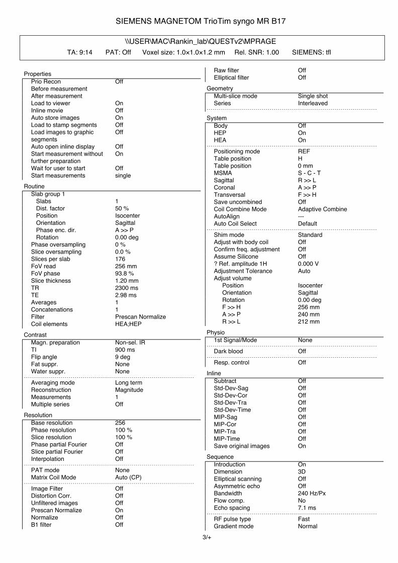
SIEMENS MAGNETOM TrioTim syngo MR B17
\\USER\MAC\Rankin_lab\QUESTv2\MPRAGETA: 9:14 PAT: Off Voxel size: 1.0×1.0×1.2 mm Rel. SNR: 1.00 SIEMENS: tfl
PropertiesPrio Recon OffBefore measurementAfter measurementLoad to viewer OnInline movie OffAuto store images OnLoad to stamp segments OffLoad images to graphicsegments
Off
Auto open inline display OffStart measurement withoutfurther preparation
On
Wait for user to start OffStart measurements single
RoutineSlab group 1 Slabs 1 Dist. factor 50 % Position Isocenter Orientation Sagittal Phase enc. dir. A >> P Rotation 0.00 degPhase oversampling 0 %Slice oversampling 0.0 %Slices per slab 176FoV read 256 mmFoV phase 93.8 %Slice thickness 1.20 mmTR 2300 msTE 2.98 msAverages 1Concatenations 1Filter Prescan NormalizeCoil elements HEA;HEP
ContrastMagn. preparation Non-sel. IRTI 900 msFlip angle 9 degFat suppr. NoneWater suppr. None
- - - - - - - - - - - - - - - - - - - - - - - - - - - - - - - - - - - - - - - - - - - - - - - - - - - - - - - - - - - - - - - - - - - - - - - - - - - - - - - - - - - -
Averaging mode Long termReconstruction MagnitudeMeasurements 1Multiple series Off
ResolutionBase resolution 256Phase resolution 100 %Slice resolution 100 %Phase partial Fourier OffSlice partial Fourier OffInterpolation Off
- - - - - - - - - - - - - - - - - - - - - - - - - - - - - - - - - - - - - - - - - - - - - - - - - - - - - - - - - - - - - - - - - - - - - - - - - - - - - - - - - - - -
PAT mode NoneMatrix Coil Mode Auto (CP)
- - - - - - - - - - - - - - - - - - - - - - - - - - - - - - - - - - - - - - - - - - - - - - - - - - - - - - - - - - - - - - - - - - - - - - - - - - - - - - - - - - - -
Image Filter OffDistortion Corr. OffUnfiltered images OffPrescan Normalize OnNormalize OffB1 filter Off
Raw filter OffElliptical filter Off
GeometryMulti-slice mode Single shotSeries Interleaved
- - - - - - - - - - - - - - - - - - - - - - - - - - - - - - - - - - - - - - - - - - - - - - - - - - - - - - - - - - - - - - - - - - - - - - - - - - - - - - - - - - - -
SystemBody OffHEP OnHEA On
- - - - - - - - - - - - - - - - - - - - - - - - - - - - - - - - - - - - - - - - - - - - - - - - - - - - - - - - - - - - - - - - - - - - - - - - - - - - - - - - - - - -
Positioning mode REFTable position HTable position 0 mmMSMA S - C - TSagittal R >> LCoronal A >> PTransversal F >> HSave uncombined OffCoil Combine Mode Adaptive CombineAutoAlign ---Auto Coil Select Default
- - - - - - - - - - - - - - - - - - - - - - - - - - - - - - - - - - - - - - - - - - - - - - - - - - - - - - - - - - - - - - - - - - - - - - - - - - - - - - - - - - - -
Shim mode StandardAdjust with body coil OffConfirm freq. adjustment OffAssume Silicone Off? Ref. amplitude 1H 0.000 VAdjustment Tolerance AutoAdjust volume Position Isocenter Orientation Sagittal Rotation 0.00 deg F >> H 256 mm A >> P 240 mm R >> L 212 mm
Physio1st Signal/Mode None
- - - - - - - - - - - - - - - - - - - - - - - - - - - - - - - - - - - - - - - - - - - - - - - - - - - - - - - - - - - - - - - - - - - - - - - - - - - - - - - - - - - -
Dark blood Off- - - - - - - - - - - - - - - - - - - - - - - - - - - - - - - - - - - - - - - - - - - - - - - - - - - - - - - - - - - - - - - - - - - - - - - - - - - - - - - - - - - -
Resp. control OffInline
Subtract OffStd-Dev-Sag OffStd-Dev-Cor OffStd-Dev-Tra OffStd-Dev-Time OffMIP-Sag OffMIP-Cor OffMIP-Tra OffMIP-Time OffSave original images On
SequenceIntroduction OnDimension 3DElliptical scanning OffAsymmetric echo OffBandwidth 240 Hz/PxFlow comp. NoEcho spacing 7.1 ms
- - - - - - - - - - - - - - - - - - - - - - - - - - - - - - - - - - - - - - - - - - - - - - - - - - - - - - - - - - - - - - - - - - - - - - - - - - - - - - - - - - - -
RF pulse type FastGradient mode Normal
3/+

SIEMENS MAGNETOM TrioTim syngo MR B17
Excitation Non-sel.RF spoiling On
4/+

SIEMENS MAGNETOM TrioTim syngo MR B17
\\USER\MAC\Rankin_lab\QUESTv2\AXIAL_T2_STARTA: 4:53 PAT: Off Voxel size: 1.0×1.0×4.0 mm Rel. SNR: 1.00 SIEMENS: gre
PropertiesPrio Recon OffBefore measurementAfter measurementLoad to viewer OnInline movie OffAuto store images OnLoad to stamp segments OffLoad images to graphicsegments
Off
Auto open inline display OffStart measurement withoutfurther preparation
On
Wait for user to start OffStart measurements single
RoutineSlice group 1 Slices 44 Dist. factor 0 % Position Isocenter Orientation Transversal Phase enc. dir. R >> L Rotation 90.00 degPhase oversampling 0 %FoV read 256 mmFoV phase 87.5 %Slice thickness 4.0 mmTR 650 msTE 20.00 msAverages 1Concatenations 2Filter Distortion Corr.(2D), Prescan
Normalize, Elliptical filterCoil elements HEA;HEP
ContrastTD 0 msMTC OffMagn. preparation NoneFlip angle 20 degFat suppr. NoneWater suppr. None
- - - - - - - - - - - - - - - - - - - - - - - - - - - - - - - - - - - - - - - - - - - - - - - - - - - - - - - - - - - - - - - - - - - - - - - - - - - - - - - - - - - -
Averaging mode Long termReconstruction MagnitudeMeasurements 1Multiple series Each measurement
ResolutionBase resolution 256Phase resolution 100 %Phase partial Fourier OffInterpolation Off
- - - - - - - - - - - - - - - - - - - - - - - - - - - - - - - - - - - - - - - - - - - - - - - - - - - - - - - - - - - - - - - - - - - - - - - - - - - - - - - - - - - -
PAT mode NoneMatrix Coil Mode Auto (CP)
- - - - - - - - - - - - - - - - - - - - - - - - - - - - - - - - - - - - - - - - - - - - - - - - - - - - - - - - - - - - - - - - - - - - - - - - - - - - - - - - - - - -
Image Filter OffDistortion Corr. OnMode 2DUnfiltered images OffUnfiltered images OffPrescan Normalize OnNormalize OffB1 filter Off
Raw filter OffElliptical filter OnMode Inplane
GeometryMulti-slice mode InterleavedSeries Interleaved
- - - - - - - - - - - - - - - - - - - - - - - - - - - - - - - - - - - - - - - - - - - - - - - - - - - - - - - - - - - - - - - - - - - - - - - - - - - - - - - - - - - -
Saturation mode StandardSpecial sat. None
- - - - - - - - - - - - - - - - - - - - - - - - - - - - - - - - - - - - - - - - - - - - - - - - - - - - - - - - - - - - - - - - - - - - - - - - - - - - - - - - - - - -- - - - - - - - - - - - - - - - - - - - - - - - - - - - - - - - - - - - - - - - - - - - - - - - - - - - - - - - - - - - - - - - - - - - - - - - - - - - - - - - - - - -
Tim CT mode OffSystem
Body OffNE2 OffHEP OnHEA OnSP4 OffSP2 OffSP8 OffSP6 OffSP3 OffSP1 OffSP7 OffSP5 Off
- - - - - - - - - - - - - - - - - - - - - - - - - - - - - - - - - - - - - - - - - - - - - - - - - - - - - - - - - - - - - - - - - - - - - - - - - - - - - - - - - - - -
Positioning mode REFTable position HTable position 0 mmMSMA S - C - TSagittal R >> LCoronal P >> ATransversal H >> FSave uncombined OffCoil Combine Mode Adaptive CombineAutoAlign ---Auto Coil Select Default
- - - - - - - - - - - - - - - - - - - - - - - - - - - - - - - - - - - - - - - - - - - - - - - - - - - - - - - - - - - - - - - - - - - - - - - - - - - - - - - - - - - -
Shim mode Tune upAdjust with body coil OffConfirm freq. adjustment OffAssume Silicone Off? Ref. amplitude 1H 0.000 VAdjustment Tolerance AutoAdjust volume Position Isocenter Orientation Transversal Rotation 0.00 deg R >> L 350 mm A >> P 263 mm F >> H 350 mm
Physio1st Signal/Mode NoneSegments 1
- - - - - - - - - - - - - - - - - - - - - - - - - - - - - - - - - - - - - - - - - - - - - - - - - - - - - - - - - - - - - - - - - - - - - - - - - - - - - - - - - - - -
Dark blood Off- - - - - - - - - - - - - - - - - - - - - - - - - - - - - - - - - - - - - - - - - - - - - - - - - - - - - - - - - - - - - - - - - - - - - - - - - - - - - - - - - - - -
Resp. control OffInline
Subtract OffLiver registration OffStd-Dev-Sag OffStd-Dev-Cor OffStd-Dev-Tra OffStd-Dev-Time Off
5/+

SIEMENS MAGNETOM TrioTim syngo MR B17
MIP-Sag OffMIP-Cor OffMIP-Tra OffMIP-Time OffSave original images On
- - - - - - - - - - - - - - - - - - - - - - - - - - - - - - - - - - - - - - - - - - - - - - - - - - - - - - - - - - - - - - - - - - - - - - - - - - - - - - - - - - - -
Wash - In OffWash - Out OffTTP OffPEI OffMIP - time Off
SequenceIntroduction OnDimension 2DPhase stabilisation OffAsymmetric echo OffContrasts 1Bandwidth 200 Hz/PxFlow comp. Slice/ReadAllowed delay 20 s
- - - - - - - - - - - - - - - - - - - - - - - - - - - - - - - - - - - - - - - - - - - - - - - - - - - - - - - - - - - - - - - - - - - - - - - - - - - - - - - - - - - -
RF pulse type FastGradient mode FastExcitation Slice-sel.RF spoiling On
6/+

SIEMENS MAGNETOM TrioTim syngo MR B17
\\USER\MAC\Rankin_lab\QUESTv2\Axial T2-FLAIRTA: 4:05 PAT: 2 Voxel size: 1.0×1.0×4.0 mm Rel. SNR: 1.00 SIEMENS: tse
PropertiesPrio Recon OffBefore measurementAfter measurementLoad to viewer OnInline movie OffAuto store images OnLoad to stamp segments OffLoad images to graphicsegments
Off
Auto open inline display OffStart measurement withoutfurther preparation
On
Wait for user to start OffStart measurements single
RoutineSlice group 1 Slices 44 Dist. factor 0 % Position Isocenter Orientation Transversal Phase enc. dir. R >> L Rotation 90.00 degPhase oversampling 0 %FoV read 256 mmFoV phase 87.5 %Slice thickness 4.0 mmTR 9000 msTE 90.0 msAverages 1Concatenations 3Filter Distortion Corr.(2D), Prescan
Normalize, Elliptical filterCoil elements HEA;HEP
ContrastTD 0.0 msMTC OffMagn. preparation Slice-sel. IRTI 2500 msFreeze suppressed tissue OffFlip angle 150 degFat suppr. NoneWater suppr. NoneRestore magn. Off
- - - - - - - - - - - - - - - - - - - - - - - - - - - - - - - - - - - - - - - - - - - - - - - - - - - - - - - - - - - - - - - - - - - - - - - - - - - - - - - - - - - -
Averaging mode Long termReconstruction MagnitudeMeasurements 1Multiple series Each measurement
ResolutionBase resolution 256Phase resolution 100 %Phase partial Fourier OffTrajectory CartesianInterpolation Off
- - - - - - - - - - - - - - - - - - - - - - - - - - - - - - - - - - - - - - - - - - - - - - - - - - - - - - - - - - - - - - - - - - - - - - - - - - - - - - - - - - - -
PAT mode GRAPPAAccel. factor PE 2Ref. lines PE 31Matrix Coil Mode Auto (Triple)Reference scan mode Integrated
- - - - - - - - - - - - - - - - - - - - - - - - - - - - - - - - - - - - - - - - - - - - - - - - - - - - - - - - - - - - - - - - - - - - - - - - - - - - - - - - - - - -
Image Filter Off
Distortion Corr. OnMode 2DUnfiltered images OffUnfiltered images OffPrescan Normalize OnNormalize OffB1 filter OffRaw filter OffElliptical filter OnMode Inplane
GeometryMulti-slice mode InterleavedSeries Interleaved
- - - - - - - - - - - - - - - - - - - - - - - - - - - - - - - - - - - - - - - - - - - - - - - - - - - - - - - - - - - - - - - - - - - - - - - - - - - - - - - - - - - -
Special sat. None- - - - - - - - - - - - - - - - - - - - - - - - - - - - - - - - - - - - - - - - - - - - - - - - - - - - - - - - - - - - - - - - - - - - - - - - - - - - - - - - - - - -- - - - - - - - - - - - - - - - - - - - - - - - - - - - - - - - - - - - - - - - - - - - - - - - - - - - - - - - - - - - - - - - - - - - - - - - - - - - - - - - - - - -
Tim CT mode OffSystem
Body OffNE2 OffHEP OnHEA OnSP4 OffSP2 OffSP8 OffSP6 OffSP3 OffSP1 OffSP7 OffSP5 Off
- - - - - - - - - - - - - - - - - - - - - - - - - - - - - - - - - - - - - - - - - - - - - - - - - - - - - - - - - - - - - - - - - - - - - - - - - - - - - - - - - - - -
Positioning mode FIXTable position HTable position 0 mmMSMA S - C - TSagittal R >> LCoronal P >> ATransversal H >> FSave uncombined OffCoil Combine Mode Adaptive CombineAutoAlign ---Auto Coil Select Default
- - - - - - - - - - - - - - - - - - - - - - - - - - - - - - - - - - - - - - - - - - - - - - - - - - - - - - - - - - - - - - - - - - - - - - - - - - - - - - - - - - - -
Shim mode StandardAdjust with body coil OffConfirm freq. adjustment OffAssume Silicone Off? Ref. amplitude 1H 0.000 VAdjustment Tolerance AutoAdjust volume Position Isocenter Orientation Transversal Rotation 90.00 deg A >> P 256 mm R >> L 224 mm F >> H 176 mm
Physio1st Signal/Mode None
- - - - - - - - - - - - - - - - - - - - - - - - - - - - - - - - - - - - - - - - - - - - - - - - - - - - - - - - - - - - - - - - - - - - - - - - - - - - - - - - - - - -
Dark blood Off- - - - - - - - - - - - - - - - - - - - - - - - - - - - - - - - - - - - - - - - - - - - - - - - - - - - - - - - - - - - - - - - - - - - - - - - - - - - - - - - - - - -
Resp. control OffInline
Subtract Off7/+

SIEMENS MAGNETOM TrioTim syngo MR B17
Std-Dev-Sag OffStd-Dev-Cor OffStd-Dev-Tra OffStd-Dev-Time OffMIP-Sag OffMIP-Cor OffMIP-Tra OffMIP-Time OffSave original images On
SequenceIntroduction OnDimension 2DCompensate T2 decay OffReduce Motion Sens. OffContrasts 1Bandwidth 222 Hz/PxFlow comp. NoAllowed delay 30 sEcho spacing 8.19 ms
- - - - - - - - - - - - - - - - - - - - - - - - - - - - - - - - - - - - - - - - - - - - - - - - - - - - - - - - - - - - - - - - - - - - - - - - - - - - - - - - - - - -
Define Turbo factorTurbo factor 16Echo trains per slice 8RF pulse type FastGradient mode Fast
8/+

SIEMENS MAGNETOM TrioTim syngo MR B17
\\USER\MAC\Rankin_lab\QUESTv2\Axial T2 TSE with Fat SatTA: 3:02 PAT: Off Voxel size: 1.0×1.0×4.0 mm Rel. SNR: 1.00 SIEMENS: tse
PropertiesPrio Recon OffBefore measurementAfter measurementLoad to viewer OnInline movie OffAuto store images OnLoad to stamp segments OffLoad images to graphicsegments
Off
Auto open inline display OffStart measurement withoutfurther preparation
On
Wait for user to start OffStart measurements single
RoutineSlice group 1 Slices 44 Dist. factor 0 % Position Isocenter Orientation Transversal Phase enc. dir. R >> L Rotation 90.00 degPhase oversampling 0 %FoV read 256 mmFoV phase 87.5 %Slice thickness 4.0 mmTR 3000 msTE 78 msAverages 1Concatenations 4Filter Distortion Corr.(2D), Prescan
Normalize, Elliptical filterCoil elements HEA;HEP
ContrastTD 0.0 msMTC OffMagn. preparation NoneFlip angle 150 degFat suppr. Fat sat.Fat sat. mode StrongWater suppr. NoneRestore magn. Off
- - - - - - - - - - - - - - - - - - - - - - - - - - - - - - - - - - - - - - - - - - - - - - - - - - - - - - - - - - - - - - - - - - - - - - - - - - - - - - - - - - - -
Averaging mode Long termReconstruction MagnitudeMeasurements 1Multiple series Off
ResolutionBase resolution 256Phase resolution 100 %Phase partial Fourier OffTrajectory CartesianInterpolation Off
- - - - - - - - - - - - - - - - - - - - - - - - - - - - - - - - - - - - - - - - - - - - - - - - - - - - - - - - - - - - - - - - - - - - - - - - - - - - - - - - - - - -
PAT mode NoneMatrix Coil Mode Auto (CP)
- - - - - - - - - - - - - - - - - - - - - - - - - - - - - - - - - - - - - - - - - - - - - - - - - - - - - - - - - - - - - - - - - - - - - - - - - - - - - - - - - - - -
Image Filter OffDistortion Corr. OnMode 2DUnfiltered images OffUnfiltered images Off
Prescan Normalize OnNormalize OffB1 filter OffRaw filter OffElliptical filter OnMode Inplane
GeometryMulti-slice mode InterleavedSeries Interleaved
- - - - - - - - - - - - - - - - - - - - - - - - - - - - - - - - - - - - - - - - - - - - - - - - - - - - - - - - - - - - - - - - - - - - - - - - - - - - - - - - - - - -
Special sat. None- - - - - - - - - - - - - - - - - - - - - - - - - - - - - - - - - - - - - - - - - - - - - - - - - - - - - - - - - - - - - - - - - - - - - - - - - - - - - - - - - - - -- - - - - - - - - - - - - - - - - - - - - - - - - - - - - - - - - - - - - - - - - - - - - - - - - - - - - - - - - - - - - - - - - - - - - - - - - - - - - - - - - - - -
Tim CT mode OffSystem
Body OffNE2 OffHEP OnHEA OnSP4 OffSP2 OffSP8 OffSP6 OffSP3 OffSP1 OffSP7 OffSP5 Off
- - - - - - - - - - - - - - - - - - - - - - - - - - - - - - - - - - - - - - - - - - - - - - - - - - - - - - - - - - - - - - - - - - - - - - - - - - - - - - - - - - - -
Positioning mode FIXTable position HTable position 0 mmMSMA S - C - TSagittal R >> LCoronal P >> ATransversal H >> FSave uncombined OffCoil Combine Mode Adaptive CombineAutoAlign ---Auto Coil Select Default
- - - - - - - - - - - - - - - - - - - - - - - - - - - - - - - - - - - - - - - - - - - - - - - - - - - - - - - - - - - - - - - - - - - - - - - - - - - - - - - - - - - -
Shim mode StandardAdjust with body coil OffConfirm freq. adjustment OffAssume Silicone Off? Ref. amplitude 1H 0.000 VAdjustment Tolerance AutoAdjust volume Position Isocenter Orientation Transversal Rotation 90.00 deg A >> P 256 mm R >> L 224 mm F >> H 176 mm
Physio1st Signal/Mode None
- - - - - - - - - - - - - - - - - - - - - - - - - - - - - - - - - - - - - - - - - - - - - - - - - - - - - - - - - - - - - - - - - - - - - - - - - - - - - - - - - - - -
Dark blood Off- - - - - - - - - - - - - - - - - - - - - - - - - - - - - - - - - - - - - - - - - - - - - - - - - - - - - - - - - - - - - - - - - - - - - - - - - - - - - - - - - - - -
Resp. control OffInline
Subtract OffStd-Dev-Sag OffStd-Dev-Cor OffStd-Dev-Tra OffStd-Dev-Time Off
9/+

SIEMENS MAGNETOM TrioTim syngo MR B17
MIP-Sag OffMIP-Cor OffMIP-Tra OffMIP-Time OffSave original images On
SequenceIntroduction OnDimension 2DCompensate T2 decay OffReduce Motion Sens. OffContrasts 1Bandwidth 186 Hz/PxFlow comp. NoAllowed delay 30 sEcho spacing 11.2 ms
- - - - - - - - - - - - - - - - - - - - - - - - - - - - - - - - - - - - - - - - - - - - - - - - - - - - - - - - - - - - - - - - - - - - - - - - - - - - - - - - - - - -
Define Turbo factorTurbo factor 16Echo trains per slice 14RF pulse type Low SARGradient mode Normal
10/+

SIEMENS MAGNETOM TrioTim syngo MR B17
\\USER\MAC\Rankin_lab\QUESTv2\Ax DWI b1000TA: 1:32 PAT: 2 Voxel size: 1.3×1.3×4.0 mm Rel. SNR: 1.00 SIEMENS: ep2d_diff
PropertiesPrio Recon OffBefore measurementAfter measurementLoad to viewer OnInline movie OffAuto store images OnLoad to stamp segments OffLoad images to graphicsegments
Off
Auto open inline display OffStart measurement withoutfurther preparation
On
Wait for user to start OffStart measurements single
RoutineSlice group 1 Slices 32 Dist. factor 20 % Position Isocenter Orientation Transversal Phase enc. dir. A >> P Rotation 0.00 degPhase oversampling 0 %FoV read 256 mmFoV phase 100.0 %Slice thickness 4 mmTR 6000 msTE 101 msAverages 3Concatenations 1Filter Raw filter, Prescan NormalizeCoil elements HEA;HEP
ContrastMTC OffMagn. preparation NoneFat suppr. Fat sat.
- - - - - - - - - - - - - - - - - - - - - - - - - - - - - - - - - - - - - - - - - - - - - - - - - - - - - - - - - - - - - - - - - - - - - - - - - - - - - - - - - - - -
Averaging mode Long termReconstruction MagnitudeDelay in TR 0 ms
ResolutionBase resolution 192Phase resolution 100 %Phase partial Fourier 6/8Interpolation Off
- - - - - - - - - - - - - - - - - - - - - - - - - - - - - - - - - - - - - - - - - - - - - - - - - - - - - - - - - - - - - - - - - - - - - - - - - - - - - - - - - - - -
PAT mode GRAPPAAccel. factor PE 2Ref. lines PE 40Matrix Coil Mode Auto (Triple)Reference scan mode Separate
- - - - - - - - - - - - - - - - - - - - - - - - - - - - - - - - - - - - - - - - - - - - - - - - - - - - - - - - - - - - - - - - - - - - - - - - - - - - - - - - - - - -
Distortion Corr. OffPrescan Normalize OnRaw filter OnIntensity WeakSlope 25Elliptical filter OffHamming Off
GeometryMulti-slice mode InterleavedSeries Interleaved
- - - - - - - - - - - - - - - - - - - - - - - - - - - - - - - - - - - - - - - - - - - - - - - - - - - - - - - - - - - - - - - - - - - - - - - - - - - - - - - - - - - -
Special sat. None- - - - - - - - - - - - - - - - - - - - - - - - - - - - - - - - - - - - - - - - - - - - - - - - - - - - - - - - - - - - - - - - - - - - - - - - - - - - - - - - - - - -
SystemBody OffHEP OnHEA OnSP4 OffSP2 OffSP8 OffSP6 OffSP3 OffSP1 OffSP7 OffSP5 Off
- - - - - - - - - - - - - - - - - - - - - - - - - - - - - - - - - - - - - - - - - - - - - - - - - - - - - - - - - - - - - - - - - - - - - - - - - - - - - - - - - - - -
Positioning mode REFTable position HTable position 0 mmMSMA S - C - TSagittal R >> LCoronal A >> PTransversal F >> HCoil Combine Mode Adaptive CombineAutoAlign ---Auto Coil Select Default
- - - - - - - - - - - - - - - - - - - - - - - - - - - - - - - - - - - - - - - - - - - - - - - - - - - - - - - - - - - - - - - - - - - - - - - - - - - - - - - - - - - -
Shim mode StandardAdjust with body coil OffConfirm freq. adjustment OffAssume Silicone Off? Ref. amplitude 1H 0.000 VAdjustment Tolerance AutoAdjust volume Position Isocenter Orientation Transversal Rotation 0.00 deg R >> L 256 mm A >> P 256 mm F >> H 153 mm
Physio1st Signal/Mode None
- - - - - - - - - - - - - - - - - - - - - - - - - - - - - - - - - - - - - - - - - - - - - - - - - - - - - - - - - - - - - - - - - - - - - - - - - - - - - - - - - - - -
Resp. control OffDiff
Diffusion mode OrthogonalDiff. weightings 2b-value 1 0 s/mm²b-value 2 1000 s/mm²Diff. weighted images OnTrace weighted images OnAverage ADC maps OnIndividual ADC maps OffFA maps OffMosaic OffTensor OffNoise level 40Diff. directions 3
SequenceIntroduction OnBandwidth 1240 Hz/PxFree echo spacing OffEcho spacing 0.89 ms
- - - - - - - - - - - - - - - - - - - - - - - - - - - - - - - - - - - - - - - - - - - - - - - - - - - - - - - - - - - - - - - - - - - - - - - - - - - - - - - - - - - -
EPI factor 19211/+

SIEMENS MAGNETOM TrioTim syngo MR B17
RF pulse type NormalGradient mode Fast
12/+

SIEMENS MAGNETOM TrioTim syngo MR B17
\\USER\MAC\Rankin_lab\QUESTv2\Cor DWI b1000TA: 2:09 PAT: 2 Voxel size: 1.3×1.3×4.0 mm Rel. SNR: 1.00 SIEMENS: ep2d_diff
PropertiesPrio Recon OffBefore measurementAfter measurementLoad to viewer OnInline movie OffAuto store images OnLoad to stamp segments OffLoad images to graphicsegments
Off
Auto open inline display OffStart measurement withoutfurther preparation
On
Wait for user to start OffStart measurements single
RoutineSlice group 1 Slices 36 Dist. factor 20 % Position Isocenter Orientation Coronal Phase enc. dir. R >> L Rotation 0.00 degPhase oversampling 0 %FoV read 256 mmFoV phase 100.0 %Slice thickness 4.0 mmTR 8500 msTE 134 msAverages 3Concatenations 1Filter Raw filter, Prescan NormalizeCoil elements HEA;HEP
ContrastMTC OffMagn. preparation NoneFat suppr. Fat sat.
- - - - - - - - - - - - - - - - - - - - - - - - - - - - - - - - - - - - - - - - - - - - - - - - - - - - - - - - - - - - - - - - - - - - - - - - - - - - - - - - - - - -
Averaging mode Long termReconstruction MagnitudeDelay in TR 0 ms
ResolutionBase resolution 192Phase resolution 100 %Phase partial Fourier 6/8Interpolation Off
- - - - - - - - - - - - - - - - - - - - - - - - - - - - - - - - - - - - - - - - - - - - - - - - - - - - - - - - - - - - - - - - - - - - - - - - - - - - - - - - - - - -
PAT mode GRAPPAAccel. factor PE 2Ref. lines PE 24Matrix Coil Mode Auto (Triple)Reference scan mode Separate
- - - - - - - - - - - - - - - - - - - - - - - - - - - - - - - - - - - - - - - - - - - - - - - - - - - - - - - - - - - - - - - - - - - - - - - - - - - - - - - - - - - -
Distortion Corr. OffPrescan Normalize OnRaw filter OnIntensity WeakSlope 25Elliptical filter OffHamming Off
GeometryMulti-slice mode InterleavedSeries Interleaved
- - - - - - - - - - - - - - - - - - - - - - - - - - - - - - - - - - - - - - - - - - - - - - - - - - - - - - - - - - - - - - - - - - - - - - - - - - - - - - - - - - - -
Special sat. None- - - - - - - - - - - - - - - - - - - - - - - - - - - - - - - - - - - - - - - - - - - - - - - - - - - - - - - - - - - - - - - - - - - - - - - - - - - - - - - - - - - -
SystemBody OffHEP OnHEA OnSP4 OffSP2 OffSP8 OffSP6 OffSP3 OffSP1 OffSP7 OffSP5 Off
- - - - - - - - - - - - - - - - - - - - - - - - - - - - - - - - - - - - - - - - - - - - - - - - - - - - - - - - - - - - - - - - - - - - - - - - - - - - - - - - - - - -
Positioning mode REFTable position HTable position 0 mmMSMA S - C - TSagittal R >> LCoronal A >> PTransversal F >> HCoil Combine Mode Adaptive CombineAutoAlign ---Auto Coil Select Default
- - - - - - - - - - - - - - - - - - - - - - - - - - - - - - - - - - - - - - - - - - - - - - - - - - - - - - - - - - - - - - - - - - - - - - - - - - - - - - - - - - - -
Shim mode StandardAdjust with body coil OffConfirm freq. adjustment OffAssume Silicone Off? Ref. amplitude 1H 0.000 VAdjustment Tolerance AutoAdjust volume Position Isocenter Orientation Coronal Rotation 0.00 deg F >> H 256 mm R >> L 256 mm A >> P 172 mm
Physio1st Signal/Mode None
- - - - - - - - - - - - - - - - - - - - - - - - - - - - - - - - - - - - - - - - - - - - - - - - - - - - - - - - - - - - - - - - - - - - - - - - - - - - - - - - - - - -
Resp. control OffDiff
Diffusion mode OrthogonalDiff. weightings 2b-value 1 0 s/mm²b-value 2 1000 s/mm²Diff. weighted images OnTrace weighted images OnAverage ADC maps OnIndividual ADC maps OffFA maps OffMosaic OffTensor OffNoise level 40Diff. directions 3
SequenceIntroduction OnBandwidth 1184 Hz/PxFree echo spacing OffEcho spacing 0.93 ms
- - - - - - - - - - - - - - - - - - - - - - - - - - - - - - - - - - - - - - - - - - - - - - - - - - - - - - - - - - - - - - - - - - - - - - - - - - - - - - - - - - - -
EPI factor 19213/+

SIEMENS MAGNETOM TrioTim syngo MR B17
RF pulse type NormalGradient mode Fast
14/-