type iii guyon syndrome in ‘b boy’ break … korean j neurotrauma 2015;11(2):183-186 guyon...
TRANSCRIPT

Copyright © 2015 Korean Neurotraumatology Society 183
Introduction
Break-dancing, a term named by a disc jockey in the South Bronx in New York City in the late 1970s, was first used to describe a style of street dance done to accompany the break-beat, or syncopated rhythm, that disc jockeys used to set the pace for a piece of hip-hop music.6) The dancers are often called ‘b-boys’ or ‘b-girls’. In contrast to the popularity of this sport, there were only case reports and small series dealing with injuries. Stress reactions and fractures of the spinal iliaca anterior superior, femur, carpal, metacarpal, and metatarsal bones, pulmonary embolism, Paget-Schroetter disease, ocular trauma, genitourinary injuries, and cervical injuries including fracture, slipped disc, and neurological deficit are reported.4)
However, a systematic epidemiological study in break-dancers is rare.1,4,8)
According to an epidemiological study of musculoskeletal injuries in break-dancers in Korea,1) the incidence of musculo-skeletal injuries were high; 95% of 42 study subjects experi-enced musculoskeletal injuries at more than one site and the most prevalent site of injuries were: wrist (69.0%), fingers (61.9%), and knee (61.9%).1) Another epidemiological study of 144 break-dancers,4) professionals had significantly more inju-ries and overuse syndromes than amateurs with significantly more injuries of the wrist, knee, hip/thigh, ankle/foot, and el-bow, and break-dancing was considered as a potentially high-risk dancing sport.4)
Although the wrist was reported to be injured with high prevalence associated with break dancing, only the musculo-skeletal injuries, such as sprain, strain, and tendinitis, were re-ported and no associate peripheral nerve injury was reported.1) This article reports an occurrence of the compression syndrome of the superficial sensory branch of the ulnar nerve, type III Guyon’s canal syndrome in an amateur ‘b boy’ break dancer.
Case Report
A 23-year-old, right handed college man presented with a
Type III Guyon Syndrome in ‘B Boy’ Break-Dancer: A Case Report
Soo-young Hu, MD1, Jin-gyu Choi, MD1, and Byung-chul Son, MD, PhD1,2
1Department of Neurosurgery, Seoul St. Mary’s Hospital, College of Medicine, The Catholic University of Korea, Seoul, Korea 2The Catholic Neuroscience Institute, College of Medicine, The Catholic University of Korea, Seoul, Korea
Although the musculoskeletal injuries associated with break-dancing which is gaining more popularity among adolescent and young people has been reported, the report regarding a peripheral nerve injury associated with breakdance is scarce. We report a rare case of a young amateur break-dancer, ‘b-boy’ who suffered from a painful paresthesia in his left hand, later diagnosed as type III Guyon’s canal syndrome. A 23-year-old, right handed college man presented with a tenderness over the left hypothenar eminence and painful paresthesia over the ring and little fingers of 3 months duration. He trained himself as an amateur ‘b boy’ break-dancer for the last 10 months. Conservative management under the diagnosis of wrist sprain before presentation did not improve his hand pain. An magnetic resonance imaging and electrodiagnostic study re-vealed that painful paresthesia was caused by type III Guyon’s canal syndrome, and 4 weeks of corticosteroid treatment was given with resolution of pain and paresthesia.
(Korean J Neurotrauma 2015;11(2):183-186)
KEY WORDS: Athletic injuries ㆍUlnar nerve ㆍUlnar nerve compression syndromes.
Received: June 24, 2015 / Revised: August 10, 2015Accepted: October 5, 2015Address for correspondence: Byung-chul Son, MD, PhD Department of Neurosurgery, Seoul St. Mary’s Hospital, The Cath-olic Neuroscience Institute, College of Medicine, The Catholic Uni-versity of Korea, 222 Banpo-daero, Seocho-gu, Seoul 06591, KoreaTel: +82-2-2258-6122, Fax: +82-2-594-4248E-mail: [email protected] cc This is an Open Access article distributed under the terms of Cre-ative Attributions Non-Commercial License (http://creativecommons.org/licenses/by-nc/3.0/) which permits unrestricted noncommercial use, distribution, and reproduction in any medium, provided the original work is properly cited.
CASE REPORTKorean J Neurotrauma 2015;11(2):183-186
pISSN 2234-8999 / eISSN 2288-2243
http://dx.doi.org/10.13004/kjnt.2015.11.2.183

184 Korean J Neurotrauma 2015;11(2):183-186
Guyon Syndrome in ‘B Boy’
tenderness over the left hypothenar eminence and painful paresthesia over the ring and little fingers of 3 months dura-tion. He trained himself as an amateur ‘b boy’ break-dancer without the supervised training for the last 10 months before presentation. The onset was gradual with repetitive training of several stylized movements including ‘toprock’, ‘foot-work’, ‘power moves’, and ‘freezes’. Upon development of left hand pain, he discontinued b-boying, however, the pain and paresthesia were not relieved. He had been treated with nonsteroidal anti-inflammatory drugs (NSAIDs) and physi-cal therapy under the clinical diagnosis of wrist sprain for two months, however, his symptoms were not relieved.
On physical examination, tenderness was noted over the hypothenar eminence. Digital pressure over the hypothenar eminence elicited a paresthesia over left ring and little fin-gers. There was no weakness on finger nor limitation of wrist motion. No hypesthesia or allodynia was observed ex-
cept paresthesia. There was no evidence of fracture or defor-mity in the X-ray films of left hand. The magnetic resonance imaging (MRI) of the left hand showed minimal swelling the ulnar nerve with subtle enhancement within the Guyon’s canal without soft tissue swelling (Figure 1). Mild effusion of the intercarpal and carpo-metacarpal joint was noticed as a traumatic change. The sensory nerve conduction study demonstrated decreased amplitude of sensory nerve action potential (SNAP) in the left ulnar nerve with abnormality of the motor nerve conduction and electromyographic exami-nation.
With the clinical symptoms and signs of tenderness and the distribution of paresthesia, combined with a decreased SNAP with preserved motor conduction on nerve conduc-tion velocity examination along with the MRI findings, a di-agnosis of type III ulnar tunnel syndrome (Guyon’s canal syndrome) was made. An exploration of the Guyon’s canal for decompression of the superficial branch of the ulnar nerve was planned after an additional course of medical treatment including corticosteroid (hydrocortisone 20 mg) with NSAIDs for 4 weeks. Month after medical treatment, the patient’s pain and paresthesia were significantly alleviat-ed and further observation with discontinuation of the medi-cation was followed. He was free from pain and paresthesia after two months of observation.
Discussion
Break-dancing reached the height of its popularity in the early and mid-1980s, faded away for a while, and then be-came popular again in the 1990s. The widespread public use of the Internet starting in the late 1990s gave break-dancers a new venue for acquiring a wider audience.1) Under the in-fluence of internet, dance was popularized around world, and an entire sub-branches has evolve around it.1) Although there has been the reports regarding the injuries in ballet dancers, modern dancers, and theatrical dancers, an epide-miological study in break-dancers is relatively rare.1,4,8)
According to Kauther et al.,4) professional break dancers was reported to have significantly more injuries and overuse syndromes with injuries of the wrist, knee, hip/thigh, ankle/foot, and elbow. No significant differences were found in the time lost per injury and the time lost per overuse syndrome. Other recent, Korean epidemiological study by Cho et al.1)
showed that 95.2% of break-dancers suffer from the muscu-loskeletal injuries at more than one site and the wrist was the most prevalent site of injury. According to Cho et al.’s study,1) the rate of injuries was significantly higher in amateurs without supervised training than professionals, and they
FIGURE 1. Break-dance posture and magnetic resonance im-aging findings of type III Guyon’s canal syndrome. A photograph showing a ‘freeze’, one of several stereotypical movements of break-dance which might be stressful to the wrist (A). T2-weighted axial (B) and fat-suppressed T1-weighted axial imag-es (C) of the Guyon’s canal showing the ulnar nerve (white ar-row) and the ulnar artery (black arrow). There was subtle enhancement of the ulnar nerve. Note right side, the T2 coronal images taken from the scout images corresponding the level of axial images.
A
B
C
Soo-young Hu, et al.
http://www.kjnt.org 185
stressed out the importance of careful screening, instruction and supervised training to prevent injuries.
The musculoskeletal injuries that have far been reported in break-dancers include sprain; strains; tendinitis; bursitis; growth plate injuries; fractures of the clavicle, radius, ulnar, carpal bone; phalanx and fifth metatarsal bone; stress fractures of the femur and calcaneus; vertebral fractures; and spinal cord injuries.1,2) And the most common type of injuries are sprain, strain and tendinitis, otherwise peripheral nerve injuries are rarely reported. It seems that several basic stylized movements including ‘toprock’, ‘uprock’, and ‘freeze’ would cause a serious burden and stress to multiple joints, especially in the wrist.
Guyon’s canal, or the ulnar-carpal canal refers to the ulnar tunnel at the wrist named for the French surgeon Jean Casimir Félix Guyon, who described this space in 1861.5) The canal is composed of medially pisiform bone and laterally hamate’s hook. The roof of Guyon’s canal is formed by palmar carpal ligament and the floor is formed by transverse carpal ligament. Guyon’s canal is approximately 4 cm long, from proximal edge of transverse carpal ligament to the aponeurotic arch of hypothenar muscles, and it contains ulnar artery and ulnar nerve.
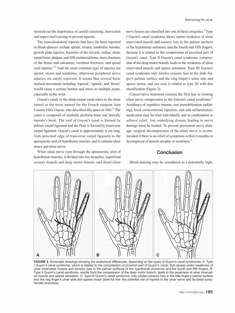
When ulnar nerve runs through the aponeurotic arch of hypothenar muscles, it divided into two branches, superficial sensory branch and deep motor branch, and distal ulnar
nerve lesions are classified into one of three categories.3) Type I Guyon’s canal syndrome shows motor weakness of ulnar innervated muscle and sensory loss in the palmar surfaces of the hypothenar eminence and the fourth and fifth fingers, because it is related to the compression of proximal part of Guyon’s canal. Type II Guyon’s canal syndrome, compres-sion of the deep motor branch, leads to the weakness of ulnar innervated muscle and spares sensation. Type III Guyon’s canal syndrome only involve sensory loss in the little fin-ger’s palmar surface and the ring finger’s ulnar side and spares motor, and our case is related to type III with this classification (Figure 2).
Conservative treatment remains the first line in treating ulnar nerve compression in the Guyon’s canal syndrome.7)
Avoidance of repetitive trauma, rest immobilization (splint-ing), local corticosteroid injection, and anti-inflammatory medication may be tried individually and in combination to achieve relief. Any underlying disease leading to nerve damage must be treated. To prevent permanent nerve dam-age, surgical decompression of the ulnar nerve is recom-mended if there is no relief of symptoms within 6 months or development of muscle atrophy or weakness.7)
Conclusion
Break-dancing may be considered as a potentially high-
FIGURE 2. Schematic drawings showing the anatomical differences, depending on the types of Guyon’s canal syndromes. A: Type I Guyon’s canal syndrome, which is related to the compression of proximal part of Guyon’s canal, that causes motor weakness of ulnar innervated muscle and sensory loss in the palmar surfaces of the hypothenar eminence and the fourth and fifth fingers. B: Type II Guyon’s canal syndrome, results from the compression of the deep motor branch, leads to the weakness of ulnar innervat-ed muscle and spares sensation. C: Type III Guyon’s canal syndrome, only causes sensory loss in the little finger’s palmar surface and the ring finger’s ulnar side and spares motor (bold full line: the potential site of injuries to the ulnar nerve and its distal symp-tomatic branches).
A B C

186 Korean J Neurotrauma 2015;11(2):183-186
Guyon Syndrome in ‘B Boy’
risk dancing sport. Since the wrist is the most commonly af-fected site of musculoskeletal injury in break-dancing, phy-sicians should be aware of the possibility of the peripheral nerve injury.
■ The authors have no financial conflicts of interest.
REFERENCES1) Cho CH, Song KS, Min BW, Lee SM, Chang HW, Eum DS.
Musculoskeletal injuries in break-dancers. Injury 40:1207-1211, 2009
2) Cho YJ, Cho SM, Sheen SH, Heo DH, Cho JH, Oh SM. Mini-mally invasive ulnar nerve decompression for cubital tunnel syn-drome. J Korean Neurotraumatol Soc 5:16-21, 2009
3) Duggal A, Anastakis DJ, Salonen D, Becker E. Compression of
the deep palmar branch of the ulnar nerve by a ganglion: a case re-port. Hand (N Y) 1:98-101, 2006
4) Kauther MD, Wedemeyer C, Wegner A, Kauther KM, von Knoch M. Breakdance injuries and overuse syndromes in ama-teurs and professionals. Am J Sports Med 37:797-802, 2009
5) Maroukis BL, Ogawa T, Rehim SA, Chung KC. Guyon canal: the evolution of clinical anatomy. J Hand Surg Am 40:560-565, 2015
6) Norman RA, Grodin MA. Injuries from break dancing. Am Fam Physician 30:109-112, 1984
7) Pecina MM, Krmpotic-Nemanie J, Markiewitz AD. Tunnel syn-dromes: peripheral nerve compression syndromes, ed 3. Boca Raton, FL: CRC Press, 2001
8) Toledo SD, Akuthota V, Drake DF, Nadler SF, Chou LH. Sports and performing arts medicine. 6. Issues relating to dancers. Arch Phys Med Rehabil 85(3 Suppl 1):S75-S78, 2004