type ii keratin cdnas from the rainbow trout: implications for keratin evolution
TRANSCRIPT

Differentiation (2002) 70:292–299 C Blackwell Verlag 2002
O R I G I N A L A R T I C L E
Michael Schaffeld ¡ Mark Haberkamp ¡ Erik BraziulisBernhard Lieb ¡ Jürgen Markl
Type II keratin cDNAs from the rainbow trout:implications for keratin evolution
Accepted in revised form: 2 May 2002
Abstract From a teleost fish, the rainbow trout On-corhynchus mykiss, we have cloned and sequencedcDNAs encoding five different type II keratins. The cor-responding protein spots, as separated by 2D-PAGE oftrout cytoskeletal preparations, have been identified bypeptide mass mapping using MALDI mass spec-trometry. Three of the sequenced keratins are expressedin the epidermis (subtype II), and two in simple epi-thelia and mesenchymal cells (subtype II). The II kera-tins are both orthologs of human K8. This leaves unse-quenced only the trace component S3 of the biochem-ically established trout keratin catalog. A phylogenetictree has been constructed from a multiple alignment ofthe rod domains of the new keratin sequences togetherwith type II sequences from other vertebrates such asshark, zebrafish, and human; lamprey K8 (recently se-quenced in our laboratory) has been used as outgroup.This tree suggests, in a highly bootstrap-supported man-ner, that the teleost II keratins diversified independentlyfrom the mammalian II keratins. In contrast, all thespecies investigated express K8-like keratins, suggestingthat the different II branches evolved from K8-like pro-genitors. The tree also indicates that the published ze-brafish sequences represent II keratins and that the bio-chemically identified K8 ortholog in zebrafish has notyet been sequenced.
Key words intermediate filaments ¡ keratins ¡cytoskeleton ¡ trout ¡ zebrafish ¡ evolution
M. Schaffeld ¡ M. Haberkamp ¡ E. Braziulis ¡ B. Lieb ¡J. Markl ( )Institute of Zoology, Johannes Gutenberg University,55099 Mainz, GermanyTel: π49 6131 392 2314, Fax: π49 6131 392 4652e-mail: markl/mail.uni-mainz.de
U.S. Copyright Clearance Center Code Statement: 0301–4681/2002/706–292 $ 15.00/0
Introduction
Cytoplasmic keratins (also termed ‘‘cytokeratins’’) areintermediate filament (IF) proteins and components ofthe cytoskeleton of vertebrate epithelial cells; in teleostfishes, they also occur in mesenchymal cell types (forreview see Markl and Schechter 1998). According totheir primary structures and assembly properties, type Iand type II keratins are distinguished; they form obli-gate heterodimers during filament assembly. Morespecifically, keratins expressed in epidermal keratino-cytes (‘‘E’’ keratins) are distinguished from keratins thatare typical for cells forming simple epithelia (‘‘S’’ kera-tins); the resulting keratin subtypes I, II, I, and IIhave been demonstrated to occur throughout the Ver-tebrata (Moll et al., 1982; Fouquet et al., 1988; Markl etal., 1989; Fuchs and Weber, 1994; Markl and Schechter,1998).
In human epithelial cells, eight different type II kera-tins have been detected and sequenced (six II keratins,termed here HsaK1 to HsaK6, and two II keratins,termed here HsaK7 and HsaK8); it should be noted,however, that the genetic diversity is higher in that, forexample, different K6 genes exist (Hesse et al., 2001). Ina striking contrast, the shark possesses only a single IIand II keratin (termed SstK1 and SstK8: Schaffeld etal., 1998). Their sequences are very distinct from thoseof human keratins, but SstK8 seems to be orthologousto HsaK8 as deduced from their similar expression pat-terns and from a common motif in the non-helical taildomain (Schaffeld et al., 1998). Several type II sequencesare also available from goldfish and zebrafish (Giordanoet al., 1989; 1990; Imboden et al., 1997; Ju et al., 1999;Chua and Lim, 2000), but the total pattern in both spe-cies is still somewhat incomplete. Our previous 2D-PAGE analysis of the rainbow trout cytoskeleton re-vealed the existence of more than half a dozen protein

293
Fig. 1 Two-dimensional polyacrylamide gel electrophoreses (2D-PAGE) of rainbow trout cytoskeletal proteins, extracted from cellline RTG-2 (a–d) and from RTG-2 plus skin (e). In the first dimen-sion, isoelectric focusing was done; in the second dimension, SDS-PAGE was applied; bovine serum albumin (B) and rabbit a-actin(A) were used as marker proteins. Note that S3 was almost invisiblein these gels (position indicated by arrow) (a) Coomassie Bluestained gel showing the entire set of trout ‘‘S’’ keratins. (b–d) Im-munoblots using monoclonal antibodies 164.4, 26.8, 79.14, respec-tively, which in Xenopus laevis are specific for keratin K8; noteexclusive staining of the type II keratin S1 in all three cases. (e)Coomassie Blue stained gel showing a comprehensive trout keratincatalog as obtained by co-electrophoresis of cytoskeletal proteinsfrom RTG-2 cells and skin (for designation of trout keratins, seealso Markl and Franke, 1988 and Markl et al., 1989). positionof actin, which was barely visible in this particular gel; arrowhead,putative degradation products of several keratins.
spots identified as II and II keratins, suggesting a kera-tin complexity in teleost fish comparable to that in mam-mals (Markl et al., 1989). The present study was de-signed to analyse, in the context of keratin evolution,whether or not direct correspondencies exist between in-dividual teleost and mammalian type II keratins and toprovide the first comprehensive view of the type II kera-tin system of a teleost fish. The rainbow trout type Ikeratins are addressed in an accompanying paper(Schaffeld et al., 2002, this issue).
Methods
Methods described in our accompanying paper (Schaffeld et al.,2002, this issue)
These include animals, RTG-2 cells, preparation of tissues andcytoskeletal proteins, two-dimensional polyacrylamide gel electro-phoresis (2D-PAGE), immunoblotting, MALDI-MS, cDNA clon-ing and libraries, sequence analysis and alignments, and phylogen-etic tree construction.
Applied antibodies, and immunofluorescene microscopy
We applied monoclonal antibodies 79.14, 164.4, and 26.8 recogniz-ing, in Xenopus laevis, ‘‘II’’ keratins (Fouquet, 1991); they wereprovided by Dr. Harald Herrmann (DKFZ, Heidelberg). In ad-dition, for cDNA library screening, we used guinea pig polyclonalantibodies raised against rainbow trout ‘‘S’’ keratins extracted fromRTG-2 cells (Markl et al., 1989). Indirect immunofluorescencemicroscopy using 5 mm thick cryostat tissue sections and RTG-2cells was performed as previously described (Markl and Franke,1988; Schaffeld et al., 1998). Texas Red-conjugated goat secondaryantibodies were obtained from Dianova (Hamburg, Germany).
In vitro transcription/translation of cDNA clones
This was performed according to the manufacturer’s instructions,using the TNTA Coupled Reticulocyte Lysate System in combi-nation with the TranscendTM Non-Radioactive Translation Detec-tion System (Promega, Mannheim/Germany). The biotinylated
Table 1 Reaction of three monoclonal anti-keratin antibodies (allspecific for keratin 8 in Xenopus laevis) on frozen tissue sections ofthe rainbow trout as deduced from immunofluorescence micro-scopy
Cell type mAb mAb mAb164.4 79.14 26.8
Hepatocytes and bile canaliculi π π πBlood vessel endothelial cells π π πBlood vessel muscle cells π π πInterstitial cells, fibroblasts π π πChondrocytes π π πScale-associated cells π π πOvary stroma and follicle cells π π πOptic nerve glial cells π π πMeninges (dura mater) cells π π πEye ciliary body cells π π πOcular lens epithelial cells π π ªRTG-2 cells π π ªIntestinal and rectal mucosa cells π π ªBile duct cells π π ªBasal cells of gill mucosa π π ªBasal cells of gill secondary lamellae π π ªGill pillar cells π π ªEpidermal Merkel cells π πEpidermal keratinocytes ª ª ªAll other cell types tested* ª ª ª
* Negative reaction includes tongue mucosa cells, neurons, sensorycells, CNS glial cells, ocular lens fiber cells, oocytes, blood cells,and all kinds of muscle cells except those surrounding blood ves-sels

294
Fig. 2 Immunofluorescencemicroscopy of cryostat sectionsof frozen trout tissues usingmonoclonal antibodiesagainst Xenopus laevis K8.(a–f, g’–l’) epifluorescence op-tics; (g–l) phase contrast op-tics. Antibodies 79.14 (a) and164.4 (b) on gill tissues, stainingblood vessel endothelia (E),chondrocytes (C), mucosa (M)and secondary lamellae (L).Antibodies 79.14 (c), 164.4 (d)and 26.8 (e) on liver tissues,showing positive reactions withhepatocytes (H), interstitialcells surrounding each triade,and the wall cells of artery (A),vein (V), and bile duct (B), asthe three triade components.(f) Antibody 164.4 on RTG-2cells, staining the network ofkeratin filaments. Antibodies79.14 (g, g’), 164.4 (h, h’), and26.8 (i, i’) on ovary tissues,showing positive reactions onstroma (St) and follicular cells(F), whereas oocytes (O) didnot react. (j, j’) Antibody 79.14on brain tissues, showing posi-tive reaction with endothelialcells of the blood capillariesand the meninges (M); nerveand glial cells were not stained.(k, k’) Antibody 79.14 on skintissues, staining dermal fibro-blasts (D) and scale associatedcells, whereas epidermal kera-tinocytes (K) remained un-stained. (l, l’) Antibody 164.4on spleen tissues, stainingstroma cells. Identical magni-fication in all cases; bar (in j),100 mm.
translation products were separated by 2D-PAGE and detectedusing the streptavidin/alkaline phosphatase reaction.
Results
Monoclonal antibodies reactive to trout keratins
In our previous studies, monoclonal antibody C10turned out to be a very useful marker for the trout typeII keratin S1 (Markl et al., 1989), but unfortunately,this antibody is no longer available. To find a substitute,we studied the reaction of three monoclonal antibodies
termed 79.14, 164.4, and 26.8 raised by Bernadette Fou-quet. In Xenopus laevis, these antibodies specifically rec-ognize keratin XK1/8 (Fouquet, 1991) which is an am-phibian ortholog of mammalian K8 (Franz et al., 1983;Franz and Franke, 1986;). 2D-PAGE immunoblots wereperformed on cytoskeletal proteins extracted from troutforehead epidermis (expressing the entire subset of Ekeratins), RTG-2 cells (expressing the entire subset of Skeratins), and intestine (containing a mixture of E andS keratins); the various keratin patterns in 2D-PAGEhave been described previously (Markl et al., 1989). Asshown in Fig. 1a–d, within the trout keratins separatedby 2D-PAGE (Fig. 1e), all three monoclonal antibodiesexclusively labeled the II keratin spot S1. In immuno-

295
Table 2 Properties of the trout type II keratins predicted from the cDNA sequences
Predicted Protein spots Expression Base Amino Mr (Da)** pI** Total number EMBLprotein in 2D-PAGE type pairs* acids** of matching Accessionsequence (Fig. 1e) masses. number(Fig. 3)
OmyK1 E1 (2 spots) II 2815 618 62,344 5.5 23 AJ272369OmyK2 E2 (2 spots) II 2755 592 60,150 5.2 15 AJ272370OmyK3 E3 II 1643 515 55,282 5.3 17 AJ315933OmyK8a S1 II 2280 538 59,103 5.3 12 AJ272373OmyK8b S2, Sx II 2176 537 58,977 5.1 33 X92522∂ OmyK8a/b S3 II ª ª ª ª 7/8 ª
* Total length of the cloned cDNA** Calculated from the protein sequence predicted from the open reading frame. Total number of masses which match the respective sequence (for details, see text)
∂ S3 is very similar to OmyK8a and OmyK8b, as deduced from peptide mass mapping
fluorescence microscopy, monoclonal antibodies 79.14and 164.4 specifically reacted with all those cell typesknown, from our previous studies (Markl and Franke,1988; Markl et al., 1989) to express S keratins (Table 1);examples are shown in Fig. 2. A different pattern wasobtained with monoclonal antibody 26.8, in that itstained only a fraction of the cell types recognized byantibodies 164.4 and 79.14 (Table 1; Fig. 2e,i,i’); appar-ently, its epitope is not accessible in all filaments contain-ing S1.
Sequencing trout type II keratins
By screening a cDNA library derived from RTG-2 cells(a rainbow trout gonadal cell line that exclusively con-tains ‘‘S’’ keratins: Markl et al., 1989) with monoclonalantibody 79.14, we isolated and sequenced a full-lengthclone (omyk8a) encoding a type II keratin; the predictedprotein sequence was termed OmyK8a (Table 2). Thissequence was assigned, by MALDI-MS, to protein spotS1 of the keratin catalog (Table 2; Fig. 1). A secondprotein sequence, which is similar to, but differs signifi-cantly from OmyK8a, was deduced from cDNA cloneomyk8b, selected by screening a trout spleen cDNA li-brary with a random primed cDNA probe from humanK8 (there is expression of ‘‘S’’ but not of ‘‘E’’ keratinsin spleen: Markl et al., 1989). This keratin sequence hasalready been shown in a previous paper (Schaffeld et al.,1998) and is termed here OmyK8b. It was assigned, byMALDI-MS, to protein spots S2 and Sx of the troutkeratin catalog (Fig. 1e and Table 2); according toMALDI-MS, S2 and Sx possess the same protein se-quence. S1 yielded four masses which also occur in atheoretical tryptic digest of OmyK8a but not ofOmyK8b. Vice versa, the peptide mass mapping of S2and Sx matched five and two masses, respectively, whichonly occur in a theoretical tryptic digest of OmyK8b butnot of OmyK8a. Moreover, the in vitro transcription/translation product of clone omyk8b comigrated with Sx
in 2D-PAGE (data not shown). Among the II keratinspots of the catalog, this leaves only the trace compo-nent S3 unsequenced. In MALDI-MS, spot S3 yieldedseven masses which matched OmyK8a as well asOmyK8b and one additional fragment which is specificfor OmyK8; moreover, several masses were obtainedfrom S3 that are specific for this keratin spot (Table 2).
By screening a cDNA library derived from trout fore-head skin with a random-primed cDNA probe ofomyk8b, we obtained two additional full-length clones(omyk1 and omyk2), each encoding a type II keratin,which we termed OmyK1 and OmyK2, respectively(Table 2). By screening the same library with guinea piganti-keratin antibodies, a third cDNA clone (omyk3) en-coding another complete type II keratin was isolated(OmyK3, Table 2). The keratin sequences OmyK1,OmyK2, and OmyK3 were identified by MALDI-MS asthe protein spots E1, E2, and E3 of the trout keratincatalog, respectively (Fig. 1e and Table 2). E1 yieldednine, E2 six, and E3 eight masses which are exclusivelycharacteristic for the respective sequence. Electrophor-etically, the situation is complicated in that E1 and E2indeed each occur as two spots (Fig. 1e). However, ac-cording to MALDI-MS, each twin spot represents asingle keratin (OmyK1 on the one hand and OmyK2 onthe other). For example, nine specific masses of OmyK1were found in the more basic E1 variant and seven ofthose in the more acidic variant.
The cDNA sequences are available in the EMBLdatabank (Table 2). A multiple alignment of the deducedprotein sequences is shown in Fig. 3, and a phylogenetictree derived from an alignment, which also includes se-quences from other species, is presented in Fig. 4.
Discussion
The numerous biochemically identified type II proteinsof the rainbow trout (Markl et al., 1989; see Fig. 1e)have now turned out to represent only six genuine kera-
296
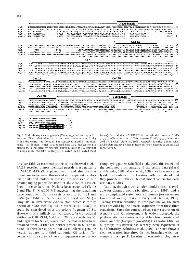
Fig. 3 Multiple sequence alignment (C_) of trout type II letters). It is similar (’’KRRF’’) in the zebrafish keratin DreK-keratins. Thick black lines mark the helical subdomains (coils) AF197909 (Chua and Lim, 2000), whereas DreKAF134850 is termin-within the central rod domain. The conserved motif in the non- ated by ‘‘SKRY’’ (Ju et al., 1999). Asterisks, identical amino acids;helical tail domain, which is proposed here as a marker for K8 double dots and single dots indicate different degrees of amino acidorthologs, is indicated by reversed staining. Note the C-terminal conservation.common motif ‘‘SRRF’’ in OmyK1, OmyK2, and OmyK3 (bold
tins (see Table 2) as several protein spots observed in 2D-PAGE revealed almost identical peptide mass patternsin MALDI-MS. (This phenomenon, and also possiblediscrepancies between theoretical and apparent isoelec-tric points and molecular masses, are discussed in ouraccompanying paper: Schaffeld et al., 2002, this issue).From these six keratins, five have been sequenced (Table2 and Fig. 3). MALDI-MS suggests that the remainingtrace component, S3, is closely related to both S1 andS2/Sx (see Table 2). As S3 is co-expressed with S1 ( ΩOmyK8a) in liver tissue cytoskeleton, which is totallydevoid of S2/Sx (see Fig. 4d in Markl et al., 1989), itcould be considered as a proteolytic fragment of S1.However, this is unlikely for two reasons: (i) Monoclonalantibodies C10, 79.14, 164.4, and 26.8 are specific for S1and negative for S3; (ii) several peptide masses have beenobtained from S3 that are neither present in S1 nor inS2/Sx. It therefore appears that S3 is indeed a genuinekeratin, apparently a third salmonid K8 variant. To-gether with the six type I keratin sequences (see our ac-
companying paper: Schaffeld et al., 2002, this issue) andthe combined biochemical and expression data (Markland Franke, 1988; Markl et al., 1989), we have now ana-lysed the rainbow trout keratins with such detail thatthey provide an efficient teleost model system for evol-utionary studies.
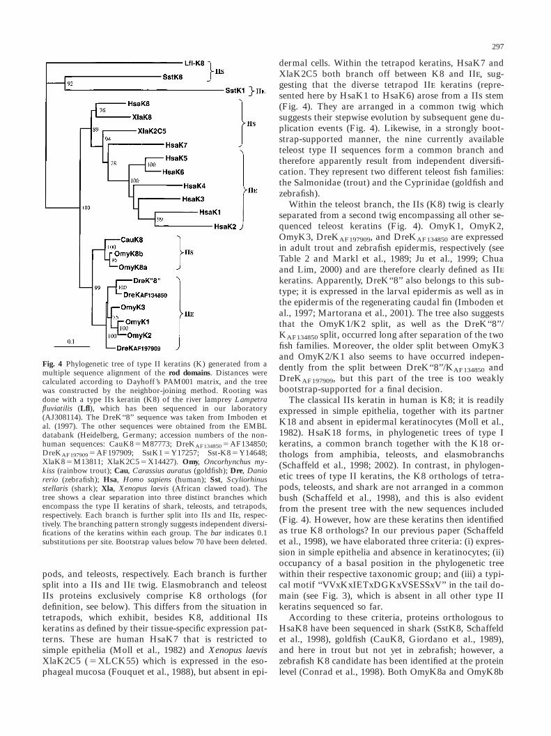
Another, though much simpler, model system is avail-able for elasmobranchs (Schaffeld et al., 1998), and amore complicated system exists in human (for review seeFuchs and Weber, 1994 and Parry and Steinert, 1999).Tracing keratin evolution is now possible on the firmbasis provided by the keratin sequences from these threeorganisms. Since the concept of an early separation ofAgnatha and Gnathostomata is widely accepted, thephylogenetic tree shown in Fig. 4 has been constructedusing lamprey (Lampetra fluviatilis) II keratin LflK8 asoutgroup; this keratin has recently been sequenced inour laboratory (Schultess et al., 2001). The tree shows aclear separation into three distinct branches which en-compass the type II keratins of elasmobranchs, tetra-
297
Fig. 4 Phylogenetic tree of type II keratins (K) generated from amultiple sequence alignment of the rod domains. Distances werecalculated according to Dayhoff ’s PAM001 matrix, and the treewas constructed by the neighbor-joining method. Rooting wasdone with a type II keratin (K8) of the river lamprey Lampetrafluviatilis (Lfl), which has been sequenced in our laboratory(AJ308114). The DreK‘‘8’’ sequence was taken from Imboden etal. (1997). The other sequences were obtained from the EMBLdatabank (Heidelberg, Germany; accession numbers of the non-human sequences: CauK8 ΩM87773; DreKAF134850 ΩAF134850;DreKAF197909 ΩAF197909; SstK1ΩY17257; Sst-K8ΩY14648;XlaK8 ΩM13811; XlaK2C5ΩX14427). Omy, Oncorhynchus my-kiss (rainbow trout); Cau, Carassius auratus (goldfish); Dre, Daniorerio (zebrafish); Hsa, Homo sapiens (human); Sst, Scyliorhinusstellaris (shark); Xla, Xenopus laevis (African clawed toad). Thetree shows a clear separation into three distinct branches whichencompass the type II keratins of shark, teleosts, and tetrapods,respectively. Each branch is further split into II and II, respec-tively. The branching pattern strongly suggests independent diversi-fications of the keratins within each group. The bar indicates 0.1substitutions per site. Bootstrap values below 70 have been deleted.
pods, and teleosts, respectively. Each branch is furthersplit into a II and II twig. Elasmobranch and teleostII proteins exclusively comprise K8 orthologs (fordefinition, see below). This differs from the situation intetrapods, which exhibit, besides K8, additional IIkeratins as defined by their tissue-specific expression pat-terns. These are human HsaK7 that is restricted tosimple epithelia (Moll et al., 1982) and Xenopus laevisXlaK2C5 ( Ω XLCK55) which is expressed in the eso-phageal mucosa (Fouquet et al., 1988), but absent in epi-
dermal cells. Within the tetrapod keratins, HsaK7 andXlaK2C5 both branch off between K8 and II, sug-gesting that the diverse tetrapod II keratins (repre-sented here by HsaK1 to HsaK6) arose from a II stem(Fig. 4). They are arranged in a common twig whichsuggests their stepwise evolution by subsequent gene du-plication events (Fig. 4). Likewise, in a strongly boot-strap-supported manner, the nine currently availableteleost type II sequences form a common branch andtherefore apparently result from independent diversifi-cation. They represent two different teleost fish families:the Salmonidae (trout) and the Cyprinidae (goldfish andzebrafish).
Within the teleost branch, the II (K8) twig is clearlyseparated from a second twig encompassing all other se-quenced teleost keratins (Fig. 4). OmyK1, OmyK2,OmyK3, DreKAF197909, and DreKAF134850 are expressedin adult trout and zebrafish epidermis, respectively (seeTable 2 and Markl et al., 1989; Ju et al., 1999; Chuaand Lim, 2000) and are therefore clearly defined as IIkeratins. Apparently, DreK‘‘8’’ also belongs to this sub-type; it is expressed in the larval epidermis as well as inthe epidermis of the regenerating caudal fin (Imboden etal., 1997; Martorana et al., 2001). The tree also suggeststhat the OmyK1/K2 split, as well as the DreK‘‘8’’/KAF134850 split, occurred long after separation of the twofish families. Moreover, the older split between OmyK3and OmyK2/K1 also seems to have occurred indepen-dently from the split between DreK‘‘8’’/KAF134850 andDreKAF197909, but this part of the tree is too weaklybootstrap-supported for a final decision.
The classical II keratin in human is K8; it is readilyexpressed in simple epithelia, together with its partnerK18 and absent in epidermal keratinocytes (Moll et al.,1982). HsaK18 forms, in phylogenetic trees of type Ikeratins, a common branch together with the K18 or-thologs from amphibia, teleosts, and elasmobranchs(Schaffeld et al., 1998; 2002). In contrast, in phylogen-etic trees of type II keratins, the K8 orthologs of tetra-pods, teleosts, and shark are not arranged in a commonbush (Schaffeld et al., 1998), and this is also evidentfrom the present tree with the new sequences included(Fig. 4). However, how are these keratins then identifiedas true K8 orthologs? In our previous paper (Schaffeldet al., 1998), we have elaborated three criteria: (i) expres-sion in simple epithelia and absence in keratinocytes; (ii)occupancy of a basal position in the phylogenetic treewithin their respective taxonomic group; and (iii) a typi-cal motif ‘‘VVxKxIETxDGKxVSESSxV’’ in the tail do-main (see Fig. 3), which is absent in all other type IIkeratins sequenced so far.
According to these criteria, proteins orthologous toHsaK8 have been sequenced in shark (SstK8, Schaffeldet al., 1998), goldfish (CauK8, Giordano et al., 1989),and here in trout but not yet in zebrafish; however, azebrafish K8 candidate has been identified at the proteinlevel (Conrad et al., 1998). Both OmyK8a and OmyK8b

298
are orthologs of human K8. As deduced from their oc-currence in all trout individuals from different sourcesstudied by us so far, OmyK8a and OmyK8b representdistinct K8 isoforms rather than different alleles of thesame keratin. They separated from each other well afterthe salmonid-cyprinid split, as suggested from the posi-tion of goldfish CauK8 in the tree (Fig. 4). The threesequenced zebrafish type II proteins (Imboden et al.,1997; Ju et al., 1999; Chua and Lim, 2000) are certainlynot K8 orthologs, but are II keratins as discussedabove. However, they share more rod sequence identitieswith HsaK8 (average 77 – 78 %) than with any other hu-man keratin (average 60 – 70 %). Apparently, the latterhave undergone drastic changes which are also evidentfrom their rather long branches (see Fig. 4). In otherwords, the molecular clock of tetrapod II keratins runsat a higher rate compared to teleost II keratins whichresemble, in this respect, teleost and tetrapod K8. There-fore, in database searches, any teleost type II sequencewill match with tetrapod K8 rather than with any ofthe tetrapod II keratins. As a practical consequence,DreK‘‘8’’ has been repeatedly interpreted as the K8 or-tholog in zebrafish (Imboden et al., 1997; Martorana etal., 2001), which according to our data has not beenconfirmed (see Fig. 4); this serves to underline the im-portance of accurately constructing the phylogenetic treeof the keratin family.
OmyK8a and OmyK8b are equally distant phylogen-etically from human K8 (76.9 versus 77.6 % rod se-quence identity), and both carry the typical K8 tail mo-tif (see Fig. 3). However, there are arguments that Omy-K8a plays more general functional roles than OmyK8b:(i) Liver cytoskeleton, which in mammals is a classicalsource of K8, in trout is completely devoid of S2/Sx butcontains S1 (see Fig. 4d,e in Markl et al., 1989). Thismeans that S2/Sx is absent not only in hepatocytes butalso in (liver) blood vessel endothelia and bile duct cellssince both are reactive to the four K8 antibodies men-tioned above (Table 1 and Fig. 2). (ii) Cytoskeletal prep-arations rich in connective tissues are rich in S2/Sx; thisis clearly shown in the fibroblastoid cell line RTG-2 (seeFig. 1a) but also in intestine, gill, or dermis (see Fig.4a,f,g in Markl et al., 1989). In this context, it should bementioned that in teleost fishes, the keratin expressionpatterns differ fundamentally from those described fortetrapods, in that many mesenchymally derived celltypes and also optic nerve glial cells express keratins(Markl and Franke, 1988). Taking all available data to-gether, we conclude that simple epithelia which in tetra-pods express K8 in trout express S1 (OmyK8a), whereasS2/Sx (OmyK8b) is a typical mesenchymal keratin inthat it occurs in cell types which in tetrapods would ex-clusively express vimentin. However, whether OmyK8bcould indeed serve as a true marker for mesenchymalkeratin in trout remains to be investigated.
According to the present data, all attempts to assignthe human II system to the fish II system are obsolete
because they did radiate independently. In a parallelstudy on the trout I keratins, a similar situation hasbeen revealed. But there is one striking difference: Tele-ost and tetrapod I keratins represent a sister group ofthe K18 orthologs which form a discrete bush that alsoincludes SstK18 from shark (accompanying paper:Schaffeld et al., 2002, this issue). In contrast, the presenttree suggests that the II keratins repeatedly evolvedfrom K8-like progenitors rather than from an ancientII root because the first step in all three branches is asplit between K8 and the other keratins (Fig. 4). Thissituation has interesting perspectives: The skin of elas-mobranchs, teleosts, amphibia, and mammals is verydifferent (Leake, 1975). It could well be that the evolu-tion of these skin types was the driving force for theevolution of different subsets of epidermal keratins, eachproviding a specific arrangement of features such as theglycine-rich loops. These repetitive glycine-rich motifs inthe head and tail domain of tetrapod epidermal keratinshave been proposed to interact with the protein loricrinof the cell membrane, which also contains glycine loopsequences. These inter- and intramolecular glycine loopinteractions are supposed to contribute substantially tothe mechanically flexible, extensible, and elastic struc-ture of tetrapod epidermis (for details see Hohl et al.,1991; Steinert et al., 1991; Parry and Steinert, 1999). Theglycine loop motifs are also present in teleost ‘‘E’’ kera-tins (see Fig. 3 and Schaffeld et al., 1998) but almostabsent in SstK1, which represents the single type IIkeratin in shark (Schaffeld et al., 1998). In shark, theepidermal keratinocytes are largely protected from ex-ternal mechanical stress by placoid scales. Also, SstK8and HsaK8 lack repetitive glycine-rich motifs which,however, are present in K8 from teleosts and Xenopuslaevis. It should be considered whether, in freshwaterfishes and amphibians, simple epithelia require specialreinforcement to combat osmotic stress. Evidence that,in vivo, epidermal and simple keratins may contribute todifferent mechanical properties and cannot simply re-place each other has already been shown by Hutton etal. (1998). Another interesting aspect is the functionalrole of the strictly conserved tail motif in K8 which isabsent in all other type II keratins and has probablybeen repeatedly abandoned. Thus, comparative func-tional analyses of the orthologous ‘‘S’’ keratins, and theconvergently evolved ‘‘E’’ keratins, in fish and manmight reveal generalized structure-function relationshipsof IF proteins.
Acknowledgements We thank Michaela Münik for cloning omyk8b,Dr. Bernadette Fouquet and Dr. Harald Herrmann for providingthe monoclonal antibodies used in this study, Thomas Schubert forexcellent technical assistance, and Dr. J. Robin Harris for polishingthe English. This work has been supported by the DFG (Ma 843/5–1).
299
References
Chua, K.L. and Lim, T.M. (2000) Type I and type II cytokeratincDNAs from the zebrafish (Danio rerio) and expression patternsduring early development. Differentiation 66:31–41.
Conrad, M., Lemb, K., Schubert, T. and Markl, J. (1998) Biochem-ical identification and tissue-specific expression patterns of kera-tins in the zebrafish Danio rerio. Cell Tissue Res 293:195–205.
Fouquet, B. (1991) Expression von Intermediärfilament-Proteinenin Xenopus laevis. Dissertation Thesis Faculty of Biology, Heidel-berg, pp 1–175.
Fouquet, B., Herrmann, H., Franz, J.K. and Franke, W.W. (1988)Expression of intermediate filament proteins during develop-ment of Xenopus laevis. III. Identification of mRNAs encodingcytokeratins typical of complex epithelia. Development 104:533–548.
Franz, J.K. and Franke, W.W. (1986) Cloning of cDNA and aminoacid sequence of a cytokeratin expressed in oocytes of Xenopuslaevis. Proc Natl Acad Sci USA 83:6475–6479.
Franz, J.K., Gall, J., Williams, M.A., Picheral, B. and Franke,W.W. (1983) Intermediate-size filaments in a germ cell: expres-sion of cytokeratins in oocytes and eggs of the frog Xenopusleavis. Proc Natl Acad Sci USA 80:6254–6258.
Fuchs, E. and Weber, K. (1994) Intermediate filaments: Structure,dynamics, function and disease. Annu Rev Biochem 63:345–382.
Giordano, S., Glasgow, E., Tesser, P. and Schechter, N. (1989) Atype II keratin is the major intermediate filament protein ex-pressed in glial cells of the goldfish visual pathway: molecularcloning and sequence analysis. Neuron 2:1507–1516.
Giordano, S., Hall, C., Quitschke, W., Glasgow, E. and Schechter,N. (1990) Keratin 8 of simple epithelia is expressed in glia of thegoldfish nervous system. Differentiation 44:163–172.
Hesse, M., Magin, T.M. and Weber, K. (2001) Genes for intermedi-ate filament proteins and the draft sequence of the human ge-nome: novel keratin genes and a surprisingly high number ofpseudogenes related to keratin genes 8 and 18. J Cell Sci114:2569–2575.
Hohl, D., Mehrel, T., Lichti, U., Turner, M.L., Roop, D.R. andSteinert, P.M. (1991) Characterization of human loricrin. Struc-ture and function of a new class of epidermal cell envelope pro-teins. J Biol Chem 226:6626–6636.
Hutton, E., Paladini, R.D., Yu, Q.C., Yen, M., Coulombe, P.A.and Fuchs, E. (1998) Functional differences between keratins ofstratified and simple epithelia. J Cell Biol 143:487–499.
Imboden, M., Goblet, C., Korn, H. and Vriz, S. (1997) Cytokeratin8 is a suitable epidermal marker during zebrafish development.C R Acad Sci III 320:689–700.
Ju, B., Xu, Y., He, J., Liao, J., Yan, T., Hew, C.L., Lam, T.J. andGong, Z. (1999) Faithful expression of green fluorescent protein(GFP) in transgenic zebrafish embryos under control of zebrafishgene promoters. Dev Genet 25:158–167.
Leake, L.D. (1975) Comparative Histology. New York Oxford Uni-versity Press, London, Toronto.
Markl, J. and Franke, W.W. (1988) Localization of cytokeratins intissues of the rainbow trout: fundamental differences in expres-sion pattern between fish and higher vertebrates. Differentiation39:97–122.
Markl, J. and Schechter, N. (1998) Fish intermediate filament pro-teins in structure, function and evolution. In: Herrmann, H. andHarris, J.R. (eds) Intermediate Filaments. Plenum Press, New,York, pp 1–33.
Markl, J., Winter, S. and Franke, W.W. (1989) The catalog and theexpression complexity of cytokeratins in a teleost fish, the rain-bow trout. Eur J Cell Biol 50:1–16.
Martorana, M.L., Tawk, M., Lapointe, T., Barre, N., Imboden,M., Joulie, C., Geraudie, J. and Vriz, S. (2001) Zebrafish keratin8 is expressed at high levels in the epidermis of regenerating cau-dal fin. Int J Dev Biol 45:449–452.
Moll, R., Franke, W.W., Schiller, D.L., Geiger, B. and Krepler, R.(1982) The catalog of human cytokeratins: patterns of expressionin normal epithelia, tumors and cultured cells. Cell 31:11–24.
Parry, D.A. and Steinert, P.M. (1999) Intermediate filaments: mol-ecular architecture, assembly, dynamics and polymorphism. QRev Biophys 32:99–187.
Schaffeld, M., Höffling, S., M., H., Conrad, M. and Markl, J.(2002) Type I keratin cDNAs from the rainbow trout: indepen-dent radiation of keratins in fish. Differentiation 70:282–291.
Schaffeld, M., Löbbecke, A., Lieb, B. and Markl, J. (1998) Tracingkeratin evolution: Catalog, expression patterns and primarystructure of shark (Scyliorhinus stellaris) keratins. Eur J Cell Biol77:69–80.
Schultess, J., Schaffeld, M. and Markl, J. (2001) Intermediatefilament protein sequences of the cyclostome Lampetra fluviatilis.Biol Cell 93: First Joint French-German Congress on Cell Bi-ology: 235.
Steinert, P.M., Mack, J.W., Korge, B.P., Gan, S.Q., Haynes, S.R.and Steven, A.C. (1991) Glycine loops in proteins: their occur-rence in certain intermediate filament chains, loricrins andsingle-stranded RNA binding proteins. Int J Biol Macromol13:130–139.