two phosphoglucomutase paralogs facilitate ionophore ... · sarcocystis neurona) have both...
TRANSCRIPT
Two Phosphoglucomutase ParalogsFacilitate Ionophore-Triggered Secretionof the Toxoplasma Micronemes
Sudeshna Saha,a Bradley I. Coleman,a Rashmi Dubey,a Ira J. Blader,b
Marc-Jan Gubbelsa
Department of Biology, Boston College, Chestnut Hill, Massachusetts, USAa; Department of Microbiology andImmunology, University at Buffalo School of Medicine, Buffalo, New York, USAb
ABSTRACT Paralogs of the widely prevalent phosphoglucomutase (PGM) proteincalled parafusin function in calcium (Ca2�)-mediated exocytosis across eukaryotes. InToxoplasma gondii, the parafusin-related protein 1 (PRP1) has been associated withCa2�-dependent microneme organelle secretion required for essential processes likehost cell invasion and egress. Using reverse genetics, we observed PRP1 to be dis-pensable for completion of the lytic cycle, including host cell invasion and egress bythe parasite. However, the absence of the gene affected increased microneme re-lease triggered by A23187, a Ca2� ionophore used to raise the cytoplasmic Ca2�
concentration mimicking the physiological role of Ca2� during invasion and egress.The basal levels of constitutive microneme release in extracellular parasites andphosphatidic acid-triggered microneme secretion were unaffected in the mutant.The phenotype of the deletion mutant of the second PGM-encoding gene in Toxo-plasma, PGM2, was similar to the phenotype of the PRP1 deletion mutant. Further-more, the ability of the tachyzoites to induce acute infection in the mice remainednormal in the absence of both PGM paralogs. Our data thus reveal that the mi-croneme secretion upon high Ca2� flux is facilitated by the Toxoplasma PGM paral-ogs, PRP1 and PGM2. However, this protein-mediated release is neither essential forlytic cycle completion nor for acute virulence of the parasite.
IMPORTANCE Ca2�-dependent exocytosis is essential for the life cycle of apicompl-exan parasites. Toxoplasma gondii harbors a phosphoglucomutase (PGM) ortholog,PRP1, previously associated with Ca2�-dependent microneme secretion. Here it isshown that genetic deletion of either PRP1, its PGM2 ortholog, or both genes is dis-pensable for the parasite’s lytic cycle, including host cell egress and invasion. Deple-tion of the proteins abrogated high Ca2�-mediated microneme secretion induced bythe ionophore A23187; however, the constitutive and phosphatidic acid-mediatedrelease remained unaffected. Secretion mediated by the former pathway is not es-sential for tachyzoite survival or acute in vivo infection in the mice.
KEYWORDS PRP1, Toxoplasma gondii, calcium, micronemes, parafusin,phosphoglucomutase
The apicomplexan parasite Toxoplasma gondii has infected one in every threehumans globally with most acute infections being mild or asymptomatic (1).
However, severe disease occurs congenitally in pregnant women (2) and in immuno-compromised patients (e.g., AIDS patients) where reactivation of a chronic infection canresult in life-threatening encephalitis or myocarditis (3). The clinical manifestation is theconsequence of tissue destruction resulting from the repetitive cycles of active inva-sion, intracellular replication, and egress that make up the lytic cycle of the parasite. Asa result, host cell invasion is a critical event in the pathogenesis of this obligate parasite(4). Unlike some other intracellular pathogens, the invasion by Toxoplasma is a parasite-
Received 2 November 2017 Accepted 5November 2017 Published 29 November2017
Citation Saha S, Coleman BI, Dubey R, Blader IJ,Gubbels M-J. 2017. Two phosphoglucomutaseparalogs facilitate ionophore-triggeredsecretion of the Toxoplasma micronemes.mSphere 2:e00521-17. https://doi.org/10.1128/mSphere.00521-17.
Editor Aaron P. Mitchell, Carnegie MellonUniversity
Copyright © 2017 Saha et al. This is an open-access article distributed under the terms ofthe Creative Commons Attribution 4.0International license.
Address correspondence to Marc-Jan Gubbels,[email protected].
RESEARCH ARTICLEHost-Microbe Biology
crossm
November/December 2017 Volume 2 Issue 6 e00521-17 msphere.asm.org 1
on Novem
ber 9, 2018 by guesthttp://m
sphere.asm.org/
Dow
nloaded from
directed event requiring its motility and sequential release of three secretory organelles(5, 6). The secretion of the first organelle, the micronemes, is regulated by intracellularcalcium (Ca2�) fluxes (7–9); however, the molecular mechanism underlying this regu-lation, especially the final events facilitating exocytosis or egress, is still incompletelyunderstood.
The phosphoglucomutase (PGM) family comprises enzymes that interconvertglucose-1-phosphate and glucose-6-phosphate, thereby linking the cytosolic processesof glycogenolysis and glycolysis. However, a PGM ortholog known as parafusin, orPFUS, is present in many eukaryotes, including ciliates, yeast Saccharomyces cerevisiae,and humans, and functions in Ca2�-mediated signaling (10–14). The Toxoplasmagenome encodes two PGM paralogs: PGM1 (accession no. TGME49_285980), alsoreferred to as parafusin-related protein 1 (PRP1), and PGM2 (TGME49_318580) (15). Inthe ciliate Paramecium, PFUS has been associated with Ca2�-mediated exocytosis ofdense core secretory vesicles (DCSVs) where it forms a scaffold on the vesicles and isinvolved in membrane fusion (16, 17, 18). Furthermore, PFUS has a role in DCSVassembly in both Paramecium (19) and Tetrahymena (20). Mechanistically, PFUS ishypothesized to impact the localized release of the matured DCSVs (17) throughtransient Ca2�-dependent phosphorylation during DCSV exocytosis; in its phosphory-lated state, PFUS associates with the vesicles closest to the plasma membrane, primingthem for secretion. For actual membrane fusion to occur, PFUS has to be dephospho-rylated through the protein phosphatase calcineurin (17).
DCSV release in ciliates has been related to Ca2�-mediated microneme secretion inapicomplexan parasites (21). Germane to Toxoplasma, the ortholog PRP1 has beenshown to localize to the most apical micronemes, and its phosphorylation status hasbeen suggested to be Ca2� dependent. Furthermore, through heterologous evaluationof PRP1 in Paramecium, an orthologous function in Ca2�-mediated microneme secre-tion has been proposed (22, 23). However, we recently reported that micronemesecretion itself is independent of calcineurin (24), thereby suggesting an incompletemechanistic orthology between ciliate DCSV release and microneme secretion inToxoplasma.
Here we directly evaluated the roles of PRP1 and PGM2 through gene deletions inToxoplasma. We show that both paralogs are required for microneme secretion inresponse to a high Ca2� concentration stimulated by an ionophore, which mimics thecellular status during host cell invasion and egress. However, both paralogs aredispensable for constitutive microneme secretion accompanying gliding motility be-tween host cells. Strikingly, tachyzoites devoid of the paralogs are completely viable invitro and during acute mouse infections. These data suggest that the Toxoplasma PGMsare dispensable for Ca2�-dependent exocytosis and for the successful completion ofthe lytic cycle.
RESULTSPRP1 is conserved across the coccidia. To establish whether PRP1 has a universal
role in exocytosis across the Apicomplexa, we first explored the conservation andphylogeny of phosphoglucomutases (PGMs) in apicomplexans and their closely relatedfree-living relatives, the chromerids. Both PRP1 (or Toxoplasma gondii PGM1 [TgPGM1])and TgPGM2 were compared to the validated PGM1/parafusins in ciliates and selectedcrown eukaryotes (Fig. 1). Consistent with the observation that PGM gene duplicationoccurred early in the emergence of the eukaryotic cell (14), these results show that allPGM1 sequences cluster together and are uniquely distinct from PGM2. The conserva-tion of the PGMs also varied among different apicomplexan subgroups. Toxoplasmaand the other cyst-forming coccidia (Hammondia hammondi, Neospora caninum, andSarcocystis neurona) have both isoforms, while the non-cyst-forming coccidia, e.g.,Eimeria spp., have only the PGM1 ortholog. Members of the genus Plasmodium haveonly PGM2, whereas Cryptosporidium spp. and the related Gregarina niphandrodes havePGM1; in fact, Cryptosporidium parvum and Cryptosporidium muris have two slightlydifferent PGM1 isotypes encoded by tandem genes in the genome. We did not find any
Saha et al.
November/December 2017 Volume 2 Issue 6 e00521-17 msphere.asm.org 2
on Novem
ber 9, 2018 by guesthttp://m
sphere.asm.org/
Dow
nloaded from
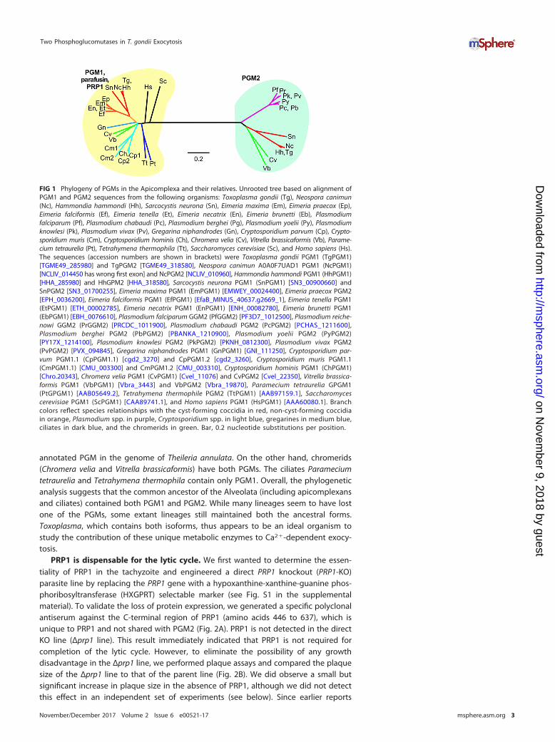
annotated PGM in the genome of Theileria annulata. On the other hand, chromerids(Chromera velia and Vitrella brassicaformis) have both PGMs. The ciliates Parameciumtetraurelia and Tetrahymena thermophila contain only PGM1. Overall, the phylogeneticanalysis suggests that the common ancestor of the Alveolata (including apicomplexansand ciliates) contained both PGM1 and PGM2. While many lineages seem to have lostone of the PGMs, some extant lineages still maintained both the ancestral forms.Toxoplasma, which contains both isoforms, thus appears to be an ideal organism tostudy the contribution of these unique metabolic enzymes to Ca2�-dependent exocy-tosis.
PRP1 is dispensable for the lytic cycle. We first wanted to determine the essen-tiality of PRP1 in the tachyzoite and engineered a direct PRP1 knockout (PRP1-KO)parasite line by replacing the PRP1 gene with a hypoxanthine-xanthine-guanine phos-phoribosyltransferase (HXGPRT) selectable marker (see Fig. S1 in the supplementalmaterial). To validate the loss of protein expression, we generated a specific polyclonalantiserum against the C-terminal region of PRP1 (amino acids 446 to 637), which isunique to PRP1 and not shared with PGM2 (Fig. 2A). PRP1 is not detected in the directKO line (Δprp1 line). This result immediately indicated that PRP1 is not required forcompletion of the lytic cycle. However, to eliminate the possibility of any growthdisadvantage in the Δprp1 line, we performed plaque assays and compared the plaquesize of the Δprp1 line to that of the parent line (Fig. 2B). We did observe a small butsignificant increase in plaque size in the absence of PRP1, although we did not detectthis effect in an independent set of experiments (see below). Since earlier reports
FIG 1 Phylogeny of PGMs in the Apicomplexa and their relatives. Unrooted tree based on alignment ofPGM1 and PGM2 sequences from the following organisms: Toxoplasma gondii (Tg), Neospora canimun(Nc), Hammondia hammondi (Hh), Sarcocystis neurona (Sn), Eimeria maxima (Em), Eimeria praecox (Ep),Eimeria falciformis (Ef), Eimeria tenella (Et), Eimeria necatrix (En), Eimeria brunetti (Eb), Plasmodiumfalciparum (Pf), Plasmodium chabaudi (Pc), Plasmodium berghei (Pg), Plasmodium yoelii (Py), Plasmodiumknowlesi (Pk), Plasmodium vivax (Pv), Gregarina niphandrodes (Gn), Cryptosporidium parvum (Cp), Crypto-sporidium muris (Cm), Cryptosporidium hominis (Ch), Chromera velia (Cv), Vitrella brassicaformis (Vb), Parame-cium tetraurelia (Pt), Tetrahymena thermophila (Tt), Saccharomyces cerevisiae (Sc), and Homo sapiens (Hs).The sequences (accession numbers are shown in brackets) were Toxoplasma gondii PGM1 (TgPGM1)[TGME49_285980] and TgPGM2 [TGME49_318580], Neospora canimun A0A0F7UAD1 PGM1 (NcPGM1)[NCLIV_014450 has wrong first exon] and NcPGM2 [NCLIV_010960], Hammondia hammondi PGM1 (HhPGM1)[HHA_285980] and HhGPM2 [HHA_318580], Sarcocystis neurona PGM1 (SnPGM1) [SN3_00900660] andSnPGM2 [SN3_01700255], Eimeria maxima PGM1 (EmPGM1) [EMWEY_00024400], Eimeria praecox PGM2[EPH_0036200], Eimeria falciformis PGM1 (EfPGM1) [EfaB_MINUS_40637.g2669_1], Eimeria tenella PGM1(EtPGM1) [ETH_00002785], Eimeria necatrix PGM1 (EnPGM1) [ENH_00082780], Eimeria brunetti PGM1(EbPGM1) [EBH_0076610], Plasmodium falciparum GGM2 (PfGGM2) [PF3D7_1012500], Plasmodium reiche-nowi GGM2 (PrGGM2) [PRCDC_1011900], Plasmodium chabaudi PGM2 (PcPGM2) [PCHAS_1211600],Plasmodium berghei PGM2 (PbPGM2) [PBANKA_1210900], Plasmodium yoelii PGM2 (PyPGM2)[PY17X_1214100], Plasmodium knowlesi PGM2 (PkPGM2) [PKNH_0812300], Plasmodium vivax PGM2(PvPGM2) [PVX_094845], Gregarina niphandrodes PGM1 (GnPGM1) [GNI_111250], Cryptosporidium par-vum PGM1.1 (CpPGM1.1) [cgd2_3270] and CpPGM1.2 [cgd2_3260], Cryptosporidium muris PGM1.1(CmPGM1.1) [CMU_003300] and CmPGM1.2 [CMU_003310], Cryptosporidium hominis PGM1 (ChPGM1)[Chro.20343], Chromera velia PGM1 (CvPGM1) [Cvel_11076] and CvPGM2 [Cvel_22350], Vitrella brassica-formis PGM1 (VbPGM1) [Vbra_3443] and VbPGM2 [Vbra_19870], Paramecium tetraurelia GPGM1(PtGPGM1) [AAB05649.2], Tetrahymena thermophile PGM2 (TtPGM1) [AAB97159.1], Saccharomycescerevisiae PGM1 (ScPGM1) [CAA89741.1], and Homo sapiens PGM1 (HsPGM1) [AAA60080.1]. Branchcolors reflect species relationships with the cyst-forming coccidia in red, non-cyst-forming coccidiain orange, Plasmodium spp. in purple, Cryptosporidium spp. in light blue, gregarines in medium blue,ciliates in dark blue, and the chromerids in green. Bar, 0.2 nucleotide substitutions per position.
Two Phosphoglucomutases in T. gondii Exocytosis
November/December 2017 Volume 2 Issue 6 e00521-17 msphere.asm.org 3
on Novem
ber 9, 2018 by guesthttp://m
sphere.asm.org/
Dow
nloaded from

reported PRP1 involvement in microneme secretion, we examined the functioning oftwo essential parasitic processes relying on microneme secretion: host cell egress andinvasion. We studied both highly elevated Ca2�-dependent and phosphatidic acid(PA)-dependent branches underlying microneme secretion (25). To induce the Ca2�-dependent branch, we utilized the A23187 Ca2� ionophore, and to induce the PA-dependent branch, we added propranolol, which activates diacylglycerol kinase 1(DGK1) and thereby raises the PA concentration (an overview of signaling pathways andsecretagogue targets is provided in Fig. 7A). We did not observe any difference inegress efficiency compared to the RHΔku80 parent line for any of the conditions tested,suggesting that micronemes are secreted sufficiently to support egress (Fig. 2C). We didnot observe any difference in invasion efficiencies, indicating that secretion of therhoptries is also likely to be unaffected in the absence of PRP1 (Fig. 2D). We directlymonitored rhoptry secretion by detecting phosphorylated STAT3 (P-STAT3) accumula-tion in the infected-host cell nucleus, as STAT3 is phosphorylated by the rhoptry proteinROP16 (26), and we did not observe any difference in accumulation of P-STAT3between parent and Δprp1 lines (Fig. S2). On the basis of these results, we concludethat PRP1 is not essential for either microneme or rhoptry secretion and is not requiredfor completing the Toxoplasma lytic cycle in vitro.
Loss of PRP1 reduces Ca2�-induced microneme secretion. To assess whether theabsence of PRP1 more subtly affected signaling events leading to microneme secretion,we used pharmacological triggers (secretagogues) acting on distinct steps in the signaltransduction pathway. The current understanding of the signaling pathway is thatprotein kinase G (PKG) acts upstream of the phosphoinositide phospholipase C (PI-PLC),whose activation results in the formation of inositol triphosphate (IP3) and diacylglyc-erol (DAG) (7). Here the pathway bifurcates, as IP3 leads to the release of Ca2� from theendoplasmic reticulum, which is further relayed by calcium-dependent protein kinases(CDPKs), whereas DAG is converted to PA, which is directly sensed by the microneme-associated sensor APH (acylated pleckstrin-homology domain-containing protein) topromote their secretion (25). We used 5-min treatments with the following. We usedzaprinast to activate PKG (27, 28), ethanol to trigger PI-PLC (29), A23187 to mimic thehigh Ca2� trigger (30, 31), and propranolol to activate DGK1, which raises the PA
FIG 2 PRP1 antiserum validation and global phenotype analysis of ΔPRP1 parasites. (A) Parental (RHΔku80), Δprp1, and endogenously YFP-tagged PRP1(gPRP1-YFP) parasites were lysed and analyzed in a Western blot using anti-PRP1 antibody. The 27-kDa shift in the molecular mass resulting from the YFP tagcan be observed in gPRP1-YFP lysate. rPRP1446-637 represents the His6-tagged recombinant protein used to generate the anti-PRP1 antiserum. An antitubulinantibody (�-tub) was used as the loading control of the parasite lysate. (B, bottom) Representative images of the plaque assay performed with Δprp1 andparental RHΔku80 parasites grown undisturbed on a host cell monolayer for 7 days. (Top) Quantification of the plaque size (in arbitrary units [A.U.]) followingdigital scanning of the plaque assays. Thirty plaques were counted per sample (n � 4). Values are means plus standard errors of the means (SEM) (error bars).Values that are significantly different (P � 0.001) by two-tailed t test indicated by the bracket and two asterisks. Statistical significance was not detected in abiological repeat of this assay (Fig. 6C). (C) Δprp1 and parent parasites were triggered to egress by the Ca2� ionophore A23187 or propranolol to activate DGK1and increase the PA concentration for 5 min at 37°C. For all samples, egress following stimulation is expressed as the percent egressed vacuoles of the totalvacuoles observed. Values are means� standard deviations (SD) (error bars) (n � 30). (D) The invasion efficiency was assessed using the red-green assay withfreshly lysed Δprp1 and parental RHΔku80 parasites. The total numbers of intracellular and extracellular parasites per microscopy field were counted, and theintracellular parasites were expressed relative to the total parasites. At least 150 parasites from three random fields per sample were scored. Values are meansplus SD (n � 3).
Saha et al.
November/December 2017 Volume 2 Issue 6 e00521-17 msphere.asm.org 4
on Novem
ber 9, 2018 by guesthttp://m
sphere.asm.org/
Dow
nloaded from

concentration and engages the micronemes directly (25, 32). Large increases in cyto-plasmic Ca2� concentration have been reported to coincide with egress and invasion,whereas intermediately elevated cytoplasmic Ca2� concentrations accompany glidingmotility between host cells (9, 33). Hence, we determined the level of so-called“constitutive” microneme secretion in the absence of any pharmacological secreta-gogues measured over 1 h, reflecting the basal level of microneme secretion duringextracellular gliding (8). Secretion was assessed through the release of the proteolyti-cally cleaved microneme protein MIC2 in the supernatant of extracellular parasites. Weobserved robust constitutive secretion regardless of the presence or absence of PRP1(Fig. 3A). Even the activation of signaling events in the PA-dependent branch of thepathway and further upstream (resulting from propranolol, ethanol, or zaprinast induc-tions) did not result in dramatic changes in MIC2 release. However, the Δprp1 parasiteswere much less responsive to the Ca2�-mediated release branch of the signalingpathway triggered by A23187; the amount of released MIC2 is sharply reduced toaround the detection limit. Thus, PRP1 plays a role only in the Ca2�-dependent signaltransduction pathway in microneme secretion and does not play a role in the PA-mediated branch contributing to secretion.
To differentiate whether the loss of the high Ca2� response is the result of an overalldeficiency or the result of reduced sensitivity, we titrated the amount of A23187 in the
FIG 3 Absence of PRP1 disrupts high-Ca2�-trigger-induced microneme secretion in tachyzoites. (A) Representative Western blotimage of the microneme secretion assay performed with Δprp1 and parent parasites. The secretion of the microneme protein MIC2was used as the marker. The 20% lane contains the nonsecreted total protein lysate from 20% of the total parasites used in thesecretion assay. Extracellular tachyzoites were treated with 0.25% (vol/vol) ethanol, 1.25 �M A23187, 500 �M zaprinast (zapr.), 500 �Mpropranolol (prop.) or DMSO as the control for 5 min at 37°C. For constitutive (const.) secretion, extracellular tachyzoites were allowedto release the protein for 1 h in the absence of a pharmacological trigger. Proteolytic processing of the secreted microneme proteincan be seen as a shift in the MIC2 band. Dense granule protein GRA2 was used as a control for microneme- and Ca2�-independentsecretion. (B) Titration of the Ca2� ionophore A23187 used for triggering microneme secretion in Δprp1 and RHΔku80 parent parasites.Extracellular tachyzoites were treated with the indicated concentrations of A23187 and zaprinast as the control for 5 min at 37°C. The10% lane contains the nonsecreted total protein lysate from 10% of the total parasites used in the secretion assay. (C) Immunoflu-orescence displaying morphology of the secretory organelles in Δprp1 and RHΔku80 parent parasites. Micronemes are shown in MIC2panels, rhoptries are shown in ROP1 panels, and GRA1 dense granules are shown in green. DAPI marks DNA. DIC, differentialinterference contrast.
Two Phosphoglucomutases in T. gondii Exocytosis
November/December 2017 Volume 2 Issue 6 e00521-17 msphere.asm.org 5
on Novem
ber 9, 2018 by guesthttp://m
sphere.asm.org/
Dow
nloaded from
microneme secretion assay (Fig. 3B). Compared to wild-type parasites that secrete MIC2at A23187 concentrations as low as 1.25 �M and reached saturation at 2.5 �M, Δprp1mutants never display any secretion even in the presence of 5 �M A23187. The defectin Δprp1 parasites therefore appears to be only in their ability in response to largeincreases in the Ca2� concentration contributing to microneme secretion.
Since PFUS in Paramecium has been shown to affect secretory organelle formation(19), we reasoned that possible mistrafficking of the microneme proteins in the Δprp1mutants could also explain its secretion defect. We tested this, as well as trafficking tothe other secretory organelles, by fluorescence imaging using MIC2 antibody alongsiderhoptry- and dense-granule-specific antisera (Fig. 3C). We observed no accumulationof MIC2 protein along the secretory pathway, and the morphology of all secretoryorganelles, including the micronemes, was normal. Thus, the loss of PRP1 does notaffect organellogenesis or protein trafficking to the micronemes but results in theinability to enhance microneme secretion in response to high Ca2� concentrations.
Where does PRP1 localize? PRP1 was previously shown to localize to the mostapical (radial) micronemes, and upon triggering microneme secretion with ethanoltransitions to the cytoplasm (22, 23). We sought to confirm this observation with ourspecific PRP1 antiserum. The polyclonal antiserum generated recognized a single bandaround 64 kDa in wild-type parasites, which is close to the predicted size of 70 kDa forthe PRP1 protein (Fig. 2A). No band was detected in the Δprp1 line, which clearlyillustrated the high specificity of our antiserum. Using previously reported 4% parafor-maldehyde (PFA) fixation, we did not observe any specific signal except a randomspotty anti-PRP1 pattern that appeared identical in wild-type and Δprp1 parasites(Fig. S3A). Methanol fixation resulted in a cytoplasmic signal that was absent in Δprp1parasites, suggesting signal specificity. Since our localization contradicts previousobservations and PRP1 has been reported to shuttle between microneme associationand cytoplasmic presence, we decided to fractionate live, wild-type parasites in mem-brane and cytosolic fractions by differential centrifugation (Fig. S3B). We used CDPK1 asa cytosolic marker (34) and MIC2 antiserum to probe for micronemal proteins. Thesedata show that a small fraction of PRP1 is present on vesicular structures (P1 in Fig. S3B)and potentially cofractionates with the micronemes (S2 in Fig. S3B; we note that MIC2appears proteolytically processed and that this fraction therefore likely representssecreted MIC2 rather than intact micronemal contents). Most PRP1 was equally distrib-uted in soluble (S2) and membrane (P2) fractions, suggesting equal amounts ofcytosolic and membrane association. Thus, these data indicate that in Toxoplasma,PRP1 is present in both soluble and membrane-associated forms, the latter of which isnot appreciated by immunofluorescence imaging. Moreover, since our antiserum isincompatible with PFA fixation, a direct comparison to previous PRP1 localizationreports is not possible (22, 23).
To further elucidate the PRP1 localization, we tagged the endogenous locus at theC terminus with a yellow fluorescent protein (YFP) (endogenously YFP-tagged PRP1[gPRP1-YFP]; Fig. 2A and 4A and B). Live imaging of intracellular parasites revealed thatYFP localized to the cortex as well as to an extended apical structure, the latter of whichwas reduced in extracellular parasites (Fig. 4C). Cotransfections targeting red fluores-cent fusion proteins to the cortical inner membrane complex (IMC) cytoskeleton (IMC1),rhoptries (TLN1), or micronemes (MIC8) showed that the cortical gPRP1-YFP signalcolocalized with the IMC, whereas the elongated apical signal overlaid the rhoptries(Fig. 4D). Surprisingly, we observed no colocalization with the micronemes under anycondition tested. We further used this YFP-tagged PRP1 line to validate our anti-PRP1antiserum upon methanol and PFA fixation through costaining with an anti-greenfluorescent protein (anti-GFP) antiserum recognizing YFP. We again observed that PFAfixation disrupted anti-PRP1 epitope recognition, since there was no overlap with theevenly distributed anti-GFP signal. Although we observed an overlap in cytoplasmicGFP and PRP1 localization upon methanol fixation, this pattern differed from the IMCand rhoptry localization evident in live parasites, suggesting a fixation artifact. We
Saha et al.
November/December 2017 Volume 2 Issue 6 e00521-17 msphere.asm.org 6
on Novem
ber 9, 2018 by guesthttp://m
sphere.asm.org/
Dow
nloaded from
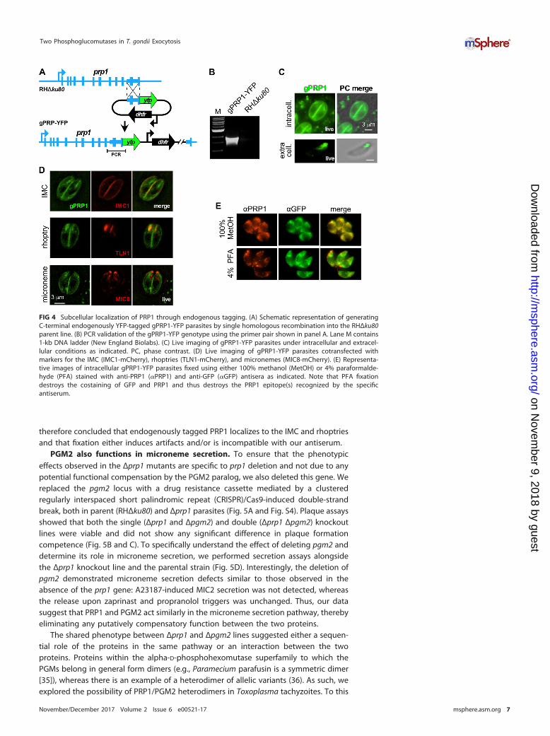
therefore concluded that endogenously tagged PRP1 localizes to the IMC and rhoptriesand that fixation either induces artifacts and/or is incompatible with our antiserum.
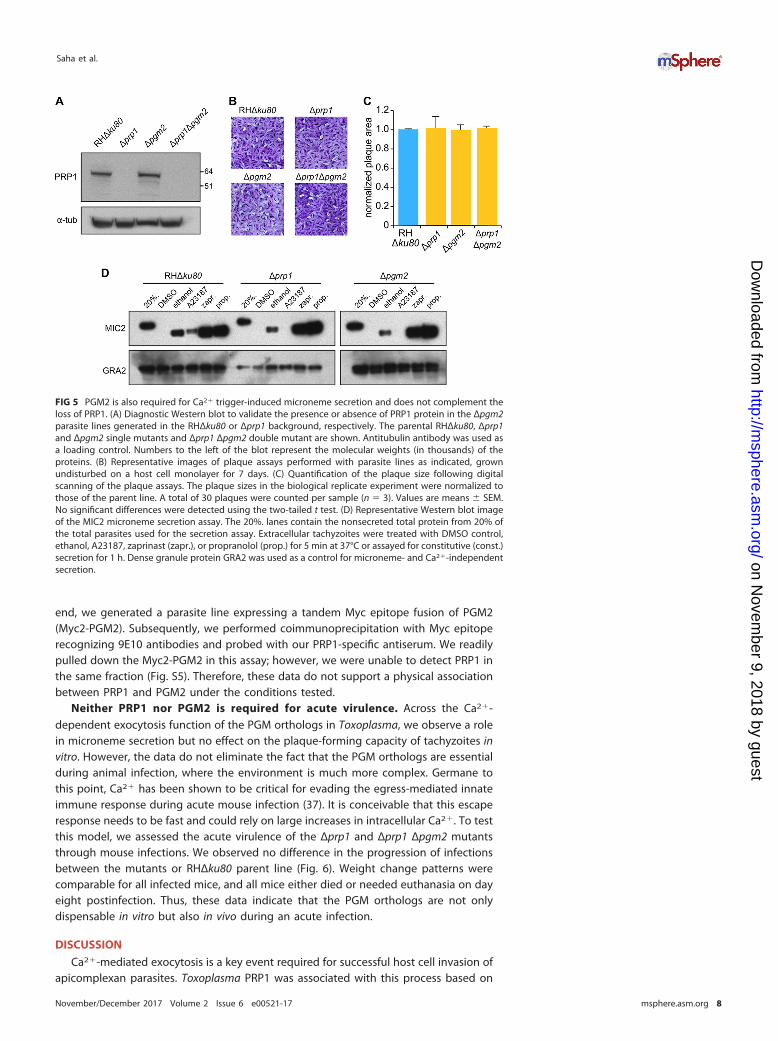
PGM2 also functions in microneme secretion. To ensure that the phenotypiceffects observed in the Δprp1 mutants are specific to prp1 deletion and not due to anypotential functional compensation by the PGM2 paralog, we also deleted this gene. Wereplaced the pgm2 locus with a drug resistance cassette mediated by a clusteredregularly interspaced short palindromic repeat (CRISPR)/Cas9-induced double-strandbreak, both in parent (RHΔku80) and Δprp1 parasites (Fig. 5A and Fig. S4). Plaque assaysshowed that both the single (Δprp1 and Δpgm2) and double (Δprp1 Δpgm2) knockoutlines were viable and did not show any significant difference in plaque formationcompetence (Fig. 5B and C). To specifically understand the effect of deleting pgm2 anddetermine its role in microneme secretion, we performed secretion assays alongsidethe Δprp1 knockout line and the parental strain (Fig. 5D). Interestingly, the deletion ofpgm2 demonstrated microneme secretion defects similar to those observed in theabsence of the prp1 gene: A23187-induced MIC2 secretion was not detected, whereasthe release upon zaprinast and propranolol triggers was unchanged. Thus, our datasuggest that PRP1 and PGM2 act similarly in the microneme secretion pathway, therebyeliminating any putatively compensatory function between the two proteins.
The shared phenotype between Δprp1 and Δpgm2 lines suggested either a sequen-tial role of the proteins in the same pathway or an interaction between the twoproteins. Proteins within the alpha-D-phosphohexomutase superfamily to which thePGMs belong in general form dimers (e.g., Paramecium parafusin is a symmetric dimer[35]), whereas there is an example of a heterodimer of allelic variants (36). As such, weexplored the possibility of PRP1/PGM2 heterodimers in Toxoplasma tachyzoites. To this
FIG 4 Subcellular localization of PRP1 through endogenous tagging. (A) Schematic representation of generatingC-terminal endogenously YFP-tagged gPRP1-YFP parasites by single homologous recombination into the RHΔku80parent line. (B) PCR validation of the gPRP1-YFP genotype using the primer pair shown in panel A. Lane M contains1-kb DNA ladder (New England Biolabs). (C) Live imaging of gPRP1-YFP parasites under intracellular and extracel-lular conditions as indicated. PC, phase contrast. (D) Live imaging of gPRP1-YFP parasites cotransfected withmarkers for the IMC (IMC1-mCherry), rhoptries (TLN1-mCherry), and micronemes (MIC8-mCherry). (E) Representa-tive images of intracellular gPRP1-YFP parasites fixed using either 100% methanol (MetOH) or 4% paraformalde-hyde (PFA) stained with anti-PRP1 (�PRP1) and anti-GFP (�GFP) antisera as indicated. Note that PFA fixationdestroys the costaining of GFP and PRP1 and thus destroys the PRP1 epitope(s) recognized by the specificantiserum.
Two Phosphoglucomutases in T. gondii Exocytosis
November/December 2017 Volume 2 Issue 6 e00521-17 msphere.asm.org 7
on Novem
ber 9, 2018 by guesthttp://m
sphere.asm.org/
Dow
nloaded from
end, we generated a parasite line expressing a tandem Myc epitope fusion of PGM2(Myc2-PGM2). Subsequently, we performed coimmunoprecipitation with Myc epitoperecognizing 9E10 antibodies and probed with our PRP1-specific antiserum. We readilypulled down the Myc2-PGM2 in this assay; however, we were unable to detect PRP1 inthe same fraction (Fig. S5). Therefore, these data do not support a physical associationbetween PRP1 and PGM2 under the conditions tested.
Neither PRP1 nor PGM2 is required for acute virulence. Across the Ca2�-dependent exocytosis function of the PGM orthologs in Toxoplasma, we observe a rolein microneme secretion but no effect on the plaque-forming capacity of tachyzoites invitro. However, the data do not eliminate the fact that the PGM orthologs are essentialduring animal infection, where the environment is much more complex. Germane tothis point, Ca2� has been shown to be critical for evading the egress-mediated innateimmune response during acute mouse infection (37). It is conceivable that this escaperesponse needs to be fast and could rely on large increases in intracellular Ca2�. To testthis model, we assessed the acute virulence of the Δprp1 and Δprp1 Δpgm2 mutantsthrough mouse infections. We observed no difference in the progression of infectionsbetween the mutants or RHΔku80 parent line (Fig. 6). Weight change patterns werecomparable for all infected mice, and all mice either died or needed euthanasia on dayeight postinfection. Thus, these data indicate that the PGM orthologs are not onlydispensable in vitro but also in vivo during an acute infection.
DISCUSSION
Ca2�-mediated exocytosis is a key event required for successful host cell invasion ofapicomplexan parasites. Toxoplasma PRP1 was associated with this process based on
FIG 5 PGM2 is also required for Ca2� trigger-induced microneme secretion and does not complement theloss of PRP1. (A) Diagnostic Western blot to validate the presence or absence of PRP1 protein in the Δpgm2parasite lines generated in the RHΔku80 or Δprp1 background, respectively. The parental RHΔku80, Δprp1and Δpgm2 single mutants and Δprp1 Δpgm2 double mutant are shown. Antitubulin antibody was used asa loading control. Numbers to the left of the blot represent the molecular weights (in thousands) of theproteins. (B) Representative images of plaque assays performed with parasite lines as indicated, grownundisturbed on a host cell monolayer for 7 days. (C) Quantification of the plaque size following digitalscanning of the plaque assays. The plaque sizes in the biological replicate experiment were normalized tothose of the parent line. A total of 30 plaques were counted per sample (n � 3). Values are means � SEM.No significant differences were detected using the two-tailed t test. (D) Representative Western blot imageof the MIC2 microneme secretion assay. The 20%. lanes contain the nonsecreted total protein from 20% ofthe total parasites used for the secretion assay. Extracellular tachyzoites were treated with DMSO control,ethanol, A23187, zaprinast (zapr.), or propranolol (prop.) for 5 min at 37°C or assayed for constitutive (const.)secretion for 1 h. Dense granule protein GRA2 was used as a control for microneme- and Ca2�-independentsecretion.
Saha et al.
November/December 2017 Volume 2 Issue 6 e00521-17 msphere.asm.org 8
on Novem
ber 9, 2018 by guesthttp://m
sphere.asm.org/
Dow
nloaded from

localization studies in Toxoplasma and heterologous functional studies in the ciliateParamecium (23). Here we directly assessed the role of PRP1 in Toxoplasma by deletingthe gene from the parasite, which surprisingly resulted in a nonlethal phenotype bothin vitro and in vivo. We did, however, identify a defect in the release of micronemestriggered by elevated Ca2� concentrations (insights are summarized in Fig. 7). Ourdetailed analysis of the prp1 deletion mutant revealed that the morphology of thesecretory organelles and cytoskeletal inner membrane complex (IMC) remain normal inΔprp1 parasites. Furthermore, we demonstrated that invasion and egress, which areboth reliant on successful Ca2�-mediated exocytosis, are independent of PRP1.
Since PRP1 was hypothesized to play a role in microneme secretion, we determinedthe dynamics of in vitro microneme secretion under different conditions and stimula-tion treatments. Our work demonstrates that the deletion of prp1 affects only mi-croneme secretion bursts triggered by a high Ca2� concentration through stimulationwith the Ca2� ionophore A23187. Titration of A23187 demonstrated that PRP1 does notact as an amplifier of low Ca2�, as the secretion boost in response to high Ca2� wasabolished regardless of the A23187 concentration. Overall, these data indicate thatPRP1 is essential to translate a high Ca2� trigger into enhanced microneme secretion(Fig. 7B), and thereby adds a new layer of complexity to the signaling pathwaysunderlying Ca2�-mediated secretion that is, to our knowledge, detected here for thefirst time.
High concentrations of intracellular Ca2� are known to occur during invasion andegress (8, 9, 33), which correlate with increased levels of microneme secretion. How-ever, since we observed no apparent growth defect and maybe even a growthadvantage of the Δprp1 parasites, the physiological function of elevated micronemesecretion is currently not clear. Constitutive microneme secretion in the absence ofpharmacological stimuli is comparable between the Δprp1 and wild-type parasites. Thissuggests that a basal level of microneme secretion is sufficient to complete the lyticcycle of the parasite. However, we cannot exclude the possibility that the ability torespond to high Ca2� fluxes and the subsequent microneme secretion response mightbe critical during other developmental stages of the Toxoplasma life cycle.
The PGM mutants allowed us to dissect the contribution of different branches of thesignaling pathway toward microneme secretion through stimulation with targetedpharmacological secretagogues (Fig. 7A). Upstream stimulation of the pathway withPKG-activating zaprinast leads to robust microneme secretion in these mutants. InPlasmodium, numerous protein substrates have been reported to be PKG substrates(38), which likely have many parallels in Toxoplasma. In Plasmodium, PKG controlsphosphoinositide biosynthesis through an unmapped mechanism, and the inhibition of
FIG 6 Loss of the PRP1/PGM2 complex does not affect acute virulence in mice. Acute virulence inC57BL/6J mice was assessed by intraperitoneal (i.p.) infection of 1,000 tachyzoites of the indicatedparasite lines. Weight changes relative to day 0 are shown for each mouse. Each symbol shows theweight change for an individual mouse. Average values for the groups are shown by the horizontal bars.The black line represents the average value for all groups. All mice in each group either died or wereeuthanized at day 8 postinfection (p.i.).
Two Phosphoglucomutases in T. gondii Exocytosis
November/December 2017 Volume 2 Issue 6 e00521-17 msphere.asm.org 9
on Novem
ber 9, 2018 by guesthttp://m
sphere.asm.org/
Dow
nloaded from
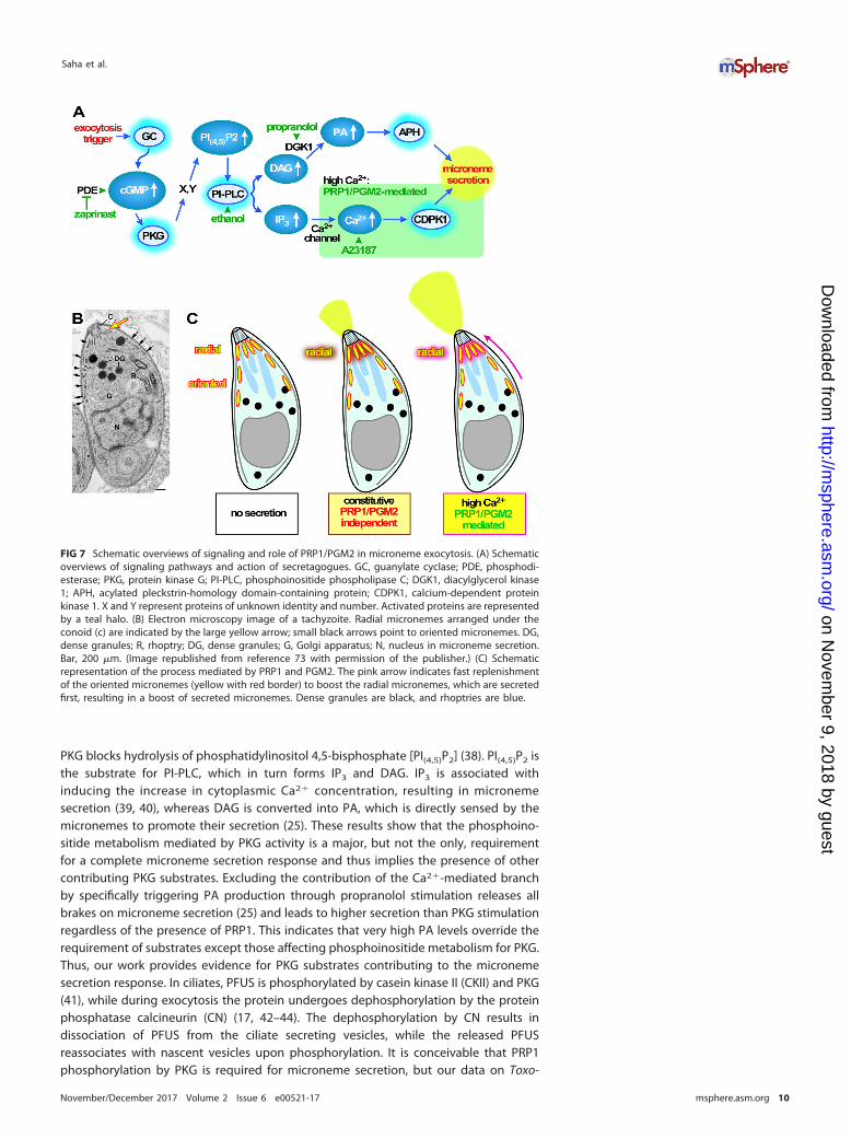
PKG blocks hydrolysis of phosphatidylinositol 4,5-bisphosphate [PI(4,5)P2] (38). PI(4,5)P2 isthe substrate for PI-PLC, which in turn forms IP3 and DAG. IP3 is associated withinducing the increase in cytoplasmic Ca2� concentration, resulting in micronemesecretion (39, 40), whereas DAG is converted into PA, which is directly sensed by themicronemes to promote their secretion (25). These results show that the phosphoino-sitide metabolism mediated by PKG activity is a major, but not the only, requirementfor a complete microneme secretion response and thus implies the presence of othercontributing PKG substrates. Excluding the contribution of the Ca2�-mediated branchby specifically triggering PA production through propranolol stimulation releases allbrakes on microneme secretion (25) and leads to higher secretion than PKG stimulationregardless of the presence of PRP1. This indicates that very high PA levels override therequirement of substrates except those affecting phosphoinositide metabolism for PKG.Thus, our work provides evidence for PKG substrates contributing to the micronemesecretion response. In ciliates, PFUS is phosphorylated by casein kinase II (CKII) and PKG(41), while during exocytosis the protein undergoes dephosphorylation by the proteinphosphatase calcineurin (CN) (17, 42–44). The dephosphorylation by CN results indissociation of PFUS from the ciliate secreting vesicles, while the released PFUSreassociates with nascent vesicles upon phosphorylation. It is conceivable that PRP1phosphorylation by PKG is required for microneme secretion, but our data on Toxo-
FIG 7 Schematic overviews of signaling and role of PRP1/PGM2 in microneme exocytosis. (A) Schematicoverviews of signaling pathways and action of secretagogues. GC, guanylate cyclase; PDE, phosphodi-esterase; PKG, protein kinase G; PI-PLC, phosphoinositide phospholipase C; DGK1, diacylglycerol kinase1; APH, acylated pleckstrin-homology domain-containing protein; CDPK1, calcium-dependent proteinkinase 1. X and Y represent proteins of unknown identity and number. Activated proteins are representedby a teal halo. (B) Electron microscopy image of a tachyzoite. Radial micronemes arranged under theconoid (c) are indicated by the large yellow arrow; small black arrows point to oriented micronemes. DG,dense granules; R, rhoptry; DG, dense granules; G, Golgi apparatus; N, nucleus in microneme secretion.Bar, 200 �m. (Image republished from reference 73 with permission of the publisher.) (C) Schematicrepresentation of the process mediated by PRP1 and PGM2. The pink arrow indicates fast replenishmentof the oriented micronemes (yellow with red border) to boost the radial micronemes, which are secretedfirst, resulting in a boost of secreted micronemes. Dense granules are black, and rhoptries are blue.
Saha et al.
November/December 2017 Volume 2 Issue 6 e00521-17 msphere.asm.org 10
on Novem
ber 9, 2018 by guesthttp://m
sphere.asm.org/
Dow
nloaded from
plasma CN demonstrate that this is not the key activity of CN, as microneme secretionremains unaffected upon CN depletion (24).
Using a peptide antibody against PFUS, which cross-reacts with PRP1, the latterwas reported to localize to the most apical micronemes from which it releases in aphosphorylation state-dependent fashion upon secretion (22). These most apicalmicronemes are known as the micronemes due to their tightly packed organizationaround the conoid at the very apical end of the parasite (45) (Fig. 7B). Thesemicronemes most likely represent a “readily releasable secretory vesicle pool” ofpredocked micronemes as have been reported for other secretory cells that can bereleased immediately upon the appropriate triggers (46, 47). For subsequent andcontinuous secretion replenishment, micronemes lying further basally along the sub-pellicular microtubules in the cell (the so-called “oriented” micronemes along themicrotubules [48]) have to be transported to the apical end. Since the first step alreadyoccurred in the parasites we tested when they egressed from the host cell, it is the fastreplenishment of releasable micronemes that appears to require PRP1/PGM2 (Fig. 7C,right schematic representation). Thus, in this model, PRP1 controls the release ofmicronemes in doses. The IMC and rhoptry localization we observe with YFP-fusedPRP1 in intracellular parasites and partial release from the rhoptries would be compat-ible to some extent with this model: PRP1 is there if the micronemes need to beactivated for transport along the IMC. Notably, protein kinase A (PKA) signalingcontribution to microneme secretion was recently reported to also be localized on theIMC (49), and therefore may serve as a signaling scaffold.
Although our PRP1 antiserum was highly specific in Western blots, unfortunately, itwas incompatible with indirect immunofluorescence assay (IFA) fixation methods,thereby restraining us from repeating the reported experiments. Membrane-associatedproteins are notoriously challenging to localize subcellularly due to fixation artifacts(50), which we also found to be the case. However, we were able to confirm that PRP1is present in soluble and membrane-associated forms in extracellular parasites, whichis consistent with the previously presented model (51). In our opinion, the YFP fusiondata are the best reflection of the natural PRP1 localization.
To ensure that the phenotypic effects observed in the Δprp1 mutants are specific toPRP1 deletion and not due to potential functional compensation by the PGM2 paralog,we also removed this gene and even made a double knockout mutant. The observationthat deletion of either one of the PGM genes simultaneously results in the samemicroneme secretion defect upon high Ca2� flux sheds a completely new light on thespecific role of PRP1 in Ca2� signaling: PRP1 is not the sole protein playing this role inthis function, as it extends to the other PGM expressed in Toxoplasma. This observationmight explain why the conservation of either ortholog in the various Apicomplexa is amishmash (Fig. 1), as either ortholog could function in Ca2�-dependent secretion.However, the fact that individual gene deletions in Toxoplasma resulted in the samephenotype suggests a functional interaction between PRP1 and PGM2 and hence alikely selective evolutionary pressure on preserving both. By coimmunoprecipitation(co-IP), we did not detect any physical interaction. Alternatively, the interaction mightbe transient upon high Ca2� levels, which is much harder to capture.
In addition, our data show that Toxoplasma tachyzoites can survive without dedi-cated phosphoglucomutase. Similar observations have been made in other organisms.For example, knockdown of PGM in Saccharomyces cerevisiae (52–54) or depletion ofthe single PGM in Paramecium and Tetrahymena (19, 20) demonstrated no apparenteffect on the growth of these organisms. In fact, there are even organisms without adedicated PGM such as Trypanosoma brucei (55). In addition, we showed that thePFUS-related PGM orthologs versus the exclusive PGM enzymes are not at all uniformlyconserved across the Apicomplexa (Fig. 1), which further supports a flexible role forthese proteins across organisms. It has been established in several systems thatalpha-D-phosphohexomutase superfamily enzymes can also have phosphoglucomutaseactivity, such as phosphomannomutase (PMM) and phospho-N-acetylglucosamine mu-tase (PAGM). Indeed, Toxoplasma has a PMM (accession no. TGME49_239710) and a
Two Phosphoglucomutases in T. gondii Exocytosis
November/December 2017 Volume 2 Issue 6 e00521-17 msphere.asm.org 11
on Novem
ber 9, 2018 by guesthttp://m
sphere.asm.org/
Dow
nloaded from

PAGM (TGME49_264650), and the amino acids involved in sugar binding are conservedbetween the Toxoplasma and T. brucei PMMs (55). These insights could thereforeexplain why the deletion of both Toxoplasma PGMs has no apparent effect on energymetabolism. However, it is conceivable that glycogen storage under the conditions wetested is not required and might become more relevant during bradyzoite differenti-ation, when storage compartments become more defined (56).
In conclusion, we show that PRP1 and PGM2 function in translating a large increasein the cytoplasmic Ca2� concentration into a burst of microneme secretion. The geneknockout lines also confirmed the bifurcation in the Ca2�- and PA-mediated control ofmicroneme secretion downstream of PI-PLC activity. The constitutive level of mi-croneme secretion is independent of PRP1 or PGM2 and sufficient to support theparasite through all steps of the lytic cycle both in vitro and in vivo. We further concludethat PRP1 and PGM2 functions control microneme secretion at a level not previouslyidentified. Considering the mosaic phylogeny pattern of PGM ortholog conservationacross the Apicomplexa, our data thus suggest that this control mechanism of themicroneme secretion pathways could be widespread among the eukaryotes.
MATERIALS AND METHODSParasites and host cells. Toxoplasma tachyzoites were maintained in human foreskin fibroblasts
(HFF) as previously described (57). The host cells were maintained in Dulbecco modified Eagle medium(DMEM) containing 10% serum. Toxoplasma strain RHΔku80ΔHX (58) was used as the basis for all mutantsin this study. Stable transgenics were obtained by selection with 1 �M pyrimethamine, 25 �g/mlmycophenolic acid (MPA) combined with 50 �g/ml xanthine or 20 �M chloramphenicol and cloned bylimiting dilutions.
Plasmids. All primer sequences are provided in Table S1 in the supplemental material. For taggingthe endogenous PRP1, 1.5-kb genomic DNA upstream of the stop codon was PCR amplified using primerpair PRP1-LIC-F and PRP1-LIC-R (PRP1-LIC-F/R) (LIC stands for ligation-independent cloning, F stands forforward, and R stands for reverse) and cloned by ligation-independent cloning into plasmid pYFP-LIC-DHFR (YFP stands for yellow fluorescent protein) (kindly provided by Vern Carruthers, University ofMichigan) (58). The plasmid was linearized with NcoI prior to transfection.
For the generation of Δprp1 plasmid, we deleted the LoxP flanked tubulin promoter and YFP cassettefrom the p5RT70-loxP-KillerRed-loxP-YFP/HX plasmid (59) (kindly provided by Markus Meissner, Univer-sity of Glasgow) by ApaI/NotI digestion, blunting the ends, and religation. The 2-kb flanks for doublehomologous recombination (HR) were PCR amplified from RHΔhx genomic DNA using primer pairs5=PRP1-KpnI-F/R for the 5= flank located 1.3 kb upstream of the ATG start codon and 3=PRP-SacI-F/R forthe 3= flank downstream of the translation stop. The 5= flank was inserted into p5RT70--/HX using KpnI,and independently, the 3= flank was inserted separately in parallel into p5RT70--/HX using SacI; the twoplasmids were conjoined using ScaI/NotI to make the final double homologous recombination plasmid.The plasmid was linearized with ScaI prior to transfection.
Plasmid ptub-Myc2-PGM2/sagCAT (tub stands for tubulin, and CAT stands for chloramphenicolacetyltransferase) was cloned by PCR amplifying the PGM2 coding sequence (CDS) from cDNA usingprimer pair PGM2-AvrII-F and PGM2-EcoRV-R and was cloned by AvrII/EcoRV digestion into the ptub-Myc2-GAPDH1/CAT (GAPDH stands for glyceraldehyde-3-phosphate dehydrogenase) (60).
Generation of PGM2 knockout. To generate Δpgm2 parasites, two CRISPR/Cas9 plasmids targetingCas9 to the pgm2 genomic locus were designed using the primer pairs 5PGM2-dKO-s/as (dKO stands fordouble knockout, s stands for sense, and as stands for antisense) and 3PGM2-dKO-s/as upstream of AUGand downstream of the stop codon, respectively. The specificity of the guide RNA was tested aspreviously described (61). The dihydrofolate reductase (DHFR) drug selectable cassette along with 20nucleotides of homologous region flanking each end, which corresponds to upstream and downstreamof the genomic locus, was PCR amplified. Forty micrograms of each pU6-5’PGM2/3’PGM2-Cas9 plasmidwas cotransfected with 40 �g of the PCR product into RHΔku80Δhx or RHΔku80�hx�prp1 parasites togenerate Δpgm2 parasites.
Immunofluorescence assays and live fluorescence microscopy. Live microscopy was performedon intracellular parasites grown overnight in six-well plates containing coverslips confluent with HFFcells. For extracellular localization, freshly lysed parasites were filtered, pelleted, and resuspended inphosphate-buffered saline (PBS). Thereafter, parasites were added to poly-L-lysine-coated coverslips andallowed to incubate for 30 min at 4°C prior to imaging. Colocalization studies with live gPRP1-YFPparasites was performed following transient cotransfection with the following plasmids: tub-IMC1mCherryRFP/sagCAT (RFP stands for red fluorescent protein) (62), TLN1(1-58)mCherryRFP/HPT(rhoptry marker) (63) (kindly provided by Peter Bradley, University of California at Los Angeles [UCLA]),and pTgMIC8-TgMIC8mycmCherryFP-nosel (pG53) (64) (kindly provided by Markus Meissner, Universityof Glasgow).
Indirect immunofluorescence assay was performed on intracellular parasites grown overnight insix-well plates containing coverslips confluent with HFF cells fixed with 100% methanol (unless statedotherwise) using the following primary antisera: rabbit anti-GFP (diluted 1:500) (Torrey Pines Biolabs),anti-MIC2 monoclonal antibody (MAb) 6D10 (1:8,000; kindly provided by David Sibley, Washington
Saha et al.
November/December 2017 Volume 2 Issue 6 e00521-17 msphere.asm.org 12
on Novem
ber 9, 2018 by guesthttp://m
sphere.asm.org/
Dow
nloaded from
University in St. Louis [65]), rabbit anti-MIC2 (1:8,000; kindly provided by David Sibley, WashingtonUniversity in St. Louis [66]), mouse anti-ROP1 (1:1,000; kindly provided by Peter Bradley, UCLA [67]),anti-GRA1 (1:20,000; kindly provided by Marie-France Cesbron-Delauw, Université Grenoble, France [68]),rat anti-IMC3 (1:2,000) (62), and guinea pig anti-PRP1 (1:1,000). Alexa Fluor 488 (A488)- or A594-conjugated goat anti-mouse, anti-rabbit, anti-rat, or anti-guinea pig were used as secondary antibodies(1:500; Invitrogen). DNA was stained with 4=,6-diamidino-2-phenylindole (DAPI). Fixation and staining ofphosphorylated STAT3 (P-STAT3) (1:120) (catalog no. 9145P; Cell Signaling) was performed exactly asdescribed previously (69). A Zeiss Axiovert 200 M wide-field fluorescence microscope equipped with a�-Plan-Fluar 100�/1.3-numerical-aperture (NA) and 100�/1.45-NA oil objectives and a HamamatsuC4742-95 charge-coupled-device (CCD) camera were used to collect images, which were deconvolvedand adjusted for phase contrast using Volocity software (Improvision/PerkinElmer).
Generation of specific PRP1 antiserum. To generate N-terminal His6-tagged fusion protein, 580 bpfrom the cDNA of PRP1 (corresponding to amino acids 446 to 637 in the C-terminal region of the protein)were PCR amplified using the primers aPRP1-LIC-F/R and cloned into the pAVA0421 plasmid usingligation-independent cloning (LIC) (70). The fusion protein was expressed in Escherichia coli BL21 using1 mM isopropyl-�-D-thiogalactopyranoside (IPTG) overnight at 37°C and purified under denaturingcondition over nickel-nitrilotriacetic acid (Ni-NTA) agarose (Invitrogen). Polyclonal antiserum was gen-erated by immunizing guinea pigs (Covance, Denver, PA).
Western blotting. Following SDS-PAGE, polyvinylidene difluoride (PVDF) membrane blots wereprobed with mouse anti-IMC1 (diluted 1:2,000; kindly provided by Gary Ward, University of Vermont),guinea pig anti-PRP1 (1:10,000), mouse monoclonal anti-GRA1 (1:20,000), or CDPK1 nanobody (1 �g/ml,a kind gift of Sebastian Lourido, Whitehead Institute [71]) followed by probing with horseradishperoxidase (HRP)-conjugated anti-mouse (1:10,000), anti-guinea pig antibody (1:3,000, Santa Cruz Bio-tech), or in case of the nanobody, anti-penta-His antibody (1:10,000; Qiagen) and detection of signal bychemiluminescent HRP substrate (Millipore).
Fractionation. Subcellular fractionation was performed as described previously (60). Essentially,extracellular parasites were lysed by 5-min freezing of the pellet in liquid nitrogen and resuspension at37°C in hypotonic buffer (10 mM Tris [pH 7.8] and 5 mM NaCl) followed by additional lyses by 40 Douncehomogenizer strokes. The lysate was centrifuged at low speed (1,000 � g for 15 min). The pellet wasresuspended in an equal volume of resuspension buffer (100 mM Tris [pH 7.8] and 150 mM NaCl) andcentrifuged at high speed (100,000 � g for 60 min). For SDS-PAGE, pellets were resuspended in an equalvolume of SDS-PAGE loading buffer, and corresponding amounts were analyzed by Western blotting.
Plaque assay. T12.5 culture flasks or six-well plates confluent with HFF cells were inoculated with 100parasites of choice and grown for 7 days. Following incubation, the monolayer was fixed with 100%ethanol for 10 min and stained with crystal violet (57). Plaque sizes were quantitated using ImageJ-win32software (72).
Microneme secretion assay. Microneme secretion assays using the MIC2 protein were performed asdescribed previously (30). Briefly, freshly lysed parasites were pelleted, washed, and resuspended to 2 �108/ml parasites in DMEM/FBS (DMEM containing 20 mM HEPES [pH 7.0] and 3% [wt/vol] fetal bovineserum [FBS]). Parasites (2 � 107) were added to each well of a 96-well plate, and secretion was inducedby the following secretagogues and concentrations unless concentrations are specifically indicatedotherwise: 1.25 �M A23187, 0.25% (vol/vol) ethanol, 500 �M zaprinast, 500 �M propranolol or dimethylsulfoxide (DMSO) control for 5 min at 37°C. For constitutive microneme secretion, we incubated parasitesat 37°C for 60 min in the absence of secretagogue. Microneme secretion was assessed in the supernatantwith mouse monoclonal anti-MIC2 6D10 (1:8,000). Ca2�-independent constitutive secretion of the densegranules was determined using mouse monoclonal anti-GRA1 (1:20,000) or rabbit anti-GRA2 antiserum(1:10,000).
Invasion assay. The red-green invasion assay was performed as previously published (73, 74) withmodifications. Tachyzoites (2.5 � 105 to 3 � 105) were added to host cells grown in 96-well black/opticalbottom plates, centrifuged (500 rpm, 3 min, room temperature [RT]), and allowed to invade the host cellsfor 1 h at 37°C. Noninvading extracellular parasites were detected using A594-conjugated anti-SAG1/P30-T41E5 antibodies (1:500; kindly provided by Jean François Dubremetz, University of Montpellier,France [75]) or anti-SAG1 MAb DG52 (kindly provided by Jeroen Saeij, Massachusetts Institute ofTechnology [76]). Following 16% formaldehyde/8% glutaraldehyde fixation and permeabilization using0.25% Triton X-100, the parasites were incubated with A488-conjugated anti-SAG1/P30 antibody tovisualize both invaded and noninvaded parasites. Each incubation with antibody was followed by threewashes with HH buffer (Hanks’ balanced salt solution containing 1 mM HEPES [pH 7.0]). CytochalasinD-treated wild-type parasites were used as a negative control. Images were taken using EVOS FL cellimaging system (Life Technologies).
Egress assay. The egress assay was performed as described previously (73, 77). Six-well platescontaining coverslips confluent with HFF cells were infected with 6 � 104 RHΔku80 or Δprp1 parasitesexpressing cytoplasmic YFP (78) and grown for 30 to 35 h. Egress was triggered by treatment withA23187 or propranolol at the concentration indicated using DMSO as a control at 37°C for 5 min,followed by 100% methanol fixation for 10 min at RT. Intact vacuoles were counted for at least 10 fieldsin two independent experiments.
Coimmunoprecipitation. Coimmunoprecipitation basically followed published procedures (79).Briefly, extracellular parasite pellets were subjected to one freeze-thaw cycle and lysed in lysis buffer (1�PBS, 0.25% NP-40, 400 mM NaCl, 250 U/ml Benzonase [Novagen], mammalian protease inhibitor cocktail[Sigma]). Lysates were precleared on protein G magnetic beads (New England Biolabs) followed by
Two Phosphoglucomutases in T. gondii Exocytosis
November/December 2017 Volume 2 Issue 6 e00521-17 msphere.asm.org 13
on Novem
ber 9, 2018 by guesthttp://m
sphere.asm.org/
Dow
nloaded from
Myc-tagged protein complex capture on 9E10 monoclonal antibody-conjugated magnetic beads (MBL).The beads were washed with three times with lysis buffer, and bound proteins eluted in Laemmli buffer.
In vivo mouse infection studies. Groups of three C57BL/6J mice each weighing between 18 and20 g were infected intraperitoneally with 1,000 tachyzoites of the RHΔku80, Δprp1, or Δprp1 Δpgm2strains. Following infection, mice were monitored daily for posture, activity level, and weight.
Sequence analysis and phylogeny. Phylogeny was performed using Geneious (80), and unrootedtrees were plotted using the neighbor-joining algorithm.
SUPPLEMENTAL MATERIALSupplemental material for this article may be found at https://doi.org/10.1128/
mSphere.00521-17.FIG S1, TIF file, 0.1 MB.FIG S2, TIF file, 2.2 MB.FIG S3, TIF file, 1.4 MB.FIG S4, TIF file, 0.4 MB.FIG S5, TIF file, 0.3 MB.TABLE S1, PDF file, 0.1 MB.
ACKNOWLEDGMENTSWe thank Tiffany Sansom for assistance with experimental infections of mice, Jayme
Henzy for assistance with phylogeny, and Giulia Bandini and Sebastian Lourido forinsightful discussions. We thank Peter Bradley, Marie-France Cesbron-Delauw, Jean-François Dubremetz, Sebastian Lourido, Markus Meissner, Jeroen Saeij, David Sibley,and Gary Ward for reagents.
S.S., B.I.C., and M.-J.G. conceived, designed, and performed most experiments. R.D.performed the fractionation experiment, and I.J.B. performed the in vivo virulenceexperiments. S.S. and M.-J.G. wrote the paper.
This work was supported by NIH grants AI108251 (B.I.C.), AI099658 (M.-J.G.),AI122923 (M.-J.G.), GM084383 (I.J.B.), and AI069986 (I.J.B.) and American Cancer Societygrant RSG-12-175-01-MPC (M.-J.G.).
The funders had no role in study design, data collection and interpretation, or thedecision to submit the work for publication.
REFERENCES1. Montoya JG, Liesenfeld O. 2004. Toxoplasmosis. Lancet 363:1965–1976.
https://doi.org/10.1016/S0140-6736(04)16412-X.2. Remington JS, McLeod R, Desmonts G. 1995. Toxoplasmosis, p 140 –267.
In Remington JS, Klein JO (ed), Infectious diseases of the fetus andnewborn infant. Saunders, Philadelphia, PA.
3. Weiss LM, Dubey JP. 2009. Toxoplasmosis: a history of clinical observa-tions. Int J Parasitol 39:895–901. https://doi.org/10.1016/j.ijpara.2009.02.004.
4. Blader IJ, Coleman BI, Chen CT, Gubbels MJ. 2015. Lytic cycle of Toxo-plasma gondii: 15 years later. Annu Rev Microbiol 69:463– 485. https://doi.org/10.1146/annurev-micro-091014-104100.
5. Baum J, Gilberger TW, Frischknecht F, Meissner M. 2008. Host-cell inva-sion by malaria parasites: insights from Plasmodium and Toxoplasma.Trends Parasitol 24:557–563. https://doi.org/10.1016/j.pt.2008.08.006.
6. Meissner M, Ferguson DJ, Frischknecht F. 2013. Invasion factors ofapicomplexan parasites: essential or redundant? Curr Opin Microbiol16:438 – 444. https://doi.org/10.1016/j.mib.2013.05.002.
7. Lourido S, Moreno SN. 2015. The calcium signaling toolkit of the Api-complexan parasites Toxoplasma gondii and Plasmodium spp. Cell Cal-cium 57:186 –193. https://doi.org/10.1016/j.ceca.2014.12.010.
8. Wetzel DM, Chen LA, Ruiz FA, Moreno SN, Sibley LD. 2004. Calcium-mediated protein secretion potentiates motility in Toxoplasma gondii. JCell Sci 117:5739 –5748. https://doi.org/10.1242/jcs.01495.
9. Sidik SM, Hortua Triana MA, Paul AS, El Bakkouri M, Hackett CG, Tran F,Westwood NJ, Hui R, Zuercher WJ, Duraisingh MT, Moreno SN, LouridoS. 2016. Using a genetically encoded sensor to identify inhibitors ofToxoplasma gondii Ca2� signaling. J Biol Chem 291:9566 –9580. https://doi.org/10.1074/jbc.M115.703546.
10. Levin S, Almo SC, Satir BH. 1999. Functional diversity of the phosphog-
lucomutase superfamily: structural implications. Protein Eng 12:737–746.https://doi.org/10.1093/protein/12.9.737.
11. Andersen AP, Wyroba E, Reichman M, Zhao H, Satir BH. 1994. The activityof parafusin is distinct from that of phosphoglucomutase in the unicel-lular eukaryote Paramecium. Biochem Biophys Res Commun 200:1353–1358. https://doi.org/10.1006/bbrc.1994.1600.
12. Subramanian SV, Wyroba E, Andersen AP, Satir BH. 1994. Cloning andsequencing of parafusin, a calcium-dependent exocytosis-related phos-phoglycoprotein. Proc Natl Acad Sci U S A 91:9832–9836. https://doi.org/10.1073/pnas.91.21.9832.
13. Satir BH, Hamasaki T, Reichman M, Murtaugh TJ. 1989. Species distribu-tion of a phosphoprotein (parafusin) involved in exocytosis. Proc NatlAcad Sci U S A 86:930 –932. https://doi.org/10.1073/pnas.86.3.930.
14. Satir BH, Wyroba E, Liu L, Lethan M, Satir P, Christensen ST. 2015.Evolutionary implications of localization of the signaling scaffold proteinparafusin to both cilia and the nucleus. Cell Biol Int 39:136 –145. https://doi.org/10.1002/cbin.10337.
15. Imada M, Kawashima S, Kanehisa M, Takeuchi T, Asai T. 2010. Charac-terization of alpha-phosphoglucomutase isozymes from Toxoplasmagondii. Parasitol Int 59:206 –210. https://doi.org/10.1016/j.parint.2010.01.007.
16. Gilligan DM, Satir BH. 1982. Protein phosphorylation/dephosphorylationand stimulus-secretion coupling in wild type and mutant Paramecium. JBiol Chem 257:13903–13906.
17. Plattner H, Kissmehl R. 2005. Molecular aspects of rapid, reversible,Ca2�-dependent de-phosphorylation of pp63/parafusin during stimu-lated exo-endocytosis in Paramecium cells. Cell Calcium 38:319 –327.https://doi.org/10.1016/j.ceca.2005.06.008.
18. Zhao H, Satir BH. 1998. Parafusin is a membrane and vesicle associated
Saha et al.
November/December 2017 Volume 2 Issue 6 e00521-17 msphere.asm.org 14
on Novem
ber 9, 2018 by guesthttp://m
sphere.asm.org/
Dow
nloaded from
protein that cycles at exocytosis. Eur J Cell Biol 75:46 –53. https://doi.org/10.1016/S0171-9335(98)80045-9.
19. Liu L, Wyroba E, Satir BH. 2011. RNAi knockdown of parafusin inhibits thesecretory pathway. Eur J Cell Biol 90:844 – 853. https://doi.org/10.1016/j.ejcb.2011.06.002.
20. Chilcoat ND, Turkewitz AP. 1997. In vivo analysis of the major exocytosis-sensitive phosphoprotein in Tetrahymena. J Cell Biol 139:1197–1207.https://doi.org/10.1083/jcb.139.5.1197.
21. Gubbels MJ, Duraisingh MT. 2012. Evolution of apicomplexan secretoryorganelles. Int J Parasitol 42:1071–1081. https://doi.org/10.1016/j.ijpara.2012.09.009.
22. Matthiesen SH, Shenoy SM, Kim K, Singer RH, Satir BH. 2001. A parafusin-related Toxoplasma protein in Ca2�-regulated secretory organelles. EurJ Cell Biol 80:775–783. https://doi.org/10.1078/0171-9335-00214.
23. Matthiesen SH, Shenoy SM, Kim K, Singer RH, Satir BH. 2003. Role of theparafusin orthologue, PRP1, in microneme exocytosis and cell invasionin Toxoplasma gondii. Cell Microbiol 5:613– 624. https://doi.org/10.1046/j.1462-5822.2003.00305.x.
24. Paul AS, Saha S, Engelberg K, Jiang RH, Coleman BI, Kosber AL, Chen CT,Ganter M, Espy N, Gilberger TW, Gubbels MJ, Duraisingh MT. 2015.Parasite calcineurin regulates host cell recognition and attachment byapicomplexans. Cell Host Microbe 18:49 – 60. https://doi.org/10.1016/j.chom.2015.06.003.
25. Bullen HE, Jia Y, Yamaryo-Botté Y, Bisio H, Zhang O, Jemelin NK, Marq JB,Carruthers V, Botté CY, Soldati-Favre D. 2016. Phosphatidic acid-mediated signaling regulates microneme secretion in Toxoplasma. CellHost Microbe 19:349 –360. https://doi.org/10.1016/j.chom.2016.02.006.
26. Saeij JP, Coller S, Boyle JP, Jerome ME, White MW, Boothroyd JC. 2007.Toxoplasma co-opts host gene expression by injection of a polymor-phic kinase homologue. Nature 445:324 –327. https://doi.org/10.1038/nature05395.
27. Donald RG, Liberator PA. 2002. Molecular characterization of a coccidianparasite cGMP dependent protein kinase. Mol Biochem Parasitol 120:165–175. https://doi.org/10.1016/S0166-6851(01)00451-0.
28. Lourido S, Tang K, Sibley LD. 2012. Distinct signalling pathways controlToxoplasma egress and host-cell invasion. EMBO J 31:4524 – 4534.https://doi.org/10.1038/emboj.2012.299.
29. Carruthers VB, Moreno SN, Sibley LD. 1999. Ethanol and acetaldehydeelevate intracellular [Ca2�] and stimulate microneme discharge inToxoplasma gondii. Biochem J 342:379 –386. https://doi.org/10.1042/bj3420379.
30. Carruthers VB, Sibley LD. 1999. Mobilization of intracellular calciumstimulates microneme discharge in Toxoplasma gondii. Mol Microbiol31:421– 428. https://doi.org/10.1046/j.1365-2958.1999.01174.x.
31. Lourido S, Shuman J, Zhang C, Shokat KM, Hui R, Sibley LD. 2010.Calcium-dependent protein kinase 1 is an essential regulator ofexocytosis in Toxoplasma. Nature 465:359 –362. https://doi.org/10.1038/nature09022.
32. Jacot D, Tosetti N, Pires I, Stock J, Graindorge A, Hung YF, Han H, TewariR, Kursula I, Soldati-Favre D. 2016. An apicomplexan actin-binding pro-tein serves as a connector and lipid sensor to coordinate motility andinvasion. Cell Host Microbe 20:731–743. https://doi.org/10.1016/j.chom.2016.10.020.
33. Borges-Pereira L, Budu A, McKnight CA, Moore CA, Vella SA, HortuaTriana MA, Liu J, Garcia CR, Pace DA, Moreno SN. 2015. Calcium signalingthroughout the Toxoplasma gondii lytic cycle: a study using geneticallyencoded calcium indicators. J Biol Chem 290:26914 –26926. https://doi.org/10.1074/jbc.M115.652511.
34. Pomel S, Luk FC, Beckers CJ. 2008. Host cell egress and invasion inducemarked relocations of glycolytic enzymes in Toxoplasma gondiitachyzoites. PLoS Pathog 4:e1000188. https://doi.org/10.1371/journal.ppat.1000188.
35. Müller S, Diederichs K, Breed J, Kissmehl R, Hauser K, Plattner H, Welte W.2002. Crystal structure analysis of the exocytosis-sensitive phosphopro-tein, pp63/parafusin (phosphoglucomutase), from Paramecium revealssignificant conformational variability. J Mol Biol 315:141–153. https://doi.org/10.1006/jmbi.2001.5168.
36. Andreotti G, Monti MC, Citro V, Cubellis MV. 2015. Heterodimerization oftwo pathological mutants enhances the activity of human phospho-mannomutase2. PLoS One 10:e0139882. https://doi.org/10.1371/journal.pone.0139882.
37. Tomita T, Yamada T, Weiss LM, Orlofsky A. 2009. Externally triggeredegress is the major fate of Toxoplasma gondii during acute infection. JImmunol 183:6667– 6680. https://doi.org/10.4049/jimmunol.0900516.
38. Brochet M, Collins MO, Smith TK, Thompson E, Sebastian S, Volkmann K,Schwach F, Chappell L, Gomes AR, Berriman M, Rayner JC, Baker DA,Choudhary J, Billker O. 2014. Phosphoinositide metabolism links cGMP-dependent protein kinase G to essential Ca2� signals at key decisionpoints in the life cycle of malaria parasites. PLoS Biol 12:e1001806.https://doi.org/10.1371/journal.pbio.1001806.
39. Lovett JL, Marchesini N, Moreno SN, Sibley LD. 2002. Toxoplasma gondiimicroneme secretion involves intracellular Ca2� release from inositol1,4,5-triphosphate (IP3)/ryanodine-sensitive stores. J Biol Chem 277:25870 –25876. https://doi.org/10.1074/jbc.M202553200.
40. Sidik SM, Huet D, Ganesan SM, Huynh MH, Wang T, Nasamu AS, Thiru P,Saeij JPJ, Carruthers VB, Niles JC, Lourido S. 2016. A genome-wideCRISPR screen in Toxoplasma identifies essential apicomplexan genes.Cell 166:1423–1435.e12. https://doi.org/10.1016/j.cell.2016.08.019.
41. Kussmann M, Hauser K, Kissmehl R, Breed J, Plattner H, Roepstorff P.1999. Comparison of in vivo and in vitro phosphorylation of theexocytosis-sensitive protein PP63/parafusin by differential MALDImass spectrometric peptide mapping. Biochemistry 38:7780 –7790.https://doi.org/10.1021/bi982888y.
42. Treptau T, Kissmehl R, Wissmann JD, Plattner H. 1995. A 63 kDa phos-phoprotein undergoing rapid dephosphorylation during exocytosis inParamecium cells shares biochemical characteristics with phosphoglu-comutase. Biochem J 309:557–567. https://doi.org/10.1042/bj3090557.
43. Subramanian SV, Satir BH. 1992. Carbohydrate cycling in signal trans-duction: parafusin, a phosphoglycoprotein and possible Ca2�-dependent transducer molecule in exocytosis in Paramecium. ProcNatl Acad Sci U S A 89:11297–11301. https://doi.org/10.1073/pnas.89.23.11297.
44. Kissmehl R, Treptau T, Hofer HW, Plattner H. 1996. Protein phosphataseand kinase activities possibly involved in exocytosis regulation in Para-mecium tetraurelia. Biochem J 317:65–76. https://doi.org/10.1042/bj3170065.
45. Paredes-Santos TC, de Souza W, Attias M. 2012. Dynamics and 3Dorganization of secretory organelles of Toxoplasma gondii. J StructBiol 177:420 – 430. https://doi.org/10.1016/j.jsb.2011.11.028.
46. Kaeser PS, Regehr WG. 2014. Molecular mechanisms for synchronous, asyn-chronous, and spontaneous neurotransmitter release. Annu Rev Physiol76:333–363. https://doi.org/10.1146/annurev-physiol-021113-170338.
47. Kaeser PS, Regehr WG. 2017. The readily releasable pool of synapticvesicles. Curr Opin Neurobiol 43:63–70. https://doi.org/10.1016/j.conb.2016.12.012.
48. Kremer K, Kamin D, Rittweger E, Wilkes J, Flammer H, Mahler S, Heng J,Tonkin CJ, Langsley G, Hell SW, Carruthers VB, Ferguson DJ, Meissner M.2013. An overexpression screen of Toxoplasma gondii Rab-GTPases revealsdistinct transport routes to the micronemes. PLoS Pathog 9:e1003213.https://doi.org/10.1371/journal.ppat.1003213.
49. Jia Y, Marq JB, Bisio H, Jacot D, Mueller C, Yu L, Choudhary J, BrochetM, Soldati-Favre D. 2017. Crosstalk between PKA and PKG controlspH-dependent host cell egress of Toxoplasma gondii. EMBO J 36:3250 –3267. https://doi.org/10.15252/embj.201796794.
50. Hannah MJ, Weiss U, Huttner WB. 1998. Differential extraction of pro-teins from paraformaldehyde-fixed cells: lessons from synaptophysinand other membrane proteins. Methods 16:170 –181. https://doi.org/10.1006/meth.1998.0664.
51. Liu L, Tucker SC, Satir BH. 2009. Toxoplasma PRP1 is an ortholog ofparafusin (PFUS) in vesicle scaffold assembly in Ca2�-regulated exocy-tosis. Eur J Cell Biol 88:301–313. https://doi.org/10.1016/j.ejcb.2008.10.004.
52. Hofmann M, Boles E, Zimmermann FK. 1994. Characterization of theessential yeast gene encoding N-acetylglucosamine-phosphate mutase.Eur J Biochem 221:741–747. https://doi.org/10.1111/j.1432-1033.1994.tb18787.x.
53. Boles E, Liebetrau W, Hofmann M, Zimmermann FK. 1994. A family ofhexosephosphate mutases in Saccharomyces cerevisiae. Eur J Biochem220:83–96. https://doi.org/10.1111/j.1432-1033.1994.tb18601.x.
54. Fu L, Miseta A, Hunton D, Marchase RB, Bedwell DM. 2000. Loss of themajor isoform of phosphoglucomutase results in altered calcium ho-meostasis in Saccharomyces cerevisiae. J Biol Chem 275:5431–5440.https://doi.org/10.1074/jbc.275.8.5431.
55. Bandini G, Mariño K, Güther ML, Wernimont AK, Kuettel S, Qiu W, AfzalS, Kelner A, Hui R, Ferguson MA. 2012. Phosphoglucomutase is absent inTrypanosoma brucei and redundantly substituted by phosphomanno-mutase and phospho-N-acetylglucosamine mutase. Mol Microbiol 85:513–534. https://doi.org/10.1111/j.1365-2958.2012.08124.x.
Two Phosphoglucomutases in T. gondii Exocytosis
November/December 2017 Volume 2 Issue 6 e00521-17 msphere.asm.org 15
on Novem
ber 9, 2018 by guesthttp://m
sphere.asm.org/
Dow
nloaded from
56. Uboldi AD, McCoy JM, Blume M, Gerlic M, Ferguson DJ, Dagley LF,Beahan CT, Stapleton DI, Gooley PR, Bacic A, Masters SL, Webb AI,McConville MJ, Tonkin CJ. 2015. Regulation of starch stores by a Ca2�-dependent protein kinase is essential for viable cyst development inToxoplasma gondii. Cell Host Microbe 18:670 – 681. https://doi.org/10.1016/j.chom.2015.11.004.
57. Roos DS, Donald RG, Morrissette NS, Moulton AL. 1994. Molecular toolsfor genetic dissection of the protozoan parasite Toxoplasma gondii.Methods Cell Biol 45:27– 63.
58. Huynh MH, Carruthers VB. 2009. Tagging of endogenous genes in aToxoplasma gondii strain lacking Ku80. Eukaryot Cell 8:530 –539. https://doi.org/10.1128/EC.00358-08.
59. Andenmatten N, Egarter S, Jackson AJ, Jullien N, Herman JP, Meissner M.2013. Conditional genome engineering in Toxoplasma gondii uncoversalternative invasion mechanisms. Nat Methods 10:125–127. https://doi.org/10.1038/nmeth.2301.
60. Dubey R, Staker BL, Foe IT, Bogyo M, Myler PJ, Ngô HM, Gubbels MJ.2017. Membrane skeletal association and post-translational allostericregulation of Toxoplasma gondii GAPDH1. Mol Microbiol 103:618 – 634.https://doi.org/10.1111/mmi.13577.
61. Sidik SM, Hackett CG, Tran F, Westwood NJ, Lourido S. 2014. Efficientgenome engineering of Toxoplasma gondii using CRISPR/Cas9. PLoSOne 9:e100450. https://doi.org/10.1371/journal.pone.0100450.
62. Anderson-White BR, Ivey FD, Cheng K, Szatanek T, Lorestani A, BeckersCJ, Ferguson DJ, Sahoo N, Gubbels MJ. 2011. A family of intermediatefilament-like proteins is sequentially assembled into the cytoskeleton ofToxoplasma gondii. Cell Microbiol 13:18 –31. https://doi.org/10.1111/j.1462-5822.2010.01514.x.
63. Hajagos BE, Turetzky JM, Peng ED, Cheng SJ, Ryan CM, Souda P,Whitelegge JP, Lebrun M, Dubremetz JF, Bradley PJ. 2012. Moleculardissection of novel trafficking and processing of the Toxoplasma gondiirhoptry metalloprotease toxolysin-1. Traffic 13:292–304. https://doi.org/10.1111/j.1600-0854.2011.01308.x.
64. Kessler H, Herm-Götz A, Hegge S, Rauch M, Soldati-Favre D, FrischknechtF, Meissner M. 2008. Microneme protein 8 – a new essential invasionfactor in Toxoplasma gondii. J Cell Sci 121:947–956. https://doi.org/10.1242/jcs.022350.
65. Wan KL, Carruthers VB, Sibley LD, Ajioka JW. 1997. Molecular character-isation of an expressed sequence tag locus of Toxoplasma gondiiencoding the micronemal protein MIC2. Mol Biochem Parasitol 84:203–214. https://doi.org/10.1016/S0166-6851(96)02796-X.
66. Labruyere E, Lingnau M, Mercier C, Sibley LD. 1999. Differential mem-brane targeting of the secretory proteins GRA4 and GRA6 within theparasitophorous vacuole formed by Toxoplasma gondii. Mol BiochemParasitol 102:311–324. https://doi.org/10.1016/S0166-6851(99)00092-4.
67. Saffer LD, Mercereau-Puijalon O, Dubremetz JF, Schwartzman JD. 1992.Localization of a Toxoplasma gondii rhoptry protein by immunoelectronmicroscopy during and after host cell penetration. J Protozool 39:526 –530. https://doi.org/10.1111/j.1550-7408.1992.tb04844.x.
68. Cesbron-Delauw MF, Guy B, Torpier G, Pierce RJ, Lenzen G, Cesbron JY,
Charif H, Lepage P, Darcy F, Lecocq JP. 1989. Molecular characterizationof a 23- kilodalton major antigen secreted by Toxoplasma gondii. ProcNatl Acad Sci U S A 86:7537–7541. https://doi.org/10.1073/pnas.86.19.7537.
69. Brown KM, Suvorova E, Farrell A, McLain A, Dittmar A, Wiley GB, MarthG, Gaffney PM, Gubbels MJ, White M, Blader IJ. 2014. Forward geneticscreening identifies a small molecule that blocks Toxoplasma gondiigrowth by inhibiting both host- and parasite-encoded kinases. PLoSPathog 10:e1004180. https://doi.org/10.1371/journal.ppat.1004180.
70. Alexandrov A, Vignali M, LaCount DJ, Quartley E, de Vries C, De Rosa D,Babulski J, Mitchell SF, Schoenfeld LW, Fields S, Hol WG, Dumont ME,Phizicky EM, Grayhack EJ. 2004. A facile method for high-throughputco-expression of protein pairs. Mol Cell Proteomics 3:934 –938. https://doi.org/10.1074/mcp.T400008-MCP200.
71. Ingram JR, Knockenhauer KE, Markus BM, Mandelbaum J, Ramek A, Shan Y,Shaw DE, Schwartz TU, Ploegh HL, Lourido S. 2015. Allosteric activation ofapicomplexan calcium-dependent protein kinases. Proc Natl Acad Sci U S A112:E4975–E4984. https://doi.org/10.1073/pnas.1505914112.
72. Abramoff MD, Magalhães PJ, Ram SJ. 2004. Image processing withImageJ. Biophotonics Int 11:36 – 42.
73. Farrell A, Thirugnanam S, Lorestani A, Dvorin JD, Eidell KP, Ferguson DJ,Anderson-White BR, Duraisingh MT, Marth GT, Gubbels MJ. 2012. ADOC2 protein identified by mutational profiling is essential for apicom-plexan parasite exocytosis. Science 335:218 –221. https://doi.org/10.1126/science.1210829.
74. Kafsack BF, Beckers C, Carruthers VB. 2004. Synchronous invasion of hostcells by Toxoplasma gondii. Mol Biochem Parasitol 136:309 –311. https://doi.org/10.1016/j.molbiopara.2004.04.004.
75. Couvreur G, Sadak A, Fortier B, Dubremetz JF. 1988. Surface antigensof Toxoplasma gondii. Parasitology 97:1–10. https://doi.org/10.1017/S0031182000066695.
76. Burg JL, Perelman D, Kasper LH, Ware PL, Boothroyd JC. 1988. Molecularanalysis of the gene encoding the major surface antigen of Toxoplasmagondii. J Immunol 141:3584 –3591.
77. Eidell KP, Burke T, Gubbels MJ. 2010. Development of a screen to dissectToxoplasma gondii egress. Mol Biochem Parasitol 171:97–103. https://doi.org/10.1016/j.molbiopara.2010.03.004.
78. Gubbels MJ, Li C, Striepen B. 2003. High-throughput growth assay forToxoplasma gondii using yellow fluorescent protein. Antimicrob AgentsChemother 47:309 –316. https://doi.org/10.1128/AAC.47.1.309-316.2003.
79. Suvorova ES, Croken M, Kratzer S, Ting LM, Conde de Felipe M, Balu B,Markillie ML, Weiss LM, Kim K, White MW. 2013. Discovery of a splicingregulator required for cell cycle progression. PLoS Genet 9:e1003305.https://doi.org/10.1371/journal.pgen.1003305.
80. Kearse M, Moir R, Wilson A, Stones-Havas S, Cheung M, Sturrock S,Buxton S, Cooper A, Markowitz S, Duran C, Thierer T, Ashton B, MeintjesP, Drummond A. 2012. Geneious Basic: an integrated and extendabledesktop software platform for the organization and analysis of sequencedata. Bioinformatics 28:1647–1649. https://doi.org/10.1093/bioinformatics/bts199.
Saha et al.
November/December 2017 Volume 2 Issue 6 e00521-17 msphere.asm.org 16
on Novem
ber 9, 2018 by guesthttp://m
sphere.asm.org/
Dow
nloaded from