two novel mutations in the antxr2 gene in a chinese
TRANSCRIPT

MOLECULAR MEDICINE REPORTS 18: 4004-4008, 20184004
Abstract. Hyaline fibromatosis syndrome (HFS; MIM 228600) is a rare autosomal recessive disorder characterized by the abnormal growth of hyalinized fibrous tissue at subcutaneous regions on the scalp, ears and neck. The disease is caused by either a homozygous or compound heterozygous mutation of the anthrax toxin receptor 2 (ANTXR2) gene. The present study describes a patient with HFS confirmed by clinical examination as well as histopathological and genetic analyses. Numerous painless and variable‑sized subcutaneous nodules were observed on the scalp, ear, trunk and four extremities of the patient. With increasing age, the number and size of the nodules gradually increased in the patient. The patient additionally presented with severe gingival thickening and developed pearly papules on the ears, back and penis foreskin. Biopsies of ear nodules revealed that the tumor was located in the dermis, and no marked alterations were observed in the epidermis compared with healthy patients. Spindle‑shaped or round tumor cells were revealed to be immersed in the eosino-philic hyaline ground substance. Furthermore, a skeletal X‑ray of the patient revealed multiple low‑density imaging on the right distal humerus. Compound heterozygous mutations in the ANTXR2 gene were identified in the patient: c.470_472del in exon 5 and c.1073 delC in exon 13. c.470_472del were revealed to be inherited from his mother and father, respec-tively. These two mutations, c.470_472del and c.1073 delC, to the best of our knowledge, have not previously been identified. Identification of the mutations in ANTXR2 may make prenatal diagnosis of HFS possible during future pregnancies.
Introduction
Hyaline fibromatosis syndrome (HFS; OMIM 228600), previously termed juvenile hyaline fibromatosis (JHF) and infantile systemic hyalinosis (ISH), is a rare autosomal reces-sive disorder. The disease is characterized by the abnormal growth of hyalinized fibrous tissue, which commonly affects subcutaneous regions on the scalp, ears and neck (1). HFS is classified into different grades according to its clinical mani-festation, resulting from the involvement of different organs; symptoms may include persistent diarrhea and recurrent pulmonary infections (2). HFS is caused by either a homo-zygous or compound heterozygous mutation in the anthrax toxin receptor 2 (ANTXR2) gene, additionally termed the capillary morphogenesis protein 2 gene, which is localized on chromosome 4q21 (1,2). ANTXR2 encodes for a type I transmembrane (TM) protein, which consists of an extra-cellular N terminal von Willebrand A (vWA) domain, an immunoglobulin‑like domain, a TM domain and a cytosolic tail (3-6). TM is able to bind to laminin and collagen IV via its vWA domain, and thus may regulate basement membrane matrix assembly (7). The cytosolic tail may be responsible for posttranslational modifications (8,9). Furthermore, TM has been suggested to be involved in collagen abnormali-ties (10) and formation defects in glycosaminoglycans (11). A diagnosis of HFS, made by the combination of clinical history findings with histopathological and genetic analyses, ought to be considered in patients exhibiting skin lesions (nodules and/or pearly papules), gingival hyperplasia and joint contracture.
In the present study, a Chinese patient with HFS exhibiting typical features characteristic of the syndrome is described. Mutation analysis revealed two novel heterozygous mutations of ANTXR2: c.470_472del in exon 5 and c.1073 delC in exon 13, which, to the best of our knowledge, have not previously been identified in ANTXR2.
Case report
Samples. In July 2016, blood and skin samples were obtained at the Department of Dermatology, Capital Institute of Pediatrics, Peking University Teaching Hospital (Beijing, China). Genomic DNA samples were extracted from the peripheral blood leukocytes obtained from the patient
Two novel mutations in the ANTXR2 gene in a Chinese patient suffering from hyaline fibromatosis syndrome: A case report
YING GAO1, JINLI BAI2, JIANCAI WANG1 and XIAOYAN LIU1
Departments of 1Dermatology and 2Medical Genetics, Capital Institute of Pediatrics, Peking University Teaching Hospital, Beijing 100020, P.R. China
Received October 30, 2017; Accepted March 14, 2018
DOI: 10.3892/mmr.2018.9421
Correspondence to: Dr Xiaoyan Liu, Department of Dermatology, Capital Institute of Pediatrics, Peking University Teaching Hospital, 2 Yabao Road, Beijing 100020, P.R. ChinaE‑mail: [email protected]
Abbreviations: HFS, hyaline fibromatosis syndrome; ISH, infantile systemic alinosis; JHF, juvenile hyaline fibromatosis; ANTXR2, anthrax toxin receptor 2 gene
Key words: hyaline fibromatosis syndrome, anthrax toxin receptor 2 gene, genetic analysis
GAO et al: TWO NOVEL MUTATIONS OF THE ANTXR2 GENE 4005
(male, aged 7.5 years old) and his parents using the QIAamp DNA Blood Mini kit (Qiagen, Inc., Valencia, CA, USA). The skin sample was embedded in paraffin and sections (5 µm thick) using a Leitz microtome (Leica Microsystems GmbH, Wetzlar, Germany). Sections were subsequently stained using a Hematoxylin‑Eosin Staining kit (Beijing Solarbio Science & Technology Co., Ltd., Beijing, China) according to the manufacturer's protocol, and then visualized using a Nikon Eclipse 80i microscope (Nikon Corporation, Tokyo, Japan) (12,13). Informed consent was obtained from the parents of the patient. The present study was approved by the Ethics Committee of the Capital Institute of Pediatrics (Beijing, China).
Targeted exome sequencing and data analysis. Extracted DNA was subjected to targeted exome sequencing and subsequent data analysis, which was performed by Chigene Translational Medicine Research Center Co. Ltd. (Beijing, China). Genomic DNA (1 µg per patient) was used to generate genomic libraries. Genomic DNA was sheared into 200‑300 base pair fragment libraries using the Covaris S2 instrument (Covaris Inc., Woburn, MA, USA). DNA fragment ends were blunted and ligated with sequencing adaptors using the KAPA HTP Library Preparation kit (Illumina, Inc., San Diego, CA, USA) according to the manufacturer's protocol. Prepared DNA was amplified via ligation‑mediated polymerase chain reaction (PCR) and sequenced using Illumina HiSeq X10 (Illumina, Inc.). Raw data were mapped to the human genome reference, build 19 (www.broadinstitute.org/ftp/pub/seq/references/Homo_sapiens_assembly19.fasta). Short Oligonucleotide Analysis Package snp software (http://soap.genomics.org.cn/soapsnp.html) and SAMtools software (http://samtools.sourceforge.net/) were used to search for single nucleotide variants, and insertion and deletion (indel) mutations.
Sanger sequencing. Sanger sequencing of exon 5 (E5) and exon 13 (E13) in ANTXR2 was performed using DNA samples isolated from peripheral blood leukocytes of the patient and his parents in order to validate the two candidate mutations (c.470_472del and c.1073 delC in E5 and E13, respectively) identified by the aforementioned targeted exome sequencing. The thermocycling conditions used for PCR were as follows: Initial denaturation at 95˚C for 5 min; followed by 30 cycles of denaturation at 95˚C for 30 sec, annealing at 55˚C for 1 min, extension at 72˚C for 30 sec; and a final extension was performed at 72˚C for 10 min. Primer sequences used are detailed in Table I. The DNA polymerase used was KAPA2G Robust DNA polymerase (KAPA2G Robust HotStart PCR kit; Sigma‑Aldrich; Merck KGaA, Darmstadt, Germany). Following this, PCR products were purified using SanPrep Column DNA Gel Extraction kit (Sangon Biotech Co., Ltd., Shanghai, China) according to the manufacturer's instruc-tions. Direct sequencing of fragments was performed using an ABI 3730 automatic sequencer (Thermo Fisher Scientific., Inc., Waltham, MA, USA). Finally, DNASTAR software (DNASTAR, Inc., Madison, WI, USA) was used to perform mutation analysis.
Clinical presentation. The patient was born at term by normal delivery, and is the second child of healthy and
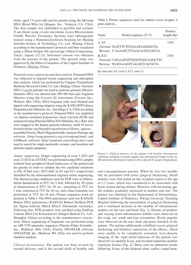
non‑consanguineous parents. When he was two months old, he presented with lower gingival thickening. Small nodules were first noted on the occipital region at the age of 1.5 years, which was considered to be associated with brain trauma during infancy. However, with increasing age, the nodules gradually increased in number and size. The patient was admitted to the Department of Dermatology, Capital Institute of Pediatrics, Peking University Teaching Hospital following the presentation of gingival thickening and a continual increase in the number of subcutaneous nodules on the ears and head (Fig. 1). Dozens of painless and varying sized subcutaneous nodules were observed on the scalp, ear, trunk and four extremities. Pearly papules were observed on the ears, back and penis foreskin of the patient. The patient additionally exhibited severe gingival thickening and bilateral contracture of the elbows, which were unable to be completely extended. Low‑density imaging of the right distal humerus of the patient was observed via skeletal X‑ray, and revealed numerous nodular osteolytic lesions (Fig. 2). There were no abnormal results following X‑rays of the bilateral ulnar, radius, carpal bone,
Figure 1. Clinical features of the patient with hyaline fibromatosis syndrome, including (A) pearly papules and subcutaneous bumps on the ear, (B) numerous subcutaneous lumps on the scalp and (C) gingival hyperplasia.
Table I. Primer sequences used for anthrax toxin receptor 2 gene analysis.
ProductName Primer sequence (5'‑3') length (bp)
A, E5 1545 Forward TAATTTCTGTAAATAAGGGCTA Reverse 5'‑AAAATCTTTAACAATCGACCA B, E13 1545 Forward CATAAATATTGGGTGGCAAGCTTC Reverse TGATAAAGTCAGATCCACGCTA
bp, base pair; E5, exon 5; E13, exon 13.

MOLECULAR MEDICINE REPORTS 18: 4004-4008, 20184006
tibia and fibula. Biopsies of ear nodules demonstrated that the tumor was located in the dermis and no significant alterations were observed in the epidermis. Spindle‑shaped or round tumor cells were revealed to be immersed in the eosinophilic hyaline ground substance by conventional histopathology (Fig. 2).
Genetic analysis. Two mutations in ANTXR2 (NM_058172.5; c.1073 delC and c.470_472del) were identified by targeted exome sequencing and further verified via Sanger sequencing. Compound heterozygous mutations of ANTXR2 were identi-fied in the patient: c.470_472del (p.Ser157del) in exon 5 and c.1073 delC (p.Pro358LeufsTer51) in exon 13 (Fig. 3). The c.470_472del in‑frame deletion mutation was additionally present within exon 5 in ANTXR2 in the mother of the patient; whereas, the frameshift mutation c.1073 delC was present within exon 13 in ANTXR2 in the father of the patient (Fig. 3). Thus, the parents of the patient were revealed to be heterozy-gous carriers of the two identified mutations.
Discussion
JHF was first reported to be molluscum fibrosum by Murray in 1873 (14). It is characterized by cutaneous lesions, gingival enlargement and osteolytic lesions (15). ISH is a disorder that is more rare and severe than JHF, and was first described by Landing and Nadorra (16). Symptoms begin to present in newborns or during infancy with the deposition of hyaline mate-rial in a number of organs, resulting in multiple skin nodules, skin thickening and joint contractures and even premature mortality. As JHF and ISH share a number of similarities, including clinical characteristics, histopathological findings and associated pathogenic genes, Nofal et al (2) suggested the use of the broader term ‘HFS’ to describe the two disorders. HFS may be classified into three grades: Grade 1 (mild type) exhibits skin involvement and gingival hypertrophy alone; grade 2 (moderate type) exhibits additional joint contractures and bone lesions; and grade 3 (severe type) presents manifestations resulting from the involvement of different organs; symptoms of which
Figure 3. Genomic DNA sequencing of the anthrax toxin receptor 2 gene in the family members of the patient. The c.470_472del mutation in exon 5, indicated by the black rectangle, was observed in the patient and the mother, although not in the father. The c.1073 delC mutation in exon 13 exhibited a heterozygous deletion in the patient and the father, although not in the mother. The two parents are heterozygous carriers.
Figure 2. Skeletal X‑ray and skin histopathology analysis of the patient. Low‑density imaging of the right distal humerus was observed by skeletal X‑ray, which revealed multiple nodular osteolytic lesions (indicated by arrow). Hematoxylin and eosin staining revealed that spindle‑shaped or round tumor cells were immersed in the eosinophilic hyaline ground substance (magnification, x100).
GAO et al: TWO NOVEL MUTATIONS OF THE ANTXR2 GENE 4007
may include persistent diarrhea and recurrent pulmonary infec-tions (2). A novel grade, known as the lethal grade, has since been introduced for the description of patients with organ failure and/or septicemia (1). At present, it is difficult to clearly differentiate the subtypes, as there is a degree of overlap in the presentation of symptoms (17). The patient in the present study exhibited symptoms associated with grade 1 (mild type) HFS, including skin lesions and gingival hypertrophy; however, the patient additionally presented certain symptoms associated with grade 2 (moderate type), including numerous nodular osteolytic lesions in the right distal humerus, thus revealing osteolytic destruction of long bones.
Pathogenic variants of ANTXR2 have been previously established to cause HFS (1,3,4,7,18-28). To the best of our knowledge, >40 pathogenic variants of ANTXR2 have been previously identified in the Human gene mutation database (http://www.hgmd.cf.ac.uk/). Of these reported variants, there are three common variants: c.1073‑1074 insC, c.1073‑1074 insCC and c.1074 delT (1,3,4,7,18-28). In the present study, a novel mutation at position c.1073 delC was revealed in the ANTXR2 gene. The aforementioned mutations occurred at c.1073‑c.1074, which may be as a result of its proximity to a low‑complexity region encoding a stretch of proline residues, which may represent a site particularly prone to error accumu-lation during DNA replication (17). The c.1073 delC variant was predicted to encode for a premature translation termination codon (PTC) at amino acid position 408 (p.Pro358LeufsTer51). It has been well established that the nonsense‑mediated decay (NMD) pathway is a post‑transcriptional mRNA quality control mechanism that targets mRNAs harboring PTC for degradation (29,30). Therefore, it may be suggested that c.1073 delC is a pathogenic variant, as it may induce the NMD pathway and thus enhance the levels of mRNA degradation. Furthermore, the c.470_472del variant may result in an amino acid deletion of the coding protein at position 157 (p.Ser157del). As the variant may result in an amino acid deletion in the vWA domain (residues 38‑218), it may be suggested that the variant interrupts the binding of laminin and collagen IV, and further destroys the basement membrane matrix assembly (31).
The correlation between the genotype and phenotype of HFS has been previously investigated, and it may be suggested that the mutational spectrum may partially explain phenotypic variability (17). Previous studies have summarized previously reported HFS mutations and have provided a phenotype/grading system of patients with HFS (1,3,4,7,18-28). Patients with mutations occurring in the vWFA domain are classified as ISH/2‑4 or JHF/2‑3. In addition, patients with c.1073‑c.1074 mutations are classified as ISH/2‑4 (1). Therefore, it may be suggested that in‑frame and missense mutations within the cytoplasmic domain may be associated with milder pheno-types, and missense and truncating mutations in the vWA domain may result in more severe phenotypes (1,7). However, this is not always consistent: For example, different patients with the same mutation (eg. p. Gly116Val) may present numerous differing phenotypes (17). In the present study, despite the patient carrying two possibly damaging variants (one frameshift mutation and one in‑frame deletion mutation), the patient only experienced mild to moderate HFS, exhibiting skin involvement, gingival hypertrophy, bone lesions and joint contractures. Thus, other variables, including age differences,
modifier genes or environmental factors, may affect the disease phenotype of patients with HFS. In conclusion, the present study described a Chinese patient suffering from HFS, whose diagnosis was based upon observed clinical manifesta-tions, histopathological analyses and molecular analysis. In addition, increased collection of genetic and clinical data from patients with HFS may further the understanding of geno-type and phenotype associations, in addition to providing an opportunity for the investigation of the underlying associated mechanisms.
Acknowledgements
Not applicable.
Funding
The present study was supported by the National Natural Science Foundation of China (grant no. 81703106) and the Beijing Municipal Administration of Hospital Youth Program (grant no. QML20151203).
Availability of data and materials
All data generated or analyzed during this study are included in this article.
Authors' contributions
YG contributed to the conception and design of the present study as well as the acquisition of clinical data. JB performed the gene analyses and data interpretation. YG and JB wrote the manuscript. JW collected patient samples and interpreted the patient data. XL contributed to the conception and design of the study, analyzed the data, critically revised the manuscript for important scientific content and provided final approval of the version to be published.
Ethics approval and consent to participate
The present study was approved by the Eth ics Committee of the Capital Institute of Pediatrics (Beijing, China). Informed consent was obtained from the parents of the patient.
Consent for publication
The patient's parents provided written informed consent for the publication of data and accompanying images.
Competing interests
The authors declare that they have no competing interests.
References
1. Denadai R, Raposo‑Amaral CE, Bertola D, Kim C, Alonso N, Hart T, Han S, Stelini RF, Buzzo CL, Raposo‑Amaral CA and Hart PS: Identification of 2 novel ANTXR2 mutations in patients with hyaline fibromatosis syndrome and proposal of a modified grading system. Am J Med Genet A 158: 732‑742, 2012.

MOLECULAR MEDICINE REPORTS 18: 4004-4008, 20184008
2. Nofal A, Sanad M, Assaf M, Nofal E, Nassar A, Almokadem S, Attwa E and Elmosalamy K: Juvenile hyaline fibromatosis and infantile systemic hyalinosis: A unifying term and a proposed grading system. J Am Acad Dermatol 61: 695‑700, 2009.
3. Rahvar M, Teng J and Kim J: Systemic hyalinosis with hetero-zygous CMG2 mutations: A case report and review of literature. Am J Dermatopathol 38: e60‑e63, 2016.
4. Deuquet J, Lausch E, Guex N, Abrami L, Salvi S, Lakkaraju A, Ramirez MC, Martignetti JA, Rokicki D, Bonafe L, et al: Hyaline fibromatosis syndrome inducing mutations in the ectodomain of anthrax toxin receptor 2 can be rescued by proteasome inhibi-tors. EMBO Mol Med 3: 208‑221, 2011.
5. Deuquet J, Lausch E, Superti‑Furga A and van der Goot FG: The dark sides of capillary morphogenesis gene 2. EMBO J 31: 3‑13, 2012.
6. Jacquez P, Avila G, Boone K, Altiyev A, Puschhof J, Sauter R, Arigi E, Ruiz B, Peng X, Almeida I, et al: The disulfide bond Cys255‑Cys279 in the immunoglobulin‑like domain of anthrax toxin receptor 2 is required for membrane insertion of anthrax protective antigen pore. PLoS One 10: e0130832, 2015.
7. Hanks S, Adams S, Douglas J, Arbour L, Atherton DJ, Balci S, Bode H, Campbell ME, Feingold M, Keser G, et al: Mutations in the gene encoding capillary morphogenesis protein 2 cause juvenile hyaline fibromatosis and infantile systemic hyalinosis. Am J Hum Genet 73: 791‑800, 2003.
8. Abrami L, Leppla SH and van der Goot FG. Receptor palmi-toylation and ubiquitination regulate anthrax toxin endocytosis. J Cell Biol 172: 309‑320, 2006.
9. Abrami L, Kunz B and van der Goot FG: Anthrax toxin triggers the activation of src‑like kinases to mediate its own uptake. Proc Natl Acad Sci USA 107: 1420‑1424, 2010.
10. El‑Maaytah M, Jerjes W, Shah P, Upile T, Murphy C and Ayliffe P: Gingival hyperplasia associated with juvenile hyaline fibromatosis: A case report and review of the literature. J Oral Maxillofac Surg 68: 2604‑2608, 2010.
11. Muniz ML, Lobo AZ, Machado MC, Valente NY, Kim CA, Lourenço SV and Nico MM: Exuberant juvenile hyaline fibro-matosis in two patients. Pediatr Dermatol 23: 458‑464, 2006.
12. Faga A, Nicoletti G, Gregotti C, Finotti V, Nitto A and Gioglio L: Effects of thermal water on skin regeneration. Int J Mol Med 29: 732‑740, 2012.
13. Liu JF, Li Y, Li K, Zhang X, Yang YN, Zhao G and Liu ZR: Neuro‑Sweet disease with positive modified acid‑fast staining of the cerebrospinal fluid: A case report. Exp Ther Med 11: 1239‑1242, 2016.
14. Murray J: On three peculiar cases of molluscum fibrosum in children. Med Chir Trans 56: 235‑254, 1873.
15. Gilaberte Y, González‑Mediero I, López Barrantes V and Zambrano A: Juvenile hyaline fibromatosis with skull‑encephalic anomalies: A case report and review of the literature. Dermatology 187: 144‑148, 1993.
16. Landing BH and Nadorra R: Infantile systemic hyalinosis: Report of four cases of a disease, fatal in infancy, apparently different from juvenile systemic hyalinosis. Pediatr Pathol 6: 55‑79, 1986.
17. Haidar Z, Temanni R, Chouery E, Jitesh P, Liu W, Al‑Ali R, Wang E, Marincola FM, Jalkh N, Haddad S, et al: Diagnosis implications of the whole genome sequencing in a large Lebanese family with hyaline fibromatosis syndrome. BMC Genet 18: 3, 2017.
18. Jaouad IC, Guaoua S, Hajjioui A and Sefiani A: Hyaline fibroma-tosis syndrome with mutation c.1074delT of the CMG2 gene: A case report. J Med Case Rep 8: 291, 2014.
19. Dowling O, Difeo A, Ramirez MC, Tukel T, Narla G, Bonafe L, Kayserili H, Yuksel‑Apak M, Paller AS, Norton K, et al: Mutations in capillary morphogenesis gene‑2 result in the allelic disorders juvenile hyaline fibromatosis and infantile systemic hyalinosis. Am J Hum Genet 73: 957‑966, 2003.
20. Narayanan DL and Phadke SR: Infant i le systemic hyalinosis with mutation in ANTXR2. Indian J Pediatr 83: 1356‑1357, 2016.
21. Lee JY, Tsai YM, Chao SC and Tu YF: Capillary morphogenesis gene‑2 mutation in infantile systemic hyalinosis: Ultrastructural study and mutation analysis in a Taiwanese infant. Clin Exp Dermatol 30: 176‑179, 2005.
22. Hatamochi A, Sasaki T, Kawaguchi T, Suzuki H and Yamazaki S: A novel point mutation in the gene encoding capillary morpho-genesis protein 2 in a Japanese patient with juvenile hyaline fibromatosis. Br J Dermatol 157: 1037‑1039, 2007.
23. Youssefian L, Vahidnezhad H, Aghighi Y, Ziaee V, Zeinali S, Abiri M and Uitto J: Hyaline fibromatosis syndrome: A novel mutation and recurrent founder mutation in the CMG2/ANTXR2 gene. Acta Derm Venereol 97: 108‑109, 2017.
24. Shieh JT, Swidler P, Martignetti JA, Ramirez MC, Balboni I, Kaplan J, Kennedy J, Abdul‑Rahman O, Enns GM, Sandborg C, et al: Systemic hyalinosis: A distinctive early childhood‑onset disorder characterized by mutations in the anthrax toxin receptor 2 gene (ANTRX2). Pediatrics 118: e1485‑e1492, 2006.
25. Sugiura K, Ohno A, Kono M, Kitoh H, Itomi K and Akiyama M: Hyperpigmentation over the metacarpophalangeal joints and the malleoli in a case of hyaline fibromatosis syndrome with ANTXR2 mutations. J Eur Acad Dermatol Venereol 30: e44‑e46, 2016.
26. Vahidnezhad H, Ziaee V, Youssefian L, Li Q, Sotoudeh S and Uitto J: Infantile systemic hyalinosis in an Iranian family with a mutation in the CMG2/ANTXR2 gene. Clin Exp Dermatol 40: 636‑639, 2015.
27. El‑Kamah GY, Fong K, El‑Ruby M, Afifi HH, Clements SE, Lai‑Cheong JE, Amr K, El‑Darouti M and McGrath JA: Spectrum of mutations in the ANTXR2 (CMG2) gene in infan-tile systemic hyalinosis and juvenile hyaline fibromatosis. Br J Dermatol 163: 213‑215, 2010.
28. Zhang Y, Li R, Li Y and Liao C: Identification of novel compound heterozygous mutations in the ANTXR2 gene in a Chinese patient with juvenile hyaline fibromatosis. Zhonghua Yi Xue Yi Chuan Xue Za Zhi 34: 866‑869, 2017.
29. Kurosaki T and Maquat L: Nonsense mediated mRNA decay in humans at a glance. J Cell Sci 129: 461‑467, 2016.
30. Hug N, Longman D and Cáceres JF: Mechanism and regulation of the nonsense‑mediated decay pathway. Nucleic Acids Res 44: 1483‑1495, 2016.
31. Wigelsworth DJ, Krantz BA, Christensen KA, Lacy DB, Juris SJ and Collier RJ: Binding stoichiometry and kinetics of the interaction of a human anthrax toxin receptor, CMG2, with protective antigen. J Biol Chem 279: 23349‑23356, 2004.