two independent s-phase checkpoints regulate … · two independent s-phase checkpoints regulate...
TRANSCRIPT

Two independent S-phase checkpoints regulateappressorium-mediated plant infection by the riceblast fungus Magnaporthe oryzaeMíriam Osés-Ruiza, Wasin Sakulkooa, George R. Littlejohna, Magdalena Martin-Urdiroza, and Nicholas J. Talbota,1
aSchool of Biosciences, University of Exeter, Exeter EX4 4QD, United Kingdom
Edited by Joan Wennstrom Bennett, Rutgers University, New Brunswick, NJ, and approved November 30, 2016 (received for review July 11, 2016)
To cause rice blast disease, the fungal pathogen Magnaporthe ory-zae develops a specialized infection structure called an appressorium.This dome-shaped, melanin-pigmented cell generates enormous tur-gor and applies physical force to rupture the rice leaf cuticle using arigid penetration peg. Appressorium-mediated infection requiresseptin-dependent reorientation of the F-actin cytoskeleton at thebase of the infection cell, which organizes polarity determinantsnecessary for plant cell invasion. Here, we show that plant infectionby M. oryzae requires two independent S-phase cell-cycle check-points. Initial formation of appressoria on the rice leaf surface re-quires an S-phase checkpoint that acts through the DNA damageresponse (DDR) pathway, involving the Cds1 kinase. By contrast,appressorium repolarization involves a novel, DDR-independentS-phase checkpoint, triggered by appressorium turgor generationand melanization. This second checkpoint specifically regulates sep-tin-dependent, NADPH oxidase-regulated F-actin dynamics to orga-nize the appressorium pore and facilitate entry of the fungus intohost tissue.
fungi | pathogen | Pyricularia | appressorium | cell cycle
To cause disease, many plant-pathogenic fungi elaborate spe-cialized infection structures called appressoria. These are
swollen, dome-shaped cells that adhere tightly to the plant cellsurface, where they develop turgor and bring about rupture of thehost cell using mechanical force, or focused secretion of lytic en-zymes. The rice blast fungus, Magnaporthe oryzae, has emerged as avaluable experimental model for understanding the biology of ap-pressorium formation (1–3). Rice blast disease claims up to 30% ofthe annual rice harvest (4) and is therefore a significant globalproblem. Understanding appressorium development in this organ-ism has the potential to lead to better rice blast disease control, bybeing able to target the earliest stages of plant infection.In this study, we set out to investigate the control of appres-
sorium development by means of cell-cycle regulation. Rice blastinfections start when a three-celled conidium of M. oryzae landson the rice leaf surface and germinates to form a highly polarizedgerm tube. The germ tube elongates and becomes flattenedagainst the plant surface, before forming a hooked tip (5). Asingle round of mitosis then occurs, and one daughter nucleusmigrates into the swollen germ tube tip (6). Growth at the germtube tip then ceases, and the tip swells isotropically, resulting information of a dome-shaped appressorium. A thick layer of melaninis synthesized in the appressorium cell wall, and glycerol rapidlyaccumulates inside the cell. At the same time, the spore collapses,and its nuclei are degraded by autophagy, with the entire sporecontents being trafficked to the appressorium (6). These coupledprocesses generate enormous hydrostatic turgor in the appresso-rium, measured at up to 8.0 MPa (7, 8). When maximum turgor isachieved, the appressorium undergoes dynamic remodeling of itsactin cytoskeleton to form a toroidal F-actin network at the base ofthe cell. Septin GTPases scaffold cortical F-actin to the appresso-rium pore, from which a rigid penetration peg emerges to rupturethe leaf cuticle (5, 9). This process requires the Nox2 NADPH
oxidase complex, which regulates septin assembly and F-actinremodeling to the point of plant infection (10).Previous work demonstrated that appressorium formation in
M. oryzae is linked to a single round of mitosis that is alwaysobserved before cellular differentiation (11). Early appressoriumdevelopment requires the nucleus in the germinating conidialcell to undergo DNA replication. Exposure to a DNA replicationinhibitor—hydroxyurea (HU), for example—or generation of atemperature-dependent mutant in the regulatory subunit of theDbf4–Cdc7 kinase complex, Δnim1ts, completely arrests devel-opment of an appressorium (11). How this cell-cycle controlpoint operates, however, is not understood. Subsequent entryinto mitosis is also clearly necessary for appressorium matura-tion; a temperature-sensitive ΔnimAts mutant blocked at the G2-Mboundary develops nonmelanized appressoria that cannot causedisease (6). Blocking the cell cycle by impairment of the ana-phase-promoting complex, using a ΔbimEts mutant, or prevent-ing mitotic exit by generating mutants expressing stabilizedB-type cyclins also prevents appressorium maturation and plantinfection, highlighting the need for fungal mitosis to precedeplant infection (11).In this study, we set out to investigate precisely how cell-cycle
control is exerted during appressorium morphogenesis by the riceblast fungus. We show here that initiation of appressorium de-velopment requires an S-phase checkpoint that operates throughthe DNA damage response (DDR) pathway, requiring the Cds1kinase. By contrast, during the next cell cycle, a second S-phasecheckpoint is triggered in the appressorium and is required for thecell to repolarize. This novel checkpoint is activated by turgor
Significance
Rice blast is a devastating fungal disease of cultivated rice, andits control is vital to ensure global food security. In an effort tounderstand how the rice blast fungus causes disease, we haveinvestigated how the cell cycle controls the early stages of plantinfection. The rice blast fungus develops a special cell, called anappressorium, to infect rice leaves. This structure generatesenormous pressure, which the fungus applies as physical force topuncture the leaf surface. We have shown that a buildup ofpressure in the appressorium is necessary to trigger an unusualcell-cycle checkpoint that is necessary for the appressorium tofunction properly. If this process is blocked, rice blast diseasecannot occur.
Author contributions: M.O.-R., W.S., and N.J.T. designed research; M.O.-R., W.S., G.R.L.,and M.M.-U. performed research; G.R.L. and M.M.-U. contributed new reagents/analytictools; M.O.-R., W.S., and N.J.T. analyzed data; and M.O.-R., W.S., and N.J.T. wrotethe paper.
The authors declare no conflict of interest.
This article is a PNAS Direct Submission.
Freely available online through the PNAS open access option.1To whom correspondence should be addressed. Email: [email protected].
This article contains supporting information online at www.pnas.org/lookup/suppl/doi:10.1073/pnas.1611307114/-/DCSupplemental.
www.pnas.org/cgi/doi/10.1073/pnas.1611307114 PNAS | Published online December 27, 2016 | E237–E244
PLANTBIOLO
GY
PNASPL
US
sensing and is linked to melanin biosynthesis. The appresso-rium S-phase checkpoint controls NADPH oxidase-activated,septin-dependent F-actin remodeling at the base of the appres-sorium. This process regulates penetration peg emergence andallows plant infection to occur.
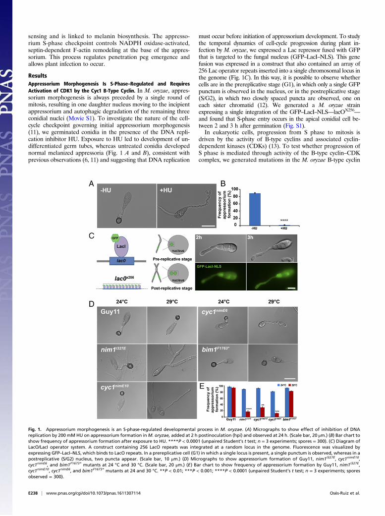
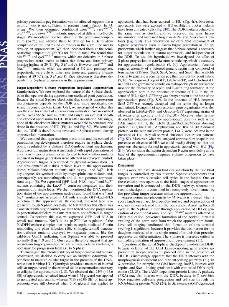
ResultsAppressorium Morphogenesis Is S-Phase–Regulated and RequiresActivation of CDK1 by the Cyc1 B-Type Cyclin. In M. oryzae, appres-sorium morphogenesis is always preceded by a single round ofmitosis, resulting in one daughter nucleus moving to the incipientappressorium and autophagic degradation of the remaining threeconidial nuclei (Movie S1). To investigate the nature of the cell-cycle checkpoint governing initial appressorium morphogenesis(11), we germinated conidia in the presence of the DNA repli-cation inhibitor HU. Exposure to HU led to development of un-differentiated germ tubes, whereas untreated conidia developednormal melanized appressoria (Fig. 1 A and B), consistent withprevious observations (6, 11) and suggesting that DNA replication
must occur before initiation of appressorium development. To studythe temporal dynamics of cell-cycle progression during plant in-fection by M. oryzae, we expressed a Lac repressor fused with GFPthat is targeted to the fungal nucleus (GFP–LacI–NLS). This genefusion was expressed in a construct that also contained an array of256 Lac operator repeats inserted into a single chromosomal locus inthe genome (Fig. 1C). In this way, it is possible to observe whethercells are in the prereplicative stage (G1), in which only a single GFPpunctum is observed in the nucleus, or in the postreplicative stage(S/G2), in which two closely spaced puncta are observed, one oneach sister chromatid (12). We generated a M. oryzae strainexpressing a single integration of the GFP–LacI–NLS—lacOX256
—
and found that S-phase entry occurs in the apical conidial cell be-tween 2 and 3 h after germination (Fig. S1).In eukaryotic cells, progression from S phase to mitosis is
driven by the activity of B-type cyclins and associated cyclin-dependent kinases (CDKs) (13). To test whether progression ofS phase is mediated through activity of the B-type cyclin–CDKcomplex, we generated mutations in the M. oryzae B-type cyclin
Fig. 1. Appressorium morphogenesis is an S-phase-regulated developmental process in M. oryzae. (A) Micrographs to show effect of inhibition of DNAreplication by 200 mM HU on appressorium formation inM. oryzae, added at 2 h postinoculation (hpi) and observed at 24 h. (Scale bar, 20 μm.) (B) Bar chart toshow frequency of appressorium formation after exposure to HU. ****P < 0.0001 (unpaired Student’s t test; n = 3 experiments; spores = 300). (C) Diagram ofLacO/LacI operator system. A construct containing 256 LacO repeats was integrated at a random locus in the genome. Fluorescence was visualized byexpressing GFP–LacI–NLS, which binds to LacO repeats. In a prereplicative cell (G1) in which a single locus is present, a single punctum is observed, whereas in apostreplicative (S/G2) nucleus, two puncta appear. (Scale bar, 10 μm.) (D) Micrographs to show appressorium formation of Guy11, nim1I327E, cyc1nimE10,cyc1nimE6, and bim1F1673* mutants at 24 °C and 30 °C. (Scale bar, 20 μm.) (E) Bar chart to show frequency of appressorium formation by Guy11, nim1I327E,cyc1nimE10, cyc1nimE6, and bim1F1673* mutants at 24 and 30 °C. **P < 0.01; ***P < 0.001; ****P < 0.0001 (unpaired Student’s t test; n = 3 experiments; sporesobserved = 300).
E238 | www.pnas.org/cgi/doi/10.1073/pnas.1611307114 Osés-Ruiz et al.
gene, CYC1, which is functionally equivalent to the Aspergillusnidulans NIME gene (11). In A. nidulans, two distinct point mu-tations inNIME, the nimE10 (originally identified as nimG10) andnimE6 alleles confer temperature sensitivity and distinct cell-cyclearrest in either the S or G2 phase, respectively (14–17). To testwhether appressorium morphogenesis is directly dependent onactivity of the CDK–cyclin B complex, we carried out targetedallelic replacements to generate cyc1nimE10 and cyc1nimE6 mutants,carrying S389R and F465P mutations, respectively, which areequivalent to the mutations found in the corresponding nimE10and nimE6 of A. nidulans (17) (Fig. S2). These mutants carried asingle targeted allelic replacement of the cyc1nimE10 and cyc1nimE6
alleles, confirmed by DNA sequencing, and showed severe growthdefects at the semirestrictive temperature of 30 °C (Fig. S3). Weevaluated the ability of these mutants to make appressoria at asemirestrictive temperature (29 °C) (Table S1). Conidia of thecyc1nimE10 strain produced hyperpolarized germ tubes that failedto develop appressoria, whereas the cyc1nimE6 mutant producedrelatively thicker germ tubes that underwent flattening andhooking of the germ tube tip, consistent with incipient appresso-rium formation (Fig. 1D). We also analyzed a nim1I327E mutant,which fails to initiate DNA synthesis and instead undergoes mi-totic catastrophe (11). The nim1I327E mutant was also unable todevelop appressoria. By contrast, a bim1F1763* mutant, which ar-rests during mitosis, before anaphase, developed appressorianormally (Fig. 1D) (6, 11). We conclude that appressoriummorphogenesis in M. oryzae requires an S-phase checkpoint thatoperates via activation of the B-type cyclin–CDK1 complex.
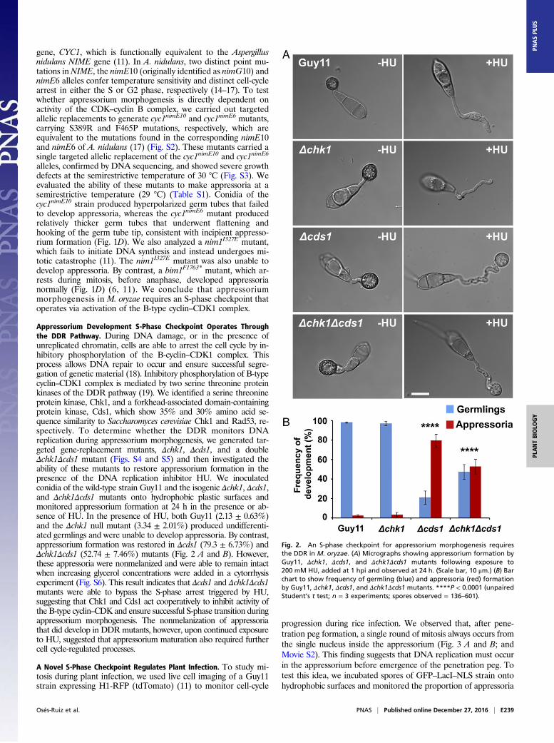
Appressorium Development S-Phase Checkpoint Operates Throughthe DDR Pathway. During DNA damage, or in the presence ofunreplicated chromatin, cells are able to arrest the cell cycle by in-hibitory phosphorylation of the B-cyclin–CDK1 complex. Thisprocess allows DNA repair to occur and ensure successful segre-gation of genetic material (18). Inhibitory phosphorylation of B-typecyclin–CDK1 complex is mediated by two serine threonine proteinkinases of the DDR pathway (19). We identified a serine threonineprotein kinase, Chk1, and a forkhead-associated domain-containingprotein kinase, Cds1, which show 35% and 30% amino acid se-quence similarity to Saccharomyces cerevisiae Chk1 and Rad53, re-spectively. To determine whether the DDR monitors DNAreplication during appressorium morphogenesis, we generated tar-geted gene-replacement mutants, Δchk1, Δcds1, and a doubleΔchk1Δcds1 mutant (Figs. S4 and S5) and then investigated theability of these mutants to restore appressorium formation in thepresence of the DNA replication inhibitor HU. We inoculatedconidia of the wild-type strain Guy11 and the isogenic Δchk1, Δcds1,and Δchk1Δcds1 mutants onto hydrophobic plastic surfaces andmonitored appressorium formation at 24 h in the presence or ab-sence of HU. In the presence of HU, both Guy11 (2.13 ± 0.63%)and the Δchk1 null mutant (3.34 ± 2.01%) produced undifferenti-ated germlings and were unable to develop appressoria. By contrast,appressorium formation was restored in Δcds1 (79.3 ± 6.73%) andΔchk1Δcds1 (52.74 ± 7.46%) mutants (Fig. 2 A and B). However,these appressoria were nonmelanized and were able to remain intactwhen increasing glycerol concentrations were added in a cytorrhysisexperiment (Fig. S6). This result indicates that Δcds1 and Δchk1Δcds1mutants were able to bypass the S-phase arrest triggered by HU,suggesting that Chk1 and Cds1 act cooperatively to inhibit activity ofthe B-type cyclin–CDK and ensure successful S-phase transition duringappressorium morphogenesis. The nonmelanization of appressoriathat did develop in DDRmutants, however, upon continued exposureto HU, suggested that appressorium maturation also required furthercell cycle-regulated processes.
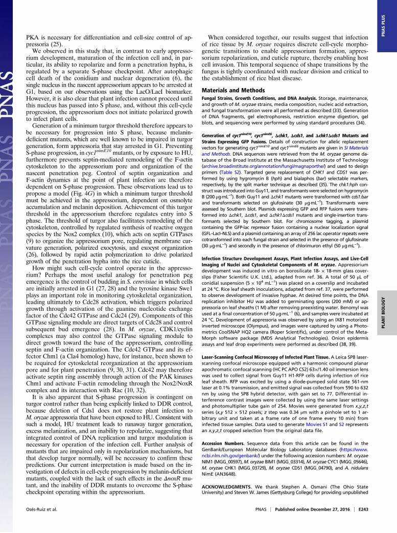
A Novel S-Phase Checkpoint Regulates Plant Infection. To study mi-tosis during plant infection, we used live cell imaging of a Guy11strain expressing H1-RFP (tdTomato) (11) to monitor cell-cycle
progression during rice infection. We observed that, after pene-tration peg formation, a single round of mitosis always occurs fromthe single nucleus inside the appressorium (Fig. 3 A and B; andMovie S2). This finding suggests that DNA replication must occurin the appressorium before emergence of the penetration peg. Totest this idea, we incubated spores of GFP–LacI–NLS strain ontohydrophobic surfaces and monitored the proportion of appressoria
Fig. 2. An S-phase checkpoint for appressorium morphogenesis requiresthe DDR in M. oryzae. (A) Micrographs showing appressorium formation byGuy11, Δchk1, Δcds1, and Δchk1Δcds1 mutants following exposure to200 mM HU, added at 1 hpi and observed at 24 h. (Scale bar, 10 μm.) (B) Barchart to show frequency of germling (blue) and appressoria (red) formationby Guy11, Δchk1, Δcds1, and Δchk1Δcds1 mutants. ****P < 0.0001 (unpairedStudent’s t test; n = 3 experiments; spores observed = 136–601).
Osés-Ruiz et al. PNAS | Published online December 27, 2016 | E239
PLANTBIOLO
GY
PNASPL
US
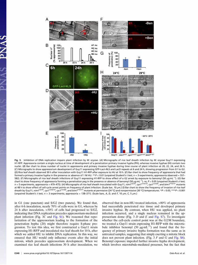
in G1 (one punctum) and S/G2 (two puncta). We found that,after 6-h inoculation, nearly 70% of cells were in G1, whereas by24 h after inoculation, >50% of cells had progressed to S/G2,indicating that DNA replication precedes appressorium-mediatedplant infection (Fig. 3C and Fig. S1). We reasoned that repo-larization of the appressorium leading to the formation of thepenetration hypha (20) might therefore require S-phase pro-gression. To test this idea, we first constructed a Guy11 strainexpressing H1-RFP and inoculated rice leaf sheath for 10 h, afterwhich we added HU to inhibit DNA replication. In this way, weensured that HU would only influence events after the initialmitosis, which precedes appressorium development. When weexamined rice leaf sheath infections 30 h after inoculation, we
observed that in non-HU-treated infection, >80% of appressoriahad successfully penetrated rice tissue and developed primaryinvasive hyphae. By contrast, when HU was applied, no plantinfection occurred, and a single nucleus remained in the ap-pressorium dome (Fig. 3 D and E and Fig. S7). To investigatewhether the cell-cycle control point was at the G2/M boundary,we treated a Guy11 strain expressing H1-RFP with the microtu-bule inhibitor benomyl (50 μg·mL−1) and found that the fre-quency of primary invasive hypha formation was the same as inuntreated samples, suggesting that simply exerting a mitotic blockdoes not prevent plant infection. (Fig. 3 F and G and Fig. S8).Benomyl exposure impeded further invasive hypha development,which involves microtubule-mediated processes, but the fact that
Fig. 3. Inhibition of DNA replication impairs plant infection by M. oryzae. (A) Micrographs of rice leaf sheath infection by M. oryzae Guy11 expressingH1-RFP. Appressoria contain a single nucleus at time of development of a penetration primary invasive hypha (PH), whereas invasive hyphae (IH) contain twonuclei. (B) Bar chart to show number of nuclei in appressoria and primary invasive hyphae during time course of plant infection at 20, 22, 24, and 26 h.(C) Micrographs to show appressorium development of Guy11 expressing GFP–LacI–NLS and LacO repeats at 6 and 24 h, showing progression from G1 to G2.(D) Rice leaf sheath observed 30 h after inoculation with Guy11 H1-RFP after exposure to HU at 10 h. (E) Bar chart to show frequency of appressoria that hadformed a primary invasive hypha in the presence or absence of 1 M HU. **P < 0.01 (unpaired Student’s t test; n = 3 experiments; appressoria observed = 557–582). (F) Micrographs of rice leaf sheath infections of Guy11 expressing H1-RFP to show effect of a G2 arrest by exposure to benomyl (50 μg·mL−1). (G) Barchart to show frequency of appressoria forming a penetration peg in the presence or absence of benomyl (50 μg·mL−1). ns, P > 0.05 (unpaired Student’s t test;n = 5 experiments, appressoria = 479–475). (H) Micrographs of rice leaf sheath inoculated with Guy11, nim1I327E, cyc1nimE10, cyc1nimE6, and bim1F1673* mutantsat 48 h to show effect of cell-cycle arrest points on frequency of plant infection. (Scale bar, 10 μm.) (I) Bar chart to show the frequency of invasion of rice leafsheath by Guy11, nim1I327E, cyc1nimE10, cyc1nimE6, and bim1F1673* mutants at permissive (24 °C) and nonpermissive (30 °C) temperatures. *P < 0.05; ***P < 0.001(unpaired Student’s t test; n = 3 experiments; appressoria = 138–371). (Scale bars, A, D, and F, 10 μm; C, 5 μm.)
E240 | www.pnas.org/cgi/doi/10.1073/pnas.1611307114 Osés-Ruiz et al.
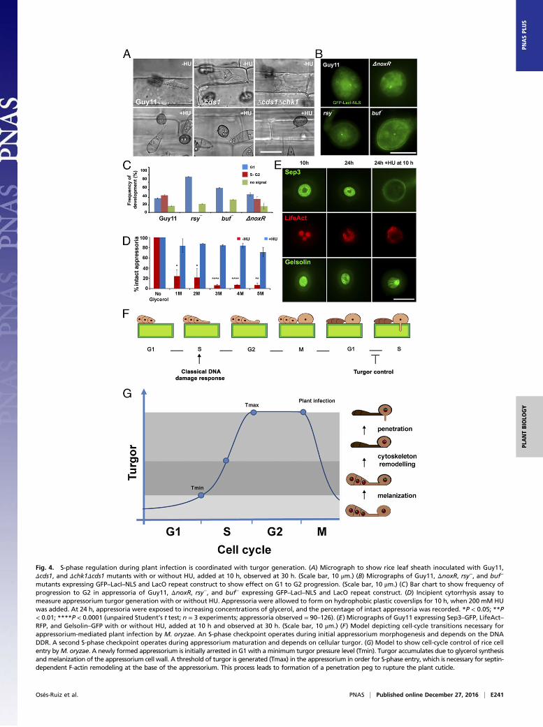
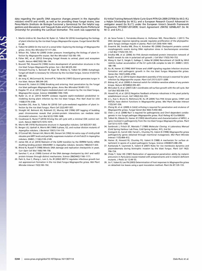
Fig. 4. S-phase regulation during plant infection is coordinated with turgor generation. (A) Micrograph to show rice leaf sheath inoculated with Guy11,Δcds1, and Δchk1Δcds1 mutants with or without HU, added at 10 h, observed at 30 h. (Scale bar, 10 μm.) (B) Micrographs of Guy11, ΔnoxR, rsy−, and buf−
mutants expressing GFP–LacI–NLS and LacO repeat construct to show effect on G1 to G2 progression. (Scale bar, 10 μm.) (C) Bar chart to show frequency ofprogression to G2 in appressoria of Guy11, ΔnoxR, rsy−, and buf− expressing GFP–LacI–NLS and LacO repeat construct. (D) Incipient cytorrhysis assay tomeasure appressorium turgor generation with or without HU. Appressoria were allowed to form on hydrophobic plastic coverslips for 10 h, when 200 mM HUwas added. At 24 h, appressoria were exposed to increasing concentrations of glycerol, and the percentage of intact appressoria was recorded. *P < 0.05; **P< 0.01; ****P < 0.0001 (unpaired Student’s t test; n = 3 experiments; appressoria observed = 90–126). (E) Micrographs of Guy11 expressing Sep3–GFP, LifeAct–RFP, and Gelsolin–GFP with or without HU, added at 10 h and observed at 30 h. (Scale bar, 10 μm.) (F) Model depicting cell-cycle transitions necessary forappressorium-mediated plant infection by M. oryzae. An S-phase checkpoint operates during initial appressorium morphogenesis and depends on the DNADDR. A second S-phase checkpoint operates during appressorium maturation and depends on cellular turgor. (G) Model to show cell-cycle control of rice cellentry byM. oryzae. A newly formed appressorium is initially arrested in G1 with a minimum turgor pressure level (Tmin). Turgor accumulates due to glycerol synthesisandmelanization of the appressorium cell wall. A threshold of turgor is generated (Tmax) in the appressorium in order for S-phase entry, which is necessary for septin-dependent F-actin remodeling at the base of the appressorium. This process leads to formation of a penetration peg to rupture the plant cuticle.
Osés-Ruiz et al. PNAS | Published online December 27, 2016 | E241
PLANTBIOLO
GY
PNASPL
US
primary penetration peg formation was not affected suggests that amitotic block is not sufficient to prevent plant infection by M.oryzae. We then examined conditional nim1I327E, cyc1nimE10,cyc1nimE6, and bim1F1763* mutants, impaired at different cell-cyclestages. We inoculated rice leaf sheath at the permissive temper-ature (24 °C) and allowed them to develop for 10 h to allowcompletion of the first round of mitosis in the germ tube and todevelop an appressorium. We then incubated them at the semi-restrictive temperature (29 °C) for 30 h in total. We found thatnim1I327E and cyc1nimE10 mutants, which are defective in S-phaseprogression, were unable to infect rice tissue and form primaryinvasive hyphae at 29 °C (Fig. 3 H and I). However, cyc1nimE6 andbim1F1763* mutants, which are arrested at G2 and preanaphase,respectively, were able to infect rice tissue and generate invasivehyphae at 29 °C (Fig. 3 H and I). Rice infection is therefore de-pendent on S-phase progression in the appressorium.
Turgor-Dependent S-Phase Progression Regulates AppressoriumRepolarization. We next explored the nature of the S-phase check-point that operates during appressorium-mediated plant penetration.Because the initial S-phase checkpoint that controls appressoriummorphogenesis depends on the DDR and, more specifically, theserine threonine protein kinase Cds1, we investigated whether thiswas the case for control of appressorium maturation. We inoculatedΔcds1, Δchk1Δcds1 null mutants, and Guy11 on rice leaf sheathand exposed appressoria to HU 10 h after inoculation. Strikingly,none of the checkpoint kinase mutants were able to penetrate ricetissue in the presence of HU (Fig. 4A and Fig. S9). We concludethat the DDR is therefore not involved in S-phase control duringappressorium maturation.We reasoned that appressorium maturation and the control of
penetration peg development therefore require an S-phase check-point, regulated by a distinct DDR-independent mechanism.Appressorium maturation is associated with rapid generation ofenormous turgor pressure, so we decided to test whether mutantsimpaired in turgor generation were affected in cell-cycle control.Appressorium turgor is generated by glycerol accumulation (7)and development of a thick melanin layer in the appressoriumdome. Melanin-deficient mutants, such as rsy1− and buf1−, lackkey enzymes for synthesis of di-hydroxynaphthalene melanin and,consequently, are nonpathogenic and do not generate appresso-rium turgor (8). We expressed GFP–LacI–NLS in rsy1− and buf1−
mutants containing the LacOx256 construct integrated into theirgenomes at a single locus. We then monitored the DNA replica-tion status of the appressorium nucleus and found that rsy1− andbuf1− mutants are arrested in G1 with a single GFP–LacI–NLSpunctum in the appressorium. By contrast, the wild type pro-gressed through S phase normally. To test whether this effect wasassociated with turgor control, we then tested S-phase progressionin penetration-deficient mutants that were not affected in turgorcontrol. To perform this test, we expressed GFP–LacI–NLS inΔnoxR null mutants. NoxR is the p67phox-like regulator of theNADPH oxidase complex that regulates septin-mediated F-actinremodeling and plant infection (10), Strikingly, ΔnoxR penetra-tion-deficient mutants displayed two separate puncta, like thewild-type Guy11, indicating that S-phase was able to proceednormally (Fig. 4 B and C). Our results therefore suggest that ap-pressorium turgor generation, which requires melanin synthesis, isnecessary for progression from G1 to S phase.To investigate the interdependency of turgor control and S-phase
progression, we decided to carry out an incipient cytorrhysis ex-periment to measure cellular turgor in the presence of the DNA-replication inhibitor HU. Cytorrhysis provides a measure of internalcellular turgor, based on the external solute concentration necessaryto collapse the appressorium (7, 8). We observed that 24% (±15.6SE) of appressoria remained intact when 1 M glycerol was appliedto nontreated appressoria, whereas 71% (±7.06 SE) of intact ap-pressoria were still observed when 5 M glycerol was applied to
appressoria that had been exposed to HU (Fig. 4D). Moreover,appressoria that were exposed to HU exhibited a thicker melaninlayer from untreated cells (Fig. S10). The DDRmutants behaved inthe same way as Guy11, and we observed the same hyper-melanization and increased turgor in Δcds1 and Δchk1Δcds1 mu-tants (Fig. S10). This observation indicates that impairment ofS-phase progression leads to excess turgor generation in the ap-pressorium, which further suggests that S-phase control is necessaryfor turgor modulation in mature appressoria, and independent ofthe DDR. To test this hypothesis, we investigated the effect ofS-phase progression on cytoskeleton remodeling, which is necessaryfor appressorium repolarization (9, 10). Appressorium functionrequires assembly of a heterooligomeric septin ring composed offour septin GTPases (Sep3, Sep4, Sep5, and Sep6) that scaffoldsF-actin to generate a penetration peg that ruptures the plant cuticle(9, 10). We expressed Sep3–GFP, LifeAct–RFP, and Gelsolin–GFPin Guy11 and germinated conidia on hydrophobic plastic surfaces tomonitor the frequency of septin and F-actin ring formation at theappressorium pore in the presence or absence of HU. In the ab-sence of HU, a Sep3–GFP ring was always present at the base of theappressorium pore (Fig. 3D). In the presence of HU, however,Sep3–GFP was severely disrupted and the septin ring no longermaintained. Disruption of appressorium pore organization was alsoobserved in LifeAct–RFP and Gelsolin–GFP expressing strains ofM. oryzae after exposure to HU (Fig. 3D). Moreover when septin-dependent components of the appressorium pore (9), such as thePAK kinase Chm1, the ERM (Ezrin/Radiixin/Moesin domain)protein Tea1, the Bim1, Amphiphysin, and Rvs167 BAR domainprotein, or the actin nucleation protein, Las17, were localized in thepresence of HU, they all showed abnormal localization patterns(Fig. S9). Moreover, when we analyzed appressorium shape in thepresence or absence of HU, we could readily distinguish that thepore was aberrantly formed in appressoria treated with HU (Fig.S10). We conclude that septin-dependent F-actin remodeling at thebase of the appressorium requires S-phase progression to havetaken place.
DiscussionIn this work, we have shown that rice infection by the rice blastfungus is controlled by two discrete S-phase checkpoints thatoperate over two successive cell cycles in the fungus. One ofthese checkpoints operates at the initial stages of appressoriumformation and is connected to the DDR pathway, whereas thesecond checkpoint is controlled in a completely novel manner bythe prevailing turgor pressure within the appressorium.Appressorium morphogenesis by M. oryzae is induced when a
spore lands on a hard, hydrophobic surface and by perception ofwax monomers released from the rice cuticle. Arresting the cellcycle at the S phase, either through application of HU or gen-eration of conditional nim1 and cyc1 nimE10 mutants affected inDNA replication, prevented formation of the hooked, terminalswelling of the germ tube from which the appressorium forms.Live cell imaging confirmed that formation of this terminalswelling is significant, because it provides the destination for thedaughter nucleus, after the single round of mitosis that precedesappressorium differentiation. The S phase is therefore critical incontrolling initiation of appressorium development (11).Operation of the initial S-phase checkpoint involves the DDR,
because deletion of the Cds1 kinase-encoding gene allows ap-pressorium morphogenesis to proceed even in the presence ofHU. It is increasingly apparent that the DDR interacts with themorphogenesis checkpoint and nutrient-sensing pathways (21). InS. cerevisiae, for example, the Cds1 homolog, Rad53, interacts withseptins and Swe1, linking cytokinesis with control of DNA repli-cation (22, 23). The cAMP-dependent protein kinase A pathway(PKA) may also interact with the DDR, because in S. cerevisiaePKA regulates cell-cycle progression and cell size through theRNA-binding protein Whi3 (24). In M. oryzae, cAMP-dependent
E242 | www.pnas.org/cgi/doi/10.1073/pnas.1611307114 Osés-Ruiz et al.
PKA is necessary for differentiation and cell-size control of ap-pressoria (25).We observed in this study that, in contrast to early appresso-
rium development, maturation of the infection cell and, in par-ticular, its ability to repolarize and form a penetration hypha, isregulated by a separate S-phase checkpoint. After autophagiccell death of the conidium and nuclear degeneration (6), thesingle nucleus in the nascent appressorium appears to be arrested atG1, based on our observations using the LacO/LacI biomarker.However, it is also clear that plant infection cannot proceed untilthis nucleus has passed into S phase, and, without this cell-cycleprogression, the appressorium does not initiate polarized growthto infect plant cells.Generation of a minimum turgor threshold therefore appears to
be necessary for progression into S phase, because melanin-deficient mutants, which are well known to be impaired in turgorgeneration, form appressoria that stay arrested in G1. PreventingS-phase progression, in cyc1nimE10 mutants, or by exposure to HU,furthermore prevents septin-mediated remodeling of the F-actincytoskeleton to the appressorium pore and organization of thenascent penetration peg. Control of septin organization andF-actin dynamics at the point of plant infection are thereforedependent on S-phase progression. These observations lead us topropose a model (Fig. 4G) in which a minimum turgor thresholdmust be achieved in the appressorium, dependent on osmolyteaccumulation and melanin deposition. Achievement of this turgorthreshold in the appressorium therefore regulates entry into Sphase. The threshold of turgor also facilitates remodeling of thecytoskeleton, controlled by regulated synthesis of reactive oxygenspecies by the Nox2 complex (10), which acts on septin GTPases(9) to organize the appressorium pore, regulating membrane cur-vature generation, polarized exocytosis, and exocyst organization(26), followed by rapid actin polymerization to drive polarizedgrowth of the penetration hypha into the rice cuticle.How might such cell-cycle control operate in the appresso-
rium? Perhaps the most useful analogy for penetration pegemergence is the control of budding in S. cerevisiae in which cellsare initially arrested in G1 (27, 28) and the tyrosine kinase Swe1plays an important role in monitoring cytoskeletal organization,leading ultimately to Cdc28 activation, which triggers polarizedgrowth through activation of the guanine nucleotide exchangefactor of the Cdc42 GTPase and Cdc24 (29). Components of thisGTPase signaling module are direct targets of Cdc28 and controlsubsequent bud emergence (28). In M. oryzae, CDK1/cyclincomplexes may also control the GTPase signaling module todirect growth toward the base of the appressorium, controllingseptin and F-actin organization. The Cdc42 GTPase and its ef-fector Chm1 (a Cla4 homolog) have, for instance, been shown tobe required for cytoskeletal reorganization at the appressoriumpore and for plant penetration (9, 30, 31). Cdc42 may thereforeactivate septin ring assembly through action of the PAK kinasesChm1 and activate F-actin remodeling through the Nox2/NoxRcomplex and its interaction with Rac (10, 32).It is also apparent that S-phase progression is contingent on
turgor control rather than being explicitly linked to DDR control,because deletion of Cds1 does not restore plant infection toM. oryzae appressoria that have been exposed to HU. Consistent withsuch a model, HU treatment leads to runaway turgor generation,excess melanization, and an inability to repolarize, suggesting thatintegrated control of DNA replication and turgor modulation isnecessary for operation of the infection cell. Further analysis ofmutants that are impaired only in repolarization mechanisms, butthat develop turgor normally, will be necessary to confirm thesepredictions. Our current interpretation is made based on the in-vestigation of defects in cell-cycle progression by melanin-deficientmutants, coupled with the lack of such effects in the ΔnoxR mu-tant, and the inability of DDR mutants to overcome the S-phasecheckpoint operating within the appressorium.
When considered together, our results suggest that infectionof rice tissue by M. oryzae requires discrete cell-cycle morpho-genetic transitions to enable appressorium formation, appres-sorium repolarization, and cuticle rupture, thereby enabling hostcell invasion. This temporal sequence of shape transitions by thefungus is tightly coordinated with nuclear division and critical tothe establishment of rice blast disease.
Materials and MethodsFungal Strains, Growth Conditions, and DNA Analysis. Storage, maintenance,and growth of M. oryzae strains, media composition, nucleic acid extraction,and fungal transformation were all performed as described (33). Generationof DNA fragments, gel electrophoresis, restriction enzyme digestion, gelblots, and sequencing were performed by using standard procedures (34).
Generation of cyc1nimE10, cyc1nimE6, Δchk1, Δcds1, and Δchk1Δcds1 Mutants andStrains Expressing GFP Fusions. Details of construction for allelic replacementvectors for generating cyc1nimE10 and cyc1nimE6 mutants are given in SI Materialsand Methods. DNA sequences were retrieved from the M. oryzae genome da-tabase of the Broad Institute at the Massachusetts Institute of Technology(archive.broadinstitute.org/annotation/fungi/magnaporthe/) and used to designprimers (Table S2). Targeted gene replacement of CHK1 and CDS1 was per-formed by using hygromycin B (hph) and bialaphos (bar) selectable markers,respectively, by the split marker technique as described (35). The chk1:hph con-struct was introduced into Guy11, and transformants were selected on hygromycinB (200 μg·mL−1). Both Guy11 and Δchk1 mutants were transformed with cds1:barand transformants selected on glufosinate (30 μg·mL−1). Transformants wereassessed by Southern blot. Plasmids expressing GFP and RFP fusions were trans-formed into Δchk1, Δcds1, and Δchk1Δcds1 mutants and single-insertion trans-formants selected by Southern blot. For chromosome tagging, a plasmidcontaining the GFP-lac repressor fusion containing a nuclear localization signal(GFL–LacI–NLS) and a plasmid containing an array of 256 lac operator repeats werecotransformed into each fungal strain and selected in the presence of glufosinate(30 μg·mL−1) and secondly in the presence of chlorimuron ethyl (50 μg·mL−1).
Infection Structure Development Assays, Plant Infection Assays, and Live-CellImaging of Nuclei and Cytoskeletal Components of M. oryzae. Appressoriumdevelopment was induced in vitro on borosilicate 18- × 18-mm glass cover-slips (Fisher Scientific U.K. Ltd.), adapted from ref. 36. A total of 50 μL ofconidial suspension (5 × 10⁴ mL−1) was placed on a coverslip and incubatedat 24 °C. Rice leaf sheath inoculations, adapted from ref. 37, were performedto observe development of invasive hyphae. At desired time points, the DNAreplication inhibitor HU was added to germinating spores (200 mM) or ap-pressoria on leaf sheaths (1 M) after removing preexisting water. Benomyl wasused at a final concentration of 50 μg·mL−1 (6), and samples were incubated at24 °C. Development of appressoria was observed by using an IX81 motorizedinverted microscope (Olympus), and images were captured by using a Photo-metrics CoolSNAP HQ2 camera (Roper Scientific), under control of the Meta-Morph software package (MDS Analytical Technologies). Onion epidermisassays and leaf drop experiments were performed as described (38, 39).
Laser-Scanning Confocal Microscopy of Infected Plant Tissue. A Leica SP8 laser-scanning confocal microscope equipped with a harmonic compound planarapochromatic confocal scanning (HC PC APO CS2) 63×/1.40 oil immersion lenswas used to collect signal from Guy11 H1-RFP cells during infection of riceleaf sheath. RFP was excited by using a diode-pumped solid state 561-nmlaser at 0.1% transmission, and emitted signal was collected from 590 to 632nm by using the SP8 hybrid detector, with gain set to 77. Differential in-terference contrast images were collected by using the same laser settingsand photomultiplier tube gain of 254. Movies were generated from x,y,z,tseries (x,y 512 × 512 pixels; z step was 0.34 μm with a pinhole set to 1 ar-bitrary unit and taken at a frame rate of one frame every 10 min) frominfected tissue samples. Data used to generate Movies S1 and S2 representsan x,y,z,t cropped selection from the original data file.
Accession Numbers. Sequence data from this article can be found in theGenBank/European Molecular Biology Laboratory databases (https://www.ncbi.nlm.nih.gov/genbank/) under the following accession numbers: M. oryzaeNIM1 (MGG_00597),M. oryzae BIM1 (MGG_03314),M. oryzae CYC1 (MGG_05646),M. oryzae CHK1 (MGG_03729), M. oryzae CDS1 (MGG_04790), and A. nidulansNimE (AN3648).
ACKNOWLEDGMENTS. We thank Stephen A. Osmani (The Ohio StateUniversity) and Steven W. James (Gettysburg College) for providing unpublished
Osés-Ruiz et al. PNAS | Published online December 27, 2016 | E243
PLANTBIOLO
GY
PNASPL
US
data regarding the specific DNA sequence changes present in the Aspergillusnidulans nimE10 and nimE6, as well as for providing these fungal strains; JosePerez-Martin (Instituto de Biología Funcional y Genómica) for the helpful sug-gestions and discussions; and Yasuyuki Kubo and Fumi Fukada (Kyoto PrefecturalUniversity) for providing the LacO/LacI biomarker. This work was supported by
EU-Initial Training NetworkMarie Curie Grant PITN-GA-2009-237936 (toM.O.-R.);a Halpin Scholarship (to W.S.); and a European Research Council Advanced In-vestigator award (to N.J.T.) under the European Union’s Seventh FrameworkProgramme FP7/2007-2013/ERC Grant Agreement 294702 GENBLAST (to M.M.-U. and G.R.L.).
1. Martin-Urdiroz M, Oses-Ruiz M, Ryder LS, Talbot NJ (2016) Investigating the biology
of plant infection by the rice blast fungusMagnaporthe oryzae. Fungal Genet Biol 90:
61–68.2. Talbot NJ (2003) On the trail of a cereal killer: Exploring the biology of Magnaporthe
grisea. Annu Rev Microbiol 57:177–202.3. Wilson RA, Talbot NJ (2009) Under pressure: Investigating the biology of plant in-
fection by Magnaporthe oryzae. Nat Rev Microbiol 7(3):185–195.4. Fisher MC, et al. (2012) Emerging fungal threats to animal, plant and ecosystem
health. Nature 484(7393):186–194.5. Bourett TM, Howard RJ (1990) Invitro development of penetration structures in the
rice blast fungus Magnaporthe grisea. Can J Bot 68:329–342.6. Veneault-Fourrey C, Barooah M, Egan M, Wakley G, Talbot NJ (2006) Autophagic
fungal cell death is necessary for infection by the rice blast fungus. Science 312(5773):
580–583.7. de Jong JC, McCormack BJ, Smirnoff N, Talbot NJ (1997) Glycerol generates turgor in
rice blast. Nature 389:244–245.8. Howard RJ, Valent B (1996) Breaking and entering: Host penetration by the fungal
rice blast pathogen Magnaporthe grisea. Annu Rev Microbiol 50:491–512.9. Dagdas YF, et al. (2012) Septin-mediated plant cell invasion by the rice blast fungus,
Magnaporthe oryzae. Science 336(6088):1590–1595.10. Ryder LS, et al. (2013) NADPH oxidases regulate septin-mediated cytoskeletal re-
modeling during plant infection by the rice blast fungus. Proc Natl Acad Sci USA
110(8):3179–3184.11. Saunders DG, Aves SJ, Talbot NJ (2010) Cell cycle-mediated regulation of plant in-
fection by the rice blast fungus. Plant Cell 22(2):497–507.12. Straight AF, Belmont AS, Robinett CC, Murray AW (1996) GFP tagging of budding
yeast chromosomes reveals that protein-protein interactions can mediate sister
chromatid cohesion. Curr Biol 6(12):1599–1608.13. Coudreuse D, Nurse P (2010) Driving the cell cycle with a minimal CDK control net-
work. Nature 468(7327):1074–1079.14. Morris NR (1976) Nucleosome structure in Aspergillus nidulans. Cell 8(3):357–363.15. Bergen LG, Upshall A, Morris NR (1984) S-phase, G2, and nuclear division mutants of
Aspergillus nidulans. J Bacteriol 159(1):114–119.16. O’Connell MJ, Osmani AH, Morris NR, Osmani SA (1992) An extra copy of nimEcyclinB
elevates pre-MPF levels and partially suppresses mutation of nimTcdc25 in Aspergillus
nidulans. EMBO J 11(6):2139–2149.17. James SW, et al. (2014) Restraint of the G2/M transition by the SR/RRM family mRNA
shuttling binding protein SNXAHRB1 in Aspergillus nidulans. Genetics 198(2):617–633.18. Rhind N, Russell P (1998) Mitotic DNA damage and replication checkpoints in yeast.
Curr Opin Cell Biol 10(6):749–758.19. Sanchez Y, et al. (1999) Control of the DNA damage checkpoint by chk1 and rad53
protein kinases through distinct mechanisms. Science 286(5442):1166–1171.20. Park G, Xue C, Zheng L, Lam S, Xu JR (2002) MST12 regulates infectious growth but
not appressorium formation in the rice blast fungus Magnaporthe grisea. Mol Plant
Microbe Interact 15(3):183–192.
21. de Sena-Tomás C, Fernández-Álvarez A, Holloman WK, Pérez-Martín J (2011) TheDNA damage response signaling cascade regulates proliferation of the phytopatho-genic fungus Ustilago maydis in planta. Plant Cell 23(4):1654–1665.
22. Enserink JM, Smolka MB, Zhou H, Kolodner RD (2006) Checkpoint proteins controlmorphogenetic events during DNA replication stress in Saccharomyces cerevisiae.J Cell Biol 175(5):729–741.
23. Smolka MB, et al. (2006) An FHA domain-mediated protein interaction network ofRad53 reveals its role in polarized cell growth. J Cell Biol 175(5):743–753.
24. Wang H, Garí E, Vergés E, Gallego C, Aldea M (2004) Recruitment of Cdc28 by Whi3restricts nuclear accumulation of the G1 cyclin-Cdk complex to late G1. EMBO J 23(1):180–190.
25. Xu JR, Hamer JE (1996) MAP kinase and cAMP signaling regulate infection structureformation and pathogenic growth in the rice blast fungus Magnaporthe grisea.Genes Dev 10(21):2696–2706.
26. Gupta YK, et al. (2015) Septin-dependent assembly of the exocyst is essential for plantinfection by Magnaporthe oryzae. Plant Cell 27(11):3277–3289.
27. Bishop AC, et al. (2000) A chemical switch for inhibitor-sensitive alleles of any proteinkinase. Nature 407(6802):395–401.
28. McCusker D, et al. (2007) Cdk1 coordinates cell-surface growth with the cell cycle. NatCell Biol 9(5):506–515.
29. Howell AS, et al. (2012) Negative feedback enhances robustness in the yeast polarityestablishment circuit. Cell 149(2):322–333.
30. Li L, Xue C, Bruno K, Nishimura M, Xu JR (2004) Two PAK kinase genes, CHM1 andMST20, have distinct functions in Magnaporthe grisea. Mol Plant Microbe Interact17(5):547–556.
31. Zheng W, et al. (2009) A Cdc42 ortholog is required for penetration and virulence ofMagnaporthe grisea. Fungal Genet Biol 46(6-7):450–460.
32. Chen J, et al. (2008) Rac1 is required for pathogenicity and Chm1-dependent conidio-genesis in rice fungal pathogen Magnaporthe grisea. PLoS Pathog 4(11):e1000202.
33. Talbot NJ, Ebbole DJ, Hamer JE (1993) Identification and characterization of MPG1, agene involved in pathogenicity from the rice blast fungus Magnaporthe grisea. PlantCell 5(11):1575–1590.
34. Sambrook J, Fritsch EF, Maniatis T (1989) Molecular Cloning: A Laboratory Manual(Cold Spring Harbour Lab Press, Cold Spring Harbor, NY), 2nd Ed.
35. Sweigard JA, Carroll AM, Farrall L, Chumley FG, Valent B (1998) Magnaporthe griseapathogenicity genes obtained through insertional mutagenesis. Mol Plant MicrobeInteract 11(5):404–412.
36. Hamer JE, Howard RJ, Chumley FG, Valent B (1988) A mechanism for surface at-tachment in spores of a plant pathogenic fungus. Science 239(4837):288–290.
37. Kankanala P, Czymmek K, Valent B (2007) Roles for rice membrane dynamics andplasmodesmata during biotrophic invasion by the blast fungus. Plant Cell 19(2):706–724.
38. Chida T, Sisler HD (1987) Restoration of appressorial penetration ability by melaninprecursors in Pyricularia oryzae treated with antipenetrants and in melanin-deficientmutants. J Pestic Sci 12:49–55.
39. Jia Y, Valent B, Lee FN (2003) Determination of host responses toMagnaporthe griseaon detached rice leaves using a spot inoculation method. Plant Dis 87:129–133.
E244 | www.pnas.org/cgi/doi/10.1073/pnas.1611307114 Osés-Ruiz et al.