two distinct regions in the model protein peb1 are
TRANSCRIPT

RESEARCH Open Access
Two distinct regions in the model protein Peb1are critical for its heterologous transport out ofEscherichia coliLena Anton1, Katariina Majander1, Harri Savilahti2, Liisa Laakkonen3, Benita Westerlund-Wikström1*
Abstract
Background: Escherichia coli is frequently the first-choice host organism in expression of heterologousrecombinant proteins in basic research as well as in production of commercial, therapeutic polypeptides. Especiallythe secretion of proteins into the culture medium of E. coli is advantageous compared to intracellular productiondue to the ease in recovery of the recombinant protein. Since E. coli naturally is a poor secretor of proteins, a fewstrategies for optimization of extracellular secretion have been described. We have previously reported efficientsecretion of the diagnostically interesting model protein Peb1 of Campylobacter jejuni into the growth medium ofEscherichia coli strain MKS12 (ΔfliCfliD). To generate a more detailed understanding of the molecular mechanismsbehind this interesting heterologous secretion system with biotechnological implications, we here analyzed furtherthe transport of Peb1 in the E. coli host.
Results: When mature Peb1 was expressed without its SecA-YEG -dependent signal sequence and without theputative signal peptidase II recognition sequence in E. coli MKS111ΔHBB lacking the flagellar secretion complex, theprotein was found in the periplasm and growth medium which indicated a flagellum-independent translocation.We assessed the Peb1 secretion proficiency by an exhaustive search for transport-affecting regions using atransposition-based scanning mutagenesis strategy. Strikingly, insertion mutagenesis of only two segments, calledTAR1 (residues 42 and 43) and TAR2 (residues 173 to 180), prevented Peb1 secretion individually. We confirmedthe importance of TAR regions by subsequent site-specific mutagenesis and verified that the secretion deficiencyof Peb1 mutants was not due to insolubility or aggregation of the proteins in the cytoplasm. We found by cellfractionation that the mutant proteins were present in the periplasm as well as in the cytoplasm of MKS12. Hence,mutagenesis of TAR regions did not affect export of Peb1 across the cytoplasmic membrane, whereas its exportover the outer membrane was markedly impaired.
Conclusions: We propose that the localization of the model protein Peb1 in the growth medium of E. coli is dueto active secretion by a still unknown pathway of E. coli. The secretion apparently is a two-step process involving aperiplasmic step and the TAR regions.
BackgroundProtein secretion is one of the main means by whichbacteria interact with their environment. The interactionmay take place in a variety of manners: bacteria secreteenzymes, toxins and other virulence factors, excretemetabolic waste products, and export binding proteinsinto the periplasm for import of nutrients or export of
toxic compounds. Bacteria also use different secretionsystems to assemble on their surface organelles formotility, adhesion and injection of effector moleculesinto host cells [1,2]. Bacterial protein secretion systemsare of great importance from a virulence-associatedviewpoint as potential targets for novel antibacterialdrugs [3] but are significant also commercially and ther-apeutically due to the use of bacteria as protein factoriesfor the delivery of polypeptides into bacterial growthmedium [4,5].
* Correspondence: [email protected] of General Microbiology, Department of Biosciences, P.O. Box 56,FIN-00014 University of Helsinki, Helsinki, FinlandFull list of author information is available at the end of the article
Anton et al. Microbial Cell Factories 2010, 9:97http://www.microbialcellfactories.com/content/9/1/97
© 2010 Anton et al; licensee BioMed Central Ltd. This is an Open Access article distributed under the terms of the Creative CommonsAttribution License (http://creativecommons.org/licenses/by/2.0), which permits unrestricted use, distribution, and reproduction inany medium, provided the original work is properly cited.

In bacteria, secretion is regarded as active transport ofproteins from the cytoplasm to the exterior of the cell[6]. In Gram-negative bacteria, the proteins to beexported have to first cross the cytoplasmic membrane(CM), usually at the cost of ATP hydrolysis, and furtherthe outer membrane (OM). Six distinct protein secretionpathways have currently been described in Gram-nega-tive bacteria, some of these to atomic detail [7-9]. Someof the secretion pathways, i. e. the type 2 and type 5secretion systems consist of separate protein complexeson the two membranes [10-12], whereas others, i. e. thetype 1, type 3, type 4 and type 6 pathways form continu-ous pores or tunnels crossing all the way from the cyto-plasm to the exterior [13,14]. In addition to thecomplexes specifically devoted to protein secretion, bac-teria possess so-called ATP-binding cassette (ABC)secretion systems, which carry a wide variety of sub-strates across the CM. Of these, the ABC importers cat-alyze the uptake of nutrients that are delivered to themby specific periplasmic substrate-binding proteins(PBPs), whereas ABC exporters are involved in thetransport of e.g. drugs, lipopolysaccharides, toxins aswell as proteins from the cytoplasm [13,15].The food-borne human gastrointestinal pathogen
Campylobacter jejuni expresses the protein Peb1 [16],which acts in distinct roles in separate compartments ofthe bacterial cell. First, Peb1 is present on the bacterialsurface of all C. jejuni isolates analyzed, which makes ita putative target for diagnostics, and is regarded as asignificant colonization and virulence factor. Antibodiesagainst Peb1 are found in sera derived from the majorityof patients convalescent from C. jejuni infection [16].The protein mediates C. jejuni adherence in vitro toHeLa cells and is required for C. jejuni colonization ofmice intestine [17,18]. Second, the major fraction ofPeb1 is present in the periplasm of C. jejuni and acts asan aspartate/glutamate -binding PBP essential for opti-mal microaerobic growth of C. jejuni on these aminoacids [19,20]. Finally, Peb1 is also found in minoramounts in the culture supernatant, but it is not presentin the CM and OM [19]. For fulfilling each of its roles,Peb1 must be efficiently exported to the correct cellcompartment. In the cytoplasm of C. jejuni, Peb1 carriesan N-terminal signal sequence, typical of proteinssecreted by the SecA-YEG pathway, which directs itsexport across the CM. A processing site typical for sig-nal peptidase II has additionally been predicted in theN-terminus of Peb1, but the biological relevance of thisprocessing site as well as the mechanism for the furthertransport of Peb1 from the periplasm across the OM inC. jejuni are still unknown [20].We have previously shown that Peb1 expressed with-
out its native SecA-YEG -dependent N-terminal secre-tion sequence and without the putative processing site
for signal peptidase II is exported very efficiently intothe growth medium at a high concentration and purityin an Escherichia coli flagellar mutant [21]. The readeris referred to Chevance and Hughes for a more detailedaccount on flagellar regulation and assembly [22]. Herewe further analyze and document this phenomenon inorder to increase the knowledge on how a heterologousmodel protein may be transported across the cell wallinto the growth medium of E. coli and to broaden theunderstanding of factors that influence heterologousprotein secretion in E. coli. We report that Peb1 isactively secreted in a flagellar-independent manner byan unknown two-step process and that the secretionis affected by mutagenesis of two distinct transport-affecting regions in Peb1.
ResultsPeb1 is transported from E. coli MKS12 alsoindependently of the flagellar secretion systemIn a previous report, we showed that the E. coli strainMKS12 (ΔfliCΔfliD), lacking the flagellar filament, isable to secrete recombinant flagellin (FliC), FliC frag-ments, FliC fusion proteins as well as heterologous pro-teins, like the model Peb1 protein, into the growthmedium [21]. We also demonstrated that the extracellu-lar location of these recombinant polypeptides was not aresult of leakage from the periplasm or cytoplasm orlysis of the cells by showing the lack of the periplasmicmaltose binding protein as well as the cytoplasmic pro-teins chloramphenicol acetyl transferase and GroEL inthe growth medium.To generate a more detailed picture of the Peb1 trans-
port event in E. coli, we here assessed the role of the fla-gellar export gate on Peb1 secretion. To construct a hoststrain lacking the flagellar export gate, the followingmodifications in strain MKS12 were made. We con-structed the E. coli strains MKS111 and MKS111ΔHBB(see Table 1 for strains used), which are isogenic withstrain MKS12; MKS111 carries additionally a deletion inthe chromosomal gene encoding the transcriptional regu-lator FlgM. The flgM deletion releases the flagellar-specific sigma factor FliA, which is required for transcrip-tion from the PfliC promotor used in this study [23]. Instrain MKS111ΔHBB, genes encoding flagellar basal bodystructures, i.e. the rings, the rod, the hook as well as com-ponents of the secretion apparatus, have additionallybeen deleted from the chromosome. Consequently, FliA-mediated transcription can proceed but flagellar filamentcomponents are not secreted from the cytoplasm acrossthe membranes in strain MKS111ΔHBB. The ΔfliC geneof the strains MKS12, MKS111 and MKS111ΔHBB wascomplemented in trans with plasmids pLA25 and pLA31(see Table 1 for plasmids used), which encode Peb1 with-out the sequences encoding its native N-terminal leader
Anton et al. Microbial Cell Factories 2010, 9:97http://www.microbialcellfactories.com/content/9/1/97
Page 2 of 16

peptide and putative processing site for signal peptidaseII, and the FliC1-183 peptide, respectively. Western blotanalyses with anti-Peb1 and anti-flagella antibodiesshowed that Peb1 was present in cell-free culture super-natant of all the strains tested, whereas the FliC1-183peptide, expressed similarly and used as a flagellum-secreted control [21], remained intracellular inMKS111ΔHBB (Figure 1A). The results showed thatPeb1 is secreted to the extracellular milieu also in thestrain lacking the flagellar export gate, whereas theC-terminally truncated flagellin and the full-length FliC(not shown) remain intracellular in the same strain. Thisfinding indicated that the secretion of Peb1 in E. coli alsocan proceed independently of the flagellum.
Peb1 is present in the periplasmic space of E. coliThe finding that Peb1 is present in the growth mediumalso in a mutant lacking the flagellar transport complexprompted us to further analyze the phenomenon. Tocompare the localization of Peb1 expressed from a mul-ticopy plasmid and from a single-copy gene from thechromosome, we first constructed the strain MKS12Plac-peb1, which carries peb1 without signal sequences undera constitutive lac promoter (Plac) that lacks the lac
operator region of Plac in the fliC deletion site on thechromosome of MKS12. We then prepared periplasmicfractions and cell samples of the E. coli strains MKS12(pLA25) and MKS12Placpeb1, both expressing Peb1without its native N-terminal signal sequences. SDS-PAGE and Western blot analysis with anti-Peb1 antibo-dies showed that the cellular concentration of Peb1 wasapproximately 50% lower in MKS12Placpeb1 than inMKS12 (pLA25), but the protein was clearly present inthe periplasm in both strains. We estimated on the basisof SDS-PAGE analysis that the percentage of intracellu-lar Peb1 found in the periplasm was 50% in MKS12(pLA25) and 25% in MKS12Placpeb1. Peb1 was notdetected in the control strains MKS12 and MKS12(pMCS3’UTR) (Figure 1B). Western blot analysis withantibodies against the intracellular RNA polymerase bsubunit (RNAP) indicated that the periplasmic fractiona-tion was successful and that the presence of Peb1 in theperiplasmic samples was not due to cytoplasmic con-tamination (bottom panel in Figure 1B). The resultsthus indicated that mature Peb1 is exported to the peri-plasm in E. coli and that the localization of Peb1 in theperiplasm is apparently not related to leakage of signifi-cantly overexpressed protein.
Table 1 Bacterial strains and plasmids used in this study
Strain or plasmid Relevant characteristics Source orreference
Strains
E. coli AAEC072A MG1655 ΔfimA-H [44]
E. coli MKS12 MG1655 ΔfimA-H ΔfliCD [21]
E. coliMKS12Placpeb1
MKS12 carrying peb1 under Plac at the ΔfliC site This study
E. coli MKS111 MG1655 ΔfimA-H ΔfliCD ΔflgM This study
E. coliMKS111ΔHBB
MG1655 ΔfimA-H ΔfliCD ΔflgM ΔfliFGH ΔflgDEFGHIJ This study
E. coli DH10B endA- RecA- Dam/Dcm+ [66]
Plasmids
pMCS3’UTR Multiple cloning site preceding the 3’UTR sequence of fliC in PvuI-EcoRI of pBR322 [21]
pPeb15’3’ 173 bp fliC promoter sequence and 702bp fragment encoding mature Peb1 in PvuI-ScaI site ofpMCS3’UTR
[21]
pLA25 PfliC and fragment encoding mature Peb1, named fliC5’UTR-peb1, in BglII-XbaI site of pMCS3’UTR This study
pLA26 3’UTR removed by digestion at ScaI-EcoRI sites of pLA25 This study
pLA31 Fragment containing PfliC and truncated fliC (encoding N-terminal 183 aa) in BglII-XbaI site ofpMCS3’UTR
This study
pLA32 Fragment containing PfliC, truncated fliC (encoding N-terminal 20 aa) and peb1 (without ss) in BglII-XbaIsite of pLA25
This study
pTAR15G Insertion of five glycines at residue D42 of Peb1. pLA25 background. This study
pTAR1P Substitution of four separate polar residues in Peb1 TAR1 region with alanine. pLA25 background. This study
pTAR2P Substitution of four separate polar residues in Peb1 TAR2 region with alanine pLA25 background. This study
pTAR1A Substitution of seven residues in Peb1 TAR1 region with alanine. pLA25 background. This study
pTAR1G Substitution of seven residues in Peb1 TAR1 region with glycine. pLA25 background. This study
pTAR2A Substitution of eight residues in Peb1 TAR2 region with alanine. pLA25 background. This study
pTAR2G Substitution of eight residues in Peb1 TAR2 region with glycine. pLA25 background. This study
Anton et al. Microbial Cell Factories 2010, 9:97http://www.microbialcellfactories.com/content/9/1/97
Page 3 of 16

To study the impact of the SecA-YEG pathway inPeb1 export, we analyzed periplasmic and cell samplesof MKS12 (pLA25) grown in the presence and absenceof subinhibitory concentration of azide, which is knownto arrest SecA-YEG dependent export across the CM[24]. Results from SDS-PAGE analysis and Westernblotting with anti-Peb1 antibodies revealed that the pre-sence of Peb1 in the cytoplasm and the periplasm wasnot affected by growth in the presence of 3 mM NaN3
(data not shown). The data thus indicate that the SecA-YEG pathway may not be involved in the export of Peb1across the CM.
Fusion of a FliC fragment to the N-terminus of Peb1 doesnot affect secretionWe next assessed whether the N-terminus of Peb1 isinvolved in the secretion process, as has been reported forflagellar proteins and effector proteins in type 3 secretionsystems [22,25]. The plasmid pLA32 encoding the fusionprotein FliC1-20Peb1 was constructed and the proteinexpressed in the E. coli strains MKS12 and MKS111ΔHBB.SDS-PAGE and Western blot analysis with anti-Peb1 anti-bodies showed that the intracellular expression and extra-cellular secretion of FliC1-20Peb1 was at the same level asthose of Peb1. FliC1-20Peb1 and Peb1 were also present inalmost equal quantities in the periplasm of all the strainsstudied (Figure 1C). The periplasmic location of Peb1 andFliC1-20Peb1 was not due to cytoplasmic contamination asindicated by the Western blot analysis with antibodiesagainst the intracellular protein RNAP (Figure 1C). Theresults thus revealed that fusion of a heterologous proteinfragment to the processed N-terminus of Peb1 doesnot interfere with Peb1 secretion in MKS12 andMKS111ΔHBB, and that this fusion protein is secretedequally efficiently in both strains.
Exhaustive search for transport-affecting regions in Peb1To identify regions in Peb1 putatively important for itstransport, we constructed an exhaustive library of 5amino acid insertion mutants. The target for the muta-genesis was the DNA fragment fliC5’UTR-peb1, inwhich peb1 is expressed from the PfliC promoter withoutthe sequences encoding the N-terminal leader peptideand the putative processing site for signal peptidase II ofPeb1. The coverage of the library was determined byDNA sequencing of the fliC5’UTR-peb1 fragment in allthe 2037 library clones. The sequence of peb1 was suc-cessfully determined in 1827 clones and 1518 of thesecarried the pentapeptide inserts in the open readingframe of peb1. We found unique inserts at 178 sites intotal along the entire amino acid sequence of Peb1,which is 233 amino acid residues in length. The longestregion lacking mutations was only four amino acid resi-dues in length (Figure 2A).
Figure 1 Analysis of localization and secretion of expressedproteins in flagellar mutant strains. (A) Western blot analyseswith anti-Peb1 and anti-flagella antibodies of whole cells and cell-free culture supernatants from the strains indicated above thepanels and expressing wt Peb1 (from pLA25) and FliC1-183 peptide(from pLA31), respectively. Note secretion of Peb1 in MKS111ΔHBBbut lack of FliC1-183 secretion in the same strain. (B) Subcellularlocalization of Peb1 expressed from a plasmid or from thechromosome. Top panel shows SDS-PAGE and middle panelWestern blot analysis with anti-Peb1 antibodies of whole -cellsamples and periplasmic extracts of MKS12 (pLA25), MKS12Placpeb1,MKS12 (pMCS3’UTR), and MKS12. Lack of cell lysis and cytoplasmicleakage was controlled by immunoblotting with anti-RNAPantibodies (bottom panel). (C) SDS-PAGE (top panel) and Westernblot (middle panel) analyses of whole cell, cell-free culturesupernatant and periplasmic samples of strains MKS12 andMKS111ΔHBB carrying the plasmids pLA25 (encodes Peb1) andpLA32 (encodes FliC1-20Peb1). Lack of cytoplasmic leakage wascontrolled by immunoblotting with anti-RNAP antibodies (bottompanel). In all the panels of the figure, the letter C indicates whole-cell samples, S cell-free culture supernatants, and PP periplasmicextracts. All the samples correspond to 100 μl of culture andmolecular weight markers in kDa are indicated on the left.
Anton et al. Microbial Cell Factories 2010, 9:97http://www.microbialcellfactories.com/content/9/1/97
Page 4 of 16
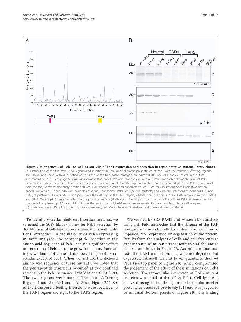
To identify secretion-deficient insertion mutants, wescreened the 2037 library clones for Peb1 secretion bydot blotting of cell-free culture supernatants with anti-Peb1 antibodies. In the majority of Peb1-expressingmutants analyzed, the pentapeptide insertion in theamino acid sequence of Peb1 had no significant effecton secretion of Peb1 into the growth medium. Interest-ingly, we found 14 clones that showed impaired extra-cellular export of Peb1. When we analyzed the deducedamino acid sequence of these mutants, we noted thatthe pentapeptide insertions occurred at two confinedregions in the Peb1 sequence: D42-V43 and S173-L180.The two regions were named Transport AffectingRegions 1 and 2 (TAR1 and TAR2; see Figure 2A). Sixof the transport-affecting insertions were localized tothe TAR1 region and eight to the TAR2 region.
We verified by SDS-PAGE and Western blot analysisusing anti-Peb1 antibodies that the absence of the TARmutants in the extracellular milieu was not due toimpaired Peb1 expression or degradation of the protein.Results from the analyses of cells and cell-free culturesupernatants of mutants representative of the entiredata set are shown in Figure 2B. According to our ana-lysis, the TAR1 mutant proteins were not degraded butexpressed intracellularly at lower quantities than wtPeb1 (see top panel of Figure 2B), which compromisedthe judgement of the effect of these mutations on Peb1secretion. The intracellular expression of TAR2 mutantproteins was equal to that of wt Peb1. Cell lysis wasanalyzed using antibodies against intracellular markerproteins as described previously [21] and was judged tobe minimal (bottom panels of Figure 2B). The finding
Figure 2 Mutagenesis of Peb1 as well as analysis of Peb1 expression and secretion in representative mutant library clones.(A) Distribution of the five-residue MGS-generated insertions in Peb1 and schematic presentation of Peb1 with the transport-affecting regionsTAR1 (pink) and TAR2 (yellow) identified on the basis of the transposon mutagenesis indicated. (B) SDS-PAGE analysis of cell-free culturesupernatant of MKS12 carrying the plasmids indicated (top panel). Western blot analysis with anti-Peb1 antibodies shows the level of Peb1expression in whole bacterial cells of the various clones (second panel from the top) and verifies that the secreted protein is Peb1 (third panelfrom the top). Western blot analysis with anti-GroEL antibodies in cells and supernatants was used for assessment of cell lysis (two bottompanels). Mutants p9G2 and p4G8 are examples of clones that secrete Peb1 well (neutral mutants) and carry the insertions at positions H25 andG106, respectively. Mutants p4G10 and p4B7 have the insertion in the TAR1 region, whereas the insertion is in the TAR2 region in mutants p3D9and p8C3. Mutant p1B6 has an insertion in the promoter region (at -87 nt) of the fliC-peb1 construct, which abolishes Peb1 expression. Wt Peb1is encoded by plasmid pLA25 and pMCS3’UTR is the vector control. Cell-free culture supernatant (S) and whole bacterial cell samples(C) corresponding to 100 μl of bacterial culture were analyzed. Molecular weight markers in kDa are indicated on the left.
Anton et al. Microbial Cell Factories 2010, 9:97http://www.microbialcellfactories.com/content/9/1/97
Page 5 of 16

that the well-expressed TAR2 mutants were not foundin the growth medium was additionally considered as aninternal control for lack of leakage from or lysis of thecells.When we compared the amino acid sequences of the
inserted pentapeptides in TAR1, TAR2 and neutralmutants, we found that the neutral mutants carriedinsertions with similar residues as the secretion-affectedmutants, but in locations outside the identified TARregions. We deduced that the secretion-deficient pheno-type of TAR1 and TAR2 mutants was not dependent onthe type of sequence inserted and that the lack of extra-cellular localization by the TAR mutants was apparentlydue to the site of insertion (See Table 2 for examples).Taken together, the exhaustive search for transport-
affecting domains in Peb1 revealed two insertion-sensitive regions in the open reading frame of Peb1,TAR1 and TAR2, which affect its localization in thegrowth medium of E. coli MKS12.
Effect of targeted mutations in TAR regions on Peb1secretionThe results obtained from the mutant library of Peb1indicated the two TAR areas in the protein as criticalfor its extracellular localization. To analyze theseregions of Peb1 in more detail, we substituted all thecharged and polar residues in the vicinity of and withinthe TAR1 region (Glu40, Asp42, Lys45 and Lys49)(encoded by plasmid pTAR1P) and in the TAR2 region(Ser173, Asp175, Lys176 and Ser177 (encoded by plas-mid pTAR2P) with alanine (Figure 3). In addition, wegenerated an insertion of five Gly between Asp42and Val43 in the TAR1 region (encoded by pTAR15G)(Figure 3). The Ala point mutations in TAR2 did notsignificantly influence the expression and secretion ofthe mutated Peb1 proteins, whereas in TAR1 the cellu-lar concentration and, as a consequence, secretion levelsof the Ala point mutants were slightly decreased. Thecellular concentration of pentaglycine insertion mutantwas also diminished and its transport was abolished(Figure 3).
We next assessed the impact of the TAR1 and TAR2regions on Peb1 secretion by creating long Ala substitu-tions (Glu40 to Leu46 and Val174 to Tyr182) and forcomparison, identical Gly substitutions were constructed(Figure 3). The mutant Peb1 proteins with nine residueslong Ala or Gly substitutions in region TAR2 (encodedby pTAR2A and pTAR2G) were synthesized at the levelof wt Peb1 but were not secreted, whereas the Peb1mutants carrying seven-residue long Ala or Gly substitu-tions in TAR1 (pTAR1A and pTAR1G) showed a loweramount of Peb1 in cells compared to the wild typestrain. The mutant with a long Gly substitution was not
Table 2 Sequence and localization of insertions present in Peb1 mutants
Localizationa)
TAR1Plasmid Sequence Localizationa)
TAR2Plasmid Sequence Localizationa)
outside TAR1 and TAR2Plasmid Sequence
D42 p4G5 DAAAV S173 p8C3 VRPHS A2 pC1B11 DAAAA
D42 p4G10 CGRID V174 p9B8 VRPHV H25 p4G8 CGRTH
D42 p8E2 CGRID D175 p2C3 CGRID R89 pC18B11 CGRKR
D42 p7F4 VAAAD L179 p3D9 LRPQL I105 p9G2 GAAAI
V43 p4B7 CGRNV L179 p3B7 LRPQL T133 pC4E1 MPRQT
V43 p9D5 AAAAV L180 p10B2 VRPQL E220 pC6B3 LRPQE
L180 p8F7 VRPQL H221 pC7B5 CGRKH
L180 p4B10 VRPQL H221 pC18H10 AAAEH
a) amino acid residue after which the pentapeptide is inserted
Figure 3 Expression and secretion of site-specifically mutatedPeb1. The amino acid sequence of mature Peb1 shows the TAR1and TAR2 regions in pink and yellow, respectively. Dots indicatesites for Ala point substitutions (encoded by plasmids pTAR1P andpTAR2P), the underlined sequences show sites for longer Ala andGly substitutions (pTAR1A, pTAR1G, pTAR2A and pTAR2G), and thearrow indicates the site of a pentaglycine insertion (pTAR15G). Everytenth residue is indicated by a vertical line. The bottom panelshows whole cells (C) and cell-free culture supernatants (S) fromstrain MKS12 carrying the plasmids indicated above the panelsanalyzed by Western blotting using anti-Peb1 antibodies (seeFig. 2A and Table 1 for details regarding the plasmids). Wt Peb1 isencoded by the plasmid pLA25. Samples correspond to 100 μl ofculture. Molecular weight markers in kDa are indicated on the left.
Anton et al. Microbial Cell Factories 2010, 9:97http://www.microbialcellfactories.com/content/9/1/97
Page 6 of 16
secreted and the mutant carrying a long Ala substitutionwas present in barely detectable amounts in the growthmedium (Figure 3).The results thus showed that selected Ala point sub-
stitutions in TAR1 and TAR2 regions do not affect Peb1secretion significantly, whereas longer Ala substitutionsin TAR1 and especially in TAR2 markedly impair Peb1secretion. Also long Gly substitutions in the TARregions and the insertion of five Gly in TAR1 affectedtransport of Peb1. In addition, the results showed thatall manipulations performed in the TAR1 region, i.e.insertions, selected point substitutions as well as longsubstitutions, affected the amount of intracellular Peb1whereas manipulations of the TAR2 region did not.
Cytosolic Peb1 proteins are soluble and not subjected toaggregationTo assess whether intracellular aggregation of mutantproteins was the reason for the lack of their presence inthe growth medium, the cytoplasmic content of E. coliMKS12 expressing Peb1 and its mutants was fractio-nated by ultracentrifugation, separated by SDS-PAGE,and analyzed by Western blotting using anti-Peb1 anti-bodies (Figure 4). The experiment included wt Peb1,Peb1 with Ala- and Gly-substituted TAR regions, Peb1carrying a pentaglycine insertion in TAR1, and one ofthe Peb1 mutants from the transposon library with thepentapeptide insertion in the TAR1 region (encoded byplasmid p9D5; see Table 2). The results showed that all
the proteins that were expressed well in the cells werealso found in the soluble fraction. The Peb1 mutantwith a long Gly substitution in TAR1 (encoded bypTAR1G) was expressed very poorly and was thereforedetected neither in the soluble nor the insoluble frac-tion, a fact hampering the analysis of its solubility. Con-clusively, the results indicated that the lack ofextracellular localization by Peb1 mutants in the ana-lyses was not due to aggregation or proteolysis of theproteins in the cytoplasm.
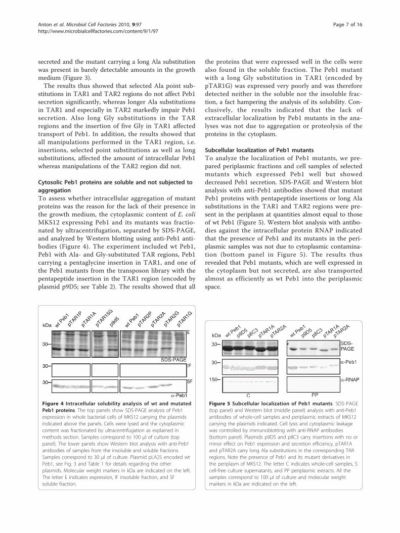
Subcellular localization of Peb1 mutantsTo analyze the localization of Peb1 mutants, we pre-pared periplasmic fractions and cell samples of selectedmutants which expressed Peb1 well but showeddecreased Peb1 secretion. SDS-PAGE and Western blotanalysis with anti-Peb1 antibodies showed that mutantPeb1 proteins with pentapeptide insertions or long Alasubstitutions in the TAR1 and TAR2 regions were pre-sent in the periplasm at quantities almost equal to thoseof wt Peb1 (Figure 5). Western blot analysis with antibo-dies against the intracellular protein RNAP indicatedthat the presence of Peb1 and its mutants in the peri-plasmic samples was not due to cytoplasmic contamina-tion (bottom panel in Figure 5). The results thusrevealed that Peb1 mutants, which are well expressed inthe cytoplasm but not secreted, are also transportedalmost as efficiently as wt Peb1 into the periplasmicspace.
Figure 4 Intracellular solubility analysis of wt and mutatedPeb1 proteins. The top panels show SDS-PAGE analysis of Peb1expression in whole bacterial cells of MKS12 carrying the plasmidsindicated above the panels. Cells were lysed and the cytoplasmiccontent was fractionated by ultracentrifugation as explained inmethods section. Samples correspond to 100 μl of culture (toppanel). The lower panels show Western blot analysis with anti-Peb1antibodies of samples from the insoluble and soluble fractions.Samples correspond to 30 μl of culture. Plasmid pLA25 encoded wtPeb1, see Fig. 3 and Table 1 for details regarding the otherplasmids. Molecular weight markers in kDa are indicated on the left.The letter E indicates expression, IF insoluble fraction, and SFsoluble fraction.
Figure 5 Subcellular localization of Peb1 mutants. SDS-PAGE(top panel) and Western blot (middle panel) analysis with anti-Peb1antibodies of whole-cell samples and periplasmic extracts of MKS12carrying the plasmids indicated. Cell lysis and cytoplasmic leakagewas controlled by immunoblotting with anti-RNAP antibodies(bottom panel). Plasmids p9D5 and p8C3 carry insertions with no orminor effect on Peb1 expression and secretion efficiency, pTAR1Aand pTAR2A carry long Ala substitutions in the corresponding TARregions. Note the presence of Peb1 and its mutant derivatives inthe periplasm of MKS12. The letter C indicates whole-cell samples, Scell-free culture supernatants, and PP periplasmic extracts. All thesamples correspond to 100 μl of culture and molecular weightmarkers in kDa are indicated on the left.
Anton et al. Microbial Cell Factories 2010, 9:97http://www.microbialcellfactories.com/content/9/1/97
Page 7 of 16

Bioinformatics analysis of Peb1 and TAR regionsWe performed sequence analysis of Peb1 in order toidentify sequences important for its transport. The soft-ware programs SignalP, TatP, LipoP and Type-III effec-tor prediction were used for prediction of putativesignal sequences in the amino acid sequence of maturePeb1 (shown in Figure 3) and thus lacking the SecA-YEG -dependent signal sequence and the putative signalpeptidase II recognition site [26-29]. We found no indi-cation of the presence of a signal sequence related toSecA-YEG, Tat, lipoprotein or type 3 secretion systempathways in the mature Peb1. Interestingly, the Secreto-meP [30] software predicted Peb1 to be a non-classicallysecreted protein.Since the TAR regions were found to have a major
impact on Peb1 secretion efficiency, we analyzed thesequence and the structure of Peb1 with emphasis onthe TAR regions in order to identify possible structuralaspects important for secretion. Mature Peb1 is a globu-lar protein of 233 amino acids that folds into twodomains both comprising of a five-stranded beta sheetsurrounded by alpha helices, and it binds two Zn2+
cations (Figure 6A). The TAR1 and TAR2 regions coin-cided with the first helix of the N-terminal core struc-ture (Na1) (Figure 6A, B) and with the end of the thirdC-terminal beta strand and the following helix (Cb3-Ca3) (Figure 6A, C), respectively. The structural analysishere considered only the Ala substitutions since thepentapeptide insertions generated by the transposonmutagenesis are challenging to analyze in structuralterms and interpretation of the effects of the Gly substi-tution in TAR1 was not reliable due to the decreasedPeb1 expression (See Figure 3).The helix Na1, that covers the TAR1 region, is a
14-residue long, straight and strongly amphiphilic helixthat interacts via Ser50 with one zinc cation but is notpart of the ligand-binding site of Peb1. The polar residuesof Na1 form strong interactions with the two terminalhelices of Peb1, Za1 and Za2, and the large apolar resi-dues pack tightly to Za2. The helix structure is stabilizedby several hydrogen bonds (see Figure 6B). By the num-ber of polar and apolar helix-helix interactions, the inter-face between Na1 and Za1 and Za2 is the strongest inthe whole protein. Substitution of all polar residues inand close to TAR1 by alanines (pTAR1P) did not signifi-cantly interfere with Peb1 transport, whereas a seven-residue run of alanines covering TAR1 (pTAR1A) did so,analogously to the results from the six amino acid trans-poson insertions (as shown in Figure 2 and 3).The TAR2 region of Peb1 is located in the very center
of the protein covering the end of Cb3 and the begin-ning of Ca3 (see Figure 6A and 6C). It forms theligand-binding pocket (residues Val174, Asp175, Ile178)along with numerous other residues from both domains.
The results from Ala substitutions in TAR2 were similarto those in TAR1: the substitution by nine consecutivealanines compromised secretion, while changes of onlythe polar residues did not (see Figure 3). Sequence com-parison between the secretion-deficient Peb1 constructsand those exhibiting a wt secretion level pointedtowards an importance of the bulky hydrophobic resi-dues Val174, Ile178, Leu179 and Leu180 for Peb1 secre-tion. Except for Leu180, these are fully buried residues,with solvent exposed surface areas smaller than 3% oftheir total area. Taken together, the structural analysisof TAR regions showed that the TAR1 region comprisesa densely packed and highly stable part of Peb1 and theTAR2 region is located in the center of protein formingthe ligand-binding pocket along with several other resi-dues. We suggest that the unifying feature of the TARsis their crucial role for proper folding of Peb1, whichapparently is important for the export across the OM.
DiscussionWhen the Peb1 protein of C. jejuni is expressed withoutits native N-terminal signal sequence and without theputative signal peptidase II cleavage site in E. coliMKS12 (ΔfliCfliD), it is still efficiently secreted into thegrowth medium. The extracellular presence of Peb1 inMKS12 is not due to leakage from or lysis of the cells,as we have demonstrated previously [21]. Here, ouraims were to analyze the molecular mechanisms behindthe secretion competency of the heterologous modelprotein Peb1 in the E. coli host in order to broaden ourunderstanding of factors that may affect secretion ofproteins in E. coli. A detailed knowledge of mechanismsof extracellular transport of heterologous proteins inE. coli is of great importance in the development ofsecretion systems for biotechnological applications [4].The present work shows that secretion of mature Peb1
into the growth medium of E. coli is facilitated also in theabsence of the flagellar machinery as shown in theMKS12 derivative MKS111ΔHBB, a strain which comple-tely lacks the flagellar secretion complex. The hypothesisof a flagellar-independent secretion mechanism wasfurther supported by the fact that fusion of an N-terminalFliC fragment to the N-terminus of Peb1 did not inter-fere with its secretion. Our finding that Peb1 is also pre-sent in the periplasm strongly indicates that the proteinis secreted by a mechanism that involves at least twoexport steps, one across the CM to the periplasm andanother across the OM. This excludes secretion depen-dent only on the flagellar machinery, which is known toproceed from the cytoplasm to the extracellular milieuwithout involvement of a periplasmic step [22].A comprehensive pentapeptide scanning mutagenesis
of Peb1 was performed to decipher transport affectingregions in the molecule itself, and the results were
Anton et al. Microbial Cell Factories 2010, 9:97http://www.microbialcellfactories.com/content/9/1/97
Page 8 of 16

confirmed by site-specific mutations. Importantly, modi-fications of only two distinct areas, named TAR1 andTAR2, in Peb1 affected secretion. This phenomenonwas not due to insolubility or aggregation of the mutantproteins in the bacterial cytoplasm as shown by cellfractionation analysis. However, manipulations in theTAR1 region affected the intracellular amount of Peb1,whereas no such effect was observed after mutagenesisof the TAR2 region. We anticipate that the reducedamount of Peb1 seen in the cells of TAR1 mutants isdue to decreased expression levels. As no degradationproducts of Peb1 were detected, we assume that signifi-cant proteolysis of Peb1 mutants did not occur. Theresults showed that also well-expressed, secretion-deficient Peb1 mutants were present in the periplasmicspace. This finding strongly indicated that the export ofthe TAR1 and TAR2 mutants across the CM was unen-cumbered, whereas their further export across the OMwas compromised and led to the observed lack of Peb1mutants in the culture supernatant.The secretion-deficiency of TAR mutants was studied
also by analyzing the Peb1 structure. The effect ofTAR1 mutations could lie in the importance of the
hydrophobic surface packing toward the rest of the pro-tein. Native-like amphiphilicity of Na1 may be neededfor the folding of Peb1, and inability to fold may be thebasic cause for secretion defects. The importance ofTAR2 in secretion is apparently related to the stabilityof the protein, as TAR2 is located in the center of Peb1and forms the ligand-binding pocket along with severalother residues. We suggest that the unifying feature ofthe TARs is their crucial role in proper folding. Wespeculate that Peb1 remains unfolded or is only partiallyfolded during its export across the CM to the periplasmsince mutagenesis of TAR regions did not affect thisstep. Correct or at least partially correct folding of Peb1may, on the contrary, be important for its export acrossthe OM into the growth medium.Searches in the literature and publically available data-
bases revealed that the host strain MKS12 used in thiswork, a ΔfliCfliD -derivative of E. coli MG1655,expresses at the culture conditions used only three char-acterized protein transport systems [31-34]. These arethe SecA-YEG and Tat systems, which export proteinswith cognate cleavable N-terminal signal sequencesacross the CM [35], and the flagellum-specific secretion
Figure 6 Structure of Peb1. (A) An overall view of the molecular structure. The first 100 residues are colored white, and the last 133 ingreyblue, highlighting the two-domain structure of Peb1. The two pseudosymmetric domains fold into sandwiches of alternating alpha helicesand beta strands. The N and C-termini are marked with spheres. The ligand binding site is situated at the domain interphase in the middle ofthe molecule, and a bound aspartate is shown as a stick model. Peb1 binds two zinc cations, which are indicated as grey spheres and markedwith Zn1 and Zn2. Pentapeptide insertion sites in the TAR1 region are colored purple, and are situated in the first helix at the core of theN-terminal domain, Na1. Pentapeptide insertion sites in the TAR2 region are colored yellow, and cover the end of the third beta-strand at thecore of the second domain, and part of the following helix, Cb3 and Ca3. The location of the long alanine substitutions in TAR1 and TAR2 arecolored green. The two terminal helices of the protein are also marked, Za1 and Za2. In the indicated aspartate, carbons are colored green,oxygens red and nitrogens blue. The same color scheme is used in all atomic structure. (B) A detailed view of helix Na1. The side chains of largehydrophobic residues of Na1 are colored brown, and interactions with the surroundings are highlighted. (C) A close-up of area of the secondcluster of disruptive substitutions at the center of the molecule, next to the the ligand-binding site. Residues of the long Ala substitution in thisregion are shown in green, except for three branched hydrophobic residues, V174, I178 and L179, that are drawn with thick brown lines. Theneighboring residues are shown in atomic colors. Val174, Asp175, Ile178 pack tightly to each other to form the ligand-binding pocket along withLys20, Ala65, Arg68, Ala82, Thr83, Phe84, Thr85, Arg90, Ala132, Thr133, Tyr157, Tyr199 from both domains.
Anton et al. Microbial Cell Factories 2010, 9:97http://www.microbialcellfactories.com/content/9/1/97
Page 9 of 16

system responsible for assembly of the flagellar filament[36]. As indicated by the bioinformatics analysis of thePeb1 amino acid sequence, none of these are apparentlyinvolved in E. coli -based Peb1 secretion. The SecA-YEG -independent secretion was here experimentallystudied by analysis of periplasmic and cell samples ofPeb1-expressing E. coli grown with or without subinhi-bitory concentrations of azide, which is known to inhibitprotein export by the pathway [24].Other well-characterized pathways dedicated for pro-
tein secretion in Gram-negative bacteria are normallynot expressed, or the genes are fragmented, deleted ortotally lacking in MKS12 as described below. The type 1secretion system of E. coli, which is responsible forsecretion of the toxin hemolysin in pathogenic E. coli, isnot found in MKS12. The strain carries the genesencoding a hemolysin-like protein named HlyE/SheA/ClyA secreted into the medium by a so far unidentifiedmechanism in H-NS mutants [37], which MKS12 is not.The genes encoding a putative type 2 secretion systemare present in MKS12, but the transcription of the twooperons encoding the majority of the type 2 secretionsystem components is repressed by the H-NS proteinexpressed in MKS12 [38]. The genes encoding a patho-genesis-related type 3 secretion system are not presentin MKS12 [32,39]. The alternative cryptic type 3 secre-tion system called ETT2 present in some pathogenicand commensal E. coli strains as well as the ancestralgene cluster encoding the alternative flagellar systemFlag-2 in 20% of E. coli strains are both nonfunctionalin MKS12 [39,40]. The strain possesses at least 16 chro-mosomal genes related to the type 4 secretion system,but the genes are not expressed in the culture condi-tions used in the study [41]. The type 5 secretion systemis represented in E. coli K-12 strains only by the SecA-YEG -dependent autotransporter protein called antigen43, which facilitates its own transport to the surface ofthe cell [42]. Finally, no orthologs of the type 6 secretionsystem proteins are present [43], the type 1 fimbriagenes have been site-specifically deleted in MKS12 [44],and the curli-encoding operons are not expressed inMKS12 at the growth conditions used [45].ABC transporters are CM-spanning proteins that
energize the transport of their substrates by bindingand hydrolysis of ATP. In bacteria, the ABC exportersfacilitate the export of various compounds from thecytoplasm and the more common ABC importersimport essential nutrients that are delivered by specificPBPs [15]. In E. coli, almost 80 ABC importer andexporter systems have been reported. These frequentlytransport only small molecules [13] and are probablynot responsible for the efficient secretion of Peb1,which is a full-length globular protein of 233 aminoacids.
The transport mechanism of the aspartate/glutamate-binding PBP Peb1 appears to differ markedly in E. colifrom that of its structurally known homologs HBP andGlnBP from E. coli [46,47] and Debp from Shigella flex-neri [48]. These ABC-transporter associated PBPs are allexported by the SecA-YEG pathway involving a cleava-ble N-terminal signal sequence. The sequence identitybetween Peb1, HBP, GlnBP and Debp is low (27% and22%) and at the structural level, root mean square devia-tion values calculated between mature Peb1, HBP andDebp are only 2.2 Å since the orientation of thedomains relative to each other varies between structures.Hence, the overall structural similarity of Peb1 withE. coli PBPs is probably not the reason for its efficientsecretion in the heterologous E. coli host.Our results show that in E. coli, mature Peb1 is pre-
sent in the extracellular milieu but cannot be visualizedon the surface of strain MKS12 (pLA25) with indirectimmunofluorecence microscopy using anti-Peb1 antibo-dies or by electron microscopy (unpublished data) indi-cating that blebbing of OM vesicles [49] may not beinvolved in the secretion. Our results also show thatPeb1 can be exported to the periplasm independently ofthe flagellar machinery, without a SecA-YEG-dependentN-terminal signal sequence independently of the Secpathway, and without a signal peptidase II recognitionsite. Taken together, we conclude that Peb1 may besecreted by a still unknown secretion route, a conclusionsupported by our result from software analysis of thePeb1 sequence indicating that the protein is secreted bya non-classical pathway.Our findings are indirectly supported by a line of inde-
pendent reports. According to Hu and colleagues [31] onethird of the 4225 protein-encoding genes of E. coli K-12are functionally unannotated, a fact which makes it possi-ble to reveal novel mechanisms even in this well-studiedorganism. Proteins denoted as non-classically secreted arefound extracellularly in various bacteria despite theabsence of a defined secretion signal or motif [50,51]. Fre-quently, these proteins called moonlighting proteins havenovel extracellular functions in addition to their well-known roles in the cytoplasm [52]. Comparison of theextracellular proteomes of E. coli K-12 and BL21 revealeda large number of proteins, both cytoplasmic and periplas-mic, to be released by unknown mechanisms into thegrowth medium when cells were grown at high cell density[53]. We assume that Peb1 is secreted in E. coli by a stilluncharacterized two-step pathway and that the structu-rally significant TAR- regions affect the secretion. We arecurrently exploring this possibility further.
ConclusionsWe have previously reported that the diagnosticallyinteresting model protein Peb1 is efficiently secreted,
Anton et al. Microbial Cell Factories 2010, 9:97http://www.microbialcellfactories.com/content/9/1/97
Page 10 of 16

without the sequences for SecA-YEG dependent secre-tion and signal peptidase II, into the growth medium ofthe secretion-competent strain E. coli MKS12 (ΔfliC-fliD). Here, we analyzed the secretion mechanismfurther and noticed that Peb1 was also transported inde-pendently of the flagellar secretion machinery. Wefound, by construction and analysis of an exhaustivelibrary of Peb1 insertion mutants, that mutagenesis oftwo segments called TAR1 (residues 42 and 43) andTAR2 (residues 173 to 180) prevented Peb1 secretionindividually. Site specific mutagenesis of the TARregions confirmed the importance of these regions forPeb1 secretion, and we verified that the secretion defi-ciency of Peb1 mutants was not due to insolubility oraggregation of the proteins in the cytoplasm. Themutant proteins were present in the periplasm as wellas in the cells of MKS12 and we deduced that mutagen-esis of TAR regions did not affect export of Peb1 acrossthe CM, whereas its export over the OM was markedlyimpaired. We conclude that the localization of themature model protein Peb1 in the growth medium ofE. coli is due to an active secretion process, whichinvolves two export steps, one across the CM into theperiplasmic space and another across the OM. Theexport across the OM apparently requires proper foldingof the TAR regions since mutagenesis of the regionsaffected Peb1 export across the OM and thereby pre-sence of the mutant proteins in the extracellular milieu.Bioinformatics analysis of the Peb1 sequence indicatedthat the protein is transported by an unknown, non-classical pathway, which is a topic for further studies.
MethodsBacterial strains and growth conditionsBacterial strains and plasmids used in this work aredescribed in Table 1. E. coli strains were cultivated byshaking in Luria broth [54] or on Luria agar plates for16 h at 37°C supplemented with antibiotics (tetracy-cline 12.5 μg/ml, streptomycin 100 μg/ml, ampicillin100 μg/ml) when appropriate. For TCA-precipitationand periplasmic isolation, the bacteria were furtherinoculated (1:30) into 3 ml and 12 ml, respectively, ofN-minimal medium [54] supplemented with 0,2% casa-mino acids and appropriate antibiotics, and cultivatedstatically for 16-24 h at 37°C for optimization of pro-tein secretion as we have described previously [21]. Forintracellular solubility assays, the strains were culti-vated by shaking in Luria broth supplemented withappropriate antibiotics for 18 h at 37°C followed bydilution 1:100, and cultivated without agitation for 20hrs at 37°C in 20 ml Luria broth with antibiotics. Foranalysis of the effect of subinhibitory concentrations ofNaN3 on Peb1 export, the strains were grown asdescribed but in the presence of 3 mM NaN3.
Antisera and reagentsRabbit anti-Peb1 antiserum (diluted 1:50 000 for immu-noblot analysis) and anti-flagellum antiserum (diluted1:2000) were available from the previous work [21]. Forcontrol of cell lysis, polyclonal anti-GroEL antibody wasused at a dilution of 1:10 000 (Sigma-Aldrich) andmouse monoclonal anti-RNAP at a dilution of 1:10 000(Neoclone). Alkaline phosphatase-conjugated secondaryantibodies were obtained from DakoCytomation anddiluted to a concentration of 1.5 μg/ml (1:1000). Allantibodies were diluted in 1% BSA/PBS. Reagents forrecombinant DNA techniques and PCR were mostlyobtained from Promega. For PCR cloning and mutagen-esis Pfu DNA polymerase (Promega) was used. ColonyPCR was performed using Dynazyme II DNA polymer-ase (Finnzymes). Primers used in this study were pur-chased from Medprobe and are shown in Table 3.
Construction of bacterial strainsBacterial strains used in this work are described in Table 1.The E. coli strains MKS111, MKS111ΔHBB andMKS12Placpeb1 were constructed from E. coli strainMKS12 [21] by site-specific mutagenesis using the pir-dependent suicide vector pCVD442 as described earlier[55]. For the construction of MKS111, a DNA fragmentcovering 2348 bp (nucleotides1128017-1130365) wasamplified by PCR to contain an in-frame deletion of flgM(367 bp, nucleotides1129426-1129059). Chromosomaldeletions of flgDEFGHIJ and fliFGH were performed tocreate the E. coli strain MKS111ΔHBB using the sameprinciple as above. The genes flgDEFGHIJ were firstdeleted from the chromosome of MKS111 using a PCR-amplified fragment covering 6782 bp (nucleotides1130748-1137529) containing a deletion of flgDEFGHIJ(4905 bp, nucleotides 1131696-1136600). From this strainfliFGH was then chromosomally deleted by the same prin-ciples; the DNA fragment of 5589 bp (nucleotides2010111-2015699) contained the deletion of fliFGH (3329bp, nucleotides 2011212-2014541). For the construction ofstrain MKS12Placpeb1, a 3066 bp DNA fragment wasamplified by PCR to contain fliC flanking regions (nucleo-tides 199936-2000133 and nucleotides 2001784-2003814with the exception of the fliD deletion, nucleotides2002082-2003109), Plac, fliC 5’ UTR and peb1 lacking itssignal sequence. This fragment was used for substitution,by homologous recombination, of fliC in strain MKS1[56]. Primers used in mutagenesis were designed on thebasis of the nucleotide sequence of E. coli MG1655 and,for the Plac promoter, E. coli DH1 (GenBank Accessionnumbers NC 000913 and CP001637.1) [57]. ChromosomalDNA of E. coli MG1655 ΔfimA-H, also called AAEC072A,and plasmid pTSF12 [58], were used as templates for PCRamplifications. The genotypes of the mutants were con-firmed by PCR and Southern blot hybridization.
Anton et al. Microbial Cell Factories 2010, 9:97http://www.microbialcellfactories.com/content/9/1/97
Page 11 of 16

Construction of plasmidsPlasmids used in this work are described in Table 1.The authenticity of the construct has been verified foreach plasmid by sequencing. Plasmids pMCS3’UTR andpPeb15’3’ were available from the previous work [21].Our former results indicated that the 5’ UTR of fliC isimportant for secretion of Peb1 in MKS12, whereasexpression of peb1 from e.g. the promoter and 5’UTR ofthe ß-lactamase-encoding gene rendered Peb1 in thecytoplasm of MKS12 [21]. Therefore, in the currentstudy all the peb1 constructs were generated to maintainthe sequence corresponding to the 5’ region of fliCmRNA and thereby facilitate secretion of the gene pro-duct. To construct pLA25 a DNA fragment containingthe peb1 gene, without the sequence encoding the leaderpeptide, fused to the 173 bp 5’UTR of fliC that includesPfliC , was constructed by PCR amplification frompPeb15’3 with primers containing BglII and XbaI restric-tions sites (primers 5’UTRfwBglIl and Peb1RXbaI). ThisDNA fragment was ligated into the BglII-XbaI digestedpMCS3’UTR and the ligation mixture was introducedinto E. coli MKS12 cells by electroporation. To con-struct the fliC5’UTR-peb1 target for the in vitro transpo-sition reaction, the plasmid pLA26 was generated. The
3’UTR of fliC in pLA25 was removed by ScaI-EcoRIdigestion and the cohesive ends were subjected to ablunting reaction with Mung Bean Nuclease at a con-centration of 5 units per μg DNA for 10 min at 37°C.The blunt ends were ligated, and the ligation mixturewas introduced by electroporation into E. coli DH10Bcells. To construct plasmid pLA31, a fragment contain-ing PfliC and the sequence encoding the N-terminal 183residues of FliC was amplified from chromosomal DNAof E. coli AAEC072A (primers 5’UTRfwBglIl andFliC183XbaIR, respectively). The BglII-XbaI-digestedPCR fragment was cloned into pMCS3’UTR and subse-quent ligation mixture was introduced into MKS12 byelectroporation. To create pLA32 a PCR-generatedDNA fragment containing the fliC 5’UTR and 60 nucleo-tides from the 5’ end of the fliC gene (p20FliCGfp5’3’[21] as template, primers 5’UTRfwBglII and peb1/pLA25RIN) was fused to a PCR-generated peb1 frag-ment (pPeb15’3’ as template, primers peb1/pLA25FINand Peb1RXbaI). This fusion fragment was cloned intopMCS3’UTR using appropriate restriction sites (BglIIand XbaI) and the ligated plasmid pLA32 was intro-duced into E. coli MKS12. For pTAR constructs, seeTable 1.
Table 3 Primers used in this study
Primer Application Sequence 5’-3’
Peb1RXbaI PCR GGCTCTAGATTATAAACCCCATTTTTTCGC
5’UTRfwBglII PCR GCCAGATCTGCGGGAATAAGGGGCAGAG
K1012R PCR GGTCTAGATTAACCCTGCAGCAGAGACA
FliC183XbaIR PCR GGTCTAGATTAAGTGGTAACTGTATCGTTAT
K1008OF PCR GGCGATCGGCGGGAATAAGGGGCAGAG
ORpLA15 PCR GCCGCATGCCGATAAACAGCCCTGCGTTA
pBRAhdIF Sequencing GCCTTCGTTCATCCATAGTTGCCT
pLA21R Sequencing GCCCAGTTAATCAGGTTACAACGATT
NotI mini Sequencing TGCGGCCGCA
028R Sequencing CAATTCAACTTGTAGGCCTGATA
peb1/pLA25FIN Construction of: pLA32 GATCACTCAAAATAATATCAACAAGGCAGAAGGTAAACTTGAGTC
peb1/pLA25RIN pLA32 GACTCAAGTTTACCTTCTGCCTTGTTGATATTATTTTGAGTGATC
HotNIF pTAR1P TAGCTGTTGCCGCATTGCTAGCTGCAAGTATATTGGGTG
HotNIR pTAR1P GCAATGCGGCAACAGCTACTGCGAAACCTTTAATTTCAC
HotCIF pTAR2P CGTTTGCTGTAGCCGCAGCAATATTGTTAGGTTATGTGG
HotCIR pTAR2P ATATTGCTGCGGCTACAGCAAACGCATCAACTCTTTTAG
PebGlyF pTAR15G GGCGGTGGTGGGGGCGTTGCCAAATTGCTAGCTAAAAG
PebGlyR pTAR15G GCCCCCACCACCGCCATCTACTTCGAAACCTTTAATTTC
PebAla1F pTAR1A GCAGCAGCGGCGGCCGCAGCGCTAGCTAAAAGTATATTGGGTG
PebAla1R pTAR1A CGCTGCGGCCGCCGCTGCTGCGAAACCTTTAATTTCACCTGTTG
PebGly1F pTAR1G GGCGGCGGTGGTGGCGGTGGCCTAGCTAAAAGTATATTGGGTG
PebGly1R pTAR1G GCCACCGCCACCACCGCCGCCGAAACCTTTAATTTCACCTGTTG
PebAla3F pTAR2A GCAGCCGCAGCAGCAGCGGCAGCGGCGGTGGATGATAAAAGTGAAATTTTG
PebAla3R pTAR2A CGCCGCTGCCGCTGCTGCTGCGGCTGCAGAAAACGCATCAACTCTTTTAG
PebGly3F pTAR2G GGCGGCGGTGGTGGCGGGGGCGGTGGTGTGGATGATAAAAGTGAAATTTTG
PebGly3R pTAR2G ACCACCGCCCCCGCCACCACCGCCGCCAGAAAACGCATCAACTCTTTTAG
Anton et al. Microbial Cell Factories 2010, 9:97http://www.microbialcellfactories.com/content/9/1/97
Page 12 of 16

Precipitation of protein in growth mediaTo analyze the presence of Peb1 and mutant proteins inthe growth medium, we precipitated culture superna-tants with TCA as described previously [21]: 1,5 ml ofculture in N-minimal medium was centrifuged in atabletop centrifuge (Biofuge 13, Heraeus Instruments) at13 000 rpm for 15 min at 4°C. The supernatant wasrecentrifuged, and 1ml of the secondary supernatant wasprecipitated with 10% TCA on ice for 30 min. Precipi-tates were pelleted (as above) and washed twice with icecold ethanol/ether (1:1). Precipitated protein wasallowed to dry before resuspension into loading dye andboiled for 5 min. An amount corresponding to 1 ml ori-ginal cultivation when analyzed by SDS-PAGE and to100 μl cultivation when analyzed by western blotting,respectively, was loaded onto the gel. Cell samples weregenerated by pelleting the cultivation followed by awash with PBS. Cells were then resuspended into appro-priate volume of loading dye for the analysis of theamount corresponding to bacterial cells from 100 μl cul-ture, and boiled.
Isolation of periplasmic contentsTo assess the presence of Peb1 and mutant proteins inthe periplasmic space, contents of the periplasm wereisolated. Bacteria were grown overnight in Luria brothand diluted in N-minimal medium (1:30) for furthercultivation for 20 hrs without agitation at 37°C. Cellswere then pelleted using a F21-rotor (FiberLite®, Pira-moon Technologies, Inc.) in a Sorvall RC-5B centrifuge(Du Pont Instruments) at 10 000 g (15 min, 4°C). Cellswere resuspended in 4 ml 20% glucose/g wet cells andincubated 5 minutes at RT. Next, the sample was cen-trifuged for 5 min at 13.000 rpm (RT) in tabletop cen-trifuge (PICO 17, Thermo Scientific). The pellet wasfurther resuspended in 1 ml ice cold 10 mM Tris/20Mm EDTA (pH 7.5)/g cells, incubated on ice 10 minand centrifuged additionally for 5 min (13 000 rpm,4°C). The remaining supernatant represented the peri-plasmic content. All SDS-PAGE and Western blot ana-lyses contained periplasmic contents corresponding to100 μl original culture as to be comparable with celland TCA-precipitation samples.
Intracellular solubility assayTo separate the soluble and insoluble protein fractionsof the bacterial cytoplasm, we performed fractionationby ultracentrifugation. The expression sample (100 μl)was removed after 18 h cultivation and centrifuged for10 min at 10 000 × g. The pellet was resuspended inSDS-PAGE sample buffer and boiled for 5 min prior toloading onto gel. The remaining portion of the over-night culture was centrifuged for 10 min 3500 rpm in atabletop centrifuge and washed once with 10 ml PBS.
The pellet was resuspended in 5 ml PBS and sonicated(in a Vibracell apparatus, Sonics & Materials Inc., tipmodel CV26) 10 × 10 sec at 20% amplitude to lyse thecells. The cytoplasmic content was cleared by centrifu-gation in a tabletop centrifuge (Biofuge 13, HeraeusInstruments) for 15 min at 6000 rpm (4°C). The super-natants were subsequently fractionated by ultracentrifu-gation for 1 h at 4°C with a Ti 50 rotor for 100 000 × g(Beckman L-60 Ultracentrifuge). The supernatant con-sisting of the soluble fraction of the cytosolic contentwas removed and the pellet, representing the insolublefraction, was resuspended in PBS. Samples for SDS-PAGE analysis, corresponding to 100 μl cultivation,were prepared by adding SDS-PAGE sample buffer andboiling for 5 min before loading onto gel. In Westernblot analysis of soluble and insoluble fractions the sam-ples corresponded to 30 μl cultivation.
Western blotting and SDS-PAGE analysisWestern blotting and SDS-PAGE analyses were carriedout essentially as described using 15% SDS-polyacryla-mide gels [21]. In Western blotting, nitrocellulose mem-brane with pore size 0.2 μm or 0.45 μm (ProtranWhatman®, Schleicher&Schuell), was used. Blockingwith 2% BSA/PBS was done for 18 h at RT, and detec-tion employed appropriate antibodies.
Generation of the insertion mutant libraryInsertion mutagenesis of the DNA fragment fli-C5’UTR-peb1 was accomplished using the MutationGeneration System (Finnzymes) that employs MuAtransposase to insert an artificial transposon randomlyinto a target plasmid [59]. Upon transposon elimina-tion by restriction digestion and plasmid recirculationby ligation, the system yields fifteen base pair inser-tions in target plasmids [60,61]. The in vitro transposi-tion reaction was essentially done according toinstructions supplied by the manufacturer. The reac-tion contained 100 ng M1-CamR transposon DNA as adonor, 480 ng plasmid pLA26 as a target, 0.2 μg MuAtransposase, 25 mM Tris-Hcl, pH 8.0, 100μg/ml BSA,15% (w/v) glycerol, 100 mM NaCl, 0, 05% Triton X-100 and 10 mM MgCl2 in a total volume of 25 μl. Thereaction was conducted at 30°C for 3 h. The DNA waspurified, precipitated, and transformed into E. coliDH10B cells by electroporation. The plasmid DNA wasisolated from a pool of 90 000 colonies using thePlasmid Maxi Kit (Qiagen). Subsequently, thefliC5’UTR-peb1 target fragment was removed by BglII-XbaI digestion and ligated with equally digestedpMCS3’UTR. The body of the transposon was removedby NotI digestion after which the plasmids were recir-culated by ligation. This pool of mutated plasmidsrepresented the insertion mutant library.
Anton et al. Microbial Cell Factories 2010, 9:97http://www.microbialcellfactories.com/content/9/1/97
Page 13 of 16

Screening of the mutant libraryFor screening purposes, plasmids of the insertionmutant library were introduced into E. coli MKS12 cellsby electroporation and transformants were randomlypicked for screening from selection plates. Primaryscreening of the mutant library was based on dot blot-ting of the cleared supernatant from cultures of libraryclones. In total 2037 transformants were cultured with-out agitation on 96-well microtiter plates in 200 μl LBsupplemented with tetracycline at 12.5 μg/ml for 16 h at37°C and 5 μl overnight culture was subsequently trans-ferred into 200 μl N-minimal medium supplementedwith 0.2% casamino acids and tetracycline (12.5 μg/ml)and cultured statically for 20 hours. The cells were pel-leted by centrifugation of the microtiter plate for 15 min-utes at approximately 2000 × g (Heraeus Megafuge 1.0,rotor model 2705, microtiter carrier model 2708) andthe remaining supernatants were cleared by centrifuging150 μl supernatant for an additional 15 min at 2000 × g.Next, 100 μl cleared supernatant was dot blotted ontonitrocellulose membrane (pore size 0.45 μm; ProtranWhatman®, Schleicher&Schuell) using the Bio-Dot®Microfiltration Apparatus (Bio-Rad Laboratories)according to manufacturers’ instructions. The mem-brane was subsequently incubated in 2% BSA/PBS for18 h at 20°C and thereafter the presence of Peb1 proteinwas detected with anti-Peb1 antibodies and alkalinephosphatase -conjugated secondary antibodies. In orderto verify that Peb1 was expressed intracellularly, the cellpellets from the microtiter plates were washed with 100μl PBS, repelleted (10 min for 1000 rpm, approx. 170 ×g) and lysed by resuspension in SDS-PAGE sample buf-fer (30 μl) and boiling for 5 min. Samples equivalent to30 μl of bacterial culture were analysed by SDS-PAGE.Secondary screening of clones expressing Peb1 intracel-lularly at the same level as wt Peb1 but showingdecreased Peb1 export was performed by precipitationof cleared supernatants with 10% TCA and Westernblotting with anti-Peb1 antibodies as described else-where [21]. Localization of the insertion site in themutated peb1 genes was done by colony PCR usingeither forward (pBRAhdIF) or reverse (pLA21R) primertogether with the NotI-miniprimer (Finnzymes, Table 3.)that recognizes the 15 bp insert originating from thetransposition event. Ultimately the insertions’ locationwas specified by sequencing (Institute of Biotechnology,University of Helsinki, Finland) using the appropriateprimers (pBRAhdIF or 028R).
Bioinformatic analysisThe amino acid sequence of mature Peb1 was analyzedfor the presence of a secretion signal using the softwaresSignalP [26], TatP [27], SecretomeP [30], LipoP [29] andType-III effector prediction [28]. The protein structures
were visualized with the program VMD [62]. Thesequences of bacterial periplasmic amino acid bindingproteins were aligned by ClustalX 2.0.5 [63] and the sol-vent exposed surface area per residue was calculatedwith the program GetArea [64].
Accession numbersThe accession numbers [65] of the cited atomic coordi-nates are Peb1 [PDB:2v25], glutamine binding proteinGlnBP from E. coli [PDB:1ggg], histidine binding proteinHBP from E. coli [PDB: 1hsl], and glutamate/aspartatebinding protein Debp from S. flexnerii [PDB: 2vha]. Thecited amino acid sequence is Peb1 [GenBank:AAA02919.1]; the genome sequences are from E. coliMG1655 [GenBank: U00096], the parental strain ofMKS12, and E. coli DH1 [GenBank: CP001637.1] [57].
List of abbreviations used(3’UTR): 3’ untranslated region; (5’UTR): 5’ untranslatedregion; (ABC): ATP-binding cassette; (CM): cytoplasmicmembrane; (PfliC): fliC promotor; (OM): outer membrane;(PBP): periplasmic binding protein; (PBS): phosphate-buffered saline, pH 7,1; (BSA/PBS): bovine serum albuminin PBS; (RNAP): RNA polymerase b subunit; (SDS-PAGE):sodium dodecylsulphate polyacrylamide gel electrophor-esis; (TCA): trichloro acetic acid; (wt): wild-type.
AcknowledgementsRaili Lameranta and Lotta Happonen are acknowledged for skilled technicalassistance and expert advice in the technical details of the transpositionmutagenesis, respectively. Elina Hienonen is thanked for providing theplasmid pTSF12 and for fruitful discussions, Lars Paulin for excellentassistance in DNA sequencing, and Janne Ravantti for advice in computerrelated questions, and for computer resources.This work was supported by the Academy of Finland (EraNet Pathogenomicsgrant number 118982 and the General research grant numbers 110716,123900 and 80690) and the European Network of Excellence inEuroPathoGenomics EPG (number LSHB-CT-2005-51206).
Author details1Division of General Microbiology, Department of Biosciences, P.O. Box 56,FIN-00014 University of Helsinki, Helsinki, Finland. 2Division of Genetics andPhysiology, Department of Biology, FIN-20014 University of Turku, Turku,Finland. 3Division of Biochemistry, Department of Biosciences, P.O. Box 56,FIN-00014 University of Helsinki, Helsinki.
Authors’ contributionsLA carried out the generation and screening of the insertion mutant library,the precipitation of proteins, the isolation of periplasmic contents, theintracellular solubility assays as well as the SDS-PAGE and Western blotanalyses. She also constructed the plasmids, participated in the study designand interpretation of data, and in drafting of the manuscript. KM carried outconstruction of the E. coli host strains and participated in revising themanuscript critically. LL carried out the structural analysis, participated in thestudy design and in revising the manuscript critically for importantintellectual content. HS participated in the design and generation of theinsertion mutant library and participated in revising the manuscript critically.BWW had the main responsibility for the study design, data interpretationand manuscript writing. All authors read and approved the final manuscript.
Competing interestsThe authors declare that they have no competing interests.
Anton et al. Microbial Cell Factories 2010, 9:97http://www.microbialcellfactories.com/content/9/1/97
Page 14 of 16

Received: 1 July 2010 Accepted: 2 December 2010Published: 2 December 2010
References1. Gerlach RG, Hensel M: Protein secretion systems and adhesins: the
molecular armory of Gram-negative pathogens. Int J Med Microbiol 2007,297(6):401-415.
2. Saier MH Jr: Protein secretion and membrane insertion systems in gram-negative bacteria. J Membr Biol 2006, 214(2):75-90.
3. Cegelski L, Marshall GR, Eldridge GR, Hultgren SJ: The biology and futureprospects of antivirulence therapies. Nat Rev Microbiol 2008, 6(1):17-27.
4. Ni Y, Chen R: Extracellular recombinant protein production fromEscherichia coli. Biotechnol Lett 2009, 31(11):1661-1670.
5. Mergulhao FJ, Summers DK, Monteiro GA: Recombinant protein secretionin Escherichia coli. Biotechnol Adv 2005, 23(3):177-202.
6. Desvaux M, Hebraud M, Talon R, Henderson IR: Secretion and subcellularlocalizations of bacterial proteins: a semantic awareness issue. TrendsMicrobiol 2009, 17(4):139-145.
7. Oldham ML, Khare D, Quiocho FA, Davidson AL, Chen J: Crystal structureof a catalytic intermediate of the maltose transporter. Nature 2007,450(7169):515-521.
8. Tsukazaki T, Mori H, Fukai S, Ishitani R, Mori T, Dohmae N, Perederina A,Sugita Y, Vassylyev DG, Ito K, Nureki O: Conformational transition of Secmachinery inferred from bacterial SecYE structures. Nature 2008,455(7215):988-991.
9. Yeo HJ, Waksman G: Unveiling molecular scaffolds of the type IVsecretion system. J Bacteriol 2004, 186(7):1919-1926.
10. Backert S, Meyer TF: Type IV secretion systems and their effectors inbacterial pathogenesis. Curr Opin Microbiol 2006, 9(2):207-217.
11. Cianciotto NP: Type II secretion: a protein secretion system for allseasons. Trends Microbiol 2005, 13(12):581-588.
12. Dautin N, Bernstein HD: Protein secretion in gram-negative bacteria viathe autotransporter pathway. Annu Rev Microbiol 2007, 61:89-112.
13. Holland IB, Schmitt L, Young J: Type 1 protein secretion in bacteria, theABC-transporter dependent pathway (review). Mol Membr Biol 2005, 22(1-2):29-39.
14. Cornelis GR: The type III secretion injectisome. Nat Rev Microbiol 2006,4(11):811-825.
15. Dawson RJ, Hollenstein K, Locher KP: Uptake or extrusion: crystalstructures of full ABC transporters suggest a common mechanism. MolMicrobiol 2007, 65(2):250-257.
16. Pei ZH, Ellison RT, Blaser MJ: Identification, purification, andcharacterization of major antigenic proteins of Campylobacter jejuni. JBiol Chem 1991, 266(25):16363-16369.
17. Pei Z, Burucoa C, Grignon B, Baqar S, Huang XZ, Kopecko DJ, Bourgeois AL,Fauchere JL, Blaser MJ: Mutation in the peb1A locus of Campylobacterjejuni reduces interactions with epithelial cells and intestinalcolonization of mice. Infect Immun 1998, 66(3):938-943.
18. Kervella M, Pages JM, Pei Z, Grollier G, Blaser MJ, Fauchere JL: Isolation andcharacterization of two Campylobacter glycine-extracted proteins thatbind to HeLa cell membranes. Infect Immun 1993, 61(8):3440-3448.
19. Leon-Kempis Mdel R, Guccione E, Mulholland F, Williamson MP, Kelly DJ:The Campylobacter jejuni PEB1a adhesin is an aspartate/glutamate-binding protein of an ABC transporter essential for microaerobic growthon dicarboxylic amino acids. Mol Microbiol 2006, 60(5):1262-1275.
20. Pei Z, Blaser MJ: PEB1, the major cell-binding factor of Campylobacterjejuni, is a homolog of the binding component in gram-negativenutrient transport systems. J Biol Chem 1993, 268(25):18717-18725.
21. Majander K, Anton L, Antikainen J, Lang H, Brummer M, Korhonen TK,Westerlund-Wikstrom B: Extracellular secretion of polypeptides using amodified Escherichia coli flagellar secretion apparatus. Nat Biotechnol2005, 23(4):475-481.
22. Chevance FF, Hughes KT: Coordinating assembly of a bacterialmacromolecular machine. Nat Rev Microbiol 2008, 6(6):455-465.
23. Hughes KT, Gillen KL, Semon MJ, Karlinsey JE: Sensing structuralintermediates in bacterial flagellar assembly by export of a negativeregulator. Science 1993, 262(5137):1277-1280.
24. Oliver DB, Cabelli RJ, Dolan KM, Jarosik GP: Azide-resistant mutants ofEscherichia coli alter the SecA protein, an azide-sensitive component ofthe protein export machinery. Proc Natl Acad Sci USA 1990,87(21):8227-8231.
25. Galan JE, Wolf-Watz H: Protein delivery into eukaryotic cells by type IIIsecretion machines. Nature 2006, 444(7119):567-573.
26. SignalP Website. [http://www.cbs.dtu.dk/services/SignalP/].27. TatP Website. [http://www.cbs.dtu.dk/services/TatP/].28. Type-III effector prediction Website. [http://www.effectors.org/].29. LipoP 1.0 Website. [http://www.cbs.dtu.dk/services/LipoP/].30. SecretomeP Website. [http://www.cbs.dtu.dk/services/SecretomeP/].31. Hu P, Janga SC, Babu M, Diaz-Mejia JJ, Butland G, Yang W, Pogoutse O,
Guo X, Phanse S, Wong P, Chandran S, Christopoulos C, Nazarians-Armavil A, Nasseri NK, Musso G, Ali M, Nazemof N, Eroukova V, Golshani A,Paccanaro A, Greenblatt JF, Moreno-Hagelsieb G, Emili A: Global functionalatlas of Escherichia coli encompassing previously uncharacterizedproteins. PLoS Biol 2009, 7(4):e96.
32. Blattner FR, Plunkett G, Bloch CA, Perna NT, Burland V, Riley M, Collado-Vides J, Glasner JD, Rode CK, Mayhew GF, Gregor J, Davis NW,Kirkpatrick HA, Goeden MA, Rose DJ, Mau B, Shao Y: The completegenome sequence of Escherichia coli K-12. Science 1997,277(5331):1453-1474.
33. BioCyc Website. [http://www.biocyc.org/ECOLI/].34. KEGG Database Website. [http://www.genome.jp/kegg-bin/show_pathway?
org_name=eco&mapno=03070&mapscale=1.0].35. Sargent F, Stanley NR, Berks BC, Palmer T: Sec-independent protein
translocation in Escherichia coli. A distinct and pivotal role for the TatBprotein. J Biol Chem 1999, 274(51):36073-36082.
36. Journet L, Hughes KT, Cornelis GR: Type III secretion: a secretorypathway serving both motility and virulence (review). Mol Membr Biol2005, 22(1-2):41-50.
37. Westermark M, Oscarsson J, Mizunoe Y, Urbonaviciene J, Uhlin BE: Silencingand activation of ClyA cytotoxin expression in Escherichia coli. J Bacteriol2000, 182(22):6347-6357.
38. Francetic O, Belin D, Badaut C, Pugsley AP: Expression of the endogenoustype II secretion pathway in Escherichia coli leads to chitinase secretion.EMBO J 2000, 19(24):6697-6703.
39. Ren CP, Chaudhuri RR, Fivian A, Bailey CM, Antonio M, Barnes WM,Pallen MJ: The ETT2 gene cluster, encoding a second type III secretionsystem from Escherichia coli, is present in the majority of strains but hasundergone widespread mutational attrition. J Bacteriol 2004,186(11):3547-3560.
40. Ren CP, Beatson SA, Parkhill J, Pallen MJ: The Flag-2 locus, an ancestralgene cluster, is potentially associated with a novel flagellar system fromEscherichia coli. J Bacteriol 2005, 187(4):1430-1440.
41. Sauvonnet N, Gounon P, Pugsley AP: PpdD type IV pilin of Escherichia coliK-12 can Be assembled into pili in Pseudomonas aeruginosa. J Bacteriol2000, 182(3):848-854.
42. van der Woude MW, Henderson IR: Regulation and function of Ag43 (flu).Annu Rev Microbiol 2008, 62:153-169.
43. Shrivastava S, Mande SS: Identification and functional characterization ofgene components of Type VI Secretion system in bacterial genomes.PLoS One 2008, 3(8):e2955.
44. Blomfield IC, McClain MS, Eisenstein BI: Type 1 fimbriae mutants ofEscherichia coli K12: characterization of recognized afimbriate strains andconstruction of new fim deletion mutants. Mol Microbiol 1991,5(6):1439-1445.
45. Barnhart MM, Chapman MR: Curli biogenesis and function. Annu RevMicrobiol 2006, 60:131-147.
46. Hsiao CD, Sun YJ, Rose J, Wang BC: The crystal structure of glutamine-binding protein from Escherichia coli. J Mol Biol 1996, 262(2):225-242.
47. Yao N, Trakhanov S, Quiocho FA: Refined 1.89-A structure of the histidine-binding protein complexed with histidine and its relationship with manyother active transport/chemosensory proteins. Biochemistry 1994,33(16):4769-4779.
48. Hu Y, Fan CP, Fu G, Zhu D, Jin Q, Wang DC: Crystal structure of aglutamate/aspartate binding protein complexed with a glutamatemolecule: structural basis of ligand specificity at atomic resolution. J MolBiol 2008, 382(1):99-111.
49. Wai SN, Lindmark B, Soderblom T, Takade A, Westermark M, Oscarsson J,Jass J, Richter-Dahlfors A, Mizunoe Y, Uhlin BE: Vesicle-mediated exportand assembly of pore-forming oligomers of the enterobacterial ClyAcytotoxin. Cell 2003, 115(1):25-35.
50. Bendtsen JD, Kiemer L, Fausboll A, Brunak S: Non-classical proteinsecretion in bacteria. BMC Microbiol 2005, 5:58.
Anton et al. Microbial Cell Factories 2010, 9:97http://www.microbialcellfactories.com/content/9/1/97
Page 15 of 16

51. Bendtsen JD, Wooldridge KG: Non-classical Secretion. In Bacterial secretedproteins: secretory mechanisms and role in pathogenesis. Edited by:Wooldridge KG. Norfolk, UK: Caister Academic Press; 2009:225-235.
52. Jeffery CJ: Moonlighting proteins: old proteins learning new tricks. TrendsGenet 2003, 19(8):415-417.
53. Xia XX, Han MJ, Lee SY, Yoo JS: Comparison of the extracellularproteomes of Escherichia coli B and K-12 strains during high cell densitycultivation. Proteomics 2008, 8(10):2089-2103.
54. Sambrook J, Russell DW: Molecular cloning: a laboratory manual. 3 edition.Cold Spring Harbor, New York, USA: CSHL Press; 2001.
55. Donnenberg MS, Kaper JB: Construction of an eae deletion mutant ofenteropathogenic Escherichia coli by using a positive-selection suicidevector. Infect Immun 1991, 59(12):4310-4317.
56. Majander K, Korhonen TK, Westerlund-Wikstrom B: Simultaneous display ofmultiple foreign peptides in the FliD capping and FliC filament proteinsof the Escherichia coli flagellum. Appl Environ Microbiol 2005,71(8):4263-4268.
57. Genbank Website. [http://www.ncbi.nlm.nih.gov/Genbank/].58. Hienonen E, Romantschuk M, Fenel F, Taira S: Transcript stabilization by
mRNA sequences from hrpA of Pseudomonas syringae. J Biotechnol 2007,128(2):258-267.
59. Haapa S, Taira S, Heikkinen E, Savilahti H: An efficient and accurateintegration of mini-Mu transposons in vitro: a general methodology forfunctional genetic analysis and molecular biology applications. NucleicAcids Res 1999, 27(13):2777-2784.
60. Poussu E, Vihinen M, Paulin L, Savilahti H: Probing the alpha-complementing domain of E. coli beta-galactosidase with use of aninsertional pentapeptide mutagenesis strategy based on Mu in vitroDNA transposition. Proteins 2004, 54(4):681-692.
61. Taira S, Tuimala J, Roine E, Nurmiaho-Lassila EL, Savilahti H, Romantschuk M:Mutational analysis of the Pseudomonas syringae pv. tomato hrpA geneencoding Hrp pilus subunit. Mol Microbiol 1999, 34(4):737-744.
62. Humphrey W, Dalke A, Schulten K: VMD: visual molecular dynamics. J MolGraph 1996, 14(1):33-8, 27-8.
63. Larkin MA, Blackshields G, Brown NP, Chenna R, McGettigan PA,McWilliam H, Valentin F, Wallace IM, Wilm A, Lopez R, Thompson JD,Gibson TJ, Higgins DG: Clustal W and Clustal × version 2.0. Bioinformatics2007, 23(21):2947-2948.
64. Fraczkiewicz R, Braun W: Exact and efficient analytical calculation of theaccessible surface areas and their gradients for macromolecules. J CompChem 1998, 19(3):319-333.
65. Protein Data Bank Website. [http://www.rcsb.org/pdb/].66. Grant SG, Jessee J, Bloom FR, Hanahan D: Differential plasmid rescue from
transgenic mouse DNAs into Escherichia coli methylation-restrictionmutants. Proc Natl Acad Sci USA 1990, 87(12):4645-4649.
doi:10.1186/1475-2859-9-97Cite this article as: Anton et al.: Two distinct regions in the modelprotein Peb1 are critical for its heterologous transport out of Escherichiacoli. Microbial Cell Factories 2010 9:97.
Submit your next manuscript to BioMed Centraland take full advantage of:
• Convenient online submission
• Thorough peer review
• No space constraints or color figure charges
• Immediate publication on acceptance
• Inclusion in PubMed, CAS, Scopus and Google Scholar
• Research which is freely available for redistribution
Submit your manuscript at www.biomedcentral.com/submit
Anton et al. Microbial Cell Factories 2010, 9:97http://www.microbialcellfactories.com/content/9/1/97
Page 16 of 16