tutorial 02 plasma membrane - uclahelper.ipam.ucla.edu/publications/cmtut/cmtut_6302.pdf ·...
TRANSCRIPT

Tutorial 2,Tutorial 2,Plasma MembranePlasma Membrane
IPAM Cells and Materials: IPAM Cells and Materials: At the Interface between Mathematics, Biology and EngineeringAt the Interface between Mathematics, Biology and Engineering
Dr. Toshikazu HamasakiDr. Toshikazu Hamasaki
Dept. Bioengineering, UCLADept. Bioengineering, UCLA

Plasma Membrane
Lipid Bi-layerCreates Hydrophobic Barrier Higher Cholesterol contents
(~20%) than Organelle membrane Glycolipid (external surface of
Plasma Membrane) Water : Poorly permeable O2, CO2 : PermeableHydrophobic agents (drugs)Detergent Hydrophobic - Hydrophilic
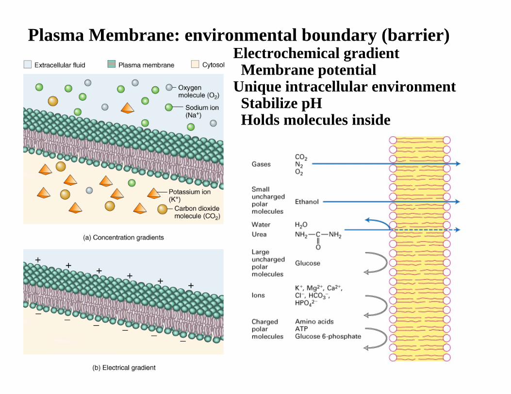
Plasma Membrane: environmental boundary (barrier) Electrochemical gradientMembrane potential
Unique intracellular environmentStabilize pHHolds molecules inside

Plasma Membrane: environmental boundary (barrier)
Ionic imbalance (particularly, Na+ and K+) between inside and outside a cell, created by membrane ionic pumps, ion exchangers and channels, establishes resting membrane potential. This is used to drive other process (such as molecule import), as well as for information processing (e.g. nerve cells).Activities of plasma membrane ionic pumps are energized by hydrolysis of ATP. All the ‘live’ cells establish and maintain the membrane potential.
-50 mV
0 mV
Na+ Na+
10 mM 145 mMK+ K+
140 mM 4 mMCalcium Calcium0.1 mM 2 mMCa2+ Ca2+
< 0.1 µM 1 mMCl- Cl-
3 mM 117 mMATP ATP1 mM < 0.1 µM
Intracellular Extracellular(Cytoplasm) (Tissue fluid)
Membrane potential
Ion Channels
Exchangers
Pumps
Importers
Porins

Plasma MembraneComponents of plasma membrane
LipidsPhospholipidsGlycolipid
CholesterolProteins; transmembrane proteins, peripheral proteinsMany proteins are glycosylated
Membrane channels, pumps : Ion concentration gradient (in out)Membrane potential
Transporters : transport molecule across plasma membrane, e.g. glucose transporterMembrane receptors : Information relay
(via particular signaling molecules, e.g. hormones, neurotransmitters) Communication between cells : Gap junctions, integrinsAdhesion molecules (Junctions); cell to extracellular matrix, cell to cellEndocytosis, Exocytosis : Intracellular membrane flow

Phospholipid structure: e.g. Phosphatidylcholine
cis-doublebond‘kink’

More kinks 1. More difficult to pack phospholipid together – membrane stays fluid at lower temp (Bacteria, yeast adjust the fatty acid composition according to temp, to maintain membrane fluidity)
2. The kinks shorten the length of hydrocarbon chains, so that the membrane is thinner.
Mobility of phospholipid

Four major phospholipids found in mammalian plasma membrane
(PE) (PS) (PC)
There are many ‘minor’ phospholipids exists, too.

Cholesterol
Unique to plasma membraneStabilize membrane
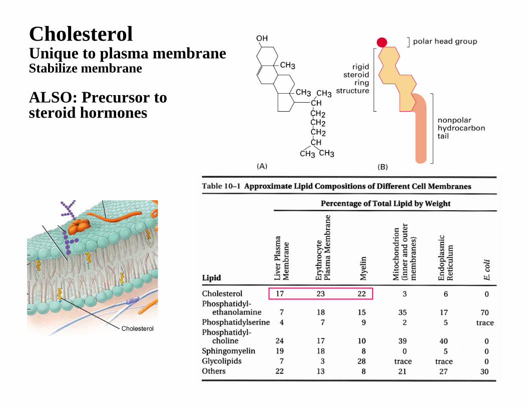
Cholesterol Unique to plasma membraneStabilize membrane
ALSO: Precursor to steroid hormones
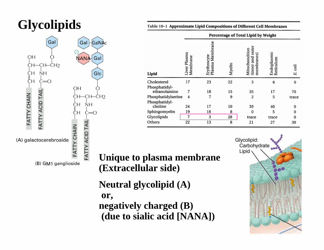
Glycolipids
Unique to plasma membrane(Extracellular side)
Neutral glycolipid (A)or, negatively charged (B)(due to sialic acid [NANA])
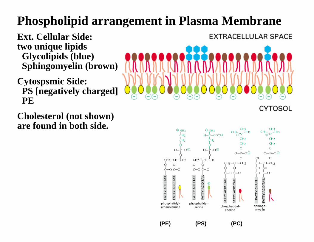
Phospholipid arrangement in Plasma MembraneExt. Cellular Side:two unique lipidsGlycolipids (blue)Sphingomyelin (brown)
Cytospsmic Side:PS [negatively charged]PE
Cholesterol (not shown) are found in both side.
(PE) (PS) (PC)

Lipid Raft
Local area of a membrane where sphingolipid, cholesterol and membrane proteins are concentrated.
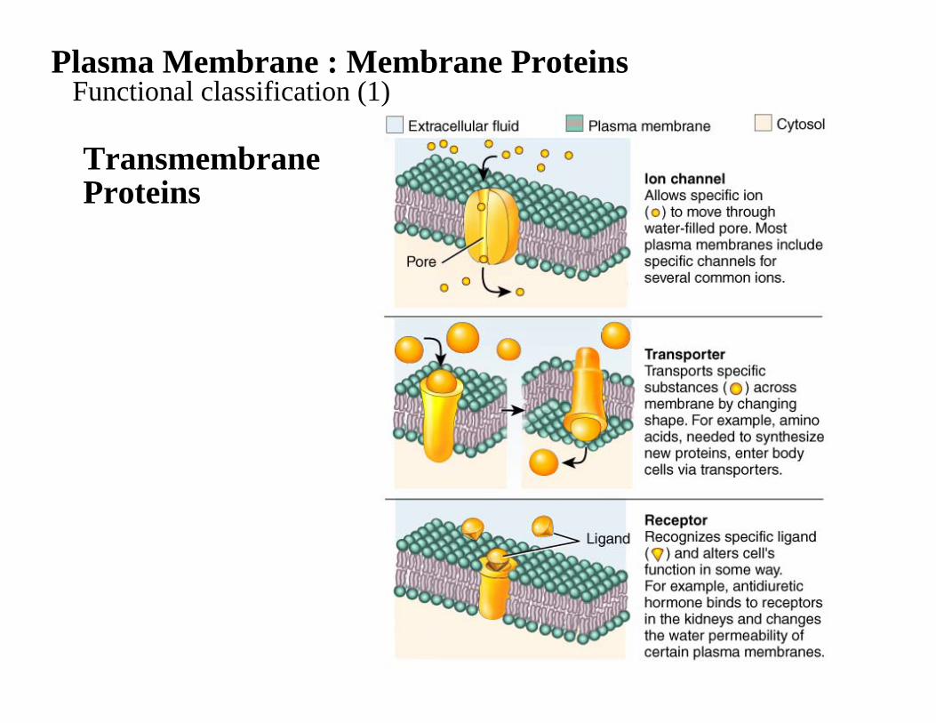
Plasma Membrane : Membrane ProteinsFunctional classification (1)
TransmembraneProteins

Plasma Membrane : Membrane ProteinsFunctional classification (2)
Transmembrane Proteins
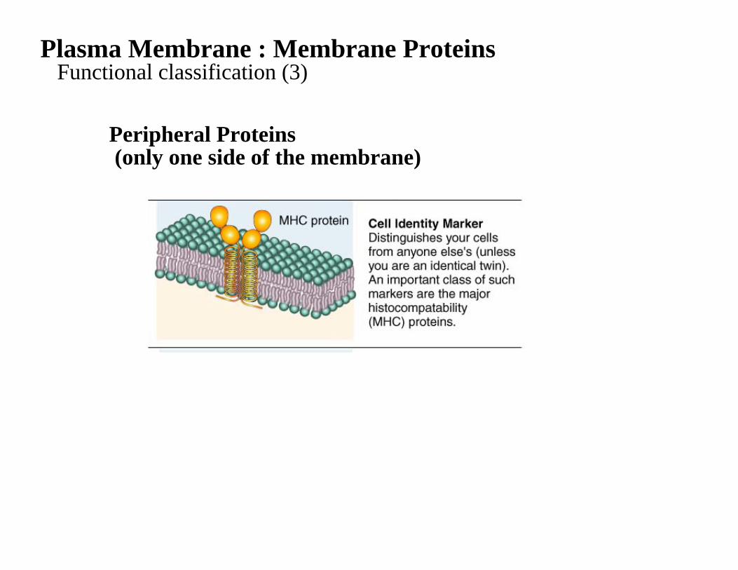
Plasma Membrane : Membrane ProteinsFunctional classification (3)
Peripheral Proteins(only one side of the membrane)

Association of membrane proteins with the lipid bilayer (1)
Transmembrane Proteins
1. A single α-helix2. Multiple α-helices3. Rolled up β-sheet
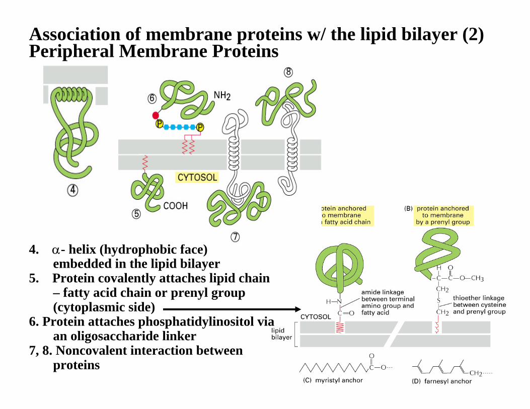
Association of membrane proteins w/ the lipid bilayer (2)Peripheral Membrane Proteins
4. α- helix (hydrophobic face)embedded in the lipid bilayer
5. Protein covalently attaches lipid chain – fatty acid chain or prenyl group(cytoplasmic side)
6. Protein attaches phosphatidylinositol via an oligosaccharide linker
7, 8. Noncovalent interaction between proteins

α helical transmembrane polypeptide chainMostly consists of hydrophobicamino acids (yellow and green)
Seven trans-membrane α helices
Hydropathy plot Prediction of transmembrane α helix bysequence of amino acids

Membrane receptor proteinsSignal transduction across the membrane
The signals are used to activate:gene transcription(s) cell differentiationcell locomotionexocytosis / endocytosis
etc Signal Transduction (an another tutorial section)

These systems exist not only on plasma membrane, but also many organelle membranes.Overview of membrane transport proteins

Na+/K+-ATPase (Na+/K+-pump)1 ATP used for exporting 3 Na+ ions and importing 2 K+ ions.Crucial for maintaining resting membrane potential.
Other pumps; e.g. Ca2+ pump
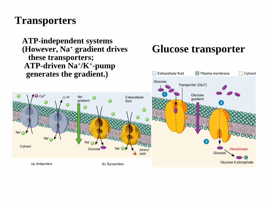
Transporters
ATP-independent systems(However, Na+ gradient drives
these transporters;ATP-driven Na+/K+-pumpgenerates the gradient.)
Glucose transporter
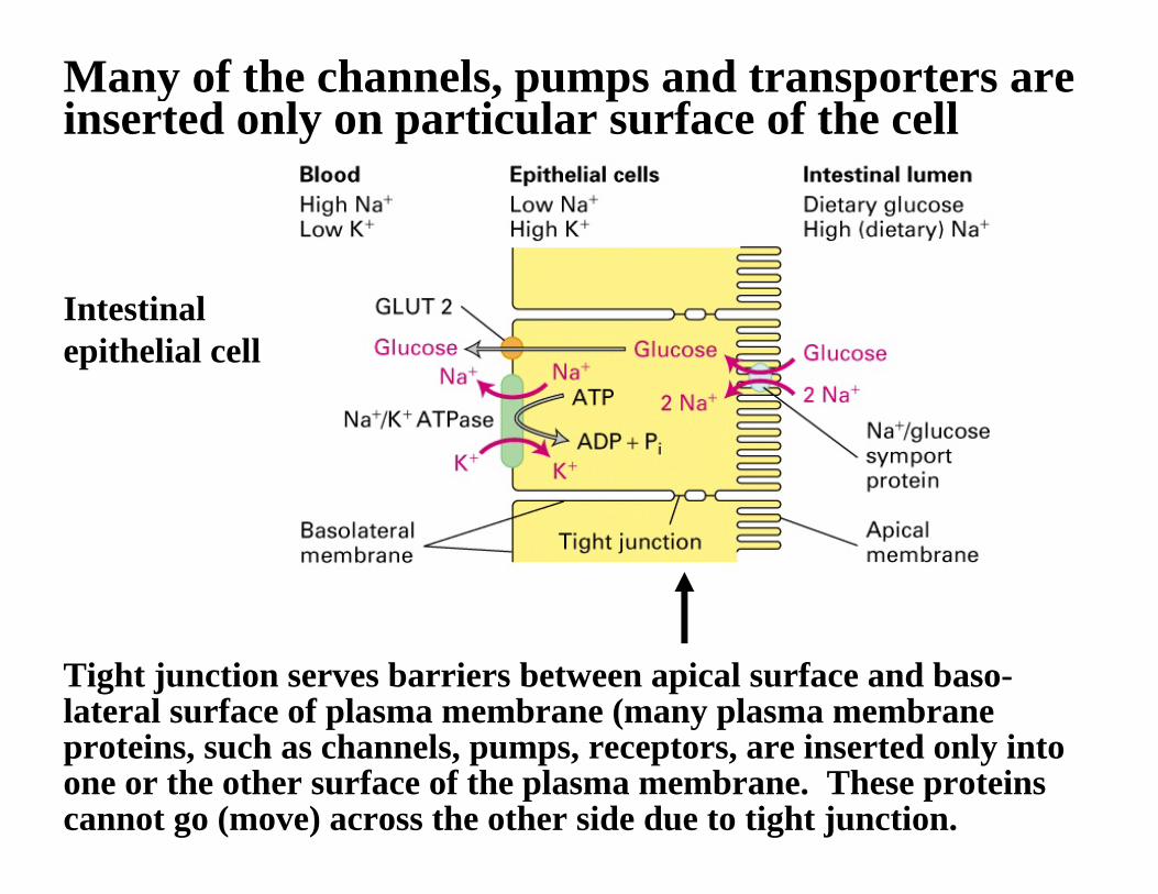
Many of the channels, pumps and transporters are inserted only on particular surface of the cell
Intestinal epithelial cell
Tight junction serves barriers between apical surface and baso-lateral surface of plasma membrane (many plasma membrane proteins, such as channels, pumps, receptors, are inserted only into one or the other surface of the plasma membrane. These proteinscannot go (move) across the other side due to tight junction.
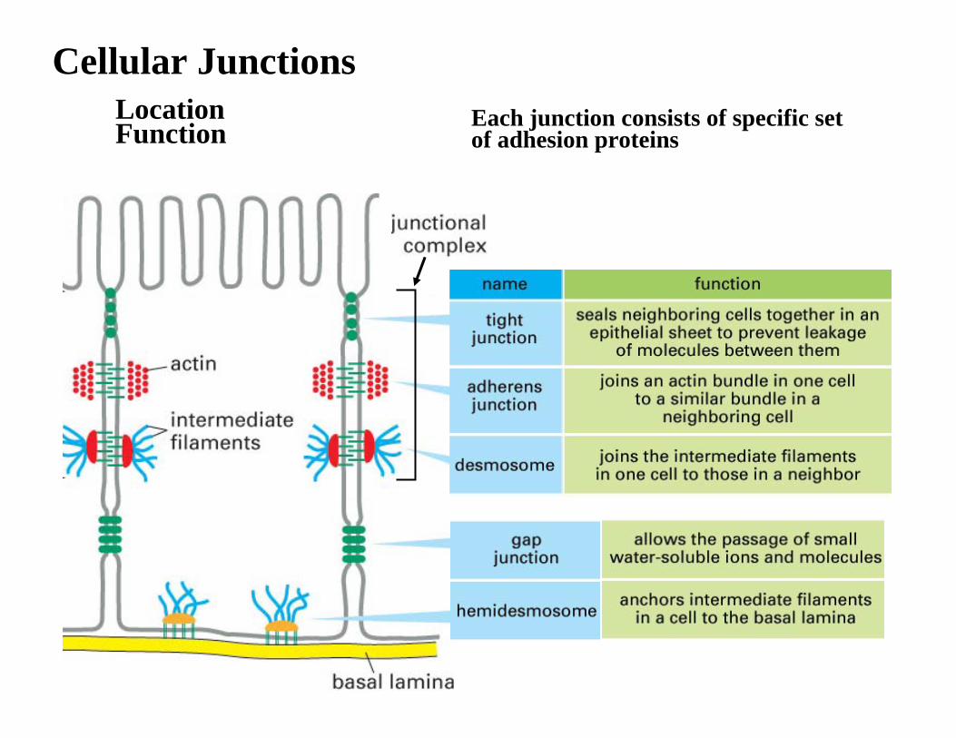
Cellular JunctionsLocation Function Each junction consists of specific set
of adhesion proteins

Occluding JunctionsTight Junction
Separates two environments
Paracellular pathway:Passage through tightjunction b/w cells
Small(er) molecules water, ions, etc

Occluding JunctionsTight Junction
Separates two environments
External body <incl. Intestine lumen, urinary-tract lumen>vs
Extracellular fluid (body fluid) <incl. Blood, lymph>

Tight JunctionTight Junction :Separation of Apical vs basolateralplasma membrane (proteins)
Sealing strands: Plasma-membrane proteins(Occludin, Claudin)Visualized with freeze-fracture EM
Ca2+-requirement
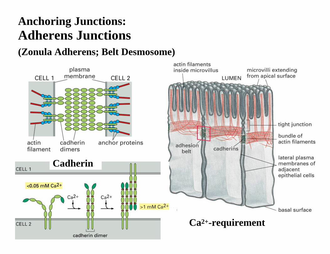
Adherens Junctions(Zonula Adherens; Belt Desmosome)
Ca2+-requirement
Cadherin
Anchoring Junctions:
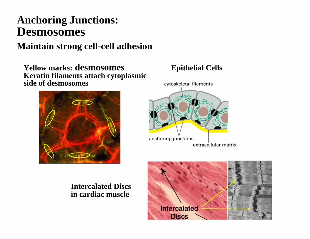
Desmosomes
Yellow marks: desmosomesKeratin filaments attach cytoplasmicside of desmosomes
Epithelial Cells
Intercalated Discsin cardiac muscle
Maintain strong cell-cell adhesion
Anchoring Junctions:
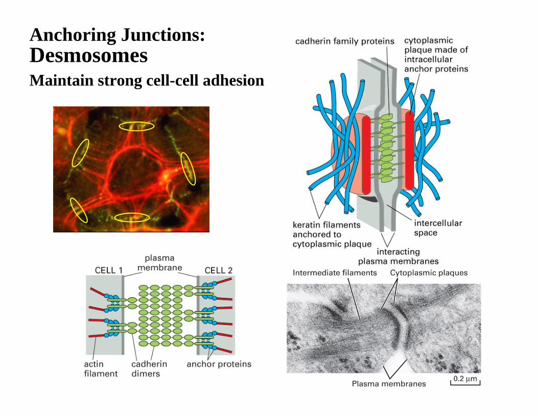
DesmosomesMaintain strong cell-cell adhesion
Anchoring Junctions:

Focal Adhesion
IntegrinCytoskeletal fibers associated:
Actin fibers– Focal adhesions– Muscle (lateral) attachment
Intermediate filaments– Hemidesmosomes
Cell-Matrix adhesion

Focal AdhesionCell-Matrix adhesion
CadherinIntegrin
Hemidesmosome (green)

Integrin holds basal lamina
Matrix-binding activity ofIntegrin is regulated bysignaling events (below)e.g. white blood cells
Integrin (cluster) triggers intracellular signalling

Gap JunctionConnexin hexamer (per cell)‘Connexon’ (hemichannel) Two connexons from adjacent cells to form intracellular channels Often found as ‘patch’ (cluster) on a plasma membrane
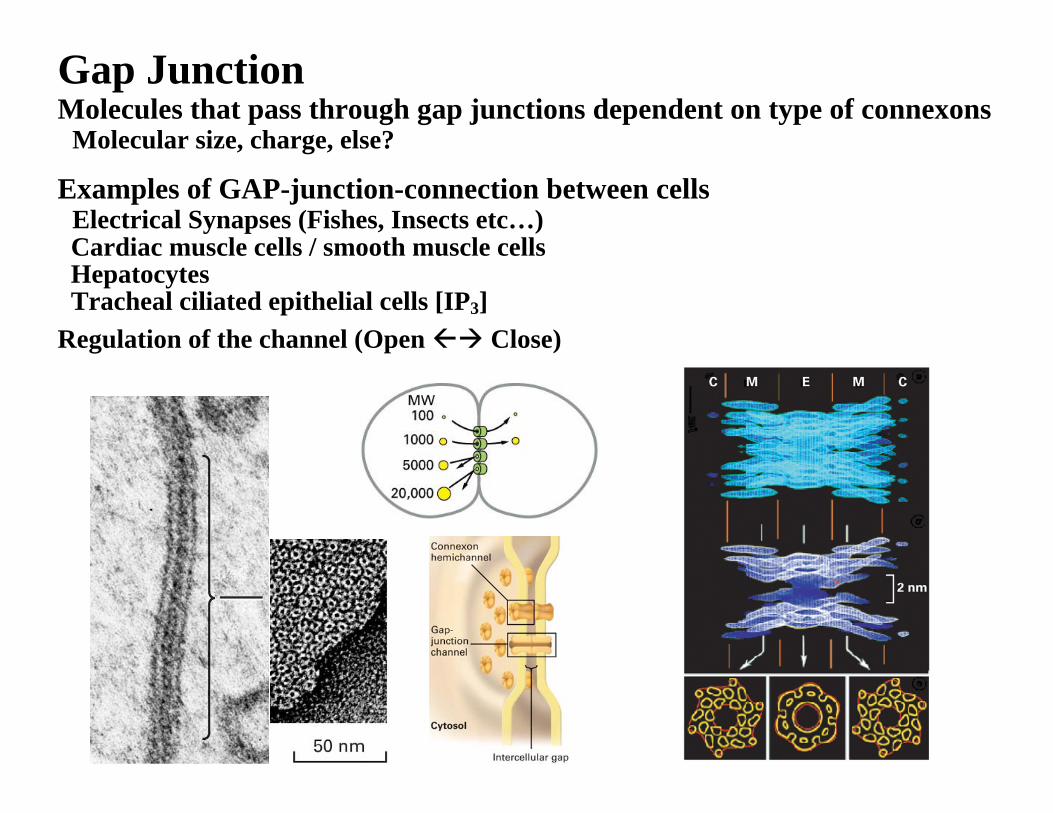
Gap JunctionMolecules that pass through gap junctions dependent on type of connexons
Molecular size, charge, else?
Examples of GAP-junction-connection between cellsElectrical Synapses (Fishes, Insects etc…) Cardiac muscle cells / smooth muscle cellsHepatocytesTracheal ciliated epithelial cells [IP3]
Regulation of the channel (Open Close)

Summary of cell-cell / cell-matrix adhesions
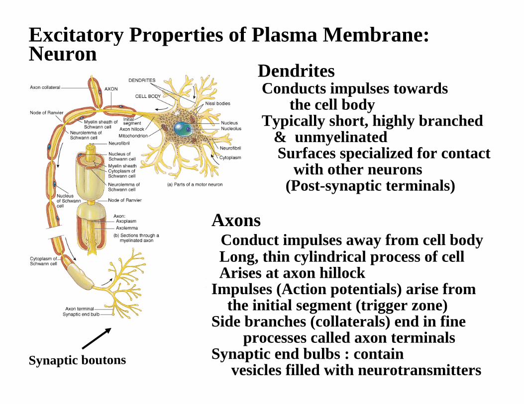
Excitatory Properties of Plasma Membrane: Neuron
DendritesConducts impulses towards
the cell bodyTypically short, highly branched
& unmyelinatedSurfaces specialized for contact
with other neurons(Post-synaptic terminals)
AxonsConduct impulses away from cell bodyLong, thin cylindrical process of cell Arises at axon hillock
Impulses (Action potentials) arise fromthe initial segment (trigger zone)
Side branches (collaterals) end in fineprocesses called axon terminals
Synaptic end bulbs : containvesicles filled with neurotransmitters
Synaptic boutons

Nerve cells, (Neurons, Grial cells)All the other living cells
Potential energy difference at rest is (about) -70 mV(depending on the cell types)
Resting Membrane Potential
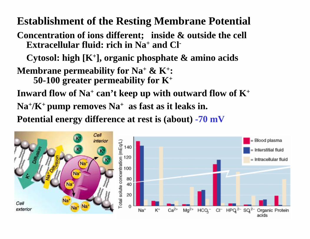
Concentration of ions different; inside & outside the cell Extracellular fluid: rich in Na+ and Cl-
Cytosol: high [K+], organic phosphate & amino acidsMembrane permeability for Na+ & K+:
50-100 greater permeability for K+
Inward flow of Na+ can’t keep up with outward flow of K+
Na+/K+ pump removes Na+ as fast as it leaks in. Potential energy difference at rest is (about) -70 mV
Establishment of the Resting Membrane Potential
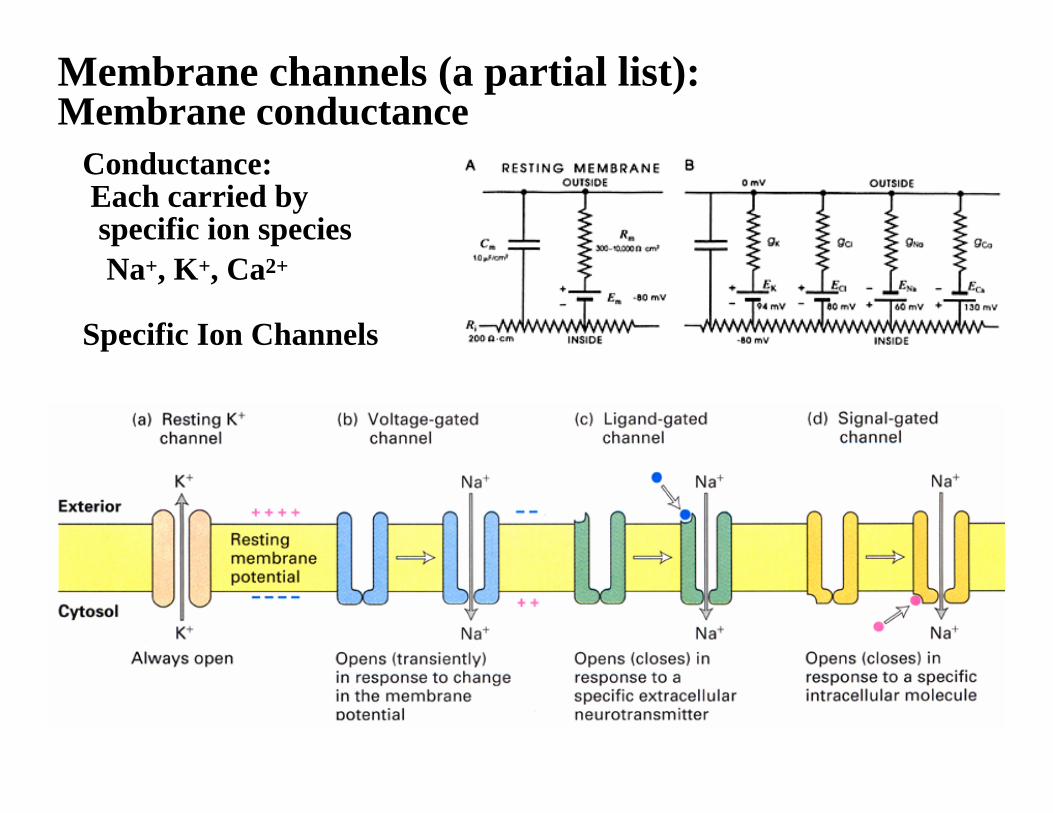
Membrane channels (a partial list): Membrane conductance
Conductance:Each carried by specific ion speciesNa+, K+, Ca2+
Specific Ion Channels

Specific ion channels are opened according to the stimulus
Strength of the stimulus ∝amount of change in membrane potential(Not always; also effective range)
Local change in membrane potential spreads through membrane with decay
Depolarization / Hyperpolarizationof the Membrane Potential
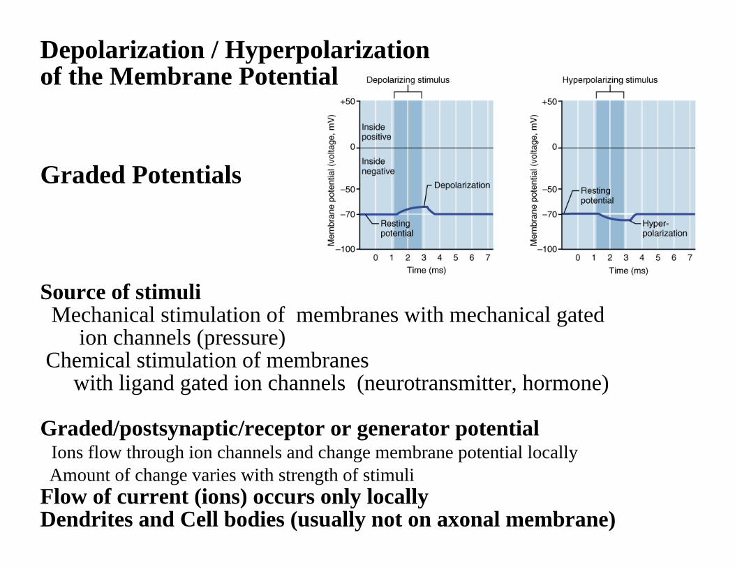
Graded Potentials
Source of stimuliMechanical stimulation of membranes with mechanical gated
ion channels (pressure)Chemical stimulation of membranes
with ligand gated ion channels (neurotransmitter, hormone)
Graded/postsynaptic/receptor or generator potentialIons flow through ion channels and change membrane potential locallyAmount of change varies with strength of stimuli
Flow of current (ions) occurs only locally Dendrites and Cell bodies (usually not on axonal membrane)
Depolarization / Hyperpolarizationof the Membrane Potential

Action PotentialProduced by voltage-gated ion channelsAll – or – NoneVoltage threshold
Voltage-gated Na+ Channel
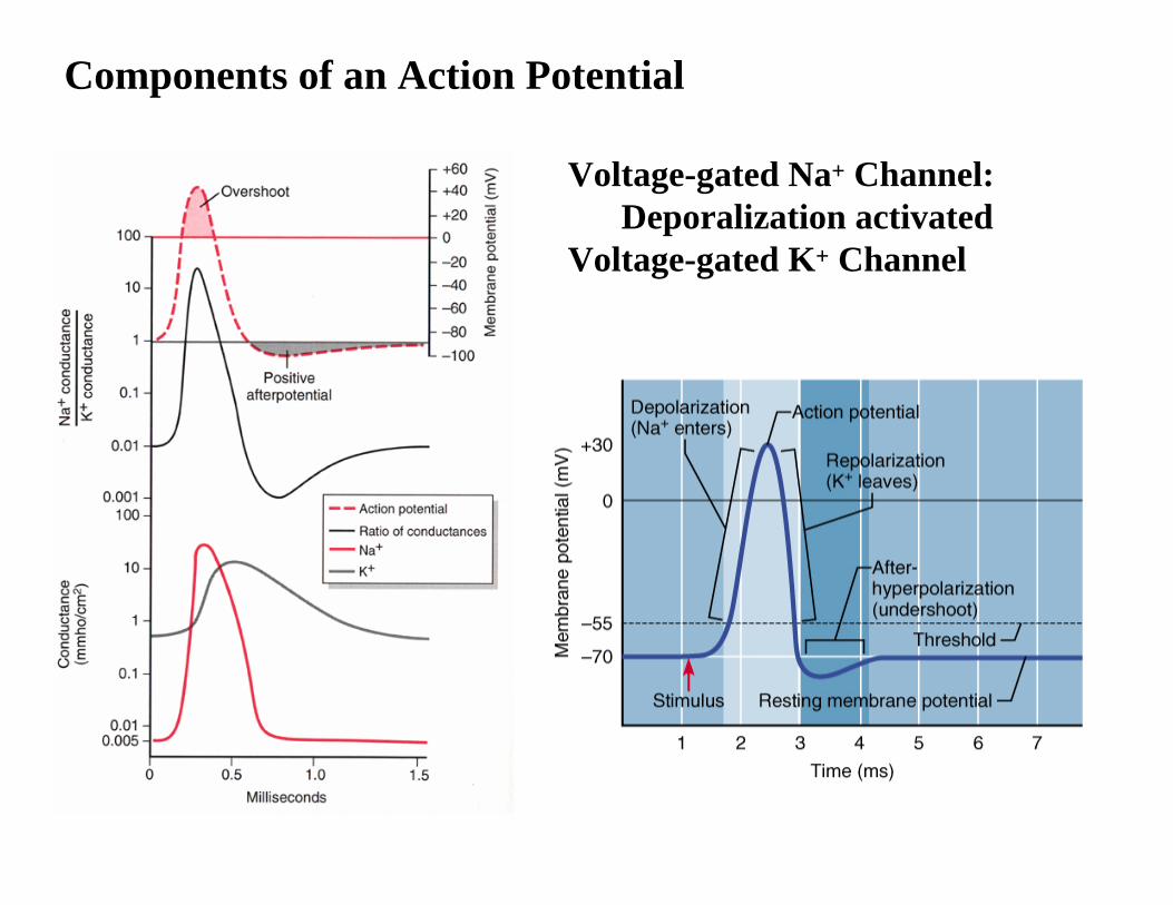
Components of an Action Potential
Voltage-gated Na+ Channel:Deporalization activated
Voltage-gated K+ Channel
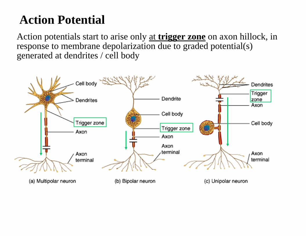
Action PotentialAction potentials start to arise only at trigger zone on axon hillock, in response to membrane depolarization due to graded potential(s) generated at dendrites / cell body

Action potential :enables information processing all-or-none
The refractory period makes action potential to propagate unidirectional
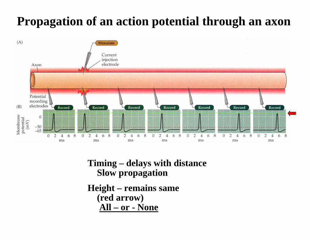
Timing – delays with distanceSlow propagation
Height – remains same (red arrow)All – or - None
Propagation of an action potential through an axon

Myelination of axon and saltatory action potential conduction at the node of Ranvier

A myelinated axon, myelin sheath [M] and aUnmyelinated axones [A] and a schwann cell [S] schwann cell [S]
Myelinated fibers: appear white jelly-rolllike wrappingsmade of lipoprotein = myelinacts as electrical insulatorspeeds conduction of nerve impulses
Unmyelinated fibers: slow,small diameter fibers
only surrounded by neurilemmabut no myelin sheath wrapping
Unmyelinated vs Myelinated axon

Saltatory action potential conduction :Speedup the action potential propagation