tumors in the dominant inferior frontal gyrus neurological
TRANSCRIPT
Page 1/16
Time to Re-Think Broca: Extent of Resection andNeurological Outcome in Patients HarboringTumors in the Dominant Inferior Frontal GyrusPaola Suarez-Meade ( [email protected] )
Mayo Clinic's Campus in Florida https://orcid.org/0000-0002-3638-6799Lina Marenco-Hillembrand
Mayo Clinic's Campus in FloridaDavid Sabsevitz
Mayo Clinic's Campus in FloridaLela Okromelidze
Mayo Clinic's Campus in FloridaBlake Perkidis
Duke UniversityWendy J. Sherman
Mayo Clinic's Campus in FloridaAlfredo Quinones-Hinojosa
Mayo Clinic's Campus in FloridaErik H. Middlebrooks
Mayo Clinic's Campus in FloridaKaisorn L. Chaichana
Mayo Clinic's Campus in Florida
Research Article
Keywords: “Broca’s area”, neuropsychological, glioma, morbidity
Posted Date: August 12th, 2021
DOI: https://doi.org/10.21203/rs.3.rs-788969/v1
License: This work is licensed under a Creative Commons Attribution 4.0 International License. Read Full License

Page 2/16
AbstractIntroduction/Purpose: There is a general lack of consensus onboth the anatomic de�nition and functionof Broca’s area. Given the belief that this region plays a critical role in expressive language, resectivesurgery is often avoided topreserve function. However, the putative role of Broca’s area in speechproduction has been recently challenged.The current study aims to investigatethe feasibilityof gliomaresection and neurological outcomes in “Broca’s area” in 15 patients.
Methods: We report a feasibility study describing the resection of gliomas within the IFG. Awake brainsurgery for resection with mapping and intraoperative neuropsychological evaluation was carried out inall individuals.
Results: All included patientshad tumors located in traditional “Broca’s area” and eight patients (53.33%)had tumors that additionally extendedinto the insula and subinsular regions. During stimulation, positivespeech-language sites within the IFG were identi�ed in ten patients. Two patients (13.33%) experienced adeclinein naming during intraoperative cognitive monitoring and thirteen (86.66%) had a stableperformance throughout surgery. With all patients had recovery of language functions at a two-weekfollow up. Extent of resection was strati�ed in anatomical regions within the IFG, being the pOr the areawith the greatest EOR (97.4%), followed by the pT (84.1%), pOp (83.8%), and vPMC (80%).
Conclusion: The belief that Broca’s area is not safe to resect is challenged. Adequate mapping andcareful patient selection allow maximum safe resection of tumors located in thetraditional “Broca’sarea”,with low risk of postoperative morbidity.
IntroductionThe understanding of the neural basis of speech-language has evolved and expanded since thetraditional models were described. The classical anatomical model of language was �rst represented inthe works of Dr. Broca [1]. He describes a localizationist theory, where discrete cortical areas of the brainare responsible for speech production, traditionally referencing “Broca’s area”; located on the parsopercularis (pOp) and pars triangularis (pT) (Brodmann’s areas 44 and 45) of the frontal operculumwithin the dominant inferior frontal gyrus (IFG) (Table 1.). Historically, Broca’s area was associated withspeech motor functions and, when damaged, resulted in non-�uent aphasia characterized by severelyreduced, effortful speech, and word-�nding di�culty. However, there is accumulating evidence thatchallenges the notion that Broca’s area is critical for speech production [2–4, 6]. We demonstrate thatmapping the IFG has important interindividual variability and that corticectomy and resection of areasthat are thought to be essential for speech is possible.
Lesions restricted to the traditional cortical Broca’s area, often result in mild and more transient non-�uentaphasia, whereas more severe and permanent speech de�cits require lesions to extend posteriorly to thevPMC and deeper to the underlying white matter tracts. The theory of a subcortical “bottleneck” of whitematter tracts deep to the cortical Broca’s area, posits that the full spectrum of severe speech de�cits from

Page 3/16
damage to IFG is likely, in large part, due to damage to multiple eloquent tracts that converge just deep tothe Broca area [5]. In fact, a recent re-examination of Broca’s original patients using high-resolution MRIrevealed extensive subcortical damage that was not originally appreciated by Dr. Broca [3].
Because of the important language eloquence in the IFG patients with tumors in or adjacent to Broca’sarea were thought to be at higher risk of postoperative language morbidity resulting in the traditionalmantra that tumors in this area are often deemed unresectable. In line with the contemporaryconnectomal description of language networks and the dynamic reorganization of language circuitsduring disease, we aimed to conduct a feasibility study on glioma surgery within the pOp and pT.
MethodsStudy Design, Setting, and Population
This study is a single-surgeon consecutive case series, where adult patients harboring tumors within theleft IFG who underwent awake surgical resection with mapping between July 2018 and August 2020.Tumor location in Broca’s area within the IFG was determined using MR imaging. Broca’s area wasanatomically de�ned to be located at the pT and the pOp, as originally described. Patients were excludedif histopathologic diagnosis was other than glioma, had multifocal disease, were left-handed or hadabsolute contraindications for awake surgery [7]. Preoperative Karnofsky Performance Status (KPS) andneuropsychological baseline language status were established the day prior to the procedure [8].Demographic, histologic, imaging, surgical, neuropsychological, and clinical outcomes were included foranalysis.
Neuropsychological Evaluation and Intraoperative Functional Language Tasks
All patients underwent baseline language testing using a tablet-based testing platform for intraoperativemapping (NeuroMapper). Administered subsets were individualized to tumor location and the affectedwhite matter tracts, with picture naming, nonword repetition, and digit repetition being the most utilizedtests. Accuracy and reaction time data were collected. Items that were answered correctly within aprespeci�ed time to allow for DCS stimulation mapping were selected for intraoperative evaluation (e.g., <5-7 seconds). Paradigms were administered during stimulation and continuously throughout activeresection of the tumors to monitor cognition. Patient performance was considered as stable when errorrate was <5% in picture naming responses and cognitive decline was de�ned as >5% error rate comparedto baseline.
Surgical Procedure and Intraoperative Mapping
All patients underwent awake surgery with brain mapping for tumor resection [9,10]. Main surgical goalswere to achieve maximal safe resection (MSR) and to obtain tissue for diagnosis. Scalp nerve block wasdone to ensure analgesia before positioning. After dural opening, direct cortical stimulation (DCS) wascarried out, starting at 3mA, increased at 1 mA intervals, until after discharges were present, positive
Page 4/16
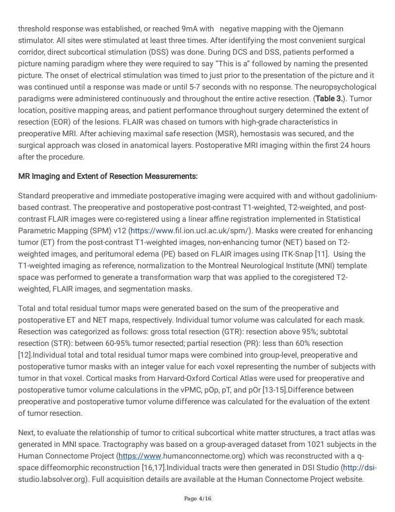
threshold response was established, or reached 9mA with negative mapping with the Ojemannstimulator. All sites were stimulated at least three times. After identifying the most convenient surgicalcorridor, direct subcortical stimulation (DSS) was done. During DCS and DSS, patients performed apicture naming paradigm where they were required to say “This is a” followed by naming the presentedpicture. The onset of electrical stimulation was timed to just prior to the presentation of the picture and itwas continued until a response was made or until 5-7 seconds with no response. The neuropsychologicalparadigms were administered continuously and throughout the entire active resection. (Table 3.). Tumorlocation, positive mapping areas, and patient performance throughout surgery determined the extent ofresection (EOR) of the lesions. FLAIR was chased on tumors with high-grade characteristics inpreoperative MRI. After achieving maximal safe resection (MSR), hemostasis was secured, and thesurgical approach was closed in anatomical layers. Postoperative MRI imaging within the �rst 24 hoursafter the procedure.
MR Imaging and Extent of Resection Measurements:
Standard preoperative and immediate postoperative imaging were acquired with and without gadolinium-based contrast. The preoperative and postoperative post-contrast T1-weighted, T2-weighted, and post-contrast FLAIR images were co-registered using a linear a�ne registration implemented in StatisticalParametric Mapping (SPM) v12 (https://www.�l.ion.ucl.ac.uk/spm/). Masks were created for enhancingtumor (ET) from the post-contrast T1-weighted images, non-enhancing tumor (NET) based on T2-weighted images, and peritumoral edema (PE) based on FLAIR images using ITK-Snap [11]. Using theT1-weighted imaging as reference, normalization to the Montreal Neurological Institute (MNI) templatespace was performed to generate a transformation warp that was applied to the coregistered T2-weighted, FLAIR images, and segmentation masks.
Total and total residual tumor maps were generated based on the sum of the preoperative andpostoperative ET and NET maps, respectively. Individual tumor volume was calculated for each mask.Resection was categorized as follows: gross total resection (GTR): resection above 95%; subtotalresection (STR): between 60-95% tumor resected; partial resection (PR): less than 60% resection[12].Individual total and total residual tumor maps were combined into group-level, preoperative andpostoperative tumor masks with an integer value for each voxel representing the number of subjects withtumor in that voxel. Cortical masks from Harvard-Oxford Cortical Atlas were used for preoperative andpostoperative tumor volume calculations in the vPMC, pOp, pT, and pOr [13-15].Difference betweenpreoperative and postoperative tumor volume difference was calculated for the evaluation of the extentof tumor resection.
Next, to evaluate the relationship of tumor to critical subcortical white matter structures, a tract atlas wasgenerated in MNI space. Tractography was based on a group-averaged dataset from 1021 subjects in theHuman Connectome Project (https://www.humanconnectome.org) which was reconstructed with a q-space diffeomorphic reconstruction [16,17].Individual tracts were then generated in DSI Studio (http://dsi-studio.labsolver.org). Full acquisition details are available at the Human Connectome Project website.

Page 5/16
Brie�y, a multishell diffusion scheme was with b-values of 1000, 2000, and 3000 s/mm2 with 90diffusion directions per shell. Images were acquired with an isotropic resolution of 1.25 mm. Adeterministic �ber tracking algorithm was used [18].Tract data was semi-automated based on priortractography atlas [19].Topology-informed pruning was applied to the tractography with 3 iterations toremove false connections. The residual tumor group map (thresholded at > 13% or n > 2) was comparedto the tract anatomy in DSI Studio [20].
Postoperative Care
Patients were evaluated two weeks after surgery and at follow-up visit at 3 months. Outcome measuresof postoperative KPS, EOR, presence of postoperative language de�cits, surgical complications, length ofhospital stay were collected. Length of hospital stay was from the time of surgery to the day ofdischarge.
Compliance with Ethical Standards:
All procedures performed in this study involving human participants were in accordance with the ethicalstandards of the institution and the national research committee. This study was carried out under theIRB number: PR16-009946-01.Informed consent was obtained from all individual participants included inthe study.
Statistical Analysis
Descriptive statistics were utilized to detail patient characteristics and postoperative outcomes. Data wasexpressed as a mean with range for continuous variables and counts with percentages for categoricalvariables. Student T-test was utilized for continuous data. P<0.05 was considered statistically signi�cant.All analyses were performed using GraphPad Prism 9.0.2 for Windows (Graphpad Software, La Jolla,California, USA).
ResultsPatient Demographics and Presentation
A total of 15 consecutive right-handed patients with tumors in the left IFG were included for analysis(Table 2). Lesion location within the IFG was most commonly in the pOp (80%), followed by the vPMC(53.33%), pT (33.33%), and pOr (6.66%). One lesion extended into the temporal lobe and eight extendedinto the insula. Fourteen patients (93.33%) showed some type of speech de�cit at diagnosis asdetermined by review of presenting clinical history or neurologic examination. Patients most oftenpresented with non-�uent aphasia (n=5, 55.55%), verbal working memory de�cits (n=8, 53.33%), andspeech arrest (n= 4, 26.66%).
Surgical Characteristics
Page 6/16
All patients underwent awake brain surgery with tailored pterional craniotomy and negative mappingtechnique (n=15) (Figure 1.). One patient presented with intraoperative seizures due to DCS that werecontrolled with cold saline. No other complications were noted. A transcortical corridor was used foraccess in 13 (86.66%) patients, to minimize exposure of the lenticulostriate arteries. Transcorticalcorridors were done through areas that showed no language disruption with DCS. Speci�cally, surgicalentry was made through the pOp (n=7, 46.66%), pT (n=1, 6.66%), pOr (n=1, 6.66%), pOr + PT (n=1, 6.66%),MFG (n=2, 13.33%), and STG (n=1, 6.66%) (Table 3).
Intraoperative Language Mapping
Patients were evaluated by neuropsychology before and during surgery. Five patients (33.33%) had noidenti�able frontal lobe language sites during DCS while ten (66.66%) presented errors in picture namingduring DCS. Disruption of speech language functions during DCS was commonly found in the vPMC(n=9, 60%), followed by the pT (n=3, 20%), and the pOp (n=3, 20%) (Table 3). For direct subcorticalstimulation (DSS), only the SLF (n=2), the IFoF (n=1), and the AF (n=1) tracts resulted in positive �ndingsfor speech arrest and phonetic paraphasias (Table No. 3). During continuous neuropsychologicalmonitoring, twelve patients (86.66%) remained stable throughout the procedure. Only two patients(13.33%) presented with mild decline in naming during surgery, as they presented >5% in error ratecompared to baseline. Neuropsychology evaluation was useful to identify the limits of the surgery, asresection was stopped in patients that presented with any kind of de�cits during active resection topreserve function.
Extent of Resection
Prior to surgery, median tumor volume was (79.42±80.14 cm3), while mean residual tumor volume was(39.83±55.24 cm3) (Figure 2.). Overall, the mean EOR was 60.35% (± 29.60%). EOR was strati�ed inanatomical regions within the IFG, being the pOr the area with the greatest EOR (97.4%), followed by thepT (84.1%), the pOp (83.8%), and then the vPMC (80%). Although not statistically signi�cant, EOR forhigh-grade gliomas (WHO grade III and IV) (72.93±17.94) was greater when compared to low-gradegliomas (WHO grade I and II) (60.75%±30.51) (p=0.4010). Resection was limited by positive mappingareas during DCS, cognitive decline, and involvement of high-risk structures. Residual tumor wascommonly located within the insular and subinsular regions as well as the deep margins within criticalwhite matter regions of the centrum semiovale (Figure 3. and Figure 4.).
Postoperative Outcome and Neurological Morbidity
Outcome data is summarized in Table 4. Patients were hospitalized for 2.33 (±1.19) days on average.Although recovery was noted in all patients at two-weeks postop compared to baseline, seven patients(46.66%) still presented with mild speech and language de�cits that were related to tumor growth and notto surgical procedure (Table 3. and Table 4.). Mean postoperative functional status (KPS: 88.66 ± 8.84)calculated at two weeks after surgery remained the same as the mean preoperative functional status(KPS:88 ± 8.32). (Table 4.).

Page 7/16
DiscussionSurgery within “Broca’s area” for tumor resection is feasible with very minimal and acceptablepostoperative morbidity. In our case series, EOR ranged between 80-97.4% in tumors located in the IFG.Resection was mainly limited by positive mapping, white matter tract involvement, and insular extensionof lesions. Resection in these areas was avoided to prevent injury to vascular and functional structures,which prevented de�ection of the results seen after resection in “Broca’s area”. Moreover, mapping the IFGhas important interindividual variability and that corticectomy and resection of areas that are thought tobe essential for speech is possible. When compared to presentation at diagnosis, all patients hadremarkable recovery of language functions two weeks after surgery and no new surgical-related de�citswere noted. These results demonstrate that resection of gliomas in what is described as “Broca’s area” isfeasible in a carefully selected subset of patients.
For more than a century, the classical model of language organization proposed by Doctors Broca andWernicke was universally accepted despite many observations that were not accounted for in thisincomplete model [1,21]. Broca’s descriptions led to a localizationist model which placed the speechoutput epicenter within the posteromedial portion of the IFG (pT, pOp; Brodmann areas 44 and 45) of thedominant hemisphere [22,23].However, Broca’s model was largely based upon lesionology and theirproponents failed to account that Leborgne’s and Lelonge’s brains – the so-called “Broca’s Brains” - showed damage beyond the cortical limits of Broca’s area, extending to the insula and to the perisylvianwhite matter [3]. These overly simpli�ed models attributed �xed functions to speci�c areas of the brainleading to the general belief that surgical intervention in these “eloquent” areas was impossible [24]. Thecurrent understanding of language has advanced towards a connectomal framework which largelyrecognizes interindividual anatomo-functional variability [25,26].Current models acknowledge thedynamic and complex subcortical circuitry that interconnect, reshape, and distribute information withinthe brain [21,27,28].
Preserving function is one of the most important principles in brain tumor surgery, as postoperativemorbidity has been demonstrated to impact patient prognosis [29,30].For that reason, surgical resectionof tumors located in the dominant IFG and adjacent subcortical areas was traditionally consideredimplausible. Nonetheless, several authors have proved that resection of Broca’s area does not alwayslead to language de�cits [31-36]. Benzgamout et al. reported a series of 7 patients with LGG whounderwent awake craniotomy for tumor resection with intraoperative mapping. Speech motor epicenterswere found within the ventral and dorsal premotor cortices, the orbitofrontal cortex, and the insula [32].Similar �ndings were described by Lubrano et al. who reported sixteen consecutive patients with lesionswithin Broca’s area [33].In this series, they correlated the morphology of the lesion to positive mappingduring stimulation of the pOp and pT, if the lesion was well-circumscribed, positive speech areas withinBroca’s area were found at a rate of 100%, whilst in�ltrative lesions presented positive mapping in Broca’sarea in only 25% of cases. In the largest series performed to date, Rolston et al. retrospectively evaluateda series of 43 patients with opercular lesions, 33 of which underwent awake craniotomy with languagemapping. After follow-up authors concluded that tumors located within the pOp did not possess any risk

Page 8/16
for developing postoperative language de�cits and identi�ed a number of risk factors associated withpostoperative de�cit appearance [34]. These included presence of preoperative seizures, preoperativelanguage de�cits, and decreased extent of resection.
The insula has also been identi�ed as an important center of coordination of the articulation regions.Studies indicate that the dominant anterior insula is likely a critical node in speech output, receiving �nalword selection from the frontal operculum, conveying it to the cerebellum and basal ganglia and relayingthis information to the vPMC and vSMC [23,38]. Neuroimaging studies and lesion to this structureindicate its key role in articulatory control and higher-order cognitive aspects of speech [37].In our work,postoperative imaging revealed that most residual tumors were located within the insula (n=9, 60%) andin the vPMC (n=6, 40%). Resection of the insular component of the tumors was avoided due to eloquenttissue and to critical microvasculature for language and motor systems that pose a high-risk of strokeduring surgery.
The vPMC has been repeatedly identi�ed as the area with the highest rate of speech arrest uponstimulation, occurring in 83% of cases after stimulation of the vPMC in the dominant hemisphere. In2014, Tate et al. challenged the traditional theories of language organization after performing aprobabilistic map of critical functional regions within the human cerebral cortex of 165 patients with LGG,including Broca's area. Interestingly, speech arrest was localized within the vPMC, not Broca’s [4]. Theirresults correlated with other studies where speech de�cits were localized in the vPMC [6, 35, 38, 39].Although the role of the left vPMC in language is poorly understood it may have a role in the control ofthe initiation of complex movements, allowing speech articulation including coordination of the tongue,lips and pharynx. The limited plastic potential of the left vPMC as well as the critical underlying whitematter tracts, makes it a high-risk area for surgical resection [40]. Thus, we conclude that stimulation ofthe vPMC causes higher rate of speech arrest than Broca’s; which challenges the prevailing notion ofBroca’s area as the quintessential motor speech epicenter.
The results presented herein differ from the classical localizationist model and support the use of thecontemporary hodotopical basis of language for tumor surgery. Given the lack of anatomic speci�city,and its historically oversimpli�ed functional role,the �eld may bene�t by no longer using the moniker of“Broca’s area”. More speci�c anatomic and functional anatomy of the IFG cortex, as well asdifferentiation from underlying white matter tracts, may add to clarity and reproducibility of surgicalliterature.
Strengths and Limitations:
While the results of our study provide insightful information on the safety pro�le of surgical resections inthe IFG, it also carries some limitations. The retrospective nature of the study, which limited the author’scapacity to homogenize available data, small sample size, and heterogeneous pathology presented in ourcohort have to be considered. This study also carries various strengths. Surgical data was obtained froma single surgeon, reducing variability and bias in our results. Real-time monitoring of speech andlanguage was used as a primary outcome variable, increasing the sensitivity of detecting surgical

Page 9/16
induced injury rather than subacute surgical effects (e.g., edema). Neuropsychological testing was carriedout a novel tablet-based platform that is OR-friendly and allows individualized testing in a speci�c andsafe manner.
Concluding Remarks:
Awake resection of tumors located within dominant inferior frontal gyrus can be safely performed withextremely low complication rates and minimal postoperative morbidity. In the presence of tumors, surgeryis limited by identi�cation of functional areas and not by the anatomical limits of what is thought to be“Broca’s area”.
DeclarationsData availability statement: The data that support the �ndings of this study are available from thecorresponding author, KLC, upon reasonable request.
Con�ict of interest: Authors declare no con�ict of interest
Author contribution:
Conceptualization and Study Design: PSM, LMH, DS, EHM, KLC
Data Acquisition: PSM, LMH, DS, LO, BP, EHM, KLC
Data Interpretation: PSM, LMH, DS, EHM, WJS, AQH, KLC
Writing: PSM, LMH, DS, EHM
Review and Editing: DS, WJS, AQH, EHM, KLC
References1. Broca P. Nouvelle Observation d’aphemie produite par une lesion de la moitie posterirure des
deuxieme et troisieme circonvolutions frontale. Bull Soc Anat. 1861. (6): 337-393.
2. Kim JH, Amankulor NM, Peck KK, Brennan N, Gutin PH, Holodny AI. Resection of glioma in an fMRI-de�ned “split” Broca’s Area. Neurocase. 2014; 20 (5): 481-486.
3. Dronkers NF, Plaisant O, Iba Zizen MT, Cabanis EA. Paul Broca’s Historic Cases: High resolution MRimaging of the brains of Leborgne and Lelong. Brain. 2007; 130 (5): 1432-1441.
4. Tate MC, Herbert G, Moritz-Gasser S, Tate JE, Duffau H. Propabilistic Map of Critical FunctionalRegions of the Human Cerebral Cortex: Broca’s Area Revisited. Brain. 2014; 137 (10): 2773-2782.
5. Gri�s JC, Nenert R, Allendorfer JB, Sza�arski JP. Damage to White Matter Bottlenecks Contributes toLanguage Impairments after Left Hemispheric Stroke. Neuroimag: Clinical. 2017; (14): 552-565

Page 10/16
�. Ferpozzi V, Fornia L, Montagna M, Siodambro C, Castellano A, Borroni P, et al. Broca’s Area as a Pre-Articulatory Phonetic Encoder: Gating the Motor Program. Front Hum Neurosci; 2018: 12:64
7. Hervey-Jumper SL, Li J, Lau D, Molinaro AM, Perry DW, Meng L, et al. Awake Craniotomy to MaximizeGlioma Resection: Methods and Technical Nuances Over a 27-year Period. J Neurosurg. 2015; 123(2): 325-39.
�. Yates JW, Chalmer B, McKegney FP. Evaluation of Patients with Advanced Cancer using theKarnofsky Performance Status. Cancer. 1980; 45 (8): 2220-2224.
9. Eseonu CI, Rincon-Torroella J, Lee YM, ReFaey K, Tripathi P, Quinones-Hinojosa, A. IntraoperativeSeizures in Awake Craniotomy for Perirolandic Glioma Resections that Undergo Cortical Mapping. JNeurol Sug A Cent Eur Neurosurg. 2018. 79 (3): 238-246.
10. ReFaey K, Tripathi S, Bhargav AG, Grewal SS, Middlebrooks EH, Sabsevitz DS, et al. PotentialDifferences Between Monolingual and Bilingual Patients in Approach and Outcome After AwakeBrain Surgery. J Neurooncol. 2020; 148 (3): 587-598.
11. Yushjevich PA, Piven J, Cody Hazlett H, Gimpel Smith R, Ho S, Gee JC, et al. User-guided 3D activecontour segmentation of anatomical structures: signi�cantly improved e�ciency and reliability.Neuroimage. 2006; 31 (3): 116-28.
12. Chaichana KL, Jusue-Torres I, Navarro-Ramirez R, Raza SM, Pascual-Gallego M, Ibrahim A, etal. Establishing percent resection and residual volume thresholds affecting survival and recurrencefor patients with newly diagnosed intracranial glioblastoma. Neuro-Oncology. 2014; 16 (1): 113-22.
13. Makris N, Goldstein JM, Kennedy D, Hodge Sm, Caviness VS, Faraone SV, et al. Decreased volume ofleft and total anterior insular lobule in schizophrenia. Schizophr Res. 2006. 83 (2-3): 155-71.
14. Frazier JA, Chiu S, Breeze JL, Makris N, Lange N, Kenney DN, et al. Structural brain magneticresonance imaging of limbic and thalamic volumes in pediatric bipolar disorder. Am J Psychiatry.2005; 162 (7): 1256-65.
15. Desikan RS, Segonne F, Fischl B, Quinn BT, Dickerson BC, Blacker D, et al. An automated labelingsystem for subdividing the human cerebral cortex on MRI scans into gyral based regions of interest.Neuroimage. 2006. 31 (2): 968-80.
1�. Yeh FC, Tseng WY. NTU-90: a high angular resolution brain atlas constructed by q-spacediffeomorphic reconstruction. Neuroimage. 2011; 58L 91-99.
17. 39. Yeh FC, Wedeen VJ, Tseng WY. Generalized q-sampling imaging. IEEE Trans Med Imaging. 2010;29 (9): 1626-35.
1�. Yeh FC, Verstynen TD, Wang Y, Fernandez-Miranda JC, Tseng WY. Deterministic diffusion �bertracking improved by quantitative anisotropy. PLoS ONE. 2013; 8 (11): 80713
19. Yeh FC, Panesar S, Fernandes D, Meola A, Yoshino M, Fernandez-Miranda JC, et al. Populationaveraged atlas of the macroscale human structural connectome and its network topology.Neuroimage. 2018; 178: 57-68.
20. Yeh FC, Panesar S, Barrios J, Fernandes D, Abhinav K, Meola A, et al. Automatic removal of falseconnections in diffusion MRI tractography using topology-informed prining (TIP). Neurotherapeutics.

Page 11/16
2019; 16 (1): 52-58.
21. Duffau H. The Error of Broca: From the Traditional Localizationist Concept to a ConnectomalAnatomy of the Human Brain. J Chem Neuroanat. 2018; 89: 73-81.
22. Vanacor CN, Isolan GR, Yu YH, et al. Microsurgical Anatomy of Language. Clin Anat. 2021; 34 (1):154-168.
23. Stinnet TJ, Reddy V, Zabel MK. Neuroanatomy, Broca. In: StatPearls. Tresure Island (FL): StatpearlsPublishing
24. Sawaya R, Hammoud M, Schoppa D, Hess KR, Wu SZ, Shi WM, et al. Neurosurgical Outcomes in aModern Series of 400 Craniotomies for Treatment of Parenchymal Tumors. Neurosurgery. 1998; 42(5): 1044-1055.
25. Middlebrooks EH, Yargmurlu K, Sza�arski JP, Rahman M, Bozjurt B. A contemporary framework oflanguage processing in the human brain in the context of preoperative and intraoperative languagemapping. Neuroradiology. 2017; 59 (1): 69-87.
2�. Yagmurlu K, Middlebrooks EH, Tanriover N, Rhoton AL Jr. Fiber tracts of the dorsal language streamin the human brain. J Neurosurg; 2016: 124 (5): 1396-405.
27. Flinker A, Korzeniewska A, Shestyuk AY, Franaszczuk PJ, Dronkers NF, Knight RT, et al. Rede�ning theRole of Broca’s Area in Speech. PNAS. 2015; 112 (9): 2871-2875.
2�. Chang EF, Raygor KP, Berger MS. Contemporary Model of Language Organization: An Overview forNeurosurgeons. Journal of Neurosurgery. 2015; 122: 250-261.
29. Rahman M, Abbatematteo J, De Leo EK, Kubulis PS, Vaziri S, Bova F, et al. The effects of new orworsened postoperative neurological de�cits on survival of patients with glioblastoma. J Neurosurg.2016; 127 (1): 123-131.
30. McGirt MJ, Mukherjee D, Chaichana KL, Than KD, Weingart JD, Quinones-Hinojosa, A. Association ofSurgically Acquired Motor and language de�cits on overall survival after resection of glioblastomamultiforme. Neurosurgery. 2009; 65 (3): 463-9.
31. Chaichana KL, Jusue-Torres I, Navarro-Ramirez R, Raza SM, Pascual-Gallego M, Ibrahim A, etal. Establishing Percent Resection and Residual Volume Thresholds Affecting Survival andRecurrence for Patients with Newly Diagnosed Intracranial Glioblastoma. Neuro Oncol. 2014; 16 (1):113-122.
32. Benzagmout M, Gatignol P, Duffau H. Resection of World Health Organization Grade II gliomasinvolving Broca’s Area: Methodological and Functional Considerations. Neurosurgery. 2007; 61 (4):741-742
33. Lubrano V, Draper L, Roux FE. What Makes Surgical Tumor Resection Feasible in Broca’s Area?Insights into Intraoperative Brain Mappin. Neurosurgery. 2010; 66 (5): 868-875.
34. Rolston JD, Englot DJ, Benet A, Li J, Cha S, Berger MS. Frontal Operculum Gliomas: LanguageOutcome Following Resection. J Neurosurg. 2015; 122 (4): 725-734.
Page 12/16
35. Duffau H, Capelle L, Denvil D, Peggy G, Sichez N, Van Effenterre R, The Role of Dominant PremotorCortex in Language: A Study Using Intraoperative Functional Mapping in Awake Patients.Neuroimage. 2003; 20 (4): 1903-1914.
3�. Gajardo-Vidal A, Lorga-Puls DL, PLORAS team, Warner H, Pshdary B,Crinion JT, et al. Damage toBroca’s Area does not contribute to long-term speech production outcome after stroke. Brain. 2021. 1-16.
37. Kim JH, Amankulor NM, Peck KK, Brennan N, Gutin PH, Holodny AI. Resection of glioma in an fMRI-de�ned "split" Broca's area. Neurocase. 2014;20(5):481-486.
3�. Sanai N, Mirzadeh Z, Berger MS. Functional Outcome after Language Mapping for glioma Resection.N Engl J Med. 2008; 358: 18-27.
39. Fiebach CJ, Schubotz RI. Dynamic Anticipatory Processing od Hierarchical Sequential Events: ACommon Role for Broca’s Area and Ventral Premotor Cortex Across Domains?. Cortex, 2006; 42 (4):499-502.
40. Van Geemen K, Herbet G, Moritz-Gasser S, Duffau H. Limited plastic potential of the left ventralpremotor cortex in speech articulation: evidence from intraoperative awake mapping in gliomapatients. Hum Brain Mapp. 2014;35(4):1587-1596.
TablesDue to technical limitations, tables are only available as a download in the Supplemental Files section.
Figures
Page 13/16
Figure 1
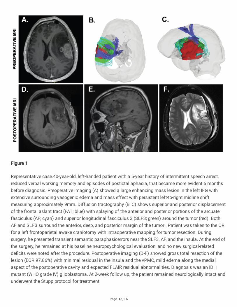
Representative case.40-year-old, left-handed patient with a 5-year history of intermittent speech arrest,reduced verbal working memory and episodes of postictal aphasia, that became more evident 6 monthsbefore diagnosis. Preoperative imaging (A) showed a large enhancing mass lesion in the left IFG withextensive surrounding vasogenic edema and mass effect with persistent left-to-right midline shiftmeasuring approximately 9mm. Diffusion tractography (B, C) shows superior and posterior displacementof the frontal aslant tract (FAT; blue) with splaying of the anterior and posterior portions of the arcuatefasciculus (AF; cyan) and superior longitudinal fasciculus 3 (SLF3; green) around the tumor (red). BothAF and SLF3 surround the anterior, deep, and posterior margin of the tumor . Patient was taken to the ORfor a left frontoparietal awake craniotomy with intraoperative mapping for tumor resection. Duringsurgery, he presented transient semantic paraphasicerrors near the SLF3, AF, and the insula. At the end ofthe surgery, he remained at his baseline neuropsychological evaluation, and no new surgical-relatedde�cits were noted after the procedure. Postoperative imaging (D-F) showed gross total resection of thelesion (EOR 97.86%) with minimal residual in the insula and the vPMC, mild edema along the medialaspect of the postoperative cavity and expected FLAIR residual abnormalities. Diagnosis was an IDHmutant (WHO grade IV) glioblastoma. At 2-week follow up, the patient remained neurologically intact andunderwent the Stupp protocol for treatment.

Page 14/16
Figure 2
.(A) Preoperative probability map of combined enhancing and non-enhancing tumor for all patients. (B)Preoperative probability map of combined enhancing and non-enhancing tumor thresholded at 93% (n >1). (C) Postoperative probability map of residual enhancing and non-enhancing tumor thresholded at the93rd percentile (n > 1) and the (D) 87th percentile (n > 2) show complete resection of tumor within the IFGin the majority of cases. Residual tumor was greatest in the insular and subinsular regions.

Page 15/16
Figure 3
Axial images at speci�ed z-axis coordinates in Montreal Neurological Institute (MNI) template spaceshowing the postoperative probability map of residual enhancing and non-enhancing tumor thresholdedat the 87th percentile (n > 2). Residual tumor was greatest in the insular and subinsular regions, as wellas the deep margins within critical white matter regions of the centrum semiovale.

Page 16/16
Figure 4
Relationship of the residual enhancing and non-enhancing tumor (red; thresholded at the 87th percentile(n > 2)) to critical left hemispheric white matter tracts. Purple = frontal aslant tract; yellow = corticospinaltract; blue = superior longitudinal fasciculus III; green = arcuate fasciculus
Supplementary Files
This is a list of supplementary �les associated with this preprint. Click to download.
Table1IFGPSM.pdf
Table2IFGPSM.pdf
Table3IFGPSM.pdf
Table4IFGPSM.pdf