tugas ebm almarchiano
DESCRIPTION
tugas ebmTRANSCRIPT

GASTROINTESTINAL
A comparison of the Accuracy of Ultrasoundand Computed Tomography in common diagnosescausing acute abdominal pain
Adrienne van Randen & Wytze Laméris & H. Wouter van Es &
Hans P. M. van Heesewijk & Bert van Ramshorst & Wim ten Hove & Willem H. Bouma &
Maarten S. van Leeuwen & Esteban M. van Keulen & Patrick M. Bossuyt &Jaap Stoker & Marja A. Boermeester & on behalf of the OPTIMA study group
Received: 9 September 2010 /Revised: 6 December 2010 /Accepted: 15 December 2010 /Published online: 2 March 2011# The Author(s) 2011. This article is published with open access at Springerlink.com
AbstractObjectives Head-to-head comparison of ultrasound and CTaccuracy in common diagnoses causing acute abdominal pain.Materials and methods Consecutive patients with abdomi-nal pain for >2 h and <5 days referred for imagingunderwent both US and CT by different radiologists/radiological residents. An expert panel assigned a finaldiagnosis. Ultrasound and CT sensitivity and predictivevalues were calculated for frequent final diagnoses. Effectof patient characteristics and observer experience on ultra-sound sensitivity was studied.
Results Frequent final diagnoses in the 1,021 patients (meanage 47; 55% female) were appendicitis (284; 28%), divertic-ulitis (118; 12%) and cholecystitis (52; 5%). The sensitivity ofCT in detecting appendicitis and diverticulitis was significantlyhigher than that of ultrasound: 94% versus 76% (p<0.01) and81% versus 61% (p=0.048), respectively. For cholecystitis,the sensitivity of both was 73% (p=1.00). Positive predictivevalues did not differ significantly between ultrasound and CTfor these conditions. Ultrasound sensitivity in detectingappendicitis and diverticulitis was not significantly negativelyaffected by patient characteristics or reader experience.
Study group members are listed in the appendix I
A. van Randen (*) : J. StokerDepartment of Radiology (suite G1-227),Academic Medical Centre,Meibergdreef 9,1105 AZ Amsterdam, The Netherlandse-mail: [email protected]
W. Laméris :M. A. BoermeesterDepartment of Surgery, Academic Medical Center,Meibergdreef 9,1105 AZ Amsterdam, The Netherlands
H. W. van Es :H. P. M. van HeesewijkDepartment of Radiology, St Antonius Hospital,Koekoekslaan 1,3435 CM Nieuwegein, The Netherlands
B. van RamshorstDepartment of Surgery, St Antonius Hospital,Koekoekslaan 1,3435 CM Nieuwegein, The Netherlands
W. ten HoveDepartment of Radiology, Gelre Hospitals,Albert Schweitzerlaan 31,7334 DZ Apeldoorn, The Netherlands
W. H. BoumaDepartment of Surgery, Gelre Hospitals,Albert Schweitzerlaan 31,7334 DZ Apeldoorn, The Netherlands
M. S. van LeeuwenDepartment of Radiology, University Medical Centre,Heidelberglaan 100,3584 CX Utrecht, The Netherlands
E. M. van KeulenDepartment of Radiology, Tergooi Hospitals,Van Riebeeckweg 212,1213 XZ Hilversum, The Netherlands
P. M. BossuytDepartment of Clinical Epidemiology, Biostatistics, andBioinformatics, Academic Medical Center,Meibergdreef 9,1105 AZ Amsterdam, The Netherlands
Eur Radiol (2011) 21:1535–1545DOI 10.1007/s00330-011-2087-5

Conclusion CT misses fewer cases than ultrasound, butboth ultrasound and CT can reliably detect commondiagnoses causing acute abdominal pain. Ultrasound sensi-tivity was largely not influenced by patient characteristicsand reader experience.
Keywords Acute abdominal pain . Computed tomography .
Ultrasound . Appendicitis . Emergency Department
Introduction
Of all patients presenting to the Emergency Department(ED), approximately 10% have complaints of acuteabdominal pain. Acute abdominal pain can be caused by awide variety of conditions. Formerly these patients werethought to have a acute abdomen, and surgery wasindicated. Nowadays, patients with acute abdominal pain,even if accompanied by abdominal tenderness and rigidity,not all of them will undergo surgery, while others withoutabdominal rigidity are operated on [1]. Diagnostic imagingis widely used in the work-up of patients with acuteabdominal pain. Ultrasound and computed tomography(CT) are both frequently used on top of clinical andlaboratory evaluation. The American College of Radiologysuggests an abdomen/pelvis CT with contrast medium inpatients with acute abdominal pain [2]. Others are in favourof ultrasound as the primary imaging technique mainlybecause ultrasound is easily accessible and does not exposepatients to ionising radiation [3, 4]. Ionising radiationexposure at CT is associated with the risk of radiation-induced cancer. This is a drawback of CT, especially as CTis increasingly being used in the diagnostic work-up ofyoung patients. This may prompt the evaluation ofalternative imaging strategies next to CT, such as ultra-sound and MRI [5]. However, diagnoses should not bemissed or delayed and thus the most accurate imagingtechnique should be used.
A previous evaluation of diagnostic strategies forunselected patients with acute abdominal pain favoureda conditional CT strategy for the detection of urgentconditions, with ultrasound first and CT after a negativeor inconclusive ultrasound [6]. For common diagnosescausing acute abdominal pain, such as appendicitisliterature suggests CT in the diagnostic work-up of thesepatients suspected with appendicitis [7]. Primarily usageof CT in patients suspected with diverticulitis is notsupported by literature, as accuracy of US and CT werecomparable in a recent published meta-analysis [8]. Thefact that ultrasound is observer-dependent is thought to bea major disadvantage. Its accuracy, as reported in theliterature, may be overestimated because in a researchenvironment ultrasound is usually performed by highly
experienced observers. Ultrasound accuracy could also belower in specific patient subgroups, such as in obesepatients, women, and in specific age groups, especiallywomen of reproductive age. CT, on the other hand hasgood inter-observer agreement in general, and evenexcellent inter-observer agreement for frequent diagnosescausing acute abdominal pain (e.g. appendicitis anddiverticulitis) [9].
Ultrasound will only be an acceptable alternative forCT if its diagnostic accuracy is comparable, i.e. if it canbe reliably used for the detection of frequent causes ofabdominal pain in unselected patients presenting at theED. In this paper we report a head-to-head comparisonof the accuracy of ultrasound and CT in detectingcommon causes of acute abdominal pain, such asappendicitis and diverticulitis, in patients presenting atthe ED with acute abdominal pain. We also evaluated towhat extent the accuracy of ultrasound was affected bypatient characteristics and observer experience.
Materials and methods
Patients
Details of the study protocol have been publishedelsewhere [6, 10]. We identified consecutive patientspresenting with acute abdominal pain for more than 2 hand less than 5 days at the emergency department (ED) oftwo university and four (large) teaching hospitals. Patientsdischarged from the ED by the treating physician withoutany diagnostic imaging (ultrasound, CT or plain radio-graphs), patients under 18 years, pregnant women, patientswith a blunt or penetrating trauma, patients with distinc-tive flank pain, suspected with renal colic,as well aspatients in haemorrhagic shock caused by a gastrointesti-nal bleeding or acute abdominal aneurysm were notinvited. Two of the teaching hospitals included patientsfrom Monday to Friday between 9 am and 5 pm. In allother hospitals, patients were included 7 days a week from8 am until 11 pm.
Eligible patients were invited to the study after beinginformed orally about the study by the treating physician.An information brochure was provided to them. Consentingpatients were included in the study. This study had beenapproved by the Institutional Review Boards of participat-ing hospitals before its initiation.
All included patients were clinically evaluated at the EDby the treating physician, usually a surgical or emergencymedicine resident, after which the patients underwent a fulldiagnostic protocol. The treating physician prospectivelyrecorded patients’ characteristics and the findings of clinicalhistory and examination in a case record form.
1536 Eur Radiol (2011) 21:1535–1545

Observers
After clinical assessment at the ED, all consenting patientsunderwent ultrasound and computed tomography (CT)within a few hours of presentation to the ED. Ultrasoundand CT were independently evaluated by two differentblinded observers. Between 5 pm and 11 pm, when oftenonly one attending radiologist or radiological resident waspresent, both ultrasound and CT were evaluated by thesame observer. The ultrasound examination was performedand evaluated by the observers: the attending radiologist orradiological resident, not by a sonographer. To guarantee ablinded evaluation for study purposes, ultrasound wasperformed first and documented in the case record form.CTwas only evaluated after finalising the ultrasound part ofthe case record form.
The CT findings with immediate treatment conse-quences were communicated to the treating physician.In cases presenting after hours, CT examinations werere-evaluated by an abdominal radiologist the nextmorning and these findings were documented in thecase record form. This radiologist was blinded to theultrasound evaluation and had access to the samedetails on clinical findings as the person evaluatingthe ultrasound examination. This second reading wasused for this comparative study, so all CT examinationswere read or supervised by a radiologist. Contrary toultrasound examinations, which were performed byradiological residents alone after hours. To evaluatethe effects of experience, all observers were asked torecord the number of abdominal ultrasounds they hadperformed (<100, 100–500, 500–1,000, 1.000–5.000,5.000–10.000 or >10.000 examinations).
Ultrasound
To standardise the ultrasound examination, a generalsurvey of the abdomen was performed and findings wererecorded on a digital case record form. In this caserecord form, the following general image characteristicsand specific radiological features were recorded: imagequality, visualisation of the painful quadrant (quadrant ofinterest), infiltration of mesenteric fat (hyperechoictissue), free fluid, abscess, free intra-peritoneal air andfistulas. Image characteristics were assessed per organ:gallbladder, bile duct, liver, pancreas, appendix, gastro-intestinal tract, lymph nodes, vascular system, kidneys,and if appropriate, the female reproductive system. In thecase of abnormalities further specification on the ob-served abnormality was warranted. All observersrecorded an ultrasound diagnosis. Observers assignedtheir diagnoses based on the imaging findings incombination with the clinical information provided by
the treating physician, no specific set of criteria wasprovided per diagnosis, reflecting daily practice. Ultra-sound cases in which the quadrant of interest could notbe visualised, were considered examinations with lowquality.
Computed tomography
Different types of CT were used in the participating centres,varying from 4- to 16-slice or more CT (Table 1). Allpatients received intravenous contrast medium; no oral orrectal contrast agents were used. In 16 (1.6%) patients anunenhanced CT was performed because of known renalfailure (n=14); Or known previous reaction to contrastagents (n=2).
The CT was evaluated in the same standardised way asthe ultrasound examinations. Approximately the samegeneral image findings and specific radiological featuresas at ultrasound were assessed for CT and recorded on adigital case record form: image quality, fat infiltration, freefluid, abscess, free intraperitoneal air and fistulas. Imageassessment per organ: gallbladder, bile duct, liver, pancreas,appendix, gastrointestinal tract, lymph nodes, vascularsystem, kidneys, and if appropriate, female genitalia. If noabnormalities were recorded, no specification was asked,but in the case of abnormalities further specification on theobserved abnormality was warranted, a CT diagnosis wasrecorded. Comparable to ultrasound, no specific set ofcriteria was provided per diagnosis to assist observers inassigning their diagnosis.
Reference standard
A final diagnosis was assigned after 6 months by anindependent expert panel, consisting of two experiencedgastrointestinal surgeons and an experienced abdominalradiologist (Appendix II) [6, 10]. Members of this panelindividually evaluated all available data for each patient,including initial clinical, laboratory and imaging findings,as well as additional clinical, laboratory, imaging findingsand if applicable, surgical and histopathological findings,and in and out-patient follow-up for at least 6 months. Thisinformation was provided to the expert panel in a stand-ardised way. In case of disagreement, consensus wasreached in a group discussion.
Analysis
The primary analysis was focused on a comparison of theaccuracy of ultrasound and CT in detecting commondiagnoses in patients with acute abdominal pain at theED, using the final diagnosis as the reference standard. Thesensitivity, specificity, positive and negative predictive
Eur Radiol (2011) 21:1535–1545 1537

values for ultrasound and CT were calculated. Differencesin sensitivity and specificity between ultrasound and CTwere evaluated with McNemar’s test statistic. Differencesbetween ultrasound and CT with regard to predictive valueswere evaluated with the Chi-squared test statistic.
The percentage of diagnoses missed at ultrasound inpatients in whom image quality was sufficient (patients inwhom the quadrant of interest was visualised) wascompared with the percentage of missed cases withinsufficient image quality. The Chi-squared test statisticfor unpaired data was used to test differences for statisticalsignificance. The percentage of diagnoses missed wascalculated as the number of false-negatives relative to thenumber of patients with the corresponding diagnosis as thefinal diagnosis (1-sensitivity).
As patient characteristics could influence the accuracy ofultrasound, potential differences in sensitivity between patientgroups were evaluated. Patient subgroups were defined bysex, age, body mass index and duration of symptoms. Inaddition, sensitivity and predictive values of ultrasound inattending radiologists including supervised residents werecompared with those of unsupervised residents. Unsupervisedresidents who had performed and evaluated less than 500ultrasound examinations were compared with unsupervisedresidents who had performed and evaluated more than500 ultrasound examinations. Subgroup differences wereevaluated with Chi-squared test statistics.
For all comparisons p values less than 0.05 were taken toindicate statistically significant differences. All analyses wereperformed in SPSS 15.0.1 (SPSS Inc. Chicago, IL, USA)
Results
Patients
Between March 2005 and November 2006, 1,101 patientswere included. Case record forms were incomplete for 80
patients (7.3%); these were excluded from the analysis. Theremaining 1,021 patients had a mean age of 47 years(range 19–94); 484 (47%) were younger than 45 years, 258(25%) were older than 65 years, 565 (55%) were female,157 (15.4%) had a body mass index over 30, 320 (31%)had prolonged ‘acute’ abdominal pain for (more than 2 daysbut still less than 5 days), and 705 (69%) a bodytemperature exceeding 38°C.
Consensus on the final diagnosis was reached afterindividual evaluation in 76% of the patients; in 24% (244)the expert panel needed a group discussion to reachconsensus. A list of the final diagnoses in the study group isprovided in Appendix III. The most frequent final diagnoseswere acute appendicitis, acute diverticulitis, bowel obstruc-tion and acute cholecystitis. Urgent gynaecological disorders(n=27) consisted of pelvic inflammatory disease (13),ovarian torsion (9), rupture or bleeding ovarian cyst (5).
Sensitivity
The sensitivity in detecting acute appendicitis and acutediverticulitis differed significantly between ultrasound andCT (both p<0.01): ultrasound sensitivity in detecting acuteappendicitis was 76% versus 94% for CT. Ultrasoundsensitivity for acute diverticulitis was 61% versus 81% onCT (Table 2). For urgent gynaecological disorders thesensitivity was also significantly higher for CT than forultrasound: 67% versus 37% (p=0.04). Likewise, thesensitivity in detecting inflammatory bowel disorders washigher for CT than for ultrasound (p=0.05). For acutecholecystitis and bowel obstruction sensitivity did not differsignificantly between ultrasound and CT (p=1.00 and 0.57,respectively (Table 2).
Predictive values
Positive predictive values did not differ significantly indetecting acute appendicitis and acute diverticulitis between
Table 1 Imaging characteristics
N Computed tomography Ultrasound
Type of system Slice thickness i.v. contrast (ml) Imaging dose Convex Mhz Linear Mhz
279 MDCT 3 mm 125 120 Kv, 165 mAs 4-5 7-8
32 MDCT 1.5 100 140 Kv, 200 mAs 5-2 12.5
285 MDCT 6.5 120 120 Kv, 165 mAs 8-5 en 5-2 12-5
180 MDCT 3 100 120 Kv, 165 mAs 5-2 12-5
108 MDCT 3 120 120 Kv, 80–140 mAs 5-2 12-5
137 MDCT 5 mm, 4 mma 120 120 Kv, 200–250 mAsb 5-2 4-7 and 5-12
a Slice thickness was 5 or 4 mm at the PACS, and 1 mm at the CT workstationb Dose adaptation was used
1538 Eur Radiol (2011) 21:1535–1545

Tab
le2
Sensitiv
ity,specificity,po
sitiv
eandnegativ
epredictiv
evalues
forUSandCTin
patientswith
acuteabdo
minal
pain
attheem
ergencydepartment
Diagn
oses
NSensitiv
ityUS(%
)Sensitiv
ityCT(%
)pvalues
Specificity
US(%
)Specificity
CT(%
)pvalue*
App
endicitis
284
76(71–81
)94
(92–97
)<0.01
*95
(94–97
)95
(94–97
)1.00
Diverticulitis
118
61(52–70
)81
(74–88
)<0.01
*99
(99–10
0)99
(98–99
)0.42
Bow
elob
struction
6863
(52–75
)69
(58–80
)0.57
99(99–10
0)99
(99–10
0)1.00
Gastrointestin
alno
n-urgent
a56
27(15–38
)36
(23–48
)0.38
99(98–10
0)99
(98–10
0)0.36
Cho
lecystitis
5273
(61–85
)73
(61–85
)1.00
97(96–98
)98
(97–99
)0.73
Hepatic-pancreatic-biliarydiseaseb
4365
(51–79
)47
(32–61
)0.08
98(97–99
)98
(97–99
)0.28
Inflam
matorybo
wel
disorder
c30
37(19–54
)67
(50–79
)0.05
97(96–98
)98
(98–99
)0.07
Pancreatitis
2839
(21–57
)68
(51–85
)0.08
100(99–
100)
100(99–10
0)1.00
Gyn
aecologicalurgent
d27
41(23–50
)70
(54–86
)0.04
*98
(98–99
)98
(97–99
)0.31
Diagn
oses
PPV
US
PPV
CT
pvalue
NPV
US
NPV
CT
pvalue*
App
endicitis
284
86(81–90
)89
(85–92
)0.35
91(89–93
)98
(97–99
)<0.01
*
Diverticulitis
118
90(83–97
)89
(83–95
)0.81
95(94–97
)98
(97–99
)<0.01
*
Bow
elob
struction
6886
(76–96
)86
(76–95
)0.94
97(96–98
)98
(97–99
)0.56
Gastrointestin
alno
n-urgent
a56
81(70–92
)78
(66–89
)0.69
98(98–9)
99(98–99
)0.72
Cho
lecystitis
5237
(22–51
)51
(36–67
)0.19
96(95–97
)96
(95–98
)0.56
Hepatic-pancreatic-biliarydiseaseb
4354
(40–67
)54
(38–70
)0.99
99(98–99
)98
(97–99
)0.21
Inflam
matorybo
wel
disorder
c30
30(15–45
)57
(41–74
)0.02
*98
(97–10
0)99
(98–10
0)0.09
Pancreatitis
2873
(51–96
)83
(67–98
)0.69
98(98–99
)99
(99–10
0)0.12
Gyn
aecologicalurgent
d27
37(19–55
)51
(36–67
)0.57
98(97–99
)99
(98–10
0)0.27
*pvalues
<0.05
wereconsidered
sign
ificant
aGastrointestinaldisordernon-urgent(n=56),consistedof
gastroenteritis
(n=27),constipation(n=12),epiploicappendagitis/om
entalinfarction(n=11),gastritis
(n=5),ulcusventriculi/duodeni(n=1)
bHPB(n=43
)consistedof;cholecystolithiasis(n=33
),choledocho
lithiasis(n=5),hepatitis(n=3),liv
ermetastases(n=1),chronicpancreatitis(n=1)
cInflam
matorybo
wel
disorder
consistedof:no
n-specifiedinflam
matorybo
wel
disorder
(n=16
);infectious
(n=11),Crohn
’sdisease(n=1),ulcerativ
ecolitis(n=2)
dUrgentgy
naecolog
ical
disorder
(n=27
)consistedof
PelvicInflam
matoryDisease
(PID
)(n=13
),adnexaltorsion(n=9),bleeding
/rup
ture
ovariancyst(n=5)
Eur Radiol (2011) 21:1535–1545 1539

ultrasound and CT (Table 2). Positive predictive values fora final diagnosis of inflammatory bowel disorder weresignificantly higher with CT (p=0.02). The negativepredictive values for acute appendicitis and acute divertic-ulitis were significantly higher for CT (both p<0.01).
Insufficient ultrasound image quality
Significantly fewer cases of acute appendicitis and of acutediverticulitis were missed in patients in whom the radiol-ogist stated that image quality was sufficient compared withcases in which image quality was insufficient (Table 3). Forall other diagnoses, the percentage of diagnoses missedwith ultrasound was not significantly lower in patients withsufficient image quality compared with those with insuffi-cient image quality (Table 3).
Patient characteristics and missed diagnoses
The percentage of acute appendicitis and acute diverticulitiscases missed by ultrasound did not differ significantly inpatient subgroups defined by sex, body mass index,duration of pain, or age (Table 4).
Observers
In the six participating hospitals, ultrasound was evaluatedby 107 different observers and CT was evaluated by 88different observers, ranging from first-year radiology
residents to a radiologist with more than 30 years ofexperience. Residents evaluated 582 (57%) of the ultra-sound examinations, of which 282 were read after hours(28%), the latter not being supervised by radiologists. Ofthese non-supervised ultrasound examinations, 187 wereperformed by residents who had evaluated and performedmore than 500 abdominal ultrasound examinations, and 95were performed by residents who had evaluated andperformed less than 500 abdominal ultrasound examina-tions. Radiologists evaluated 439 (43%) of the ultrasoundexaminations. CT were evaluated by supervised residents in299 patients (29%); in 722 patients (71%) CT wereevaluated by radiologists.
The sensitivity of ultrasound for acute appendicitis andacute cholecystitis was somewhat lower—with no signifi-cant difference—for unsupervised residents compared withattending radiologists including supervised residents: 73%versus 78% (p=0.33) and 60% versus 62% (p=0.43),respectively (Fig. 1).
Ultrasound sensitivity in detecting acute appendicitisand acute diverticulitis
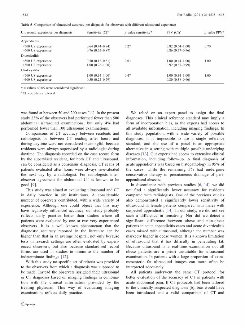
There were no significant differences between unsupervisedresidents who had evaluated (and performed) more than500 ultrasound examinations and those who had evaluatedless than 500 ultrasound examinations for these twodiagnoses (Table 5). Unsupervised residents had a highersensitivity than attending radiologists, including supervised
Table 3 Sensitivity of ultrasound with sufficient image quality versus insufficient image quality
Diagnoses N Missed diagnoses sufficientimage qualitya (%)
N Missed diagnoses insufficientimage qualitya (%)
p value
Appendicitis 241 16 (11–20) 43 67 (53–81) <0.01
Diverticulitis 96 30 (21–39) 22 77 (57–90) <0.01
Bowel obstruction 37 32 (17–48) 31 42 (26–59) 0.46
Gastrointestinal Non-Urgentb 38 71 (57–85) 18 78 (55–91) 0.75
Cholecystitis 45 22 (10–34) 7 57 (25–84) 0.08
Hepatic-pancreatic-biliary diseasec 31 29 (13–45) 12 50 (25–75) 0.29
Inflammatory bowel disorderd 21 52 (31–74) 9 89 (56–98) 0.10
Pancreatitis 11 45 (16–75) 17 71 (47–87) 0.25
Gynaecological urgente 30 53 (30–75) 8 88 (53–98) 0.19
a Insufficient image quality is defined as ultrasound examinations in which the region of interest could not be visualisedb gastrointestinal disorder non-urgent (n=56), gastroenteritis (n=27), constipation (n=12), epiploic appendagitis/omental infarction (n=11),gastritis (n=5), ulcus ventriculi/duodeni (n=1)c HPB (n=43) consisted of; cholecystolithiasis (n=33), choledocholithiasis (n=5), hepatitis (n=3), liver metastases (n=1), chronic pancreatitis (n=1)d Inflammatory bowel disorder consisted of: non-specified inflammatory bowel disorder (n=16); infectious (n=11), Crohn’s disease (n=1),ulcerative colitis (n=2)e Urgent gynaecological disorder (n=27) consisted of Pelvic Inflammatory Disease (PID) (n=13), adnexal torsion (n=9), bleeding/rupture ovariancyst (n=5)
1540 Eur Radiol (2011) 21:1535–1545

residents for the diagnosis of diverticulitis with ultrasound,83% versus 57% (p=0.04). Here, the sensitivity wassignificantly higher for more experienced unsupervisedresidents (Table 5).
Positive predictive values for common diagnoses such asacute appendicitis, acute diverticulitis and acute cholecys-titis were comparable for non-supervised residents andattending radiologists, including supervised residents(Fig. 1).
Discussion
In this study we found that the sensitivity of CT wassignificantly higher than that of ultrasound in detectingappendicitis and diverticulitis. Fewer cases of acute
appendicitis and acute diverticulitis were missed by CT,but positive predictive values of ultrasound and CT werecomparable. For acute cholecystitis and bowel obstruc-tion there were no significant differences in accuracybetween ultrasound and CT. No subgroup differences inultrasound sensitivity in detecting acute appendicitis andacute diverticulitis were found for any of the evaluatedpatient characteristics: BMI, age and duration of pain.There were no statistically significant differences be-tween obese women and men. The sensitivity ofultrasound performed by non-supervised radiologicalresidents was not significantly lower than that ofultrasound performed by attending radiologists, includingsupervised residents. The percentage of missed acuteappendicitis and acute diverticulitis cases was lower ifthe observer was able to visualise the region of interestcompared with the percentage of missed cases of acuteappendicitis or diverticulitis with insufficient imagequality. For all other diagnoses, such a reduction in thenumber of missed diagnoses was not found.
A number of potential limitations of this analysisshould be acknowledged. One could object that thesensitivity of US was underestimated, because ultrasoundwas partly performed and interpreted by unsupervisedradiological residents. Unsupervised residents did nothave a significantly lower sensitivity in detecting diseasein this study compared with attending radiologists. In aprevious study, the overall sensitivity of ultrasoundperformed by unsupervised residents for detecting urgentdiagnoses was significantly lower than that of ultrasoundperformed by attending radiologists, without a significantdifference in positive predictive value [6], indicating thatresidents more often missed an urgent diagnosis. When-ever an urgent diagnosis was assigned, however, this wasmost likely correct. In a study by Hertzberg et al. trainingin ultrasound was evaluated and a significant improvement
Fig. 1 Comparison of sensitivity and positive predictive value (PPV)for subgroups of observers
Patient characteristics Appendicitis Diverticulitis
N Missed (%) p value N Missed (%) p value
Female 121 27 0.21 65 43 0.31Male 163 21 53 34
BMI >30 29 21 0.70 19 26 0.22BMI <30 255 24 99 41
BMI >30 female 14 29 0.39 7 43 0.31BMI >30 male 15 13 12 17
Duration pain >2 days 214 22 0.42 39 33 0.38Duration pain <2 days 70 27 79 42
Age <45 111 22 0.53 n.a.Age >45 173 25 n.a.
Age <60 n.a. 73 40 0.32Age >60 n.a. 45 38
Table 4 Missed diagnoses ofappendicitis and diverticulitis atultrasound
n.a. not applicable
Eur Radiol (2011) 21:1535–1545 1541
was found at between 50 and 200 cases [11]. In the presentstudy 23% of the observers had performed fewer than 500abdominal ultrasound examinations, but only 4% hadperformed fewer than 100 ultrasound examinations.
Comparisons of CT accuracy between residents andradiologists or between CT reading after hours andduring daytime were not considered meaningful, becauseresidents were always supervised by a radiologist duringdaytime. The diagnosis recorded on the case record formby the supervised resident, for both CT and ultrasound,can be considered as a consensus diagnosis. CT scans ofpatients evaluated after hours were always re-evaluatedthe next day by a radiologist. For radiologists inter-observer agreement for abdominal CT is known to begood [9].
This study was aimed at evaluating ultrasound and CTin daily practice in six institutions. A considerablenumber of observers contributed, with a wide variety ofexperience. Although one could object that this mayhave negatively influenced accuracy, our study probablyreflects daily practice better than studies where allpatients were evaluated by one or two very experiencedobservers. It is a well known phenomenon that thediagnostic accuracy reported in the literature can behigher than that in an average hospital, not only becausetests in research settings are often evaluated by experi-enced observers, but also because standardised recordforms are used in studies to minimise the number ofindeterminate findings [12].
With this study no specific set of criteria was providedto the observers from which a diagnosis was supposed tobe made. Instead the observers assigned their ultrasoundor CT diagnoses based on imaging findings in combina-tion with the clinical information provided by thetreating physician. This way of evaluating imagingexaminations reflects daily practice.
We relied on an expert panel to assign the finaldiagnoses. This clinical reference standard may imply aform of incorporation bias, as the experts had access toall available information, including imaging findings. Inthis study population, with a wide variety of possiblediagnoses, it is impossible to use a single referencestandard, and the use of a panel is an appropriatealternative in a setting with multiple possible underlyingdiseases [13]. Our experts had access to extensive clinicalinformation, including follow-up. A final diagnosis ofacute appendicitis was based on histopathology in 95% ofthe cases, while the remaining 5% had undergoneconservative therapy or percutaneous drainage of peri-appendiceal abscess.
In discordance with previous studies [6, 14], we didnot find a significantly lower accuracy for residentscompared with radiologists. One of the previous studiesalso demonstrated a significantly lower sensitivity ofultrasound in female patients compared with males withsuspected appendicitis [14]. In our study, we did not seesuch a difference in sensitivity. Nor did we detect asignificant difference between obese and non-obesepatients in acute appendicitis cases and acute diverticulitiscases missed with ultrasound, although the number wasmarkedly higher in obese women. It is a known limitationof ultrasound that it has difficulty in penetrating fat.Because ultrasound is a real-time examination not allobese patients are a priori unsuitable for ultrasoundexamination. In patients with a large proportion of extra-mesenteric fat ultrasound images can more often beinterpreted adequately.
All patients underwent the same CT protocol forbetter evaluation of the accuracy of CT in patients withacute abdominal pain. If CT protocols had been tailoredto the clinically suspected diagnosis [6], bias would havebeen introduced and a valid comparison of CT and
Table 5 Comparison of ultrasound accuracy per diagnosis for observers with different ultrasound experience
Ultrasound experience per diagnosis Sensitivity (CI)a p value sensitivity* PPV (CI)a p value PPV*
Appendicitis
<500 US experience 0.64 (0.44–0.84) 0.27 0.82 (0.64–1.00) 0.70>500 US experience 0.76 (0.65–0.87) 0.86 (0.77–0.96)
Diverticulitis
<500 US experience 0.50 (0.18–0.81) 0.03 1.00 (0.44–1.00) 1.00>500 US experience 1.00 (0.76–1.00) 0.92 (0.67–0.99)
Cholecystitis
<500 US experience 1.00 (0.34–1.00) 0.47 1.00 (0.34–1.00) 1.00>500 US experience 0.50 (0.22–0.79) 0.80 (0.38–0.96)
* p values <0.05 were considered significanta CI: confidence interval
1542 Eur Radiol (2011) 21:1535–1545

ultrasound would not have been possible. Recent researchhas shown that usage of oral contrast agent does notincrease the accuracy of diagnosing appendicitis with CT[15, 16]. For the evaluation of acute diverticulitis a widevariety of CT protocols is described in the literature,ranging from solely intravenous contrast to a combinationof oral, rectal and intravenous contrast. The CT protocolsolely using iv contrast agent did not achieve loweraccuracy values compared with studies with extendedcontrast agent usage [8].
We observed a low prevalence in our study group of anumber of important disorders, such as perforated viscusor bowel ischaemia and other common diagnoses causingacute abdominal pain such as pancreatitis and urinarytract calculus (patients with distinctive flank pain,suspected with renal colic, were not eligible for thisstudy). This low prevalence limited any comparison ofCT or ultrasound accuracy for the full range of diagnosesin patients presenting with acute abdominal pain.
The study reported here was not designed toseparately evaluate the sensitivity and specificity ofspecific complications of any of the diagnoses causingacute abdominal pain. We only aimed to study theaccuracy of ultrasound and CT in assigning the correctdiagnosis.
A meta-analysis did not show any significant differ-ence in accuracy between ultrasound and CT in detectingdiverticulitis, although CT is more likely to detectcomplications of acute diverticulitis [8]. We did not finda significant difference in the accuracy of detecting bowelobstruction between ultrasound and CT; the aetiology ofthe obstruction is better evaluated with CT than with US.Likewise, a better accuracy for CT has been described indetecting complicated bowel obstruction [17–21], althoughthe accuracy of CT in the detection of bowel ischaemia is atbest mediocre [22].
Some of the accuracy estimates for ultrasound in this studyare lower than those reported elsewhere in the literature. Thereported sensitivities for ultrasound in experienced hands indetecting appendicitis have been as high as 90% [23]. Inrecent meta-analyses of diagnostic imaging in acute appen-dicitis, ultrasound sensitivity varied between 86% [24] and78% [7], which is comparable to the estimates in the presentstudy. The accuracy in detecting acute diverticulitis is lowerthan in the aforementioned recent meta-analysis. Summarysensitivity of 92% for ultrasound was reported, which ismuch higher than the sensitivity of 68% [8]. The most likelyexplanation for this difference might be that we includedunselected patients with acute abdominal pain, whereas thestudies included in the meta-analysis more often hadrecruited selected patients with a clinically suspected acutediverticulitis. A higher pre-test likelihood of disease isknown to result in a higher accuracy [25].
We observed the significantly higher sensitivity of CTcompared with ultrasound with regard to urgent gynaeco-logical disorders. This result may be counterintuitive tosome as ultrasound is the imaging technique of choice inthese patients [26]. Our findings may be explained by thefact that we used abdominal ultrasound performed byradiologists, not trans-vaginal ultrasound performed by thegynaecologist. Gynaecologists can be expected to be moreexperienced in the evaluation of gynaecological disorders;they can probably achieve a higher sensitivity with trans-vaginal ultrasound than radiologists can with transabdomi-nal ultrasound. Unfortunately patients directly referred togynaecologists are not routed through the emergencydepartment and therefore not included in this study.
In summary, we observed that CT sensitivity is higher thanthat of ultrasound in detecting appendicitis and diverticulitis inunselected patients presenting with acute abdominal pain, butpositive predictive values are comparable. Accuracy of bowelobstruction and acute cholecystitis were not significantlydifferent. The percentage of cases missed on ultrasound wasnot influenced by patient characteristics and observer experi-ence at large with regard to common diagnoses. Theproportion of missed acute appendicitis and acute diverticu-litis was significantly lower in the subgroup of patients inwhom the radiologist could adequately visualise the region ofinterest. These results indicate that ultrasound is a good first-line technique.
Acknowledgements The Dutch Organization for Health Researchand Development, Health Care Efficiency Research Programme,funded the study (ZonMw, grant number 945-04-308).
Open Access This article is distributed under the terms of theCreative Commons Attribution Noncommercial License which per-mits any noncommercial use, distribution, and reproduction in anymedium, provided the original author(s) and source are credited.
Appendix I
Members of the OPTIMA study group:Academic Medical Center, AmsterdamA. van Randen, Department of RadiologyW. Laméris, Department of SurgeryJ. Stoker, Department of RadiologyM.A. Boermeester, Department of SurgeryP.M.M. Bossuyt, Department of Clinical epidemiology,
Biostatistics, and BioinformaticsSt Antonius Hospital NieuwegeinB. van Ramshorst, Department of SurgeryJ.P.M. van Heesewijk, Department of RadiologyM.P. Gorzeman, Department of Emergency MedicineGelre Hospitals, ApeldoornW.H. Bouma, Department of Surgery
Eur Radiol (2011) 21:1535–1545 1543

W. ten Hove, Department of RadiologyJ. Winkelhagen, Department of SurgeryUniversity Medical Center UtrechtH.G. Gooszen, Department of Surgery,M.S. van Leeuwen, Department of RadiologyD.E.J.G.J. Dolmans, Department of SurgeryTergooi Hospitals, HilversumE. van Keulen, Department of RadiologyJ.W. Juttmann, Department of SurgeryM.J. van der Laan, Department of SurgeryOnze Lieve Vrouwe Gasthuis, AmsterdamS.C. Donkervoort, Department of SurgeryV.P.M. van der Hulst, Department of Radiology
Appendix II
OPTIMA trial expert panel members:Academic Medical CenterO.R.C. Busch, Department of SurgeryT.M. van Gulik, Department of SurgeryO.D. Henneman, Department of Radiology, Bronovo
Hospital, Den HaagTergooi HospitalsA.A.W. van Geloven, Department of Surgery, Tergooi
Hospitals, HilversumJ.W. Juttmann, Department of Surgery, Tergooi Hospi-
tals, HilversumE. van Keulen, Department of Radiology, Tergooi
Hospitals, HilversumOnze Lieve Vrouwe GasthuisS.C. Donkervoort, Department of Surgery, Onze Lieve
Vrouwe Gasthuis, AmsterdamM.P. Simons, Department of Surgery, Onze Lieve
Vrouwe Gasthuis, AmsterdamJ. Peringa, Department of Radiology, Onze Lieve
Vrouwe Gasthuis, AmsterdamSt Antonius Hospital NieuwegeinH.W. van Es, Department of Radiology, St Antonius
Hospital, NieuwegeinP.M.N.Y.H Go, Department of Surgery, St Antonius
Hospital, NieuwegeinM.J. Wiezer, Department of Surgery, St Antonius
Hospital, NieuwegeinGelre HospitalsW.H. Bouma, Department of Surgery, Gelre Hospitals,
ApeldoornE.J. Hesselink, Department of Surgery, Gelre Hospitals,
ApeldoornW. ten Hove, Department of Radiology, Gelre Hospitals,
Apeldoorn
Appendix III [6]
References
1. Stoker J, van Randen A, Laméris W, Boermeester MA (2009)Imaging patients with acute abdominal pain. Radiology 253:31–46
2. Shuman WP, Ralls PW, Balfe DM et al (2000) Imaging evaluationof patients with acute abdominal pain and fever. AmericanCollege of Radiology. ACR Appropriateness Criteria. Radiology215(Suppl):209–212
3. Puylaert JB (2003) Ultrasonography of the acute abdomen:gastrointestinal conditions. Radiol Clin North Am 41:1227–1242, vii
Table 6 Final diagnoses in 1021 patients assigned by the expert panel
Diagnoses N %
Acute appendicitis 284 27.8
Non-specific abdominal paina 183 17.9
Acute diverticulitis 118 11.6
Bowel obstruction 68 6.7
Gastro-intestinal disorder non-urgent 56 5.5
Acute cholecystitis 52 5.1
HPBb 43 4.2
Inflammatory bowel disorder 30 2.9
Acute pancreatitis 28 2.7
Gynaecological disorder; urgent 27 2.6
Urinary tract disorder; urgent 22 2.2
Urinary tract disorder 20 0.2
Abscess 14 1.4
Perforated viscus 13 1.3
Bowel ischaemia 12 1.2
Pneumonia 11 1.1
Gynaecological disorder; non-urgent 9 0.9
Retro-peritoneal or abdominal wall bleeding 9 0.9
Malignancy 5 0.5
Acute peritonitisc 3 0.3
Herniationd 2 0.2
Othere 12 1.2
Total 1,021 100
a non-specific abdominal pain was abbreviated as NSAP, which is nottruly a diagnosis but merely a negative patient, without diseaseb 33 cholecystolithiasis, 5 common bile duct stones, 3 hepatitis, 1 livermetastasisc Peritonitis not caused by perforationd Hernia without strangulation, otherwise it would have been classifiedas bowel ischaemiae Other diagnoses were abdominal wall infiltration, oesophagitis (2),renal infarction (2), gastric band problem (2), SLE, mesentericlymphadenitis, post-procedural pain, uterine haemorrhage and atesticular torsion
1544 Eur Radiol (2011) 21:1535–1545

4. The 2007 Recommendations of the International Commission onRadiological Protection (2007) ICRP publication 103. Ann ICRP37:1–332
5. Stoker J (2008) Magnetic resonance imaging and the acuteabdomen. Br J Surg 95:1193–1194
6. Laméris W, van Randen A, van Es HW et al (2009) Imagingstrategies for detection of urgent conditions in patients with acuteabdominal pain: diagnostic accuracy study. BMJ 338:b2431
7. van Randen A, Bipat S, Zwinderman AH, Ubbink DT, Stoker J,Boermeester MA (2008) Acute appendicitis: meta-analysis ofdiagnostic performance of CT and graded compression US relatedto prevalence of disease. Radiology 249:97–106
8. Laméris W, van Randen A, Bipat S, Bossuyt PM, BoermeesterMA, Stoker J (2008) Graded compression ultrasonography andcomputed tomography in acute colonic diverticulitis: meta-analysis of test accuracy. Eur Radiol 18:2498–2511
9. van Randen A, Laméris W, Nio CY et al (2009) Inter-observeragreement for abdominal CT in unselected patients with acuteabdominal pain. Eur Radiol 19:1394–1407
10. Laméris W, van Randen A, Dijkgraaf MG, Bossuyt PM, Stoker J,Boermeester MA (2007) Optimization of diagnostic imaging usein patients with acute abdominal pain (OPTIMA): design andrationale. BMC Emerg Med 7:9
11. Hertzberg BS, Kliewer MA, Bowie JD, Carroll BA, DeLong DH,Gray L et al (2000) Physician training requirements in sonogra-phy: how many cases are needed for competence? AJR Am JRoentgenol 174:1221–1227
12. Cuschieri J, Florence M, Flum DR et al (2008) Negative appendec-tomy and imaging accuracy in the Washington State Surgical Careand Outcomes Assessment Program. Ann Surg 248:557–563
13. Rutjes AWS, Reitsma JB, Coomarasamy A, Khan KS, BossuytPMM (2007) Evaluation of diagnostic tests when there is no goldstandard. A review of methods. Health Technol Assess 11(50)
14. Gaitini D, Beck-Razi N, Mor-Yosef D et al (2008) Diagnosingacute appendicitis in adults: accuracy of color Doppler sonogra-phy and MDCT compared with surgery and clinical follow-up.AJR Am J Roentgenol 190:1300–1306
15. Anderson SW, Soto JA, Lucey BC et al (2009) Abdominal 64-MDCTfor suspected appendicitis: the use of oral and IV contrast material
versus IV contrast material only. AJR Am J Roentgenol 193:1282–1288
16. Gurusamy K, Samraj K, Gluud C, Wilson E, Davidson BR (2010)Meta-analysis of randomized controlled trials on the safety andeffectiveness of early versus delayed laparoscopic cholecystectomyfor acute cholecystitis. Br J Surg 97:141–150
17. Hainaux B, Agneessens E, Bertinotti R et al (2006) Accuracy ofMDCT in predicting site of gastrointestinal tract perforation. AJRAm J Roentgenol 187:1179–1183
18. Lazarus DE, Slywotsky C, Bennett GL, Megibow AJ, Macari M(2004) Frequency and relevance of the “small-bowel feces” signon CT in patients with small-bowel obstruction. AJR Am JRoentgenol 183:1361–1366
19. Maglinte DD, Howard TJ, Lillemoe KD et al (2008) Small-bowelobstruction: state-of-the-art imaging and its role in clinicalmanagement. Clin Gastroenterol Hepatol 6:130–139
20. Schmutz GR, Benko A, Fournier L, Peron JM, Morel E, Chiche L(1997) Small bowel obstruction: role and contribution ofsonography. Eur Radiol 7:1054–1058
21. Silva AC, Pimenta M, Guimarães LS (2009) Small bowelobstruction: what to look for. Radiographics 29:423–439
22. Sheedy SP, Earnest F, Fletcher JG, Fidler JL, Hoskin TL (2006)CT of small-bowel ischemia associated with obstruction inemergency department patients: diagnostic performance evalua-tion. Radiology 241:729–736
23. Puylaert JB, Rutgers PH, Lalisang RI et al (1987) A prospectivestudy of ultrasonography in the diagnosis of appendicitis. N EnglJ Med 317:666–669
24. Terasawa T, Blackmore CC, Bent S, Kohlwes RJ (2004)Systematic review: computed tomography and ultrasonographyto detect acute appendicitis in adults and adolescents. Ann InternMed 141:537–546
25. Leeflang MM, Moons KG, Reitsma JB, Zwinderman AH (2008)Bias in sensitivity and specificity caused by data-driven selectionof optimal cutoff values: mechanisms, magnitude, and solutions.Clin Chem 54:729–737
26. Potter AW, Chandrasekhar CA (2008) US and CT evaluation ofacute pelvic pain of gynaecologic origin in nonpregnant premen-opausal patients. Radiographics 28:1645–1659, Review
Eur Radiol (2011) 21:1535–1545 1545

Copyright of European Radiology is the property of Springer Science & Business Media B.V. and its content
may not be copied or emailed to multiple sites or posted to a listserv without the copyright holder's express
written permission. However, users may print, download, or email articles for individual use.