tudo junto com folha de rosto - estudo geral: home mestrado... · ix rim regulating synaptic...
TRANSCRIPT

I
Agradecimentos
Gostaria de começar por agradecer a todos os meus familiares e amigos que, de
variadíssimas formas e cada qual à sua maneira, contribuíram para o meu bem-estar e
boa disposição no decorrer do mestrado. Um obrigado às gentes de Braga, Marco de
Canaveses, Porto, Lisboa, Guimarães, e claro, Coimbra.
Um obrigado especial aos meus pais e irmãos por todo o apoio, confiança e,
principalmente, interesse pelo meu trabalho. Não posso deixar de agradecer a amável
paciência com que sempre lideram com uma Joana renitente e teimosa adormecida no
seu sofá predilecto!
Gostaria igualmente de agradecer a todos os membros do laboratório toda a
ajuda imediata e apoio, e pela forma agradável como me receberam.
Um obrigado muito especial ao “Chefinho” Graciano por toda a preciosa ajuda
dispensada na primeira fase do meu trabalho no laboratório. Obrigado pelos supremos
ensinamentos, pela boa disposição, pela paciência e, claro, pelas capitais!
Agradeço ao Professor Carlos Duarte e à Professora Ana Luísa Carvalho todo o
conhecimento transmitido, apoio e disponibilidade.
Ao Dr. Ramiro Almeida um grande obrigado pela excelente orientação.
Agradeço toda a ajuda, todos os conhecimentos transmitidos, toda a confiança,
interesse, entusiasmo e, acima de tudo, dedicação.
E por último, um grande beijinho de obrigado à minha avó!

II
III
Table of contents
Abbreviations VII-IX
Abstract 1
Resumo 3
Chapter 1 – Introduction 7
1.1. The cell body hypothesis vs local protein synthesis 9
1.2. mRNA trafficking and regulation of local protein synthesis in neurons 11
1.3. Local protein synthesis in dendrites 18
1.4. Local protein synthesis in axons 20
1.4.1. Early evidences from invertebrates 20
1.4.2. Emerging evidences in vertebrates 21
1.5. Role of local protein synthesis in axons 23
1.5.1. Developing axons 23
1.5.1.1. Axonal protein synthesis in axon guidance 24
1.5.1.2. Axonal protein synthesis in synaptogenesis 28
1.5.2. Mature axons 29
1.5.2.1. Axonal protein synthesis in axon regeneration 30
1.5.2.2. Axonal protein synthesis in synaptic plasticity 31
1.6. Balancing local protein synthesis and local protein degradation 33
1.7. Presynaptic differentiation 35
1.7.1 Presynaptic organizing molecules 41
1.8. Objectives 47
IV
Chapter 2 – Materials and Methods 49
2.1. Reagents 51
2.1.1. Antibodies 52
2.2. Hippocampal neurons 53
2.2.1. Preparation of microfluidic devices 53
2.2.2. Culture of embryonic rat hippocampal neurons 54
2.3. Stimulation and total extracts preparation 55
2.3.1. Electrophoresis and Western Blot 56
2.3.2. Stripping and reprobing 57
2.4. Stimulation and protein synthesis inhibition 57
2.4.1. Immunocytochemistry 58
2.4.2. Fluorescence microscopy and quantification 59
2.5. Statistical analysis
60
Chapter 3 – Results and Discussion 61
3.1. Induction of presynaptic differentiation by FGF22 63
3.1.1. FGF22 stimulation of 293T cells and primary cultures of hippocampal
neurons induces ERK1/2 phosphorylation
63
3.1.2. Presynaptic differentiation assessed by synapsin clustering 66
3.1.3. FGF22 and BNDF have a role in the differentiation of the presynaptic
terminal of embryonic hippocampal neurons when globally applied
67
3.2. FGF22-induced presynaptic differentiation and axonal protein synthesis 73
3.2.1. Microfluidic devices for neuron cell culture 73
3.2.1.1. Growth of embryonic rat hippocampal neurons in microfluidic
chambers
75
V
3.2.1.2. Microfluidic chambers: fluidic isolation of pure axonal
preparations
79
3.2.2. Local FGF22-presynaptogenic effect is specific 82
3.2.3. FGF22-induced presynaptic differentiation is dependent on axonal
protein synthesis
87
Chapter 4 – Closing remarks 93
4.1. Conclusion 95
4.2. Future Perspectives
97
Chapter 5 - References 101

VI
VII
Abbreviations
5-FDU 5-fluoro-2’-deoxiuridina
ALS Axonal localization element
AMPA α-Amino-3-hydroxy-5-methyl-4-isoxazole-propionic acid
AP3 Adaptor complex 3
APS Ammonium persulfate
AraC Cytosine arabinose
Arc Activity-regulated cytoskeleton-associated protein
BC1 Brain cytoplasmic 1
BNDF Brain-derived neurotrophic factor
BSA Bovine Serum Albumin
CA3 Cornu Ammonis region 3
CAM Cell-adhesion molecule
CaMKII Ca2+/calmodulin-dependent protein kinase II
CASK Calcium/calmodulin-dependent serine protein kinase
CAZ Cytomatrix at the active zone
CGRP Calcitonin gene-related peptide
CNS Central nervous system
CPE Cytoplasmic polyadenylation element
CPEB Cytoplasmic polyadenylation element binding protein
CPR-2 Conopressin receptor 2
CREB cAMP response element binding
DCC Deleted in colorectal carcinoma
DIV Days in vitro
DMEM Dulbecco’s modified Eagle’s medium
DMSO Dimethyl sulfoxide
DTE Dendritic targeting element
DTT Dithiothreitol
E1 Ubiquitin-activating enzyme
ECF Enhanced chemifluorescence substrate
eIF Eukaryotic translation initiation factor
eIF4EBP Eukaryotic translation initiation factor 4E binding protein
ELH Egg-laying hormone
EphA2 Ephrin type A receptor 2
ERK Extracellular signal-regulated kinase
FAK Focal adhesion kinase
FBS Fetal bovine serum
VIII
FGF Fibroblast growth factor
FGF22 Fibroblast growth factor-22
FGFR Fibroblast growth factor receptor
FMRP Fragile-X mental retardation protein
GABA Gamma-aminobutyric acid
GFP Green fluorescence protein
Glu Glutamate
Grb7 Growth factor receptor-bound protein 7
GSK-3 Glycogen synthase kinase 3
HEPES 4-(2-hydroxyethyl)-1-piperazineethanesulfonic acid
HFS High frequency stimulation
hnRNP Heterogeneous nuclear ribonucleoproteins
IGF-II Insulin-like growth factor
IRES Internal ribosomal entry sites
KIF Kinesin
LFS Low-frequency stimulation
LTD Long-term depression
LTF Long-term facilitation
LTP Long-term potentiation
MAP2 Microtubule associated protein-2
MAPK Mitogen-activated protein kinase
MEM Minimum essential medium eagle
miRNAs MicroRNAs
mTOR Mammalian target of rapamycin
MW Multi-well
Na3VO4 Sodium orthovanadate
NaF Sodium fluoride
NGF Nerve growth factor
NLS Nuclear localization signal
NMDA N-methyl-D-aspartate
PACAP Plasma cell induced ER protein 1
PBS Phosphate buffered saline
PDL Poly-D-lysine
PDMS Poly-dimethlysiloxane
PKMζ Protein kinase M ζ
PSD-95 Postsynaptic density protein 95
PTV Piccolo-Bassoon transport vesicle
PVDF Polyvinylidene difluoride
IX
RIM Regulating synaptic membrane exocytosis
RISC RNA-induced silencing complex
RNP Ribonucleotide protein
SDS Sodium dodecyl sulfate
Sema3A Semaphoring-3A
SIRP Signal regulatory proteins
SNAP-25 Synaptosomal-associated protein-25
SNARE Soluble NSF attachment receptor
STV Synaptic vesicles transport particle
SV Synaptic vesicle
SynCAM Synaptic cell-adhesion molecule
TBS Tris buffered saline
TBS-T Tris-buffered saline with 0.1 % Tween 20
TEMED N,N,N’,N’-Tetramethylethylenediamine
TGS Tris-glycine-SDS buffer
TLS Translocation in liposarcoma
Tris 2-Amino-2-(hydroxymethyl)-1,3-propanediol
TSP Thrombospondin
TUJ1 Neuronal Class III β-tubulin
UTR Untranslated region
VAMP Vesicle SNARE synaptobrevin
VGLUT1 Vesicular glutamate transporter 1
WB Western Blot
ZBP1 Zip-code binding protein 1
κor κ-opioid receptor

X
1
Abstract
Neurons are highly complex and polarized cells with an incredible network of
functionally active processes that extend outwards the cell body. Communication
between neurons occurs at a specialized structure, the synapse, frequently distant from
the soma, the neuron trophic center. For long, proteins were thought to be synthesized in
the cell body and then guided by microtubule-mediated transport to specific sites in
dendrites and axons. However, this model has been challenged by accumulating
evidences suggesting local translation of specific mRNAs selectively localized to
neurites.
Local protein synthesis in axons has not been object of intense studies in the
past and its functional significance is not clear. It is believed to be essential for several
neurodevelopmental events like axonal guidance, synapse formation, synaptic plasticity,
axonal regeneration and retrograde signaling. However, the involved mechanisms are
far from being understood.
In the present work we investigated the potential role of axonal translation in
presynaptogenesis, the mechanism by which a functional presynaptic terminal is
formed. During presynaptic differentiation, freely moving packets containing
presynaptic material such as synaptic vesicles, vesicular and fusion proteins, recycling
machinery and active zone components, accumulate at pre-defined sites along the axon
shaft. Despite this, transition of nascent synapses to mature synapses also involves
correct alignment between pre- and postsynaptic terminals, presynaptic growth and
cytoskeletal restructuring. Lately, the required action of presynaptic organizing
molecules, such as synaptic cell adhesion molecules (SynCAMs), fibroblast growth
2
factor-22 (FGF22), neuroligins, WNTs and thrombospondins, has been highlighted by
several studies.
In this work we focus our attention on FGF22-induced presynaptic
differentiation, a target-derived soluble molecule recently proven to promote synaptic
vesicles clustering in central nervous system (CNS) synapses. This work comprises two
distinct parts. Firstly, we stimulated primary cultures of rat embryo hippocampal
neurons with recombinant human FGF22; assessment of synapsin clustering
demonstrated an increase in both number and size of presynaptic sites. Secondly, we
tested whether this presynaptogenic effect of FGF22 was dependent on axonal
translated proteins. For that, we cultured dissociated hippocampal neurons in
microfluidic devices capable of physically and fluidically isolating axons. We then
stimulated axons, under localized protein synthesis inhibition, with FGF22. Our results
clearly show an abrogation of FGF22-induced presynaptic assembly when protein
synthesis inhibitors are added to the medium, proving that FGF22 depends on
translation of axonal mRNAs to exert its function. We present the first evidence that,
not only axonal translation has a role in presynaptic formation in a mammalian system,
but also that presynaptic organizing molecules action might rely on newly and locally
synthesized proteins.
Further studies will allow us to understand how FGF22 acts, revealing the
intracellular events (signaling pathways, induction and regulation of local protein
synthesis and mRNAs involved) that lead to the appropriate formation of presynaptic
terminals.
Keywords: FGF22, BDNF, presynaptic differentiation, axonal protein
synthesis, microfluidic devices.
3
Resumo
Os neurónios são células altamente complexas e polarizadas com uma incrível
rede de prolongamentos funcionalmente activos que se estendem do corpo celular. A
comunicação entre neurónios ocorre numa estrutura especializada, a sinapse,
normalmente distante do centro trófico do neurónio, o corpo celular. Durante vários
anos, assumiu-se que as proteínas eram sintetizadas no corpo celular e enviadas para
locais específicos nas dendrites e axónios, através do transporte mediado pelos
microtúbulos. Contudo, novas evidências têm desafiado este modelo ao demonstrarem a
ocorrência de tradução local de mRNAs, selectivamente localizados nas neurites.
No passado, poucos estudos se têm focado na síntese proteica em axónios, e
consequentemente, a sua relevância funcional não é clara. Actualmente, a tradução
axonal parece ser essencial para vários eventos do desenvolvimento do sistema nervoso,
como condução axonal, formação sináptica, plasticidade sináptica, regeneração axonal e
sinalização retrógrada. Contudo, os mecanismos envolvidos estão longe de ser
descortinados.
O presente trabalho tem como objectivo investigar o papel da tradução axonal
na pré-sinaptogénese, o mecanismo pelo qual se forma um terminal pré-sináptico.
Durante a diferenciação pré-sináptica, estruturas móveis contendo material pré-sináptico
como vesículas sinápticas, proteínas vesiculares e de fusão, maquinaria de reciclagem
de vesículas e componentes da zona activa, acumulam-se em locais pré-definidos ao
longo do eixo axonal. Para além disso, a formação de sinapses maduras também requer
um correcto alinhamento entre os terminais pré- e pós-sinápticos, crescimento pré-
sináptico e remodelação do citosqueleto. Ultimamente, vários estudos têm salientado a

4
acção de moléculas organizadoras do terminal pré-sináptico, como SynCAMs, FGF22,
neuroliguinas, WNTs e trombospondinas.
Neste trabalho, focámo-nos na diferenciação pré-sináptica induzida por FGF22,
uma molécula solúvel secretada pelo terminal pós-sináptico alvo cuja acção indutora da
agregação das vesículas sinápticas foi demonstrada recentemente em sinapses do
sistema nervoso central. Este trabalho compreende duas partes distintas. Em primeiro
lugar, estimulámos culturas primárias de neurónios de hipocampo de embrião de rato
com FGF22 e avaliámos a agregação da sinapsina, observando-se um aumento no
número e tamanho dos locais pré-sinápticos. Em segundo lugar, procurámos testar se
este efeito presinaptogénico do FGF22 está dependente de proteínas traduzidas
localmente. Para tal, plaqueámos neurónios de hipocampo em câmaras microfluídicas
capazes de isolar, física e fluidicamente, axónios. De seguida, estimulámos axónios com
FGF22, sob inibição local de síntese proteica, ou seja, apenas a nível axonal. Os nossos
resultados mostram que na presença de inibidores da síntese proteica o efeito do FGF22
na formação sináptica é abolido, provando, deste modo, que o FGF22 depende da
tradução de mRNAs nos axónios para exercer a sua função. Neste trabalho
apresentamos a primeira evidência de que a tradução proteica axonal está envolvida na
formação pré-sináptica em neurónios do sistema nervoso central de mamíferos. Por
outro lado, demonstrámos pela primeira vez que moléculas organizadoras do terminal
pré-sináptico requerem novas proteínas sintetizadas localmente.
Experiências futuras permitir-nos-ão compreender o modo de acção do FGF22,
ajudando-nos a revelar os eventos intracelulares (vias de sinalização, indução e
regulação da síntese proteica local e mRNAs envolvidos) que conduzem à formação do
terminal pré-sináptico.

5
Palavras-chave: FGF22, BDNF, diferenciação pré-sináptica, síntese proteica
axonal, câmaras microfluídicas.

6
7
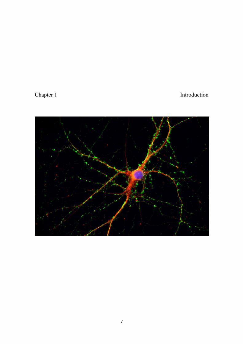
Chapter 1 Introduction

8
9
1.1. The cell body hypothesis vs local protein synthesis
The mammalian brain is characterized by a tremendous and complex
interconnectivity of its neurons. The communication between neurons occurs at a
specialized structure: the synapse, frequently far away from the cell body. The
formation of this distal connections and their synaptic activity requires a specific set of
proteins. According to this, a question has been puzzling neurobiologists since the 19th
century: how do neurites acquire this indispensable set of proteins during development
and functional maintenance?
After the discovery that axons degenerate when separated from their cell body
(Waller, 1851) and that they directly grow out of the neuronal cell body (Harrison,
1910), the concept of the exclusive trophic role of the neuron soma arose, firstly stated
by Ramón y Cajal in his “Neuron theory” (reviewed in López-Muñoz et al., 2006). In
the following years, new findings concerning protein transport in neurons helped to
solidify this theory – the “cell body hypothesis”. In 1963, Droz and Leblond observed,
by autoradiography, migration of proteins in axons of the central nervous system, and in
1980 the intracellular transport of proteins in neurons was already undoubtedly accepted
(Grafstein and Foreman, 1980). Protein synthesis and posttranslational processing
machinery was believed to be present in neuronal cell bodies (Einarson, 1933). It was
thought that the protein building blocks of axons and dendrites were synthesized in the
cell body and then transported to specific places within the neuron. However, this view
was challenged by posterior accumulating evidences suggesting that translation of
mRNAs occurs locally in dendrites. The discovery of polyribosomes under the base of
dendritic spines in granule cells (Steward and Levy, 1982), along with the detection of
specific mRNAs within dendrites, such as microtubule associated protein 2 (MAP2)
10
(Garner et al., 1988) and Ca2+/calmodulin-dependent protein kinase II (CaMKII) alpha
subunit (Burgin et al., 1990), first gave rise to the possibility that protein synthesis could
occur at a synaptic level. Nowadays, local protein synthesis in dendrites is widely
accepted and is postulated to provide the basic mechanisms underlying synaptic
plasticity and regulation of synaptic activity (Skup, 2008).
On the contrary, the idea of axonal protein synthesis in vertebrates has
remained controversial, despite the accumulating evidences for their occurrence in
vertebrate axons (summarized in Giuditta et al., 2008). Early studies demonstrating
absence of ribosomes in axons (Lasek et al., 1973) are now disbelieved by the discovery
of intermittently localized ribosomes along the axon shaft (Koenig et al., 2000) and the
detection of mRNAs in axons of vertebrate and invertebrate nerve cells (Mohr and
Richter, 2000). In a different perspective, local protein synthesis in axons can explain
theoretical inconsistencies inherent in the “cell body hypothesis”. Firstly, there is a great
discrepancy between proteins half-lives and the duration of slow axonal transport of
cytoskeletal and cytosolic proteins, resulting in protein loss-of-function during their
transport to the distal-most targets (Alvarez et al., 2000). Secondly, the exclusive
somatic origin of axonal and presynaptic proteins fails to ensure the two-way signaling
between axon and neuron soma that would be required for plastic events occurring at
growth cones or presynaptic terminals. For example, Šatkauskas and Bagnard (2007)
emphasized the role of growth cone protein synthesis in the acquisition of adaptative
properties that allow growth cones to respond rapidly and autonomously (soma
independently) to spatiotemporal regulation of growing processes.
Why some proteins must be synthesized locally remains mostly speculative,
but some possible rationales had been pointed out by Lin and Holt (2008). For example,
mRNAs have regulatory elements located in the 5’ untranslated region (5’UTR) and
11
3’UTR that encode information regarding localization and activation, and therefore
mRNAs are more easily regulated than the functional protein. Another advantage of
translationally dormant mRNAs in relation to inactive proteins is the less space they
require to be stored. Upon activation, a single mRNA serves as template for the
synthesis of a huge amount of protein. Moreover, in addition to the obvious acquisition
of independence and autonomy, the dendrites and axons are able to respond more
promptly to extracellular stimuli, without having to wait for protein delivery from the
cell body. Local protein synthesis is also characterized by a precise spatial and temporal
regulation, meaning that instead of being constitutively synthesized, proteins are formed
only where and when needed.
This emerging field is under intense investigation and axonal protein synthesis
is gradually gaining acceptance. The mechanisms of mRNA transport, local regulation
of translation and the roles of axonal protein synthesis will be discussed in later
sections. We also include a small section concerning presynaptic differentiation and
presynaptic organizing molecules.
1.2. mRNA trafficking and regulation of local protein synthesis in neurons
The mechanism by which specific mRNAs are trafficked and locally regulated
is not yet fully characterized. The knowledge acquired until now resulted, almost
completely, from investigation in dendrites. Supposing that the mechanisms underlying
mRNA trafficking and regulation of local protein synthesis in axons might be similar to
the ones in dendrites, in this section we describe these findings mainly in dendrites.
However, there are accumulating evidences suggesting similar mechanisms to occur in
axons, which will also be described along the text.
12
An important aspect of local protein synthesis within a microdomain of the
neuron is the targeting and transport of mRNA to the appropriate compartment. This
localized transport of mRNAs is essential for the establishment and maintenance of
subcelular locations in charge of specific and unique functions (recall the functional
heterogeneity within a neuron). Actually, neurons have developed a selective
mechanism able to specifically sort mRNAs into various compartments. It must be
emphasized the crucial effect of this sorting in the selection of mRNAs to dendrites and
axons, which are functionally distinct, therefore requiring different mRNAs.
In general, mRNAs contain segments that codify information for specific
functionality (such as subcellular targeting), denominated cis-acting elements. Active
targeting involves their recognition by trans-acting RNA-binding factors, which then
interact with motor proteins, thus promoting traveling of these newly formed
ribonucleotide protein (RNP) complexes along cytoskeletal filaments (Kindler et al.,
2005). This happens to be the case in neuronal cells and it is accepted that most mRNAs
are transported into dendrites as part of large RNPs, commonly referred as RNA
granules (for a review see Bramham and Wells, 2007).
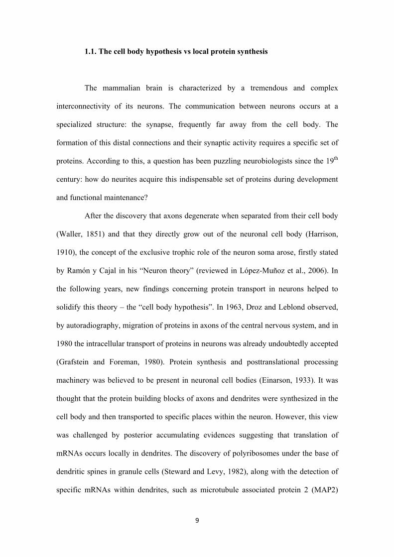
Kosik and colleagues firstly observed endogenous RNA granules in dendrites
using the fluorescence vital RNA dye SYTO14 (Knowles et al., 1996). Later, it was
discovered that RNA granules include translationally silenced mRNAs and clusters of
ribosomes, which upon stimulation (such as synaptic activation in the form of
depolarization) move to the polysome fraction to be translated (Krichevsky and Kosik,
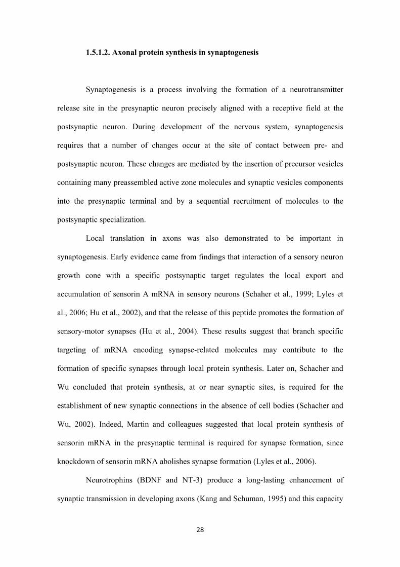
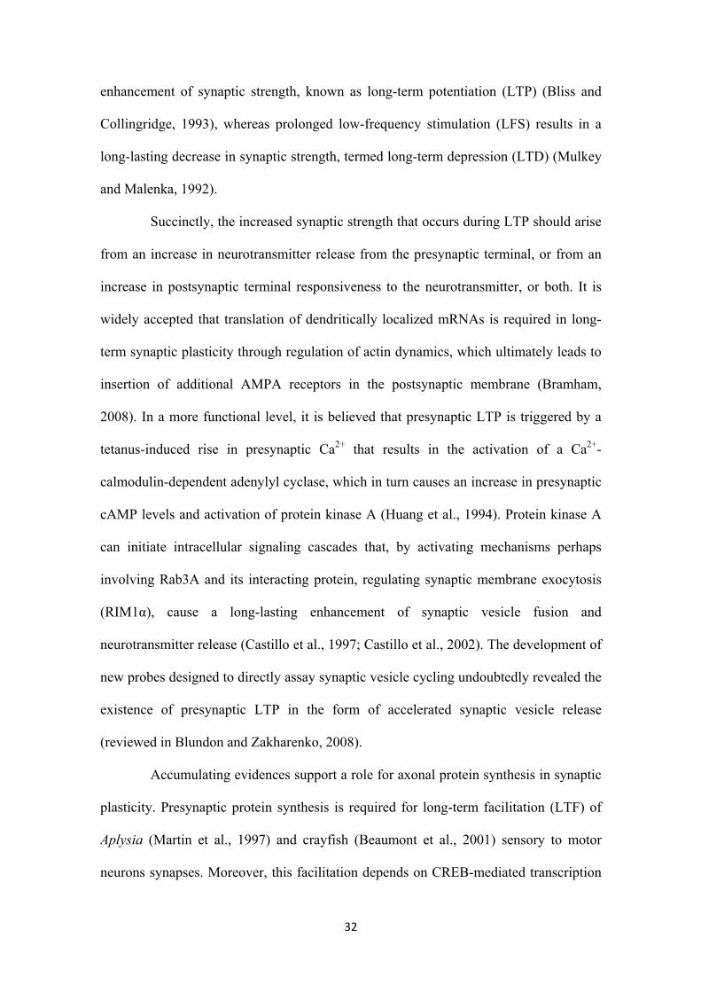
2001) (figure 1, step 5). Besides this activity-regulated translation, there seems to be an
activity-regulated transport of RNA granules. Indeed, studies showed that neuronal
activity, in the form of depolarization, N-methyl-D-aspartate (NMDA) activation or
metabotropic glutamate receptor activation, promotes: an increase in the number of
13
dendritic localized αCaMKII mRNA-containing granules (Rook et al., 2000), an
increase in the movement of zip-code binding protein 1 (ZBP1)-containing granules
(Tiruchinapalli et al., 2003) or the localization of mRNAs encoding α-Amino-3-
hydroxy-5-methyl-4-isoxazole-propionic acid (AMPA) receptor subunits GluR1 and
GluR2 into dendrites (Grooms et al., 2006), respectively. These results suggest that
synaptic activity increases dendritic protein synthesis.
It should be noted that nuclear proteins (such as heterogeneous nuclear
ribonucleoproteins (hnRNPs)) reside in RNA granules, suggesting that the formation of
RNA granules begins in the nucleus. Once in the cytoplasm RNA granules are
transported via microtubules to dendrites through interaction with the C-terminal tail of
the motor protein kinesin5 (KIF5), whose overexpression enhances granules movement
(Kanai et al., 2004). This microtubule mediated transport of mRNAs has been proved to
occur in axons too: firstly, selective perturbation of the cytoskeleton revealed that the
presence of axonal mRNA was dependent on microtubules (Olink-Coux et al., 1996,
Muslimov et al., 2002) and secondly, HuD and kinesin KIF3A were identified as
components of the tau RNP granules in neuronal axons and growth cones (Aronov et
al., 2002). Another important contribution came from Tiedge and colleagues that
suggested a two-step process in the delivery of brain cytoplasmic 1 (BC1) RNA to local
axonal sites, a long-range axial transport along microtubules and a local radial transfer
to cortical domains via actin filaments (Muslimov et al., 2002). In fact, the RNA
binding protein translocation in liposarcoma (TLS) transport to dendrites was shown to
be dependent on actin filaments (Fujii et al., 2005), what contributed to the speculation
that myosin may promote the transport of the mRNAs within the spine (figure 1, step 2).
RNA binding proteins are key regulators of cellular functioning due to their
ability to specifically bind to cis-acting elements in mRNAs and thereby regulate the

14
transport of the bound RNA. Dendritic targeting element (DTE), which is a cis-acting
element that instructs mRNA transport to dendrites, was shown to be present in several
mRNAs, such as CaMKII, Shank1, vasopressin and protein kinase M ζ (PKMζ)
(Kindler et al., 2005). By analogy, axonal localization cis element (ALS) in the 3’UTR
of tau mRNA are essential for the localization of this mRNA into developing axons
(Behar et al., 1995).
Several RNA binding proteins have been reported to be involved in sorting and
targeting of mRNAs to dendrites and axons (figure 1, step 1). For example, the
cytoplasmic polyadenylation element binding protein (CPEB) binds to mRNAs and
facilitates their transport to dendrites (Huang et al., 2003; Bramham and Wells, 2007).
In axons, the requirement of a cytoplasmic polyadenylation element (CPE) sequence in
3’UTR for axonal translation of ephrin type A receptor 2 (EphA2) (Brittis et al., 2002),
suggests a role for CPEB in regulating axonal mRNAs transport in a way similar to
dendrites.
Another well studied examples are: the fragile-X mental retardation protein
(FMRP) and ZBP1 (reviewed in Bramham and Wells, 2007; Wells, 2006). The activity
and requirement of these mRNA binding proteins in axonal RNA targeting has also
been suggested (Antar et al., 2006; Wu et al., 2005; Sotelo-Silveira et al., 2008; Zhang
et al., 2001). On the other hand, in a functional level, the formation of an RNP complex
between β-actin and ZBP1 seems to be required for growth cone response to netrin-1
(Leung et al., 2006) or brain-derived neurotrophic factor (BDNF) (Yao et al., 2006)
gradient. Furthermore, FMRP is involved in protein synthesis-dependent growth cone
collapse induced by semaphorin 3A (Sema3A) (Li et al., 2009).
In addition to specific mRNA transport, if a message is to have a local effect,
translation during transport must be repressed, thus preventing delocalized synthesis en
15
route to its destination. Actually, the mRNAs being trafficked into dendrites have been
shown to be in a translationally dormant state (figure 1, step 3).
Translational repression of mRNAs can be achieved by multiple mechanisms
(reviewed in Sossin and DesGroseillers, 2006; Bramham and Wells, 2007): repression
of translation initiation by eukaryotic translation initiation factor 4E binding protein
(eIF4EBP) that prevents eIF4E binding to eIF4G and recruitment of the ribosome
(Richter et al., 2005); cytoplasmic deadenylation, which is the removal of the poly(A)
tail and thus reduction of the translatability capacity of the mRNA (de Moore et al.,
2005); and maintainance of mRNAs in a translational dormant state by mRNA binding
proteins, such as ZBP1, FMRP and CPEB (Huttelmaier et al., 2005; Li et al., 2001 and
reviewed in Wells et al., 2006, respectively). Another common mechanism of protein
synthesis regulation is the RNA interference pathway1, in which microRNAs (miRNAs)
retained within the RISC complex identify mRNAs to be cleaved or repressed
translationally, rendering them incapable of protein synthesis (Mello and Conte, 2004).
Several studies raise the possibility that miRNAs might function to maintain mRNAs in
a translationally dormant state, and to thereby negatively regulate translation both in
axons (Hengst et al., 2006; Murashov et al., 2007; Wu et al., 2005) and dendrites
(Schratt et al., 2006).
In summary, the transport of mRNAs in a repressed state prevents ectopic
expression. Furthermore, the maintenance of those repressed mRNAs at the destination
site (for example a postsynaptic spine, a growth cone or an axonal varicosity)
guarantees that translation only occurs upon certain stimulation and in a particular time.
1 RNAi pathway uses small endogenous RNA molecules produced by Dicer, denominated microRNAs (miRNAs). These strands are incorporated in RNA-induced silencing complexes (RISCs), which, according to the complementarity between mRNA and miRNA, induce mRNA catalytic cleavage or translational repression (Mello and Conte, 2004).
16
Upon arrival of a synaptic stimulation, translation of localized mRNAs is induced, and
so de-repression of dormant mRNAs must take place (figure 1, step 4).
Regulation of local protein synthesis may occur at the level of translation
initiation. As seen previously, eIF4E and eIF4EBP are regulators of translation initiation
known to function in dendrites and axons. Evidences in axons attribute great importance
to this kind of de-repression in guidance cues-regulated translation of localized mRNAs.
It was reported that netrin-1 and Sema3A induce phosphorylation of eIF4EBP via
mitogen-activated protein kinases (MAPK) and mammalian target of rapamycin
(mTOR) (Campbell and Holt, 2001, 2003). Moreover, guidance cues activate eIF4E by
phosphorylation via MAPKs (Campbell and Holt 2003; Piper et al., 2006). Another
mechanism for specifically regulating translation initiation of dendritic mRNAs uses
internal ribosomal entry sites (IRES). Internal initiation of translation occurs in five
dendritically localized mRNAs, such as activity-regulated cytoskeleton-associated
protein (Arc), αCaMKII and BC1 (Pinkstaff et al., 2001).
The control of local protein synthesis discussed so far involves mRNA
translational machinery, and so it is not mRNA specific. The regulation of local protein
synthesis in an mRNA specific manner results from RNA binding proteins, the principal
examples being ZBP1, FMRP and CPEB (Wells, 2006).
Firstly, translational dormant β-actin, due to ZBP1 binding, arrives at the base
of spines. In these spots, ZBP1 on the β-actin mRNA is released through protein kinase
Src-dependent phosphorylation of ZBP1 whose affinity for β-actin mRNA decreases
and consequently promotes β-actin protein synthesis (Huttelmaier et al., 2005). This
kind of local regulation of translation was also detected in axons by Zheng and
colleagues, who reported that ZBP1-mediated localization of β-actin mRNA and its
translation are essential for bidirectional turning (Yao et al., 2006).
17
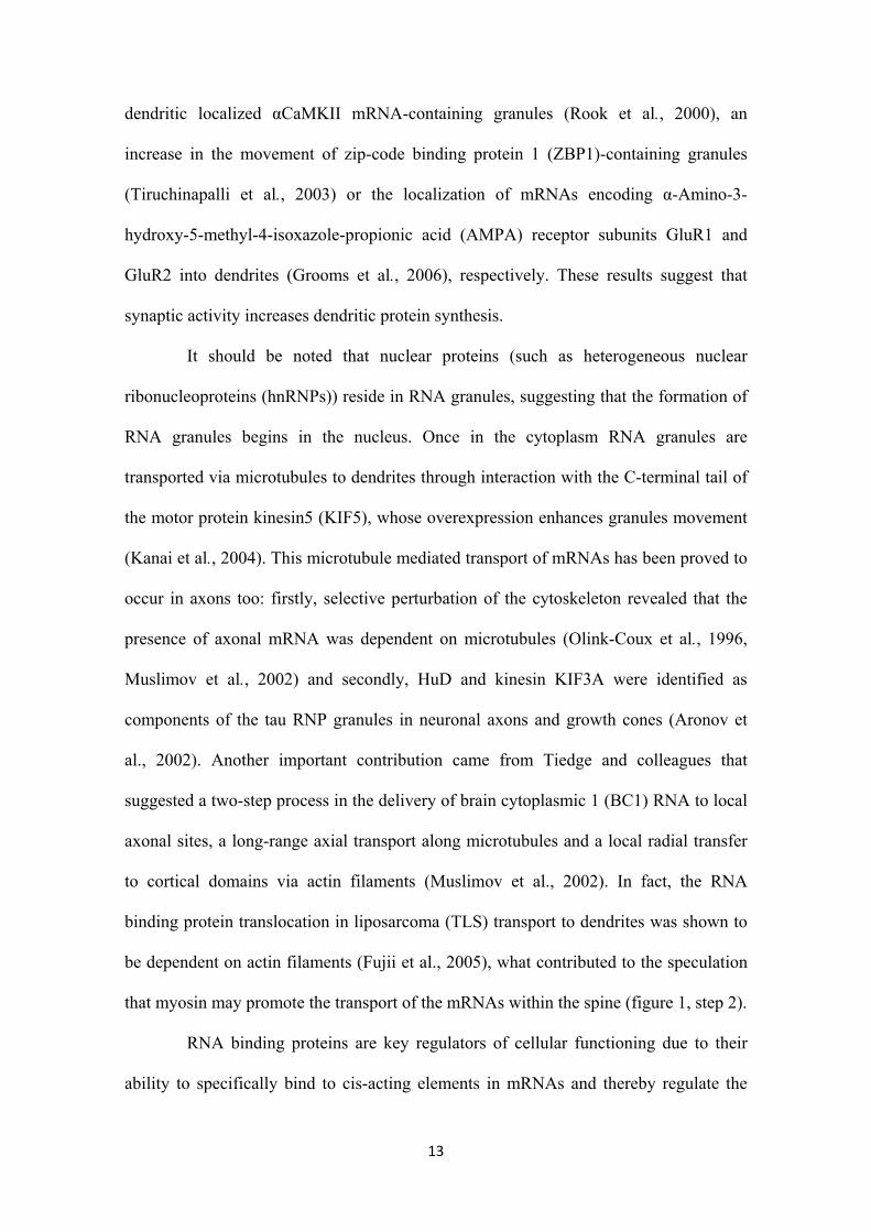
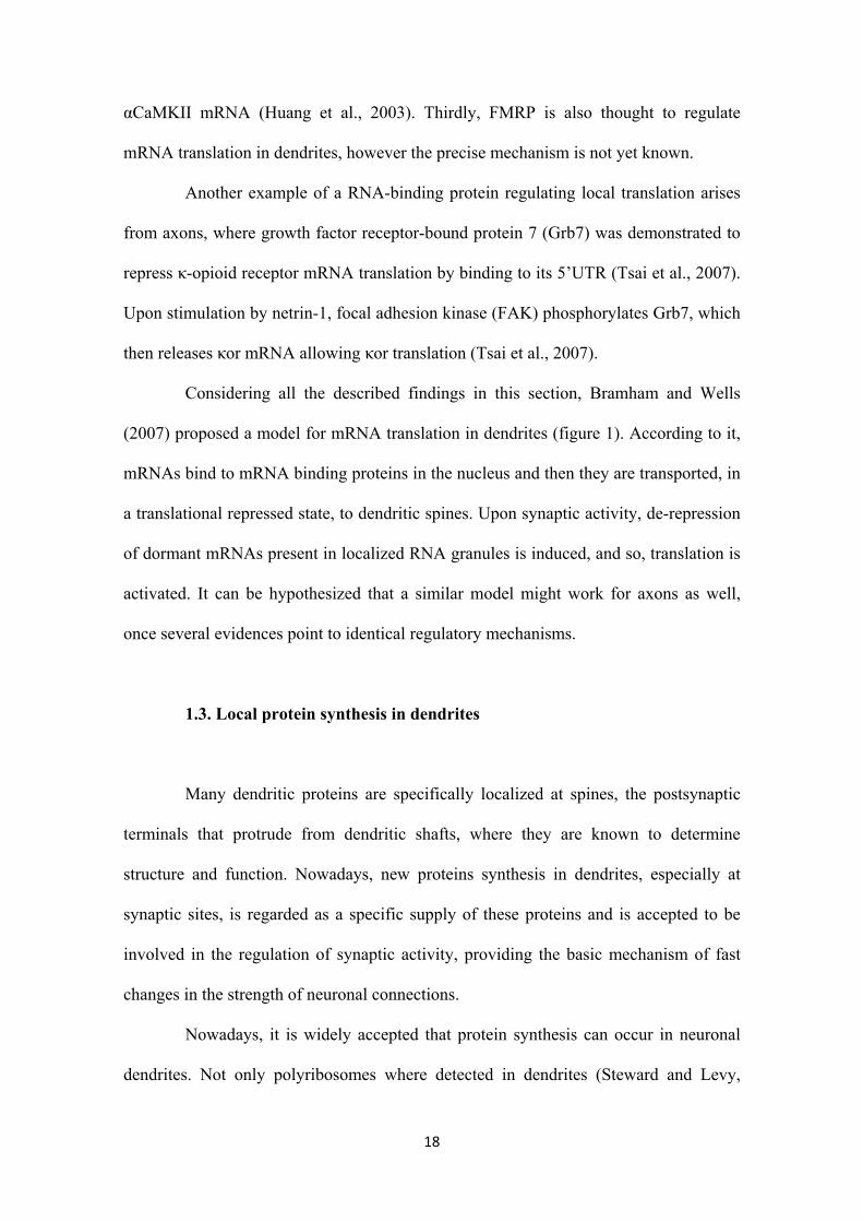
Figure 1 - Proposed model for mRNA translation in neuronal dendrites.
In the nucleus, an mRNA binding protein (R) binds to a specific mRNA (1).
Binding of other proteins will eventually form a RNA granule with motor
proteins for translocation along cytoskeletal elements (kinesin for
microtubules and myosin for actin filaments) (2). The mRNA bound to
mRNA binding proteins in the granule is translationally silenced during
transport (3). Following synaptic activation, de-repression of translation (4)
promotes movements of the mRNAs to polysome particles and local
dendritic translation (5) (Adapted from Bramham and Wells, 2007).
Secondly, once repressed mRNAs bound to CPEB get to sites of translation,
CPEB is phosphorylated by Aurora A kinase. This phosphorylation converts CPEB into
a translation activator that recruits an atypical poly(A) polymerase Gld-2, and thereby
elongates the poly(A) tail of the mRNA it binds. The maskin-eIF4E association is
disrupted after poly(A) tail elongation, therefore allowing the binding of eIF4G and
translation (reviewed in Wells, 2006). Indeed, NMDA activation in synaptosomes
increases Aurora activity, CPEB phosphorylation and, as a result, polyadenylation of
18
αCaMKII mRNA (Huang et al., 2003). Thirdly, FMRP is also thought to regulate
mRNA translation in dendrites, however the precise mechanism is not yet known.
Another example of a RNA-binding protein regulating local translation arises
from axons, where growth factor receptor-bound protein 7 (Grb7) was demonstrated to
repress κ-opioid receptor mRNA translation by binding to its 5’UTR (Tsai et al., 2007).
Upon stimulation by netrin-1, focal adhesion kinase (FAK) phosphorylates Grb7, which
then releases κor mRNA allowing κor translation (Tsai et al., 2007).
Considering all the described findings in this section, Bramham and Wells
(2007) proposed a model for mRNA translation in dendrites (figure 1). According to it,
mRNAs bind to mRNA binding proteins in the nucleus and then they are transported, in
a translational repressed state, to dendritic spines. Upon synaptic activity, de-repression
of dormant mRNAs present in localized RNA granules is induced, and so, translation is
activated. It can be hypothesized that a similar model might work for axons as well,
once several evidences point to identical regulatory mechanisms.
1.3. Local protein synthesis in dendrites
Many dendritic proteins are specifically localized at spines, the postsynaptic
terminals that protrude from dendritic shafts, where they are known to determine
structure and function. Nowadays, new proteins synthesis in dendrites, especially at
synaptic sites, is regarded as a specific supply of these proteins and is accepted to be
involved in the regulation of synaptic activity, providing the basic mechanism of fast
changes in the strength of neuronal connections.
Nowadays, it is widely accepted that protein synthesis can occur in neuronal
dendrites. Not only polyribosomes where detected in dendrites (Steward and Levy,

19
1982), but also incubation of isolated dendrites with radiolabeled aminoacids, with or
without transfection with mRNAs, resulted in a strong labeling of proteins, indication
that local protein synthesis in dendrites actually takes place (Torre and Steward, 1992;
Crino and Eberwine, 1996). Furthermore, using radioactive uridine precursors, Steward
and colleagues demonstrated that mRNAs were transported into dendrites of cultured
hippocampal neurons (Davis et al., 1987). Recently, dendritic ‘hot spots’ of translation,
regions where translation seems to occur repeatedly over time, were observed by
transfecting dendrites with mRNA (Eberwine et al., 2001). Nowadays, these dendritic
‘hot spots’ are known to be located at the base of spines.
In the past years, the hypothesis of dendritic protein synthesis has received
further support in the identification of hundreds of mRNAs localized in dendrites. Some
examples are: structural proteins (MAP2 and Arc), enzymes (αCaMKII), growth factors
(BDNF and NT3), growth factor receptors (TrkA and TrkB), ligand-gated ion channels
(gamma-aminobutyric acid (GABA) and NMDA receptor subunits and glycine receptor
α subunit), voltage-gated ion channels (calcium channels) and transcription factors
(cAMP response element binding protein (CREB)) (reviewed in Steward and Schuman,
2003).
Local protein synthesis in dendrites is believed to play a decisive role in
synaptic plasticity, induction of both late long-term potentiation (LTP) (Bramham,
2008) and late long-term depression (LTD) (Bramham and Wells, 2007), in synaptic
scaling2 and synaptic tagging3 (topics reviewed in Skup, 2008; Barrett and Eberwine,
2008, respectively).
2 Synaptic scaling is a form of homeostatic plasticity that scales synaptic strengths up or down to compensate for prolonged changes in activity (Turrigiano, 2008). 3 Synaptic tagging refers to the idea that the induction of LTP is associated with the setting of a ‘synaptic tag’ at activated synapses, which sequesters the relevant proteins to establish late LTP, by stabilizing temporary synaptic changes and so extend their persistence (Frey and Morris, 1998).
20
It has also been proposed a role for dendritic translation in synaptogenesis.
First evidence came from findings by Burry (1985), who reported an essential role for
dendritic protein synthesis in synapse formation. Later on, accumulation of ribosomes in
dendrites during synaptogenesis (Steward and Falk, 1986; McCarthy, 2003) and
posterior reduction with synapse maturation (Steward and Falk, 1986) strengthened this
idea. More recently, acethylcoline receptor was shown to be synthesized in the
postsynaptic muscle during neuromuscular differentiation (Hall and Sanes, 1993). Lack
of novel evidences may be due to difficulty in isolating dendrites and in assessing
postsynaptic differentiation.
1.4. Local protein synthesis in axons
A growing body of evidence supports the idea that local translation in axons, as
it was seen for the postsynaptic terminal, is important for the development and plasticity
of neural circuits. This section considers the direct evidence that proteins can be
synthesized in axons and its possible functional significance.
1.4.1 - Early evidences from invertebrates
Although being a controversial area, protein synthesis in invertebrate axons is
already well accepted, in part due to the presence of translation machinery (Black and
Lasek, 1977; Lasek et al., 1973; Giuditta et al., 1977; Giuditta et al., 1980; Martin et al.,
1998; van Minnen and Syed, 2001) and several mRNAs, such as β-actin and β-tubulin
(Kaplan et al., 1992), kinesin (Gioio et al., 1994), enolase (Chun et al., 1995) and
neurofilament proteins (Giuditta et al., 1991), among many others.

21
The capacity for translation in truncated neurites and growth cones was
demonstrated by Kater and colleagues through the incorporation of radiolabeled amino
acids into unspecific synthesized proteins in a soma-independent manner (Davis et al.,
1992). Later on, new studies demonstrated the occurrence of de novo protein synthesis
in isolated axons by the synthesis of a foreign mRNA encoding the peptide egg-laying
hormone (ELH) (van Minnen et al., 1997), or the synthesis and functional integration of
the membrane-bound conopressin receptor 2 (CPR-2) from axonally injected mRNA
(Spencer et al., 2000). These results show that the axon possesses the machinery to
produce a functional and appropriately trafficked membrane protein.
1.4.2 - Emerging evidences in vertebrates
As for invertebrates, emerging evidences prove the existence of local protein
synthesis in vertebrate axons, despite initial failure in detecting ribosomes in these
processes (Lasek et al., 1973).
Firstly, different papers demonstrate the presence of components of the protein
synthesis machinery: ribosomes in developing axons (Tennyson, 1970); ribosomal RNA
and tRNA in Mauhtner axon (Koenig, 1979); identification of polysomal aggregates
stained by YOYO-1 and antiribosomal antibodies in Mauhtner axon (Koenig and
Martin, 1996) and in rabbit and rat root fibers (Koenig et al., 2000); identification of
initiation factors (Zheng et al., 2001) and 7S RNA, a key component of the signal
recognition particle (Walter and Johnson, 1994). The detection of several mRNAs in
mature (adult) and developing axons (embryonic or newborn) also constitutes a precious
evidence for axonal translation (see Table 1) – the role of their local translation will be
highlighted in the next sections. It should be noted the prevalence of mRNAs coding
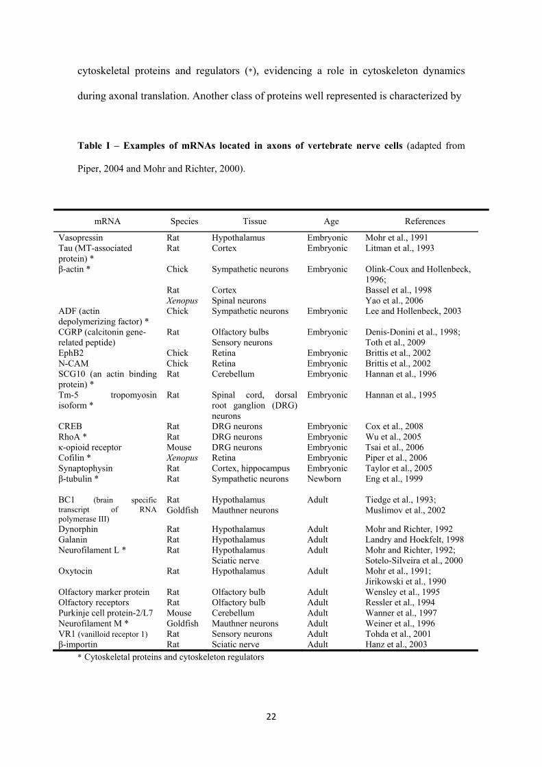
22
cytoskeletal proteins and regulators (*), evidencing a role in cytoskeleton dynamics
during axonal translation. Another class of proteins well represented is characterized by
Table I – Examples of mRNAs located in axons of vertebrate nerve cells (adapted from
Piper, 2004 and Mohr and Richter, 2000).
mRNA Species Tissue Age References
Vasopressin Rat Hypothalamus Embryonic Mohr et al., 1991 Tau (MT-associated protein) *
Rat Cortex Embryonic Litman et al., 1993
β-actin * Chick Rat Xenopus
Sympathetic neurons Cortex Spinal neurons
Embryonic Olink-Coux and Hollenbeck, 1996; Bassel et al., 1998 Yao et al., 2006
ADF (actin depolymerizing factor) *
Chick Sympathetic neurons Embryonic Lee and Hollenbeck, 2003
CGRP (calcitonin gene-related peptide)
Rat Olfactory bulbs Sensory neurons
Embryonic Denis-Donini et al., 1998; Toth et al., 2009
EphB2 Chick Retina Embryonic Brittis et al., 2002 N-CAM Chick Retina Embryonic Brittis et al., 2002 SCG10 (an actin binding protein) *
Rat Cerebellum Embryonic Hannan et al., 1996
Tm-5 tropomyosin isoform *
Rat Spinal cord, dorsal root ganglion (DRG) neurons
Embryonic Hannan et al., 1995
CREB Rat DRG neurons Embryonic Cox et al., 2008 RhoA * Rat DRG neurons Embryonic Wu et al., 2005 κ-opioid receptor Mouse DRG neurons Embryonic Tsai et al., 2006 Cofilin * Xenopus Retina Embryonic Piper et al., 2006 Synaptophysin Rat Cortex, hippocampus Embryonic Taylor et al., 2005 β-tubulin * Rat Sympathetic neurons Newborn Eng et al., 1999
BC1 (brain specific transcript of RNA polymerase III)
Rat Goldfish
Hypothalamus Mauthner neurons
Adult Tiedge et al., 1993; Muslimov et al., 2002
Dynorphin Rat Hypothalamus Adult Mohr and Richter, 1992 Galanin Rat Hypothalamus Adult Landry and Hoekfelt, 1998 Neurofilament L * Rat Hypothalamus
Sciatic nerve Adult Mohr and Richter, 1992;
Sotelo-Silveira et al., 2000 Oxytocin Rat Hypothalamus Adult Mohr et al., 1991;
Jirikowski et al., 1990 Olfactory marker protein Rat Olfactory bulb Adult Wensley et al., 1995 Olfactory receptors Rat Olfactory bulb Adult Ressler et al., 1994 Purkinje cell protein-2/L7 Mouse Cerebellum Adult Wanner et al., 1997 Neurofilament M * Goldfish Mauthner neurons Adult Weiner et al., 1996 VR1 (vanilloid receptor 1) Rat Sensory neurons Adult Tohda et al., 2001 β-importin Rat Sciatic nerve Adult Hanz et al., 2003
* Cytoskeletal proteins and cytoskeleton regulators
23
their involvement in neuron activity and includes peptide hormones (vasopressin and
calcitonin gene-related peptide (CGRP)), the opioid peptide dynorphin and its receptor,
the neuropeptide galanin, the neurotransmitter oxytocin and the ion selective channel
vanilloid receptor. Thus, a role for axonal translation in synaptic function can also be
hypothesized.
Secondly, experiments using radiolabeled amino acids (Edström, 1966;
Alvarez and Chen, 1972; Koenig, 1967) revealed protein synthesis-dependent
incorporation of labeled metabolic precursors into dissected axons. An improvement in
this basic methodology for investigating protein synthesis consists in the extraction and
SDS-PAGE analysis of radiolabeled axonal proteins (exclusively synthesized in axons
once cell bodies had been removed). The use of this technique led to the identification
of actin, tubulin (Koenig, 1991; Eng et al., 1999) and neurofilament proteins (Koenig,
1991) as the principal locally synthesized proteins.
To sum up, the presence of mRNAs and protein synthetic machinery, the
incorporation of amino acids and production of a specific set of proteins in isolated
axons firmly demonstrate that axonal translation in vertebrates is not a fictitious process
and that it must be associated with a functional role.
1.5. Role of local protein synthesis in axons
1.5.1. Developing axons
During development of the nervous system, axons pass through distinct stages,
characterized by profound morphological and functional changes. In a first stage, the
developing axons seek for a synaptic target. In order to achieve this objective,
24
developing axons undergo elongation and possess at their tips an actin-rich growth
cone, which is a highly motile structure. During axon navigation, the growth cone
senses the environment by detecting extracellular guidance cues and morphologically
responding to them (Farrar and Spencer, 2008), a process that promotes axonal
pathfinding. In a final stage, axon navigation halts in an area where connection to the
target postsynaptic terminal must occur. In this moment, the growth cone undergoes a
profound transition from a highly motile structure to a functional presynaptic terminal,
and by the end of synaptogenesis, a functional synaptic connection is established.
An important issue that has received considerable attention is the level of
dependence of the axon tip on the cell body during both axon guidance and
synaptogenesis. As a matter of fact, protein translation at axon tips offers a higher
degree of autonomy during axon development.
1.5.1.1. Axonal protein synthesis in axon guidance
During development and navigation toward target cells, the growth cone senses
spatially and temporally distributed guidance cues and subsequently steers the axon in
the appropriate direction (Tessier-Lavigne and Goodman, 1996). These extracellular
guidance cues, including netrins, Slits, semaphorins and ephrins, can either attract or
repel growth cones or even induce their branching or collapse (Tessier-Lavigne and
Goodman, 1996). Guidance cues act by binding to surface receptors in growth cones:
deleted in colorectal carcinoma (DCC) and UNC-5 receptors for netrins (Culotti and
Merz, 1998; Keleman and Dickson, 2001), Robo receptors for Slits (Kidd et al., 1999),
multimeric receptor complexes with plexin protein for semaphorins (Tamagnone et al.,
1999) and Eph family of receptor tyrosine kinases for ephrins (Drescher et al., 1997)
25
(reviewed in Dickson, 2002). The activation of these surface receptors elicits localized
intracellular signaling events, which ultimately control cytoskeletal activities to steer the
growth cone, such as reorganization of actin filaments or stabilization of microtubules.
Growth cone responses to guidance cues have been proposed to be dependent
on local protein synthesis, what could explain specific protein requirements, altered
responsiveness along the way and specificity of an axon’s trajectory. For example,
growth cone turning is caused by extension on one side and collapse on the other, and is
triggered by local attractive and repulsive cues. Initial experiments revealed that in vitro
chemotropic responses to netrin-1 and Sema3A (attractive or repulsive and collapse,
respectively) of isolated axons were dependent on local protein synthesis (Campbell and
Holt, 2001). This study also demonstrated that Sema3A and netrin-1 trigger a burst of
protein synthesis within the growth cone. Later on, new studies identified other
guidance cues-induced chemotropic responses dependent on local axonal translation:
plasma cell induced ER protein 1 (PACAP)-induced growth cone attraction (Guirland et
al., 2003), Engrailed-2-induced turning responses (Brunet et al., 2005), Slit-2-induced
collapse and repulsion (Piper et al., 2006) and BDNF-induced attraction (Yao et al.,
2006).
Considering that guidance cues ultimately modulate cytoskeletal activities in
growth cones, it is reasonable to assume that activation of their receptors leads to local
translation of cytoskeletal proteins or regulators. In fact, an attractive gradient of netrin-
1 or BDNF induces β-actin synthesis in growth cones (Leung et al., 2006; Yao et al.,
2006); Slit2 causes a protein synthesis dependent increase in the actin-depolymerizing
protein cofilin (Piper et al., 2006) and Sema3A induces intra-axonal translation of RhoA
mRNA, which is a critical upstream regulator of the cytoskeleton and whose activation
leads to growth cone collapse (Wu, et al., 2005).
26
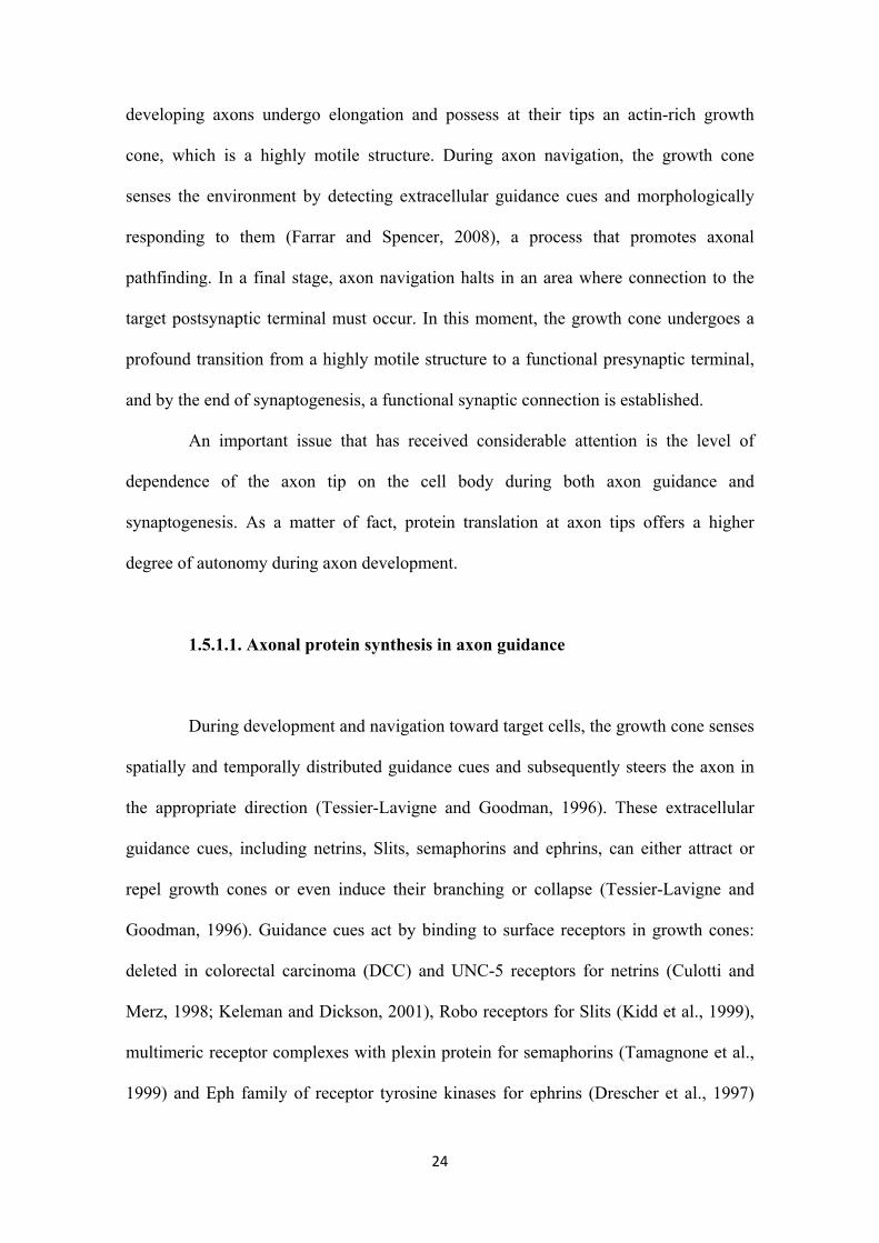
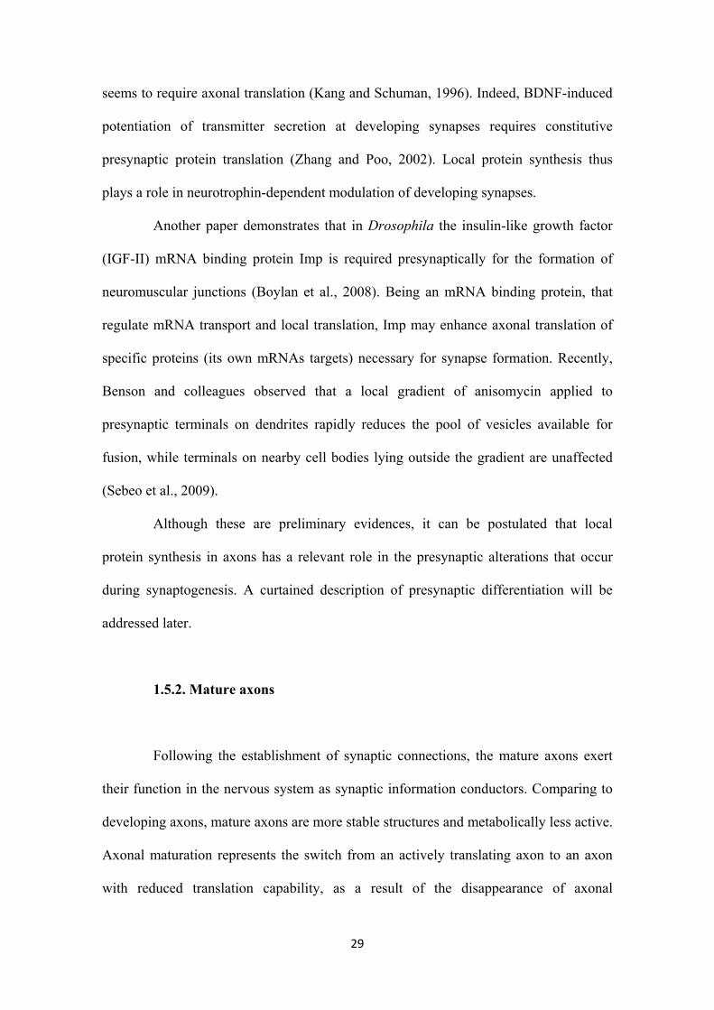
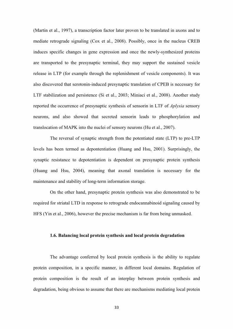
Based in these evidences, the “differential translation model” has emerged,
postulating that attractive and repulsive cues induce asymmetrical translation of proteins
that ultimately promotes assembly or disassembly of the cytoskeleton, respectively (Lin
and Holt, 2008) (see Figure 2). However, a recent study contradicts these findings
stating that protein synthesis inhibition does not alter ephrin-A2, slit-3 and Sema3A-
induced growth cone collapse and loss of actin filaments; neither NGF nor
neurotrophin-3-induced growth cone protrusion and increased actin filaments (Roche et
al., 2009).
Figure 2 - The “differential translation” model for local translation in
growth cones. This model states that (a) a gradient of an attractive guidance
cue induces local translation of mRNAs (such as β-actin) that cause actin
polymerization and thus attractive turning; on the other hand (b) a gradient
of a repulsive guidance cue induces local translation of mRNAs (such as
cofilin) that cause actin depolymerization and consequently repulsive turning
(Adapted from Lin and Holt, 2008).
27
In their navigation, axons reach intermediate targets, and once they pass
through them, they must change their responsiveness to the guidance cues. For example,
as the vertebrate commissural axons cross the floor plate they lose sensitivity to the
midline attractant, netrin, and acquire sensitivity to Slit and semaphorin repellents (Stein
and Tessier-Lavigne 2001). Recently, it has been proposed that local translation may
also mediate changes in axonal responsiveness. Flanagan and colleagues revealed a
mechanism that could promote local synthesis of receptors for guidance cues. Focusing
on EphA2 receptor and elaborating a construct made up of 3’UTR of EphA2 and green
fluorescence protein (GFP), they observed that the fusion protein was expressed at high
levels in the distal segments of the axons that had passed the midline (Brittis et al.,
2002). This result suggests that signals from an intermediate target trigger the
upregulation of a different set of receptors that are necessary for guiding the axon to the
next intermediate point.
Desensitization and resensitization are mechanisms through which axons alter
their sensitivity to external guidance cues. These mechanisms of regulation of growth
cone behavior and responsiveness are dependent on local protein synthesis, since it was
demonstrated that resensitization to netrin-1, BDNF or Sema3A requires axonal
translation (Ming et al., 2002; Piper et al., 2005). The regained sensitivity to guidance
cues is due to a recovery of functional surface receptors. Considering that axons have
the ability to synthesize and translocate a functional surface receptor (Spencer et al.,
2000), it can be suggested that local protein synthesis dependent-resensitization results
from guidance cue receptor synthesis and local insertion into the plasma membrane. In
fact, axons can promote synthesis and functional membrane integration of the κ-opioid
receptor in response to netrin-1 (Tsai et al., 2006).
28
1.5.1.2. Axonal protein synthesis in synaptogenesis
Synaptogenesis is a process involving the formation of a neurotransmitter
release site in the presynaptic neuron precisely aligned with a receptive field at the
postsynaptic neuron. During development of the nervous system, synaptogenesis
requires that a number of changes occur at the site of contact between pre- and
postsynaptic neuron. These changes are mediated by the insertion of precursor vesicles
containing many preassembled active zone molecules and synaptic vesicles components
into the presynaptic terminal and by a sequential recruitment of molecules to the
postsynaptic specialization.
Local translation in axons was also demonstrated to be important in
synaptogenesis. Early evidence came from findings that interaction of a sensory neuron
growth cone with a specific postsynaptic target regulates the local export and
accumulation of sensorin A mRNA in sensory neurons (Schaher et al., 1999; Lyles et
al., 2006; Hu et al., 2002), and that the release of this peptide promotes the formation of
sensory-motor synapses (Hu et al., 2004). These results suggest that branch specific
targeting of mRNA encoding synapse-related molecules may contribute to the
formation of specific synapses through local protein synthesis. Later on, Schacher and
Wu concluded that protein synthesis, at or near synaptic sites, is required for the
establishment of new synaptic connections in the absence of cell bodies (Schacher and
Wu, 2002). Indeed, Martin and colleagues suggested that local protein synthesis of
sensorin mRNA in the presynaptic terminal is required for synapse formation, since
knockdown of sensorin mRNA abolishes synapse formation (Lyles et al., 2006).
Neurotrophins (BDNF and NT-3) produce a long-lasting enhancement of
synaptic transmission in developing axons (Kang and Schuman, 1995) and this capacity
29
seems to require axonal translation (Kang and Schuman, 1996). Indeed, BDNF-induced
potentiation of transmitter secretion at developing synapses requires constitutive
presynaptic protein translation (Zhang and Poo, 2002). Local protein synthesis thus
plays a role in neurotrophin-dependent modulation of developing synapses.
Another paper demonstrates that in Drosophila the insulin-like growth factor
(IGF-II) mRNA binding protein Imp is required presynaptically for the formation of
neuromuscular junctions (Boylan et al., 2008). Being an mRNA binding protein, that
regulate mRNA transport and local translation, Imp may enhance axonal translation of
specific proteins (its own mRNAs targets) necessary for synapse formation. Recently,
Benson and colleagues observed that a local gradient of anisomycin applied to
presynaptic terminals on dendrites rapidly reduces the pool of vesicles available for
fusion, while terminals on nearby cell bodies lying outside the gradient are unaffected
(Sebeo et al., 2009).
Although these are preliminary evidences, it can be postulated that local
protein synthesis in axons has a relevant role in the presynaptic alterations that occur
during synaptogenesis. A curtained description of presynaptic differentiation will be
addressed later.
1.5.2. Mature axons
Following the establishment of synaptic connections, the mature axons exert
their function in the nervous system as synaptic information conductors. Comparing to
developing axons, mature axons are more stable structures and metabolically less active.
Axonal maturation represents the switch from an actively translating axon to an axon
with reduced translation capability, as a result of the disappearance of axonal
30
polyribosomes and mRNAs. Thus, the axonal synthesis of proteins in mature
mammalian axons is controversial even though several mRNAs (see Table I) and
ribosomes were detected in adult mammalian axons (Koenig et al., 2000). Moreover,
studies demonstrating the participation of axonal translation in events occurring in
mature axons, such as axon regeneration (reviewed in Willis and Twiss, 2006) and
synaptic plasticity (Martin, 2004), are gaining acceptance.
1.5.2.1. Axonal protein synthesis in axon regeneration
During regeneration, in order to restore the connectivity with target tissues, the
cut end of the axon must be remodeled and initiate a growth program to reform a new
growth cone. This process has been shown to occur in isolated axons, which could
regenerate new growth cones after injury, suggesting autonomous capacity to synthesize
molecules needed for regeneration (Shaw and Bray, 1977).
Studies of regeneration in adult sensory axons demonstrated the functional
importance of local intra-axonal protein synthesis in growth cone initiation after
axotomy. The neurite regeneration of PC12 cells was shown to be dependent on the
translation of the mRNA encoding the ribosomal L4 protein (Twiss et al., 2000).
Furthermore, blocking synthesis in regenerating sensory axons causes a rapid retraction
of their growth cones when separated from the cell body (Zheng et al., 2001) and reduce
the proportion of transected axons able to reform growth cones (Verma et al., 2005).
Twiss and colleagues showed that cytoskeletal, chaperone, metabolic and anti-oxidant
proteins are synthesized by regenerating sensory axons (Willis et al., 2005).
Theoretically, this huge set of proteins provides the axon with cytoskeletal elements to
reform the growth cone, and provides autonomy in responding to environmental cues.
31
Amazingly, Toth and colleagues (2009) demonstrated that a specific protein, calcitonin
gene-related peptide (CGRP), is synthesized during regeneration and its mRNA is
transported to regenerating axons (Toth et al., 2009).
Retrograde signaling is also important for axonal regeneration, as the cell body
has to be updated on the injured status of the axon to modulate a program of repair
(Hanz and Fainzilber, 2006). The involvement of local translation in retrograde
signaling was highlighted by the axonal synthesis of importin-β (a protein that
transports nuclear localization signal (NLS) bearing proteins to the nucleus) after nerve
lesion and its retrograde transport by the motor protein dynein (Hanz et al., 2003). The
activated MAP kinase (Erk 1/2) has been proposed to be transported by importins via
vimentin binding after nerve injury (Perlson et al., 2005), thus promoting changes in the
cell body that support regeneration. Indeed, a detailed model of axonal retrograde
signaling after nerve lesion regulated by RanGTPase and importin-β was proposed
(Yudin et al., 2008). Another study identified a signaling role for axonally derived
CREB in mediating the response to the neurotrophin nerve growth factor (NGF) (Cox et
al., 2008). These studies raise the possibility that newly synthesized transcription factors
can be retrogradely transported on microtubules to the cell body where they influence
transcription of genes for axonal repair.
1.5.2.2. Axonal protein synthesis in synaptic plasticity
Synaptic plasticity refers to the capacity of neurons to modulate the strength of
their synaptic connections. Synaptic plasticity contributes to a variety of physiological
processes in the adult brain, including memory, learning and age-related memory loss. It
can exist in two distinct forms: high frequency stimulation (HFS) triggers a long-lasting
32
enhancement of synaptic strength, known as long-term potentiation (LTP) (Bliss and
Collingridge, 1993), whereas prolonged low-frequency stimulation (LFS) results in a
long-lasting decrease in synaptic strength, termed long-term depression (LTD) (Mulkey
and Malenka, 1992).
Succinctly, the increased synaptic strength that occurs during LTP should arise
from an increase in neurotransmitter release from the presynaptic terminal, or from an
increase in postsynaptic terminal responsiveness to the neurotransmitter, or both. It is
widely accepted that translation of dendritically localized mRNAs is required in long-
term synaptic plasticity through regulation of actin dynamics, which ultimately leads to
insertion of additional AMPA receptors in the postsynaptic membrane (Bramham,
2008). In a more functional level, it is believed that presynaptic LTP is triggered by a
tetanus-induced rise in presynaptic Ca2+ that results in the activation of a Ca2+-
calmodulin-dependent adenylyl cyclase, which in turn causes an increase in presynaptic
cAMP levels and activation of protein kinase A (Huang et al., 1994). Protein kinase A
can initiate intracellular signaling cascades that, by activating mechanisms perhaps
involving Rab3A and its interacting protein, regulating synaptic membrane exocytosis
(RIM1α), cause a long-lasting enhancement of synaptic vesicle fusion and
neurotransmitter release (Castillo et al., 1997; Castillo et al., 2002). The development of
new probes designed to directly assay synaptic vesicle cycling undoubtedly revealed the
existence of presynaptic LTP in the form of accelerated synaptic vesicle release
(reviewed in Blundon and Zakharenko, 2008).
Accumulating evidences support a role for axonal protein synthesis in synaptic
plasticity. Presynaptic protein synthesis is required for long-term facilitation (LTF) of
Aplysia (Martin et al., 1997) and crayfish (Beaumont et al., 2001) sensory to motor
neurons synapses. Moreover, this facilitation depends on CREB-mediated transcription
33
(Martin et al., 1997), a transcription factor later proven to be translated in axons and to
mediate retrograde signaling (Cox et al., 2008). Possibly, once in the nucleus CREB
induces specific changes in gene expression and once the newly-synthesized proteins
are transported to the presynaptic terminal, they may support the sustained vesicle
release in LTP (for example through the replenishment of vesicle components). It was
also discovered that serotonin-induced presynaptic translation of CPEB is necessary for
LTF stabilization and persistence (Si et al., 2003; Miniaci et al., 2008). Another study
reported the occurrence of presynaptic synthesis of sensorin in LTF of Aplysia sensory
neurons, and also showed that secreted sensorin leads to phosphorylation and
translocation of MAPK into the nuclei of sensory neurons (Hu et al., 2007).
The reversal of synaptic strength from the potentiated state (LTP) to pre-LTP
levels has been termed as depontentiation (Huang and Hsu, 2001). Surprisingly, the
synaptic resistance to depotentiation is dependent on presynaptic protein synthesis
(Huang and Hsu, 2004), meaning that axonal translation is necessary for the
maintenance and stability of long-term information storage.
On the other hand, presynaptic protein synthesis was also demonstrated to be
required for striatal LTD in response to retrograde endocannabinoid signaling caused by
HFS (Yin et al., 2006), however the precise mechanism is far from being unmasked.
1.6. Balancing local protein synthesis and local protein degradation
The advantage conferred by local protein synthesis is the ability to regulate
protein composition, in a specific manner, in different local domains. Regulation of
protein composition is the result of an interplay between protein synthesis and
degradation, being obvious to assume that there are mechanisms mediating local protein
34
degradation as well, possibly with outstanding importance for neuronal function. In fact,
recent evidences support this idea (for a review Steward and Schuman, 2003; Hedge,
2004; Gumy et al., 2009). Indeed, the detection of ubiquitinated proteins in adult rat
forebrain synaptic fractions (Chapman et al., 1994) and the detection of the machinery
responsible for ubiquitin-dependent degradation, such as ubiquitin, ubiquitin-activating
enzyme E1 and proteasome subunits in retinal growth cones (Campbell and Holt, 2001)
and in presynaptic terminals (Speese et al., 2003), suggest that both the attachment of
ubiquitin to substrate proteins and their subsequent degradation may occur locally. It is
important to notice that local protein degradation is critical in neuronal functions
already proved to be dependent upon local protein synthesis, and thus revealing a
possible balance between these mechanisms in neuronal activities occurring far from the
cell body.
Campbell and Holt observed that proteasome inhibitors prevented netrin-1-
induced chemotropic responses and that these guidance cues elicit rises in ubiquitin-
protein conjugates on growth cones (Campbell and Holt, 2001). In addition, loss of
function mutations of the ubiquitin-activating enzyme (E1) or proteasome subunits in
neurons of Drosophila mushroom bodies block axon pruning4 (Watts et al., 2003).
These findings suggest that protein degradation is important for both axon guidance and
pruning.
As it is described in the section 1.5.2.1., regeneration of axons requires the re-
creation of the growth cone. Besides local protein synthesis, local degradation has
gained attention in the past years. In regenerating sciatic nerves, an increase in
ubiquitinated proteins was observed (Jack et al., 1992). On the other hand, Fawcett and
colleagues reported that application of proteasome inhibitors resulted in a reduction in
4 Axon pruning is widely used for the refinement of neural circuits, and consists in the development of exuberant axonal branches, followed by a selective pruning of a subset of these branches.
35
the proportion of transected axons to regenerate growth cones (Verma et al., 2005).
Recently, it was observed that mammalian CNS axons after a stretch injury accumulate
ubiquitin associated with cytoskeleton elements (Staal et al., 2009), suggesting UPS-
dependent reorganization of the cytoskeleton in axons after injury.
With respect to synaptic plasticity, several studies indicate that the ubiquitin-
proteasome pathway plays a role in long-term and short-term effects. For example, the
presynaptic protein DUNC-13 (a synaptic vesicle priming protein) is ubiquitinated and
degraded by the proteasome in Drosophila neuromuscular synapse (Speese et al., 2003),
and so acute inhibition of proteasome activity promotes rapid strengthening of
neurotransmition (Speese et al., 2003). Two other synaptic vesicle proteins,
synaptophysin and syntaxin have been shown to be substrates for ubiquitin-proteasome-
mediated degradation (Wheeler et al., 2002). On the other synaptic side, it has been
shown that ubiquitin-proteasome pathway is involved in receptor internalization,
modulating synaptic transmission through endocytosis of AMPA receptor subunits
(Patrick et al., 2003). In addition to the neurotransmitter receptors, proteins of the
postsynaptic density are substrates of the ubiquitin-proteasome degradation (Schwartz,
2003) in an activity-dependent manner (Ehlers, 2003).
1.7. Presynaptic differentiation
Following axon outgrowth and arrival at the target area, the growth cone
suffers tremendous morphological and functional alterations as it differentiates into a
presynaptic terminal. In a first stage, the growth cone loses its highly motile properties:
axon navigation slows down and the growth cone flattens (Yoshihara et al., 1997; Jontes
et al., 2000), probably due to an increase in axonal membrane adhesiveness and
36
alteration of axonal cytoskeleton dynamics. It is interesting to point out the possible
involvement of axonal translation in mediating these alterations, since several axonal
localized mRNAs code for cytoskeleton proteins and regulators of cytoskeleton
dynamics (see table 1).
The initial formation of axonal varicosities, which resemble immature
presynaptic terminals with neurotransmitter containing vesicles, is accompanied by
axonal branching (Alsina et al., 2001). Within minutes, a functional synapse (with a
relatively immobile axon and a fully functional neurotransmitter-releasing presynaptic
terminal) is formed, suggesting that axons can autonomously form a synaptic contact,
and do not depend on soma-trafficked proteins.
Presynaptic formation involves coordinated action of several inter-dependent
events: precise alignment between postsynaptic density and the presynaptic active zone;
clustering and maturation of synaptic vesicles (SVs); establishment of the active zone;
presynaptic growth; cytoskeletal restructuring and assembly of vesicle recycling
machinery.
Synaptic vesicle genesis, including maturation and clustering, is crucial for
presynaptic formation. In a functional presynaptic site, synaptic vesicles are clustered
into two distinct pools: the reserve pool and the readily releasable pool, that allow for
their rapid availability in case of stimulation (Dillon and Goda, 2005). These pools are
maintained by actin filaments by forming a physical barrier to prevent vesicles
dispersion (Dillon and Goda, 2005), and therefore the formation of an actin network
during presynaptic differentiation is absolutely crucial for synaptic vesicles clustering.
Indeed, it has been proposed that formation of actin networks beneath nascent synaptic
sites leads to passive accumulation of mobile synaptic vesicles transport particles
(STVs) (McAllister, 2007), which are vesicular structures carrying several SV-
37
associated proteins and other proteins critical for exo- and endocytosis (Ahmari et al.,
2000 and Zhai et al., 2001).
Other proteins are involved in synaptic vesicles biogenesis and clustering. For
example, the adaptor complex 3 (AP3), which associates with endosomes, is believed to
regulate SV maturation at nascent synapses: mutant mice lacking AP3 exhibit abnormal
SVs of GABA-inhibitory synapses (Nakatsu et al., 2004) and mistargeting of several
SV membrane proteins (Salazar et al., 2004a,b). Another example is the exocyst, a
protein complex composed of eight proteins, believed to be involved in targeting
secretory vesicles to specific domains in the plasma membrane. Results showed that
exocyst subunit mutants fail to maintain golgi-derived immature synaptic vesicles at the
tips of axons (Murthy et al., 2003; Mehta et al., 2005). On the other hand, growth factor
signaling is also involved in the modulation of synaptic vesicles clustering. Wnt7
interaction with its receptor, Frizzled, prevents β-catenin degradation and promotes
phosphorylation of the microtubule-associated protein MAP1B leading to microtubule
stabilization (Krylova et al., 2000; Ciani et al., 2004), thus contributing to axonal
branching and recruitment of presynaptic vesicles (Hall et al., 2000) (figure 3, right
image). Another example is the fibroblast growth factors (FGFs)-induced synaptic
vesicle clustering through activation of FGF receptors in the presynaptic membrane
(Dai and Peng, 1995).
In order to guarantee the perfect alignment between pre- and postsynaptic
sides, trans-synaptic interactions are maintained by neuronal adhesion molecules that
bind cytoplasmic scaffolding proteins. The neuroligin-neurexin complex (figure 3, left
image), which directly bridges the synaptic cleft, induces the nucleation of
calcium/calmodulin-dependent serine protein kinase (CASK) in the presynaptic site
(Dean et al., 2003) and postsynaptic density protein 95 (PSD-95) in the postsynaptic site
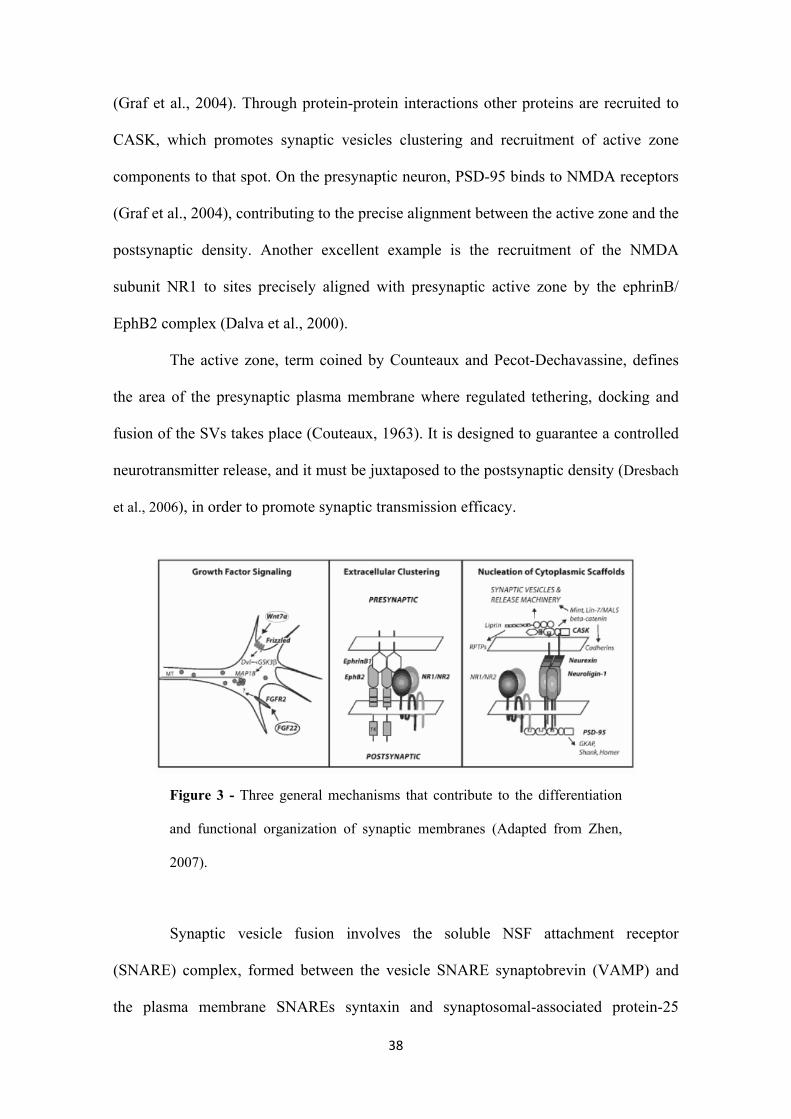
38
(Graf et al., 2004). Through protein-protein interactions other proteins are recruited to
CASK, which promotes synaptic vesicles clustering and recruitment of active zone
components to that spot. On the presynaptic neuron, PSD-95 binds to NMDA receptors
(Graf et al., 2004), contributing to the precise alignment between the active zone and the
postsynaptic density. Another excellent example is the recruitment of the NMDA
subunit NR1 to sites precisely aligned with presynaptic active zone by the ephrinB/
EphB2 complex (Dalva et al., 2000).
The active zone, term coined by Counteaux and Pecot-Dechavassine, defines
the area of the presynaptic plasma membrane where regulated tethering, docking and
fusion of the SVs takes place (Couteaux, 1963). It is designed to guarantee a controlled
neurotransmitter release, and it must be juxtaposed to the postsynaptic density (Dresbach
et al., 2006), in order to promote synaptic transmission efficacy.
Figure 3 - Three general mechanisms that contribute to the differentiation
and functional organization of synaptic membranes (Adapted from Zhen,
2007).
Synaptic vesicle fusion involves the soluble NSF attachment receptor
(SNARE) complex, formed between the vesicle SNARE synaptobrevin (VAMP) and
the plasma membrane SNAREs syntaxin and synaptosomal-associated protein-25
39
(SNAP-25) (Murthy and De Camilli, 2003; Rizo and Sudhof, 2002). The formation of
this complex is believed to provide the driving force for fusion (Chen and Scheller,
2001; Jahn et al., 2003) and therefore these proteins must be located in the active zone.
Even though this is actually true (Garcia et al., 1995), syntaxin and SNAP-25 also locate
in other regions of neuronal plasma membrane (Garcia et al., 1995), showing the need
for a specific machinery that restricts neurotransmitter release to active zones. In fact,
the cytomatrix at the active zone (CAZ) is an electron-dense structure of specialized
proteins that controls and promotes neurotransmitter release. CAZ is composed of
several proteins: Munc13 confers fusion competence to docked SVs (Rosenmund et al.,
2003; Varoqueaux et al., 2002); RIM is necessary in normal SV exocytosis and in
synaptic plasticity (Kaeser and Sudhof, 2005; Calakos et al., 2004); CAST/ERC,
Bassoon, Piccolo and liprin are scaffolding proteins or adaptors capable of homomeric
interactions with other CAZ proteins, or the synaptic plasma membrane components of
the synaptic cytoskeleton and synaptic vesicle release machinery (Fenster et al., 2000;
Ohtsuka et al., 2002). This capability suggests that the presynaptic molecular scaffolds
must exert spatial constraints on the distribution of synaptic vesicles and on the
connection of SVs with the plasma membrane.
It has been shown that functional active zones can form within 30-60 minutes
after initial axodendritic contact. In cultured hippocampal neurons most identified active
zone proteins are preassembled and packaged in cytoplasmic transport vesicles, derived
from the trans-Golgi network, that are delivered to the nascent synaptic sites (Ahmari et
al., 2000; Shapira et al., 2003; Zhai et al., 2001). These vesicles are termed Piccolo-
Bassoon transport vesicles (PTVs) and contain, besides Piccolo and Bassoon, SNAREs
syntaxin and SNAP25, RIM1, Munc13, N-type voltage-gated calcium channels and
Munc18 (Shapira et al., 2003; Zhai et al., 2001). The active zone transport vesicle
40
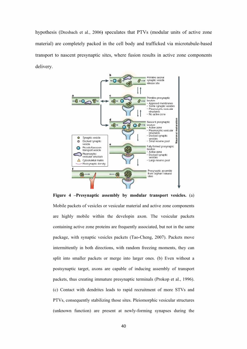
hypothesis (Dresbach et al., 2006) speculates that PTVs (modular units of active zone
material) are completely packed in the cell body and trafficked via microtubule-based
transport to nascent presynaptic sites, where fusion results in active zone components
delivery.
Figure 4 –Presynaptic assembly by modular transport vesicles. (a)
Mobile packets of vesicles or vesicular material and active zone components
are highly mobile within the developin axon. The vesicular packets
containing active zone proteins are frequently associated, but not in the same
package, with synaptic vesicles packets (Tao-Cheng, 2007). Packets move
intermittently in both directions, with random freezing moments, they can
split into smaller packets or merge into larger ones. (b) Even without a
postsynaptic target, axons are capable of inducing assembly of transport
packets, thus creating immature presynaptic terminals (Prokop et al., 1996).
(c) Contact with dendrites leads to rapid recruitment of more STVs and
PTVs, consequently stabilizing those sites. Pleiomorphic vesicular structures
(unknown function) are present at newly-forming synapses during the

41
assembly of presynaptic boutons (Ahmari et al., 2000). (d) Fusion of PTVs
with presynaptic membrane leads to formation of the active zone (Dresbach
et al., 2006). (e) Within days, a totally mature presynaptic boutton is formed
with a large reserve pool of SVs and a ready releasable pool (Dillon and
Goda, 2005). (f) Portions of existing mature terminals can split off and
mobilize to form new immature presynaptic sites (Krueger et al., 2003)
(Adapted from Ziv and Garner, 2004).
Presynaptic differentiation is a highly complex event, and accounts for the
involvement of various cellular events. Besides the described mechanisms, several
others contribute to the formation of a properly organized and functional presynaptic
terminal. A simple model for presynaptic assembly has been proposed (figure 4).
Succinctly, it states that presynaptic components are constitutively generated in the cell
body and then packed into vesicular intermediates in the trans-Golgi network (Jin and
Garner, 2008). Freely-moving packets along the immature axon (McAllister, 2007),
such as STVs and PTVs (Zhai et al., 2001 and Sabo et al., 2006, respectively), are
transported and assembled in nascent synaptic sites upon contact between presynaptic
axon and postsynaptic target (Jin and Garner, 2008; Ziv and Garner, 2004).
1.7.1. Presynaptic organizing molecules
Presynaptic differentiation is known to depend on presynaptogenic factors,
commonly referred to as presynaptic organizing molecules. Axon itself has the capacity
to initialize the recruitment of presynaptic contents (Prokop et al., 1996), however, a
mature and functional presynaptic terminal will only form after postsynaptic contact
(Ziv and Garner, 2004). This observation has led scientists to speculate that probably
42
factors from the postsynaptic side would interact with the presynaptic membrane, thus
affecting and promoting presynaptic differentiation. In fact, postsynaptic-derived
soluble and membrane proteins and glia-derived molecules are known to be
fundamental for presynaptic differentiation (Fox et al., 2006). We will briefly review
the presynaptic organizing function of those factors at synapses of the central nervous
system.
Neuroligin, a postsynaptic transmembrane protein that binds across the
synaptic cleft to the presynaptic cell surface receptor β-neurexin (reviewed in Craig and
Kang, 2007), is able to induce formation of presynaptic specializations. Preliminary
data arose from co-cultures with neuroligin-transfected non neuronal cells and basal
pontine neurons. In these cultures, neuroligin 1 and 2 showed a remarkable capacity to
induce synaptic vesicles clustering along overlying axons (Scheifelle et al., 2000 and Fu
et al., 2003). Later on, Dean and colleagues (2003) concluded that neuroligin effect on
synaptic vesicles distribution is mediated by clustering of β-neurexin in the presynaptic
membrane. Moreover, this clustering seemed to promote the formation of functional
active zones as assessed by the ability of synaptic vesicles at these sites to recycle the
styryl dye FM1-43 (Dean et al., 2003). Further published data helped to solidify the idea
that neuroligin heterophilically binds presynaptic neurexin to form functional
presynaptic terminals (Graf et al., 2004; Levinson et al., 2005; Prange et al., 2004; Sara
et al., 2005; Chih et al.,2005). Interestingly, the β-neurexin/neuroligin complex is bi-
directionally active regarding that presynaptic β-neurexin is also capable of triggering
assembly of postsynaptic receptors through neuroligins (Nam and Chen, 2005 and
Sudhof, 2008) (see also figure 3 right image). Against all odds, recent data contradicts
the proposed role of neuroligins in the initial establishment of synapses. Using knockout
mice for neuroligin 1, 2 and 3, different research groups observed an impaired synaptic
43
signaling and transmission, but not a decrease in synapse formation (Varoqueaux et al.,
2006; Chubykin et al., 2007; Tabuchi et al., 2007), thus concluding that neuroligins
must regulate the proper maturation of synapses. Further experiments should be
performed to clarify neuroligin’s function.
Synaptic cell-adhesion molecule (SynCAM) is a another cell surface adhesion
molecule capable of inducing pre-synaptic differentiation. SynCAM, a transmembrane
protein containing three Ig-domains and an intracellular C-terminal PDZ-binding motif,
is a homophilic calcium-independent cell-adhesion molecule (CAM) expressed on both
sides of the synapse (Biederer et al., 2002). Early studies revealed that contact with
SynCAM induce the formation and assemble of presynaptic terminals capable of both
spontaneous and evoked neurotransmitter release (Biederer et al., 2002; Sara et al.,
2005), thus suggesting that these terminals are functionally active. Although being
unusual, heterophilic adhesion between the various SynCAM isoforms can occur
(Shingai et al., 2003) and, in fact, the transynaptic SynCAM1/2 complex promotes
organization of presynaptic terminals and enhanced neurotransmitter release (Fogel et
al., 2007). Another interesting feature of SynCAM is the fact that, after axotomy,
SynCAM mRNA levels are elevated during restoration of central synapses on
motoneurons, thus revealing their possible involvement in the re-formation of synaptic
connections in the adult brain (Zelano et al., 2009). In a way similar to neuroligins,
SynCAM is believed to trigger active zone formation (it binds CASK and contains PDZ
binding motifs – Biederer et al., 2002), however the precise mechanism is far from
being uncovered.
WNT family, composed of postsynaptic-secreted glycoproteins, is known to
induce growth cone remodeling that would promote synaptic differentiation. Indeed,
exposure of cerebellar granule cells or pontine mossy fibers to Wnt-7a induces
44
spreading of the axonal growth cone (Lucas and Salinas, 1997) and clustering of
synapsin I (Hall et al., 2000). Similarly, Wnt-3 was shown to enhance clustering of
synapsin I in spinal sensory neurons (Krylova et al., 2002). Wnt-7a acts by inactivating
glycogen synthase kinase 3 (GSK-3) (Hall et al., 2000), which ultimately controls the
phosphorylation of MAP-1B and thereby the stability of microtubules in the growth
cone (Lucas et al., 1998) (see figure 3 left image and descriptive text). Recently,
Frizzled-5 was reported to be the specific receptor that mediates the synaptogenic effect
of Wnt-7a (Sahones et al., 2010). In a different perspective, Wnts in the developing
hippocampus were shown to promote, not only pro-synaptogenic, but also anti-
synaptogenic effects (Davis et al., 2008). This differential synaptogenic activity was
proposed to result from activation of different signaling pathways downstream Frizzled
receptors (Davis et al., 2008). Analogously, Wnts were shown to be required for correct
synapse formation and target recognition in neuromuscular junction, both as pro-
(Packard et al., 2002) and antisynaptogenic molecules (Inaki et al., 2007; Klassen and
Shen, 2007).
FGFs constitute a large family of polypeptide growth factors implicated in the
regulation of a wide range of processes. Their role in presynaptic rearrangement was
early highlighted by Dai and Peng (1995), who clearly showed clustering of synaptic
vesicles, as evidenced by anti-synaptotagmin labeling and electron microscopy, at the
sites of contact between FGFs-coated beads and Xenopus spinal cord neurites. Later on,
FGF22 and related family members FGF7 and FGF10 were identified as target secreted
proteins capable of promoting synaptic vesicles clustering in axons of young cerebellum
neurons (Umemori et al., 2004). They proposed that FGF22 was secreted by
postsynaptic granule cells and activated fibroblast growth factor receptor 2 (FGFR2)
located at the presynaptic terminals of mossy fibers, thus leading to presynaptic
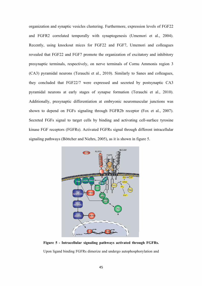
45
organization and synaptic vesicles clustering. Furthermore, expression levels of FGF22
and FGFR2 correlated temporally with synaptogenesis (Umemori et al., 2004).
Recently, using knockout mices for FGF22 and FGF7, Umemori and colleagues
revealed that FGF22 and FGF7 promote the organization of excitatory and inhibitory
presynaptic terminals, respectively, on nerve terminals of Cornu Ammonis region 3
(CA3) pyramidal neurons (Terauchi et al., 2010). Similarly to Sanes and colleagues,
they concluded that FGF22/7 were expressed and secreted by postsynaptic CA3
pyramidal neurons at early stages of synapse formation (Terauchi et al., 2010).
Additionally, presynaptic differentiation at embryonic neuromuscular junctions was
shown to depend on FGFs signaling through FGFR2b receptor (Fox et al., 2007).
Secreted FGFs signal to target cells by binding and activating cell-surface tyrosine
kinase FGF receptors (FGFRs). Activated FGFRs signal through different intracellular
signaling pathways (Böttcher and Niehrs, 2005), as it is shown in figure 5.
Figure 5 - Intracellular signaling pathways activated through FGFRs.
Upon ligand binding FGFRs dimerize and undergo autophosphorylation and
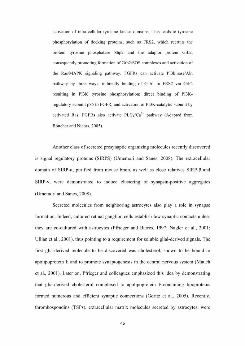
46
activation of intra-cellular tyrosine kinase domains. This leads to tyrosine
phosphorylation of docking proteins, such as FRS2, which recruits the
protein tyrosine phosphatase Shp2 and the adaptor protein Grb2,
consequently promoting formation of Grb2/SOS complexes and activation of
the Ras/MAPK signaling pathway. FGFRs can activate PI3kinase/Akt
pathway by three ways: indirectly binding of Gab1 to FRS2 via Grb2
resulting in PI3K tyrosine phosphorylation; direct binding of PI3K-
regulatory subunit p85 to FGFR; and activation of PI3K-catalytic subunit by
activated Ras. FGFRs also activate PLCγ/Ca2+ pathway (Adapted from
Böttcher and Niehrs, 2005).
Another class of secreted presynaptic organizing molecules recently discovered
is signal regulatory proteins (SIRPS) (Umemori and Sanes, 2008). The extracellular
domain of SIRP-α, purified from mouse brain, as well as close relatives SIRP-β and
SIRP-γ, were demonstrated to induce clustering of synapsin-positive aggregates
(Umemori and Sanes, 2008).
Secreted molecules from neighboring astrocytes also play a role in synapse
formation. Indeed, cultured retinal ganglion cells establish few synaptic contacts unless
they are co-cultured with astrocytes (Pfrieger and Barres, 1997; Nagler et al., 2001;
Ullian et al., 2001), thus pointing to a requirement for soluble glial-derived signals. The
first glia-derived molecule to be discovered was cholesterol, shown to be bound to
apolipoprotein E and to promote synaptogenesis in the central nervous system (Mauch
et al., 2001). Later on, Pfrieger and colleagues emphasized this idea by demonstrating
that glia-derived cholesterol complexed to apolipoprotein E-containing lipoproteins
formed numerous and efficient synaptic connections (Goritz et al., 2005). Recently,
thrombospondins (TSPs), extracellular matrix molecules secreted by astrocytes, were
47
demonstrated to be necessary and sufficient for synapse formation both in vitro and in
vivo (Christopherson et al., 2005). This synaptogenic effect of TSPs seems to be
specific for glutamatergic synapses, since no change is registered when the secreted
molecule is applied to GABAergic synapses (Hughes et al., 2010). Recently, scientists
identified the neuronal thrombospondin receptor involved in CNS synapse formation as
alpha2delta-1, which was shown to be required postsynaptically for thrombospondin-
and astrocyte-induced synapse formation in vivo (Eroglu et al., 2009). Interestingly, a
link between glia-derived factors and adhesion molecules in induction of synapse
formation was revealed by Xu and colleagues (2010), who reported that TSP1 increase
the speed of synapse formation in young hippocampal neurons through interaction with
neuroligin 1. This work wonderfully reveals that presynaptic organizers must work in
synchrony to consummate their duties. Concluding, astrocytes are active participants in
CNS synaptogenesis, and it is now understandable why throughout the CNS the main
phase of synaptogenesis starts instantaneously after glia differentiation.
1.8. Objectives
The objective of this work is to establish the role of axonal protein synthesis in
FGF22-induced presynaptic differentiation, the mechanism by which a functional
presynaptic terminal is formed. To our knowledge, no attemps were made to determine
how inhibition of axonal translation will affect or impair the formation of the
presynaptic terminal.
Neuronal mechanisms like axon guidance, synaptic plasticity, axonal
regeneration and retrograde signaling all require intra-axonal protein synthesis.
Moreover, recent data have shown that synapse formation also relies on proteins
48
axonally synthesized (Sebeo et al., 2009, Schacher and Wu, 2002, Lyles et al., 2006),
but strong evidences are still lacking. The majority of the work published until now
addressed this question by looking into the changes in potentiation of synaptic
transmission in developing synapses by inhibition of protein synthesis (Zhang and Poo,
2002) or by specifically knocking-down an mRNA localized to neurites of Aplysia
sensory neurons (Lyles et al., 2006). However, we currently do not know how local
protein synthesis regulates presynaptogenesis.
In order to accomplish our goal, we first aim at characterizing FGF22
presynaptogenic effect on rat embryo hippocampal neurons. This factor was already
reported to trigger presynaptic differentiation (Umemori et al., 2004; Terauchi et al.,
2010). We will assess synaptic vesicles clustering by immunolabeling cultured neurons
for synapsin, a membrane protein localized to the cytosolic side of SVs. Then, by using
microfluidic chambers and taking advantage of their fluidic properties, we propose to
look into the presynaptogenic effect of FGF22 when applied to axons. Having
established an in vitro model of local induction of presynaptic differentiation by FGF22,
we will then add protein synthesis inhibitors to isolated axons and evaluate the
dependence on axonal protein synthesis of a developing axon during differentiation of
the presynaptic terminal.
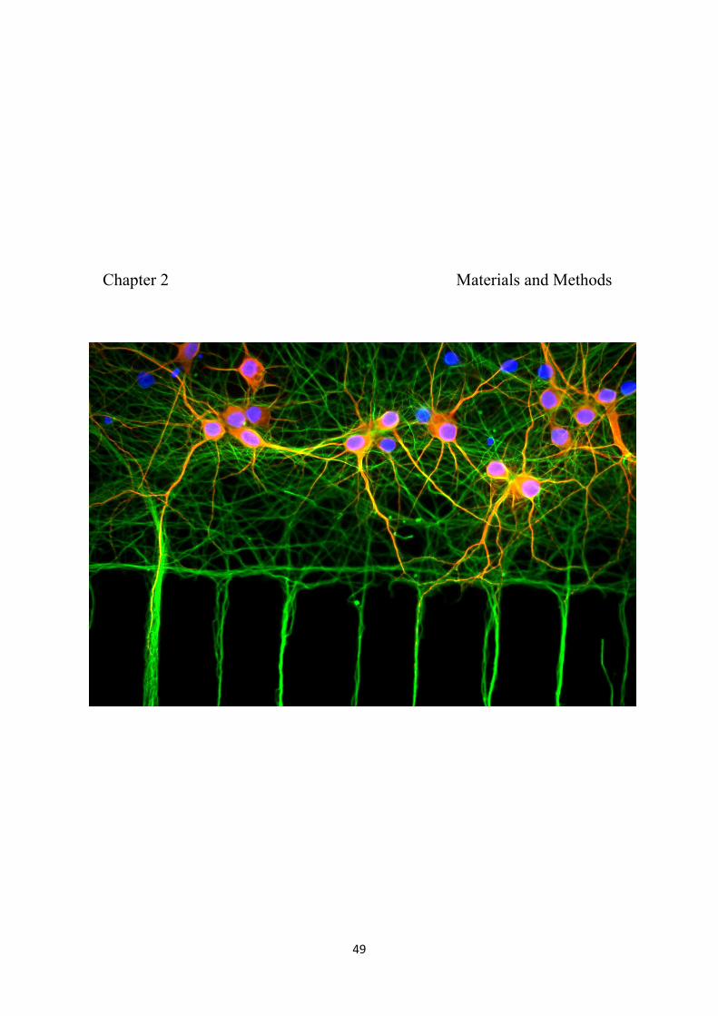
49
Chapter 2 Materials and Methods

50
51
2.1. Reagents
Bovine Serum Albumin (BSA), deoxyribonuclease I from bovine pancreas,
cytosine arabinose (AraC), poly-D-lysine (PDL), Dulbecco’s modified Eagle’s medium
(DMEM), Minimum essential medium eagle (MEM), 5-fluoro-2’-deoxiuridina (5-
FDU), paraformaldehyde, L-glutamic acid, N,N,N’,N’-Tetramethylethylenediamine
(TEMED), ammonium persulfate (APS), the protein synthesis inhibitor emetine
dihydrochloride hydrate and the phosphatases inhibitor sodium orthovanadate were all
purchased from Sigma-Aldrich (Saint Louis, Missouri, USA). MEM-Non essential
amino acids Neurobasal medium, penicillin-streptomycin, B27 supplement, sodium
pyruvate, fetal bovine serum (FBS), trypsin, glutamine and goat serum were purchased
from GIBCOTM Invitrogen Corporation (Carlsbad, California, USA). Mouse laminin I
was obtained from Cultrex®, as part of Trevigen, Inc (Helgerman Court, Gaithersburg,
USA). The protein synthesis inhibitor anisomycin was obtained from Calbiochem®, a
division of Merck KGaA (Darmstadt, Germany). Mounting media ProLong Gold
antifade reagent with DAPI was obtained from Molecular Probes®, as part of
Invitrogen Life Technologies (Eugene, Oregon, USA). Restore western blot stripping
buffer was purchased from Thermo Fisher Scientific (Rockford, Illinois, USA). Sodium
fluoride (NaF) was purchased from Merck (Darmstadt, Germany). Complete protease
inhibitor cocktail tablets was obtained from Roche Applied Science (Carnaxide,
Portugal). Enhanced chemifluorescence (ECF) was obtained from GE Healthcare
(Uppsala, Sweden). Recombinant human fibroblast growth factor-22 (FGF22) was
purchased from R&D Systems, Inc (Minneapolis, USA). Recombinant human brain-
derived neurotrophic factor (BNDF) was from Peprotech (Rocky Hill, New Jersey,
USA). All other reagents of high degree of purity were from Sigma (Saint Louis,
52
Missouri, USA), from Merck (Darmstadt, Germany), Panreac Quimica Sau (Castellar
del Vallès, Barcelona, Spain), Fisher Scientific (Rockford, Illinois, USA) and from Bio-
Rad Laboratories, Inc (Amadora, Portugal).
Stock solutions of FGF22 and BNDF were made in phosphate buffered saline
(PBS) with 0.1% BSA. Stock solution of orthovanadate was made in dimethyl sulfoxide
(DMSO). All other reagents were kept in aqueous stock.
2.1.1. Antibodies
Anti-p44/42 MAPK (Erk1/2) polyclonal antibody (rabbit) (9102) and anti-
phospho-p44/42 MAPK (Erk1/2) (Thr202/Tyr204) monoclonal antibody (rabbit) (4377)
used for western blots were both purchased from Cell Signaling Technology (Danvers,
Massachusetts, USA). The secondary anti-rabbit IgG alkaline phosphatase linked whole
antibody (from goat) (NIF1317) was obtained from GE Healthcare (Uppsala, Sweden).
The antibodies used for immunocytochemistry are listed below. Anti-β-tubulin I
monoclonal antibody (mouse) (T7816) and anti-MAP2 (2a + 2b) monoclonal antibody
(mouse) (M1406) were acquired from Sigma Aldrich (Saint Louis, Missouri, USA).
Anti-synpasin I polyclonal antibody (rabbit) (AB1543) was obtained from Millipore
(Billerica, Massachusetts, USA). Anti-neuronal Class III β-tubulin (TUJ1) monoclonal
antibody (mouse) (MMS-435P) was obtained from Covance (Emeryville, California,
USA). The secondary antibodies used for immunocytochemistry, TexasRed goat anti-
mouse IgG (T862) and AlexaFluor 488 goat anti-rabbit IgG (A-11008) were both
acquired from Molecular Probes®, as part of Invitrogen Life Technologies (Eugene,
Oregon, USA).

53
2.2. Hippocampal neurons
2.2.1. Preparation of microfluidic devices
The microfluidic device for neuron cell culture consists of a molded PDMS (poly-
dimethlysiloxane) chamber placed against a glass coverslip. The glass slides (Corning
No.1 24 mm × 40 mm) were cleaned with nitric acid 65% for 24 h in constant agitation
and then washed 5 times 30 min each with mQ H2O. The glass slides were rinsed twice
in pure ethanol and then placed in the oven at 50oC until they were dry (approximately
15-20 min). Exposure to ultraviolet radiation for 10 min guaranteed the sterilization of
the glass slides, which were then maintained sterile for neuron culture.
The PDMS devices were kindly fabricated and offered by Noo Li Jeon (School of
Mechanical & Aerospace Engineering, Seoul National University, Seoul 151-472,
Korea). The PDMS mold (containing several chambers) was cut in individual pieces
each bearing a single PDMS molded chamber. We punched holes in the place of the
media reservoirs with a sharpened rounded tool and we cleaned each chamber with 3M
Scotch Brand 471 tape to lift off any remaining debris. The chambers were rinsed once
with filtered 75% ethanol. The following steps were carried out in sterile conditions.
The chambers were always constructed in the plating day. Glass slides placed in a
10cm dish were coated with 0.1 mg/ml PDL overnight at 37oC and then washed 3 times
with sterile mQ H2O (1 hour in the last wash). As soon as the glass slides were dry, the
microfluidic chambers were assembled on top of the PDL coated slides, and 2 μg/ml of
laminin in neurobasal plain media was added to the medium reservoirs making sure that
the microgrooves connecting the compartments were totally filled. After an incubation
of at least 2h at 37oC the chambers were washed once with plating media (minimum
54
essential medium eagle (MEM) supplemented with 0.026 M NaHCO3, 3.5 g/L glucose
(to achieve a final concentration of 0.025 M), 1 mM sodium pyruvate and 10% FBS).
2.2.2. Culture of embryonic rat hippocampal neurons
Primary cultures of rat hippocampal neurons were prepared from the hippocampus
of E17-18 Wistar rat embryos. After dissection, hippocampi were treated for 15 min at
37oC with trypsin (0.045%) and deoxyribonuclease (0.01% v/v) in Hank’s balanced salt
solution (HBSS) (5.36 mM KCl, 0.44 mM KH2PO4, 137 mM NaCl, 4.16 mM NaHCO3,
0.34 mM Na2HPO4.2H2O, 5 mM glucose, 1 mM sodium pyruvate, 10 mM HEPES and
0,001% phenol red). After centrifugation for 1 min at 1000 rpm to deposit the
hippocampi, Hank’s solution with trypsin was removed and the hippocampi were
washed with plating medium containing 10% FBS to stop trysin activity. In order to
obtain a homogeneous cell suspension the hippocampi were mechanically dissociated
with a P1000-pipette and then with a Pasteur pipette. Cells were then plated in multi-
wells and microfluidic chambers coated with PDL (0.1 mg/ml) and laminin (2 μg/ml).
In microfluidic chambers, 10 μl of a 4 × 10⁶ cells/ml cell suspension were added to the
somal side (see microfluidic chamber scheme, figure 9). In order to create pseudo-
explants of hippocampal neurons, 50 μl of a 2 × 10⁵ cells/ml cell suspension
(approximately 10000 cells) were plated inside a cloning cylinder (6 mm diameter × 8
mm length) placed in the middle of each coverslip-containing well of a 24 multi-well
plate. For the preparation of hippocampal neurons extracts, neurons were plated at a
density of 3.5 × 10⁵ cells/ml in 12 multi-well plates. After 4 h incubation at 37oC, the
plating medium was removed and replaced for culture medium (Neurobasal medium
supplemented with 2% B27, 25 μM glutamate, 0.5 mM glutamine and 1:400 penicillin-
55
streptomycin). In the microfluidic chambers, the reservoirs of the axonal compartment
were filled with glutamate-free culture medium (Neurobasal medium supplemented
with 2% B27, 0.5 mM glutamine and penicillin-streptomycin), in order to reduce the
exposure to high glutamate concentrations that could induce excitotoxicity in growing
axons.
At DIV 3/4, Ara C or 5-FDU (according to lab availability) was added to the
cultures at a final concentration of 1.825 μg/ml or 10 μM, respectively. The volume of
medium added in each type of culture depended on the medium evaporation and on the
final volume used at DIV0.
To determine the percentage of viable cells in microfluidic devices and in 24
multi-well plates, we counted the number of apoptotic nuclei present in a total of 400-
500 cells per independent experiments from cultures at DIV8-7, respectively.
2.3. Stimulation and total extracts preparation
Total extracts of 293T cells and hippocampal neurons after FGF22 stimulation
were prepared for Western Blot (WB) analysis of Erk1/2 phosphorylation. Total cell
extracts of hippocampal neurons were prepared at DIV8. Cells were stimulated with
increasing doses of FGF22 (as indicated in the results section) for 5 min (293T cells) or
10 min (hippocampal primary cultures). To reduce the basal levels of Erk
phosphorylation, 293T cells were subjected to a starvation period of 3 h in DMEM with
0.5% BSA prior to FGF22 stimulation. Cells were washed once with cold phosphate
buffered saline (PBS) (137 mM NaCl, 2.7 mM KCl, 10 mM Na2HPO4, 1.8 mM
KH2PO4, pH 7.4) and then ice-cold denaturing lysis buffer (0.125 mM Tris pH 6.8, 2%
sodium dodecyl sulfate (SDS), 100 mM dithiothreitol (DTT), 10% glycerol)
56
supplemented immediately before use with proteases inhibitors cocktail and
phosphatases inhibitors (1.5 mM sodium orthovanadate (Na3VO4) and 50 mM sodium
fluoride (NaF)) was added to the cells, which were then scraped. The lysates obtained
were sonicated. Depending on the lysates volume, the sonication was performed in an
ultrasonic bath for 15 min or using an ultrasonic probe with 2 pulses of 15 sec each (30
sec on ice between sonication pulses). Samples were denaturated at 95oC for 5 min and
then left on ice for at least 5 min before electrophoresis.
2.3.1. Electrophoresis and Western Blot
Total cell extracts obtained were electrophoresed in tris-glycine-SDS (TGS)
buffer (25 mM Tris, 192 mM glycine, 0.1% (w/v) SDS, pH 8.3) in a 7.5% (v/v)
acrylamide gel 1.5 mm thick at 120-140 V. Each well was loaded with 20 μl of cell
extract. Electrotransference onto a PVDF (polyvinylidene difluoride) membrane was
performed overnight at 40 V at 4oC. Membranes were washed once with tris-buffered
saline (TBS) (20 mM Tris, 137 mM NaCl) with 0.1% Tween 20 (TBS-T), and then
blocked for 1 h at room temperature in TBS-T with 5% non-fat dry milk. Membranes
were then washed three times (2×5 min and 1×15 min) with TBS-T and then incubated
overnight at 4oC with the primary antibody diluted in TBS-T with 5% BSA. The
following dilutions of primary antibodies were used: monoclonal anti-phospho-p44/42
MAPK (Erk1/2) 1:2000 and polyclonal anti-p44/42 MAPK (Erk1/2) 1:2000 (the first
antibody applied was always for phospho-Erk1/2).
Membranes were then washed with TBS-T three times (2×5 min and 1×15 min)
and incubated for 1 h at room temperature with alkaline phosphatase-conjugated anti-
rabbit secondary antibody (1:20000) diluted in TBS-T with 3% non-fat dry milk. After
57
incubation with the secondary antibody, membranes were washed again with TBS-T
(2×5 min and 1×15 min) and then resolved with ECF substrate for a maximum of 5 min
or until the protein bands were visible. We scanned the membranes on Storm 860 Gel
and Blot Imaging System (Amersham Biosciences, Buckinghamshire, UK).
2.3.2. Stripping and reprobing
In order to reprobe the membranes for total Erk1/2, they were stripped with
Restore western blot stripping buffer for 15 min and then washed several times with
TBS-T. Membranes were blocked for 1 h in TBS-T with 5% non-fat dry milk and then
washed twice briefly to remove the milk. The incubations with the primary and the
secondary antibody (polyclonal anti-p44/42 MAPK (Erk1/2) 1:2000 and alkaline
phosphatase-conjugated anti-rabbit 1:20000, respectively) and the membrane resolution
were performed as described above.
2.4. Stimulation and protein synthesis inhibition
To look into the presynaptogenic effect of FGF22 and BNDF on hippocampal
neurons, primary cultures at DIV6 (pseudo-explants on MW24) and DIV7 (microfluidic
devices) were stimulated for 14h at 37oC in conditioned medium. Recombinant human
FGF22 was used at 10 nM (in MW24) and 20 nM or 100 nM (in microfluidic devices).
Recombinant human BDNF was used at 100ng/ml. In microfluidic devices, these
factors were added only to the axonal compartment. A minute volume difference
between the somal compartment and the axonal compartment (≈25µl) was maintained
during the incubation to prevent the diffusion of the applied factors from the axonal to
58
the somal side. The slightly higher volume on the somal side will cause a slow net flow
of liquid from the somal to the axonal compartment and not the other direction.
When protein synthesis inhibitors were added, we performed 15 min incubation at
37oC prior FGF22 stimulation, so that the drug could enter the cell before FGF22
exposure. FGF22 stimulation was carried in protein synthesis inhibitors containing
medium. The inhibitors used were emetine and anysomicin at 2 μM (MW24) and 10 μM
(microfluidic chambers). In microfluidic chambers, the inhibitors were added to the
axonal compartment thus inhibiting protein synthesis only in axons.
2.4.1. Immunocytochemistry
After 14 h stimulation cells were fixed in 4% paraformaldehyde (in PBS with 4%
sucrose) for 10 min at room temperature. In microfluidic chambers we did a pre-fixation
of 5 min in 1% paraformaldehyde in order to reduce the aggressive effect of
paraformalehyde in fragile axons of the axonal compartment. We washed cultures three
times with tris-buffered saline (TBS) and then permeabilized them using TBS with
0.25% Triton X-100 for 5 min at room temperature. We washed once with TBS and we
blocked non-specific binding with TBS with 5% goat serum and 5% FBS for 30 min at
room temperature. We incubated overnight at 4oC with primary antibodies in TBS with
5% goat serum and 5% fetal bovine serum. We used the following dilutions of primary
antibodies: polyclonal anti-synapsin I 1:2000 in microfluidic devices and 1:4000 in
MW24 coverslips, monoclonal anti-β-tubulin I 1:2000, monoclonal anti-neuronal class
III β-tubulin (TUJ1) 1:1000 and monoclonal anti-MAP2 (2a + 2b) 1:200.
After incubation with the primary antibody, we washed preparations three times, 5
min each, with TBS to remove primary antibody, then incubated them with secondary
59
antibody in TBS with 5% goat serum and 5% FBS for 1 h at room temperature. We
washed preparations twice, 5 min each, with TBS with 0.1% Triton X-100, then one 5-
min wash in TBS, then we rinsed the glass slides with mQ H2O and mounted them in
prolong mounting media with DAPI. All the immunocytochemistry steps were
performed with the microfluidic devices assembled, only after the last wash the PDMS
mold was carefully removed from the coverslip. The preparations were cured overnight
at 4oC protected from light, sealed with nailpolish and kept at 4oC until microscopy
analysis. For all experiments we used Texas Red anti-mouse 1:200 and Alexa Fluor 488
anti-rabbit 1:500.
2.4.2. Fluorescence microscopy and quantification
Fluorescent images were taken using an inverted microscope Zeiss Axiovert 200,
an AxioCam HRm camera and AxioVision 4.8 software. Images were acquired with a
Plan-Neofluar 63× oil objective (numerical aperture 1.3) and exposure times conserved
in single experiments. In microfluidic chambers images were taken on the axonal side,
and to be more accurate we selected regions equally distant from the microgrooves’
entrance (approximately 300-400 μm). In pseudo-explants, images were taken on
regions surrounding the central bulk of neurons, where growing axons could be found
isolated.
The images of the entire microfluidic chamber (both axonal and somal
compartment) were taken using a confocal microscope Zeiss LSM 510 Meta and LSM
510 software. Images were acquired with an EC-PlanNeofluar 40× oil objective
(numerical aperture 1.3) or with a Plan-ApoChromat 20× objective (numerical aperture
0.8).
60
Synapsin clustering was measured with Image J 1.42 software. Axons were
randomly chosen from exported 16-bit images and their lengths determined. It should be
noted that selected axons had similar thickness and appearance, and that fragmented,
bead-bearing axons or terminal regions were rejected. Moreover, this selection was
carried out in β-tubulin images, without observation of synapsin puncta pattern. Images
were thresholded (synapsin and background threshold values conserved in single
experiments) and the number and area of synapsin puncta were quantified. The
background intensity of each image was subtracted. Per condition, 25 or 30 axons were
analyzed in each experiment. Per each selected axon we determined number of synapsin
puncta/μm and total synapsin puncta area/μm. The values obtained per axon were
normalized against the control mean of that single experiment. The number of
experiments and the number of axons analyzed are indicated in the graphs.
2.5. Statistical analysis
Results are presented as normalized means ± SEM of the number of axons
indicated. Graphs and statistical analysis were performed in Graph Pad Prism 5
software. Statistical significance was assessed by unpaired t-test or one-way ANOVA
analysis followed by the Bonferroni´s post test.
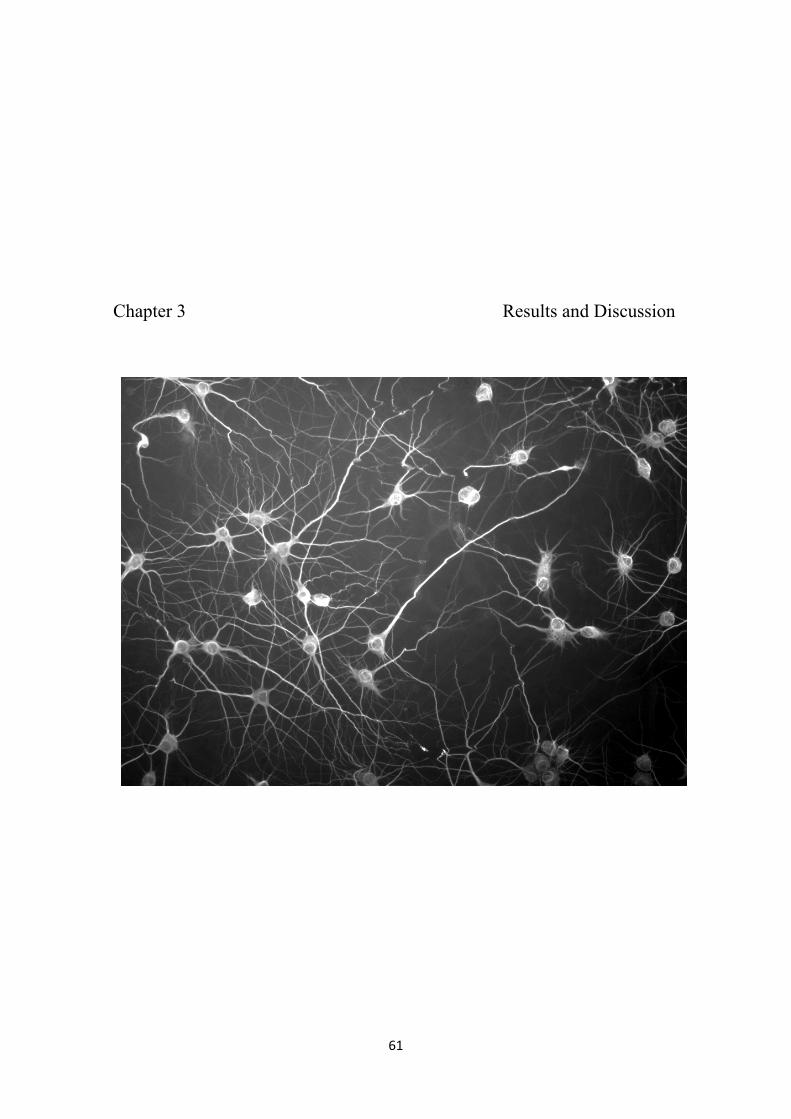
61
Chapter 3 Results and Discussion

62
63
3.1. Induction of presynaptic differentiation by FGF22
3.1.1. FGF22 stimulation of 293T cells and primary cultures of rat
hippocampal neurons induces ERK1/2 phosphorylation
In humans, fibroblast growth factors (FGFs) family comprises twenty-two
secreted proteins, commonly referred as FGF1-14 and FGF16-23, and arranged into
seven subfamilies according to evolutionary distance (Itoh, 2007). The multifunctional
FGF signaling has been demonstrated to be involved in a variety of biological
processes, such as: wound healing (Komi-Kuramochi et al., 2005), bone formation
(Lazarus et al., 2006), perimplantation and early placental function (Zhong et al., 2006),
morphogenesis of embryonic tissues (for example Revest et al., 2001), proliferation of
gland epithelial cells (Steinberg et al., 2004), among many others. FGF signaling has
been suggested to be involved in CNS development and fcuntion, such as, in neural
plate formation, patterning of specific CNS regions (neocortex and spinal cord),
establishment of functional neuro-circuits, and repair of damaged tissues (reviewed in
Dono, 2003). The recently discovered member of the FGF family, FGF22, is involved
in neural differentiation of granule cells and acts as an organizer of presynaptic activity
(Umemori et al., 2004).
FGF receptors (FGFR1-4) display extracellular immunoglobulin-like domains
and cytoplasmic tyrosine kinase domain (Katoh, 2008). FGFR2 has two isoforms,
FGFR2b and FGFR2c, with different affinities for FGFs (Katoh, 2008). FGFR2b is a
high affinity receptor for FGF1, FGF7, FGF10 and FGF22 (Ornitz et al., 2001; Yeh et
al., 2003; Umemori et al., 2004), while FGFR2c for FGF1, FGF2, FGF4, FGF6, FGF9,
FGF16 and FGF20 (Ornitz et al., 1996; Zhang et al., 2006). The transduction of FGF
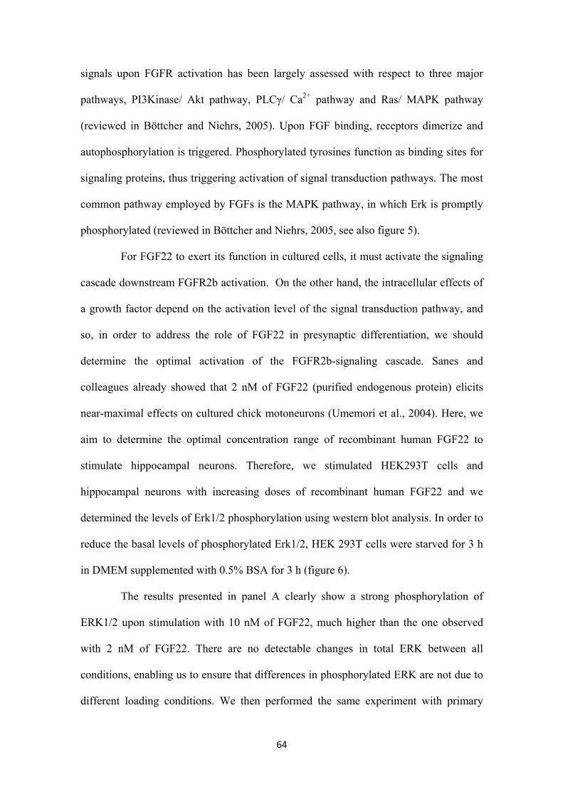
64
signals upon FGFR activation has been largely assessed with respect to three major
pathways, PI3Kinase/ Akt pathway, PLCγ/ Ca2+ pathway and Ras/ MAPK pathway
(reviewed in Böttcher and Niehrs, 2005). Upon FGF binding, receptors dimerize and
autophosphorylation is triggered. Phosphorylated tyrosines function as binding sites for
signaling proteins, thus triggering activation of signal transduction pathways. The most
common pathway employed by FGFs is the MAPK pathway, in which Erk is promptly
phosphorylated (reviewed in Böttcher and Niehrs, 2005, see also figure 5).
For FGF22 to exert its function in cultured cells, it must activate the signaling
cascade downstream FGFR2b activation. On the other hand, the intracellular effects of
a growth factor depend on the activation level of the signal transduction pathway, and
so, in order to address the role of FGF22 in presynaptic differentiation, we should
determine the optimal activation of the FGFR2b-signaling cascade. Sanes and
colleagues already showed that 2 nM of FGF22 (purified endogenous protein) elicits
near-maximal effects on cultured chick motoneurons (Umemori et al., 2004). Here, we
aim to determine the optimal concentration range of recombinant human FGF22 to
stimulate hippocampal neurons. Therefore, we stimulated HEK293T cells and
hippocampal neurons with increasing doses of recombinant human FGF22 and we
determined the levels of Erk1/2 phosphorylation using western blot analysis. In order to
reduce the basal levels of phosphorylated Erk1/2, HEK 293T cells were starved for 3 h
in DMEM supplemented with 0.5% BSA for 3 h (figure 6).
The results presented in panel A clearly show a strong phosphorylation of
ERK1/2 upon stimulation with 10 nM of FGF22, much higher than the one observed
with 2 nM of FGF22. There are no detectable changes in total ERK between all
conditions, enabling us to ensure that differences in phosphorylated ERK are not due to
different loading conditions. We then performed the same experiment with primary
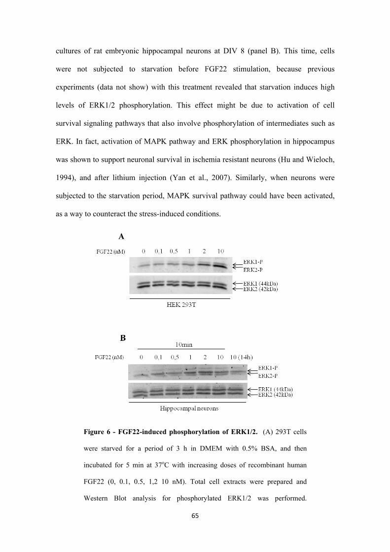
65
cultures of rat embryonic hippocampal neurons at DIV 8 (panel B). This time, cells
were not subjected to starvation before FGF22 stimulation, because previous
experiments (data not show) with this treatment revealed that starvation induces high
levels of ERK1/2 phosphorylation. This effect might be due to activation of cell
survival signaling pathways that also involve phosphorylation of intermediates such as
ERK. In fact, activation of MAPK pathway and ERK phosphorylation in hippocampus
was shown to support neuronal survival in ischemia resistant neurons (Hu and Wieloch,
1994), and after lithium injection (Yan et al., 2007). Similarly, when neurons were
subjected to the starvation period, MAPK survival pathway could have been activated,
as a way to counteract the stress-induced conditions.
Figure 6 - FGF22-induced phosphorylation of ERK1/2. (A) 293T cells
were starved for a period of 3 h in DMEM with 0.5% BSA, and then
incubated for 5 min at 37oC with increasing doses of recombinant human
FGF22 (0, 0.1, 0.5, 1,2 10 nM). Total cell extracts were prepared and
Western Blot analysis for phosphorylated ERK1/2 was performed.
66
Membranes were reprobed for total ERK1/2. (B) Primary cultures of rat
embryonic hippocampal neurons at DIV8 were incubated for 10 min at 37oC
with increasing doses of recombinant human FGF22 (0, 0.1, 0.5, 1,2 10 nM).
To evaluate the long-term effect of FGF22 on ERK1/2 phosphorylation we
incubated neurons at DIV7 for 14 h at 37oC with the higher dose of FGF22
used (10nM). Total cell extracts were prepared and Western Blot analysis for
phosphorylated ERK1/2 was performed. Membranes were reprobed for total
ERK1/2.
The results obtained (figure 6B) show considerable levels of ERK
phosphorylation when primary cultures of rat embryonic hippocampal neurons are
stimulated with 1, 2 and 10 nM of FGF22. The signal obtained with 2 nM stimulation is
slightly stronger than with 10 nM, difference that could probably be abolished if more
experiments were done. Once more, there are no detectable changes in total ERK
between all conditions, enabling us to ensure that differences in phosphorylated ERK
are not due to different loading conditions. Concluding, figure 6B indicates that
maximal activation of FGFR2b-signaling cascade in rat embryonic hippocampal
neurons can be achieved by applying concentrations of recombinant human FGF22
equal or higher than 2 nM. So, taking both biological systems in consideration
(HEK293T cells and hippocampal neurons), we decided 10 nM to be the ideal
concentration to stimulate cultured cells.
3.1.2. Presynaptic differentiation assessed by synapsin clustering
Presynaptic differentiation comprises several events to occur in a coordinated
manner: clustering of synaptic vesicles, establishment of the active zone, post- and pre-
67
synaptic membranes alignment, cytoskeletal restructuring and assembly of vesicle
recycling machinery (Jin and Garner, 2008). In this work, we used synaptic vesicle
clustering (synapsin I immunolabeling) as a marker of presynaptic differentiation.
Synapsins, the most abundant family of neuron-specific synaptic vesicle (SV)-
associated phosphosproteins (Fornasiero et al., 2010), localize to the cytoplasmic
surface of small SVs (figure 7C). Synapsin has long been used as a marker protein of
synaptic vesicles (Thiel et al., 1993; Bixby et al., 1985), and nowadays it turned out to
be a very useful tool to assess synaptic vesicles clustering in the study of presynaptic
differentiation (Hall et al., 2000; Umemori et al., 2004; Fox et al., 2007; Umemori et al.,
2008).
Cultured embryonic hippocampal neurons immunolabeled for synapsin exhibit
a pronounced punctuated pattern (figure 7A). The magnified axonal segment in figure
2B shows that synapsin is arranged in distinct puncta along the axonal shaft, revealing
clustering of synaptic vesicles. In this work, FGF22 effect on presynaptic differentiation
was assessed by measuring synapsin puncta, a direct indicator of synaptic vesicle
assembly and a hallmark of presynaptogenesis.
3.1.3. FGF22 and BNDF induce presynaptic differentiation of rat
embryonic hippocampal neurons
To this date, several membrane and soluble proteins with functional roles in
synaptogenesis have been identified (described in section 1.7.1). Fibroblast growth
factors were recently associated to modulation of synaptogenesis. FGF22 was reported
to induce presynaptic differentiation in cultured chick motoneurons and in cerebellum
cultures (Umemori et al., 2004). Moreover, in vivo presynaptic differentiation of
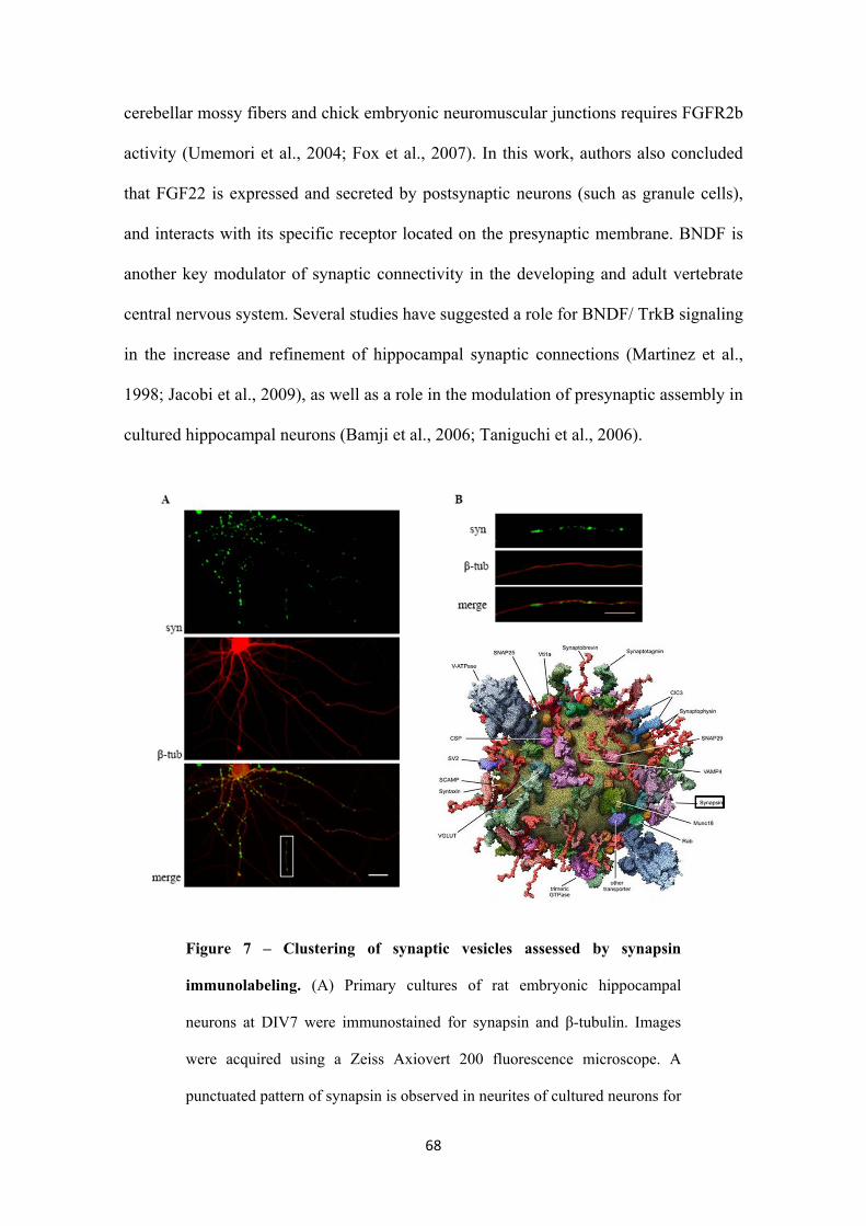
68
cerebellar mossy fibers and chick embryonic neuromuscular junctions requires FGFR2b
activity (Umemori et al., 2004; Fox et al., 2007). In this work, authors also concluded
that FGF22 is expressed and secreted by postsynaptic neurons (such as granule cells),
and interacts with its specific receptor located on the presynaptic membrane. BNDF is
another key modulator of synaptic connectivity in the developing and adult vertebrate
central nervous system. Several studies have suggested a role for BNDF/ TrkB signaling
in the increase and refinement of hippocampal synaptic connections (Martinez et al.,
1998; Jacobi et al., 2009), as well as a role in the modulation of presynaptic assembly in
cultured hippocampal neurons (Bamji et al., 2006; Taniguchi et al., 2006).
Figure 7 – Clustering of synaptic vesicles assessed by synapsin
immunolabeling. (A) Primary cultures of rat embryonic hippocampal
neurons at DIV7 were immunostained for synapsin and β-tubulin. Images
were acquired using a Zeiss Axiovert 200 fluorescence microscope. A
punctuated pattern of synapsin is observed in neurites of cultured neurons for
69
seven days. The scale bar is 25 μm. (B) Magnification of the area defined in
Amerge. Bar is 10 μm. (C) Molecular model of an average synaptic vesicle
(outside view) exhibiting synapsin to the cytosolic side (Takamori et al.,
2006).
The main aim of this work is to elucidate the role of axonal protein synthesis in
presynaptic differentiation. Thus, to pursue our studies, we need to establish a
functional, reliable and reproducible system of induction of presynaptic differentiation
in vitro. Taking into consideration the highly pronounced presynaptogenic effect of
FGF22 reported at both CNS and PNS level, and the fact that FGF22 is a target-derived
soluble molecule, we decided to test the presynaptic organizing effect of this molecule
in embryonic rat hippocampal neurons. Simultaneously, we also decided to stimulate
hippocampal neurons with BDNF, neurotrophin already proven to affect hippocampal
connectivity and synaptic formation, and whose receptor is highly expressed in
hippocampus (Fryer et al., 1996) and present in the surface of hippocampal dendrites
and axons (Yan et al., 1997).
In order to address the possible involvement of FGF22 and BNDF on the
induction of presynaptic differentiation in hippocampus, we prepared primary cultures
of embryonic rat hippocampal neurons in the form of pseudo-explants. Cultures at DIV6
were subjected to a 14 h-stimulation at 37oC with 10 nM of FGF22 or 100 ng/ml of
BNDF in conditioned medium. Cultures were then fixed and immunocytochemistry for
synapsin and β-tubulin was performed. Fluorescence microscope images were taken at
peripheral regions of the pseudo-explants, where extending axons are isolated.
Quantification of number and area of synapsin puncta per axon length was carried out in
Image J software.
70
The results obtained (figure 8) show that both FGF22 and BNDF significantly
increase the number of synapsin puncta per axon length (figure 8B) (141.6%, p<0.001
and 132.7%, p<0.05, respectively), suggesting that these factors augment the number of
synaptic vesicles-accumulating sites along the axon shaft. These sites with synaptic
vesicles clusters represent potential spots for the formation of a functional and mature
presynaptic terminal provided that one presynaptic differentiation event had already
took place. Moreover, FGF22 also increased total synapsin puncta area per axon length
(figure 8C) (123.27%, p<0.05), suggesting that FGF22 enlarges the area occupied by
synapsin-positive puncta, or, in other words, increases the size of synaptic vesicles
clusters. Conversely, BNDF does not increase synapsin puncta area per axon length
(figure 8C) as statistical analysis using unpaired t-test showed no significant differences
between control and BNDF-stimulated condition for total synapsin puncta area/μm.
In line with what has been reported, our results indicate that FGF22 and
BNDF, when globally applied, induce presynaptogenesis in rat embryonic hippocampal
neurons, with a specific role in the organization, distribution and clustering of synaptic
vesicles in presynaptic terminals. The effect of FGF22 seems to be more robust than
BNDF effect (considering that it alters two properties of SV clusters, number and area).
A very recent paper from Umemori and colleagues strongly supports and
validates the results obtained and discussed for FGF22. Using hippocampal cultures of
FGF22-knockout mice, authors observed that lack of FGF22 is characterized by a
decrease in number and size of vesicular glutamate transporter 1 (VGLUT1)-positive
puncta (Terauchi et al., 2010). This phenotype was reversed if FGF22 was subsequently
added to cultures. Concluding, FGF22 induces differentiation of excitatory pre-synaptic
terminals in CA3 pyramidal neurons (Terauchi et al., 2010). These results are in total
agreement with our data (figure 8). However, FGF22 involvement in presynaptogenesis
71
was analyzed in different developmental stages. The authors, claiming that
synaptogenesis peak is at P14, decided to use post-natal mice at P14. In this work, we
used embryonic mice (E17-18), because dependence on axonal protein synthesis
decreases with maturation of the neuronal network. It is known that mature presynaptic
terminals show decreased sensitivity to protein synthesis inhibitors (Sebeo et al., 2009).
Despite these differences, FGF22 induce similar responses, suggesting that hippocampal
neurons conserve the ability to respond equally to extracellular FGF22 from embryonic
state until, at least, 14 days after birth. Considering this, we can predict that in vivo
FGF22 effect on presynaptic differentiation in CNS might occur before and/or after
birth. In cerebellum (Umemori et al., 2004) and hippocampus (Terauchi et al., 2010)
FGF22 and FGFR2 are highly expressed at P8, but decrease at P23 to barely detectable
levels (Umemori et al., 2004). So, FGF22 presynaptogenic effect must be relevant only
in the first weeks after birth. On the other hand, in mice neuromuscular junction FGF22
mRNA and FGFR2 levels are relatively high at E18 and decrease dramatically with
birth (Fox et al., 2007). Summarizing, expression of FGF22 and its receptor seems to be
transient and to correspond temporally to synaptogenesis period. Efforts should be made
to analyze the expression levels of FGF22 and FGFR2 in hippocampus and cerebellum
at embryonic stages.
To sum up, we were successful in establishing an in vitro system of
presynaptogenesis induction by FGF22 in embryonic hippocampal neurons. Our data is
validated by other studies, thus increasing its credibility and reliability.
Interestingly, the results obtained for BDNF are in total agreement with the
working model proposed by Reichardt and colleagues. According to this model,
activation of TrkB receptors localized on the presynaptic membrane culminates in
disruption of the signals that localize SVs to the presynaptic compartment (Bamji et al.,
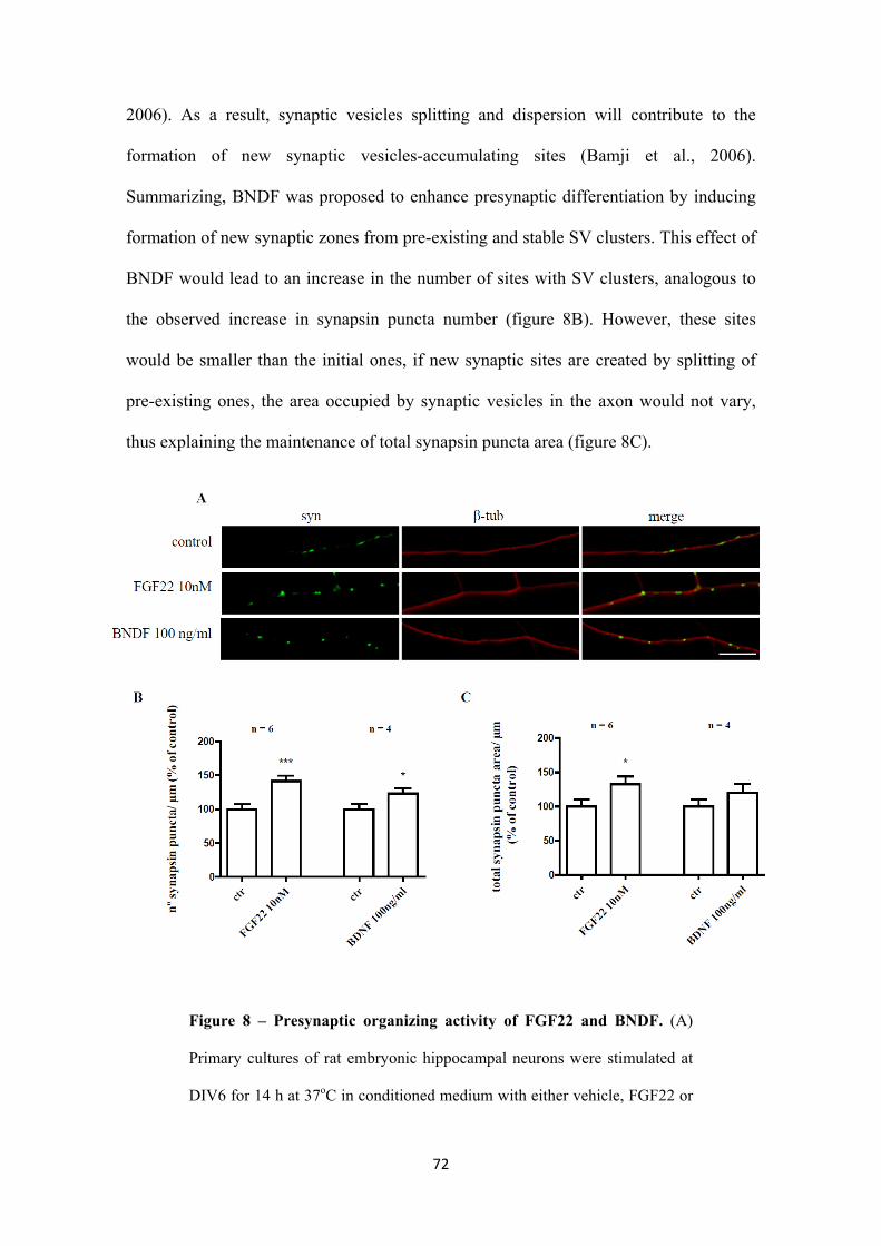
72
2006). As a result, synaptic vesicles splitting and dispersion will contribute to the
formation of new synaptic vesicles-accumulating sites (Bamji et al., 2006).
Summarizing, BNDF was proposed to enhance presynaptic differentiation by inducing
formation of new synaptic zones from pre-existing and stable SV clusters. This effect of
BNDF would lead to an increase in the number of sites with SV clusters, analogous to
the observed increase in synapsin puncta number (figure 8B). However, these sites
would be smaller than the initial ones, if new synaptic sites are created by splitting of
pre-existing ones, the area occupied by synaptic vesicles in the axon would not vary,
thus explaining the maintenance of total synapsin puncta area (figure 8C).
Figure 8 – Presynaptic organizing activity of FGF22 and BNDF. (A)
Primary cultures of rat embryonic hippocampal neurons were stimulated at
DIV6 for 14 h at 37oC in conditioned medium with either vehicle, FGF22 or
73
BNDF. Immunostaining for synapsin and β-tubulin was performed and
images were taken using a Zeiss Axiovert 200 fluorescent microscope.
Representative images of axons segments revealing a clear difference in
synapsin clustering between analyzed conditions. Results show that global
application of recombinant human FGF22 (10nM) or recombinant human
BNDF (100 ng/ml) to hippocampal cultures induces a change in synapsin
clustering/distribution pattern. The scale bar is 10 μm. (B, C) The number of
synapsin puncta/ axon length and the total synapsin puncta area/ axon length
were measured with Image J 1.42 software in randomly selected axons (25-
30 axons per experiment). Values were normalized to the control mean of
each experiment. Bars represent the mean ± SEM of 120-160 axons of at
least four independent experiments (* represents p<0.05 by unpaired t-test
and *** represents p<0.001 by unpaired t-test, when compared with control).
3.2. FGF22-induced presynaptic differentiation and axonal translation
3.2.1. Microfluidic devices for neuron cell culture
In the previous section, we observed that global application of FGF22 induces
an increase in both number and total area of synapsin puncta per axon length. These
results led us to conclude that FGF22 has a presynaptogenic effect when globally
applied to cultured hippocampal neurons. Our next goal is to uncover the effect of
FGF22 when it is locally applied, in other words, when only axons are exposed to the
growth factor. In order to fulfill the need to only expose axons to different stimuli, we
made use of microfluidic chambers, small multicompartment devices capable of
creating physical and fluidical independent compartments.
74
Microfluidics can be defined as the study of flows (liquids or gases) that are
circulating in tiny artificial microsystems (Tabeling, 2005). This area deals with the
behavior, precise control and manipulation of small amounts of fluid (10-9 to 10-8 litres)
that are constrained to sub-millimeter channels and valves (Tabeling, 2005). The
accurate control of fluids dynamics and behavior allows microfluidic systems to
monitor and regulate microenvironments, thereby allowing users to know exactly the
content of individual compartments. In recent years, fluid management in microsystems
has been very useful for cell biologists and neuroscientists, and several devices have
been designed for in vitro cell cultures and biological studies.
The application of microfluidics in neuroscience is recent; nevertheless,
microsystems have been employed in studies of: axon injury and regeneration (Taylor et
al., 2005), localization and identification of mRNAs in axons (Taylor et al., 2005),
synapse-to-nucleus signaling (Taylor et al., 2010), intracellular pH regulation in
neuronal soma and neurites (Vitzhum et al., 2010), and axonal navigation and network
formation (Millet et al., 2010). On the other hand, manipulation of this technology
contributed to the appearance of microdevices specifically designed to visualize and
manipulate synapses (Taylor et al., 2010), to orient growth of neuron processes through
substrate patterning (Taylor et al., 2003), and to integrate networks of neurons with
multielectrode arrays in an attempt to create neuron-based functional biosensors (Morin
et al., 2005). Concluding, microfluidics is giving great contributions to neuroscience. In
this work, we make use of these microfluidic devices to study presynaptic
differentiation and axonal protein synthesis.
In the next sub-sections, I will describe succinctly the type of microfluidic
chambers used, highlighting the special features that render them the ideal tool to
address our goal.
75
3.2.1.1. Growth of embryonic rat hippocampal neurons in microfluidic
chambers
In this work, we determined the effect of FGF22 on the presynaptic
differentiation of rat embryonic hippocampal neurons (see 3.1.3). Since the next
proposed aim is to look into the presynaptogenic effect of FGF22 in pure axonal
populations, it was imperative to optimize long-term primary cultures of rat embryo
hippocampal neurons in microfluidic devices. Up to now, few studies reported
successful culturing of CNS neurons in microfluidic devices. Despite the difficulties,
investigators were able to culture PNS and CNS neurons, such as chick dorsal root
ganglion (DRG) (Park et al., 2009) and rat cortical neurons (Taylor et al., 2003, 2005;
Vitzhum et al., 2010; Rhee et al., 2006; Park et al., 2009), respectively. While this study
was being conducted, two independent studies also managed to culture viable primary
hippocampal neurons in microfluidic chambers (Taylor et al., 2010; Millet et al., 2010).
Each microfluidic device is composed of a molded PDMS piece (figure 9A),
bearing desired surface embossed designs, placed against a properly coated-glass
coverslip where neurons adhere. Poly-dimethylsiloxane (PDMS) has been shown to be
the most suitable material for the fabrication of microsystems in which to grow cells.
PDMS’s suitability for the job can be explained by a huge list of advantages: optical
transparency, high permeability to oxygen and carbon dioxide, low cost and easy
fabrication using standard soft-lithography (simple protocols described in Rhee et al.,
2004 and Park et al., 2009), thermal stability5, physiological and chemical inertness, and
the most important biocompatibility (Wang et al., 2008 and Whitesides, 2006).
5 Thermal stability is defined as the resistance to permanent change in properties caused by heat.
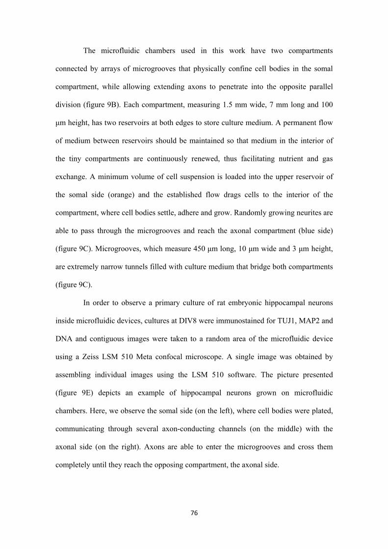
76
The microfluidic chambers used in this work have two compartments
connected by arrays of microgrooves that physically confine cell bodies in the somal
compartment, while allowing extending axons to penetrate into the opposite parallel
division (figure 9B). Each compartment, measuring 1.5 mm wide, 7 mm long and 100
μm height, has two reservoirs at both edges to store culture medium. A permanent flow
of medium between reservoirs should be maintained so that medium in the interior of
the tiny compartments are continuously renewed, thus facilitating nutrient and gas
exchange. A minimum volume of cell suspension is loaded into the upper reservoir of
the somal side (orange) and the established flow drags cells to the interior of the
compartment, where cell bodies settle, adhere and grow. Randomly growing neurites are
able to pass through the microgrooves and reach the axonal compartment (blue side)
(figure 9C). Microgrooves, which measure 450 μm long, 10 μm wide and 3 μm height,
are extremely narrow tunnels filled with culture medium that bridge both compartments
(figure 9C).
In order to observe a primary culture of rat embryonic hippocampal neurons
inside microfluidic devices, cultures at DIV8 were immunostained for TUJ1, MAP2 and
DNA and contiguous images were taken to a random area of the microfluidic device
using a Zeiss LSM 510 Meta confocal microscope. A single image was obtained by
assembling individual images using the LSM 510 software. The picture presented
(figure 9E) depicts an example of hippocampal neurons grown on microfluidic
chambers. Here, we observe the somal side (on the left), where cell bodies were plated,
communicating through several axon-conducting channels (on the middle) with the
axonal side (on the right). Axons are able to enter the microgrooves and cross them
completely until they reach the opposing compartment, the axonal side.
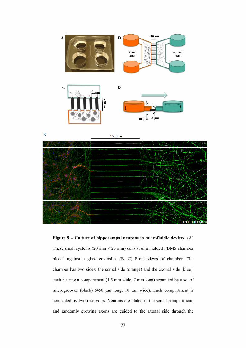
77
Figure 9 – Culture of hippocampal neurons in microfluidic devices. (A)
These small systems (20 mm × 25 mm) consist of a molded PDMS chamber
placed against a glass coverslip. (B, C) Front views of chamber. The
chamber has two sides: the somal side (orange) and the axonal side (blue),
each bearing a compartment (1.5 mm wide, 7 mm long) separated by a set of
microgrooves (black) (450 μm long, 10 μm wide). Each compartment is
connected by two reservoirs. Neurons are plated in the somal compartment,
and randomly growing axons are guided to the axonal side through the
78
microgrooves. (D) Chamber side view: microgrooves’ height (3 μm) is
extremely small comparing to compartments’ height (100 μm). If a volume
difference is maintained (dotted lines), the high fluidic resistance of the
microgrooves guarantees fluidic isolation for 20 h between
microenvironments (Taylor et al., 2005). (E) Primary cultures of
hippocampal neurons at DIV8 were immunostained for TUJ1, MAP2 and
DNA. Contiguous images were taken from a random area of the microfluidic
chamber using a Zeiss LSM 510 confocal microscope with an EC-
PlanNeofluar 40× objective and assembled into a single image using the
LSM 510 software. In this work, compartmentalization and isolation of
axons was achieved by the use of microfluidic chambers.
It is important to emphasize the fact that conditions inside the chambers are not
as appropriate to cell culture as conditions in multi-wells. Firstly, efficient gas exchange
is compromised due to reduced contact between medium and surroundings. Secondly,
the reduced size of the compartments (especially height – 100 μm) partially restricts
fluidity, and so, accumulation of metabolism-derived toxic compounds in the interior of
the compartment, as well as, dead cells, is likely to occur. Considering this, neurons
confined to microfluidic chambers grow in a less pleasant environment, and so they are
more sensitive to external stresses, such as: medium evaporation in the reservoirs,
ineffective coating of the glass slides, temperature variations, substrate roughness,
among others. Therefore, this extremely sensitive type of culture must be highly
controlled and monitored.
Here, we report the well-succeeded growth of hippocampal neurons in
microfluidic chambers. Our results show that neurons grow, develop and establish a
complex and normal network of neurites when culture on microfluidic chambers (figure
79
10A). Moreover, cell viability inside the chambers, assessed by analysis of nuclear
morphology, was shown unaffected when compared to control cultures (figure 10B and
C). Hippocampal neurons were plated in microfluidic devices (40.000 cells in the somal
compartment) and in coverslip-containing multi-wells (10.000 cells inside cloning
cylinders). At DIV8 and DIV7, respectively, cultures were fixed and mounted in
prolong mounting media with DAPI, and the number of apoptotic nuclei was counted.
Our data shows that cells in the devices are morphologically equivalent to cells in the
control culture (figure 10B) with only a slight non-statistically significant decrease in
the percentage of viable cells cultured in the microfluidic chambers (figure 10C). The
adverse conditions in microfluidic chambers do not affect neurons’ viability and
welfare. Moreover, our results show higher levels of cell viability than the pioneer
studies performed with cortical neurons, where the authors only obtained survival rates
around 70% (Taylor et al., 2003).
On the other hand, as it is shown in figure 9E, after the long crossing confined
to the microgrooves’ limits, when axons reach the axonal side they spread in every
direction and cover the whole surface of the axonal compartment. We observed no axon
bundles or bead-bearing axons in the axonal side which reveals that axons are healthy,
stable and in good conditions.
3.2.1.2. Microfluidic chambers: fluidic isolation of pure axonal populations
Microfluidic chambers usefulness to the field of neuroscience is due to two
distinct features: axonal isolation and fluidic isolation. In this section we will explain
succinctly these properties and their relevance to the proposed work.
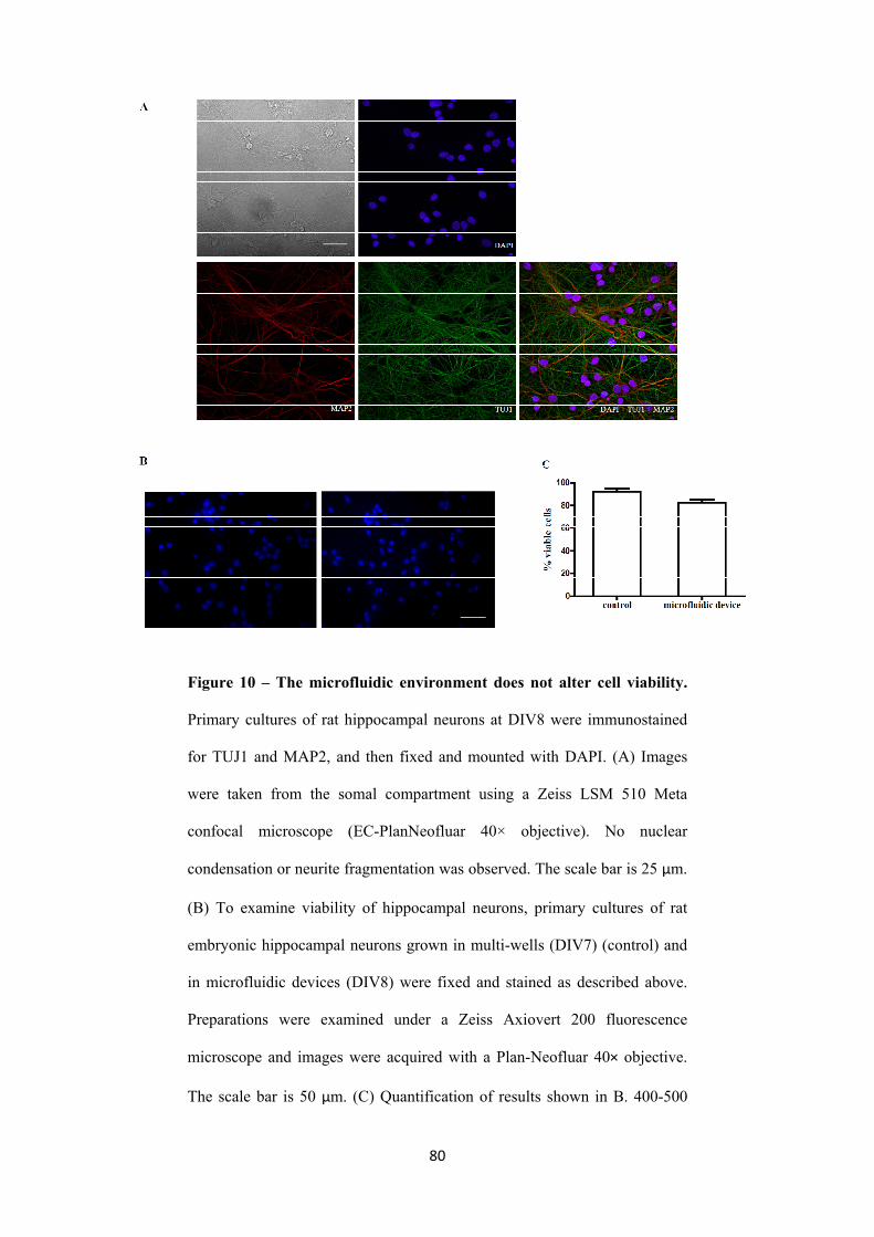
80
Figure 10 – The microfluidic environment does not alter cell viability.
Primary cultures of rat hippocampal neurons at DIV8 were immunostained
for TUJ1 and MAP2, and then fixed and mounted with DAPI. (A) Images
were taken from the somal compartment using a Zeiss LSM 510 Meta
confocal microscope (EC-PlanNeofluar 40× objective). No nuclear
condensation or neurite fragmentation was observed. The scale bar is 25 μm.
(B) To examine viability of hippocampal neurons, primary cultures of rat
embryonic hippocampal neurons grown in multi-wells (DIV7) (control) and
in microfluidic devices (DIV8) were fixed and stained as described above.
Preparations were examined under a Zeiss Axiovert 200 fluorescence
microscope and images were acquired with a Plan-Neofluar 40× objective.
The scale bar is 50 μm. (C) Quantification of results shown in B. 400-500
81
cells were counted per experiment and the percentage of viable cells
determined. Bars represent the mean ± SEM of three independent
experiments. Statistical analysis was preformed with unpaired t-test.
As discussed in the previous section (3.2.1.1), dissociated neurons are plated in
the somal compartment and randomly growing axons arise in the axonal compartment
by traversing narrow microchannels. Neuron cell bodies are known to vary from 4 to
100 μm in diameter and microgrooves height is 3 μm, thus preventing passage of cell
bodies through these microtunnels. As a result, cell bodies get trapped on the somal side
(figure 11A). Importantly, when neurons are plated, axonal reservoirs are totally filled
with medium, in contrast to somal reservoirs, inducing the formation of an
unidirectional flow (from axonal to somal side). This flow counteracts completely
diffusion of suspended components from the somal to the axonal side.
Oppositely, dendrites are as narrower as axons, and they can easily enter the
microgrooves. However, dendrites length is much smaller. Garruto and colleagues
reported that the mean length of longest radiolabeled dendrite at DIV12 of hippocampal
neurons was 168.3 μm (Wakayama et al., 1997). According to this, the presence of
dendrites in a 450μm-distant axonal compartment is surely impossible. Indeed, we can
detect dendrites inside the microgrooves, but they were never detected beyond the
middle of the microgrooves, or more importantly, in the axonal compartment (figure
11B). These results are consistent with observations reported by Jeon and colleagues for
cultures of embryonic rat cortical neurons in microfluidic devices with 450 μm
micropores (Taylor et al., 2005).
On the other hand, fluidic isolation of the axonal microenvironment can be
achieved if a minute volume difference between the two compartments is established
(Taylor et al., 2003 and 2005). The reduced height of the microgrooves (3 μm) creates a
82
high fluidic resistance between compartments (whose height is 100 μm), thus reducing
fluidic flow to minimal levels between microenvironments. Theoretically (see figure
9D), if a compartment has higher volume (dotted line, orange side) than the other
(dotted line, blue side), a small but sustained unidirectional flow of liquids arises, thus
ensuring retention of soluble components that might be present only in the blue side.
Aiming at testing this hypothesis, Texas Red and [35S] methionine were added to the
axonal compartment of a microfluidic device under a volume difference of 25 μl
between compartments. After 20 h there was no trace of these components in the somal
side (Taylor et al., 2005). Considering these experiments, we can stimulate or apply a
particular drug only to the axonal side, thus producing a localized effect.
Concluding, a pure population of functional and living axons without
contamination of somata or dendrites can be obtained inside the axonal compartment of
a microfluidic device. Furthermore, microfluidic chambers allow us to fluidically isolate
axons for up to 20 h, thus enabling localized exposure to insults. In the next sections, we
make great use of this amazing property to induce local stimulation with FGF22 and
local inhibition of protein synthesis.
3.2.2. Local FGF22-presynaptogenic effect is specific
Before, we have shown that FGF22 and BNDF, when globally applied, induce
presynaptic differentiation in hippocampal neurons. Both factors were capable of
inducing changes in synaptic vesicles clustering pattern (figure 8). These results lead us
to ask if FGF22 and BNDF presynaptogenic effect occurs locally. So, are these soluble
molecules able to induce presynaptic differentiation when applied only to axons? In
fact, FGF22 was proven to induce presynaptic differentiation in fluidically isolated
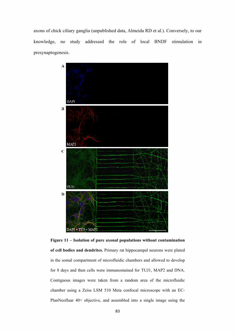
83
axons of chick ciliary ganglia (unpublished data, Almeida RD et al.). Conversely, to our
knowledge, no study addressed the role of local BNDF stimulation in
presynaptogenesis.
Figure 11 – Isolation of pure axonal populations without contamination
of cell bodies and dendrites. Primary rat hippocampal neurons were plated
in the somal compartment of microfluidic chambers and allowed to develop
for 8 days and then cells were immunostained for TUJ1, MAP2 and DNA.
Contiguous images were taken from a random area of the microfluidic
chamber using a Zeiss LSM 510 Meta confocal microscope with an EC-
PlanNeofluar 40× objective, and assembled into a single image using the
84
LSM 510 software. Cell bodies are unable to pass through the narrow
microgrooves, and become trapped on the somal side. Dendrites are able to
enter the microgrooves, but they are not long enough to reach the axonal
side. As a result, only axons are able to cross to the opposite compartment
(the axonal compartment) making these microfluidic devices an essential
tool to compartmentalize and isolate axons in vitro. The scale bar is 200 μm.
In order to clarify this question, we prepared primary cultures of rat embryonic
hippocampal neurons in microfluidic chambers. At DIV7 neurons were stimulated with
either FGF22 or BNDF in conditioned medium. A volume difference of 25 μl was kept
between compartments to guarantee total fluidic isolation. Cultures were then fixed and
immunocytochemistry was performed.
The results obtained clearly show an increase in number (288%, p<0,001)
(figure 12B) and total area of synapsin puncta (328%, p<0,001) (figure 12C) per axon
length when 100 nM of FGF22 is applied to the axonal compartment of microfluidic
devices. However, stimulation of hippocampal neuron axons with BNDF 100 ng/ml or
FGF22 20 nM doesn’t alter synapsin clustering, neither number of synpasin puncta/μm
or total puncta area/μm showed significant differences when compared to control
conditions, indicating that FGF22, but not BNDF, is able to induce presyaptogenesis
when locally applied.
It should be noted that FGF22-induced presynaptic differentiation was obtained
at a concentration of 100 nM, higher than what we previously reported to induce
presynaptic differentiation when globally applied (figure 8).
An adequate explanation for this discrepancy lies on PDMS properties.
Succinctly, PDMS, which is the main component of microfluidic chambers, is a
polymer consisting of repeating -OSi(CH3)2- units. The huge number of CH3 groups
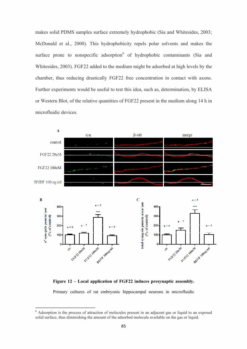
85
makes solid PDMS samples surface extremely hydrophobic (Sia and Whitesides, 2003;
McDonald et al., 2000). This hydrophobicity repels polar solvents and makes the
surface prone to nonspecific adsorption6 of hydrophobic contaminants (Sia and
Whitesides, 2003). FGF22 added to the medium might be adsorbed at high levels by the
chamber, thus reducing drastically FGF22 free concentration in contact with axons.
Further experiments would be useful to test this idea, such as, determination, by ELISA
or Western Blot, of the relative quantities of FGF22 present in the medium along 14 h in
microfluidic devices.
Figure 12 – Local application of FGF22 induces presynaptic assembly.
Primary cultures of rat embryonic hippocampal neurons in microfluidic
6 Adsorption is the process of attraction of molecules present in an adjacent gas or liquid to an exposed solid surface, thus diminishing the amount of the adsorbed molecule available on the gas or liquid.
86
devices were stimulated at DIV7 for 14 h at 37oC in conditioned medium
with either vehicle, FGF22 (20 nM and 100 nM) or BNDF (100 ng/ml).
Stimuli were added to the axonal compartment only. Immunostaining for
synapsin and β-tubulin was performed and images were taken from the
axonal compartment using a Zeiss Axiovert 200 fluorescent microscope. (A)
Representative images of axons segments revealing a clear difference in
synapsin clustering when conditioned medium of the axonal compartment is
supplemented with 100 nM of FGF22 for 14 h. Results show that local
application of recombinant human FGF22 to hippocampal cultures, as
opposed to local application of recombinant human BNDF, induces a
pronounced increase in synapsin clustering. The scale bar is 10 μm. (B,C)
The number of synapsin puncta/axon length and total synpasin puncta
area/axon length were measured with Image J 1.42 software in randomly
selected axons (30 axons per experiment). Values were normalized to the
control mean of each experiment. Bars represent the mean ± SEM of 90-200
axons of at least three independent experiments (*** represents p<0.001 by
ANOVA using Bonferroni’s post test when compared to control).
On the other hand, based on the working model proposed by Umemori and
colleagues, FGF22 is a post-synaptic derived soluble molecule and so secreted locally
upon synaptic contact (Terauchi et al., 2010). As a result of this local secretion,
concentration of FGF22 must be elevated on the areas surrounding the presynaptic
terminal and gradually smaller in remote places. According to this, local stimulation
might require extremely high concentrations of FGF22, thus partially justifying the need
of 100 nM of FGF22 to induce presynaptogenesis in fluidically isolated axons. It would
be interesting to look into FGFR2b activation by FGF22 in pure axonal populations and
determine the concentration of FGF22 that would lead to optimal activation of FGFR2b,
87
although this would be a very challenging experiment due to the small amounts of
protein that can be obtained from the axonal compartment.
Unlike FGF22 our results show that local stimulation with BNDF does not
induce presynaptic differentiation, enabling us to reason that BNDF contact with axons
is not sufficient to trigger remodeling of synaptic vesicles clusters (figure 12).
To sum up, when applied specifically to axons FGF22 induces differentiation
of the presynaptic terminal as determined by clustering of synaptic vesicles, a hallmark
of presynaptogenesis. Moreover, this localized presynaptogenic effect of FGF22 is
specific because when axons are stimulated with BNDF no increase in synapsin puncta
number is observed. We predict that this different local responsiveness to FGF22 and
BNDF results from differential localization and distribution of the corresponding
receptor. To date, no efforts have been made to determine the localization of FGFR2b
receptors in neurons, a question that could be addressed by ultrastructural studies of the
synapse.
3.2.3. FGF22-induced presynaptic differentiation is dependent on axonal
protein synthesis
We next decided to study the role of intra axonal translation in the formation of
synapses, particularly the differentiation of the presynaptic terminal. Preliminary
evidences suggest that local translation in axons might be a fundamental mechanism in
synaptogenesis (Schacher and Wu, 2002; Zhang and Poo, 2002; Lyles et al., 2006;
Sebeo et al., 2009). So, considering that FGF22 is capable of inducing presynaptic
assembly when locally applied to axons of hippocampal neurons (figure 12), we next
asked if this local FGF22-induced presynaptic differentiation depends on axonal protein
88
synthesis. In fact, local protein synthesis was shown to be required for FGF22-induced
presynaptic assembly in chick cilliary ganglia (unpublished data, Almeida RD et al.).
To this purpose, we prepared primary cultures of rat embryonic hippocampal
neurons in microfluidic devices. The axonal compartment of cultures at DIV7 was
subjected to a 14 h stimulation with 100 nM of FGF22 in the absence or presence of
protein synthesis inhibitors (emetine or anisomycin, 10 μM). A pre-incubation of 15
min with the inhibitors was performed, so that the drug could enter the cells prior to
FGF22 sitmulation. Total fluidic isolation was guaranteed by a volume difference of 25
μl between compartments. Cultures were then fixed and immunocytochemistry for
synapsin and β-tubulin was performed. Images were taken from the axonal
compartment and quantification of number and area of synapsin puncta per axon length
was carried out in Image J software.
The results obtained show that when protein synthesis inhibitors are present in
the axonal compartment at the time of FGF22 stimulation, presynaptic differentiation is
abolished (figure 13). Emetine decreased synapsin puncta number and area to 84.95%
(p<0.001) and 73.92% (p<0.001) respectively, and anisomycin decreased synapsin
puncta number and area to 107% (0<0.01) and 89.98% (p<0.001) respectively. These
inhibitors exert their action through different mechanisms, while emetine blocks protein
synthesis by binding to the 40S ribosomal subunit, anisomycin binds to the 60S subunit,
indicating that the loss FGF22-induced puncta is protein synthesis-dependent and not
due to a non-specific effect of the pharmacological inhibitor. It is also important to
notice that 14h-incubation with the inhibitors does not alter presynaptic assembly
pattern (neither number nor area of synapsin puncta). Thus, guaranteeing that the
observed effect is entirely due to abrogation of FGF22-induced synapsin clustering.
Therefore, we can conclude that FGF22-induced presynaptic assembly requires axonal

89
translation, indicating that local protein translation can regulate the formation of new
synapses.
Figure 13 – Intra-axonal protein synthesis is required for FGF22-
induced presynaptic differentiation. (A) Primary cultures of rat embryonic
hippocampal neurons in microfluidic devices were allowed to develop for 7
days, then emetine or anisomycin were added to the axonal compartment for
15 min. Axons located in the axonal compartment were then stimulated with
100 nM of FGF22 in protein synthesis inhibitor-containing medium for 14 h
at 37oC. Immunostaining for synapsin and β-tubulin was performed and
images were taken to the axonal compartment using a Zeiss Axiovert 200
90
fluorescent microscope. Representative images of axons segments revealing
a clear reduction in synapsin clustering when protein synthesis inhibitors are
added. Results show that local inhibition of protein synthesis with emetine or
anisomycin abolishes FGF22-induced synapsin clustering. The scale bar is
10 μm. (B, C) The number of synapsin puncta/axon length and total synpasin
puncta area/axon length were measured with Image J 1.42 software in
randomly selected axons (30 axons per experiment). Values were normalized
to the control mean of each experiment. Bars represent the mean ± SEM of
120 axons of four independent experiments (*** represents p<0.001 by
ANOVA using Bonferroni´s post test when compared to control; ##
represents p<0.01 and ### represents p<0.001 by ANOVA using Bonferroni´s
post test when compared to FGF22 100nM).
If FGF22 induces synapsin clustering in a local protein synthesis dependent
manner, then it depends on the presence of local mRNAs to fulfill its presynaptogenic
effect. Thus, we may wonder if FGF22 is capable of redistributing mRNAs, promoting
their recruitment to synaptic sites, and their translation in a spatial and temporal
regulated manner. Indeed, using Aplysia sensory neurons it was reported that a specific
mRNA (sensorin) immediately localizes to sites of contact between pre- and
postsynaptic neurons, in other words, to sites of newly forming synapses (Lyles et al.,
2006). Moreover, knockdown of sensorin mRNA abolishes synapse formation (Lyles et
al., 2006). According to this study and the results shown here, it is reasonable to assume
that, upon synaptic contact, specific targeting of mRNAs to presynaptic sites could be
promoted by target derived presynaptogenic molecules (such as FGF22, WNT-7a,
neuroligin and SynCAM).
91
Interestingly, Benson and colleagues showed that rat hippocampal neurons
require local protein synthesis to maintain the pool of synaptic vesicles available for
fusion, thus regulating presynaptic function (Sebeo et al., 2009). However, it is not clear
if the protocol used by the authors to block axonal protein synthesis is only specific to
axons and does not inhibit somal protein synthesis, compromising the entire study. To
our knowledge, this study is the first work to report an indispensable role for intra-
axonal translation in the differentiation of the presynaptic terminal in a mammalian
system. Furthermore, this study represents the first attempt to establish a link between
the activity of presynaptogenic molecules and protein synthesis occurring at the
axoplasm.
In order to further extend our findings it would be interesting to show, using a
puromycin derivative assay, that FGF22 induces local protein synthesis in live neurons.
Puromycin, a common translation reporter, is covalently bound to a fluorescence group
that shows no signal in this form. When protein translation is initiated, puromycin
inserts itself into the nascent peptide and releases its fluorescence group. Consequently,
an increase in fluorescence intensity correlates to an increase in protein translation. We
would expect to observe an increase in fluorescence intensity when FGF22 is added to
fluidically isolated axons.

92
93
Chapter 4 Closing Remarks

94
95
4.1. Conclusion
In this study, using fluidically isolated axons, we have examined the
requirement of axonal protein synthesis in FGF22-induced presynaptic differentiation of
hippocampal neurons in culture. We found that (1) FGF22 induces activation of
FGFR2b signaling cascade in embryonic rat hippocampal neurons and, consequently,
phosphorylation of ERK1/2 (figure 1); (2) FGF22 and BNDF induce differentiation of
the presynaptic terminal (figure 3); (3) FGF22 has a localized presynaptogenic effect
(figure 7); and (4) FGF22-induced presynaptic assembly is dependent on axonal protein
synthesis (figure 8).
Taken together, our data suggest that the spatially directed action of FGF22 on
presynaptic assembly is dependent on axonal translation. This dependence was also
previously observed in chick ciliary ganglia (unpublished data, Almeida RD et al.) thus
revealing a conserved mechanism of action of FGF22 between different species (rat and
chicken) and between different types of neurons (hippocampal neurons (CNS) and
ciliary ganglia (PNS)).
Firstly, this work aimed at characterizing FGF22 effect on the differentiation of
presynaptic terminals in cultured hippocampal neurons. For some time, there is
evidence that FGFs play a role in the formation of the presynaptic terminal, particularly
in synaptic vesicles clustering (Dai and Peng, 1995). Application of exogenous FGFs,
FGF7, FGF10 and FGF22 promote vesicle clustering and neurite branching (Umemori
et al., 2004). Moreover, FGF22-knockout mice have impaired differentiation of
excitatory nerve terminals (Terauchi et al., 2010). The present study demonstrates that
FGF22 acts locally to induce presynaptic differentiation. Upon contact to nascent
presynaptic sites, FGF22 acts as a localized neuromodulator capable of inducing
96
synaptic vesicles assembly. This is consistent with findings from Sanes and Umemori,
who showed that FGF22 is secreted by the postsynaptic terminal, thus promoting a
spatially restricted effect on closely located nascent presynaptic sites (Umemori et al.,
2004; Terauchi et al., 2010). In order to reinforce the idea of a localized presynaptic
action, it would be interest to determine the exact localization of FGFR2b receptors in
neurons. In a similar way, potentiation of neurotransmitter secretion at developing
synapses by BNDF was shown to occur in a spatially restricted manner (Zhang and Poo,
2002).
Secondly, we aimed to determine the dependence of FGF22-induced
presynaptogenesis on proteins synthesized locally. Neurons are highly complex cells,
capable of extending axons to remote places, frequently far away from the cell body.
Developing axons undergo long-range pathfinding to seek for a synaptic target, and,
upon initial contact, morphological and functional remodeling of terminals guarantees
the perfect establishment of synaptic connections. Formation of the neuronal network is
a highly demanding period in terms of requirement of newly synthesized components.
Indeed, during development, protein synthesis levels peak during synaptogenesis
(Phillips et al., 1990), suggesting that immature and newly-forming synapses have a
particular high demand for new proteins. The required set of newly synthesized proteins
could be generated at the cell body and then translocated to nascent synaptic sites, or,
alternatively, synthesized locally along the axon shaft. In these last years, accumulating
evidences support a role for axonal protein synthesis in synaptogenesis (Schacher and
Wu, 2002; Lyles et al., 2006; Zhang and Poo, 2002; Boylan et al., 2008; Sebeo et al.,
2009 – for a detailed description see introduction, section 1.5.1.2.). Our work
strengthens this emerging hypothesis. Here, we demonstrate that FGF22-induced
presynaptogenesis relies on axonally synthesized proteins. We can speculate that
97
extracellular presynaptogenic factors, by interacting with membrane receptors, activate
specific intracellular signaling pathways that culminate in modulation of local
translation initiation. In fact, it was already reported that activation of guidance cues
receptors leads to eIF-4E and eIF4EBP phosphorylation (Campbell and Holt, 2003;
Piper et al., 2006), known to de-repress local translation, and so, contribute with newly-
synthesized proteins needed for the structural alterations inherent to the induced growth
cone response.
Based on the current knowledge and on the new findings presented here, an
interesting working model could be formulated where the postsynaptic cell secrets
FGF22, which in turn interacts with FGFR2b, located at the presynaptic membrane,
activating downstream signaling pathways that will probably induce translation of
localized mRNAs. We may speculate that activation of FGFRs induces de-repression of
local translation and/or translocation of specific mRNAs, present at RNA granules, at
the nascent presynaptic site. Newly-synthesized proteins will then promote the
clustering of freely-moving synaptic vesicles at these specific sites along the axon shaft
leading to presynaptic assembly (figure 14). Future work will be necessary to elucidate
how FGF22 signaling leads to the translation of specific mRNAs necessary for the
assembly of the presynaptic terminal.
4.2. Future perspectives
In this work, we investigated the role of local protein synthesis in synapse
formation using fluidically isolated axons. We assessed synaptic vesicles clustering by
immunolabeling primary cultures of rat embryonic hippocampal neurons for synapsin,
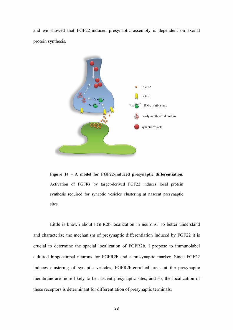
98
and we showed that FGF22-induced presynaptic assembly is dependent on axonal
protein synthesis.
Figure 14 – A model for FGF22-induced presynaptic differentiation.
Activation of FGFRs by target-derived FGF22 induces local protein
synthesis required for synaptic vesicles clustering at nascent presynaptic
sites.
Little is known about FGFR2b localization in neurons. To better understand
and characterize the mechanism of presynaptic differentiation induced by FGF22 it is
crucial to determine the spacial localization of FGFR2b. I propose to immunolabel
cultured hippocampal neurons for FGFR2b and a presynaptic marker. Since FGF22
induces clustering of synaptic vesicles, FGFR2b-enriched areas at the presynaptic
membrane are more likely to be nascent presynaptic sites, and so, the localization of
these receptors is determinant for differentiation of presynaptic terminals.
99
Protein synthesis, both in dendrites and axons, is known to be tightly regulated.
It has been shown that external stimuli activate intracellular cascades that de-repress
translation of dormant mRNAs (Campbell and Holt, 2003; Piper et al., 2006). Similarly,
FGF22/FGFR2b signaling must modulate local protein synthesis. It would be interesting
to unravel the intracellular signaling pathways activated by FGF22 that, in turn, activate
translation pathways and/or machinery. We propose to use specific inhibitors for
intermediates of the intracellular cascades known to be activated by FGFR2b (figure 5),
and evaluate their requirement for FGF22-induced presynaptic differentiation and for
FGF22-induced intra axonal protein synthesis.
Having characterized the requirement of local protein synthesis on FGF22-
induced presynaptic differentiation, it would be interesting to find out which mRNAs
are translated upon this stimulus. This information would give us important clues to
understand the mechanism of action of FGF22 and the mechanisms leading to
presynaptogenesis. To achieve this, I propose to purify distal axons and growth cones of
rat embryonic hippocampal neurons, subjected or not to FGF22 stimulation, and screen
for mRNAs using microarray technology. Differences in mRNAs expression between
conditions will indicate which mRNAs are locally regulated, and possibly translated,
upon FGFR2b activation by FGF22.
If we succeed on identifying mRNAs whose axonal expression is upregulated
upon FGF22 stimulation, several questions may then be raised: how are these mRNAs
related to presynaptogenesis? Is their local translation required for FGF22-induced
presynaptic terminal formation? These questions can then be answered by knocking
down specific mRNAs located in axons by adding siRNAs to the axonal compartment
of microfluidic devices, determining how and when presynaptogenesis is affected.
100
Further understanding the signaling pathways and mechanism of FGF22-
induced presynaptogenesis, and the exact role of axonal protein synthesis in synapse
formation may prove relevant to better understand the formation and establishment of
neuronal connections in the central nervous system, both in neurodevelopmental and
pathophysiological conditions.
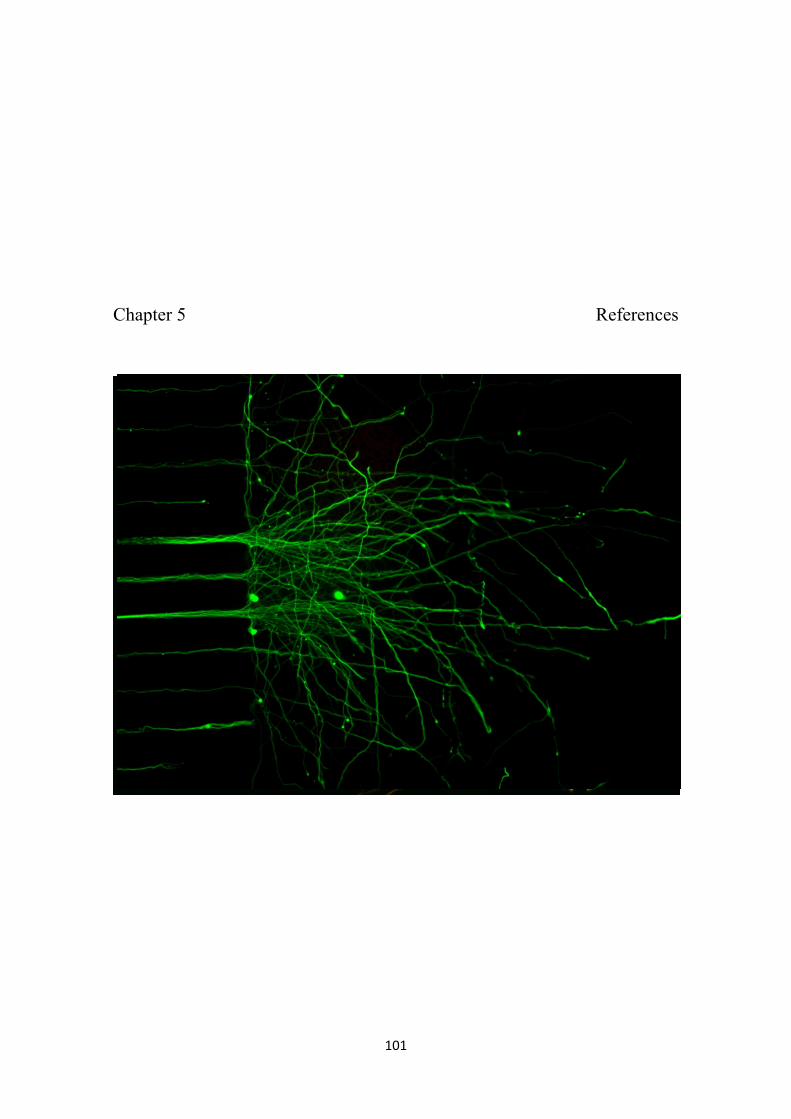
101
Chapter 5 References

102
103
Ahmari S.E., Buchanan J. & S.J. Smith (2000). Assembly of presynaptic active zones
from cytoplasmic transport packets. Nature Neuroscience 3: 445-451.
Alsina B., Vu T. & S. Cohen-Cory (2001). Visualizing synapse formation in arborizing
optic axons in vivo: dynamics and modulation by BDNF. Nature Neuroscience
4: 1093-1101.
Alvarez J. & W.Y. Chen (1972). Injection of leucine into a myelinic axon: incorporation
in the axoplasm and transfer to associated cells. Acta Physiologica Latino
Americana 22: 266–269.
Alvarez J., Giuditta A. & E. Koenig (2000). Protein synthesis in axons and terminals:
significance for maintenance, plasticity and regulation of phenotype - With a
critique of slow transport theory. Progress in Neurobiology 62: 1-62.
Antar L.N., Li C., Zhang H., Carroll R.C. & G.J. Bassell (2006). Local functions for
FMRP in axon growth cone motility and activitydependent regulation of
filopodia and spine synapses. Molecular Cellular Neuroscience 32: 37–48.
Aronov S., Aranda G., Behar L. & I. Ginzburg (2002). Visualization of translated tau
protein in the axons of neuronal P19 cells and characterization of tau RNP
granules. Journal of Cell Science 115: 3817-3827.
Bamji S.X., Rico B., Kimes N. & L.F. Reichardt (2006). BDNF mobilizes synaptic
vesicles and enhances synapse formation by disrupting cadherin–β-catenin
interactions. The Journal of Cell Biology 174: 289–299.
Barrett L. & J. Eberwine (2008). RNA targeting, trafficking and translation in dendrites
– Contribution to synaptic tagging and plasticity, in Dendrites, (Stuart, G.,
Spruston, N. and Häusser, M., eds), pp. 117-130. Oxford University Press,
Oxford.
104
Bassell G.J., Zhang H., Byrd A.L., Femino A.M., Singer R.H., Taneja K.L., Lifshitz
L.M., Herman I.M. & K.S. Kosik (1998). Sorting of b-Actin mRNA and Protein
to Neurites and Growth Cones in Culture. The Journal of Neuroscience 18: 251–
265.
Beaumont V., Zhong N., Fletcher R., Froemke R.C. & R.S. Zucker (2001).
Phosphorylation and Local Presynaptic Protein Synthesis in Calcium- and
Calcineurin-Dependent Induction of Crayfish Long-Term Facilitation. Neuron
32: 489–501.
Behar L., Marx R., Sadot E., Barg J. & I. Ginzburg (1995). Cis-acting signals and trans-
acting proteins are involved in tau mRNA targeting into neurites of
differentiating neuronal cells. International Journal of Developmental
Neuroscience 13: 113-127.
Biederer T., Sara Y., Mozhayeva M., Atasoy D., Liu X., Kavalali E.T. & T.C. Sudhöf
(2002). SynCAM, a synaptic adhesion molecule that drives synapse assembly.
Science 297: 1525–1531.
Bixby J.L. & L.F. Reichardt (1985). The expression and localization of synaptic vesicle
antigens at neuromuscular junctions in vitro. The Journal of Neuroscience 5:
3070-3080.
Black M.M. & R.J. Lasek (1977). The presence of transfer RNA in the axoplasm of the
squid giant axon. Journal of Neurobiology 8: 229–237.
Bliss T.V. & G.L. Collingridge (1993). A synaptic model of memory: long-term
potentiation in the hippocampus. Nature 361: 31-39.
Blundon J.A. & S.S. Zakharenko (2008). Dissecting the Components of Long-Term
Potentiation. Neuroscientist 14: 598–608.
105
Böttcher R.T. & C. Niehrs (2005). Fibroblast Growth Factor Signaling during Early
Vertebrate Development. Endocrine Reviews 26: 63–77.
Boylan K.L.M., Mische S., Li M., Marqués G., Morin X., Chia W. & T.S. Hays (2008).
Motility screen identifies Drosophila IGF-II mRNA-binding protein—zipcode-
binding protein acting in oogenesis and synaptogenesis. PLoS Genetics 4: 1-13.
Bramham C.R. (2008). Local protein synthesis, actin dynamics, and LTP consolidation.
Current Opinion in Neurobiology 18: 524–531.
Bramham C.R. & Wells D.G. (2007). Dendritic mRNA: transport, translation and
function. Nature Reviews 8: 776-789.
Brittis P.A., Lu Q. & J.G. Flanagan (2002). Axonal protein synthesis provides a
mechanism for localized regulation at an intermediate target. Cell 110: 223–235.
Brunet I., Weinl C., Piper M., Trembleau A., Volovitch M., Harris W., Prochiantz1 A.
& C. Holt (2005). The transcription factor Engrailed-2 guides retinal axons.
Nature 438: 94-98.
Burgin K.E., Waxham M.N., Rickling S., Westgate S.A., Mobley W.C. & P.T. Kelly
(1990). In situ hybridization histochemistry of Ca2+/calmodulin-dependent
protein kinase in developing rat brain. The Journal of Neuroscience 10: 1788–
1798.
Burry, R. (1985). Protein synthesis requirement for the formation of synaptic elements.
Brain Research. 344: 109–119.
Calakos N., Schoch S., Sudhof T.C. & R.C. Malenka (2004). Multiple roles for the
active zone protein RIM1alpha in late stages of neurotransmitter release. Neuron
42: 889-896.
106
Campbell D.S. & C.E. Holt (2001). Chemotropic responses of retinal growth cones
mediated by rapid local protein synthesis and degradation. Neuron 32: 1013–
1026.
Campbell D.S. & C.E. Holt (2003). Apoptotic Pathway and MAPKs Differentially
Regulate Chemotropic Responses of Retinal Growth Cones. Neuron 37: 939–
952.
Castillo P.E., Janz R., Sudhof T.C., Tzounopoulos T., Malenka R.C. & R.A. Nicoll
(1997). Rab3A is essential for mossy fibre long-term potentiation in the
hippocampus. Nature 388: 590-593.
Castillo P.E., Schoch S., Schmitz F., Sudhof T.C. & R.C. Malenka (2002). RIM1α is
required for presynaptic long-term potentiation. Nature 415: 327-330.
Chen Y.A. & R.H. Scheller (2001). SNARE-mediated membrane fusion. Nature
Reviews Molecular Cell Biology 2: 98-106.
Chih B., Engelman H. & P. Scheiffele (2005). Control of excitatory and inhibitory
synapse formation by neuroligins. Science 307: 1324-1328.
Christopherson K.S., Ullian E.M., Stokes C.C., Mullowney C.E., Hell J.W., Agah A.,
Lawler J., Mosher D.F., Bornstein P. & B.A. Barres (2005). Thrombospondins
are astrocyte-secreted proteins that promote CNS synaptogenesis. Cell 120: 421–
433.
Chubykin A.A., Atasoy D., Etherton M.R., Brose N., Kavalali E.T., Gibson J.R. &
Sudhof T.C. (2007). Activitydependent validation of excitatory versus inhibitory
synapses by neuroligin-1 versus neuroligin-2. Neuron 54: 919–931.
Chun J.T., Gioio A.E., Crispino M., Giuditta A. & Kaplan B.B. (1995). Characterization
of squid enolase mRNA: sequence analysis, tissue distribution, axonal
localization. Neurochemical Research 20: 923–930.
107
Ciani L., Krylova O., Smalley M.J., Dale T.C. & Salinas P.C. (2004). A divergent
canonical WNT-signaling pathway regulates microtubule dynamics: disheveled
signals locally to stabilize microtubules. The Journal of Cell Biology 164: 243-
253.
Couteaux R. (1963). The differentiation of synaptic areas. Proceedings of the Royal
Society of London Series B 158: 457–480.
Cox L.J., Hengst U., Gurskaya N.G., Lukyanov K.A. & S.R. Jaffrey (2008). Intra-
axonal translation and retrograde trafficking of CREB promotes neuronal
survival. Nature cell biology 10: 149-159.
Craig A.M. & Y. Kang (2007). Neurexin–neuroligin signaling in synapse development.
Current Opinion in Neurobiology 17: 43–52.
Crino P.B. & J. Eberwine (1996). Molecular characterization of the dendritic growth
cone: regulated mRNA transport and local protein synthesis. Neuron 17: 1173–
1187.
Culotti J.G. & D.C. Merz (1998). DCC and netrins. Current Opinion in Cell Biology 10:
609-13.
Dai Z. & H.B. Peng (1995). Presynaptic Differentiation Induced in Cultured Neurons by
Local Application of Basic Fibroblast Growth Factor. The Journal of
Neuroscience 75: 5466-5475.
Dalva M.B., Takasu M.A., Lin M.Z., Shamah S.M., Hu L., Gale N.W. & M.E.
Greenberg (2000). EphB receptors interact with NMDA receptors and regulate
excitatory synapse formation. Cell 103: 945-956.
Davis E.K., Zou Y. & A. Ghosh (2008). Wnts acting through canonical and
noncanonical signaling pathways exert opposite effects on hippocampal synapse
formation. Neural Development 3: 1-17.
108
Davis L., Banker G.A. & O. Steward (1987). Selective dendritic transport of RNA in
hippocampal neurons in culture. Nature 330: 477– 479.
Davis L., Dou P., DeWit M. & S.B. Kater (1992). Protein synthesis within neural
growth cones. The Journal of Neuroscience 12: 4867–4877.
de Moor C.H., Meijer H. & S. Lissendem (2005). Mechanisms of translational control
by the 3' UTR in development and differentiation. Seminars in Cell and
Developmental Biology 16: 49-58.
Dean C., Scholl F.G., Choih J., DeMaria S., Berger J., Isacoff E. & P. Scheiffele (2003).
Neurexin mediates the assembly of presynaptic terminals. Nature Neuroscience
6: 708-716.
Denis-Donini S., Branduardi P., Campiglio S. & M.D. Carnevali (1998). Localization of
calcitonin gene-related peptide mRNA in developing olfactory axons. Cell
Tissue Res. 294: 81–91.
Dickson B.J. (2002). Molecular mechanisms of axon guidance. Science 298: 1959-
1964.
Dillon C. & Y. Goda (2005). The actin cytoskeleton: integration form and function in
synapses. Annual Review of Neuroscience 28: 25-55.
Dono R. (2003). Fibroblast growth factors as regulators of central nervous system
development and function. American journal of physiology: regulatory,
integrative and comparative physiology 284: R867–R881.
Dresbach T., Fejtová A. & E. D. Gundelfinger (2006) Assembly of preynaptic active
zones, in Molecular mechanisms of synaptogenesis (Dityatev, A. and El-
Husseini, A., eds), pp. 235-245. Springer Science+Business Media, LLC, New
York.
109
Drescher U., Bonhoeffer F. & B.K. Müller (1997). The Eph family in retinal axon
guidance. Current Opinion in Neurobiology 7: 75-80.
Droz B. & C.P. Leblond (1963). Axonal migration of proteins in the central nervous
system and peripheral nerves as ahown by radioautography. The Journal of
Comparative Neurology. 121: 325–346.
Eberwine J., Miyashiro K., Kacharmina J.E. & C. Job (2001). Local translation of
classes of mRNAs that are targeted to neuronal dendrites. Proceedings of the
National Academy of Sciences of the United States of America 98: 7080-7085.
Edström A. (1966). Amino acid incorporation in isolated Mauthner nerve fiber
components. Journal of Neurochemistry 13: 315–321.
Einarson L. (1933). Notes on the morphology of the chromophil material of nerve cells
and its relation to nuclear substances. American Journal of Anatomy 53: 141–
175.
Eng H., Lund K. & R.B. Campenot (1999). Synthesis of β-tubulin, actin, other proteins
in axons of sympathetic neurons in compartmented cultures. The Journal of
Neuroscience 19: 1–9.
Eroglu C., Allen N.J., Susman M.W., O'Rourke N.A., Park C.Y., Ozkan E.,
Chakraborty C., Mulinyawe S.B., Annis D.S., Huberman A.D., Green E.M.,
Lawler J., Dolmetsch R., Garcia K.C., Smith S.J., Luo Z.D., Rosenthal A.,
Mosher D.F. & B.A. Barres (2009). Gabapentin receptor alpha2delta-1 is a
neuronal thrombospondin receptor responsible for excitatory CNS
synaptogenesis. Cell 16: 380-392.
Farrar N.R. & G.E. Spencer (2008). Pursuing a ‘turning point’ in growth cone research.
Developmental Biology 318: 102–111.
110
Fenster S.D., Chung W.J., Zhai R., Cases-Langhoff C., Voss B. & A.M. Garner (2000).
Piccolo, a presynaptic zinc finger protein structurally related to bassoon. Neuron
25: 203-214.
Fogel A.I., Akins M.R., Krupp A.J., Stagi M., Stein V. & T. Biederer (2007). SynCAMs
organize synapses through heterophilic adhesion. The Journal of Neuroscience
27: 12516-12530.
Fornasiero E.F., Bonanomi D., Benfenati F. & F. Valtorta (2010). The role of synapsins
in neuronal development. Cellular and Molecular Life Sciences 67: 1383–1396.
Fox M.A., Sanes J.R., Borza D.B., Eswarakumar V.P., Fassler R., Hudson B.G., John
S.W.M., Ninomiya Y., Pedchenko V., Pfaff S.L., Rheault M.N., Sado Y., Segal
Y., Werle M.J. & H. Umemori (2007). Distinct Target-Derived Signals Organize
Formation, Maturation, and Maintenance of Motor Nerve Terminals. Cell 129:
179–193.
Fox M.A. & H. Umemori (2006). Seeking long-term relationship: axon and target
communicate to organize synaptic differentiation. Journal of Neurochemistry 97:
1215–1231.
Fryer R.H., Kaplan D.R., Feinstein S.C., Radeke M.J., Grayson D.R. & Kromer L.F.
(1996). Developmental and mature expression of full-length and truncated TrkB
receptors in the rat forebrain. The Journal of Comparative Neurology 374: 21–
40.
Fu Z.Y., Washbourne P., Ortinski P. & S. Vicini (2003). Functional excitatory synapses
in HEK293 cells expressing neuroligin and glutamate receptors. Journal of
Neurophysiology 90: 3950 –3957.
Fujii R., Okabe S., Urushido T., Inoue K., Yoshimura A., Tachibana T., Nishikawa T.,
Hicks G.G. & T. Takumi (2005). The RNA binding protein TLS is translocated
111
to dendritic spines by mGluR5 activation and regulates spine morphology.
Current Biology 15: 587–593.
Garcia E.P., McPherson P.S., Chilcote T.J., Takei K. & P. De Camilli (1995). rbSec1A
and B colocalize with syntaxin 1 and SNAP-25 throughout the axon, but are not
in a stable complex with syntaxin. The Journal of Cell Biology 129: 105-120.
Garner C.C., Tucker R.P. & Matus A. (1988). Selective localization of messenger RNA
for cytoskeletal protein MAP2 in dendrites. Nature 336: 674–677.
Gioio A.E., Chun J.T., Crispino M., C. Perrone Capano, Giuditta A. & B.B. Kaplan
(1994). Kinesin mRNA is present in the squid giant axon. Journal of
Neurochemistry 63: 13–18.
Giuditta A., Chun J.T., Eyman M., Cefaliello C., Bruno A.P. & M. Crispino (2008).
Local gene expression in axons and nerve endings: the glia-neuron unit.
Physiological Reviews 88: 515–555.
Giuditta A., Cupello A. & G. Lazzarini (1980). Ribosomal RNA in the axoplasm of the
squid giant axon. Journal of Neurochemistry 34: 1757–1760.
Giuditta A., Menichini E., Perrone Capano C., Langella M., Martin R., Castigli E. &
B.B. Kaplan (1991). Active polysomes in the axoplasm of the squid giant axon.
Journal of Neuroscience Research 26: 18–28.
Giuditta A., Metafora S., Felsani A. & A. Del Rio (1977). Factors for protein synthesis
in the axoplasm of squid giant axons. Journal of Neurochemistry 28: 1393–1395.
Goritz C., Mauch D.H. & Pfrieger F.W. (2005). Multiple mechanisms mediate
cholesterol-induced synaptogenesis in a CNS neuron. Molecular and Cellular
Neuroscience 29: 190–201.
112
Graf E.R., Zhang X., Jin S.X., Linhoff M.W. & A.M. Craig (2004). Neurexins induce
differentiation of GABA and glutamate postsynaptic specializations via
neuroligins. Cell 119: 1013-1026.
Grafstein B. & D.S. Foreman (1980). Intracellular transport in neurons. Physiological
Reviews 60: 1167–1283.
Grooms S.Y., Noh K.M., Regis R., Bassell G.J., Bryan M.K., Carroll R.C. & R.S. Zukin
(2006). Activity bidirectionally regulates AMPA receptor mRNA abundance in
dendrites of hippocampal neurons. The Journal of Neuroscience 26: 8339–8351.
Guirland C., Buck K.B., Gibney J.A., DiCicco-Bloom E. & J.Q. Zheng (2003). Direct
cAMP signaling through G-protein-coupled receptors mediates growth cone
attraction induced by pituitary adenylate cyclase-activating polypeptide. The
Journal of Neuroscience 23: 2274 –2283.
Hall A.C., Lucas F.R. & P.C. Salinas (2000). Axonal Remodeling and Synaptic
Differentiation in the Cerebellum Is Regulated by WNT-7a Signaling. Cell 100:
525–535.
Hall Z.W. & J.R. Sanes (1993). Synaptic structure and development: neuromuscular
junction. Cell 72: 99–121.
Hannan A.J., Henke R.C., Weinberger R.P., Sentry J.W. & P.L. Jeffrey (1996).
Differential induction and intracellular localization of SCG10 messenger RNA is
associated with neuronal differentiation. Neuroscience 72: 889–900.
Hannan A.J., Schevzov G., Gunning P., Jeffrey P.L. & R.P. Weinberger (1995).
Intracellular localization of tropomyosin mRNA and protein is associated with
development of neuronal polarity. Molecular and Cellular Neuroscience 6: 397–
412.
113
Hanz S. & M. Fainzilber (2006). Retrograde signaling in injured nerve - the axon
reaction revisited. Journal of Neurochemistry 99, 13-19.
Hanz S., Perlson E., Willis D., Zheng J.Q., Massarwa R., Huerta J.J., Koltzenburg M.,
Kohler M., van-Minnen J., Twiss J.L. & M. Fainzilber (2003). Axoplasmic
importins enable retrograde injury signaling in lesioned nerve. Neuron 40: 1095–
1104.
Harrison R.G. (1910). The outgrowth of the nerve fiber as a mode of protoplasmic
growth. Journal of Experimental Zoology 9: 787–846.
Hengst U., Cox L.J., Macosko E.Z. & S.R. Jaffrey (2006). Functional and Selective
RNA Interference in Developing Axons and Growth Cones. The Journal of
Neuroscience 26: 5727–5732.
Hu J.Y., Chen Y. & S. Schacher (2007). Protein Kinase C Regulates Local Synthesis
and Secretion of a Neuropeptide Required for Activity-Dependent Long-Term
Synaptic Plasticity. The Journal of Neuroscience 27: 8927– 8939.
Hu J.Y., Glickman L., Wu F. & S. Schacher (2004). Serotonin regulates the secretion
and autocrine action of a neuropeptide to activate MAPK required for long-term
facilitation in Aplysia. Neuron 43: 373–385.
Hu J.Y., Meng X. & S. Schacher (2002). Target interaction regulates distribution and
stability of specific mRNAs. The Journal of Neuroscience 22: 2669–2678.
Huang C.C. & K.S. Hsu (2001). Progress in understanding the factors regulating
reversibility of long-term potentiation. Reviews in Neuroscience 12: 51-68.
Huang C.C. & K.S. Hsu (2004). Local protein synthesis and GABAB receptors regulate
the reversibility of long-term potentiation at murine hippocampal mossy fibre-
CA3 synapses. The Journal of Physiology 561: 91-108.
114
Huang Y.S., Carson J.H., Barbarese E. & J.D. Richter (2003). Facilitation of dendritic
mRNA transport by CPEB. Genes and development 17: 638-653.
Huang Y.Y., Li X.C. & E.R. Kandel (1994). cAMP contributes to mossy fiber LTP by
initiating both a covalently mediated early phase and macromolecular synthesis-
dependent late phase. Cell 79: 69-79.
Hughes E.G., Elmariah S.B. & R.J. Balice-Gordon (2010). Astrocyte secreted proteins
selectively increase hippocampal GABAergic axon length, branching, and
synaptogenesis. Molecular and Cellular Neuroscience 43: 136-45.
Huttelmaier S., Zenklusen D., Lederer M., Dictenberg J., Lorenz M., Meng X., Bassell
G.J., Condeelis J. & R.H. Singer (2005). Spatial regulation of beta-actin
translation by Src-dependent phosphorylation of ZBP1. Nature 438: 512–515.
Inaki M., Yoshikawa S., Thomas J.B., Aburatani H. & A. Nose (2007). Wnt4 is a local
repulsive cue that determines synaptic target specificity. Current Biology 17:
1574-1579.
Itoh N. (2007). The Fgf Families in Humans, Mice, and Zebrafish: Their Evolutional
Processes and Roles in Development, Metabolism, and Disease. Biological and
Pharmaceutical Bulletin 30: 1819-1825.
Jacobi S., Soriano J., Segal M. & E. Moses (2009). BDNF and NT-3 increase excitatory
input connectivity in rat hippocampal neurons. European Journal of
Neuroscience 30: 998-1010.
Jahn R., Lang T. & T.C. Sudhof (2003). Membrane fusion. Cell 112: 519-533.
Jin Y. & C.C. Garner (2008). Molecular mechanisms of presynaptic differentiation.
Annual Review of Cell and Developmental Biology 24: 237–262.
115
Jirikowski G.F., Sanna P.P. & F.E. Bloom (1990). mRNA coding for oxytocin is present
in axons of the hypothalamo-neurohypophysial tract. Proceedings of the
National Academy of Sciences USA 87: 7400–7404.
Jontes J.D., Buchanan J. & S.J. Smith (2000). Growth cone and dendrite dynamics in
zebrafish embryos: early events in synaptogenesis imaged in vivo. Nature
Neuroscience 3: 231-237.
Kaeser P.S. & T.C. Sudhof (2005). RIM function in short- and long-term synaptic
plasticity. Biochemical Society Transactions 33: 1345-1349.
Kanai Y., Dohmae N. & N. Hirokawa (2004). Kinesin transports RNA: isolation and
characterization of an RNA-transporting granule. Neuron 43: 513–525.
Kang H. & E.M. Schuman (1995). Long-lasting neurotrophin induced enhancement of
synaptic transmission in the adult hippo campus. Science 267: 1658–1662.
Kang H. & E.M. Schuman (1996). A requirement for local protein synthesis in
neurotrophin-induced hippocampal synaptic plasticity. Science 273: 1402–1406.
Kaplan B.B., Gioio A.E., Perrone Capano C., Crispino M. & A. Giuditta (1992). β-
Actin and β-tubulin are components of a heterogeneous mRNA population
present in the squid giant axon. Molecular and Cellular Neuroscience 3: 133–
144.
Katoh M. (2008). Cancer genomics and genetics of FGFR2. International journal of
oncology 33: 233-237.
Keleman K. & B.J. Dickson (2001). Short- and long-range repulsion by the Drosophila
Unc5 netrin receptor. Neuron 32: 605-617.
Kidd T., Bland K.S. & C.S. Goodman (1999). Slit is the midline repellent for the robo
receptor in Drosophila. Cell 96: 785-794.
116
Kindler S., Wang H., Richter D. & H. Tiedge (2005). RNA Transport and Local Control
of Translation. Annual Review of Cell and Developmental Biology 21: 223–245.
Klassen M.P. & K. Shen (2007). Wnt signaling positions neuromuscular connectivity by
inhibiting synapse formation in C. elegans. Cell 130: 704-716.
Knowles R.B., Sabry J.H., Martone M.E., Deerinck T.F., Ellisman M.H., Bassell G.J. &
K.S. Kosik (1996). Translocation of RNA granules in living neurons. The
Journal of Neuroscience 16: 7812–7820.
Koenig E. (1967). Synthetic mechanisms in the axon. IV. In vitro incorporation of
[3H]precursors into axonal protein and RNA. Journal of Neurochemistry 14:
437–446.
Koenig E. (1979). Ribosomal RNA in Mauthner axon: implications for a protein
synthesizing machinery in the myelinated axon. Brain Research 174: 95–107.
Koenig E. (1991). Evaluation of local synthesis of axonal proteins in the goldfish
Mauthner cell axon and axons of dorsal and ventral rootsof the rat in vitro.
Molecular and Cellular Neuroscience 2: 384–394.
Koenig E. & R. Martin (1996). Cortical plaque-like structures identify ribosome-
containing domains in the Mauthner cell axon. The Journal of Neuroscience 16:
1400–1411.
Koenig E., Martin R., Titmus M. & J.R. Sotelo-Silveira (2000). Cryptic peripheral
ribosomal domains distributed intermittently along mammalian myelinated
axons. The Journal of Neuroscience 20: 8390-8400.
Komi-Kuramochi A., Kawano M., Oda Y., Asada M., Suzuki M., Oki J. & T. Imamura
(2005). Expression of fibroblast growth factors and their receptors during full-
thickness skin wound healing in young and aged mice. Journal of Endocrinology
186: 273–289.
117
Krichevsky A.M. & K.S. Kosik (2001). Neuronal RNA Granules: A Link between RNA
Localization and Stimulation-Dependent Translation. Neuron 32: 683–696.
Krueger S.R., Kolar A. & M.R. Fitzsimonds (2003). The presynaptic release apparatus
is functional in the absence of dendritic contact and highly mobile within
isolated axons. Neuron 40: 945–57.
Krylova O., Herreros J., Cleverley K.E., Ehler E., Henriquez J.P., Hughes S.M. & P.C.
Salinas (2002). WNT-3, expressed by motoneurons, regulates terminal
arborization of neurotrophin-3-responsive spinal sensory neurons. Neuron 35:
1043–1056.
Krylova O., Messenger M.J. & P.C. Salinas (2000). Dishevelled-1 regulates
microtubule stability: a new function mediated by glycogen synthase kinase-
3beta. The Journal of Cell Biology 151: 83-94.
Landry M. & T. Hoekfelt (1998). Subcellular localization of preprogalanin messenger
RNA in perikarya and axons of hypothalamo-posthypophyseal magnocellular
neurons: an in situ hybridization study. Neuroscience 84: 897–912.
Lasek R.J., Dabrowski C. & R. Nordlander (1973). Analysis of axoplasmic RNA from
invertebrate giant axons. Nature New Biology 244: 162–165.
Lazarus J.E., Hegde A., Andrade A.C., Nilsson O. & J. Baron (2006). Fibroblast growth
factor expression in the postnatal growth plate. Bone 40: 577-86.
Lee S.K. & P.J. Hollenbeck (2003). Organization and translation of mRNA in
sympathetic axons. Journal of Cell Science 116: 4467–4478.
Leung K.M., van Horck F.P.G., Lin A.C., Allison R., Standart N. & C.E. Holt (2006).
Asymmetrical β-actin mRNA translation in growth cones mediates attractive
turning to netrin-1. Nature Neuroscience 9: 1247–1256.
118
Levinson J.N., Chery N., Huang K., Wong T.P., Gerrow K., Kang R., Prange O., Wang
Y.T. & A. El-Husseini (2005). Neuroligins mediate excitatory and inhibitory
synapse formation: involvement of PSD-95 and neurexin-1beta in neuroligin-
induced synaptic specificity. The Journal of Biological Chemistry 280: 17312-
17319.
Li C., Bassell G.J. & Y. Sasaki (2009). Fragile X mental retardation protein is involved
in protein synthesis-dependent collapse of growth cones induced by
Semaphorin-3A. Frontiers in Neural Circuits 3: 1-10.
Li Z., Zhang Y., Ku L., Wilkinson K.D., Warren S.T. & Y. Feng (2001). The fragile X
mental retardation protein inhibits translation via interacting with mRNA.
Nucleic Acids Research 29: 2276-2283.
Lin A.C. & C.E. Holt (2008). Function and regulation of local axonal translation.
Current Opinion in Neurobiology 18: 60–68.
Litman P., Barg J., Rindzoonski L. & I. Ginzburg (1993). Subcellular localization of tau
mRNA in differentiating neuronal cell culture: implications for neuronal
polarity. Neuron 10: 627–638.
López-Muñoz F., Boya J. & C. Alamo (2006). Neuron theory, the cornerstone of
neuroscience, on the centenary of the Nobel Prize award to Santiago Ramón y
Cajal. Brain Research Bulletin 70: 391-405.
Lucas F.R., Goold R.G., Gordon-Weeks P.R. & P.C. Salinas (1998). Inhibition of GSK-
3beta leading to the loss of phosphorylated MAP-1B is an early event in axonal
remodelling induced byWNT-7a or lithium. Journal of Cell Science 111: 1351–
1361.
Lucas F.R. & P.C. Salinas (1997). WNT-7a induces axonal remodeling and increases
synapsin I levels in cerebellar neurons. Developmental Biology 192: 31–44.
119
Lyles V., Zhao Y. & K.C. Martin (2006). Synapse formation and Mrna localization in
cultured Aplysia neurons. Neuron 49: 349-356.
Martin K.C. (2004). Local protein synthesis during axon guidance and synaptic
plasticity. Current Opinion in Neurobiology 14: 305–310.
Martin K.C., Casadio A., Zhu H., Yaping E., Rose J.C., Chen M., Bailey C.H. & E.R.
Kandel (1997). Synapse-Specific, Long-Term Facilitation of Aplysia Sensory to
Motor Synapses: A Function for Local Protein Synthesis in Memory Storage.
Cell 91: 927–938.
Martin R., Vaida B., Bleher R., Crispino M. & A. Giuditta (1998). Protein synthesizing
units in presynaptic and postsynaptic domains of squid neurons. Journal of Cell
Science 111: 3157–3166.
Martínez A., Alcántara S., Borrell V., Del Río J.A., Blasi J., Otal R., Campos N.,
Boronat A., Barbacid M., Silos-Santiago I. & E. Soriano (1998). TrkB and TrkC
Signaling Are Required for Maturation and Synaptogenesis of Hippocampal
Connections. The Journal of Neuroscience 18: 7336–7350.
Mauch D.H., Nagler K., Schumacher S., Goritz C., Muller E.C., Otto A., & F.W.
Pfrieger (2001). CNS synaptogenesis promoted by glia-derived cholesterol.
Science 294: 1354–1357.
McAllister A.K. (2007). Dynamic Aspects of CNS Synapse Formation. Annual Review
of Neuroscience 30: 425-450.
McCarthy J.B. & T.A. Milner (2003). Dendritic ribosomes suggest local protein
synthesis during estrous synaptogenesis. Neuroreport 18: 1357-60.
McDonald J.C., Duffy D.C., Anderson J.R., Chiu D.T., Wu H., Schueller O.J. & G.M.
Whitesides (2000). Fabrication of microfluidic systems in
poly(dimethylsiloxane). Electrophoresis 21: 27–40.
120
Mehta S.Q., Hiesinger R.P., Beronja S., Zhai G.R. & L.K. Schulze (2005). Mutations in
Drosophila sec15 reveal a function in neuronal targeting for a subset of exocyst
components. Neuron 46: 219-232.
Mello C.C. & J.D. Conte (2004). Revealing the world of RNA interference. Nature 431:
338 –342.
Millet L.J., Stewart M.E., Nuzzo R.G. & M.U. Gillette (2010). Guiding neuron
development with planar surface gradients of substrate cues deposited using
microfluidic devices. Lab Chip 10: 1525–1535.
Ming G., Wong S.T., Henley J., Yuan X., Songk H., Spitzer N.C. & M. Poo (2002).
Adaptation in the chemotactic guidance of nerve growth cones. Nature 417: 411-
418.
Miniaci M.C., Kim J.H., Puthanveettil S.V., Si K., Zhu H., Kandel E.R. & C.H. Bailey
(2008). Sustained CPEB-Dependent Local Protein Synthesis Is Required to
Stabilize Synaptic Growth for Persistence of Long-Term Facilitation in Aplysia.
Neuron 59: 1024–1036.
Mohr E., Fehr S. & D. Richter (1991). Axonal transport of neuropeptide encoding
mRNAs within the hypothalamo-hypophyseal tract of rats. The EMBO Journal
10: 2419–2424.
Mohr E. & D. Richter (1992). Diversity of messenger RNAs in the axonal compartment
of peptidergic neurons in the rat. European Journal of Neuroscience 9: 870–876.
Mohr E. & D. Richter (2000). Axonal mRNAs: functional significance in vertebrates
and invertebrates. Journal of Neurocytology 29: 783–791.
Morin F., Nishimura N., Griscom L., LePioufle B., Fujita H., Takamura Y. & E. Tamiya
(2005). Constraining the connectivity of neuronal networks cultured on
121
microelectrode arrays with microfluidic techniques: A step towards neuron-
based functional chips. Biosens. Bioelectron. 21: 1093–1100.
Mulkey R.M. & R.C. Malenka (1992). Mechanisms underlying induction of
homosynaptic long-term depression in area CA1 of the hippocampus. Neuron 9:
967-975.
Murashov A.K., Chintalgattu V., Islamov R., Lever T.E., Pak E.S., Sierpinski P.L.,
Katwa L.C. & M.R. Van Scott (2007). RNAi pathway is functional in peripheral
nerve axons. The Faseb Journal 21: 656–670.
Murthy V.N. & P. De Camilli (2003). Cell biology of the presynaptic terminal. Annual
Review of Neuroscience 26: 701-728.
Muslimov I.A., Titmus M., Koenig E. & H. Tiedge (2002). Transport of neuronal BC1
RNA in Mauthner axons. The Journal of Neuroscience 22: 4293–4301.
Nagler K.., Mauch D.H. & F.W. Pfrieger (2001). Glia-derived signals induce synapse
formation in neurones of the rat central nervous system. The Journal of
Physiology 533: 665–679.
Nakatake Y., Hoshikawa M., Asaki T., Kassai Y. & N. Itoh (2001). Identification of a
novel fibroblast growth factor, FGF-22, preferentially expressed in the inner root
sheath of the hair follicle. Biochimica et Biophysica Acta 1517: 460-463.
Nakatsu F., Okada M., Mori F., Kumazawa N. & H. Iwasa (2004). Defective function of
GABA-containing synaptic vesicles in mice lacking the AP-3B clathrin adaptor.
The Journal of Cell Biology 167: 293-302.
Nam C.I. & L. Chen (2005). Postsynaptic assembly induced by neurexinneuroligin
interaction and neurotransmitter. Proceedings of the National Academy of
Sciences of the United States of America 102: 6137-6142.
122
Ohtsuka T., Takao-Rikitsu E., Inoue E., Inoue M., Takeuchi M. & K. Matsubara (2002).
Cast: A novel protein of the cytomatrix at the active zone of synases that forms a
ternary complex with RIM1 and munc13-1. The Journal of Cell Biology 158:
577-590.
Olink-Coux M. & P.J. Hollenbeck (1996). Localization and active transport of mRNA
in axons of sympathetic neurons in culture. The Journal of Neuroscience 16:
1346–1358.
Ornitz D.M., Xu J., Colvin J.S., McEwen D.G., MacArthur C.A., Coulier F., Gao G. &
M. Goldfarb (1996). Receptor Specificity of the Fibroblast Growth Factor
Family. The journal of biological chemistry 271: 15292–15297.
Packard M., Koo E.S., Gorczyca M., Sharpe J., Cumberledge S. & V. Budnik (2002).
The Drosophila Wnt, wingless, provides an essential signal for pre- and
postsynaptic differentiation. Cell 111: 319-330.
Park J., Koito H., Li J. & A. Han (2009). A multi-compartment CNS neuron-glia co-
culture microfluidic platform. Journal of Visualized Experiments 10: 1-7.
Perlson E., Hanz S., Ben-Yaakov K., Segal-Ruder Y., Segar R. & M. Fainzilber (2005).
Vimentin-dependent spatial translocation of an activated MAP kinase in injured
nerve. Neuron 45: 715-726.
Pfrieger F.W. & B.A. Barres (1997). Synaptic efficacy enhanced by glial cells in vitro.
Science 277: 1684–1687.
Phillips L.L., Pollack A.E. & O. Steward (1990). Protein synthesis in the neuropil of the
rat dentate gyrus during synapse development. Journal of Neuroscience
Research 26: 474–482.
Pinkstaff J.K., Chappell S.A., Mauro V.P., Edelman G.M. & L.A. Krushel (2001).
Internal initiation of translation of five dendritically localized neuronal mRNAs.
123
Proceedings of the National Academy of Sciences of the United States of
America 98: 2770-2775.
Piper M., Anderson R., Dwivedy A., Weinl C., van Horck F., Leung K.M., Cogill E. &
C. Holt (2006). Signaling mechanisms underlying Slit2-induced collapse of
Xenopus retinal growth cones. Neuron 49: 215–228.
Piper M. & C. Holt (2004). RNA translation in axons. Annual Review of Cell and
Developmental Biology 20: 505–523.
Piper M., Salih S., Weinl C., Holt C.E. & W.A. Harris (2005). Endocytosis-dependent
desensitization and protein synthesis–dependent resensitization in retinal growth
cone adaptation. Nature Neuroscience 8: 179-186.
Prange O., Wong T.P., Gerrow K., Wang Y.T. & A. El-Husseini (2004). A balance
between excitatory and inhibitory synapses is controlled by PSD-95 and
neuroligin. Proceedings of the National Academy of Sciences of the United
States of America 101: 13915-13920.
Prokop A., Landgraf M., Rushton E., Broadie K. & M. Bate (1996). Presynaptic
development at the Drosophila neuromuscular junction: assembly and
localization of presynaptic active zones. Neuron 17: 617–26.
Ressler K.J., Sullivan S.L. & L.B. Buck (1994). Information coding in the olfactory
system: evidence for a stereotyped and highly organized epitope map in the
olfactory bulb. Cell 79: 1245–1255.
Revest J.M., Spencer-Dene B., Kerr K., De Moerlooze L., Roswell I. & C. Dickson
(2001). Fibroblast growth factor receptor-2-IIIb acts upstream of Shh and Fgf4
and is required for limb bud maintenance but not for the induction of Fgf8,
Msx1, or Bmp4. Developmental Biology 231: 47-62.
124
Rhee S.W., Taylor A.M., Tu C.H., Cribbs D.H., Cotman C.W. & N.L. Jeon (2006).
Patterned cell culture inside microfluidic devices. Lab Chip 5: 102–107.
Richter J.D. & N. Sonenberg (2005). Regulation of cap-dependent translation by eIF4E
inhibitory proteins. Nature 433: 477–480.
Rizo J. & T.C. Sudhof (2002). Snares and Munc18 in synaptic vesicle fusion. Nature
Reviews in Neuroscience 3: 641-653.
Roche F.K., Marsick B.M. & P.C. Letoumeau (2009). Protein synthesis in distal axons
is not required for growth cone responses to guidance cues. The Journal of
Neuroscience 29: 638-652.
Rook M.S., Lu M. & K.S. Kosik (2000). CaMKIIα 3′ untranslated region-directed
mRNA translocation in living neurons: visualization by GFP linkage. The
Journal of Neuroscience 20: 6385–6393.
Rosenmund C., Rettig J. & N. Brose (2003). Molecular mechanisms of active zone
function. Current Opinion in Neurobiology 13: 509-519.
Sabo S.L., Gomes A.R. & K.A. McAllister (2006). Formation of presynaptic terminals
at predefined sites along axons. The Journal of Neuroscience 26: 10813–10825.
Sahores M., Gibb A. & P.C. Salinas (2010). Frizzled-5, a receptor for the synaptic
organizer Wnt7a, regulates activity-mediated synaptogenesis. Development.
137: 2215-2225.
Salazar G., Love R., Styers L.M., Werner E. & A. Peden (2004a). AP-3-dependent
mechanisms control the targeting of a chloride channel (ClC-3) in neuronal and
non-neuronal cells. The Journal of Biological Chemistry 279: 25430–25439.
Salazar G., Love R., Werner E., Doucette M.M. & S. Cheng (2004b). The zinc
transporter ZnT3 interacts with AP-3 and it is preferentially targeted to a distinct
synaptic vesicle subpopulation. Molecular Biology of the Cell 15: 575–587.
125
Sara Y., Biederer T., Atasoy D., Chubykin A., Mozhayeva M.G., Sudhöf T.C. & E.T.
Kavalali (2005). Selective capability of SynCAM and neuroligin for functional
synapse assembly. The Journal of Neuroscience 25: 260–270.
Šatkauskas S. & D. Bagnard (2007). Local Protein Synthesis in Axonal Growth Cones-
What is Next? Cell Adhesion & Migration 1: 179-184.
Schacher S. & F. Wu (2002). Synapse Formation in the Absence of Cell Bodies
Requires Protein Synthesis. The Journal of Neuroscience 22: 1831–1839.
Schacher S., Wu F., Panyko J.D., Sun Z.Y. & D. Wang (1999). Expression and branch-
specific export of mRNA are regulated by synapse formation and interaction
with specific postsynaptic targets. The Journal of Neuroscience 19: 6338–6347.
Scheiffele P., Fan J., Choih J., Fetter R. & T. Serafini (2000). Neuroligin expressed in
nonneuronal cells triggers presynaptic development in contacting axons. Cell
101: 657– 669.
Schratt G.M., Tuebing F., Nigh E.A., Kane C.G., Sabatini M.E., Kiebler M. & M.E.
Greenberg (2006). A brain-specific microRNA regulates dendritic spine
development. Nature 439: 283–289.
Sebeo J., Hsiao K., Bozdagi O., Dumitriu D., Ge Y., Zhou Q. & D.L. Benson (2009).
Requirement for Protein Synthesis at Developing Synapses. The Journal of
Neuroscience 29: 9778 –9793.
Shapira M., Zhai R.G., Dresbach T., Bresler T., Torres V.I. & E.D. Gundlfinger (2003).
Unitary assembly of presynaptic active zones from Piccolo-Bassoon transport
vesicles. Neuron 38: 237-252.
Shaw G. & D. Bray (1977). Movement and extension of isolated growth cones.
Experimental Cell Research 104: 55– 62.
126
Shingai T., Ikeda W., Kakunaga S., Morimoto K., Takekuni K., Itoh S., Satoh K.,
Takeuchi M., Imai T., Monden M. & Y. Takai (2003). Implications of nectin-
like molecule-2/IGSF4/RA175/SgIGSF/TSLC1/SynCAM1 in cell-cell adhesion
and transmembrane protein localization in epithelial cells. The Journal of
Biological Chemistry 278: 35421–35427.
Si K., Giustetto M., Etkin A., Hsu R., Janisiewicz A.M., Miniaci M.C., Kim J.H., Zhu
H. & E.R. Kandel (2003). A Neuronal Isoform of CPEB Regulates Local Protein
Synthesis and Stabilizes Synapse-Specific Long-Term Facilitation in Aplysia.
Cell 115: 893–904.
Sia S.K. & G.M. Whitesides (2003). Microfluidic devices fabricated in
poly(dimethylsiloxane) for biological studies. Electrophoresis 24: 3563–3576.
Skup M. (2008). Dendrites as separate compartment – local protein synthesis. Acta
Neurobiologiae Experimentalis 68: 305–321.
Sossin W.S. & L. DesGroseillers (2006). Intracellular Trafficking of RNA in Neurons.
Traffic 7: 1581–1589.
Sotelo-Silveira J.R., Calliari A., Kun A., Benech J.C. & C. Sanguinetti (2000).
Neurofilament mRNAs are present and translated in the normal and severed
sciatic nerve. Journal of Neuroscience Research 62: 65–74.
Sotelo-Silveira J.R., Crispino M., Puppo A., Sotelo J.R. & E. Koenig (2008).
Myelinated axons contain beta-actin mRNA and ZBP-1 in periaxoplasmic
ribosomal plaques and depend on cyclic AMP and F-actin integrity for in vitro
translation. Journal of Neurochemistry 104: 545–557.
Spencer G.E., Syed N.I., van Kesteren E., Lukowiak K., Geraerts W.P. & J. van Minnen
(2000). Synthesis and functional integration of a neurotransmitter receptor in
isolated invertebrate axons. Journal of Neurobiology. 44: 72–81.

127
Stein E. & M. Tessier-Lavigne (2001). Hierarchical organization of guidance receptors:
Silencing of netrin attraction by slit through a Robo/DCC receptor complex.
Science 291: 1928-1938.
Steinberg Z., Myers C., Heim V.M., Lathrop C.A., Rebustini I.T., Stewart J.S., Larsen
M. & M.P. Hoffman (2004). FGFR2b signaling regulates ex vivo submandibular
gland epithelial cell proliferation and branching morphogenesis. Development
132: 1223-1234.
Steward O. & W.B. Levy (1982). Preferential localization of polyribosomes under the
base of dendritic spines in granule cells of the dentate gyrus. The Journal of
Neuroscience 2: 284–291.
Steward O. & E.M. Schuman (2003). Compartmentalized synthesis and degradation of
proteins in neurons. Neuron 40: 347–359.
Steward O. & P.M. Falk (1986). Protein-synthetic machinery at postsynaptic sites
during synaptogenesis: a quantitative study of the association between
polyribosomes and developing synapses. The Journal of Neuroscience 6: 412–
423.
Sudhof T.C. (2008). Neuroligins and neurexins link synaptic function to cognitive
disease. Nature 455: 903–911.
Tabeling P. (2005). Introduction to microfluidics. Oxford University Press, New York.
Tabuchi K., Blundell J., Etherton M.R., Hammer R.E., Liu X., Powell C.M. & T.C.
Sudhof (2007). A neuroligin-3 mutation implicated in autism increases
inhibitory synaptic transmission in mice. Science 318: 71–76.
Takamori S., Holt M., Stenius K., Lemke E.A., Grønborg M., Riedel D., Urlaub H.,
Schenck S., Brugger B., Ringle P., Muller S.A., Rammner B., Grater F., Hub
J.S., De Groot B.L., Mieskes G., Moriyama Y., Klingauf J., Grubmuller H.,
128
Heuser J., Wieland F. & R. Jahn (2006). Molecular Anatomy of a Trafficking
Organelle. Cell 127: 831–846.
Tamagnone L., Artigiani S., Chen H., He Z., Ming G.I., Song H., Chedotal A., Winberg
M.L., Goodman C.S., Poo M., Tessier-Lavigne M. & P.M. Comoglio (1999).
Plexins are a large family of receptors for transmembrane, secreted, and GPI-
anchored semaphorins in vertebrates. Cell 99: 71-80.
Taniguchi N., Shinoda Y., Takei N., Nawa H., Ogura A. & K. Tominaga-Yoshino
(2006). Possible involvement of BDNF release in long-lasting synapse formation
induced by repetitive PKA activation. Neuroscience Letters 406: 38–42.
Tao-Cheng J.H. (2007). Ultrastructural localization of active zone and synaptic vesicle
proteins in a preassembled multi-vesicle transport aggregate. Neuroscience 150:
575–584.
Taylor A.M., Blurton-Jones M., Rhee S.W., Cribbs D.H., Cotman C.W. & N.L. Jeon
(2005). A microfluidic culture platform for CNS axonal injury, regeneration and
transport. Nature Methods 2: 599–605.
Taylor A.M., Dieterich D.C., Ito H.T., Kim S.A. & E.M. Schuman (2010). Microfluidic
Local Perfusion Chambers for the Visualization and Manipulation of Synapses.
Neuron 66: 57–68.
Taylor A.M., Rhee S.W., Tu C.H., Cribbs D.H., Cotman C.W. & N.L. Jeon (2003).
Microfluidic Multicompartment Device for Neuroscience Research. Langmuir
19: 1551-1556.
Tennyson V.M. (1970). The fine structure of the axon and growth cone of the dorsal
root neuroblast of the rabbit embryo. The Journal of Cell Biology 44: 62–79.
129
Terauchi A., Johnson-Venkatesh E.M., Toth A.B., Javed D., Sutton M.A. & H.
Umemori (2010). Distinct FGFs promote differentiation of excitatory and
inhibitory synapses. Nature
Tessier-Lavigne M. & C.S. Goodman (1996). The molecular biology of axon guidance.
Science 274: 1123-1133.
Thiel G. (1993). Synapsin I, synapsin II, and synaptophysin: marker proteins of synaptic
vesicles. Brain Pathology 3: 87-95.
Tiedge H., Zhou A., Thorn N.A. & J. Brosius (1993). Transport of BC1 RNA in
hypothalamo-neurohypophyseal axons. The Journal of Neuroscience 13: 4214–
4219.
Tiruchinapalli D.M., Oleynikov Y., Kelic S., Shenoy S.M., Hartley A., Stanton P.K.,
Singer R.H. & G.J. Bassell (2003). Activity-dependent trafficking and dynamic
localization of zipcode binding protein 1 and β-actin mRNA in dendrites and
spines of hippocampal neurons. The Journal of Neuroscience 23: 3251–3261.
Tohda C., Sasaki M., Konemura T., Sasamura T., Itoh M. & Y. Kuraishi (2001). Axonal
transport of VR1capsaicin receptor mRNA in primary afferents and its
participation in inflammation induced increase in capsaicin sensitivity. Journal
of Neurochemistry 76: 1628–1635.
Torre E.R. & O. Steward (1992). Demonstration of local protein synthesis within
dendrites using a new cell culture system that permits the isolation of living
axons and dendrites from their cell bodies. The Journal of Neuroscience 12:
762–772.
Toth C.C., Willis D., Twiss J.L., Walsh S., Martinez J.A., Liu W.Q., Midha R. & D.W.
Zochodne (2009). Locally synthesized calcitonin gene-related peptide has a
130
critical role in peripheral nerve regeneration. Journal of Neuropathology and
Experimental Neurology 68: 326-337.
Tsai N.P., Bi J., Loh H.H. & L.N. Wei (2006). Netrin-1 signaling regulates de novo
protein synthesis of κ-opioid receptor by facilitating polysomal partition of its
mRNA. The Journal of Neuroscience 26: 9743–9749.
Tsai N.P., Bi J. & L.N. Wei (2007). The adaptor Grb7 links netrin-1 signaling to
regulation of mRNA translation. The EMBO Journal 26: 1522–1531.
Twiss J.L., Smith D.S., Chang B. & E.M. Shooter (2000). Translational Control of
Ribosomal Protein L4 mRNA Is Required for Rapid Neurite Regeneration.
Neurobiology of Disease 7: 416–428.
Ullian E.M., Sapperstein S.K., Christopherson K.S. & B.A. Barres (2001). Control of
synapse number by glia. Science 291: 657–661.
Umemori H., Linhoff M.W., Ornitz D.M. & J.R. Sanes (2004). FGF22 and Its Close
Relatives Are Presynaptic Organizing Molecules in the Mammalian Brain. Cell
118: 257–270.
Umemori H. & J.R. Sanes (2008). Signal Regulatory Proteins (SIRPS) Are Secreted
Presynaptic Organizing Molecules. The journal of biological chemistry 283:
34053–34061.
van Minnen J., Bergman J.J., Van Kesteren E.R., Smit A.B. & W.P. Geraerts (1997). De
novo protein synthesis in isolated axons of identified neurons. Neuroscience 80:
1–7.
van Minnen J. & N.I. Syed (2001). Local protein synthesis in invertebrate axons: from
dogma to dilemma. Results and Problems in Cell Differentiation 34: 175–196.
131
Varoqueaux F, Aramuni G, Rawson RL, Mohrmann R, Missler M, Gottmann K, Zhang
W,TC Südhof, & N. Brose (2006). Neuroligins Determine Synapse Maturation
and Function. Neuron 51: 741–754.
Varoqueaux F., Sigler A., Rhee J.S., Brose N., Enk C., Reim K. & C. Rosenmund
(2002). Total arrest of spontaneous and evoked synaptic transmission but normal
synaptogenesis in the absence of Munc13-mediated vesicle priming. Proceedings
of the National Academy of Sciences of the United States of America 99: 9037-
9042.
Verma P., Chierzi S., Codd A.M., Campbell D.S., Meyer R.L., Holt C.E. & J.W.
Fawcett (2005). Axonal protein synthesis and degradation are necessary for
efficient growth cone regeneration. The Journal of Neuroscience 25: 331–342.
Vitzthum L., Chen X., Kintner D.B., Huang Y., Chiu S.Y., Williams J. & D. Sun
(2010). Study of Na+/H+ exchange-mediated pHi regulations in neuronal soma
and neurites in compartmentalized microfluidic devices. Integrative Biology 2:
58–64.
Wakayama I., Song K.J., Nerurkar V.R., Yoshida S. & R.M. Garruto (1997). Slow
dendritic transport of dissociated mouse hippocampal neurons exposed to
aluminium. Brain Research 748: 237–240.
Waller A. (1851). Experiments on the section of the glossopharygeal and hypoglossal
nerves of the frog, and observations of the alterations produced thereby in the
structure of the primative fibers. Philosphical Transactions of the Royal Society
(London) Series B 140: 423– 429.
Walter P. & A.E. Johnson (1994). Signal sequence recognition and protein targeting to
the endoplasmic reticulum membrane. Annual Review of Cell Biology 10: 87–
119.
132
Wang J., Ren L., Li L., Liu W., Zhou J., Yu W., Tong D. & S. Chen (2008).
Microfluidics: A new cosset for neurobiology. Lab Chip 9: 644–652.
Wanner I., Baader S.L., Brich M., Oberdick J. & K. Schilling (1997). Subcellular
localization of specific mRNAs and their protein products in Purkinje cells by
combined fluorescence in situ hybridization and immunocytochemistry.
Histochemical Cell Biology 108: 345–357.
Weiner O.D., Zorn A.M., Krieg P.A. & G.D. Bittner (1996). Medium weight
neurofilament mRNA in goldfish Mauthner axoplasm. Neuroscience Letters
213: 83–86.
Wells D.G. (2006). RNA-Binding Proteins: A Lesson in Repression. The Journal of
Neuroscience 26: 7135–7138.
Wensley C.H., Stone D.M., Baker H., Kauer J.S., Margolis F.L. & D.M. Chikaraishi
(1995). Olfactory marker proteinmRNAis found in axons of olfactory receptor
neurons. The Journal of Neuroscience 15: 4827–4837.
Whitesides G.M. (2006). The origins and the future of microfluidics. Nature 442: 368-
370.
Willis D., Li K.W., Zheng J-Q., Smith A.B., Kelly T.K., Merianda T.T., Sylvester J.,
van Minnen J. & J.L. Twiss (2005). Differential transport and local translation of
cytoskeletal, injury-response, and nuerodegeneration protein mRNAs in axons.
The Journal of Neuroscience 25: 778-791.
Willis D.E. & J.L. Twiss (2006). The evolving roles of axonally synthesized proteins in
regeneration. Current Opinion in Neurobiology 16: 111–118.
Windvoel T., Mbanjwa M., Mokone N., Mogale A. & K. Land (2010). Surface Analysis
of Polydimethylsiloxane Fouled with Bovine Serum Albumin. Engineering and
Technology 61: 354-356.
133
Wu K.Y., Hengst U., Cox L.J., Macosko E.Z., Jeromin A., Urquhart E.R. & S.R. Jaffrey
(2005). Local translation of RhoA regulates growth cone collapse. Nature 436:
1020–1024.
Xu J., Xiao N. & J. Xia (2010). Thrombospondin 1 accelerates synaptogenesis in
hippocampal neurons through neuroligin 1. Nature Neuroscience 13: 22-24.
Yan Q., Radeke M.J., Matheson C.R., Talvenheimo J., Welcher A.A. & S.C. Feinstein
(1997). Immunocytochemical localization of TrkB in the central nervous system
of the adult rat. The Journal of Comparative Neurology 378: 135–157.
Yao J., Sasaki Y., Wen Z., Bassell G.J. & J.Q. Zheng (2006). An essential role for β-
actin mRNA localization and translation in Ca2+-dependent growth cone
guidance. Nature Neuroscience 9: 1265-1273.
Yeh B.K., Igarashi M., Eliseenkova A.V., Plotnikov A.N., Sher I., Ron D., Aaronson
S.A. & M. Mohammadi (2003). Structural basis by which alternative splicing
confers specificity in fibroblast growth factor receptors. PNAS 5: 2266–2271.
Yin H.H., Davis M.I., Ronesi J.A. & D.M. Lovinger (2006). The Role of Protein
Synthesis in Striatal Long-Term Depression. The Journal of Neuroscience 26:
11811–11820.
Yoshihara M., Rheuben M.B. & Y. Kidokoro (1997). Transition from growth cone to
functional motor nerve terminal in Drosophila embryos. The Journal of
Neuroscience 17: 8408-8426.
Yudin D., Hanz S., Yoo S., Iavnilovitch E., Willis D., Gradus T., Vuppalanchi D.,
Segal-Ruder Y., Ben-Yaakov K., Hieda M., Yoneda Y., Twiss J.L. & M.
Fainzilber (2008). Localized Regulation of Axonal RanGTPase Controls
Retrograde Injury Signaling in Peripheral Nerve. Neuron 59: 241–252.
134
Zelano J., Berg A., Thams S., Hailer N.P. & S. Cullheim (2009). SynCAM1 expression
correlates with restoration of central synapses on spinal motoneurons after two
different models of peripheral nerve injury. The Journal of Comparative
Neurology 517: 670-82.
Zhai R.G., Vardinon-Friedman H., Cases-Langhoff C., Beker B., Gundelfinger E.D.,
Ziv N.E. & C.C. Garner (2001). Assembling the presynaptic active zone: a
characterization of an active one precursor vesicle. Neuron 29: 131-143.
Zhang H.L., Eom T., Oleynikov Y., Shenoy S.M., Liebelt D.A., Dictenberg J.B., Singer
R.H. & G.J. Bassell (2001). Neurotrophin-Induced Transport of a β-Actin mRNP
Complex Increases β-Actin Levels and Stimulates Growth Cone Motility.
Neuron 31: 261–275.
Zhang X., Ibrahimi O.A., Olsen S.K., Umemori H., Mohammadi M. & D.M. Ornitz
(2006). Receptor Specificity of the Fibroblast Growth Factor Family - the
complete mammalian fgf family. The journal of biological chemistry 281:
15694–15700.
Zhang X. & M. Poo (2002). Localized synaptic potentiation by BDNF requires local
protein synthesis in the developing axon. Neuron 36: 675–688.
Zhen M. (2007) Presynaptic terminal differentiation, in Protein trafficking in neurons
(Bean, A., ed), pp. 75-94. Elsevier Academic Press.
Zheng J.Q., Kelly T.K., Chang B., Ryazantsev S., Rajasekaran A.K., Martin K.C. & J.L.
Twiss (2001). A functional role for intra-axonal protein synthesis during axonal
regeneration from adult sensory neurons. The Journal of Neuroscience 21: 9291–
9303.
Zhong W., Wang Q.T., Sun T., Wang F., Liu J., Leach R., Johnson A., Puscheck E.E. &
D.A. Rappolee (2006). FGF Ligand Family mRNA Expression Profile for
135
Mouse Preimplantation Embryos, Early Gestation Human Placenta, and Mouse
Trophoblast Stem Cells. Molecular reproduction and development 73: 540–550.
Ziv N.E. & C.C. Garner (2004). Celular and molecular mechanisms of presynaptic
assembly. Nature Neuroscience Reviews 5: 385-399.