tubuloampullar structures associated with the endoplasmic reticulum in pancreatic b-cells
TRANSCRIPT
Cell Tissue Res. 208, 507-510 (1980) Cell and Tissue Research �9 by Springer-Verlag 1980
Short Communication
Tubuloampullar Structures Associated with the Endoplasmic Reticulum in Pancreatic B-Cells
R. G r a f and Ph. U. Heitz*
Anatomisches Institut der Universit/it Tfibingen, Bundesrepublik Deutschland, und *Institut for Pathologie der Universit/it Basel, Schweiz
Summary. Unusual aggregations of ampullar dilated tubules (tubuloampullar structures, TAS) were observed in pancreatic B-cells of rats and in one case of human malignant insulinoma. The TAS were invariably associated with dilated rough endoplasmic reticulum, but lacked ribosomes. They were frequently seen to be closely associated with the Golgi apparatus. Furthermore, TAS were located mainly in degranulated areas of B-cells.
Key words: Pancreatic B-cell - Tubuloampullar s t ructures- Rough endoplasmic reticulum - Ultrastructure.
Various structures of undulating and aggregating tubules in cells of normal and pathologic tissues (e.g., endothelial cells, lymphoid cells, tumor cells) have been described in various species, including man. At present their origin and significance are not clear (for review, see Uzman et al. 1971 and Schaff et al. 1972). Despite differences in the description of the morphology of the tubular formations, their association with the endoplasmic reticulum (ER) is well established.
In the present study aggregated tubules occurring in pancreatic B-cells of the rat and man are described that were found to display a structure similar to those described previously in cardiac muscle cells of patients with cardiac hypertrophy. In the latter cells aggregated tubules derived from and continuous with the sarcoplasmic reticulum are considered to represent an unusual type of proliferation of the sarcoplasmic reticulum (Maron and Ferrans 1974).
Send offprint requests to: Renate Graf, Dipl.-Biol., Anatomisches Institut der Universit~it Tfibingen, Postfach 1103, OsterbergstraBe 3, D-7400 Tfibingen 1, Federal Republic of Germany
Dedicated to Prof. W. Graumann on the occasion of his 65th birthday
The expert assistance of Mrs. M. T61ken is gratefully acknowledged
This work was supported in part by the deutsche Forschungsgemeinschaft, Re 225/7 and K1426/1
0302-766X/80/0208/0507/$01.00
508 R. Graf and Ph.U. Heitz
Materials and Methods
10 male Sprague Dawley and 5 Wistar rats (200-300 g) were used. Intracardial perfusion was carried out using 1.5 % glutaraldehyde in 0.1 M cacodylate buffer (pH 7.2) with 4 % PVP (polyvinylpyrrolidone). Tissue samples of a human malignant insulinoma causing severe hypoglycemia were fixed in 3 % glutaraldehyde in 0.1 M phosphate buffer (pH 7.2) and embedded in Epon-Araldite. Thin sections were stained with lead citrate and examined in a Philips 300 electron microscope, equipped with a goniometer stage for tilting (+ 60 ~ and rotating (360 ~ the specimen holder.
Results and Discussion
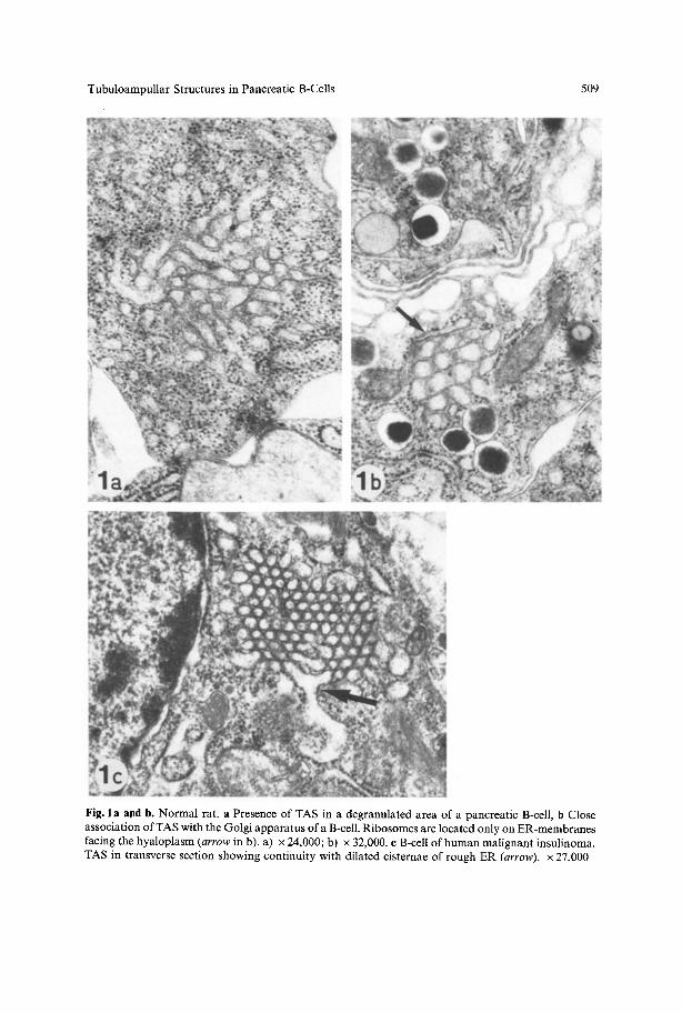
Number , size, and morphology of the secretory granules in the pancreatic islets of rats were considered to be normal. The malignant human insulinoma contained insulin-producing B-cells and glucagon-producing A-cells. In both pancreatic tissues, honeycomb-like structures were found to occur exclusively in B-cells (Fig. 1 a-c). They consist of aggregated and ampullar dilated tubules, were closely associated with dilated rough ER and were frequently seen in close contact with the Golgi apparatus. Such tubuloampullar structures (TAS) are usually found in degranulated B-cell areas, containing abundant free ribosomes, but are sometimes also observed in well-granulated areas. By use of the goniometer in tilting ultrathin sections + 30 ~ the undulated form and branching of the aggregated tubules could be demonstrated. In their surroundings, narrow tubules of ER were often seen bearing ribosomes only on the hyaloplasmic side of their membrane. A direct contact between the aggregated tubules and the surrounding narrow ER was not obvious. The TAS in pancreatic B-cells are always found closely assoc(ated with the ER, and they appear to arise by aggregation of ampullar dilated cisternae of the ER. These findings suggest that TAS may represent a special form of the ER.
In secretory epithelial cells in the prostate of aged rats such a special form of the rough ER, called transitional elements ofendoplasmic reticulum, is assumed to be related to decreased testosterone production (Ichihara and Kawamura 1979). Similar aggregations of tubules associated with the ER were also found in digestive cells of a mollusc (McLean 1978), in neurons of the supraoptic nucleus of the hibernating dormouse (Machin-Santamaria 1978), and in chief cells of the gastric mucosa of hibernating bats (Ito and Winchester 1963).
The origin of these structures remains to be established. In the endocrine pancreas TAS occur exclusively in B-cells. They were described in various viral infections and may form "crystalline aggregates", and were considered to be of viral origin (Blizinger et al. 1969). It is well known that viruses are able to damage specifically pancreatic B-cells and to cause diabetes (Craighead 1977). However, the lack of virus or virus-like particles and of other inclusions within B-cells of the rats does not favor the hypothesis of a viral origin of TAS. Moreover, Baringer and Swoveland (1972) found that tubular aggregates in the endoplasmic reticulum originate as invaginations of the ER membrane, a fact which they consider to be evidence against their viral nature.
Tubuloampullar Structures in Pancreatic B-Cells 509
Fig. l a and b. Normal rat. a Presence of TAS in a degranulated area o f a pancreatic B-cell, b Close association of TAS with the Golgi apparatus of a B-cell. Ribosomes are located only on ER-membranes facing the hyaloplasm (arrow in b). a) x 24,000; b) x 32,000. c B-cell of h u m a n malignant insulinoma. TAS in transverse section showing continuity with dilated cisternae o f rough ER (arrow). x 27,000
510 R. Graf and Ph.U. Heitz
A g g r e g a t e d tubu les a s soc i a t ed wi th the E R were to da t e desc r ibed on ly in cells in a p a r t i c u l a r p h y s i o l o g i c o r p a t h o p h y s i o l o g i c state, such as h ibe rna t ion . T h e f ind ing
o f T A S in d e g r a n u l a t e d a reas a n d in c lose c o n t a c t w i th the G o l g i a p p a r a t u s in
p a n c r e a t i c B-cel ls o f n o r m a l ra t s o f t w o d i f fe ren t s t ra ins and in ac t ive ly secre t ing
t u m o r o u s B-cells o f the h u m a n p a n c r e a s suggests an a s soc i a t i on o f T A S wi th n o r m a l o r p a t h o l o g i c sec re to ry processes o f the B-cell .
References
Baringer JR, Swoveland P (1972) Tubular aggregates in endoplasmic reticulum: evidence against their viral nature. J Ultrastruct Res 41:270-276
Blizinger K, Simon J, Magrath D, Boulger L (1969) Poliovirus crystals within the endoplasmic reticulum of endothelial and mononuclear cells in the monkey spinal cord. Science 136:1336-1337
Craighead JE (1977) Viral diabetes In: BW Volk, The diabetic pancreas. KF Wellman (eds) Bailli6re Tindall, London
Ichihara J, Kawamura H (1979) The fine structure of ventral prostatic secretory epithelial cells in older rats. Cell Tissue Res 203:181-188
I to S, Winchester RJ (1963) The fine structure of the gastric mucosa in the bat. J Cell Biol 16:541 577 Machin-Santamaria C (1978) Ultrastructure of the hypothalamic neurosecretory nuclei of the dormouse
(Eliomys quercinus L.) in the awakening and hibernating states. J Anat 127:239-249 Maron BJ, Ferrans VJ (1974) Aggregates of tubules in human cardiac muscle cells. J Mol Cell Cardiol
6: 249-264 McLean N (1978) Unusual aggregations in tubules associated with endoplasmic reticulum in digestive
cells of Alderia modesta (Mollusca: Gastropoda: Saccoglossa). Cell Tissue Res 194:179-182 Schaff Z, Heine U, Dalton AJ (1972) Ultramorphological and ultracytochemical studies on
tubuloreticular structures in lymphoid cells. Cancer Res 32:2696-2706 Uzman BG, Saito H, Kasac M (1971) Tubular arrays in the endoplasmic reticulum in human tumor
cells. Lab Invest 24:492498
Accepted April 3, 1980