tubular up-regulation of clusterin mrna in murine
TRANSCRIPT
American Journal of Pathology, Vol. 152, No. 4, April 1998
Copyright X American Societyfor Investigative Pathology
Tubular Up-Regulation of Clusterin mRNA in MurineLupus-Like Nephritis
Solange Moll,1 Pierre-Alain Menoud,*Lars French,§ Andre-Pascal Sappino,*Yves Pastore,* Jurg A. Schifferli,t and Shozo Izui*From the Departments of Pathology,* Internal Medicinet andDertnatology,5 University of Geneva Medical School, Geneva, andthe Department ofMedicine,t University ofBasel Medical School,Basel, Switzerland
Clusterin, a widely distributed glycoprotein, is de-tected in most tissues and in numerous physiologicalfluids. In the kidney, this protein is constitutivelyexpressed in tubular epithellal cells, and its expres-sion is enhanced following tubular injuries. In addi-tion, clusterin has been detected in glomerular im-mune deposits of glomerulonephritis. The presentstudy was designed to define the sites of clusterinmRNA accumulation in murine lupus-like nephritis incomparison with murine tubulopathies. In lupus-likenephritis, a significant increase of clusterin mRNAabundance was demonstrated. This up-regulation waslocalized exclusively in tubular epithelial cells exhib-iting tubulointerstitial alterations, whereas no clus-terin mRNA was detectable in diseased glomeruli, ex-cluding an active synthesis of clusterin by glomerularcells. A similar tubular increase of clusterin mRNAabundance was observed in myeloma-like cast ne-phropathy induced by IgG3 monoclonal cryoglobu-lins and even in the absence of any detectable histo-logical alterations in a model of septic shock inducedby the injection of bacterial lipopolysaccharides. Ourresults suggest that tubular epithelial cells are theonly sites of clusterin mRNA accumulation during thecourse of lupus-Uike nephritis and that the tubularup-regulation of clusterin gene expression may re-flect the cellular response to various types of tubularinjuries. (AmJ Pathol 1998, 152:953-962)
Clusterin, a widely distributed heterodimeric glycoproteinwith a molecular mass of 80 kd, is detected in mosttissues, primarily in epithelial cells, and is found in nu-merous physiological fluids including semen, urine, milk,cerebrospinal fluid, and plasma.1'2 The expression ofclusterin can be enhanced or induced in many organs attimes of tissue injury or remodeling.3 Although its biolog-ical role(s) has not yet been defined, it has been sug-gested that it may be involved in numerous physiologicalfunctions, including reproduction, lipid transport, com-
plement regulation, endocrine secretion, initiation of ap-optosis, morphological transformation, membrane pro-tection, and promotion of cellular interactions.4
In the kidney, constitutive expression of clusterin isprimarily observed in epithelial cells of distal tubules andin transitional epithelium lining the renal pelvis.56 En-hancement of renal clusterin mRNA or protein expressionhas been reported in experimental models of acute renalinjury (ureter obstruction, ischemia/reperfusion, folic acidnephropathy, gentamicin nephrotoxicity, and myoglobin-uric renal failure), chronic models of renal disease (renalablation, tubulointerstitial diseases induced by dietarydeficiency of the antioxidants, vitamin E, and selenium),and a murine model of polycystic kidney disease.6-12Despite the demonstration of marked tubular epithelialcell induction of clusterin in experimental models of renalinjury, studies in human renal diseases have been lim-ited. Dvergsten et al13 have described the expression ofclusterin, by immunohistochemical staining, in renal tu-bular epithelium in a number of human renal diseases,including polycystic kidney diseases, multifocal cysts,neoplastic cysts, renal hypoplasia/dysplasia, and renaltransplant rejection. Increased expression of clusterinhas been also noted in some cases of renal clear cellcarcinoma.13'14 Moreover, deposition of clusterin, to-gether with the terminal complement component, on themembrane of tubular epithelial cells and in the tubularlumina was observed in renal infarction lesions.15 Nota-bly, no clusterin staining was detected in glomeruli in anyof those pathological conditions.
In contrast, clusterin has been shown to be localized inglomeruli in association with the terminal complementcomplex and immunoglobulin deposits in a majority of thecases of human glomerulonephritis including lupus ne-phritis.16-18 Although tubular staining for clusterin ap-peared not to be a predominant feature in the case ofglomerulonephritis, an increased tubular staining of clus-terin was reported in 24% of the renal biopsies in patientsdeveloping different types of glomerulonephritis includ-ing lupus nephritis.18 However, details regarding suchtubular staining, particularly in relation to the presence orabsence of tubular lesions, have not been studied. As the
This work was supported by grants from the Swiss National Foundation forScientific Research.
Accepted for publication January 20, 1998.Address reprint requests to Dr. Shozo Izui, Department of Pathology,
C. M. U., 1211 Geneva 4, Switzerland. E-mail: [email protected].
953
954 Moll et alAJP April 1998, Vol. 152, No. 4
presence of clusterin in glomerular immune deposits hasbeen preferentially associated with the terminal comple-ment complex,1718 it has been suggested that clusterin,as an inhibitor of the membrane attack complex,19'20 maymodulate complement-mediated glomerular injury duringthe course of immune complex glomerulonephritis.21Moreover, a recent study has shown that clusterin mRNAcan be induced in glomerular epithelial and mesangialcells stimulated by thrombin in vitro.22 However, it is notknown whether clusterin is indeed expressed in diseasedglomeruli during the course of glomerulonephritis. Toaddress these questions, we have determined the sites ofclusterin mRNA accumulation in relation to the presenceof glomerular and tubular lesions in three different exper-imental models of renal disease: chronic lupus-like ne-phritis occurring spontaneously in three different lupus-prone mice, acute nephropathies induced by murineIgG3 monoclonal cryoglobulins, and acute tubular necro-sis induced by bacterial lipopolysaccharides (LPS).
Materials and Methods
Mice(NZB x NZW)F1, BXSB, MRL-/pr/lpr, and MRL+/+ micewere purchased from Harlan Olac (Oxon, UK). C57BL/6and BALB/c mice were obtained from BomholtgardBreeding and Research Center (Ry, Denmark). (MRL+/+x BALB/c)F1 hybrid (MRL x BALB) mice were bred inour own animal facilities.
Monoclonal AntibodiesMurine IgG3 monoclonal antibodies (mAb) with cryoglobu-lin activity (6-19 and 1-10B4 (anti-lgG2a rheumatoid factor(RF)), 1G3 (anti-DNA), 6-19J, 6-E4, 6-H6, 6-G3, 2-2G (un-known specificity)) were obtained from unmanipulatedMRL-lpr/lpr mice.23 IgG3 antitrinitrophenyl mAb (9A6 andCB1) were established from trinitrophenyl-immunizedC57BL/6 and BALB/c mice, respectively.23 The 6-191gM orIgGl class switch variant lacking cryoglobulin activity weregenerated as described previously.2324 Three-month-oldMRL x BALB mice were injected intraperitoneally with 107hybridoma cells. Animals were killed between 7 and 14days after the injection of hybridoma cells.
LPS Treatment
Mice were injected intraperitoneally with a single 100-,gdose of LPS from Escherichia coil (Sigma, St. Louis, MO).Animals were killed 3, 8, and 48 hours after injection.Alternatively, a first injection of 100 gg of LPS was fol-lowed 24 hours later by a second 25-gg injection withmice killed 18 hours thereafter.
Molecular Cloning of the MurineMegalin/Glycoprotein 330 (gp330) Partial cDNA
Degenerate primers were designed from the conserved
amino acid sequences, KLGLMFWT and WLTQVR, lo-
cated in the ectodomain close to the transmembranedomain of the human and rat megalin/gp330, a memberof the low density lipoprotein receptor family.2526 Theprimer sequences used were: 5'-AA/GC/TTNGGNC/TTNATGTTC/TTGG-3' and 5'-CGNACC/TTGNGTNAA/GCCA-3'. A 370-bp reverse transcription-polymerasechain reaction product27 was amplified from murine kid-ney total mRNA and cloned into the EcoRV site of pBSKS.Cloned polymerase chain reaction products were se-quenced on both strands by the chain terminationmethod using T3 and T7 primers.28 The nucleotide se-quence has been deposited into European MolecularBiology Laboratory data bank under the accession num-ber Y08566.
Probe PreparationsClusterin antisense RNA probe was transcribed in vitrowith T3 polymerase using pBSKS containing the 638-bpmurine clusterin cDNA fragment5 and linearized with Stulfor RNase protection assays and BamHl for in situ hybrid-izations. Clusterin sense RNA probe was transcribed withSP6 RNA polymerase using the murine clusterin cDNAlinearized with Hindlil. The megalin/gp330 antisense RNAprobe was transcribed in vitro with T3 RNA polymeraseusing the pBSKS containing the 378 bp of the murinemegalin/gp330 partial cDNA and linearized with EcoRI forRNase protection assays and in situ hybridizations.Megalin/gp330 sense RNA probe was transcribed withT7 RNA polymerase using the murine megalin/gp330partial cDNA linearized with Xhol. The glyceraldehyde-3-phosphate-dehydrogenase (GAPDH) antisense RNAprobe was prepared as described previously.23
RNase Protection AssayRNase protection assays were performed as de-scribed.29 Briefly, five ,ug of total RNA extracted fromwhole kidneys were hybridized for 16 hours at 42°C witha[32P]UTP-labeled antisense clusterin, megalin/gp330,and GAPDH riboprobes. Yeast tRNA was used as thenegative control. Unhybridized RNA was digested withRNase A (Sigma) for 1 hour at 250C. The RNase was thendigested with proteinase K (Sigma) for 20 minutes at350C. After phenol-chloroform extraction and sodium ac-etate precipitation, the protected RNA was denatured at900C and electrophoresed on a urea-6% polyacrylamidegel. The dried gels were exposed to autoradiographicfilm (Kodak X-Omat film, Eastman Kodak, Rochester, NY)and subjected to phosphor imager analysis for quantifi-cation (Image Quant Software FAST SCAN, MolecularDynamics, Kemsing, UK).
In Situ HybridizationFive-,um cryostat kidney sections were fixed in 4% glu-taraldehyde in phosphate-buffered saline for 1 minute.Prehybridizations, hybridizations, and posthybridizationwashes were carried out as described.30 Two x 106 cpmof 32P-labeled clusterin cRNA or 0.4 to 1 x 10⁶ cpm of
Clusterin in Murine Lupus Nephritis 955AJP April 1998, Vol. 152, No. 4
3H-labeled cRNA were applied to each cryostat kidneytissue section in 30 ,ul of hybridization mixture. Aftergraded ethanol dehydration, sections hybridized to 32P-labeled cRNA were directly exposed to X-ray films (SB5;Eastman Kodak) between intensifying screens, and thefilms were developed after 2 days of exposure at roomtemperature. Sections hybridized to 3H-labeled cRNAwere immersed in NTB-2 emulsion (Eastman Kodak) anddiluted 1:1 in deionized water. After 16 weeks of expo-sure, they were developed in Kodak D-19 developer,fixed in 30% sodium thiosulfate, and counterstained withmethylene blue.
Renal PathologySamples of kidney tissues were fixed in 10% formalin,and sections were stained with hematoxylin and eosin orperiodic acid-Schiff (PAS). A number of histologicalchanges in three major compartments (glomeruli, tu-bules, and interstitium) in lupus-prone mice were individ-ually assessed as to type and degree according to Piraniand Salinas-Madrigal.31 The degree of each histopatho-logical feature is graded on a semiquantitative scale of 0to 4+ as follows: a grade of 1 corresponded to mildchanges; 2+ to moderate changes; 3+ to moderatelysevere changes; and 4+ to severe changes. Based onthe sum of the total degree of damage for each compart-ment, renal lesions were scored on a 0 to 4+ scale.
Statistical AnalysisStatistical analysis was performed with the Wilcoxon two-sample test. Probability values <0.05 were consideredsignificant.
Results
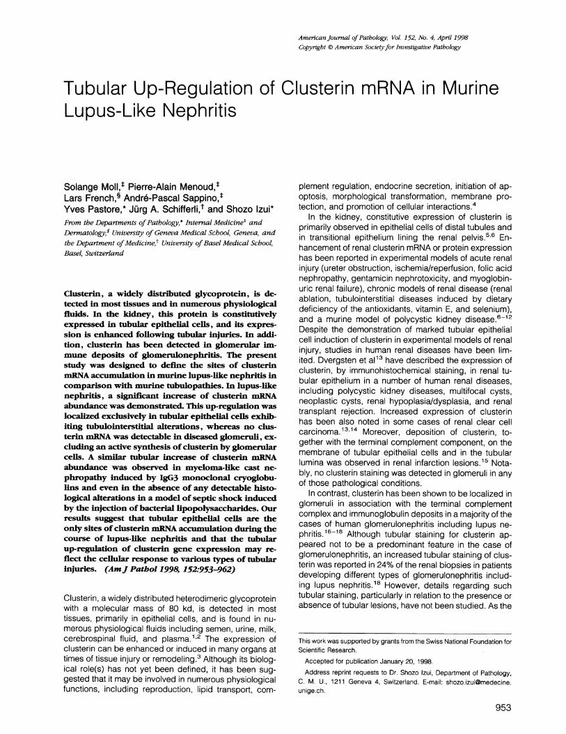
Enhanced Tubular Accumulation of ClusterinmRNA in Chronic Lupus Nephritis OccurringSpontaneously in Lupus-Prone MiceUsing three different lupus-prone mice ((NZB x NZW)F1,MRL-Ipr/lpr, and BXSB), renal abundance of clusterinmRNA was assessed in relation to the development oftheir histopathological lesions. For this purpose, totalRNA was extracted from kidneys of lupus-prone mice ofvarious ages, and clusterin and GAPDH mRNA abun-dance was simultaneously analyzed by RNase protectionassay. All three lupus-prone mice exhibited an increasedabundance of clusterin mRNA concomitantly with thedevelopment of glomerular and tubulointerstitial lesions,ie, at 9 months for (NZB x NZW)F1 females, 4 months forMRL-Ipr/lpr females, and 5 to 8 months for BXSB males(Figure 1A). Notably, intensity and extent of histopatho-logical changes of tubular lesions (tubular atrophy, inter-stitial mononuclear infiltration, tubular cast formation,andinterstitial fibrosis) correlated with histological grades ofglomerular lesions in three lupus-prone mice. Quantita-tive analysis, normalized to GAPDH mRNA signals, re-
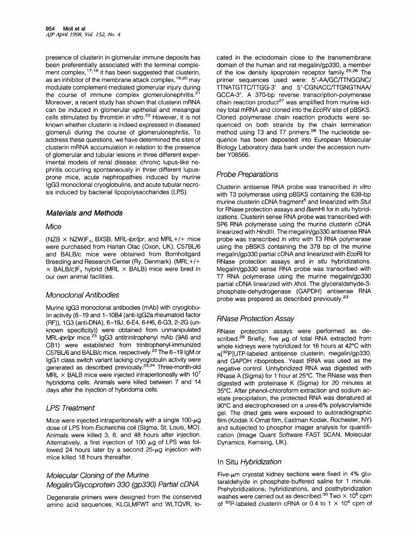
vealed a clear correlation between clusterin mRNA levelsand histological grades of renal lesions in individual lu-pus-prone mice (Figure 2). Increases in clusterin mRNAwere highly significant when the histological grade was 4(means of 13 mice = 6.0 + 4.0) as compared with micehaving the histological grade of 3 (means of 17 mice =2.6 ± 1.5; P < 0.005) and of less than 2 (means of 32mice = 1.4 ± 0.8; P < 0.001); differences between thelast two groups were also significant (P < 0.01). In con-trast, no changes in the abundance of clusterin mRNAwas observed in nonautoimmune C57BL/6 mice (Fig-ure 1A).The sites of clusterin mRNA accumulation were local-
ized by hybridizing kidney sections with 32P-labeled clus-terin cRNA probes. Markedly enhanced accumulation ofclusterin mRNA was confined to the cortex in the threediseased lupus-mice (Figure 1A). Cellular sites of corticalclusterin mRNA accumulation, as disclosed by kidneysections hybridized to 3H-labeled clusterin cRNA, wereidentified as tubular epithelial cells (Figure 1 B). However,no clusterin mRNA was detected in diseased glomeruli.
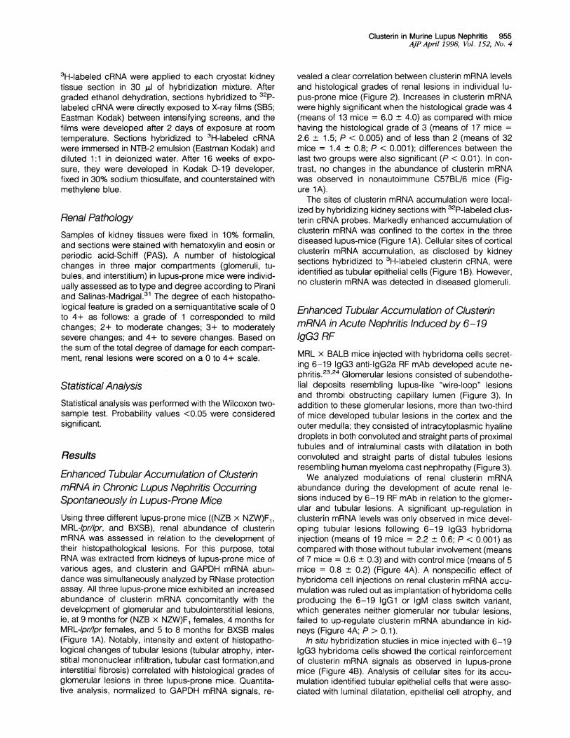
Enhanced Tubular Accumulation of ClusterinmRNA in Acute Nephritis Induced by 6-19IgG3 RFMRL x BALB mice injected with hybridoma cells secret-ing 6-19 IgG3 anti-IgG2a RF mAb developed acute ne-phritis.23'24 Glomerular lesions consisted of subendothe-lial deposits resembling lupus-like "wire-loop" lesionsand thrombi obstructing capillary lumen (Figure 3). Inaddition to these glomerular lesions, more than two-thirdof mice developed tubular lesions in the cortex and theouter medulla; they consisted of intracytoplasmic hyalinedroplets in both convoluted and straight parts of proximaltubules and of intraluminal casts with dilatation in bothconvoluted and straight parts of distal tubules lesionsresembling human myeloma cast nephropathy (Figure 3).We analyzed modulations of renal clusterin mRNA
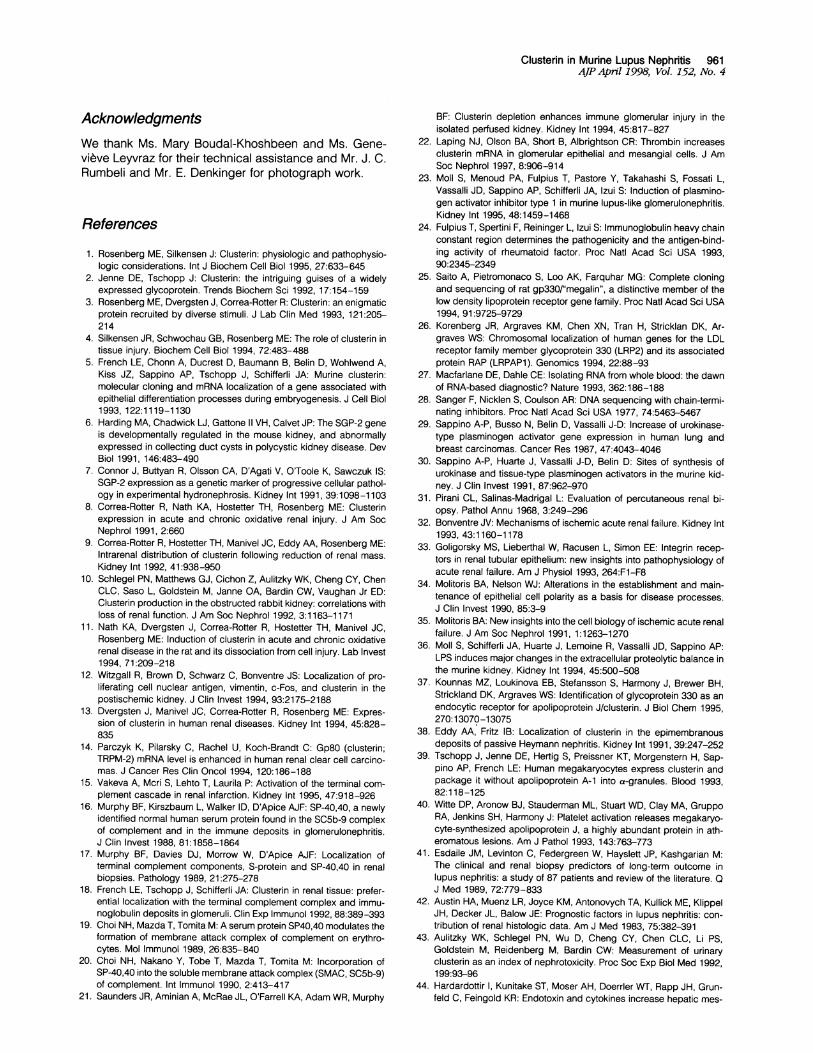
abundance during the development of acute renal le-sions induced by 6-19 RF mAb in relation to the glomer-ular and tubular lesions. A significant up-regulation inclusterin mRNA levels was only observed in mice devel-oping tubular lesions following 6-19 IgG3 hybridomainjection (means of 19 mice = 2.2 ± 0.6; P < 0.001) ascompared with those without tubular involvement (meansof 7 mice = 0.6 + 0.3) and with control mice (means of 5mice = 0.8 ± 0.2) (Figure 4A). A nonspecific effect ofhybridoma cell injections on renal clusterin mRNA accu-mulation was ruled out as implantation of hybridoma cellsproducing the 6-19 IgGl or IgM class switch variant,which generates neither glomerular nor tubular lesions,failed to up-regulate clusterin mRNA abundance in kid-neys (Figure 4A; P > 0.1).
In situ hybridization studies in mice injected with 6-19IgG3 hybridoma cells showed the cortical reinforcementof clusterin mRNA signals as observed in lupus-pronemice (Figure 4B). Analysis of cellular sites for its accu-mulation identified tubular epithelial cells that were asso-ciated with luminal dilatation, epithelial cell atrophy, and

956 Moll et alAJP April 1998, Vol. 152, No. 4
A
(NZBx NZW) Fi2mo 4lmo mo 9mo 0
GAPDH . - .-- - -- - - -.S - X i
MRL- 4p,lIjr2 MO 4mo.
GAPDH . *
BXSB Mak
nb 2 me 5mo 8m
GAPDH* -' 9dSg..9IIt9
C57B16Femalk Male BXSB
2mo 6 2mm Gm. F M 0
"istrn -a*_ ftom Mftft_ _ 4za gl
GAPDH '**4D db04 bf - bd * a '*Clwleu1n~a..,. .I
Figure 1. A: Clusterin mRNA accumulation and localization in kidneys from three different lupus-prone mice: (NZB X NZW)Fj, MRL-lpr/lpr, and BXSB. TotalRNA of kidneys from lupus-prone and nonautoimmune (C57BL/6) mice of different ages were analyzed by the RNase protection assay using clusterin and GAPDHcRNA probes simultaneously; two different RNase-protected bands (315 bp for clusterin and 260 bp for GAPDH) are revealed. RNA extracted from yeast (*) wasused as a negative control in each assay. Cryostat kidney sections from lupus-prone mice having severe glomerulonephritis were analyzed by in situ hybridizationwith 32P-labeled clusterin cRNA antisense probe. Note a marked up-regulation of clusterin accumulation with a speckled cortical localization in diseased kidneysfrom three different lupus mice as compared with 6-month-old nonautoimmune C57BU/6 mice. B: Tubular up-regulation of clusterin mRNA abundance inlupus-prone mice. Cryostat kidney sections from nephritic BXSB mice were hybridized with 3H-labeled-clusterin cRNA antisense probe. Upper panel is bright-fieldmicrograph (X20), and lower panel is the corresponding dark-field micrograph. Note an increased accumulation of clusterin mRNA in tubules without anydetectable signals in glomeruli.
intraluminal casts; no signals were detected in diseasedglomeruli (Figure 5A). In addition, clusterin mRNA tran-scripts were newly induced in the outer medulla where nosignificant signals were detectable in control mice (Fig-ures 4B and 5). Because of the limitation of morphologi-cal resolution on cryostat tissue sections, the positivetubular structures in the cortex and the outer medullacould not be identified with certainty.
Enhanced Accumulation of Clusterin mRNA inTubulopathies Induced by IgG3 mAbTo further determine whether the up-regulation of clus-terin mRNA in kidneys of mice receiving hybridoma cellswas associated with the development of tubular and/orglomerular lesions, we analyzed the effect of IgG3 hy-bridoma cells inducing distinct types of nephropathies.First, IgG3 monoclonal cryoglobulins (6-19J, 6-E4, and6-H6) induce only tubular lesions, resembling myelomacast nephropathy, without glomerular involvement. Sec-ond, IgG3 monoclonal cryoglobulins (1-10B4, CB1, and6-G3) provoke only glomerular lesions (wire-loop lesions
or intracapillary thrombotic lesions) without tubular in-volvement. Third, IgG3 mAb (9A6, 1G3, and 2-2G) gen-erate neither glomerular nor tubular lesions. The firstgroup of IgG3 mAb generating tubular lesions withoutglomerular lesions was the only one to induce up-regu-lation of renal clusterin mRNA levels (Figure 6). In con-trast, renal clusterin mRNA levels remained at baselinevalues with the second and the third groups of IgG3 mAb,which failed to induce tubular lesions, independent of thedevelopment of glomerular lesions.
Enhanced Accumulation of Clusterin mRNAduring the Course ofAcute Tubular NecrosisInduced by LPSAs cytoskeletal, biochemical, and functional alterationscharacterize renal tubular epithelium after acute renalinjury, eg, ischemic or toxic, even in the absence ofhistological evidence of tubular lesions,32 35 we deter-mined whether the development of acute tubular injuryfollowing the injection of LPS was associated with mod-
L----

Clusterin in Murine Lupus Nephritis 957AJP April 1998, Vol. 152, No. 4
Histological gradesFigure 2. Quantitative analysis of renal clusterin mRNA abundance in rela-tion to the intensity of renal lesions in three different lupus-prone (NZB X
NZW)F1, MRL-lp r/lp r, and BXSB mice. Total RNA of kidneys from differentages of lupus-prone mice were analyzed by the RNase protection assay usingclusterin and GAPDH cRNA probes simultaneously. The ratio of the radio-activity associated with the clusterin signal relative to that of the GAPDHsignal in individual animals was computed from the intensity of each signaldetermined by phosphor imager analysis. Results obtained from differentages of three lupus-prone (NZB X NZW)F1 (0), MRL-lp r/lp r(@), and BXSB(0) mice were pooled, and clusterin/GAPDH mRNA ratios were correlatedwith the severity of renal lesions, which are expressed as histological gradesbased on intensity and extent of glomerular and tubulointerstitial changes inindividual animals.
ulations in renal clusterin mRNA abundance. Micewere injected with a single dose of 100 ,ug of LPS andkilled at 3, 8, and 48 hours after injection. Although nodetectable histological lesions at the level of opticalmicroscopy were observed, renal clusterin mRNA was
significantly enhanced following the LPS injection.Comparative measurements of clusterin and GAPDHmRNA levels revealed a rapid increase at 3 hours (P <0.02) to reach a maximum at 8 hours (P < 0.001) andreturn toward baseline values at 48 hours (Figure 7A).In situ hybridization studies showed a marked rein-forcement of cortical signals with an induction of clus-terin mRNA in the outer medulla similar to those ob-served in mice injected with 6-19 hybridoma cells(Figure 7B). Cortical and medullary clusterin mRNAtranscripts were additionally elevated in kidneys ofanimals killed 18 hours after two LPS injections. Nota-bly, they developed histologically detectable alter-ations in tubules (dilatation of cortical tubules withflattening of epithelial cells and presence of intralumi-nal desquamated debris), as previously described.36
Megalin/gp330 mRNA Levels in MiceDeveloping Tubular LesionsAs clusterin has recently been shown to be a ligand ofmegalin/gp330, which is assumed to function as an en-
docytic receptor,37 we studied whether the tubular up-regulation of clusterin mRNA was associated with renalmodulation of megalin/gp330 mRNA. However, indepen-
Figure 3. Histology of glomerular and tubular lesions in MRL X BALB miceinjected with hybridoma cells secreting 6-19 IgG3 anti-IgG2a RF mAb. A totalof 107 hybridoma cells were injected intraperitoneally into MRL X BALB miceon day 0, and mice were killed at day 7 after the injection of hybridoma cells.A and B: Representative histological appearance of glomerular and tubularlesions in the cortex. Glomerular lesions are characterized by the presence ofPAS-positive deposits along the glomerular capillary walls and by the plug-ging of glomerular capillaries by voluminous PAS-positive thrombi. Tubularlesions are characterized by the casts, dilatation of cortical tubules (mostlikely convoluted parts of distal tubules), and the presence of intracytoplas-mic hyaline droplets in convoluted parts of proximal tubules, lesions resem-
bling human myeloma cast nephropathy. C: Representative histological ap-
pearance of tubular lesions with casts and intracytoplasmic hyaline dropletsin the outer medulla. PAS; magnification, X 100 (A), X 200 (B and C).
dently of the presence of tubular lesions, no significantchanges in renal megalin/gp330 mRNA abundance weredetected in mice receiving 6-19 IgG3 hybridoma cells(means of 16 mice with tubular lesions = 1.3 + 0.6;
lI r4 i
10
1:
Rll.
'IC
0It00
0.1
8 z+ I T J w0
<2 3 4i i
.,
.w4V Alp.

958 Moll et alAJP April 1998, Vol. 152, No. 4
A
. 0.
619Y3 6-19Y 6-19 i Central
B
Figure 4. A: Quantitative analysis of clusterin mRNA abundance in kidneysfrom mice injected with hybridoma cells secreting 6-19 Ig class switchvariants. Animals were killed 7 to 14 days after the injection of hybridomacells secreting 6-19 IgG3 (y3), IgGl (yl), or IgM (IL) switch variant. Clusterinand GAPDH mRNA were analyzed in individual mice by the RNase protec-tion assay using clusterin and GAPDH cRNA probes simultaneously. Resultsare expressed as a ratio of the radioactivity associated with the clusterinsignal relative to that of the GAPDH signal in individual animals. Note asignificant up-regulation of clusterin mRNA levels in kidneys developingboth glomerular and tubular lesions (0) but neither in those having onlyglomerular lesions (0) from mice injected with hybridoma cells secreting6-19 IgG3 (y3) nor in those from mice injected with hybridoma cellssecreting 6-19 IgGl (yl) or IgM (IL) switch variant (P < 0.001). B: Renallocalization of clusterin mRNA accumulation in mice injected with hybridomacells secreting 6-19 IgG3 anti-IgG2a RF mAb. Cryostat kidney sections from6-19 IgG3 hybridoma-injected mice at day 0 (DO) and day 7 (D7) were
hybridized with 32P-labeled clusterin cRNA antisense probe. Note a markedaccumulation of clusterin mRNA in cortex and outer medulla at day 7 (D7)after the 6-19 hybridoma injection.
means of 7 mice without tubular lesions = 1.0 ± 0.6; P >0.4). Moreover, renal megalin/gp330 mRNA levels re-mained at baseline values in three different lupus-pronemice and in mice injected with LPS (data not shown).
DiscussionIn the present study, we have demonstrated that an en-
hanced expression of clusterin mRNA was localized ex-
clusively in tubular epithelial cells exhibiting tubulointer-stitial alterations during the course of chronic lupusnephritis in three different lupus-prone mice; no clusterinmRNA was detectable in diseased glomeruli. A similartubular up-regulation of clusterin mRNA abundance wasobserved in mice developing myeloma-like cast ne-phropathy (induced by the injection of hybridoma cellssecreting murine IgG3 monoclonal cryoglobulins) andeven in the absence of detectable histological alterationsin a model of septic shock (induced by the injection ofbacterial LPS).
In situ hybridization experiments have disclosed thattubules are the only sites of clusterin mRNA accumulation
during the course of chronic lupus-like nephritis occur-ring spontaneously in lupus-prone mice. Analysis of theacute model of lupus-like nephritis induced by IgG3 RFmAb confirmed that clusterin mRNA abundance is in-creased only when tubular lesions are developed inde-pendently of the presence of glomerular lesions. Thisexcludes an active synthesis of clusterin by glomerularand/or infiltrating inflammatory cells following glomerularinjury, although our results do not rule out the possibilitythat glomerular cells can be a potential source of clus-terin under certain pathological conditions, as shown bya recent in vitro study.22 Thus, glomerular localization ofclusterin observed in both experimental and human glo-merulonephritis including lupus nephritis16-1838 is likelyto have resulted from a deposition of plasma- and/orplatelet-derived clusterin into the membrane attackcomplex.3940
It is known that glomerulonephritis is often accompa-nied by tubular and interstitial lesions. In fact, tubuloin-terstitial abnormalities have been recognized to be themost valuable biopsy marker in predicting renal outcomein a number of glomerulonephritis including lupus nephri-tis.41'42 In the chronic model of lupus-like glomerulone-phritis, we have demonstrated a close correlation be-tween the tubular clusterin mRNA abundance and theseverity of nephritic lesions in the three different strains oflupus-prone mice in which intensity and extent of tubulo-interstitial changes parallel to the development of glomer-ular lesions. These results suggest that clusterin could bea biological index of tubular injury associated with glo-merulonephritis. As levels of clusterin secreted in urinehave been shown to increase in experimental models oftubulopathies,43 the measurement of urinary clusterincould be a potentially useful clinical marker in themanagement and prognosis of patients with glomerulo-nephritis.
Clusterin mRNA abundance is elevated and/or in-duced in tubular epithelial cells exhibiting different kindsof alterations, such as luminal dilatation, atrophy, intracy-toplasmic hyaline droplets, and cast formation, in micedeveloping chronic lupus-like nephritis, myeloma-likecast nephropathy, or LPS-induced acute tubular necro-sis. In addition, it is possible that an increased clusteringene expression can be a consequence of tubular stim-ulation by cytokines released by infiltrating inflammatorycells or activated tubular cells. Although it has neverbeen shown if the clusterin expression in tubular epithe-lial cells can be modulated by any cytokines, severalcytokines, such as tumor necrosis factor, interleukin-1(IL-1), IL-2, and transforming growth factor (31, have beenshown to induce clusterin mRNA or protein expression inlivers and cultured astrocytes.4446 It is of particular in-terest to note that the injection of LPS or tumor necrosisfactor/IL-1, the major cytokines inducible by LPS, is ableto increase hepatic clusterin mRNA levels and serumconcentrations of clusterin in hamsters.44 Such a mech-anism can be in part responsible for a rapid increase intubular clusterin mRNA abundance in mice receiving asingle injection of LPS, even without any histologicallydetectable tubular lesions. Nevertheless, our demonstra-tion, tubular clusterin up-regulation in three different

Clusterin in Murine Lupus Nephritis 959AJP April 1998, Vol. 152, No. 4
CORTEX MEDULLA
A
B
CORTEX MEDULLA
Figure 5. Cellular localization of clusterin mRNA accumulation in mice injected with hybridoma cells secreting 6-19 IgG3 anti-IgG2a RF mAb. A: Cryostat kidneysections obtained 7 days after the injection of 6-19 hybridoma cells were hybridized with 3H-labeled clusterin cRNA antisense probe. Upper panel is bright-fieldmicrograph, and lower panel is the corresponding dark-field micrograph. Note an increased accumulation of clusterin mRNA in epithelial cells of pathologicaltubules (intraluminal casts and dilatation) in the cortex; no detectable signal is observed in pathological glomeruli. In addition, clusterin mRNA signal is newlydetectable in tUbUlar epithelial cells of the outer imiedulla. B: Cryostat kidney sections from control mice were hybridized with 3H-labeled clusterin cRNA antisenseprobe. Note the constituLtive expression of cluLsterin nmRNA localized in epithelial cells of tuLbuLles in the cortex, but the lack of its expression in glomeruli and intuLbuLlar epithelial cells of the outer medulll.a. Magnification, X40 (cortex), X20 (medulla).

960 Moll et alAJP April 1998, Vol. 152, No. 4
I.Q
6-19J 6-E4 6-H6 1-IOB4 CB1 6-G3 9A6 1G3 2-2G Control
Tub+ Tub- Tub-Glom - Glom + Glom -
Figure 6. Quantitative analysis of clusterin mRNA abundance in kidneys frommice injected with different hybridoma cells. Mice were injected with hy-bridoma cells secreting the following mAb: 6-19J, 6-E4, and 6-H6, whichgenerate tubular lesions (Tub') without glomerular lesions (Glom-);1-10B4, CB1, and 6-G3, which generate glomerular lesions (Glom+) withouttubular lesions (Tub-); 9A6, 1G3, and 2-2G, which generate neither tubular(Tub-) nor glomerular (Glom-) lesions. Animals were killed 7 to 14 dayslater. Clusterin and GAPDH mRNA were analyzed in individual mice (2 to 5mice for each mAb) by the RNase protection assay using clusterin andGAPDH cRNA probes simultaneously. Results are expressed as a ratio of theradioactivity associated with the clusterin signal relative to that of the GAPDHsignal in individual animals.
types of tubular injuries, is consistent with observations ofothers on various acute and chronic models of tubulopa-thies.7 1247 This indicates that numerous physiopatho-logical mechanisms responsible for different tubularlesions can result in an up-regulation of clusteringene expression, whatever the cause of tubular injuriesmight be.The physiological role of clusterin in kidneys remains to
be defined. However, based on its expression site andmolecular characteristics, several possible roles of clus-terin can be postulated. Clusterin is expressed at thefluid-tissue interface and secreted in urine,43 and severalpotential binding sites including heparin-binding and am-phiphilic helix domains are identified in clusterin mole-cules.1 In addition, recent studies have shown that mega-lin/gp330 expressed in renal proximal tubules apparentlyacts as an endocytic receptor for clusterin.37 An attrac-tive hypothesis is that tubular clusterin may be involved inthe elimination of pathological excessive proteins or des-quamated cellular debris to maintain tubular patency andfluidity. A possible interaction of tubular clusterin withamphiphilic domains in proteins or lipids in membranedebris and subsequent endocytosis through megalin/gp330 may help prevent intraluminal precipitation ofpathogenic molecules, hence tubular obstruction. Thedemonstration of clusterin in tubular casts9 15,48 is con-sistent with this hypothesis. Although we did not find anysignificant changes in megalin/gp330 mRNA abundancein mice developing tubular lesions, it should be men-tioned that megalin/gp330 is one of the most abundantmembrane proteins constitutively expressed in proximaltubules.49 An alternative or additional role of clusterin isthat it may promote intercellular and cell-matrix interac-tions to maintain tubular integrity. In vitro experiments
31k a 48
BCantd LFS3h
*LSa 1Sh wIwLtS
Figure 7. A: Quantitative analysis of clusterin mRNA abundance in kidneysfrom mice injected with LPS. Mice injected with 100 ,ug of LPS or 0.1 ml ofNaCI were killed after 3, 8, or 48 hours after injection. Clusterin and GAPDHmRNA were analyzed in individual mice (3 mice for each time point) by theRNase protection assay using clusterin and GAPDH cRNA probes simulta-neously, and results are expressed as a ratio of the radioactivity associatedwith the clusterin signal relative to that of the GAPDH signal in individualanimals. B: Renal localization of clusterin mRNA accumulation in mice in-jected with LPS. Cryostat kidney sections from LPS-injected mice, 3 and 8hours after the single injection and 18 hours after the two injections, were
hybridized with 32P-labeled clusterin cRNA antisense probe. Note a markedaccumulation of clusterin mRNA in cortex and medulla from mice injectedwith LPS.
have shown that the inhibition of cell-substratum interac-tion stimulated the synthesis of clusterin, which in turnpromoted the formation of cellular aggregates.50 In fact,subtle alterations in cell-cell and cell-substratum adhe-sions observed in tubular lesions resulting from hypoxiahave been shown to be accompanied by the induction ofclusterin.1247We suggest that an increased expression of tubular
clusterin in three different types of nephropathies (chron-ic lupus-like nephritis, myeloma-like cast nephropathy,and LPS-induced acute tubular necrosis) may be a hostresponse to tubular injury to maintain tubular patency andfluidity to prevent the development tubular obstructivelesions and/or promote cell-cell or cell-matrix interactionsto maintain tubular integrity. Thus, clusterin could play a
cytoprotective role during tubular injury. It is important togain further insights into the physiopathological role(s) ofclusterin in the hope of obtaining a new clinical marker,and a therapeutic agent in various glomerulonephritis aswell as tubulopathies.
12-0
10
8-
6-0
4- 0
2 0
A
r3
1-
4 .
u
.0000 0
S~~~~48 ~ 8
O col.i
* +LPS
11
L
11
Clusterin in Murine Lupus Nephritis 961AJP April 1998, Vol. 152, No. 4
AcknowledgmentsWe thank Ms. Mary Boudal-Khoshbeen and Ms. Gene-vibve Leyvraz for their technical assistance and Mr. J. C.Rumbeli and Mr. E. Denkinger for photograph work.
References
1. Rosenberg ME, Silkensen J: Clusterin: physiologic and pathophysio-logic considerations. Int J Biochem Cell Biol 1995, 27:633-645
2. Jenne DE, Tschopp J: Clusterin: the intriguing guises of a widelyexpressed glycoprotein. Trends Biochem Sci 1992, 17:154-159
3. Rosenberg ME, Dvergsten J, Correa-Rotter R: Clusterin: an enigmaticprotein recruited by diverse stimuli. J Lab Clin Med 1993, 121:205-214
4. Silkensen JR, Schwochau GB, Rosenberg ME: The role of clusterin intissue injury. Biochem Cell Biol 1994, 72:483-488
5. French LE, Chonn A, Ducrest D, Baumann B, Belin D, Wohlwend A,Kiss JZ, Sappino AP, Tschopp J, Schifferli JA: Murine clusterin:molecular cloning and mRNA localization of a gene associated withepithelial differentiation processes during embryogenesis. J Cell Biol1993, 122:1119-1130
6. Harding MA, Chadwick LJ, Gattone II VH, Calvet JP: The SGP-2 geneis developmentally regulated in the mouse kidney, and abnormallyexpressed in collecting duct cysts in polycystic kidney disease. DevBiol 1991, 146:483-490
7. Connor J, Buttyan R, Olsson CA, D'Agati V, O'Toole K, Sawczuk IS:SGP-2 expression as a genetic marker of progressive cellular pathol-ogy in experimental hydronephrosis. Kidney Int 1991, 39:1098-1103
8. Correa-Rotter R, Nath KA, Hostetter TH, Rosenberg ME: Clusterinexpression in acute and chronic oxidative renal injury. J Am SocNephrol 1991, 2:660
9. Correa-Rotter R, Hostetter TH, Manivel JC, Eddy AA, Rosenberg ME:Intrarenal distribution of clusterin following reduction of renal mass.Kidney Int 1992, 41:938-950
10. Schlegel PN, Matthews GJ, Cichon Z, Aulitzky WK, Cheng CY, ChenCLC, Saso L, Goldstein M, Janne OA, Bardin CW, Vaughan Jr ED:Clusterin production in the obstructed rabbit kidney: correlations withloss of renal function. J Am Soc Nephrol 1992, 3:1163-1171
11. Nath KA, Dvergsten J, Correa-Rotter R, Hostetter TH, Manivel JC,Rosenberg ME: Induction of clusterin in acute and chronic oxidativerenal disease in the rat and its dissociation from cell injury. Lab Invest1994, 71:209-218
12. Witzgall R, Brown D, Schwarz C, Bonventre JS: Localization of pro-liferating cell nuclear antigen, vimentin, c-Fos, and clusterin in thepostischemic kidney. J Clin Invest 1994, 93:2175-2188
13. Dvergsten J, Manivel JC, Correa-Rotter R, Rosenberg ME: Expres-sion of clusterin in human renal diseases. Kidney Int 1994, 45:828-835
14. Parczyk K, Pilarsky C, Rachel U, Koch-Brandt C: Gp8O (clusterin;TRPM-2) mRNA level is enhanced in human renal clear cell carcino-mas. J Cancer Res Clin Oncol 1994, 120:186-188
15. Vakeva A, Mcri S, Lehto T, Laurila P: Activation of the terminal com-plement cascade in renal infarction. Kidney Int 1995, 47:918-926
16. Murphy BF, Kirszbaum L, Walker ID, D'Apice AJF: SP-40,40, a newlyidentified normal human serum protein found in the SC5b-9 complexof complement and in the immune deposits in glomerulonephritis.J Clin Invest 1988, 81:1858-1864
17. Murphy BF, Davies DJ, Morrow W, D'Apice AJF: Localization ofterminal complement components, S-protein and SP-40,40 in renalbiopsies. Pathology 1989, 21:275-278
18. French LE, Tschopp J, Schifferli JA: Clusterin in renal tissue: prefer-ential localization with the terminal complement complex and immu-noglobulin deposits in glomeruli. Clin Exp Immunol 1992, 88:389-393
19. Choi NH, Mazda T, Tomita M: A serum protein SP40,40 modulates theformation of membrane attack complex of complement on erythro-cytes. Mol Immunol 1989, 26:835-840
20. Choi NH, Nakano Y, Tobe T, Mazda T, Tomita M: Incorporation ofSP-40,40 into the soluble membrane attack complex (SMAC, SC5b-9)of complement. Int Immunol 1990, 2:413-417
21. Saunders JR, Aminian A, McRae JL, O'Farrell KA, Adam WR, Murphy
BF: Clusterin depletion enhances immune glomerular injury in theisolated perfused kidney. Kidney Int 1994, 45:817-827
22. Laping NJ, Olson BA, Short B, Albrightson CR: Thrombin increasesclusterin mRNA in glomerular epithelial and mesangial cells. J AmSoc Nephrol 1997, 8:906-914
23. Moll S, Menoud PA, Fulpius T, Pastore Y, Takahashi S, Fossati L,Vassalli JD, Sappino AP, Schifferli JA, Izui S: Induction of plasmino-gen activator inhibitor type 1 in murine lupus-like glomerulonephritis.Kidney Int 1995, 48:1459-1468
24. Fulpius T, Spertini F, Reininger L, Izui S: Immunoglobulin heavy chainconstant region determines the pathogenicity and the antigen-bind-ing activity of rheumatoid factor. Proc Natl Acad Sci USA 1993,90:2345-2349
25. Saito A, Pietromonaco S, Loo AK, Farquhar MG: Complete cloningand sequencing of rat gp330/"megalin", a distinctive member of thelow density lipoprotein receptor gene family. Proc NatI Acad Sci USA1994, 91:9725-9729
26. Korenberg JR, Argraves KM, Chen XN, Tran H, Stricklan DK, Ar-graves WS: Chromosomal localization of human genes for the LDLreceptor family member glycoprotein 330 (LRP2) and its associatedprotein RAP (LRPAP1). Genomics 1994, 22:88-93
27. Macfarlane DE, Dahle CE: Isolating RNA from whole blood: the dawnof RNA-based diagnostic? Nature 1993, 362:186-188
28. Sanger F, Nicklen S, Coulson AR: DNA sequencing with chain-termi-nating inhibitors. Proc Natl Acad Sci USA 1977, 74:5463-5467
29. Sappino A-P, Busso N, Belin D, Vassalli J-D: Increase of urokinase-type plasminogen activator gene expression in human lung andbreast carcinomas. Cancer Res 1987, 47:4043-4046
30. Sappino A-P, Huarte J, Vassalli J-D, Belin D: Sites of synthesis ofurokinase and tissue-type plasminogen activators in the murine kid-ney. J Clin Invest 1991, 87:962-970
31. Pirani CL, Salinas-Madrigal L: Evaluation of percutaneous renal bi-opsy. Pathol Annu 1968, 3:249-296
32. Bonventre JV: Mechanisms of ischemic acute renal failure. Kidney Int1993, 43:1160-1178
33. Goligorsky MS, Lieberthal W, Racusen L, Simon EE: Integrin recep-tors in renal tubular epithelium: new insights into pathophysiology ofacute renal failure. Am J Physiol 1993, 264:F1-F8
34. Molitoris BA, Nelson WJ: Alterations in the establishment and main-tenance of epithelial cell polarity as a basis for disease processes.J Clin Invest 1990, 85:3-9
35. Molitoris BA: New insights into the cell biology of ischemic acute renalfailure. J Am Soc Nephrol 1991, 1:1263-1270
36. Moll S, Schifferli JA, Huarte J, Lemoine R, Vassalli JD, Sappino AP:LPS induces major changes in the extracellular proteolytic balance inthe murine kidney. Kidney Int 1994, 45:500-508
37. Kounnas MZ, Loukinova EB, Stefansson S, Harmony J, Brewer BH,Strickland DK, Argraves WS: Identification of glycoprotein 330 as anendocytic receptor for apolipoprotein J/clusterin. J Biol Chem 1995,270:13070-1 3075
38. Eddy AA, Fritz IB: Localization of clusterin in the epimembranousdeposits of passive Heymann nephritis. Kidney Int 1991, 39:247-252
39. Tschopp J, Jenne DE, Hertig S, Preissner KT, Morgenstern H, Sap-pino AP, French LE: Human megakaryocytes express clusterin andpackage it without apolipoprotein A-1 into a-granules. Blood 1993,82:118-125
40. Witte DP, Aronow BJ, Stauderman ML, Stuart WD, Clay MA, GruppoRA, Jenkins SH, Harmony J: Platelet activation releases megakaryo-cyte-synthesized apolipoprotein J, a highly abundant protein in ath-eromatous lesions. Am J Pathol 1993, 143:763-773
41. Esdaile JM, Levinton C, Federgreen W, Hayslett JP, Kashgarian M:The clinical and renal biopsy predictors of long-term outcome inlupus nephritis: a study of 87 patients and review of the literature. QJ Med 1989, 72:779-833
42. Austin HA, Muenz LR, Joyce KM, Antonovych TA, Kullick ME, KlippelJH, Decker JL, Balow JE: Prognostic factors in lupus nephritis: con-tribution of renal histologic data. Am J Med 1983, 75:382-391
43. Aulitzky WK, Schlegel PN, Wu D, Cheng CY, Chen CLC, Li PS,Goldstein M, Reidenberg M, Bardin CW: Measurement of urinaryclusterin as an index of nephrotoxicity. Proc Soc Exp Biol Med 1992,199:93-96
44. Hardardottir I, Kunitake ST, Moser AH, Doerrler WT, Rapp JH, Grun-feld C, Feingold KR: Endotoxin and cytokines increase hepatic mes-
962 Moll et alAJP April 1998, Vol. 152, No. 4
senger RNA levels and serum concentrations of apolipoprotein J(clusterin) in Syrian hamsters. J Clin Invest 1994, 94:1304-1309
45. Zwain IH, Grima J, Cheng CY: Regulation of clusterin secretion andmRNA expression in astrocytes by cytokines. Mol Cell Neurosci 1994,5:229-237
46. Laping NJ, Morgan TE, Nichols NR, Rozovsky I, Young-Chan CS,Zarow C, Finch CE: Transforming growth factor-,1l induces neuronaland astrocyte genes: tubulin al, glial fibrillary acidic protein andclusterin. Neuroscience 1994, 58:563-572
47. Rosenberg ME, Silkensen J: Clusterin and the kidney. Exp Nephrol1995, 3:9-14
48. Rosenberg ME, Manivel JC, Carone FA, Kanwar YS: Genesis of renalcysts is associated with clusterin induction. J Am Soc Nephrol 1995,5:1669-1674
49. Zheng G, Bachinsky DR, Stamenkovic I, Strickland DK, Brown D,Andres G, McCluskey RT: Organ distribution in rats of two membersof the low-density lipoprotein receptor gene family, gp330 and LRP/a2MR, and the receptor-associated protein (RAP). J Histochem Cyto-chem 1994, 42:531-542
50. Dvergsten J, Rosenberg ME: The role of clusterin in renal epithelialcell-cell and cell-substratum interactions. J Am Soc Nephrol 1993,4:650