tuberculosis - srm dental college
TRANSCRIPT

Dr. K. Viswaja Profesor
SRM DENTAL COLLEGE
DEPT. OF GENERAL PATHOLOGY
TUBERCULOSIS
Profesor & HOD
SRM DENTAL COLLEGE
DEPT. OF GENERAL PATHOLOGY
TUBERCULOSIS

TUBERCULOSIS
PRESENTED BY : DR.M.J.KAVICHITHRAA
TUBERCULOSIS
PRESENTED BY : DR.M.J.KAVICHITHRAAI YEAR MDS
DEPARTMENT OF ORTHODONTICSBatch: 2018

GRANULOMATOUS DISEASES
DEFINITION :It is an inherited primary immuno
disease where certain cells involved with immunity (Phagocytes) are unable to destroy bacteria and hence the patient suffers repeated bacterial infections.
• A recessive X-linked defect of leukocyte function in which phagocytic cells ingest but fail to digest bacteria resulting in recurring bacterial infections with
GRANULOMATOUS DISEASES
It is an inherited primary immuno-deficiency disease where certain cells involved with immunity
) are unable to destroy bacteria and hence the patient suffers repeated bacterial infections.
linked defect of leukocyte function in which phagocytic cells ingest but fail to digest bacteria resulting in recurring bacterial infections with granuloma formation.
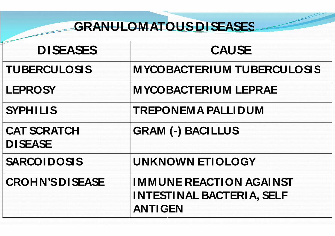
GRANULOMATOUS DISEASES
DISEASESTUBERCULOSIS MYCOBACTERIUM TUBERCULOSIS
LEPROSY MYCOBACTERIUM LEPRAE
SYPHILIS TREPONEMA
CAT SCRATCHDISEASE
GRAM (
SARCOIDOSIS UNKNOWN ETIOLOGY
CROHN’S DISEASE IMMUNE REACTION AGAINST INTESTINAL BACTERIA,ANTIGEN
GRANULOMATOUS DISEASES
CAUSEMYCOBACTERIUM TUBERCULOSIS
MYCOBACTERIUM LEPRAE
TREPONEMA PALLIDUM
GRAM (-) BACILLUS
UNKNOWN ETIOLOGY
IMMUNE REACTION AGAINST INTESTINAL BACTERIA, SELF ANTIGEN

INTRODUCTION :
Tuberculosis is caused by tubercle bacilli which belong to the genus MYCOBACTERIUM.
The burden of TB in many countries is compounded in those who have co-infection with the human immunodeficiency virus (HIV).
In 2006, the emergence of extensively drugtuberculosis (XDR-TB) was first reported.
Tuberculosis is caused by tubercle bacilli which belong to the
The burden of TB in many countries is compounded in those infection with the human immunodeficiency virus
In 2006, the emergence of extensively drug-resistant TB) was first reported.

ROBERT KOCH –Discoverer of Mycobacterium tuberculosis

INCIDENCE :
In spite of great advances in chemotherapy and immunology, tuberculosis still continues to be worldwide in distribution.
More common in developing countries like Africa, Latin America and Asia.
Other factors are malnutrition, inadequate medical care, poverty, crowding, chronic debilitating conditions like uncontrolled diabetes, alcoholism and immunocompromised states like AIDS.
In spite of great advances in chemotherapy and immunology, tuberculosis still continues to be worldwide in
More common in developing countries like Africa, Latin
Other factors are malnutrition, inadequate medical care, poverty, crowding, chronic debilitating conditions like uncontrolled diabetes, alcoholism and immunocompromised states like AIDS.

CAUSATIVE ORGANISMS :
Mycobacterium tuberculosis ( HUMANS)Mycobacterium bovis ( ANIMALS)
OTHER CAUSATIVE ORGANISMS
• Mycobacterium africanum• Mycobacterium microti
ETIOLOGY :
Mycobacterium tuberculosis ( HUMANS)( ANIMALS)
OTHER CAUSATIVE ORGANISMS

NON-MYCOBACTERIUM GENUS (Atypical tuberculosis)
• Mycobacterium leprae – causes leprosy• Mycobacterium avium – common in patients with HIV/ AIDS• Mycobacterium asiaticum
M.Tuberculosiscomplex
- These are acid – fast.
MYCOBACTERIUM GENUS (Atypical tuberculosis)
causes leprosycommon in patients with HIV/ AIDS
M.AfricanumM.Bovis
M.CanettiM.microti

RISK FACTORS :
Length of exposure time to contaminated air.
Immune status of the exposed individual.
Infected persons living in crowded or closed environments pose a particular risk to non-infected individuals.
Microepidemics have occurred in closed environments such as submarines and on transcontinental flights, hospital employees, inner city residents, nursing home residents and prisoners.
Length of exposure time to contaminated air.
Immune status of the exposed individual.
Infected persons living in crowded or closed environments infected individuals.
have occurred in closed environments such as submarines and on transcontinental flights, hospital employees, inner city residents, nursing home residents and

Factors that increase the risk of developing TB :
HIV positive.
Injecting drug users
Solid organ transplantation
Haematological malignancy , for example leukemia and lymphomas.
Silicosis
Factors that increase the risk of developing TB :
malignancy , for example leukemia and lymphomas.

MODE OF TRANSMISSION :
Human beings acquire infection with tubercle bacilli by one of the following routes:
Inhalation of organisms present in fresh cough droplets or in dried sputum from an open case of pulmonary tuberculosis.
Ingestion of the organisms leads to development of tonsillar tuberculosis or intestinal tuberculosis and selfinfected sputum of an open case of pulmonary tuberculosis or ingestion of bovine tubercle bacilli from milk of diseased cows.
Human beings acquire infection with tubercle bacilli by one of the
of organisms present in fresh cough droplets or in dried sputum from an open case of pulmonary tuberculosis.
of the organisms leads to development of tonsillar tuberculosis or intestinal tuberculosis and self-swallowing of infected sputum of an open case of pulmonary tuberculosis or ingestion of bovine tubercle bacilli from milk of diseased cows.

Inoculation of the organisms into the skin may rarely occur from infected postmortem tissue.
Transplacental route results in development of congenital tuberculosis in foetus from infected mother and is a rare mode of transmission.
of the organisms into the skin may rarely occur from infected postmortem tissue.
results in development of congenital from infected mother and is a rare

SPREAD OF TUBERCULOSIS :
The disease spreads in the body by various routes :
Local spread : This takes place by macrophages carrying the bacilli into the surrounding tissues.
Lymphatic spread : Tuberculosis is primarily an infection of lymphoid tissues.
The bacilli may pass into lymphoid follicles of pharynx, bronchi, intestines or regional lymph nodes resulting in regional tuberculosis lymphadenitis.
SPREAD OF TUBERCULOSIS :
The disease spreads in the body by various routes :
: This takes place by macrophages carrying the bacilli into the surrounding tissues.
: Tuberculosis is primarily an infection of
The bacilli may pass into lymphoid follicles of pharynx, bronchi, intestines or regional lymph nodes resulting in regional tuberculosis lymphadenitis.

Haematogenous spread : This occurs either as a result of tuberculous bacillaemia because of the drainage of lymphatics into the venous system.
This produces millet seed-sizedthe body like lungs, liver, kidneys, bones and other tissues and is known as miliary tuberculosis
This occurs either as a result of because of the drainage of lymphatics
sized lesions in different organs of the body like lungs, liver, kidneys, bones and other tissues and
tuberculosis.

By the natural passages : Lung lesions into pleura( tuberculous pleurisy)
• Trans bronchial spread into the adjacent lung segments.
Lung lesions into pleura(
Trans bronchial spread into the adjacent lung

• Tuberculous salphingitis into peritoneal cavity (tuberculous peritonitis)
• Infected sputum into larynx (tuberculous laryngitis)
• Swallowing of infected sputum (
• Renal lesions into ureter and down to trigone of bladder.
into peritoneal cavity (tuberculous
Infected sputum into larynx (tuberculous laryngitis)
Swallowing of infected sputum ( ileocaecal tuberculosis)
Renal lesions into ureter and down to trigone of bladder.

CLASSIFICATION OF TUBERCULOSIS :
Lung is the main organ affected in tuberculosis.
Depending upon the type of tissue response and age , the infection with tubercle bacilli is of two main types:
Primary tuberculosis
2. Secondary tuberculosis
CLASSIFICATION OF TUBERCULOSIS :
Lung is the main organ affected in tuberculosis.
Depending upon the type of tissue response and age , the infection with tubercle bacilli is of two main types:

EVOLUTION OF TUBERCLE :
The sequence of events which take place when the tubercle bacilli are introduced into the tissue are as follows :

After about 12 hours there is progressive infiltration by macrophages (C2a and C2b) which act as
The macrphages start phagocytosing the tubercle bacilli and either kill the bacteria or die away themselves.
In the latter case, they further proliferate locally as well as there is increased recruitment of macrophages from blood monocytes.
After about 12 hours there is progressive infiltration by macrophages (C2a and C2b) which act as opsonins.
start phagocytosing the tubercle bacilli and either kill the bacteria or die away themselves.
In the latter case, they further proliferate locally as well as there is increased recruitment of macrophages from blood monocytes.

As a part of the body’s immune response , T and B cells are activated. Activated CD4+ T cells develop the cell mediated delayed type hypersensitivity reaction formation of antibodies which play no role in body’s against the tubercle bacilli.
In 2-3 days , the macrophages undergo structural changes as a result of immune mechanisms to
As a part of the body’s immune response , T and B cells are activated. Activated CD4+ T cells develop the cell mediated delayed type hypersensitivity reaction while B cells result in formation of antibodies which play no role in body’s defence
3 days , the macrophages undergo structural changes as a result of immune mechanisms to epithelioid cells.

The epithelioid cells in time aggregate into tight clusters or granulomas. Release of cytokinesCD4+ T cells and some constituents of mycobacterial cell wall play a role in the formation of granuloma.
Some of the macrophages form fusion of adjacent cells. The giant cells may be having peripherally arranged nuclei in the form of horseshoe or ring.
The epithelioid cells in time aggregate into tight clusters or Release of cytokines in response to sensitized
CD4+ T cells and some constituents of mycobacterial cell wall play a role in the formation of granuloma.
Some of the macrophages form multinucleated giant cells by fusion of adjacent cells. The giant cells may be Langhans’ type having peripherally arranged nuclei in the form of horseshoe or

Around the mass of epithelioid cells and giant cells is a zone of lymphocytes, plasma cells and fibroblasts.
The lesion at this stage is called central necrosis.
Within 10-14 days, the centre of the cellular mass begins to undergo caseation necrosis, characterisedand high lipid content.
Around the mass of epithelioid cells and giant cells is a zone of lymphocytes, plasma cells and fibroblasts.
The lesion at this stage is called hard tubercle due to absence of
of the cellular mass begins to characterised by cheesy appearance

• This stage is called soft tubercle tuberculous lesions.
• The development of caseation necrosis is possibly due to interaction of mycobacteria with activated T cells (CD4+ helper T cells via IFN-γ and CD8+ suppressor T cells directly).
soft tubercle which is the hallmark of
The development of caseation necrosis is possibly due to interaction of mycobacteria with activated T cells (CD4+
and CD8+ suppressor T cells

Microscopically, caseation necrosis is structure less, eosinophilic and granular material with nuclear debris.
The soft tubercle which is a fully developed granuloma with caseous centre does not favourbacilli.
Microscopically, caseation necrosis is structure less, eosinophilic and granular material with nuclear debris.
The soft tubercle which is a fully developed granuloma with favour rapid proliferation of tubercle

THE FATE OF A GRANULOMA
The caseous material may undergo liquefaction, extends and discharge.
In tuberculosis of tissues like bones, joints, lymph nodes, epididymis, sinuses are formed and the sinus tracts are lined by tuberculous granulation tissue.
THE FATE OF A GRANULOMA
material may undergo liquefaction, extends and
In tuberculosis of tissues like bones, joints, lymph nodes, epididymis, sinuses are formed and the sinus tracts are lined by tuberculous granulation tissue.

May coalesce together enlarging the lesion which is surrounded by progressive fibrosis.
In the granuloma enclosed by fibrous tissue , calcium salts may get deposited in the caseous material ( calcification) and sometimes the lesion may even get ossified over the years.
enlarging the lesion which is surrounded by progressive fibrosis.
In the granuloma enclosed by fibrous tissue , calcium salts may material ( dystrophic
) and sometimes the lesion may even get ossified

PRIMARY TUBERCULOSIS
The infection of an individual who has not been previously infected or immunised is called primary tuberculosisor childhood tuberculosis.
The most commonly involved tissues are the lungs and the lymph nodes.
PRIMARY TUBERCULOSIS
The infection of an individual who has not been previously infected primary tuberculosis or Ghon’s complex
The most commonly involved tissues are the lungs and the

• Other tissues involved are tonsils and cervical lymph nodes, lesions may be found in small intestine and mesenteric lymph nodes.
• Progressive primary tuberculosis is particularly high in immunocompromised host.
Other tissues involved are tonsils and cervical lymph nodes, lesions may be found in small intestine and
Progressive primary tuberculosis is particularly high in immunocompromised host. Eg: in patients with AIDS.

Microscopically, the lung lesion consists of tuberculous granulomas with caseation necrosis.
2. Lymphatic vessel componentlung lesion contain phagocytes containing bacilli and may develop beaded, miliary tubercles along the path of hilar lymph nodes.
3. Lymph node component : This consists of enlarged hilar and tracheo-bronchial lymph nodes in the area drained. The affected lymph nodes are matted and show caseation necrosis.
the lung lesion consists of tuberculous granulomas with caseation necrosis.
Lymphatic vessel component: The lymphatics draining the lung lesion contain phagocytes containing bacilli and may
tubercles along the path of hilar lymph
This consists of enlarged hilar and bronchial lymph nodes in the area drained. The affected
lymph nodes are matted and show caseation necrosis.

FATE OF PRIMARY TUBERCULOSIS:
The lesions do not progress but instead heals by fibrosis and undergo calcification.
In some cases , the primary focus continues to grow and the caseous material is disseminated through bronchi to the other parts of the same lung or the opposite lung. This is called progressive primary tuberculosis.
At times, bacilli may enter the circulation organs. This is called primary
FATE OF PRIMARY TUBERCULOSIS:
The lesions do not progress but instead heals by fibrosis and
In some cases , the primary focus continues to grow and the material is disseminated through bronchi to the other
parts of the same lung or the opposite lung. This is called progressive primary tuberculosis.
At times, bacilli may enter the circulation ans spread to other organs. This is called primary miliary TB.

TUBERCULOSIS OF THE SPINE
• Most common site of skeletal TB• Pott’s disease.
Pathology : 1. Blood borne infection2. Vertebral body involved3. Destruction and caseation necrosis4. Spreads to disc space and next vertebra5. Vertebral bodies collapse.6. Cold abscess formation
TUBERCULOSIS OF THE SPINE
Most common site of skeletal TB
Destruction and caseation necrosisSpreads to disc space and next vertebra

PATHOGENESIS OF COLD ABSCESS :
Phagocytosis of tubercle bacilli by RES macrophages
Tuberculous granulomas (
Small patches of central
Coalesce into a large yellow mass
Breakdown of centre
PATHOGENESIS OF COLD ABSCESS :
Phagocytosis of tubercle bacilli by RES ( monocytes,
Tuberculous granulomas (Langhan’s giant cells)
Small patches of central caseous necrosis
a large yellow mass
to form a cold abscess

SECONDARY TUBERCULOSIS
The infection of an individual who has been previously infected or sensitized is called secondaryprimary or reinfection, or chronic
SECONDARY TUBERCULOSIS
The infection of an individual who has been previously secondary, or post-
chronic tuberculosis.

SOURCE OF INFECTION:
The infection may be acquired from:
- exogeneous source such as fresh dose of reinfection by the tubercle bacilli.
- endogeneous source such as reactivation of dormant primary complex.
The infection may be acquired
such as by the
such as of dormant primary

SITES INVOLVED :
Secondary tuberculosis occurs most commonly in lungs in the region of apex.Other sites and tissues which can be involved are tonsils, pharynx, larynx, small intestine and skin.
It begins as 1-2cm area of consolidation of the lung and develop a small area of central caseation necrosis and peripheral fibrosis. It occurs by infection from primary complex to the apex to the apex of the affected lung where the oxygen tension is high and favourable for growth of aerobic tubercle bacilli.
Secondary tuberculosis occurs most commonly in lungs in the
Other sites and tissues which can be involved are tonsils, pharynx, larynx, small intestine and skin.
2cm area of consolidation of the lung and develop a small area of central caseation necrosis and peripheral fibrosis. It occurs by heamatogenous spread of infection from primary complex to the apex to the apex of the affected lung where the oxygen tension is high and
for growth of aerobic tubercle bacilli.

Patients with HIV infection previously exposed to tuberculous infection have particularly reactivation of primary tuberculosis and the pattern of lesions in such cases is similar to that of primary tuberculosis i.e. with involvement of hilar lymph nodes rather than cavitary and apical lesions in the lung.
In addition, opportunistic infection with M.aviumintracellulare can occur in cases of
previously exposed to tuberculous infection have particularly high incidence of
of primary tuberculosis and the pattern of lesions in such cases is similar to that of primary tuberculosis i.e. with involvement of hilar lymph nodes rather than
and apical lesions in the lung.
In addition, opportunistic infection with can occur in cases of AIDS.

FATE OF SECONDARY PULMONARY TUBERCULOSIS
The subapical lesions in lungs can have the following courses:
The lesions may heal with fibrous scarring and calcification The lesions may coalesce together
tuberculous pneumonia and produce progressive secondary pulmonary tuberculosis with following pulmonary and extrapulmonary involvements:
Fibrocaseous tuberculosisTuberculous caseous pneumonia Miliary tuberculosis
FATE OF SECONDARY PULMONARY TUBERCULOSIS
The subapical lesions in lungs can have the following
The lesions may heal with fibrous scarring and calcification coalesce together to form larger area of
and produce progressive secondary pulmonary tuberculosis with following pulmonary and
pneumonia
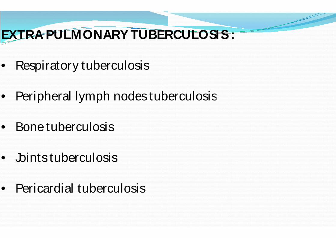
EXTRA PULMONARY TUBERCULOSIS :
• Respiratory tuberculosis
• Peripheral lymph nodes tuberculosis
• Bone tuberculosis
• Joints tuberculosis
• Pericardial tuberculosis
EXTRA PULMONARY TUBERCULOSIS :
Peripheral lymph nodes tuberculosis

FIBROCASEOUS TUBERCULOSIS
The original area of tuberculous pneumonia undergoes massive central caseation necrosis which may:
Either break into a bronchus from a cavity (open fibrocaseous tuberculosis)
Remain, as a soft caseous lesion without drainage into a bronchus or bronchiole to produce a non(chronic fibrocaseous tuberculosis).
The original area of tuberculous pneumonia undergoes massive central caseation necrosis which may:
Either break into a bronchus from a cavity (cavitary or tuberculosis)
lesion without drainage into a bronchus or bronchiole to produce a non-cavitary lesion
tuberculosis).

• The cavity provides favourableof tubercle bacilli due to high oxygen tension. The cavity may communicate with bronchial tree and becomes the source of spread of infection ( ‘ open tuberculosis
• It may implant tuberculous lesion on the mucosal lining of air passages producing endobronchial and endotracheal tuberculosis.
favourable environment for proliferation of tubercle bacilli due to high oxygen tension. The cavity may communicate with bronchial tree and becomes the source of
open tuberculosis ‘ )
It may implant tuberculous lesion on the mucosal lining of air passages producing endobronchial and endotracheal

COMPLICATIONS OF CAVITARY SECONDARY TUBERCULOSIS ARE:
- Aneurysms of patent arteries causing
- Bronchopleural fistula
- Tuberculous empyema
- Thickened pleura from adhesions of parietal pleura
COMPLICATIONS OF CAVITARY SECONDARY TUBERCULOSIS
Aneurysms of patent arteries causing haemoptysis
Thickened pleura from adhesions of parietal pleura

The caseous material from a case of secondary tuberculosis in an individual with high degree of hypersensitivity may spread to rest of the lung producing
material from a case of secondary tuberculosis in an individual with high degree of hypersensitivity may spread to rest of the lung producing caseous pneumonia.

Microscopically, the lesions show exudative reaction with oedema, fibrin, polymorphs and monocytes but numerous tubercle bacilli can be demonstrated in the exudates.
the lesions show exudative reaction with , fibrin, polymorphs and monocytes but numerous
tubercle bacilli can be demonstrated in the exudates.

MILIARY TUBERCULOSIS
This is lymphoheamotogenousinfection from primary focus or later stage of tuberculosis.
MILIARY TUBERCULOSIS
lymphoheamotogenous spread of tuberculous infection from primary focus or later stage of tuberculosis.

SPREAD OF INFECTION:
The spread is either by entry of infection into vein producing disseminated or isolated organ lesion in different extra-pulmonary sites( brain, meninges, genitourinary tract and bone marrow) or into pulmonary artery restricting the development of miliary lesions within the lung.
The spread is either by entry of infection into pulmonary producing disseminated or isolated organ lesion in
pulmonary sites( eg: liver, spleen, kidney, brain, meninges, genitourinary tract and bone marrow) or
restricting the development of lesions within the lung.

MILIARY LESION
The miliary lesions are millet seedareas without grossly visible caseation necrosis
MICROSCOPICALLY, the lesions show the structure of tubercles with minute areas of caseation necrosis.
lesions are millet seed-sized, yellowish, firm areas without grossly visible caseation necrosis
MICROSCOPICALLY, the lesions show the structure of tubercles with minute areas of caseation necrosis.

CLINICAL FEATURES :
Referable to lungs such as:productive cough( may be with pleural effusion, dyspnoea,orthopnoea etc. Chest X-ray may show typical apical changes like pleural effusion, nodularity and miliarylung parenchyma.
productive cough( may be with haemoptysis),
ray may show typical apical changes like pleural miliary or diffuse infiltrates in the

Systemic features :–1. such as low grade fever, 2. night sweats, 3. fatigue,4. loss of weight and appetite. 5. Long-standing and untreated cases of tuberculosis may
develop systemic secondary amyloidosis.Causes of death in pulmonary tuberculosis are usually pulmonary insufficiency, pulmonary to disseminated miliary tuberculosis, secondary amyloidosis
standing and untreated cases of tuberculosis may develop systemic secondary amyloidosis.
Causes of death in pulmonary tuberculosis are usually pulmonary insufficiency, pulmonary haemorrhage, sepsis due
tuberculosis, corpulmonale or