tuberculosis and lung damage: from epidemiology to...
TRANSCRIPT
Tuberculosis and lung damage: fromepidemiology to pathophysiology
Shruthi Ravimohan1, Hardy Kornfeld2, Drew Weissman1 andGregory P. Bisson1,3
Affiliations: 1Dept of Medicine, Division of Infectious Diseases, Perelman School of Medicine at the Universityof Pennsylvania, Philadelphia, PA, USA. 2Dept of Medicine, University of Massachusetts Medical School,Worcester, MA, USA. 3Dept of Biostatistics and Epidemiology, Center for Clinical Epidemiology andBiostatistics, Perelman School of Medicine at the University of Pennsylvania, Philadelphia, PA, USA.
Correspondence: Shruthi Ravimohan, Dept of Medicine, Division of Infectious Diseases, University ofPennsylvania, 423 Guardian Drive, 930 Blockley Hall, Philadelphia, PA, USA.E-mail: [email protected]
@ERSpublicationsHost factors driving lung injury in TB likely contribute to variable patterns of pulmonary impairmentafter TB http://ow.ly/a3of30hBsxB
Cite this article as: Ravimohan S, Kornfeld H, Weissman D, et al. Tuberculosis and lung damage: fromepidemiology to pathophysiology. Eur Respir Rev 2018; 27: 170077 [https://doi.org/10.1183/16000617.0077-2017].
ABSTRACT A past history of pulmonary tuberculosis (TB) is a risk factor for long-term respiratoryimpairment. Post-TB lung dysfunction often goes unrecognised, despite its relatively high prevalence andits association with reduced quality of life. Importantly, specific host and pathogen factors causing lungimpairment remain unclear. Host immune responses probably play a dominant role in lung damage, asexcessive inflammation and elevated expression of lung matrix-degrading proteases are common duringTB. Variability in host genes that modulate these immune responses may determine the severity of lungimpairment, but this hypothesis remains largely untested. In this review, we provide an overview of theepidemiological literature on post-TB lung impairment and link it to data on the pathogenesis of lunginjury from the perspective of dysregulated immune responses and immunogenetics.
IntroductionA third of the world’s population is infected with Mycobacterium tuberculosis (MTB), and over 9 millionnew cases of tuberculosis (TB) are reported annually [1]. Treatment of drug-susceptible pulmonary TB ishighly effective, with 85% (66 million cases) of reported cases estimated to have been successfully treatedbetween 1995 and 2015 [1]. However, up to half of TB survivors have some form of persistent pulmonarydysfunction despite microbiologic cure [2–5]. Pulmonary dysfunction, ranging from minor abnormalitiesto severe breathlessness, can increase the risk of death from respiratory causes [6–9]. Furthermore, treatedTB patients appear to contribute substantially to the growing worldwide burden of chronic obstructivepulmonary disease (COPD) [10–12]. These findings call for strategies to address pulmonary impairmentafter TB (PIAT).
Copyright ©ERS 2018. ERR articles are open access and distributed under the terms of the Creative CommonsAttribution Non-Commercial Licence 4.0.
Received: July 05 2017 | Accepted after revision: Oct 28 2017
Support statement: This study was supported by the National Center for Advancing Translational Sciences(KL2TR001879) and the National Institute of Allergy and Infectious Diseases (R01AI120821) of the National Institutesof Health. Funding information for this article has been deposited with the Crossref Funder Registry.
Conflict of interest: Disclosures can be found alongside this article at err.ersjournals.com
Provenance: Submitted article, peer reviewed.
https://doi.org/10.1183/16000617.0077-2017 Eur Respir Rev 2018; 27: 170077
REVIEWTUBERCULOSIS
A notable feature of lung involvement in TB is its striking heterogeneity. This is observed on formal lungfunction testing in terms of the magnitude of pulmonary function, ranging from no impairment to severedysfunction [3, 7, 8] and the specific types of ventilatory defects [3, 11, 13]. Patients may present withcavitation, fibrosis or nodular infiltrates, or have a mix of these pulmonary pathologies [14, 15]. Thisimmense variability may relate to host–pathogen interactions and the diverse immunological events thatcan follow. We also hypothesise that heterogeneity in lung damage may be partially attributed to variationin genes coding for or regulating host immune responses. Elucidating the immune pathways and geneticrisk factors for TB-associated lung injury could inform therapies that specifically target immunologicalfactors responsible for lung injury.
This review summarises the epidemiology of PIAT, examines TB-associated lung pathology potentiallylinked to lung dysfunction, and reviews the immunological and plausible genetic correlates of lung tissuedamage in TB. We also describe several processes, such as pulmonary cavitation, fibrosis andbronchiectasis, which probably contribute collectively to lung remodelling in TB and PIAT. These termsare defined in table 1.
Search strategyFor this review we identified references in PubMed that were published up to May 2017. We specificallysearched for studies describing the epidemiology of impaired lung function post-TB treatment using thesearch terms “epidemiology”, “pulmonary tuberculosis”, “pulmonary function”, “obstruction AND/ORrestrictive”, “bronchiectasis”, “fibrosis”, “cavitation” and “TB treatment”. For studies on lung pathologyand mediators of inflammation in TB we used the terms “pulmonary tuberculosis”, “lung pathology”,“immunopathology”, “matrix metalloproteinase”, “inflammatory biomarkers OR inflammation”,“neutrophils” and “CD4 T cells”. Genetic studies were queried using the terms “pulmonary tuberculosis”,“genetics”, “COPD” and “idiopathic pulmonary fibrosis”. Relevant articles published in English thatresulted from the searches, and references cited therein, were reviewed.
PIATEpidemiologyInitial studies conducted among untreated or incompletely treated TB patients highlighted that lungdisability was a relatively common outcome [16, 17]. Many studies since have reported on lungimpairment at TB treatment completion [5, 7, 8, 18], with persistence of defects several years post cure(table 2) [2, 4, 13]. For example, a South African study observed airflow obstruction in 68% of patientswith a history of TB treated up to 16 years (mean 5.6 years) prior to assessment [13]. Althoughlongitudinal studies have shown improvement in pulmonary function with TB treatment, a considerableproportion of patients have irreversible and often progressive pulmonary defects [3, 4, 7, 8, 20]. In aprospective study of 74 hospitalised patients with newly diagnosed TB, 54% had improved lung functionwith treatment and the rest had either no change or worsening pulmonary function [3]. Even in the
TABLE 1 Definitions for processes contributing to lung remodelling during pulmonary tuberculosis (TB) and pulmonaryimpairment after TB
Term Definition
Pulmonary cavitation Process by which normal pulmonary tissue is obliterated, becoming gas-filled spaces or cavities in the lung.This process initially involves caseous necrosis of lipid pneumonia lesions, producing caseous pneumonia.During caseation, alveolar cells and septa are destroyed along with neighbouring vessels and bronchi. Cavitiesform when these regions of caseous pneumonia liquefy, fragment and are released upon coughing.
Pulmonary fibrosis Results from long-term lung tissue injury that is characterised by excessive extracellular matrix deposition inthe lung. Replacement of normal lung parenchyma with collagenous tissue results in architectural changes inthe lung, such as thickening and stiffening of the lung walls.
Bronchiectasis Manifests as irreversible bronchial dilatation and thickening of the bronchial wall. Elastic and muscularcomponents of the bronchial wall are destroyed in bronchiectasis. Bronchial dilatation associated withbronchiectasis in TB may be due to multiple factors, including traction from surrounding tissue fibrosis,caseous necrosis that makes its way into the bronchi, and elevated luminal pressure due to coughing.Bronchiectasis can also predispose to recurrent exacerbations of purulent sputum production and possiblybacterial pneumonia in subsequent years.
Pulmonary impairmentafter TB
A broad term we use in this review to refer to lung dysfunction that includes airflow obstruction, restrictiveventilatory defects and impaired gas exchange. Pulmonary impairment after TB is probably downstream of awide variety of lung remodelling events, some of which are described above. Given the lung’s considerablereserve, these structural changes may manifest as symptoms and pulmonary disability over a period of time.
https://doi.org/10.1183/16000617.0077-2017 2
TUBERCULOSIS | S. RAVIMOHAN ET AL.
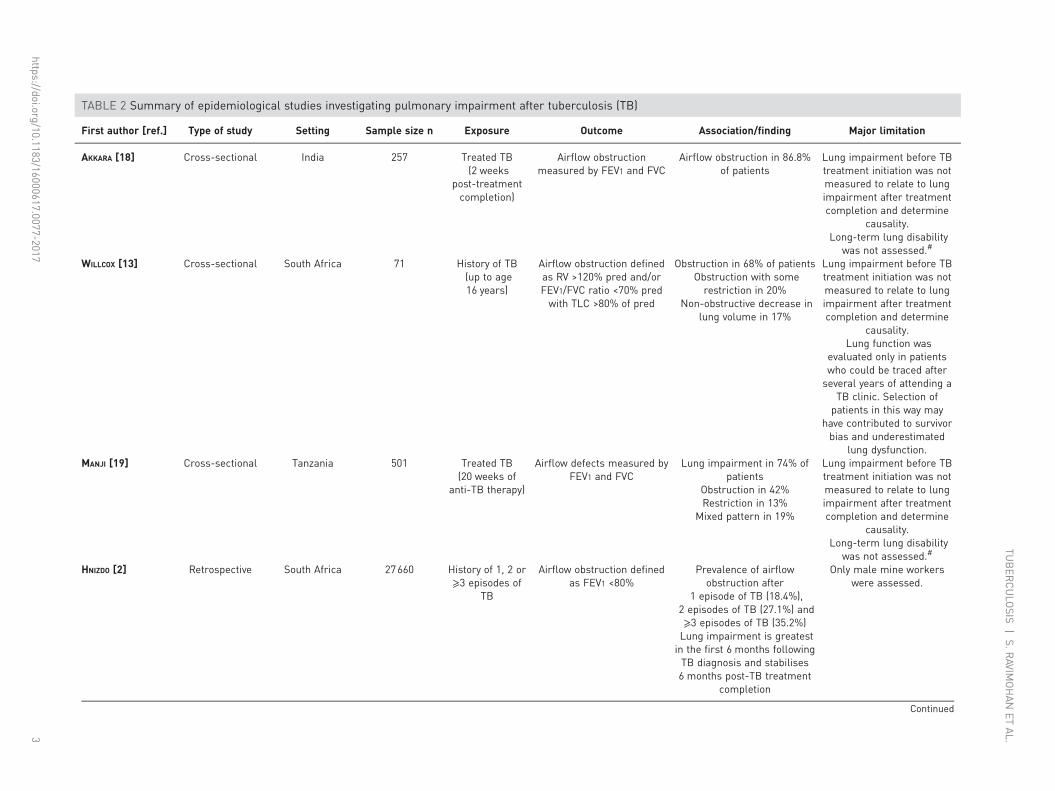
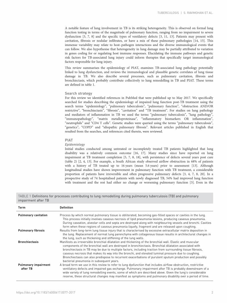
TABLE 2 Summary of epidemiological studies investigating pulmonary impairment after tuberculosis (TB)
First author [ref.] Type of study Setting Sample size n Exposure Outcome Association/finding Major limitation
AKKARA [18] Cross-sectional India 257 Treated TB(2 weeks
post-treatmentcompletion)
Airflow obstructionmeasured by FEV1 and FVC
Airflow obstruction in 86.8%of patients
Lung impairment before TBtreatment initiation was notmeasured to relate to lungimpairment after treatmentcompletion and determine
causality.Long-term lung disability
was not assessed.#
WILLCOX [13] Cross-sectional South Africa 71 History of TB(up to age16 years)
Airflow obstruction definedas RV >120% pred and/orFEV1/FVC ratio <70% predwith TLC >80% of pred
Obstruction in 68% of patientsObstruction with somerestriction in 20%
Non-obstructive decrease inlung volume in 17%
Lung impairment before TBtreatment initiation was notmeasured to relate to lungimpairment after treatmentcompletion and determine
causality.Lung function was
evaluated only in patientswho could be traced afterseveral years of attending a
TB clinic. Selection ofpatients in this way may
have contributed to survivorbias and underestimated
lung dysfunction.MANJI [19] Cross-sectional Tanzania 501 Treated TB
(20 weeks ofanti-TB therapy)
Airflow defects measured byFEV1 and FVC
Lung impairment in 74% ofpatients
Obstruction in 42%Restriction in 13%
Mixed pattern in 19%
Lung impairment before TBtreatment initiation was notmeasured to relate to lungimpairment after treatmentcompletion and determine
causality.Long-term lung disability
was not assessed.#
HNIZDO [2] Retrospective South Africa 27660 History of 1, 2 or⩾3 episodes of
TB
Airflow obstruction definedas FEV1 <80%
Prevalence of airflowobstruction after
1 episode of TB (18.4%),2 episodes of TB (27.1%) and⩾3 episodes of TB (35.2%)Lung impairment is greatestin the first 6 months followingTB diagnosis and stabilises6 months post-TB treatment
completion
Only male mine workerswere assessed.
Continued
https://doi.org/10.1183/16000617.0077-20173
TUBER
CULO
SIS|S.R
AVIMOHAN
ETAL.
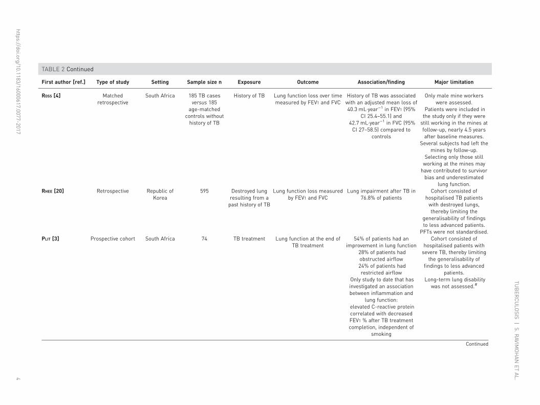
TABLE 2 Continued
First author [ref.] Type of study Setting Sample size n Exposure Outcome Association/finding Major limitation
ROSS [4] Matchedretrospective
South Africa 185 TB casesversus 185
age-matchedcontrols withouthistory of TB
History of TB Lung function loss over timemeasured by FEV1 and FVC
History of TB was associatedwith an adjusted mean loss of40.3 mL·year−1 in FEV1 (95%
CI 25.4–55.1) and42.7 mL·year−1 in FVC (95%CI 27–58.5) compared to
controls
Only male mine workerswere assessed.
Patients were included inthe study only if they werestill working in the mines atfollow-up, nearly 4.5 yearsafter baseline measures.
Several subjects had left themines by follow-up.
Selecting only those stillworking at the mines mayhave contributed to survivorbias and underestimated
lung function.RHEE [20] Retrospective Republic of
Korea595 Destroyed lung
resulting from apast history of TB
Lung function loss measuredby FEV1 and FVC
Lung impairment after TB in76.8% of patients
Cohort consisted ofhospitalised TB patientswith destroyed lungs,thereby limiting the
generalisability of findingsto less advanced patients.
PFTs were not standardised.PLIT [3] Prospective cohort South Africa 74 TB treatment Lung function at the end of
TB treatment54% of patients had an
improvement in lung function28% of patients hadobstructed airflow24% of patients hadrestricted airflow
Only study to date that hasinvestigated an associationbetween inflammation and
lung function:elevated C-reactive proteincorrelated with decreasedFEV1 % after TB treatmentcompletion, independent of
smoking
Cohort consisted ofhospitalised patients withsevere TB, thereby limiting
the generalisability offindings to less advanced
patients.Long-term lung disability
was not assessed.#
Continued
https://doi.org/10.1183/16000617.0077-20174
TUBER
CULO
SIS|S.R
AVIMOHAN
ETAL.
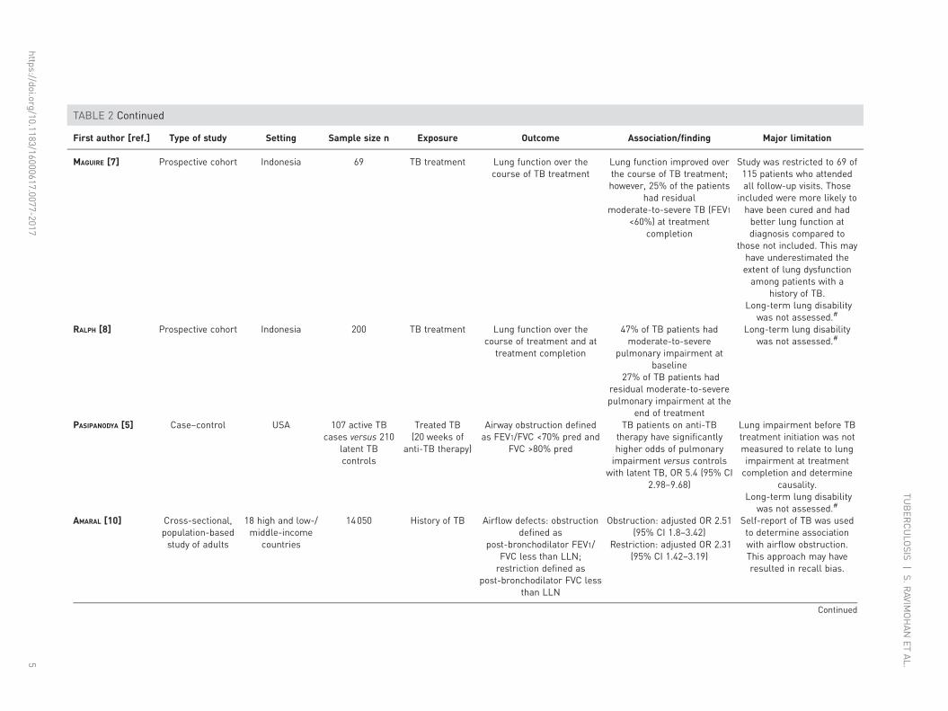
TABLE 2 Continued
First author [ref.] Type of study Setting Sample size n Exposure Outcome Association/finding Major limitation
MAGUIRE [7] Prospective cohort Indonesia 69 TB treatment Lung function over thecourse of TB treatment
Lung function improved overthe course of TB treatment;however, 25% of the patients
had residualmoderate-to-severe TB (FEV1
<60%) at treatmentcompletion
Study was restricted to 69 of115 patients who attendedall follow-up visits. Those
included were more likely tohave been cured and hadbetter lung function atdiagnosis compared to
those not included. This mayhave underestimated theextent of lung dysfunctionamong patients with a
history of TB.Long-term lung disability
was not assessed.#
RALPH [8] Prospective cohort Indonesia 200 TB treatment Lung function over thecourse of treatment and at
treatment completion
47% of TB patients hadmoderate-to-severe
pulmonary impairment atbaseline
27% of TB patients hadresidual moderate-to-severepulmonary impairment at the
end of treatment
Long-term lung disabilitywas not assessed.#
PASIPANODYA [5] Case–control USA 107 active TBcases versus 210
latent TBcontrols
Treated TB(20 weeks of
anti-TB therapy)
Airway obstruction definedas FEV1/FVC <70% pred and
FVC >80% pred
TB patients on anti-TBtherapy have significantlyhigher odds of pulmonaryimpairment versus controls
with latent TB, OR 5.4 (95% CI2.98–9.68)
Lung impairment before TBtreatment initiation was notmeasured to relate to lungimpairment at treatmentcompletion and determine
causality.Long-term lung disability
was not assessed.#
AMARAL [10] Cross-sectional,population-basedstudy of adults
18 high and low-/middle-income
countries
14050 History of TB Airflow defects: obstructiondefined as
post-bronchodilator FEV1/FVC less than LLN;restriction defined as
post-bronchodilator FVC lessthan LLN
Obstruction: adjusted OR 2.51(95% CI 1.8–3.42)
Restriction: adjusted OR 2.31(95% CI 1.42–3.19)
Self-report of TB was usedto determine associationwith airflow obstruction.This approach may haveresulted in recall bias.
Continued
https://doi.org/10.1183/16000617.0077-20175
TUBER
CULO
SIS|S.R
AVIMOHAN
ETAL.
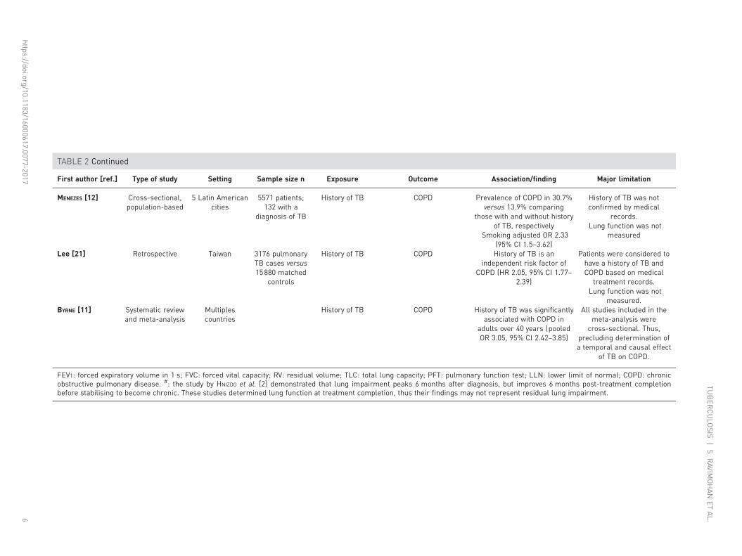
TABLE 2 Continued
First author [ref.] Type of study Setting Sample size n Exposure Outcome Association/finding Major limitation
MENEZES [12] Cross-sectional,population-based
5 Latin Americancities
5571 patients;132 with a
diagnosis of TB
History of TB COPD Prevalence of COPD in 30.7%versus 13.9% comparing
those with and without historyof TB, respectively
Smoking adjusted OR 2.33(95% CI 1.5–3.62)
History of TB was notconfirmed by medical
records.Lung function was not
measured
Lee [21] Retrospective Taiwan 3176 pulmonaryTB cases versus15880 matched
controls
History of TB COPD History of TB is anindependent risk factor of
COPD (HR 2.05, 95% CI 1.77–2.39)
Patients were considered tohave a history of TB andCOPD based on medical
treatment records.Lung function was not
measured.BYRNE [11] Systematic review
and meta-analysisMultiplescountries
History of TB COPD History of TB was significantlyassociated with COPD in
adults over 40 years (pooledOR 3.05, 95% CI 2.42–3.85)
All studies included in themeta-analysis were
cross-sectional. Thus,precluding determination ofa temporal and causal effect
of TB on COPD.
FEV1: forced expiratory volume in 1 s; FVC: forced vital capacity; RV: residual volume; TLC: total lung capacity; PFT: pulmonary function test; LLN: lower limit of normal; COPD: chronicobstructive pulmonary disease. #: the study by HNIZDO et al. [2] demonstrated that lung impairment peaks 6 months after diagnosis, but improves 6 months post-treatment completionbefore stabilising to become chronic. These studies determined lung function at treatment completion, thus their findings may not represent residual lung impairment.
https://doi.org/10.1183/16000617.0077-20176
TUBER
CULO
SIS|S.R
AVIMOHAN
ETAL.

outpatient setting, where patients are presumably healthier than those who are hospitalised, a quarter [7]to one-third [8] of the patients had moderate-to-severe airway limitation at treatment completion. Othergroups have corroborated these findings in multiple other settings [5, 19, 22, 23]. A limitation of many ofthe studies described above, however, was their small-to-moderate sample size (study-specific limitationsare described in table 2).
An increasing number of population-based studies have demonstrated that a history of TB increases riskfor airflow obstruction and COPD [10–12, 22]. A study of 14050 patients from 18 countries revealed thata history of TB increased risk for obstructive airway disease by 2.5-fold independent of smoking and otherclinical factors [10]. Another study (n=5571) showed a higher prevalence of COPD in those with a historyof TB (30.7%) versus those without (13.9%) [12]. In a large study of 13522 adults aged ⩾40 years set inSouth Korea, a history of TB and lesions on chest radiographs were associated with 4.47 increased odds ofairflow obstruction (95% CI 3.07–6.51) after adjusting for age, smoking, body mass index (BMI) and otherconfounders [24]. Furthermore, a meta-analysis demonstrated that a history of treated TB was a risk factorfor COPD (pooled OR 3.05, 95% CI 2.42–3.85) independent of smoking and age [11].
Risk factors associated with PIAT have not been fully elucidated and the relationships are probablycomplicated. It has been suggested that smoking, which is an established risk factor for COPD, maycontribute to PIAT [2, 3, 5]. However, some reports have found no such association [4, 7, 8]. For example,in one study individuals with a history of TB were twice as likely to be current smokers than those in thenon-TB group; yet, greater lung dysfunction over time in the TB group was independent of smoking in amultivariate analysis [4]. A similar lack of association between smoking and impaired lung function wasobserved among patients completing TB treatment in Indonesia [7, 8]. HIV co-infection may be anadditional risk factor for lung function decline. Several studies have now shown that HIV-infected patientsare at increased risk for impaired gas exchange as well as airway obstruction [25–27]. However, data areconflicting and sparse with respect to lung function in HIV/TB co-infected patients [2, 8]. Other riskfactors for poor lung function after TB may include comorbidities such as diabetes, as well asenvironmental factors like indoor smoke from biomass fuels. Although these risks have been linked toCOPD [26, 28], they remain to be investigated in the context of post-TB lung disability.
Variability in lung function deficitsPIAT can involve airflow obstruction and/or restrictive ventilatory defects, as well as impairment ingas exchange (figure 1) [3, 11, 13, 15, 20]. Airflow defects can be detected by spirometry, whichquantitates the flow and volume of air inhaled and exhaled [29]. Impaired gas exchange is determined bymeasuring the diffusing capacity of the lung for carbon monoxide (DLCO) [30]. The American ThoracicSociety and the European Respiratory Society provide standardised guidelines on conducting and
Bronchogenic spread andendobronchial diseaseCaseous necrosis leads
to break down of cavitary
lesions, which pass through
bronchial walls
Pulmonary cavitationDistortion of airways
Obstruction
Reduced capacity
to expel air out of
the lungs
Decrease in FEV1
Restriction
Reduced capacity
to inhale to full
potential
Decrease in FVC
and/or increase in
FEV1/FVC ratio
Airway narrowingStiffening of lung
parenchyma
Bronchovascular
distortion
Excessive inflammation Excessive fibrosis
Fibrotic bands
Pleural thickeningBronchiectasisDestruction of elastic and
muscular components of
bronchial walls
FIGURE 1 Mechanisms and radiographic features associated with airflow obstruction and restrictiveventilatory defects in patients with a history of tuberculosis. FEV1: forced expiratory volume in 1 s; FVC: forcedvital capacity.
https://doi.org/10.1183/16000617.0077-2017 7
TUBERCULOSIS | S. RAVIMOHAN ET AL.
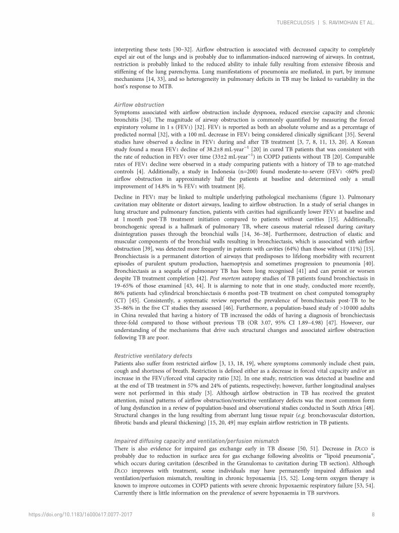
interpreting these tests [30–32]. Airflow obstruction is associated with decreased capacity to completelyexpel air out of the lungs and is probably due to inflammation-induced narrowing of airways. In contrast,restriction is probably linked to the reduced ability to inhale fully resulting from extensive fibrosis andstiffening of the lung parenchyma. Lung manifestations of pneumonia are mediated, in part, by immunemechanisms [14, 33], and so heterogeneity in pulmonary deficits in TB may be linked to variability in thehost’s response to MTB.
Airflow obstructionSymptoms associated with airflow obstruction include dyspnoea, reduced exercise capacity and chronicbronchitis [34]. The magnitude of airway obstruction is commonly quantified by measuring the forcedexpiratory volume in 1 s (FEV1) [32]. FEV1 is reported as both an absolute volume and as a percentage ofpredicted normal [32], with a 100 mL decrease in FEV1 being considered clinically significant [35]. Severalstudies have observed a decline in FEV1 during and after TB treatment [3, 7, 8, 11, 13, 20]. A Koreanstudy found a mean FEV1 decline of 38.2±8 mL·year−1 [20] in cured TB patients that was consistent withthe rate of reduction in FEV1 over time (33±2 mL·year−1) in COPD patients without TB [20]. Comparablerates of FEV1 decline were observed in a study comparing patients with a history of TB to age-matchedcontrols [4]. Additionally, a study in Indonesia (n=200) found moderate-to-severe (FEV1 <60% pred)airflow obstruction in approximately half the patients at baseline and determined only a smallimprovement of 14.8% in % FEV1 with treatment [8].
Decline in FEV1 may be linked to multiple underlying pathological mechanisms (figure 1). Pulmonarycavitation may obliterate or distort airways, leading to airflow obstruction. In a study of serial changes inlung structure and pulmonary function, patients with cavities had significantly lower FEV1 at baseline andat 1 month post-TB treatment initiation compared to patients without cavities [15]. Additionally,bronchogenic spread is a hallmark of pulmonary TB, where caseous material released during cavitarydisintegration passes through the bronchial walls [14, 36–38]. Furthermore, destruction of elastic andmuscular components of the bronchial walls resulting in bronchiectasis, which is associated with airflowobstruction [39], was detected more frequently in patients with cavities (64%) than those without (11%) [15].Bronchiectasis is a permanent distortion of airways that predisposes to lifelong morbidity with recurrentepisodes of purulent sputum production, haemoptysis and sometimes progression to pneumonia [40].Bronchiectasis as a sequela of pulmonary TB has been long recognised [41] and can persist or worsendespite TB treatment completion [42]. Post mortem autopsy studies of TB patients found bronchiectasis in19–65% of those examined [43, 44]. It is alarming to note that in one study, conducted more recently,86% patients had cylindrical bronchiectasis 6 months post-TB treatment on chest computed tomography(CT) [45]. Consistently, a systematic review reported the prevalence of bronchiectasis post-TB to be35–86% in the five CT studies they assessed [46]. Furthermore, a population-based study of >10000 adultsin China revealed that having a history of TB increased the odds of having a diagnosis of bronchiectasisthree-fold compared to those without previous TB (OR 3.07, 95% CI 1.89–4.98) [47]. However, ourunderstanding of the mechanisms that drive such structural changes and associated airflow obstructionfollowing TB are poor.
Restrictive ventilatory defectsPatients also suffer from restricted airflow [3, 13, 18, 19], where symptoms commonly include chest pain,cough and shortness of breath. Restriction is defined either as a decrease in forced vital capacity and/or anincrease in the FEV1/forced vital capacity ratio [32]. In one study, restriction was detected at baseline andat the end of TB treatment in 57% and 24% of patients, respectively; however, further longitudinal analyseswere not performed in this study [3]. Although airflow obstruction in TB has received the greatestattention, mixed patterns of airflow obstruction/restrictive ventilatory defects was the most common formof lung dysfunction in a review of population-based and observational studies conducted in South Africa [48].Structural changes in the lung resulting from aberrant lung tissue repair (e.g. bronchovascular distortion,fibrotic bands and pleural thickening) [15, 20, 49] may explain airflow restriction in TB patients.
Impaired diffusing capacity and ventilation/perfusion mismatchThere is also evidence for impaired gas exchange early in TB disease [50, 51]. Decrease in DLCO isprobably due to reduction in surface area for gas exchange following alveolitis or “lipoid pneumonia”,which occurs during cavitation (described in the Granulomas to cavitation during TB section). AlthoughDLCO improves with treatment, some individuals may have permanently impaired diffusion andventilation/perfusion mismatch, resulting in chronic hypoxaemia [15, 52]. Long-term oxygen therapy isknown to improve outcomes in COPD patients with severe chronic hypoxaemic respiratory failure [53, 54].Currently there is little information on the prevalence of severe hypoxaemia in TB survivors.
https://doi.org/10.1183/16000617.0077-2017 8
TUBERCULOSIS | S. RAVIMOHAN ET AL.
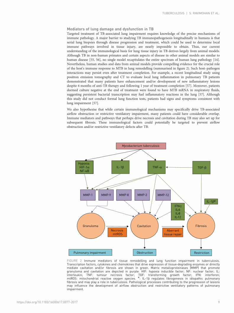
Mediators of lung damage and dysfunction in TBTargeted treatment of TB-associated lung impairment requires knowledge of the precise mechanisms ofimmune pathology. A major barrier to studying TB immunopathogenesis longitudinally in humans is thatserial lung biopsies through disease progression and treatment, which could be used to determine localimmune pathways involved in tissue injury, are nearly impossible to obtain. Thus, our currentunderstanding of the immunological basis for lung tissue injury in TB derives largely from animal models.Although TB in non-human primates and certain aspects of disease in other animal models are similar tohuman disease [55, 56], no single model recapitulates the entire spectrum of human lung pathology [14].Nevertheless, human studies and data from animal models provide compelling evidence for the crucial roleof the host’s immune response to MTB in lung remodelling (summarised in figure 2). Such host–pathogeninteractions may persist even after treatment completion. For example, a recent longitudinal study usingpositron emission tomography and CT to evaluate local lung inflammation in pulmonary TB patientsdemonstrated that many patients have enhancement and/or development of new inflammatory lesionsdespite 6 months of anti-TB therapy and following 1 year of treatment completion [57]. Moreover, patientsdeemed culture negative at the end of treatment were found to have MTB mRNA in respiratory fluids,suggesting persistent bacterial transcription may fuel inflammatory reactions in the lung [57]. Althoughthis study did not conduct formal lung function tests, patients had signs and symptoms consistent withlung impairment [57].
We also hypothesise that while certain immunological mechanisms may specifically drive TB-associatedairflow obstruction or restrictive ventilatory impairment, many patients could have considerable overlap.Immune mediators and pathways that perhaps drive necrosis and cavitation during TB may also set up forsubsequent fibrosis. These immunological factors could potentially be targeted to prevent airflowobstruction and/or restrictive ventilatory defects after TB.
IL-1βHypoxia:
HIF-1αNF-κB
TNF-α TGF-β
MMP-1 MMP-8MMP-9
Granuloma Cavitation Fibrosis
Obstruction
MMP-3 MMP-12
Mycobacterium tuberculosis
Restriction
IFN-γIL-6IL-8
IL-12
NecrosismtROS
Aberrant
tissue repair
Pulmonary impairment
IL-1β#
FIGURE 2 Immune mediators of tissue remodelling and lung function impairment in tuberculosis.Transcription factors, cytokines and chemokines that drive expression of tissue-degrading enzymes or directlymediate cavitation and/or fibrosis are shown in green. Matrix metalloproteinases (MMP) that promotegranuloma and cavitation are depicted in purple. HIF: hypoxia inducible factor; NF: nuclear factor; IL:interleukin; TNF: tumour necrosis factor; TGF: transforming growth factor; IFN: interferon;mtROS: mitochondrial reactive oxygen species. #: IL-1β regulates fibrogenesis in idiopathic pulmonaryfibrosis and may play a role in tuberculosis. Pathological processes contributing to the progression of lesionsmay influence the development of airflow obstruction and restrictive ventilatory patterns of pulmonaryimpairment.
https://doi.org/10.1183/16000617.0077-2017 9
TUBERCULOSIS | S. RAVIMOHAN ET AL.
Granulomas to cavitation during TBThe host’s immune system responds to the invading mycobacterium and triggers granuloma formationduring primary infection [14, 58]. A granuloma is a highly organised structure consisting of manyimmune cell types (e.g. macrophages, neutrophils, natural killer cells and T- and B-cells) that surround acaseous necrotic core of MTB-infected alveolar macrophages [33, 58]. The granuloma is traditionallythought to be host-protective by sequestering and preventing dissemination of MTB, but studies using thezebrafish model for TB have demonstrated that granulomas can be conducive to MTB proliferation andspread [59, 60]. Moreover, there appears to be substantial heterogeneity in the bacterial load, size andinflammatory profile between granulomatous lesions within a single host based on recent non-humanprimate studies [61–63]. It has been shown that a single or few granulomas that fail to controlmycobacterial proliferation can dramatically influence disease progression and clinical outcome [64].
A widely held view, based on data from animal models, is that these granulomas coalesce and breakdownvia liquefactive necrosis, leaving behind a cavity during active disease [56, 65]. However, human studiessuggest that cavities originate from lipid pneumonia during post-primary TB [14, 66]. These lipidpneumonia lesions may develop into areas of caseous pneumonia as a result of caseous necrosis [14, 66].During caseous necrosis, alveolar cells are destroyed, along with nearby structures such as vessels andbronchi [14]. However, elastic fibres of the alveolar walls and vessels appear to remain intact [14, 67]. Thisnecrotic tissue begins to soften and fissure and is eventually coughed out [14, 67]. Gas-filled spacessurrounded by a collagen capsule in turn replace normal lung tissue following cavitation.
Although the precise immune mechanisms underlying liquefactive or caseous necrosis are not fullyunderstood, robust immune responses probably play a significant role [68, 69]. Initial evidence for thiscomes from Robert Koch’s observations over a century ago. He noted that when guinea pigs previouslyinfected with MTB are rechallenged, they develop necrotic lesions that expand in circumference at the siteof cutaneous injection [70]. The Koch Phenomenon has been described by others [71], including MOREIRA
et al. [72], where MTB-infected mice were treated with a recombinant bacillus Calmette–Guerin (BCG)vaccine that released cytokines (e.g. tumour necrosis factor (TNF)-α) shown to reduce bacterial burden ina pre-infection vaccine model. Paradoxically, post-infection treatment with BCG–TNF-α exacerbated lungpathology without decreasing MTB load in these mice [72]. Furthermore, SHWARTZMAN [73] found thatrabbits previously injected with Gram-negative bacteria in the skin developed haemorrhagic necrosis at theinitial site of injection following an intravenous injection of the same bacteria 24 h later. This reaction maybe explained, in part, by T-cell mediated MTB-specific immune responses, given that depleting CD4+
T-cells in pre-immunised mice inhibited this reaction [74]. This study also highlighted an important rolefor TNF-α in perpetuating necrosis [74]. Other studies have corroborated these findings [75]. In additionto an excessive and tissue-damaging immune response directed towards viable and nonviable mycobacteria[65, 76], dysregulation of host lipid metabolism has recently been hypothesised to influence caseousnecrosis [77]. We expand our discussion on possible immune mechanisms underlying lung pathology anddysfunction in the sections below.
Regardless of the way granulomas and cavities form, they can have variable trajectories of resolutionthrough the course of disease or treatment, and may undergo abnormal repair resulting in focal orextensive tissue fibrosis [14, 78]. Thus, it is plausible that host immune responses that drive inflammation,cavitation and fibrosis contribute to the variable patterns of lung healing, manifesting as persistent airflowobstruction and/or restrictive ventilatory defects. Additionally, differences in the quality and quantity ofimmune effector responses underlying these processes may also contribute to variability in PIAT.
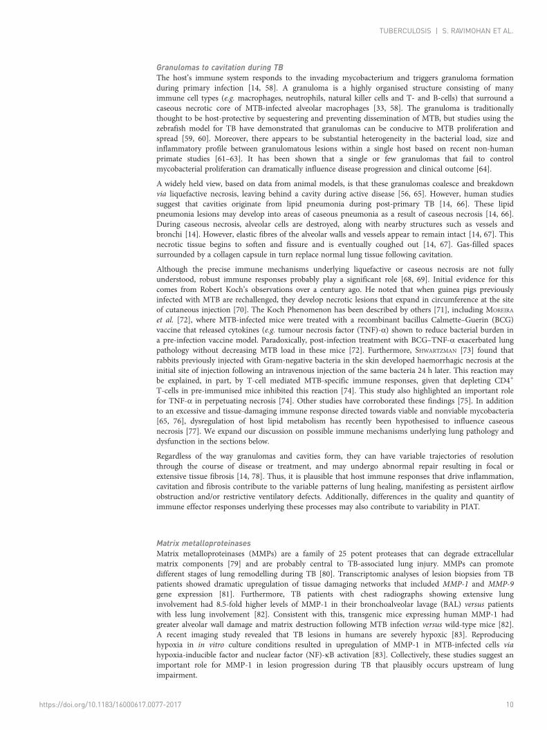
Matrix metalloproteinasesMatrix metalloproteinases (MMPs) are a family of 25 potent proteases that can degrade extracellularmatrix components [79] and are probably central to TB-associated lung injury. MMPs can promotedifferent stages of lung remodelling during TB [80]. Transcriptomic analyses of lesion biopsies from TBpatients showed dramatic upregulation of tissue damaging networks that included MMP-1 and MMP-9gene expression [81]. Furthermore, TB patients with chest radiographs showing extensive lunginvolvement had 8.5-fold higher levels of MMP-1 in their bronchoalveolar lavage (BAL) versus patientswith less lung involvement [82]. Consistent with this, transgenic mice expressing human MMP-1 hadgreater alveolar wall damage and matrix destruction following MTB infection versus wild-type mice [82].A recent imaging study revealed that TB lesions in humans are severely hypoxic [83]. Reproducinghypoxia in in vitro culture conditions resulted in upregulation of MMP-1 in MTB-infected cells viahypoxia-inducible factor and nuclear factor (NF)-κB activation [83]. Collectively, these studies suggest animportant role for MMP-1 in lesion progression during TB that plausibly occurs upstream of lungimpairment.
https://doi.org/10.1183/16000617.0077-2017 10
TUBERCULOSIS | S. RAVIMOHAN ET AL.
MMPs are tightly regulated at the level of transcription and proteolytic maturation, as well as by tissueinhibitors of metalloproteinases (TIMPs) [84]. In a rabbit model for cavitary TB disease, MTB was shownto drive an imbalance in MMP-1 and its specific inhibitor, TIMP-3, which associated with the progressionof consolidated regions in the lungs to cavities [85]. Also, dysregulation in MMPs/TIMPs wasdemonstrated in the plasma [84] and respiratory fluids [82] of patients with active TB. Thus, uninhibitedMMP expression and activity may lead to tissue destruction that ultimately contributes to PIAT.
Multiple other MMPs have been implicated in perpetuating TB-associated lung injury. For example,neutrophil-derived MMP-8 and -9 were associated with cavitary disease in TB patients [86]. In a pilotstudy of advanced HIV/TB co-infected patients, rapid increase in MMP-8 levels following antiretroviraltherapy (ART) initiation was associated with impaired lung function nearly 2 years after TB treatmentcompletion [87]. These preliminary findings are in line with the tissue destructive role of MMP-8 [87].MMP-3 [82] and -12 [88] may also induce lung injury during TB. Although TB treatment appears todecrease sputum MMP-1, -3 and -8, they do return to normal levels [89]. Several other proteases such asneutrophil-associated elastase, proteinase-3 and cathepsin G have also been linked to matrix degradation inCOPD and may participate in destroying lung tissue during TB [90]. Taken together, it is conceivable thata complex network of MMPs and other proteases mediate TB-associated lung injury with long-termimpact on lung function.
Inflammatory cytokinesFew studies have directly investigated the relationship between inflammation and lung function in TB.A study by PLIT et al. [3] demonstrated that elevated C-reactive protein, which is a non-specificinflammatory marker, correlated with decreased FEV1 in TB patients after treatment completion. In clinicaltrials of adjunctive corticosteroid versus standard therapy alone for TB [91–93], lung disability was generallythe same between groups; however, steroid administration increased vital capacity in one report [93]. Itshould be noted that adjunctive steroid treatment in TB patients has been associated overall with a trendtowards improved clinical outcome [94–96], but its prolonged use at higher doses should be approachedwith caution given the long-term cardiovascular and metabolic risks. Furthermore, corticosteroids broadlysuppress inflammation by inhibiting NF-κB signalling [97, 98], which regulates the expression of severalcytokines including interleukin (IL)-1, IL-2, IL-6, IL-8, TNF-α and interferon (IFN)-γ. Thus, preventing ortreating TB-associated lung impairment may require a more targeted approach.
TNF-α is a key regulator of host immune responses to TB with pleiotropic effects [99]. Intracellularpathogen clearance via macrophage activation is a crucial host-protective role of TNF-α [99, 100]. Thiscytokine can also promote apoptosis [101]. Apoptosis is a non-inflammatory mode of cell death thateliminates infected cells; however, TNF-α-induced apoptosis appears suboptimal for MTB control [100].Moreover, MTB can stimulate expression of an inhibitor of TNF-α, soluble TNF-receptor II (sTNF-RII),and evade apoptosis [102]. Low levels of TNF-α may be problematic, as this has been shown to lead toinefficient macrophage activation and reduced microbicidal activity [103]. With uninhibited MTBreplication, excessive inflammation and necrosis can ensue [103]. Conversely, elevated TNF-α levels maydrive necrosis through the induction of mitochondrial reactive oxygen species, despite reduced MTBgrowth [103, 104]. The hyper-inflammatory nature of necrotic cell death can ultimately cause cavitationand lung tissue damage. Of note, a higher ratio of TNF-α to sTNF-RI and -RII correlated with largercavity size [105]. Moreover, TNF-α is necessary for MMP-1 and -9 expression by monocyte–bronchialepithelial cell networks [106, 107]. Taken together, TNF-α perhaps potentiates tissue destruction inmultiple ways during TB.
In addition to TNF-α, elevated IL-6, IL-8 and IL-12 levels in the BAL have been correlated with cavities,bronchial wall thickening and fibrotic bands in active TB patients [108]. In another pulmonary TB study,several cytokines were compared between patients classified as early or late responders based onimprovement in chest radiographs after 2 or 6 months of TB treatment, respectively [109]. Late respondershad higher levels of IL-1β, TNF-α and IFN-γ versus early responders [109], implicating these cytokines inlung tissue injury. Notably, TNF-α levels can rapidly increase following TB treatment [110]. Given thatinflammatory cytokines levels are highly dynamic soon after TB treatment initiation [89, 111], lungremodelling is perhaps ongoing during and after TB treatment completion.
Fibrogenic cytokinesPermanent changes in lung architecture after TB may be, in part, due to aberrant wound-healingprocesses. Excessive collagen deposition and fibrotic scarring can occur through the course of TB diseaseand treatment [14, 112]. TNF-α may play a role in tissue fibrosis after TB. Treatment of MTB-infectedrabbits with etanercept, a TNF-α antagonist, reduced expression of several genes involved in fibrosis andcollagen metabolism [113]. Transforming growth factor (TGF)-β is considered the principal mediator of
https://doi.org/10.1183/16000617.0077-2017 11
TUBERCULOSIS | S. RAVIMOHAN ET AL.
fibrogenesis [114]. Activation of the TGF-β signalling pathway correlated with elevated collagen levels inlung lesions before and during TB treatment [112]. In pleural TB patients, higher TGF-β levels in thepleural fluid associated with greater pleural thickening before and after anti-TB therapy [49]. Additionally,IL-1β has been linked to fibrosis in patients with idiopathic pulmonary fibrosis (IPF), which is aprogressive and fatal lung disease characterised by restrictive ventilatory defects [115]. Specifically, animbalance in IL-1β and its receptor antagonist (IL-1RA) was implicated in propagating a pro-fibroticmilieu in IPF [116]. TB patients were reported to have a similar imbalance in IL-1β and IL-1RA thatcorrelated with enhanced cavity size [105]. Although this study did not evaluate the relationship betweenIL-1β/IL-1RA imbalance and fibrosis [105], it is plausible that such dysregulation may contribute toaberrant tissue repair in TB patients. Collectively, TNF-α, TGF-β and IL-1β mediated fibrogenesis maycontribute to restrictive ventilatory defects in patients with a history of TB and requires furtherinvestigation.
Neutrophils and CD4 T-cellsMultiple cell types have been implicated in orchestrating the development and progression of lesions andultimately lung damage in TB. During primary infection, the first cell type infected with MTB is thoughtto be the lung-resident alveolar macrophage [14, 117]. These cells release inflammatory cytokines andchemokines upon activation, in turn recruiting both innate (natural killer cells, neutrophils, γ/δ T-cells anddendritic cells) and adaptive immune cells to the site of infection [65, 118]. While these early events areessential for containing the pathogen, dysregulation of immune responses probably drive caseation andcavitation [58, 119]. Evaluating the contribution of each of these cell types in TB-associated lung damageis beyond the scope of this review; however, we will discuss the role of neutrophils and CD4+ T-cells inmediating tissue injury during TB disease.
Studies using murine models of TB have attributed a protective role for neutrophils very early in infection;however, these cells appear to play an adverse role during chronic, poorly controlled TB disease [120–122].For example, MTB-infected neutrophils were found to facilitate host-protective adaptive immune responseby delivering TB antigens to dendritic cells shortly after infection in mice [121]. Depletion of neutrophilsled to decreased migration of dendritic cells to lymph nodes, as well as delayed activation and reducedproliferation of naïve TB-specific CD4 T-cells in these mice [121]. In contrast, massive infiltration andaccumulation of neutrophils in the lungs is associated with increased pathology later in disease [123–125].Consistently, depletion of neutrophils reduced lung injury in a hyper-susceptible mouse model of TB [126].
In humans, neutrophils are perhaps the predominant cell type in the lungs that are infected withreplicating MTB during active pulmonary TB [127]. Moreover, it has been reported that cavitary lesions inhumans are primarily lined by neutrophils [86, 127]. Several different tissue-damaging pathways may betriggered by neutrophils. In one study that examined lung biopsies, the cavity walls stained positive forneutrophils expressing extracellular matrix destroying MMP-8 and -9 [86]. Another study demonstratedthat lung pathology resulting from neutrophilic inflammation during TB was dependent on calprotectin(S100A8/A9) secretion by these cells [122]. S100A8/A9 may not only be a surrogate for lunginflammation, but may also stimulate influx of destructive neutrophils to the lungs [122], along with otherchemokines such as CXCL5 [128].
There is a growing body of evidence for the induction of neutrophil extracellular traps in TB [129, 130],which could fuel tissue damage and pulmonary dysfunction as observed in other lung diseases [131–133].Neutrophil extracellular traps are released upon neutrophil activation and function to capture and killbacteria [134]. They are composed of chromatin fibres, histones and proteases such as myeloperoxidase(MPO), capthesin G and neutrophil-associated elastase [134, 135]. Although these proteases haveantimicrobial properties, they can potentially drive severe lung pathology [136–138]. Of note, MMP-8 andcalprotectin have recently been found within neutrophil extracellular traps [86, 139]. Collectively, thesestudies underscore the contribution of inflammatory neutrophils in driving lung injury.
CD4 T-cells mount protective TB-specific responses; however, these cells may perpetuate tissue damage iftheir responses go unchecked [87, 140]. The association between CD4 T-cells and lung damage is evidentfrom studies of advanced HIV/TB co-infected patients. In one study, HIV/TB patients with CD4 counts<150 cells·μL-1 were five times more likely to have a normal chest radiograph than HIV-negative TBpatients [141]. Moreover, ART-mediated reversal of CD4 lymphopenia in advanced HIV/TB patients isassociated with incident lung involvement, particularly in those who experience TB-immune reconstitutioninflammatory syndrome (IRIS) [142]. In a small study, TB-IRIS and robust CD4 T-cell recovery after ARTinitiation were associated with lower FEV1 post-TB treatment completion [87]. Mechanistically,TB-specific CD4 T-cells that secrete TNF-α and IFN-γ perhaps trigger multiple downstream pathways andthe activation of effectors like MMPs. These responses may converge on excessive inflammation and tissueinjury and subsequent lung disability.
https://doi.org/10.1183/16000617.0077-2017 12
TUBERCULOSIS | S. RAVIMOHAN ET AL.
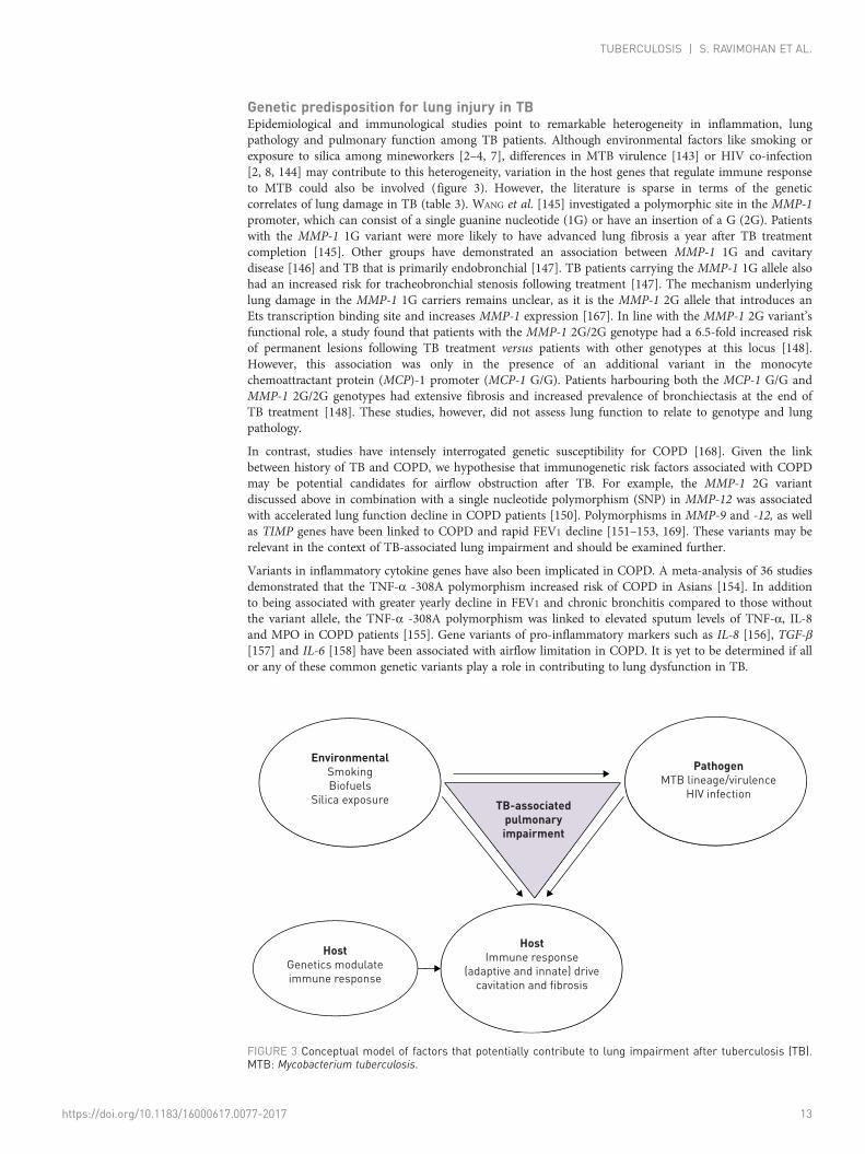
Genetic predisposition for lung injury in TBEpidemiological and immunological studies point to remarkable heterogeneity in inflammation, lungpathology and pulmonary function among TB patients. Although environmental factors like smoking orexposure to silica among mineworkers [2–4, 7], differences in MTB virulence [143] or HIV co-infection[2, 8, 144] may contribute to this heterogeneity, variation in the host genes that regulate immune responseto MTB could also be involved (figure 3). However, the literature is sparse in terms of the geneticcorrelates of lung damage in TB (table 3). WANG et al. [145] investigated a polymorphic site in the MMP-1promoter, which can consist of a single guanine nucleotide (1G) or have an insertion of a G (2G). Patientswith the MMP-1 1G variant were more likely to have advanced lung fibrosis a year after TB treatmentcompletion [145]. Other groups have demonstrated an association between MMP-1 1G and cavitarydisease [146] and TB that is primarily endobronchial [147]. TB patients carrying the MMP-1 1G allele alsohad an increased risk for tracheobronchial stenosis following treatment [147]. The mechanism underlyinglung damage in the MMP-1 1G carriers remains unclear, as it is the MMP-1 2G allele that introduces anEts transcription binding site and increases MMP-1 expression [167]. In line with the MMP-1 2G variant’sfunctional role, a study found that patients with the MMP-1 2G/2G genotype had a 6.5-fold increased riskof permanent lesions following TB treatment versus patients with other genotypes at this locus [148].However, this association was only in the presence of an additional variant in the monocytechemoattractant protein (MCP)-1 promoter (MCP-1 G/G). Patients harbouring both the MCP-1 G/G andMMP-1 2G/2G genotypes had extensive fibrosis and increased prevalence of bronchiectasis at the end ofTB treatment [148]. These studies, however, did not assess lung function to relate to genotype and lungpathology.
In contrast, studies have intensely interrogated genetic susceptibility for COPD [168]. Given the linkbetween history of TB and COPD, we hypothesise that immunogenetic risk factors associated with COPDmay be potential candidates for airflow obstruction after TB. For example, the MMP-1 2G variantdiscussed above in combination with a single nucleotide polymorphism (SNP) in MMP-12 was associatedwith accelerated lung function decline in COPD patients [150]. Polymorphisms in MMP-9 and -12, as wellas TIMP genes have been linked to COPD and rapid FEV1 decline [151–153, 169]. These variants may berelevant in the context of TB-associated lung impairment and should be examined further.
Variants in inflammatory cytokine genes have also been implicated in COPD. A meta-analysis of 36 studiesdemonstrated that the TNF-α -308A polymorphism increased risk of COPD in Asians [154]. In additionto being associated with greater yearly decline in FEV1 and chronic bronchitis compared to those withoutthe variant allele, the TNF-α -308A polymorphism was linked to elevated sputum levels of TNF-α, IL-8and MPO in COPD patients [155]. Gene variants of pro-inflammatory markers such as IL-8 [156], TGF-β[157] and IL-6 [158] have been associated with airflow limitation in COPD. It is yet to be determined if allor any of these common genetic variants play a role in contributing to lung dysfunction in TB.
EnvironmentalSmoking
Biofuels
Silica exposure
HostGenetics modulate
immune response
HostImmune response
(adaptive and innate) drive
cavitation and fibrosis
TB-associatedpulmonaryimpairment
PathogenMTB lineage/virulence
HIV infection
FIGURE 3 Conceptual model of factors that potentially contribute to lung impairment after tuberculosis (TB).MTB: Mycobacterium tuberculosis.
https://doi.org/10.1183/16000617.0077-2017 13
TUBERCULOSIS | S. RAVIMOHAN ET AL.
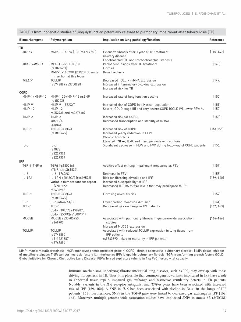
Immune mechanisms underlying fibrotic interstitial lung diseases, such as IPF, may overlap with thosedriving fibrogenesis in TB. Thus, it is plausible that common genetic variants implicated in IPF have a rolein abnormal tissue repair, impaired gas exchange and restrictive ventilatory defects in TB patients.Notably, variants in the IL-1 receptor antagonist and TNF-α genes have been associated with increasedrisk of IPF [159, 160]. A SNP in IL-6 has been associated with decline in DLCO in the lungs of IPFpatients [161]. Furthermore, SNPs in the TGF-β gene were linked to decreased gas exchange in IPF [162,163]. Moreover, multiple genome-wide association studies have implicated SNPs in mucin 5B (MUC5B)
TABLE 3 Immunogenetic studies of lung dysfunction potentially relevant to pulmonary impairment after tuberculosis (TB)
Biomarker/gene Polymorphism Implication on lung pathology/function Reference
TBMMP-1 MMP-1 -1607G (1G) (rs1799750) Extensive fibrosis after 1 year of TB treatment
Cavitary diseaseEndobronchial TB and tracheobronchial stenosis
[145–147]
MCP-1+MMP-1 MCP-1 -2518G (G/G)(rs1024611)MMP-1 -1607GG (2G/2G) Guanineinsertion at this locus
Permanent lesions after TB treatmentFibrosisBronchiectasis
[148]
TOLLIP TOLLIPrs5743899 rs3750920
Decreased TOLLIP mRNA expressionIncreased inflammatory cytokine expressionIncreased risk for TB
[149]
COPDMMP-1+MMP-12 MMP-1 2G+MMP-12 nsSNP
(rs652438)Increased rate of lung function decline [150]
MMP-9 MMP-9 -1562C/T Increased risk of COPD in a Korean population [151]MMP-12 MMP-12
rs652438 and rs2276109Severe (GOLD stage III) and very severe COPD (GOLD IV), lower FEV1 % [152]
TIMP-2 TIMP-2+853G/A-418G/C
Increased risk for COPDDecreased transcription and stability of mRNA
[153]
TNF-α TNF-α -308G/A(rs1800629)
Increased risk of COPDIncreased yearly reduction in FEV1Chronic bronchitisElevated TNF-α, IL-8, and myeloperoxidase in sputum
[154,155]
IL-8 IL-8rs4073rs2227306rs2227307
Significant decrease in FEV1 and FVC during follow-up of COPD patients [156]
IPFTGF-β+TNF-α TGFβ (rs1800469)
+TNF-α (rs361525)Additive effect on lung impairment measured as FEV1 [157]
IL-6 IL-6 -174G/C Decrease in FEV1 [158]IL-1RA IL-1RN +2018C/T (rs419598)
Variable number tandem repeat(VNTR)*2
rs2637988
Risk for fibrosing alveolitis and IPFIncreased susceptibility for IPFDecreased IL-1RA mRNA levels that may predispose to IPF
[159, 160]
TNF-α TNF-α -308G/A(rs1800629)
Fibrosing alveolitis risk [159]
IL-6 IL-6 intron 4A/G Lower carbon monoxide diffusion [161]TGF-β TGF-β
Codon 10T/C(rs1982073)Codon 25G/C(rs1800471)
Decreased gas exchange in IPF patients [162, 163]
MUC5B MUC5B rs35705950rs868903
Associated with pulmonary fibrosis in genome-wide associationstudies
Increased MUC5B expression
[164–166]
TOLLIP TOLLIPrs5743890rs111521887rs5743894
Associated with reduced TOLLIP expression in lung tissue fromIPF patients
rs5743890 linked to mortality in IPF patients
MMP: matrix metalloproteinase; MCP: monocyte chemoattractant protein; COPD: chronic obstructive pulmonary disease; TIMP: tissue inhibitorof metalloproteinase; TNF: tumour necrosis factor; IL: interleukin; IPF: idiopathic pulmonary fibrosis; TGF: transforming growth factor; GOLD:Global Initiative for Chronic Obstructive Lung Disease; FEV1: forced expiratory volume in 1 s; FVC: forced vital capacity.
https://doi.org/10.1183/16000617.0077-2017 14
TUBERCULOSIS | S. RAVIMOHAN ET AL.
and TOLLIP, which modulate innate immune responses, in IPF [164–166]. Genetic variation in TOLLIP isparticularly interesting, given its role in negatively regulating the TGF-β [170] and Toll-like receptorsignalling cascade [149]. TOLLIP deficiency leads to an increase in IL-6 and TNF-α secretion in mice andhumans [149, 171]. Furthermore, individuals who harboured the TOLLIP rs5743899 or rs3750920 SNP haddiminished TOLLIP mRNA expression, elevated levels of inflammatory cytokines and at an increased riskfor TB [149]. However, these SNPs have not been examined in the context of lung pathology in TB or PIAT.
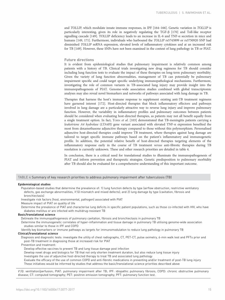
Future directionsIt is evident from epidemiological studies that pulmonary impairment is relatively common amongpatients with a history of TB. Clinical trials investigating new drug regimens for TB should considerincluding lung function tests to evaluate the impact of these therapies on long-term pulmonary morbidity.Given the variety of lung function abnormalities, management of TB can potentially be pulmonaryimpairment specific and could target specific underlying immunopathological mechanisms. Furthermore,investigating the role of common variants in TB-associated lung injury may provide insight into theimmunopathogenesis of PIAT. Genome-wide association studies combined with global transcriptomicanalyses may also reveal novel biomarkers and networks of pathways associated with lung damage in TB.
Therapies that harness the host’s immune response to supplement existing anti-TB treatment regimenshave garnered interest [172]. Host-directed therapies that block inflammatory effectors and pathwaysinvolved in lung damage are a particularly attractive way to reverse lung injury and improve pulmonaryfunction. However, the variability in inflammatory profiles and pulmonary outcomes between patientsshould be considered when evaluating host-directed therapies, as patients may not all benefit equally froma single treatment option. In fact, TOBIN et al. [103] demonstrated that TB-meningitis patients carrying aleukotriene A4 hydrolase (LTA4H) gene variant associated with elevated TNF-α expression benefited themost from dexamethasone adjunctive therapy compared to those without this polymorphism. Personalisedadjunctive host-directed therapies could improve TB treatment, where therapies against lung damage aretailored to target specific immune pathways based on the patient’s inflammatory and immunogeneticprofile. In addition, the potential relative benefit of host-directed therapies targeting elements of theinflammatory response early in the course of TB treatment versus anti-fibrotic therapies during TBresolution is currently unknown. These and other research priorities are detailed in table 4.
In conclusion, there is a critical need for translational studies to illuminate the immunopathogenesis ofPIAT and inform prevention and therapeutic strategies. Genetic predisposition to pulmonary morbidityafter TB should also be evaluated for a comprehensive understanding of this important outcome.
TABLE 4 Summary of key research priorities to address pulmonary impairment after tuberculosis (TB)
Epidemiological studiesPopulation-based studies that determine the prevalence of: 1) lung function defects by type (airflow obstruction, restrictive ventilatorydefects, gas exchange abnormalities, V′/Q mismatch and mixed defects); and 2) lung damage by type (cavitation, fibrosis andbronchiectasis)
Investigate risk factors (host, environmental, pathogen) associated with PIATMeasure impact of PIAT on quality of lifeDetermine the prevalence of PIAT and characterise lung deficits in specific patient populations, such as those co-infected with HIV, who havediabetes mellitus or are infected with multidrug-resistant TB
Basic/translational scienceDelineate the immunopathogenesis of pulmonary cavitation, fibrosis and bronchiectasis in pulmonary TBDetermine the immunogenetic correlates of hyper-inflammation and tissue damage in pulmonary TB utilising genome-wide associationstudies similar to those in IPF and COPD
Identify key biomarkers or immune pathways as targets for immunomodulation to reduce lung pathology in pulmonary TBClinical/translational scienceDiagnosis and diagnostic tests: investigate the utility of chest radiographs, CT, PET-CT, pulse oximetry, 6-min walk test and PFTs prior andpost-TB treatment in diagnosing those at increased risk for PIAT
Prevention and treatment:Develop effective vaccines to prevent TB and lung tissue damage post infectionDevelop novel drugs and biologics for TB that not only shorten treatment duration, but also reduce lung tissue injuryInvestigate the use of adjunctive host-directed therapy to treat TB and associated lung pathologyEvaluate the efficacy of the use of common COPD and anti-fibrotic medications in preventing and/or treatment of post-TB lung injuryThese initiatives would be informed by studies that address the basic/translational science priorities described above
V′/Q: ventilation/perfusion; PIAT: pulmonary impairment after TB; IPF: idiopathic pulmonary fibrosis; COPD: chronic obstructive pulmonarydisease; CT: computed tomography; PET: positron emission tomography; PFT: pulmonary function test.
https://doi.org/10.1183/16000617.0077-2017 15
TUBERCULOSIS | S. RAVIMOHAN ET AL.
References1 World Health Organization. Global Tuberculosis Report 2015. 20th edition. Geneva, WHO, 2015.2 Hnizdo E, Singh T, Churchyard G. Chronic pulmonary function impairment caused by initial and recurrent
pulmonary tuberculosis following treatment. Thorax 2000; 55: 32–38.3 Plit ML, Anderson R, Van Rensburg CE, et al. Influence of antimicrobial chemotherapy on spirometric
parameters and pro-inflammatory indices in severe pulmonary tuberculosis. Eur Respir J 1998; 12: 351–356.4 Ross J, Ehrlich RI, Hnizdo E, et al. Excess lung function decline in gold miners following pulmonary
tuberculosis. Thorax 2010; 65: 1010–1015.5 Pasipanodya JG, Miller TL, Vecino M, et al. Pulmonary impairment after tuberculosis. Chest 2007; 131:
1817–1824.6 Pasipanodya JG, McNabb SJ, Hilsenrath P, et al. Pulmonary impairment after tuberculosis and its contribution to
TB burden. BMC Public Health 2010; 10: 259.7 Maguire GP, Anstey NM, Ardian M, et al. Pulmonary tuberculosis, impaired lung function, disability and quality
of life in a high-burden setting. Int J Tuberc Lung Dis 2009; 13: 1500–1506.8 Ralph AP, Kenangalem E, Waramori G, et al. High morbidity during treatment and residual pulmonary
disability in pulmonary tuberculosis: under-recognised phenomena. PLoS One 2013; 8: e80302.9 Schunemann HJ, Dorn J, Grant BJ, et al. Pulmonary function is a long-term predictor of mortality in the general
population: 29-year follow-up of the Buffalo Health Study. Chest 2000; 118: 656–664.10 Amaral AF, Coton S, Kato B, et al. Tuberculosis associates with both airflow obstruction and low lung function:
BOLD results. Eur Respir J 2015; 46: 1104–1112.11 Byrne AL, Marais BJ, Mitnick CD, et al. Tuberculosis and chronic respiratory disease: a systematic review. Int J
Infect Dis 2015; 32: 138–146.12 Menezes AM, Hallal PC, Perez-Padilla R, et al. Tuberculosis and airflow obstruction: evidence from the
PLATINO study in Latin America. Eur Respir J 2007; 30: 1180–1185.13 Willcox PA, Ferguson AD. Chronic obstructive airways disease following treated pulmonary tuberculosis. Respir
Med 1989; 83: 195–198.14 Hunter RL. Pathology of post primary tuberculosis of the lung: an illustrated critical review. Tuberculosis (Edinb)
2011; 91: 497–509.15 Long R, Maycher B, Dhar A, et al. Pulmonary tuberculosis treated with directly observed therapy: serial changes
in lung structure and function. Chest 1998; 113: 933–943.16 Gaensler EA, Lindgren I Chronic bronchitis as an etiologic factor in obstructive emphysema; preliminary report.
Am Rev Respir Dis 1959; 80: 185–193.17 Birath G, Caro J, Malmberg R, et al. Airways obstruction in pulmonary tuberculosis. Scand J Respir Dis 1966; 47:
27–36.18 Akkara SA, Shah AD, Adalja M, et al. Pulmonary tuberculosis: the day after. Int J Tuberc Lung Dis 2013; 17:
810–813.19 Manji M, Shayo G, Mamuya S, et al. Lung functions among patients with pulmonary tuberculosis in Dar es
Salaam – a cross-sectional study. BMC Pulm Med 2016; 16: 58.20 Rhee CK, Yoo KH, Lee JH, et al. Clinical characteristics of patients with tuberculosis-destroyed lung. Int J Tuberc
Lung Dis 2013; 17: 67–75.21 Lee CH, Lee MC, Lin HH, et al. Pulmonary tuberculosis and delay in anti-tuberculous treatment are important
risk factors for chronic obstructive pulmonary disease. PLoS One 2012; 7: e37978.22 de la Mora IL, Martinez-Oceguera D, Laniado-Laborin R. Chronic airway obstruction after successful treatment
of tuberculosis and its impact on quality of life. Int J Tuberc Lung Dis 2015; 19: 808–810.23 Pefura-Yone EW, Kengne AP, Tagne-Kamdem PE, et al. Clinical significance of low forced expiratory flow
between 25% and 75% of vital capacity following treated pulmonary tuberculosis: a cross-sectional study. BMJOpen 2014; 4: e005361.
24 Choi CJ, Choi WS, Lee SY, et al. The definition of past tuberculosis affects the magnitude of association betweenpulmonary tuberculosis and respiratory dysfunction: Korea National Health and Nutrition Examination Survey,2008–2012. J Korean Med Sci 2017; 32: 789–795.
25 Gingo MR, George MP, Kessinger CJ, et al. Pulmonary function abnormalities in HIV-infected patients duringthe current antiretroviral therapy era. Am J Respir Crit Care Med 2010; 182: 790–796.
26 van Zyl Smit RN, Pai M, Yew WW, et al. Global lung health: the colliding epidemics of tuberculosis, tobaccosmoking, HIV and COPD. Eur Respir J 2010; 35: 27–33.
27 Gingo MR, He J, Wittman C, et al. Contributors to diffusion impairment in HIV-infected persons. Eur Respir J2014; 43: 195–203.
28 Glaser S, Kruger S, Merkel M, et al. Chronic obstructive pulmonary disease and diabetes mellitus: a systematicreview of the literature. Respiration 2015; 89: 253–264.
29 Crapo RO. Pulmonary-function testing. N Engl J Med 1994; 331: 25–30.30 Macintyre N, Crapo RO, Viegi G, et al. Standardisation of the single-breath determination of carbon monoxide
uptake in the lung. Eur Respir J 2005; 26: 720–735.31 Miller MR, Hankinson J, Brusasco V, et al. Standardisation of spirometry. Eur Respir J 2005; 26: 319–338.32 Pellegrino R, Viegi G, Brusasco V, et al. Interpretative strategies for lung function tests. Eur Respir J 2005; 26:
948–968.33 Dorhoi A, Kaufmann SH. Pathology and immune reactivity: understanding multidimensionality in pulmonary
tuberculosis. Semin Immunopathol 2016; 38: 153–166.34 Rabe KF, Hurd S, Anzueto A, et al. Global strategy for the diagnosis, management, and prevention of chronic
obstructive pulmonary disease: GOLD executive summary. Am J Respir Crit Care Med 2007; 176: 532–555.35 Cazzola M, MacNee W, Martinez FJ, et al. Outcomes for COPD pharmacological trials: from lung function to
biomarkers. Eur Respir J 2008; 31: 416–469.36 Hatipoglu ON, Osma E, Manisali M, et al. High resolution computed tomographic findings in pulmonary
tuberculosis. Thorax 1996; 51: 397–402.37 Murata K, Itoh H, Todo G, et al. Centrilobular lesions of the lung: demonstration by high-resolution CT and
pathologic correlation. Radiology 1986; 161: 641–645.
https://doi.org/10.1183/16000617.0077-2017 16
TUBERCULOSIS | S. RAVIMOHAN ET AL.

38 Im JG, Itoh H, Shim YS, et al. Pulmonary tuberculosis: CT findings – early active disease and sequential changewith antituberculous therapy. Radiology 1993; 186: 653–660.
39 Roberts HR, Wells AU, Milne DG, et al. Airflow obstruction in bronchiectasis: correlation between computedtomography features and pulmonary function tests. Thorax 2000; 55: 198–204.
40 Milliron B, Henry TS, Veeraraghavan S, et al. Bronchiectasis: mechanisms and imaging clues of associatedcommon and uncommon diseases. Radiographics 2015; 35: 1011–1030.
41 Grancher J. La dilatation des bronches chez les tuberculeux. Gazz Med de Paris 1878; 146.42 Ko JM, Kim KJ, Park SH, et al. Bronchiectasis in active tuberculosis. Acta Radiol 2013; 54: 412–417.43 Jones EM, Peck WM. Relationships between tuberculosis and bronchiectasis; a study of clinical and of
post-mortem material. Am Rev Tuberc 1950; 61: 387–398.44 Salkin D. Tuberculosis as a cause of upper lobe bronchiectasis. Calif Med 1950; 73: 577–580.45 Capone RB, Capone D, Mafort T, et al. Tomographic aspects of advanced active pulmonary tuberculosis and
evaluation of sequelae following treatment. Pulm Med 2017; 2017: 9876768.46 Meghji J, Simpson H, Squire SB, et al. A systematic review of the prevalence and pattern of imaging defined
post-TB lung disease. PLoS One 2016; 11: e0161176.47 Zhou YM, Wang C, Yao WZ, et al. [The prevalence and risk factors of bronchiectasis in residents aged 40 years
old and above in seven cities in China]. Zhonghua Nei Ke Za Zhi 2013; 52: 379–382.48 Ehrlich RI, Adams S, Baatjies R, et al. Chronic airflow obstruction and respiratory symptoms following
tuberculosis: a review of South African studies. Int J Tuberc Lung Dis 2011; 15: 886–891.49 Seiscento M, Vargas FS, Antonangelo L, et al. Transforming growth factor β-1 as a predictor of fibrosis in
tuberculous pleurisy. Respirology 2007; 12: 660–663.50 Malmberg R. Gas exchange in pulmonary tuberculosis. II. Review of literature clinical significance and
conclusions. Scand J Respir Dis 1966; 47: 277–305.51 Pipavath SN, Sharma SK, Sinha S, et al. High resolution CT (HRCT) in miliary tuberculosis (MTB) of the lung:
correlation with pulmonary function tests and gas exchange parameters in north Indian patients. Indian J MedRes 2007; 126: 193–198.
52 Kim MA, Kim SH, Zo JH, et al. Right heart dysfunction in post-tuberculosis emphysema. Int J Tuberc Lung Dis2004; 8: 1120–1126.
53 Continuous or nocturnal oxygen therapy in hypoxemic chronic obstructive lung disease: a clinical trial.Nocturnal Oxygen Therapy Trial Group. Ann Intern Med 1980; 93: 391–398.
54 Long term domiciliary oxygen therapy in chronic hypoxic cor pulmonale complicating chronic bronchitis andemphysema. Report of the Medical Research Council Working Party. Lancet 1981; 1: 681–686.
55 Flynn JL, Gideon HP, Mattila JT, et al. Immunology studies in non-human primate models of tuberculosis.Immunol Rev 2015; 264: 60–73.
56 Helke KL, Mankowski JL, Manabe YC. Animal models of cavitation in pulmonary tuberculosis. Tuberculosis(Edinb) 2006; 86: 337–348.
57 Malherbe ST, Shenai S, Ronacher K, et al. Persisting positron emission tomography lesion activity andMycobacterium tuberculosis mRNA after tuberculosis cure. Nat Med 2016; 22: 1094–1100.
58 Ramakrishnan L. Revisiting the role of the granuloma in tuberculosis. Nat Rev Immunol 2012; 12: 352–366.59 Volkman HE, Pozos TC, Zheng J, et al. Tuberculous granuloma induction via interaction of a bacterial secreted
protein with host epithelium. Science 2010; 327: 466–469.60 Davis JM, Ramakrishnan L. The role of the granuloma in expansion and dissemination of early tuberculous
infection. Cell 2009; 136: 37–49.61 Lin PL, Ford CB, Coleman MT, et al. Sterilization of granulomas is common in active and latent tuberculosis
despite within-host variability in bacterial killing. Nat Med 2014; 20: 75–79.62 Gideon HP, Phuah J, Myers AJ, et al. Variability in tuberculosis granuloma T cell responses exists, but a balance
of pro- and anti-inflammatory cytokines is associated with sterilization. PLoS Pathog 2015; 11: e1004603.63 Martin CJ, Cadena AM, Leung VW, et al. Digitally barcoding Mycobacterium tuberculosis reveals in vivo
infection dynamics in the macaque model of tuberculosis. MBio 2017; 8: e00312-17.64 Coleman MT, Maiello P, Tomko J, et al. Early changes by 18fluorodeoxyglucose positron emission tomography
coregistered with computed tomography predict outcome after Mycobacterium tuberculosis infection inCynomolgus macaques. Infect Immun 2014; 82: 2400–2404.
65 Dannenberg AM Jr, Sugimoto M. Liquefaction of caseous foci in tuberculosis. Am Rev Respir Dis 1976; 113:257–259.
66 Hunter RL, Jagannath C, Actor JK. Pathology of postprimary tuberculosis in humans and mice: contradiction oflong-held beliefs. Tuberculosis (Edinb) 2007; 87: 267–278.
67 Grosset J. Mycobacterium tuberculosis in the extracellular compartment: an underestimated adversary. AntimicrobAgents Chemother 2003; 47: 833–836.
68 Rook GA, al Attiyah R. Cytokines and the Koch phenomenon. Tubercle 1991; 72: 13–20.69 Dheda K, Booth H, Huggett JF, et al. Lung remodeling in pulmonary tuberculosis. J Infect Dis 2005; 192:
1201–1209.70 Koch R. Fortsetzung uber ein Heilmittel gegen Tuberculose. Dtsch Med Wochenschr 1891; 17: 101–102.71 Taylor JL, Turner OC, Basaraba RJ, et al. Pulmonary necrosis resulting from DNA vaccination against
tuberculosis. Infect Immun 2003; 71: 2192–2198.72 Moreira AL, Tsenova L, Aman MH, et al. Mycobacterial antigens exacerbate disease manifestations in
Mycobacterium tuberculosis-infected mice. Infect Immun 2002; 70: 2100–2107.73 Shwartzman G. Phenomenon of local skin reactivity to Bacillus tuberculosis: I. Skin-preparatory and reacting
potencies of tuberculin, O.T., and Bacillus tuberculosis culture filtrates. J Exp Med 1935; 61: 369–382.74 al Attiyah R, Moreno C, Rook GA. TNFα-mediated tissue damage in mouse footpads primed with mycobacterial
preparations. Res Immunol 1992; 143: 601–610.75 Cardona PJ, Llatjos R, Gordillo S, et al. Towards a “human-like” model of tuberculosis: intranasal inoculation of
LPS induces intragranulomatous lung necrosis in mice infected aerogenically with Mycobacterium tuberculosis.Scand J Immunol 2001; 53: 65–71.
https://doi.org/10.1183/16000617.0077-2017 17
TUBERCULOSIS | S. RAVIMOHAN ET AL.
76 Yamamura Y, Ogawa Y, Maeda H, et al. Prevention of tuberculous cavity formation by desensitization withtuberculin-active peptide. Am Rev Respir Dis 1974; 109: 594–601.
77 Kim MJ, Wainwright HC, Locketz M, et al. Caseation of human tuberculosis granulomas correlates with elevatedhost lipid metabolism. EMBO Mol Med 2010; 2: 258–274.
78 Barry CE 3rd, Boshoff HI, Dartois V, et al. The spectrum of latent tuberculosis: rethinking the biology andintervention strategies. Nat Rev Microbiol 2009; 7: 845–855.
79 Greenlee KJ, Werb Z, Kheradmand F. Matrix metalloproteinases in lung: multiple, multifarious, and multifaceted.Physiol Rev 2007; 87: 69–98.
80 Salgame P. MMPs in tuberculosis: granuloma creators and tissue destroyers. J Clin Invest 2011; 121: 1686–1688.81 Subbian S, Tsenova L, Kim MJ, et al. Lesion-specific immune response in granulomas of patients with
pulmonary tuberculosis: a pilot study. PLoS One 2015; 10: e0132249.82 Elkington P, Shiomi T, Breen R, et al. MMP-1 drives immunopathology in human tuberculosis and transgenic
mice. J Clin Invest 2011; 121: 1827–1833.83 Belton M, Brilha S, Manavaki R, et al. Hypoxia and tissue destruction in pulmonary TB. Thorax 2016; 71:
1145–1153.84 Arpino V, Brock M, Gill SE. The role of TIMPs in regulation of extracellular matrix proteolysis. Matrix Biol
2015; 44–46: 247–254.85 Kubler A, Luna B, Larsson C, et al. Mycobacterium tuberculosis dysregulates MMP/TIMP balance to drive rapid
cavitation and unrestrained bacterial proliferation. J Pathol 2015; 235: 431–444.86 Ong CW, Elkington PT, Brilha S, et al. Neutrophil-derived MMP-8 drives AMPK-dependent matrix destruction
in human pulmonary tuberculosis. PLoS Pathog 2015; 11: e1004917.87 Ravimohan S, Tamuhla N, Kung SJ, et al. Matrix metalloproteinases in tuberculosis-immune reconstitution
inflammatory syndrome and impaired lung function among advanced HIV/TB co-infected patients initiatingantiretroviral therapy. EBioMedicine 2016; 3: 100–107.
88 Subbian S, Tsenova L, O’Brien P, et al. Phosphodiesterase-4 inhibition combined with isoniazid treatment ofrabbits with pulmonary tuberculosis reduces macrophage activation and lung pathology. Am J Pathol 2011; 179:289–301.
89 Ugarte-Gil CA, Elkington P, Gilman RH, et al. Induced sputum MMP-1, -3 & -8 concentrations duringtreatment of tuberculosis. PLoS One 2013; 8: e61333.
90 Owen CA. Roles for proteinases in the pathogenesis of chronic obstructive pulmonary disease. Int J ChronObstruct Pulmon Dis 2008; 3: 253–268.
91 Angel JH, Chu LS, Lyons HA. Corticotropin in the treatment of tuberculosis. A controlled study. Arch InternMed 1961; 108: 353–369.
92 Marcus H, Yoo OH, Akyol T, et al. A randomized study of the effects of corticosteroid therapy on healing ofpulmonary tuberculosis as judged by clinical, roentgenographic, and physiologic measurements. Am Rev RespirDis 1963; 88: 55–64.
93 Malik SK, Martin CJ. Tuberculosis, corticosteroid therapy, and pulmonary function. Am Rev Respir Dis 1969;100: 13–18.
94 Dooley DP, Carpenter JL, Rademacher S. Adjunctive corticosteroid therapy for tuberculosis: a critical reappraisalof the literature. Clin Infect Dis 1997; 25: 872–887.
95 Critchley JA, Young F, Orton L, et al. Corticosteroids for prevention of mortality in people with tuberculosis: asystematic review and meta-analysis. Lancet Infect Dis 2013; 13: 223–237.
96 Wallis RS. Corticosteroid effects on sputum culture in pulmonary tuberculosis: a meta-regression analysis. OpenForum Infect Dis 2014; 1: ofu020.
97 Auphan N, DiDonato JA, Rosette C, et al. Immunosuppression by glucocorticoids: inhibition of NF-kappa Bactivity through induction of I kappa B synthesis. Science 1995; 270: 286–290.
98 Scheinman RI, Cogswell PC, Lofquist AK, et al. Role of transcriptional activation of I kappa B alpha inmediation of immunosuppression by glucocorticoids. Science 1995; 270: 283–286.
99 Dorhoi A, Kaufmann SH. Tumor necrosis factor alpha in mycobacterial infection. Semin Immunol 2014; 26:203–209.
100 Ray JC, Flynn JL, Kirschner DE. Synergy between individual TNF-dependent functions determines granulomaperformance for controlling Mycobacterium tuberculosis infection. J Immunol 2009; 182: 3706–3717.
101 Laster SM, Wood JG, Gooding LR. Tumor necrosis factor can induce both apoptic and necrotic forms of celllysis. J Immunol 1988; 141: 2629–2634.
102 Fratazzi C, Arbeit RD, Carini C, et al. Macrophage apoptosis in mycobacterial infections. J Leukoc Biol 1999; 66:763–764.
103 Tobin DM, Roca FJ, Oh SF, et al. Host genotype-specific therapies can optimize the inflammatory response tomycobacterial infections. Cell 2012; 148: 434–446.
104 Roca FJ, Ramakrishnan L. TNF dually mediates resistance and susceptibility to mycobacteria via mitochondrialreactive oxygen species. Cell 2013; 153: 521–534.
105 Tsao TC, Hong J, Li LF, et al. Imbalances between tumor necrosis factor-alpha and its soluble receptor forms,and interleukin-1beta and interleukin-1 receptor antagonist in BAL fluid of cavitary pulmonary tuberculosis.Chest 2000; 117: 103–109.
106 Elkington PT, Green JA, Emerson JE, et al. Synergistic up-regulation of epithelial cell matrix metalloproteinase-9secretion in tuberculosis. Am J Respir Cell Mol Biol 2007; 37: 431–437.
107 Elkington PT, Emerson JE, Lopez-Pascua LD, et al. Mycobacterium tuberculosis up-regulates matrixmetalloproteinase-1 secretion from human airway epithelial cells via a p38 MAPK switch. J Immunol 2005; 175:5333–5340.
108 Casarini M, Ameglio F, Alemanno L, et al. Cytokine levels correlate with a radiologic score in active pulmonarytuberculosis. Am J Respir Crit Care Med 1999; 159: 143–148.
109 Su WL, Perng WC, Huang CH, et al. Association of reduced tumor necrosis factor α, γ interferon, andinterleukin-1β (IL-1β) but increased IL-10 expression with improved chest radiography in patients withpulmonary tuberculosis. Clin Vaccine Immunol 2010; 17: 223–231.
https://doi.org/10.1183/16000617.0077-2017 18
TUBERCULOSIS | S. RAVIMOHAN ET AL.
110 Bekker LG, Maartens G, Steyn L, et al. Selective increase in plasma tumor necrosis factor-alpha and concomitantclinical deterioration after initiating therapy in patients with severe tuberculosis. J Infect Dis 1998; 178: 580–584.
111 Riou C, Perez Peixoto B, Roberts L, et al. Effect of standard tuberculosis treatment on plasma cytokine levels inpatients with active pulmonary tuberculosis. PLoS One 2012; 7: e36886.
112 DiFazio RM, Mattila JT, Klein EC, et al. Active transforming growth factor-β is associated with phenotypicchanges in granulomas after drug treatment in pulmonary tuberculosis. Fibrogenesis Tissue Repair 2016; 9: 6.
113 Tsenova L, O’Brien P, Holloway J, et al. Etanercept exacerbates inflammation and pathology in a rabbit model ofactive pulmonary tuberculosis. J Interferon Cytokine Res 2014; 34: 716–726.
114 Wynn TA. Integrating mechanisms of pulmonary fibrosis. J Exp Med 2011; 208: 1339–1350.115 Wilson MS, Madala SK, Ramalingam TR, et al. Bleomycin and IL-1β-mediated pulmonary fibrosis is IL-17A
dependent. J Exp Med 2010; 207: 535–552.116 Barlo NP, van Moorsel CH, Korthagen NM, et al. Genetic variability in the IL1RN gene and the balance between
interleukin (IL)-1 receptor agonist and IL-1β in idiopathic pulmonary fibrosis. Clin Exp Immunol 2011; 166:346–351.
117 Russell DG, Cardona PJ, Kim MJ, et al. Foamy macrophages and the progression of the human tuberculosisgranuloma. Nat Immunol 2009; 10: 943–948.
118 Guirado E, Schlesinger LS, Kaplan G. Macrophages in tuberculosis: friend or foe. Semin Immunopathol 2013; 35:563–583.
119 Martin CJ, Carey AF, Fortune SM. A bug’s life in the granuloma. Semin Immunopathol 2016; 38: 213–220.120 Pedrosa J, Saunders BM, Appelberg R, et al. Neutrophils play a protective nonphagocytic role in systemic
Mycobacterium tuberculosis infection of mice. Infect Immun 2000; 68: 577–583.121 Blomgran R, Ernst JD. Lung neutrophils facilitate activation of naive antigen-specific CD4+ T cells during
Mycobacterium tuberculosis infection. J Immunol 2011; 186: 7110–7119.122 Gopal R, Monin L, Torres D, et al. S100A8/A9 proteins mediate neutrophilic inflammation and lung pathology
during tuberculosis. Am J Respir Crit Care Med 2013; 188: 1137–1146.123 Eruslanov EB, Lyadova IV, Kondratieva TK, et al. Neutrophil responses to Mycobacterium tuberculosis infection
in genetically susceptible and resistant mice. Infect Immun 2005; 73: 1744–1753.124 Pichugin AV, Yan BS, Sloutsky A, et al. Dominant role of the sst1 locus in pathogenesis of necrotizing lung
granulomas during chronic tuberculosis infection and reactivation in genetically resistant hosts. Am J Pathol2009; 174: 2190–2201.
125 Repasy T, Martinez N, Lee J, et al. Bacillary replication and macrophage necrosis are determinants of neutrophilrecruitment in tuberculosis. Microbes Infect 2015; 17: 564–574.
126 Yeremeev V, Linge I, Kondratieva T, et al. Neutrophils exacerbate tuberculosis infection in genetically susceptiblemice. Tuberculosis (Edinb) 2015; 95: 447–451.
127 Eum SY, Kong JH, Hong MS, et al. Neutrophils are the predominant infected phagocytic cells in the airways ofpatients with active pulmonary TB. Chest 2010; 137: 122–128.
128 Nouailles G, Dorhoi A, Koch M, et al. CXCL5-secreting pulmonary epithelial cells drive destructive neutrophilicinflammation in tuberculosis. J Clin Invest 2014; 124: 1268–1282.
129 Repasy T, Lee J, Marino S, et al. Intracellular bacillary burden reflects a burst size for Mycobacterium tuberculosisin vivo. PLoS Pathog 2013; 9: e1003190.
130 Ramos-Kichik V, Mondragon-Flores R, Mondragon-Castelan M, et al. Neutrophil extracellular traps are inducedby Mycobacterium tuberculosis. Tuberculosis (Edinb) 2009; 89: 29–37.
131 Narasaraju T, Yang E, Samy RP, et al. Excessive neutrophils and neutrophil extracellular traps contribute to acutelung injury of influenza pneumonitis. Am J Pathol 2011; 179: 199–210.
132 Grabcanovic-Musija F, Obermayer A, Stoiber W, et al. Neutrophil extracellular trap (NET) formationcharacterises stable and exacerbated COPD and correlates with airflow limitation. Respir Res 2015; 16: 59.
133 Dicker AJ, Crichton ML, Pumphrey EG, et al. Neutrophil extracellular traps are associated with disease severityand microbiota diversity in chronic obstructive pulmonary disease. J Allergy Clin Immunol 2018; 141: 117–127.
134 Brinkmann V, Reichard U, Goosmann C, et al. Neutrophil extracellular traps kill bacteria. Science 2004; 303:1532–1535.
135 Kaplan MJ, Radic M. Neutrophil extracellular traps: double-edged swords of innate immunity. J Immunol 2012;189: 2689–2695.
136 Guyot N, Wartelle J, Malleret L, et al. Unopposed cathepsin G, neutrophil elastase, and proteinase 3 cause severelung damage and emphysema. Am J Pathol 2014; 184: 2197–2210.
137 Cheng OZ, Palaniyar N. NET balancing: a problem in inflammatory lung diseases. Front Immunol 2013; 4: 1.138 Hoenderdos K, Condliffe A. The neutrophil in chronic obstructive pulmonary disease. Am J Respir Cell Mol Biol
2013; 48: 531–539.139 Urban CF, Ermert D, Schmid M, et al. Neutrophil extracellular traps contain calprotectin, a cytosolic protein
complex involved in host defense against Candida albicans. PLoS Pathog 2009; 5: e1000639.140 Kwan CK, Ernst JD. HIV and tuberculosis: a deadly human syndemic. Clin Microbiol Rev 2011; 24: 351–376.141 Chamie G, Luetkemeyer A, Walusimbi-Nanteza M, et al. Significant variation in presentation of pulmonary
tuberculosis across a high resolution of CD4 strata. Int J Tuberc Lung Dis 2010; 14: 1295–1302.142 Meintjes G, Lawn SD, Scano F, et al. Tuberculosis-associated immune reconstitution inflammatory syndrome:
case definitions for use in resource-limited settings. Lancet Infect Dis 2008; 8: 516–523.143 Via LE, Weiner DM, Schimel D, et al. Differential virulence and disease progression following Mycobacterium
tuberculosis complex infection of the common marmoset (Callithrix jacchus). Infect Immun 2013; 81: 2909–2919.144 Walker NF, Clark SO, Oni T, et al. Doxycycline and HIV infection suppress tuberculosis-induced matrix
metalloproteinases. Am J Respir Crit Care Med 2012; 185: 989–997.145 Wang CH, Lin HC, Lin SM, et al. MMP-1(-1607G) polymorphism as a risk factor for fibrosis after pulmonary
tuberculosis in Taiwan. Int J Tuberc Lung Dis 2010; 14: 627–634.146 Ninomiya S, Niimi T, Shimizu S, et al. Matrix metalloproteinase-1 polymorphism of promoter region in
sarcoidosis and tuberculosis patients. Sarcoidosis Vasc Diffuse Lung Dis 2004; 21: 19–24.147 Kuo HP, Wang YM, Wang CH, et al. Matrix metalloproteinase-1 polymorphism in Taiwanese patients with
endobronchial tuberculosis. Tuberculosis (Edinb) 2008; 88: 262–267.
https://doi.org/10.1183/16000617.0077-2017 19
TUBERCULOSIS | S. RAVIMOHAN ET AL.
148 Ganachari M, Guio H, Zhao N, et al. Host gene-encoded severe lung TB: from genes to the potential pathways.Genes Immun 2012; 13: 605–620.
149 Shah JA, Vary JC, Chau TT, et al. Human TOLLIP regulates TLR2 and TLR4 signaling and its polymorphismsare associated with susceptibility to tuberculosis. J Immunol 2012; 189: 1737–1746.
150 Joos L, He JQ, Shepherdson MB, et al. The role of matrix metalloproteinase polymorphisms in the rate of declinein lung function. Hum Mol Genet 2002; 11: 569–576.
151 Lee SY, Kim MJ, Kang HG, et al. Polymorphisms in matrix metalloproteinase-1, -9 and -12 genes and the risk ofchronic obstructive pulmonary disease in a Korean population. Respiration 2010; 80: 133–138.
152 Haq I, Chappell S, Johnson SR, et al. Association of MMP-2 polymorphisms with severe and very severe COPD:a case control study of MMPs-1, 9 and 12 in a European population. BMC Med Genet 2010; 11: 7.
153 Hirano K, Sakamoto T, Uchida Y, et al. Tissue inhibitor of metalloproteinases-2 gene polymorphisms in chronicobstructive pulmonary disease. Eur Respir J 2001; 18: 748–752.
154 Zhang L, Gu H, Gu Y, et al. Association between TNF-α -308 G/A polymorphism and COPD susceptibility: ameta-analysis update. Int J Chron Obstruct Pulmon Dis 2016; 11: 1367–1379.
155 Sapey E, Wood AM, Ahmad A, et al. Tumor necrosis factor-α rs361525 polymorphism is associated withincreased local production and downstream inflammation in chronic obstructive pulmonary disease. Am J RespirCrit Care Med 2010; 182: 192–199.
156 Cordoba-Lanus E, Baz-Davila R, Espinoza-Jimenez A, et al. IL-8 gene variants are associated with lung functiondecline and multidimensional BODE index in COPD patients but not with disease susceptibility: a validationstudy. COPD 2015; 12: 55–61.
157 Chiang CH, Chuang CH, Liu SL. Transforming growth factor-β1 and tumor necrosis factor-α are associated withclinical severity and airflow limitation of COPD in an additive manner. Lung 2014; 192: 95–102.
158 He JQ, Foreman MG, Shumansky K, et al. Associations of IL6 polymorphisms with lung function decline andCOPD. Thorax 2009; 64: 698–704.
159 Whyte M, Hubbard R, Meliconi R, et al. Increased risk of fibrosing alveolitis associated with interleukin-1receptor antagonist and tumor necrosis factor-α gene polymorphisms. Am J Respir Crit Care Med 2000; 162:755–758.
160 Korthagen NM, van Moorsel CH, Kazemier KM, et al. IL1RN genetic variations and risk of IPF: a meta-analysisand mRNA expression study. Immunogenetics 2012; 64: 371–377.
161 Pantelidis P, Fanning GC, Wells AU, et al. Analysis of tumor necrosis factor-α, lymphotoxin-α, tumor necrosisfactor receptor II, and interleukin-6 polymorphisms in patients with idiopathic pulmonary fibrosis. Am J RespirCrit Care Med 2001; 163: 1432–1436.
162 Xaubet A, Marin-Arguedas A, Lario S, et al. Transforming growth factor-β1 gene polymorphisms are associatedwith disease progression in idiopathic pulmonary fibrosis. Am J Respir Crit Care Med 2003; 168: 431–435.
163 Son JY, Kim SY, Cho SH, et al. TGF-β1 T869C polymorphism may affect susceptibility to idiopathic pulmonaryfibrosis and disease severity. Lung 2013; 191: 199–205.
164 Seibold MA, Wise AL, Speer MC, et al. A common MUC5B promoter polymorphism and pulmonary fibrosis.N Engl J Med 2011; 364: 1503–1512.
165 Fingerlin TE, Murphy E, Zhang W, et al. Genome-wide association study identifies multiple susceptibility locifor pulmonary fibrosis. Nat Genet 2013; 45: 613–620.
166 Noth I, Zhang Y, Ma SF, et al. Genetic variants associated with idiopathic pulmonary fibrosis susceptibility andmortality: a genome-wide association study. Lancet Respir Med 2013; 1: 309–317.
167 Rutter JL, Mitchell TI, Buttice G, et al. A single nucleotide polymorphism in the matrix metalloproteinase-1promoter creates an Ets binding site and augments transcription. Cancer Res 1998; 58: 5321–5325.
168 Marciniak SJ, Lomas DA. Genetic susceptibility. Clin Chest Med 2014; 35: 29–38.169 Hegab AE, Sakamoto T, Uchida Y, et al. Association analysis of tissue inhibitor of metalloproteinase2 gene
polymorphisms with COPD in Egyptians. Respir Med 2005; 99: 107–110.170 Zhu L, Wang L, Luo X, et al. Tollip, an intracellular trafficking protein, is a novel modulator of the transforming
growth factor-β signaling pathway. J Biol Chem 2012; 287: 39653–39663.171 Didierlaurent A, Brissoni B, Velin D, et al. Tollip regulates proinflammatory responses to interleukin-1 and
lipopolysaccharide. Mol Cell Biol 2006; 26: 735–742.172 Zumla A, Rao M, Wallis RS, et al. Host-directed therapies for infectious diseases: current status, recent progress,
and future prospects. Lancet Infect Dis 2016; 16: e47–e63.
https://doi.org/10.1183/16000617.0077-2017 20
TUBERCULOSIS | S. RAVIMOHAN ET AL.