tube leukocyte adherence inhibition assay bhupendra.pdf · tube leukocyte adherence inhibition...
TRANSCRIPT

TUBE LEUKOCYTE ADHERENCE INHIBITION ASSAY The assessment of tumor immunity in cancer patients and in rats
DE LEUKOCYTEN-ADHERENTIE-REMMINGS TEST Bepaling van tumor immuniteit bij kanker patienten en ratten
PROEFSCHRIFT
TER VERKRIJG!r<G VA:--J DE GRAAD YAK DOCTOR IN DE GEKEESKUNDE
AAK DE ERASMUS CNIVERSITEIT ROTTERDAM OP GEZAG VAN DE RECTOR MAGNIFICUS
PROF. DR. M.W. YAK HOF E:--J VOLGEKS BESLUIT VAN HET COLLEGE VAN DEKANEN.
DE OPE:--JBARE YERDEDIGING ZAL PLAATSYINDEN OP WOENSDAG 19 .JUNI 1985 TE 14.00 UUR
DOOR
BHUPENDRA TANK
GEBORE\1 TE NAIROBI. KE:"JY A
1985 DRUKKERIJ J.H. PAS:vtANS B.V. 's-GRJ\ VENHAGE

Begeleidingscommissie:
Promotor:
Overige leden:
Prof. Dr. D .L. Westbroek
Prof. Dr. R. Benner Prof. Dr. D.W. van Bekkum Prof. Dr. J. Jeekel
The investigations described in this thesis were performed at the Laboratory for Experimental Surgery, Erasmus University Rotterdam, and were financed through a grant from the Dutch Cancer Foundation (Koningin Wilhelmina Fonds).

To my Parents For Ria


5
CONTENTS
CHAPTER 1 GENERAL INTRODUCTION
1.1 1.1.1 1.2 1.2.1 1.2.2 1.2.2.1 1.2.2.2 1.2.2.3 1.3 1.3.1 1.3.2 1.3.3 1.3.4 1.3.5 1.3.6 1.3.7 1.3.8 1.4 1.5
Brief history of tumor immunology Tumor-associated antigens (T AA) I\.1ethods for the detection of tumor immunity In vitro tests for humoral immunity In vitro tests for cell-mediated immunity Antibody-dependent cell-mediated cytotoxicity (ADCC) Lymphocyte-mediated cytotoxicity Migration inhibition assays Leukocyte Adherence Inhibition (LAI) assays A brief review General methodology Hemocytometer LAI assay Microplate LA! assay Tube LAJ assay Humoral LAI (H-LAI) assay Comparison of LAI assays with other assays Comparison of hemocytometer, microplate and tube LAI assays The objectives of this study References
CHAPTER 2 EVALUATION OF THE TUBE LEUKOCYTE ADHERENCE INHIBITION (LAI) ASSAY IN BREAST AND GASTROINTESTINA ~
9
9 10 11 11 12 12 12 13 14 14 15 16 17 18 21 21 22 23 24
CANCER PATIENTS 32
2.1 2.2 2.2.1 2.2.2 2.2.3 2.2.4 2.3 2.3.1 2.3.2 2.3.3 2.3.4 2.3.5 2.4 2.5
Introduction Materials and methods Patients Preparation of tumor extracts Isolation of peripheral blood leukocytes (PBL) Tube LAI assay Results Non-specific inhibition of adherence Antigen titration NAI values for control subjects Selection of tumor extracts Clinical results of the tube LA! assay Discussion References
32 32 32 33 33 33 34 34 34 35 35 36 37 38

6
CHAPTER 3 TUBE LEUKOCYTE ADHERENCE INHIBITION (LA!) ASSAY IN PATIENTS WITH NON-MALIGNANT DISORDERS OF THE COLON AND COLORECTAL CANCER 40
3.1 3.2 3.2.1 3.2.2 3.2.3 3.2.4 3.3 3.3.1 3.3.2 3.3.3 3.4 3.5
Introduction Materials and Methods Patients Preparation of tissue extracts Isolation of peripheral blood leukocytes (PBL) Tube LA! assay Results Selection of tumor extracts Nonadherent Index (NAI) values in control groups Clinical results Discussion References
CHAPTER 4 TUBE LEUKOCYTE ADHERENCE INHIBITION (LA!) ASSAY IN COLO RECTAL CANCER PATIENTS USING PARTIALLY PURIFIED
40 40 40 40 41 41 41 41 41 42 43 44
TUMOR EXTRACTS 46
4.1 4.2 4.2.1 4.2.2 4.2.3 4.2.4 4.2.5 4.3 4.3.1 4.3.2 4.3.3
4.4 4.5
Introduction Materials and Methods Patients Preparation of tissue extracts Sepbacryl S-200 column chromatography Isolation of peripheral blood leukocytes (PBL) Tube LA! assay Results Selection of fraction reactive in the LAI LA! results using partially purified extracts Comparison of LA! results obtained using crude and partially purified extracts Discussion References
46 46 46 46 47 47 47 47 47 48
49 51 52

7
CHAPTER 5 LEUKOCYTE ADHERENCE INHIBITION (LA!) IN RATS BEARING TRANSPLANTABLE SYNGENEIC TUMORS OF DIFFERENT IMMUNOGENICITY 53
5.1 5.2 5.2.1 5.2.2 5.2.2.1 5.2.2.2 5.2.2.3 5.2.2.4 5.2.2.5 5.2.3 5.2.4 5.2.5 5.3 5.4 5.5
Introduction Materials and Methods Rats Tumors Liposarcoma (LS175) Colon adenocarcinoma (CC531) Skin basal cell carcinoma (1618) Tumor implantation Sensitization of WAG rats with irradiated 1618 tumor cells Preparation of tissue extracts Isolation of peripheral blood leukocytes (PBL) Tube LA! assay Results Discussion References
CHAPTER 6 LEUKOCYTE ADHERENCE INHIBITION (LA!) IN RATS USING
53 53 53 53 53 54 54 54 54 55 55 55 55 58 60
PARTIALLY PURIFIED TUMOR EXTRACTS 62
6.1 6.2 6.2.1 6.2.2 6.2.2.1 6.2.2.2 6.2.2.3 6.2.3 6.2.4 6.2.5 6.2.6 6.2.7 6.3 6.4 6.5
Introduction Materials and Methods Rats Tumors Liposarcoma (LS 175) Colon adenocarcinoma ( CC53l) Tumor implantation Preparation of tissue extracts Sephacryl S-200 column chromatography Isolation of peripheral blood leukocytes (PBL) Peritoneal cells Tube LA! assay Results Discussion References
62 62 62 63 63 63 63 63 64 64 64 64 64 67 68

CHAPTER 7 GENERAL DISCUSSION
7.1 7.2 7.3 7.4 7.5
Introduction Tube LA! in patients Tube LA! in rats Concluding remarks References
CHAPTER 8 SUMMARY
CHAPTER 9 SAMENV A TTING
ACKNOWLEDGEMENTS
CURRICULUM VITAE
8
69
69 69 71 72 73
75
77
79
80
9
CHAPTER I
GENERAL INTRODUCTION
1.1 Brief history of tumor immunology
The history of tumor immunology can be considered to have evolved over three eras. The first era began towards the end of the 19th century when it was observed that cancer could sometimes be transmitted from one animal to another by transplantation of tumor tissue ( 1 ,2). Subsequently, it was observed that while some tumors like Ehrlich mouse carcinoma and Jensen rat carcinoma grew progressively resulting in the eventual death of the host, many other tumors grew only for a brief period and then regressed completely. The investigations of Ehrlich (3,4) demonstrated that mice with an already established tumor transplant often failed to produce a further tumor when reinoculated with tumor cells, suggesting that the tumors possessed particular antigens not occurring on normal cells. These observations were confirmed and extended by other investigators in several laboratories, and sometimes, using different terminology for the tumor-associated antigens (TAA). However, many false conclusions on the same observations that were made in various laboratories could be attributed to lack of genetically homogeneous strains of laboratory animals. One of the most valuable contributions of this era came from Murphy in 1926 on the role lymphocytes played in resistance to tissue grafts, and malignant diseases (5).
The second era began with the development of inbred strains of laboratory animals by C.C. Little at Bar Harbour. This initiated a tremendous surge in immunogenetic investigations by several prominent investigators (6). As a result, the genetic basis of histocompatibility was firmly established, specific immunological tolerance and the graft-versus-host (GVH) reaction were discovered and a vaste amount of data on the mechanisms of allograft rejection, methods of immunosuppression, and biochemistry of transplantation antigens was further accumulated.
Since considerable discrepancies exist in the correct terminology in the literature, the following definitions are introduced. Tumor-associated antigens (T AA) are defined as determinants which are present on tumor cells but are either totally absent or present to a limited extent only on normal cells. Tumor-specific antigens are defined as determinants that are present on a spontaneous tumor of a particular type in an individual and are totally absent on the cells of the same type of tumor in another individual and on normal cells.
The discovery of TAA in 1943 by Gross marked the birth of the third era. It was firmly established that a variety of tumors possessed T AA (7). Furthermore, T AA
10
of some tumors included a subset of cell-surface antigens that were termed tumorassociated transplantation antigens (TA TA) which could evoke an immune response in other animals syngeneic with the animal in which the tumor originated (7). It was also established that autotransplants of some tumors induced resistance in the animal in which the tumor originated. Klein and Oettgen (8) reported that autotransplants of some tumors encountered a stronger barrier than similar tumor transplants in untreated syngeneic recipients suggesting that such tumors possessed the above defined tumor-specific antigens with a rejection-inducing potential in the autochthonous host.
1.1.1 Tumor-associated antigens (T AA)
The existence ofT AA in most animal tumor systems has been confirmed on the basis of the rejection of experimentally induced tumors inoculated into previously immunized syngeneic recipients (9,10). Oncofetal antigens (OF A) are found in fetal and malignant tissues, but are totally absent or only present in very small amounts in normal adult tissues (ll). These normal antigens in the foetus are thought to be repressed as the process of intrauterine development proceeds towards birth and then de-repressed during the malignant transformation process. The existence of OF A supports the idea that cancer represents a de-differentiation to a more primitive cell type. The relationship between neoplasms, specific tumor antigens and fetal antigens is not clear. Carcinoembryonic antigen (CEA) and a-fetoprotein (AFP) are the most extensively investigated OF A ( 12, 13). The most apparent importance of these antigens is their ability to serve as markers for various cancers. Both CEA and AFP are neither disease specific nor do they have any clear correlation with the prognosis for patients as a whole, but they do have some prognostic value for individual patients. Little information exists as to whether the OFA are organ specific. In recent studies, Thomson has reported that fetal organs expressed the organ specific neoantigens (OSN) at about 13-19 weeks (14).
However, todate, no true tumor-specific antigen or TAA has ever been isolated from spontaneous tumors in contrast to numerous virus induced tumors. This thesis is confined to spontaneous tumors only and therefore tumor-specific antigens and TAA will be discussed in this context. Comparable direct evidence for the existence of TAA in human tumors is also not available.
The existence of TAA in many human tumors is suggested by the accumulation of lymphocytes and plasma cells in the tumor stroma, the presence of immunoglobulin on tumor cells, and histological changes characteristic of an immunological reaction in the regional lymph nodes. The presence of TAA has been indirectly established by the demonstration of antigenic differences between tumor cells and normal cells on xenogeneic immunization, and in vitro evidence of immunity to tumor cells or extracts mediated by the patient's serum or lymphocytes. Recently it has also been proposed that TAA displayed by some tumor cells may be closely related to normally occurring major histocompatibility complex (MHC) coded structures ( 15).

11
The principal data supporting the concept that human tumors express TAA similar to those detected on experimental tumors has been obtained from in vitro assays of cell-mediated and humoral antibody responses to tumor cells (16). However, the validity of the in vitro approaches for assessing whether tumors do indeed express neoantigens capable of host recognition has been questioned on the basis of a complete lack of histologic type-specific reactivity by cancer patients (17,18,19). Recently, direct evidence confirming the existence of TAA in human tumors has been obtained by the successful development and use of monoclonal antibodies which were capable of demonstrating antigens that were gained in malignant transformation (20-23).
TAA represent a wide spectrum of antigens with different implications in terms of immunogenicity in the autochthonous host, uniformity of expression and specificity. For effective immunotherapy, it is desirable to have an immunogenic antigen which is uniformly found in all tumors of a given histological type, is not found in benign disease and is located on the surface of all tumor cells. Unfortunately, TAA are rarely ideal. An antigen that is immunogenic in one patient with cancer, may not be immunogenic in another because of genetic variations in immune-responsiveness and what is considered "self". Since TAA of some experimentally induced animal tumors appear to be weakly immunogenic, the antitumor immune response evoked by the tumor host would be correspondingly weak (24). Nevertheless, these immune responses are more tumor-directed than those which may be evoked by the immunization of xenogeneic animals.
The host's anti-tumor immune response has been used to identify TAA (23,25,26,27). ln vitro assays of anti-tumor immunity of the human host have until recently, been found unreliable to monitor the isolation of the antigen(s) involved in the response. Furthermore, studies dealing with the detection of human tumor antigens by the immunization of xenogeneic animals were fraught with problems (28).
1.2 Methods for the detection of tumor immunity
1.2.1 In vitro tests for humoral immunity
In vitro tests for humoral immunity in tumor-bearing animals or cancer patients include complement fixation, various tests based on the precipitin reaction, lysis of tumor cells in the presence of complement (10), immunofluorescence on fixed cells, membrane immunofluorescence (29), the indirect radioactivity labeled antibody technique (30), immune adherence (31), the mixed antiglobulin reaction (32) and the related mixed hemadsorption test (33) and inhibition of tumor cell mobility (34). The standard complement (C) fixation test and immunofluorescence with fixed cells detect antigens within as well as on the surface of the cell. The modified CIa fixation and transfer test (35) and membrane immunofluorescence, however, detect only cell surface antigens.
The reactivity of sera from cancer patients with intracellular antigen of their own
12
or a histologically similar tumor, or cell lines which are derived from such tumors has been demonstrated by complement fixation (36), precipitin reaction with antigenic tumor extracts (37) and immunofluorescence on fixed cells (38) in Burkitt's lymphoma, leukemias, sarcomas, melanoma and carcinomas of the kidney, colon, skin, nasopharynx and cervix (36,37,38). On the whole, using these techniques, considerable cross-reactivity between tumors of the same histological type was observed although there was also some evidence for some specific antigens in individual human colon carcinomas (39). The literature concerning crossreactivity between T AA from tumors of different origins is complex and confusing.
1.2.2 In vitro tests for cell-mediated immunity
1.2.2.1 Antibody-dependent cell-mediated cytotoxicity (ADCC)
ADCC is a test in which a variety of target cells can be lysed by cells from nonimmunized donors in the presence of specific antiserum or immunoglobulin G (IgG) antibody raised in xenogeneic and allogeneic animals in the absence of complement. This phenomenon has also been demonstrated with antiserum from mice immunized with a syngeneic tumor and human effector cell ( 40). Various types of cells (K cells, monocytes, macrophages and polymorphonuclear leukocytes) bearing Fe receptors that can bind IgG function as effectors in ADCC (41). Subclasses ofT and B cells also mediate ADCC via immunoglobulin M (lgM) Fe receptors that bind lgM-antigen complexes (42). Lymphocytes with T-cell markers can also co-operate with IgG antibodies in the lysis of human tumor cells in vitro ( 43).
1.2.2.2 Lymphocyte-mediated cytotoxicity
Lymphocyte-mediated cytotoxicity can be assessed using three types of assays: a. The colony inhibition (Cl) assay: This assay was developed by Hellstrom in 1967 (44). In this assay a comparison is made between the number of colonies which develop in tissue culture plates seeded with tumor cells as target and lymphocytes as effectors and the number of colonies which develop on plates seeded with tumor cells only. In the cloning inhibition test, a modified version of CI, the target cells and the effector cells are preincubated together in bulk before being seeded into microplates ( 45).
b. Long-term (20-72 hours) cytotoxicity assays Several different techniques have been developed and discussed in details elsewhere (46). The microcytotoxicity assay of Takasugi and Klein (47) is based on the enumeration of adherent cells remaining in the wells of a microtest plate at the end of the incubation period with effector lymphocytes. The enumeration can be done manually after Giemsa or some other staining or it can also be done electronically (48). Other methods are based on labeling the target cells with 3H-thymidine (49),
13
3H-uridine (50), 1251-iododeoxy-uridine (1 25 1 UDR) (51) or 3H-proline (45), and the measurement of the amount of radioactivity in the supernatant. The assays are usually set up with various effector to target cell ratios, depending on the type of the assay.
c. Short-term (4-8 hours) cytotoxicity assays These assays are based on the release of 51 Chromium C1Cr) from labeled target cells. This method of labeling is unsatisfactory for long-term assays since, there is a high amount of spontaneous leakage of the isotope from the tumor cells incubated in the absence of effector cells.
Clinical results A large number of patients with a wide range of tumors including carcinomas of the ovary, thyroid, skin, buccal cavity, colon, breast and testis have been found to give frequent positive reactions when their lymphocytes were tested in CI or long-term cytotoxic assays with their own tumor, or with allogeneic tumors of the same histological type including cell lines (52). The results were similar when both tests were used simultaneously. Lymphocytes which are cytotoxic for autochthonous tumors have usually been found to be cytotoxic also for allogeneic tumors of the same histological type, but not for other tumors (45,52). While this observation clearly suggests the existence of TAA that are found on all tumors of a particular type (group-specific T AA), the existence of some antigens specific for individual tumors from individual patients is not excluded, and there is evidence that these are possessed by some tumors. Some of the antigens found on tumors may also occur in benign lesions; thus cross-reactivity has been reported between carcinoma and benign hyperthrophy of the prostate (53) and carcinoma of the breast and benign fibrocystic disease (54). In some instances a patient's lymphocytes cease to be reactive in cytotoxic tests when his/her tumor becomes widely disseminated (55).
Short-term cytotoxic assays have performed rather poorly. Inconsistent results with an increased non-specific cytotoxicity and a poor correlation with long-term assays has been frequently observed (56). However, since most of the short-term assays have been performed with allogeneic tumor cell lines, the results using autochthonous tumor material could well be of better consistency.
l.2.2.3 Migration inhibition assays
Migration inhibition assays are based on the observation that when sensitized T cells come in contact with the sensitizing antigen, lymphokines are released by the T-cells which inhibit the migration of leukocytes and macrophages. Two types of assays have been developed: The direct migration inhibition assay (57) and the indirect migration inhibition assay (58).
In the direct assay, the migration of blood leukocytes is measured in the presence or absence of antigen; if sensitized T cells are present, the migration inhibition

14
factor (MIF) they release, inhibits the migration of other leukocytes in the population which serve as indicator.
In the indirect assay, lymphocytes are incubated with or without antigen, and the capacity of the culture supernatants to inhibit the migration of guinea pig peritoneal exudate cells is measured.
The main limitation of the direct test is the determination of optimum concentration of tumor extract at which the concentration of toxic substances in the extract is minimal (59). Despite this technical difficulty, evidence of sensitization of lymphocytes to TAA in patients with carcinoma of the breast (60), colon (61), malignant melanoma (62) and other tumors (63) was obtained using direct tests in which antigenic extracts of autochthonous tumors and allogeneic tumors of the same histological type were used. In the majority of these studies, there was evidence of cross-reactivity with allogeneic tumors of the same type.
The indirect assay would appear to be less prone to errors and shows a good correlation with skin hypersensitivity tests using soluble protein antigens, but surprisingly has been less widely used than the direct assay. In a small study, positive results with autochthonous tumor antigens were observed in 4 of 7 patients using the indirect assay (64). However, in the same study, no definite evidence of sensitivity to carcinoembryonic antigen in nine patients with colon carcinoma was obtained using the indirect assay.
A modification of the migration inhibition assay was devised by Field and his collegues (65). In this version, human blood lymphocytes were mixed with guinea pig macrophages, with or without the addition of antigenic material derived from human brain, sciatic nerve or any of a variety of tumors. The rate of migration of individual marophages under the influence of an electric field was then measured with a cytopherometer. After extensive investigations these authors came to the conclusion that lymphocytes from patients with a wide variety of tumors were sensitized to a basic histone-like protein which can be extracted from human brain and peripheral nervous tissue. It was suggested that this test could provide a suitable diagnostic tool of malignancy. Todate this work has neither been confirmed nor refuted by other independent investigators.
In 1972, Halliday and Miller reported on the phenomenon known as leukocyte adherence inhibition (LA!) in tumor bearing mice (66).
1.3 Leukocyte Adherence Inhibition (LA!) assays
1.3.1 A brief revie11·
The Leukocyte Adherence Inhibition (LA!) assay has evolved directly from the macrophage migration inhibition (MMI) assay which was used with some success by Halliday and coworkers (67,68,69). The MMI test is based on the reactivity of specific antigen with immune lymphocytes which then release substance(s)capable of inhibiting the migration of macrophages. However, since this assay was both tedious and time consuming, the idea that perhaps, the adherence of macrophages

15
to a solid surface during a brief incubation period might be analogous to their migration during longer periods as an indicator of lymphocyte activity, was conceived and the leukocyte adherence inhibition (LA!) test was born. This test is based on the phenomenon that sensitized leukocytes, not necessarily containing macrophages, lose their ability to adhere to glass or plastic SJ..Irfaces when exposed to the sensitizing antigens in vitro. The first results utilizing this assay to demonstrate specific cell-mediated immunity and serum blocking factors in inbred mice with transplanted chemically induced tumors were published by Halliday and Miller in 1972 (66).
Since the first introduction of the original hemocytometer LAI (66), several modified versions of the assay have been developed during the last decade aiming at a simple, automated and reproducible method. All existing variations of the LAI assay fall into one of the three general categories; hemocytometer LAI (66), microplate LA! (70) and tube LA! (71) methods. In all the three categories, adherence has been studied in relation to polystyrene and glass surfaces on which interaction occurs. Both these materials have been used in various forms such as glass hemocytometers (66), glass tubes (72), 96-well polystyrene tissue culture microtest plates (73,74) and small-sized polystyrene tissue culture microtest plates (70). For an accurate estimation of adherent and nonadherent cells, machine aided techniques which utilize an automated light microscope (75, 76, 77) or a coulter counter (74,78) have also been introduced. A radio-isotopic method in which radioactive chromium (' 1Cr) prelabeled cells are used and radioactivity is measured instead of the enumeration of cells has also been successfully developed and used (73, 79). Recently, a micro-glass-tube LAI assay in which the microscopic enumeration of the specific monoclonal antibody reagents bound to the adherent cells in glass tubes has been developed and successfuly used (27 ,80).
The individual merits of the three general categories of the LAI assays have also been the subject of an international workshop held in 1978 (81). Although, the three categories of LAI assays were found to have different mechanisms, the general consensus of the workshop was that the LAI was a promising aid for monitoring cell mediated immunity (CMI) in cancer. In comparison to the other methods for the detection of CMI (43,44,45,57,58), the LA! assays were found not only to be simpler and faster, but could also detect serum blocking factors (antigens, antibodies and/or immune complexes). In addition, they also had the essential properties of specificity, reproducibility and correlation with the stage of disease for minitoring cancer patients. An extensive review on the development of various LAI techniques, their mechanisms and their applications in man an domestic animals has been published by Thomson (82). The general consensus of this review is that LAI assay is a valuable tool to study tumor immunity in man and animals.
1.3.2 General methodology
The LA! test is based on the phenomenon of decreased adherence of leukocytes to glass or plastic surface when exposed to tumor associated antigens against which

16
the leukocyte donors (tumor-bearers) have been sensitized. The test hinges on the existence of organ specific TAA. The general source of these antigens is crude extracts that are prepared by homogenization or 3M potassium chloride (KCI) treatment of allogeneic tumor material. Alternative sources of these antigens include serum, urine and spent tissue culture media of tumor cell lines.
1.3.3 Hemocytometer LA! assay
The LAI-reaction that is observed in this technique is based on the production of a soluble mediator, a lymphokine, called the Leukocyte Adherence Inhibition Factor (LAIF). The action of LAIF on adherence can be detected in a one-stage, direct assay or in a two-stage, indirect assay.
Direct assay The hemocytometer LA! as originally described by Halliday and Miller (66) consists of reaction mixtures containing tumor extract and leukocytes suspended in medium supplemented with normal serum in plastic tubes. The mixtures are incubated for 30 minutes at 37°C with intermittent shaking to prevent the cells from adhering. The mixtures are then transferred to hemocytometer chambers and incubated for an additional one hour at 37°C to permit adherence. At the end of this period the cells are counted. This is followed by a gentle rinsing procedure which appears to be very criticle and requires some dexterity and routine. The adherent cells are then counted.
Jndireci assay An active supernatant is prepared by preincubation of specifically reactive leukocytes with tumor extract. The LAIF in the supernatant is then detected by its action on normal indicator cells in a second stage. An alternative method of LA IF preparation is to "pulse" the leukocytes with tumor extract for two hours. After centrifugation, the supernatant is discarded and fresh medium is added and the cells are incubated for a further 24 hours. The resulting supernatant is harvested and can be stored frozen. This method has the advantage of having no tumor extract present in the supernatants at the time of testing with indicator cells. The non-specific effect of the tumor extract on adherence is thus eliminated.
The indirect assay has several advantages. LAIF can be quantitated and several supernatants can be evaluated using a single batch of indicator cells. The LA IF containing supernatants can be stored frozen and used when required.
Putative mechanisms Investigations into the putative mechanisms of the hemocytometer assay have been conducted using several kinds of defined antigens as well as tumor extracts. Species that were examined include mice, rats, guinea pigs and humans. LAIF as a soluble mediator in the hemocytometer LAI assay both in experimental animals and man has been confirmed by several independent investigators (83-90). In all these

17
studies, the presence ofT-lymphocytes with or without macrophages as accessory cells for the generation of LAIF was found to be imperative. In addition, Dunn and Halliday (88) using spleen cells of sensitized mice showed for the first time that two populations of B cells were also involved; one population that specifically produced LAIF while the second, in the presence of specific antigen, suppressed the LAIreaction, They also obtained evidence for a suppressor T cell that regulated LAIF production by B cells in mice. These findings have not been confirmed yet.
Todate there are no reports of the cell types involved in LAIF production with human tumor antigens, although LAI response to these antigens is mediated by a soluble factor. Noonan et al. (91) have demonstrated an adherence stimulating factor in response to tumor antigens, and it is possible that the LAI observed is the net result of the action of both LAIF and the adherence stimulating factor under appropriate conditions. Murine blood leukocytes from tumor-sensitized animals reacting with tumor extracts resulted in a well-defined indirect LAI-reactions similar to those observed with murine peritoneal cells (86).
The adherence inhibiting activity of LAIF also seems to be very aselective, effecting all major types of allogeneic leukocytes (92), syngeneic leukocytes and xenogeneic leukocytes (39),
An important feature of serum inclusion in this assay enables the investigation of blocking and other serum factors. Blocking factors (BF) are specific inhibitors of in vitro reactions of cell mediated immunity deteted in sera of animals and humans with cancer (94). It has been demonstrated in numerous studies that the sera of tumor-bearers blocked the LA! specifically (66,85,95,96,97), whereas the sera of extumor-bearers often show unblocking properties. BF are believed to be circulating antigen-antibody complexes (98,99) or excess shed tumor antigens in the circulation which block the leukocyte-antigen interaction (100,101), Sera from mice or man, obtained soon after tumor removal or regression, contain substances that inhibit BF, These have been suggested to be antibodies to BF (102),
Since the presence of BF or unblocking serum factors may directly reflect the tumor state and prognosis, their detection could play a significant role in the immunological monitoring of treated cancer patients for recurrence or for residual disease. However, the hemocytometer LAI assay does not discriminate between the early or late stage of disease or recurrent disease (103). Specific leukocyte activity was observed in all stages of tumor growth using this assay. These findings contradict those obtained using the tube LAI, where leukocytes from patients with disseminated disease were constantly non-reactive ( 10 I).
L3A Microplate LA! assay
The microplate LAI assay was originally developed by Holt and coworkers in 1974 (70,104), Similar to the hemocytometer LA! assay, this assay is also based on the release of a soluble mediator(s) from sensitized lymphocytes upon contact with specific antigen.

18
The microplate assay is performed in microtest tissue culture plates. The test is set up in triplicate wells. Each well contains 1 ,ul of tumor extract and 5 X 10³ viable leukocytes in 10 Ill of RPM! 1640 medium supplemented with 10% serum, usually fetal calf serum (FCS) or serum syngeneic to the experimental animal under test. The plates are then incubated at 37°C in a humidified C02 incubator for 2 hours, at the end of which they are inverted for 15 minutes to allow the sedimentation of the bulk of nonadherent cells. The plates are then carefully rinsed, and the adherent cells at the bottom of the well are fixed with methanol, stained with 0.1% toludine blue and counted.
Putative mechanisms Variables such as the incubation time, the rinsing procedures and the counting devices that were used in the microplate LA! assay (73,75,105,106) may play a considerable role in the various putative mechanisms of this assay. Holt and coworkers (104), using bacterial and viral antigens demonstrated the obligatory role ofT-lymphocytes which released LAIF. Creemers (107) showed by means of an indirect LA! assay, using a murine mammary tumor virus model, the involvement ofT-lymphocytes and three apparently distinct soluble mediators that were sequentially produced. Russo et al. (73) and Goldrosen et al. (105) using a murine colon adenocarcinoma (MCA-38) model demonstrated that in this system the mediator was an immunoglobulin that was released from sensitized B cells. This immunoglobulin then inhibited the adherence of monocytes. This putative mechanism is similar to that of the tube LAI (72). These discrepancies in the postulated mechanism(s) of the microplate assay were investigated by Holt eta]. ( 108) who pointed out that Goldrosen eta!. ( 105) assayed a more strongly adherent population and also cells of a greater average diameter than those used by Holt et a!. (104) and Creemers ( !07). The confirmation that both T and B lymphocytes were essential in the microplate assay was reported by Mortensen and Elson ( 106).
The microplate LAI assay was found to be more sensitive than the cytotoxicity assays for the detection of blocking factors ( 109,110) and compared favourably in most respects with other established tests for the assessment of host cell reactivity to a variety of tumor, bacterial and viral antigens in mouse and man (Ill). Leveson et al. ( 112) and Mortensen ( 113) also observed that the leukocytes from patients with large tumor burdens were non-reactive in the microplate assay. These findings are similar to those observed with the tube LA! assay (101).
1.3.5 Tube LA! assay
The tube LAI assay was first described by Holan and coworkers in 1974 using various antigen extracts in the rat (71 ). They used soluble alloantigens and tumor extracts that were prepared by the extraction of normal tissues (spleen and muscle) or tumors (sarcomas RSL and L W 13K2) and peritoneal cells as LA! indicator cells. They detected specific alloimmune reactivity after skin grafting. This specific reactivity was maintained even across a xenogeneic barrier. Tumor specificity was

19
also observed, but the LAI seemed to manifest itself more strongly in rats with stationary tumors than in those with progressively growing tumors. This seems to be characteristic for this assay when used in tumor systems. Holan eta!. (114) als_o observed a poor correlation between the LAI-reactivity and leukocyte activity as measured in the long term cytotoxicity assays (1.2.2.2.b).
The further development of the tube LA! assay since then, has been the work of Thomson and coworkers. The tube LAI assay as described by Grosser and Thomson (115) is performed in triplicate in 16 X 150 mm glass tubes. Each tube contains 1 X 10⁶ viable leukocytes in 0.1 ml of medium, 0.1 ml of buffer or of specific tumor extract or unrelated tissue extract and 0.3 ml of medium. The tubes are then incubated horizontally so that the contents cover 75% of the lower surface of each tube, in a himidified C02 incubator at 37°C for 2 hours after which the tubes are placed vertically and their contents gently agitated. The nonadherent cells are sampled easily and then counted by manual, automated ( 115, 116), enzymatic ( 112) or isotopic (79,80) means.
Putative mechanisms In their initial study (71), Holan and coworkers indicated that the tube LA! system differed from the other LA! systems in that no lymphokine could be demonstrated. They suggested that the LA! was mediated by sensitized macrophage-like cells which possessed antigen receptor that may or may not have originated from some sort of cytophilic antibody.
The detailed analysis of the possible mechanism(s) of the tube LA! system has been performed by Thomson and his collegues. They verified many of the previous observations of Holan eta!. (71) and extended them further. Thomson and Grosser (72) found that only mononuclear cells from peripheral blood leukocytes were reactive in the LA I. Systematic removal of either phagocytic cells, or cells with Fe receptors or monocytes from a reactive mononuclear cell suspension resulted in the abrogation of LAI-reactivity. T-cell enriched, monocyte-depleted preparations were equally nonreactive, whereas lymphocyte-poor, monocyte-enriched populations reacted well in this assay. Hence, it was concluded that the blood monocyte was the reactive cell in the tube LAI assay, but the source or specificity was not known. The findings of Grosser eta!. (118) and Eccles eta!. (119) supported the earlier finding of Holan et a!. (71) that macrophage-like cells were involved in the tube LAI assay. Since no lymphokine seemed to be involved in the tube LAI assay (71, 72,115), Marti et a!. ( 120), investigated the role of monocytes and confirmed that no lymphokine was involved, but conclusively demonstrated that normal monocytes could be made specifically reactive to tumor extracts by means of a cytophilic IgG obtained from sera of the patients with the relevant tumor. In addition, they also demonstrated that arming of monocytes was optimal when sera from patients with moderate tumor burdens rather than those with disseminated disease were used. This observation is reminiscent of a similar observation in rats by Holan et a!. (71). However, in their study, Holan et a!. (71) observed that the macrophage reacted directly with antigen and not through a cytophilic antibody.

20
The absence of reactivity of cells from donors with tumor overload apparently relates to their inability to bind free cytophilic anti-tumor antibody (121). In addition, sera of such patients have no free cytophilic antibody. This is as a result of large quantities of tumor antigen in circulation and the formation of immune complexes ( 10 I). If the non-reactive cells of individuals with tumor overload are treated with trypsin (101,123) or with prostaglandin E2 (122), the ability to bind IgG and reactivity in the tube LAI assay is restored. This is because both trypsin and prostaglandin E2 increase the level of intracellular cyclic adenosine monophosphate (AMP) by activating the cell surface enzyme adenylate cyclase. The importance of circulating free antigen in human cancer may be crucial as an "escape" mechanism whereby the tumor evades the normal consequences of an immunological response.
An alternative putative mechanism of the tube LAI assay demonstrating the presence of an active lymphokine and the obligatory role of weakly adherent or nonadherent cells in humans and guinea pigs was reported by Yagawa and collegues ( 117). Although their LA! assay appeared to be similar to tube assay, 't resembled the closest to microplate or hemocytometer assay. However, Tong et al. ( 123), using a tube LA! assay similar to that of Holan et al. (71 ), and Grosser and Thomson ( 115). could not confirm that an active lymphokine was involved. On the other hand, they found various cell types (T-cells, B-cells and other non-rosetted cells) reac~ive in the tube LAI assay. Their findings could not be satisfactorily evaluated since their methodology did not describe accurately the assay conditions that were used. In 1982, Thomson et al. ( 124, 125) demonstrated the involvement of leukotrienes in the tube LAI assay, whereby the assay can be divided into three parts: immunologic recognition of tumor antigen, generation of leukotrienes from monocytes and leukotriene-induced inhibition of the adherence of leukocytes to glass ( 118,120,124, 125). Adherence to substrate by leukocytes is generally regarded as an active cellular event (126), thus. tumor extract-induced inhibition of adherence to glass was viewed as a negation of an active cellular process. Thomson et al. ( 127), however, showed that the adherence of leukocytes to glass was a comparatively passive event, since neither oxidative metabolism. an intact cytoskeletal microtubular system, nor calcium movement was needed, whereas, tumor extract-induced LAI required all these cellular components. Since tumor extract-induced LAI depends upon the generation ofleukotriene chemoattractants and since other chemoattractants added to normal leukocytes inhibit their adherence to glass, Thomson et al. ( 127) have proposed that the induced locomotion from glass would be more appropriately named as "mobility" instead of "adherence inhibition".
A micro-glass-tube LAI assay was used by Morizane and SjOgren (27,80) who demonstrated that both T-lymphocytes and monocytes functioned as indicator cells in the tube LAI using a rat colon adenocarcinoma model.
Recently, in 1984, Shenouda eta!. (128) demonstrated that human T-cells react ·,ecifically to autologous cancer extract in the tube LAI assay and suggested that · ' tumor extract recognition could be major histocompatibility complex (MHC)

21
restricted. They confirmed this in studies (129) using monoclonal antibodies to class-! MHC antigens and observed that while the response of T-cells to the autologous cancer extract in the tube LAI was inhibited by the monoclonal antibodies, the antibody dependent response to allogeneic cancer extracts using the same assay in the same patient remained unaffected and therefore was not MHC restricted.
1.3.6 Humoral LA! (H-LAJ) assay
An essentially different LAI assay was reported in 1981 by Kotlar and Sanner (130, 131 ). Their "Humoral Leukocyte Adherence Inhibition test" (H-LAI) was performed using trypsinized peripheral blood leukocytes (PBL) from control subjects as indicator cells and 0.25% serum from the patient was added to the assay system together with the relevant tumor extract. Anti-tumor antibody in the patient's serum (if any) reacted specifically with the appropriate tumor antigen and by the reaction with Fe receptors of the Fe receptor bearing PBL caused their nonadherence. This assay was claimed to have accuracy, specificity and sensitivity comparable to the conventional LA! assay (131).
1.3.7 Comparison of LA! assays 'rt-·ith other assays
Cell-mediated tumor immunity can be assessed using a variety of assays that have been developed over the years.
The delayed cutaneous hypersensitivity test (DCH) is the only in- vivo test, all others such as lymphotoxin (LTOX), various long term and short term microcytotoxicity assays (MCA), macrophage and leukocyte migration inhibition assays (MMI and LMI) and leukocyte stimulation assays are all in vitro tests. Practical disadvantages of some of these tests include the large number of reactive cells that are necessary, long assay incubation periods, tedious time consuming manipulations, subjective endpoint determinations and the requirement for tissue culture techniques necessary for cultured tumor cells. In comparison. the LAI assay is a relatively simple and fast assay.
Holt et al. (108) obtained better results with the microplate LA\ test than with the LMI assay in detecting the primary and secondary responses to defined antigens in mice. In the same study, the development of spleen cell reactivity toT AA from B 16 tumor, following subcutaneous inoculation with viable Bl6 melanoma cells in mice, was equally well detected by the MCA as by the L TOX and LA! assays. The leukocyte stimulation and LMI assays proved to be considerably less accurate. Overall, the LAI assay compared favourably with the other established techniques.
Rudczynski et al. ( 132) compared the microplate LA! and the LMI tests in breast cancer patients using breast carcinoma (cell line MCF-7) extract as antigen. The LAI assay had a positive response in 69% of the patients as compared to less than 50% using the LMI assay. There was no correlation between the LA! and LMI

22
indices or between the positive and negative responses. The results suggested that the two tests measured the production of different mediators (86,104) or that these mediators exert their effect on various cell populations (133,134). Fukada eta!. (135) and McCoy et a!. (I 36) reported high sensitivity of 75 to 84% using the LMI assay in breast cancer patients. They observed false positive reactions in 5 to 10% of the controls. Comparable LA! results were reported by Sanneret a!. (137), Flores et a!. (138) and Halliday eta!. (139) in breast cancer with sensitivity ranging from 69 to 89%.
In colorectal cancer patients, Burtin et al. (140) obtained very poor results using the LMI assay. They observed a sensitivity of only 43% whereas false positive reactions were observed in 65% of the patients with non-malignant colorectal disorders. In sharp contrast to these results, Halliday eta!. (141) and Tataryn eta!. (142) reported excellent sensitivity of 95% and a specificity of 90% in colorectal cancer patients using the LAI assays.
Skin tests with tumor extracts may be useful in the diagnosis of malignant diaseases. The theoretical possibility, that the extracts contain viruses or other oncogenic components limit the use of these tests. Burger eta!. (116) and Vetto eta!. (143) compared the LA! assays with the DCH assay in a group of patients with melanomary squamous cell carcinoma of head and neck or neuroblastoma. They observed a good correlation between the LAI results and the dermal response to melanoma, epidermoid carcinoma and neuroblastoma tumor extracts. The sensitivity of the LA! tests appeared to be five to ten fold greater than that ofDCH tests in terms of the amount of protein required to demonstrate a positive response. There was no correlation between the size of the dermal reaction and the LAI indices.
1.3.8 Comparison of hemocytometer, microplate and tube LA! assays
The originally described hemocytometer assay (66) is based on the adherence of cells to the surface of a hemocytometer. The only significant improvement in this assay has been the automation of the counting procedures by the use of electronic devices (77). This allows the collection of large amount of results which are necessary for the quantitative assessment of LAI data. The assay can be modified by using a specific lymphokine which has been generated previously by the interaction of leukocytes with tumor extract and had been stored. Another advantage of this assay is that it can be used to detect serum factors that block specific LAIreactions (71 ,91 ,95-97).
In the studies of melanoma, colorectal carcinoma and breast cancer, this assay showed excellent specificity (139). However, a high proportion of patients with benign breast diasease reacted with the breast cancer extracts as well, though normal subjects or patients with unrelated tumors were unreactive. This suggests the existence of common antigens generating cell-mediated immunoreactivity in benign and malignant breast tumor patients. The hemocytometer a:;say differs from the microplate and tube LAI assays with regard to its sensitivity. Maluish eta!.

23
(103) using the hemocytometer assay observed specific leukocyte activity at all stages of tumor growth. Noonen eta!. (91) found the assay to be of no value in monitoring the patients during chemo- and/or immunotherapy since the results of the assay did not correlate with the clinical effect of these types of therapy.
The microplate LAI assay like the hemocytometer assay is sensitive to lymphokine(s) (104, 107), is inhibited by sera containing blocking factors (75) and can detect arming factors in the sera (105). Since, the LA! response in the microplate assay like the tube LAI assay can be abrogated by excessive circulating tumor antigens, there is a correlation of LAI response with the stage of the disease (74, 132,144).
The microplate assay has been less well investigated than either the hemocytometer or the tube assays. There is also considerable disagreement in the reported results with different variations of the microplate LAI test. The putative mechanism(s) of this assay is also not yet satisfactorily resolved. Holt eta!. (104) showed that in mice the effect is mediated by lymphokine produced by T cells, whereas Russo et al. (73) showed that in humans it is dependent on B cells and monocytes armed with antibody. Using the microplate assay, Fritze et al. (147) failed to discriminate between high-risk patients with and without breast cancer.
The tube LA! assay differs considerably from both the hemocytometer and the microplate LA I. The test is performed in tubes and in medium without serum. The adherence inhibition in this assay is lymphokine independent but does depend on the release of immunopharmacologic mediators similar to leukotrienes by leukocytes when they specifically react with tumor extract.
Although, the tube LA! has not been extensively demonstrated with defined aritigens, several authors have obtained excellent results using ciude allogeneic extracts (138,142,145,146). In addition, in these studies, the LA! response correlated well with the stage of the disease. The normally unreactive cells from patients with disseminated disease were made reactive in the tube LAI assay by pretreatment of their cells with prostaglandin E2 (122). The negative results with the tube LA! assay have also been reported by various authors ( 148, 149). This assay was found to be unsuitable for the diagnosis of Huntington's chorea (148). Vose et al.(l49) reported the failure of this assay to discriminate between benign and malignant breast disease. Although, the tube LAI assay on its own has a sensitivity of about 70%, it can be used very effectively in combination with carcinoembryonic antigen (CEA) determinations as reported recently by Payne et a!. (150) who diagnosed colorectal carcinoma with 91% sensitivity and 68o/c specificity. Their results provide additional evidence that CEA antigens are not the antigens that trigger the LAI reaction.
1.4 The objectives of this study
For the past two decades, intensive search has been made for the existence of tumor-specific antigens of human cancer. The recent succesful development of

24
monoclonal antibodies against TAA on human cell membrane has not yet resulted in the identification of any tumor-specific determinant(s) on cancer cells.
An alternative approach for the identification of tumor-specific antigens has been to study the immune response of the host to cancer. Cell-mediated cytotoxicity was initially investigated using microcytotoxicity assays (47). Specific cytotoxicity against a variety of cultured cells from human tumors was observed with lymphoid cells from tumor-bearers or individuals whose tumor had been resected. The whole concept of specific cell-mediated cytotoxicity in human cancer was doubted when natural cytotoxicity was discovered (18, 19). However, investigators working with the tumor extract-induced leukocyte adherence inhibition (LA I) phenomenon have successfully provided much of the existing evidence for specific anti-tumor immunity in animals and human cancers (81,82).
The ultimate objective of the present study was to use the tube LA! assay to monitor the purification of human TAA from crude tumor extracts. On the assumption that T AA are foreign or modified human major histocompatibility complex antigens (HLA antigens) or are closely associated with these antigens, the biochemical techniques used to study the nature of HLA antigens could be applied to obtain an insight into the biochemical nature of TAA and their relation (if any) to HLA antigens.
The tube LAI assay was chosen since it was claimed to be simple, rapid and reproducible. In the first instance it was necessary to develop a reliable tube LAI technique of high sensitivity. In the initial studies it was investigated whether this could be achieved by using partially purified tumor extracts. Since the amount of patient tumor material severely restricted the amount of crude extracts that could be chroma to graphed and purified further using other physico-chemical techniques, LAI studies in rats were also pursued.
1.5 References
I. Han au A. Erfolgreiche experimentelle Uebertragung von Krazinom. Frotschr. Med., 1889, 7, 321-329.
2. Jensen CO. Experimentelle Untersuchungen i.iber Krebs bei Mausen. Centralbl. Bakt., 1903.34, 28-34. 122-143.
3. Ehrlich P. Experimentelle carcinomestudien an Mausen. Inst. Exp. Ther. Frankf. 1906, 1, 75-102. 4. Ehrlich P. Experimentelle Studien an Mausetumoren. Z. Krebsforsch., 1907, 5, 59-81. 5. Murphy JB. The lymphocyte in resistance to tissue grafting, malignant disease and tuberculosis
infection. Monogr. Rockefeller Inst. Med. Res., 1926. no. 21. 6. Woodruff MFA. The transplantation of tissues and organs. Editor C. C. Thomas (Springfield),
1960. 7. Gross L. Intradermal immunization of C3H mice against sarcoma that originated in animal of
same line. Cancer Res., 1943, 3, 326-333. 8. Klein G, Oettgen HF. Immunologic factors involved in the growth of primary tumors in human or
animal hosts. Cancer Res., 1969, 29, 1741-1746. 9. Foley EJ. Antigenic properties of methylcholanthrene-induced tumors in mice of strain of origin.
Cancer Res., 1953, 13, 835-837. 10. Old LJ, Royse EA. Immunology of experimental tumors. Annu. Rev. Med., 1964, 15, 167-186. 11. Alexander P. Foetal "antigens" in cancer. Nature, 1972, 235, 137-140.

25
12. Herrera MA, Chu TM, Holyoke ED, Mittleman A. CEA monitoring of palliative treatment for colorectal carcinoma. Ann. Surg .. 1977, 185. 23-30.
13. Nishioka M, Ibata T, Okita K, Harada T. Fujita T. Localization of a-fetoprotein in hepatoma tissues by immunofluorescence. Cancer Res .. 1972, 32, 162-166.
14. Thomson DMP. Features of the colorectal organ-specific cancer neoantigens. In: S.R. Wolman and A.J. Mastromarino (Eds), Progress in Cancer Research and Therapy. (Raven Press New York), 1984, 29. 169-183.
15. Pierotti MA. Parmiani G. Alien antigens on tumor cells revisited. J. of Immunogenetics. 1984, II, 1-7.
16. Shuster .1, Thomson DMP. Gold P. Immunodiagnosis In: JE Castro (Ed). Immunological aspects of cancer (MTP Press, Lancaster, England). 1978, chapter 12, 283-312.
17. Takasugi M. Mickey MR, Terasaki PJ. Studies on specificity of cell mediated immunity to human tumors. JI\CI, 1974, 53. 1527-1538.
18. Baldwin RW. In vitro assays of cell mediated immunity to human solid tumors: problems of quantitation, specificity and interpretation. JNCL 1975, 55. 745-748.
!9. Herberman RB. Oldham RK. Problems associated with study of cell mediated immunity to human tumors by microcytotoxicity assays. JNCL 1975, 55, 749-753.
20. Epenetos AA. Nimmon CC Ark lie J, Elliott AT, Hawkins LA, Knowles RW, Britton KE, Bodmer WF. Detection of human cancer in an animal model using radio-labelled tumor-associated monoclonal antibodies. Br. J. Cancer. !982, 46, l-8.
21. Ferrands PA. Pimm MY. Em belton MJ, Perkins AC, Hardy JD, Baldwin RW, Hardcastle JD. Radioimmunodetection of human colorectal cancers by an anti-tumor monoclonal antibody. Lancet, 1982. 8295. 397-400.
22. Arends JW, Wiggers T, Schutte B, Thijs CT, Vcrstijnen C. Hilgers J, Blijham GH, Bosman FT. Monoclonal antibody (116 NS.l9-9) defined monosialoganglioside (GICA) in colorectal carcinoma in relation to stage histopathology' and DNA flow cytometry. Int. J. Cancer, !983, 32, 289-
293. 23. Herlyn M, Shen JW, Sears HF, Civin CL Verrill H. Goldberg EM. Koprowski H. Detection of a
circulating gastrointestinal cancer antigen in sera of patients with gastrointestin.al malignancies by a double determinant immunoassay with monoclonal antibodies against human blood group determinants. Clin .Exp. lmmunol. 1984. 55, 23-35.
24. Thomson DMP, Sellens V. Eccles S, Alexander PA. Radio-immunoassay of tumor specific transplantation antigen of rat sarcomata: circulating tumor specific antigen in tumor-bearers. Br. J. Cancer. 1973, 28, 377-388.
25. Baldwin RW, Em belton MJ. Detection and isolation of tumor specific antigen associated with a spontaneously arising rat mammary carcinoma. Int. J. Cancer, 1970, 6, 373-382.
26. Thomson DMP. Alexander PA. Cross-reacting embryonic antigen in the membrane of rat sarcoma cells which is immunogenic in the syngeneic host. Br. J. Cancer, 1973. 27, 35-47.
27. Morizane T, SjOgren H. A Leukocyte adherence inhibiton (LA!) assay of anti-tumor immunity in rats using selective radio-immunological assessment of adherence of T-lymphocytes and monocytes. Int. J. Cancer, 1983.31.803-812.
28. Gold P, Freedman SO. Demonstration of tumor specific antigens in human colonic carcinomata by immunological tolerance and absorption techniques. J. Exp. Med. 1965, 121. 439-462.
29. Old LL Boyse EA. Oettgen HF. de Harven E. Geering G. Williamson B. Clifford P. Precipitating antibody in human serum to an antigen present in cultured Burkitt's lymphoma cells. Proc. Nat!. Acad. Sci. USA, 1966,56. 1699-1704.
30. Harder FH. McKhann CF. Demonstration of cellular antigens on sarcoma cells by an indirect 1''1-labelled antibody technique. JNCI. 1968. 40, 231-241.
31. Tachibana T, Klein E. Detection of cell surface antigens on monolayer cells. Immunology, 1970, 19, 771-782.
32. Gillespie A V. Detection of a tumour-specific antigen (Gross) with the mixed antiglobulin reaction using erythrocytes from NZB/BL mice. Immunology, 1968. 15. 855-862.

26
33. Tachibana T, Worst P, Klein E. Detection of cell surface antigens on monolayer cells. II. The application of mixed haemadsorption on a microscale. Immunology. \970. 19, 809-816.
34. Cochran AJ, Klein E, Kiessling R. Effect of immunefactors on the motility of lymphoma cells. JNC1, 1972, 48, 1657-1661.
35. Borsos T, Colton HR, Spalter JS, Rogentine N, Rapp HJ. The C Ia fixation and transfer test: Examples of its applicability to the detection and enumeration of antigens and antibodies at ceil surfaces. J. ImmunoL, 1968, 101. 392-398.
36. Eilber FR, Morton DL. Sarcoma-specific antigens: Detection by complement fixation with serum from sarcoma patients. JNCI, 1970, 44, 651-656.
37. Roberts M, Jones-Williams W. The delayed hypersensitivity reaction in breast cancer. Br. J. Surgery, 1974, 61, 549-552.
38. Nelson DS. Antigens of carcinoma of the cervix uteri. A study by means of immunofluorescence. Clin. Exp. Immunol. 1974, 16. 53-62.
39. Nairn RC, Nind APP. Gu1i EPG. Davies DJ, Rolland JM, McGiven AR. Hughes ESR. Immunological reactivity in patients with carcinoma of colon. Br. Med. J. 1971, 4, 706-709.
40. Gale RP, Zighelboim J. Polymorphonuclear leucocytes in antibody-dependent cellular cytotoxicity. J. Immunol., 1975. 114, 1047-1051.
41. Hersey P, Edwards A, Edwards J. Characterization of mononuclear effector cells in human blood. Clin. Exp. Immunol., 1976, 23. 104-113.
42. Lamon EW, Whitten HD, Skurzak HM, Andersson B, Lindin B. IgM antibody-dependent cellmediated cytotoxicity in the Moloney sarcoma virus system. The involvement ofT- and 8-lymphocytes as effector cells. J. Immunol., 1975, 115, 1288-1294.
43. Saal JG, Kieber EP. Hadam M. Reithmuller G. Lymphocytes with T-cell markers co-operate with IgG antibodies in the lysis of human tumour cells. Nature, 1977, 265, 158-160.
44. HellstrOm I. A colony inhibition (CI) technique for demonstration of tumor cell destruction by lymphoid cells in vitro. Int. J. Cancer, 1967, 2. 65-68.
45. Bataillon G, Pross H, Klein G. Comparative in vitro sensitivity of two methylcholanthreneinduced murine sarcoma lines to humoral and cellular immune cytotoxicity. Int. J. Cancer. 1975, 16, 255-265.
46. Herberman RB, Gaylord CE (Eds): Conference and workshop on cel!ular immune reactions to human tumor-associated antigens, 1973, Natl. Cancer Inst.
47. Takasugi M, Klein E. The methodology of micro-assay for cell-mediated immunity (MCI). In Bloom BRand Glade PR (Eds): In vitro methods in cell-mediated immunity.(NewYork,Acad. Press) 1971, 415-422.
48. Takasugi M, Mickey MR and Tarasaki PI. Quantitation of the microassay for cell mediated immunity through electronic image analysis. J. Nat!. Cancer Inst. Monogr.. 1973. 37, 77-84.
49. Jagarlamoody SM. Aust JC. Tew RH, McKhann, CF. In vitro detection of Cytotoxic Cellular immunity against tumor-specific antigens by a radio isotope technique. Proc. Natl. Acad. Sci. USA, 1971. 68, 1346-1350.
50. McKhann CF, Cleveland PH, Burk MW. Some problems involving in vitro cellular cytotoxicity assay. Nat!. Cancer Inst. Monogr. 1973, 37, 37-39.
51. Oldham RK, Siwarski D, McCoy JL, Plata EJ, Herberman RB. Evaluation of a cell-mediated cytotoxicity assay utilizing 125 Iodedeoxiuridine~labelled tissue-culture target cells. Nat!. Cancer Inst. Monogr., 1973, 37, 49-58.
52. HellstrOm I, HellstrOm KE, SjOgren HO, Warner GA. Demonstration of cell-mediated immunity to human neoplasms of various histological types. Int. J. Cancer, 1971. 7. 1-6.
53. A vis F, A vis I, Cole AT, Fried F, Haughton G. Antigenic cross-reactivity between benign prostatic hyperplasia and adenocarcinoma of the prostate. Urology, 1975, 5. 122-130.
54. A vis F. A vis I, Newsome JF, Haughton G. Antigenic cross-reactivity between adenocarcinoma of the breast and fibrocystic disease of the breast. JNCI, 1976, 56. 17-25.
55. Byrne M, Heppner G. Stolbach F, Cummings F, McDonough E, Ca!abresi P. Tumor immunity in melanoma patients as assessed by colony inhibition and microcytotoxicity methods: A preliminary report. Nat!. Cancer Inst. Monogr.. 1973. 37, 3-8.
27
56. Peter HH, Kalden JR, Seeland P, Diehl V, Eckert G. Humoral and cellular immune reactions in vitro against allogeneic and autologous human melanoma cells. Clin. Exp. Immunol., 1975,20, 193-207.
57. Rosenberg SA, David JR. Inhibition of leucocyte migration: An evaluation of this in vitro assay of delayed hypersensitivity in man to a soluble antigen. J. Immunol. 1970, 104, 1447-1452.
58. Rocklin RE. Meyers OL, Davis JR. An in vitro assay for cellular hypersensitivity in man. J. ImmunoL 1970. 104. 95-!02.
59. Rosenburg SA. Problems with the leucocyte migration inhibition technique in the study ofhuman tumor immunity. Nat!. Cancer Inst. Monogr., 1973, 37, 139-140.
60. Black MM. Zachra RE. Shore B, Leis HP. Biological considerations oftumor~specific and virusassociated antigens of human breast cancer. Cancer Res., 1976, 36,769-774.
6 I. Elias EG, Eli a.~ LL. Some immunologic characteristics of carcinoma of the colon and rectum. Surg. Gy .. bec. Obstct.. \975. 141, 715-718.
62. Cochran AJ, Mackie RN, Thomas CE. Grant RM. Cameron-Mowat DE, Spilg WGS. Cellular immunity to breast carcinoma and malignant melanoma. In Moore M, Nisbet NW, Haigh MV (Eds): Immunology of malignancy. Br. .J. Cancer, 1973. 28 (suppl. 1), 77-82.
63. Segall A. Weiller 0. Genin J, Lacour J. Lacour F. In vitro study of cellular immunity against autochthonous human cancer. Int. J. Cancer, 1972. 9, 417-425.
64. Churchill WH. Rocklin RE. Studies on the detection of cellular immunity to human tumors by inhibition or macrophage migration. Nat!. Cancer Inst. Monogr., 1973, 37, 135-137.
65. Field EJ. Caspary EA. Smith KS. Macrophage electrophoretic mobility (MEM)test in cancer: A critical evaluation. Br. J. Cancer, 1973, 28, (suppl. I) 208-218.
66. Halliday WJ. Miller S. Leucocyte adherence inhibition: A simple test for cell-mediated tumor immunity and serum blocking factors. Int. .J. Cancer, 1972, 9, 477-483.
67. Halliday WJ, Webb M. Delayed hypersensitivity to chemically induced tumors in mice and correlation with an in vitro test. .J:--JCI. 1969, 43. 141-149.
68. Halliday \V.I. Blocking effect of serum from tumor-bearing animals on macrophage migration inhibition \Vith tumor antigens . .1. lmmunol.. 1971, 106. 853-857.
69. Halliday \V.I. Macrophage migration inhibition with mouse tumor antigens: properties of serum and peritoneal cells during tumor growth and after tumor loss. Cell Immunol.. 1972. 3, 113-123.
70. Holt PG. Bray AE, Roberts LM. Keast D. Detection of cell~mediated immunity and associated blocking factors employing inhibition of leucocyte adherence. J. Int. Res. Comm. (IRCS), 1974, 2.
1484. 71. Holan V, Hasek M. Buenik .I. Chutna J. Antigen mediated macrophage adherence inhibition. Cell
Immunol.. !974, 13, 107-116. 72. Thomson DMP. Grosser f\:. Immunological mechanisms of tube leukocyte adherence inhibition.
Cancer Res .. 1979,39,576-581. 73. Rus.~o AJ, Nordin AA. Goldrosen MH. A radio C 1 Cr) micro-tube leukocyte adherence inhibition
assay: specific tumor-associated immunity in three murine tumor systems. J. ImmunoL Met h .. 1979.31.259-269.
74. Fritze D. Schulte-Uentrop C. Kaufmann M. Leukocyte adherence inhibition (LA!) tests in patients clinically suspected of having breast cancer using a panel of breast carcinoma extracts. Eur. J. Cancer. 1979, 15. 1491-1496.
75. Levenson SH. Howell JH. Holyoke ED. Goldrosen MH. Leukocyte adherence inhibition: An automated microassay demonstrating specific antigen recognition and blocking activity in two murine tumor systems. J. Immunol. Meth., 1977, 17, 153-163.
76. Thomson DMP. Tataryn Dl\'. Lopez M, Schwartz R. MacFarlane JK. Human tumor-specific immunity assayed by a computerized tube leukocyte adherence inhibition. Cancer Res., 1979,39,
638-643. 77. Waldman SR. Yoncmoto RH. Automation of the leukocyte adherence inhibition assay. Counting
live mononuclear cells with an automated light microscope system. J. Immunol. Meth. 1980.34.
269-278.
28
78. Morizane T, Kumagai N, Tsuchimoto K, Watanake T, Tsuchiya M. Specific immunodiagnosis of hepatoma by tube leukocyte adherence inhibition assay and a modified method of repeated tube leukocyte adherence inhibition assay. Cancer Res .. 1980, 40, 2928-2934.
79. Tsang PH, Holland JF, Bekesi JG. Central role ofT-lymphocytes in specific recognition of tumor antigen in 51 Cr-leukocyte adherence inhibition. CelL Immunol., 1982, 73, 365-375.
80. Morizane T. SjOgren HO. Detection of different states of immunity to colon cancer in rats by a
leukocyte adherence inhibition (LAI) assay assessing the adherence of T-lymphocytes and monocytes selectively. Int. J. Cancer, 1983.31,813-816.
81. Goldrosen MH, Howell JH (Eds). International workshop on leukocyte adherence inhibition.
Cancer Res., 1979, 39, 551-662. 82. Thomson DMP (Ed.). Assessment of immune status by the leukocyte adherence inhibition test.
(Academic Press, New York), 1982. 83. Aaskov JG, Halliday WJ. Cell-mediated immunity to bacterial flagellin as assessed by leukocyte
adherence inhibition. Med. Microbial. Immunol.. 1978, 165, 191-207. 84. Noonan FP, Halliday WJ. Studies of contact hypersensitivity and tolerance in vivo and in vitro. I.
Basic characteristics of the reactions and confirmation of an immune response in tolerant mice. Int. Arch. Allergy Appl. Immunol.. 1978. 56, 523-526.
85. Koppi T A, Halliday W J. Regulation of cell-mediated immunologic reactivity to molone:y· murine sarcoma virus-induced tumors. I. Cell and serum activity detected by leukocyte adherence
inhibition. JNCI., 1981,66, 1089-1096.
86. Maluish AE, Halliday WJ. Quantitation of anti-tumor cell-mediated immunity by a lymphokine
dependent reaction using small volumes of blood. Cell. Immunol.. 1975, 17, 131-140. 87. Dunn IS, Halliday WJ. Interaction between T and B lymphocytes and macrophagcs in the
production of leukocyte adherence inhibition factor. Cell. Immunol., 1980, 52. 48-61. 88. Dunn IS, Halliday W ]. Subpopu1ations of splenic T- and B-lymphocytes producing and regulating
leukocyte adherence inhibiting factor. Cell. Immunol.. 1980, 56. 465-477.
89. Powell AE, Sloss AM. Smith RN. Leukocyte adherence inhibition. A specific assay of cell
mediated immunity dependent on lymphokine-mediated collaboration between T-lymphocytes. J.
Immunol.. 1978, 120, 1957-1966. 90. Powell AE, Sloss AM, Smith RN, Murell H. Antigenic specificity and cellular mechanism in
leukocyte adherence inhibition analysis of immunity to simple proteins and hapten-protein conjugates. Cancer Res., 1979, 39, 570-575.
91. Noonan FP, Halliday WJ, Wall DR. Clunie GJA. Cell-mediated immunity and serum blocking
factors in cancer patients during chemotherapy and immunotherapy. Cancer Res., 1977. 37, 2473-
2480. 92. Koppi T A, Maluish AE, Halliday W J. The cellular mechanism of leukocyte adherence inhibition.
The Journal of Jmmunol.. 1979, 123. 2255-2260.
93. Schimke R, Ambrosius H. Detection of anti-tumor immunity in man by· indirect macrophage adherence inhibition assay using guinea pig peritoneal cells as indicator cells. 1\'eoplasma. 1981. 28,
103-110. 94. HellstrOm I, HellstrOm KE, Evans CA, Heppner GH. Pierce GE, Yang JP. Serum-mediated
protection of neoplastic cells from inhibition by lymphocytes immune to their tumor-specific antigens. Proc. Nat!. Acad. Sci. USA., 1969, 62, 362-368.
95. Halliday W J, Maluish A, MillerS. Blocking and unblocking of cell-mediated antitumor immunity in mice as detected by the leukocyte adherence inhibition test. Cell. Immunol., ]974, 10,467-475.
96. Halliday WJ. Detection of anti-tumor cell-mediated immunity and serum blocking !'actors in
cancer patients by the leukocyte adherence inhibition test. Br. J. Cancer, 1974, 29. 31-35.
97. Maluish AE, Halliday WJ. Cell-mediated immunity and specific serum factors in human cancer: the leukocyte adherence inhibition test. JNCI, 1974, 52. 1415-1420.
98. SjOgren HO, HellstrOm I, Bansal SC, HellstrOm KE. Suggesting evidence that the ''blocking antibodies" of tumor bearing individuals may be antigen-antibody complexes. Proc. Nat!. A cad. Sci. USA, 1971, 68, 1372-1375.
29
99. Baldwin RW, Price MR, Robbins RA. Blocking of lymphocyte-mediated cytotoxicity for rat hepatoma cells by tumor specific antigen-antibody complexes. Nature (London) New Bioi.. 1972, 238. 185-187.
100. Currie GA. Basham C. Serum mediated inhibition of the immunological reactions of the patient io his own tumor: a possible role for circulating antigen. Br. J. Cancer, 1972, 26. 427-438.
101. Thomson DMP, Tataryn DN. Schwartz R. MacFarlane JK. Abrogation of the phenomenon of leukocyte adherence inhibition by excess circulating tumor antigen. Eur. J. Cancer, 1979, 15, 1095-1106.
102. HellstrOm KE. HellstrOm I. 1'\epom JT. Specific blocking factors -are they important? Biochem. Biophys. Acta, 1977. 47), 121-148.
103. Maluish AE, Halliday WJ. Hemocytometer leukocyte adherence inhibition technique. Cancer Res., 1979, 39, 625-626.
104. Holt PG. Roberts LM, Fimmel PJ, Keast D. The LAI microtest: A rapid and sensitive procedure for the demonstration of cell-mediated immunity in vitro. J. Immunol. Methods, 1975, 8. 277-288.
105. Gold rosen MH. Russo AJ. Howe! JH, Levcson SH. Holyoke ED. Cellular and humoral factors in the mechanism of the micro-leukocyte adherence inhibition reaction. Cancer Res., 1979, 39, 587-592.
106. Mortensen RF, Elson LM. Leukocyte adherence inhibition response to murine sarcoma virusinduced tumors. J. Immunol., 1980. 124. 2316-2323.
107. Creemers P. The role of subpopulations in the indirect leukocyte adherence inhibition assay in the mammary tumor virus system. Eur. J. Immunol.. 1977, 7. 48-53.
108. Holt PG. Fimmel PJ. Fin lay-Jones LM. Flower RL Evaluation of the microplate leukocyte adherence inhibition test and its reproducibility, sensitivity and relationship to other tests of cellular immunity. Cancer Res .. 1979, 39, 564-569.
109. Bray AE, Holt PG. Serum blocking factor as an index of metastatic spread following primary tumor excision. Eur. J. Cancer, 1975, 11, 855-860.
110. Bmy AE, Holt PG. Roberts LM, Keast D. Early onset of serum blocking in a murine melanoma model. Int. J. Cancer. 1975. 16, 607-615.
111. Holt PG. Comparison of microplate LA! with other in vitro assays of cell-mediated immunity: assessment of response:. to bacterial, viral and tumor antigens in man and experimental animals. In D.M .P. Thomson (Ed): Assessment of immune status by the leukocyte adherence inhibition test. (Academic Pres.<,, New York). !982, Chapter 1 L 278-283.
112. Leveson SH. Howell JH, Paolini NS. Tan MH, Holyoke ED, Goldrosen MH. Correlations between the leukocyte adherence inhibition microassay and in vivo test of transplantation resistance. Cancer Res .. 1979, 39. 582-586.
113. Mortensen RF. Detection of cellular immunity to murine sarcoma virus-induced tumors by leukocyte adherence inhibition. JNCI. 1979, 62. 157-163.
114. Holan V. Chutna J. Hasek M. Cross-reactivity between Bacillus Calmette-Guerin and Rou.<, virusinduced sarcoma detected in rats by tube kukoc;.'te adherence inhibition assay. Cancer Res .. 1979, 39, 593-596.
115. Grosser N. Thomson DMP. Cell-mediated antitumor immunity in breast cancer patients evaluated by antigen-induced leukocy·te adherence inhibition in test-tubes. Cancer Res .. 1975. 35, 2571~2579.
116. Burger DR. Vanden bark AA. Finke P. Assessment of reactivity to tumor extract by leukocyte adherence inhibition and dermal testing. J:-.J'CL 1977, 59.317-323.
117. Yagawa K. Kaku M. \Ilana be H. Yasumoto K. Mashiba H, lribe H. Yamagata J, Takenaka K, :.Jomura Y. Superoxide assay-leukocyte adherence inhibition test and a soluble factor which stimulates the adherence of macrophages. Cancer Res .. 1980, 40, 4791-4795.
118. Grosser N. :vtarti JH. Procter JW. Thomson OM P. Tube leukocyte adherence inhibition assay for the detection of anti-tumor immunity. I. Monocyte is the reactive cell. Int. J. Cancer. 1976. 18,39-47.
119. Eccles SA. Alexander PA. Macrophage content of tumors in rel<~tion to metastatic spread and host immune reaction. 1\aturc. 1974. 250. 667-669.

30
120. Marti JH, Grosser:'-!, Thomson DMP. Tube leukocyte adherence inhibition assay for the detection of anti-tumor immunity·. II. Monocyte reacts with tumor antigen via cytophilic anti-tumor antibody. Int. J. Cancer. 1976. 18. 48-57.
121. Grosser N. Thomson DMP. Tube leukocyte (monocyte) adherence inhibition assay for the detection of anti-tumor immunity. III. "Blockade" of monocy'te reactivity by excess free antigen and immune complexes in advanced cancer patients. Int. J. Cancer, 1976, 18. 58-66.
122. Kancti J, Thomson DMP, Reid EC. Prostaglandin E2 affects the tumor immune response in prostatic carcinoma. J. UraL, 1981. 126.65-70.
123. Tong A W, Burger DR, Finke P, Barney C Vandenbark AA. Markvetto R. Assessment of the mechanism of the leukocyte adherence inhibition test. Cancer Res., 1979, 39, 597-603.
124. Thomson DMP. Phelan K. Morton DG. Bach BK. Armed human monocytes challenged with sensitizing cancer extract release substances pharmacologically similar to leukotrienes. Int. J. Cancer. 1982. 30, 299~306.
125. Thomson DMP, Phelan K. Scanzano R, Fink A. Modulation of antigen-induced leukocyte adherence inhibition by metabolites of arachidonic acid and intracellular nuckotides. Int. J. Cancer. 1982. 30, 311-319.
126. Stossel TP. The mechanism of leukocyte locomotion. In: J. I. Gall in and P.G. Quie (eds). leukocyte chemotaxis (Raven Press, New York). 1978. 143-160.
127. Thomson DMP, Phelan K, Scanzano R. Oxidative metabolism, cytoskeletul system and calcium entry of leukocytes in the phenomenon of sensitizing cancer extract-induced leukocyte adherence inhibition. Cancer Res., 1983. 43. 1066-1073.
128. Shenouda G. Thomson DMP. MacFarlane JK. Requirement for autologous cancer extract:. and lipoxygenation of arachidonic acid from human T-cell responses in leukocyte adherence inhibition and transmembrane potential change assays. Cancer Res., !9X4, 44. 123~-1245.
129. Shenouda G. Thomson DMP. Blocking of the response by human T-lymphocytes to extracts of autologous cancer by monoclonal antibody to class-! major histocompatibility complex gene products in the lcukoc;:..-te adherence inhibition assay. Cancer Res .. 19X4. 44. 2762-276X.
130. Kotlar H Kr, Sanner T. Humoral antitumor immune response in patients with breast cancer measured with the leukocyte adherence inhibition technique. JI\CI, 19Xl, 66. 265-271.
131. Kotlar H Kr, SannerT. Rokofimmune complexes in the humoral leukocyte adherence inhibition test. Cancer Immunol. Immunother .. 1981. II. 109-113.
132. Rudczynski AB, Dyer(' A. Mortensen RF. Detection of cell-mediated immune reactivity of hre;lst cancer patients by the leukocyte adherence response to MCF-7 extracts. Cancer Res .. 197X. 3X, 3590-3594.
133. Cohen S. David J. Feldmann M, Glade PR, Mayer M. Oppenheim JJ, Papermastcr B\V. Pick E. Pierce CW. Rosenstreich DL. Waksman BH. Current .qate of studies of mediators of cellular immunity: A progress report. Cell. Immunol.. !977, 33. 233-244.
134. Ross CE, Cochran AJ, Jackson AM. Mackie RM, Ogg L. The mechanism of tumor cell induced inhibition of human leukocyte migration. Eur. J. Cancer. 1979, 15. 995-1000.
135. Fukada MD, \Vanebo HJ, Tsuel L. Ashikari R, Sarkas NH. Leukocyte migration inhibition among breast cancer patients in response to various oncogenic viruses. JNCL 1980.64.431-437.
136. McCoy JL. Jerome LF. Andersen C. Cannon GB. Alford TC. Connor RJ. Oldham RK, Herbcrman RB. Leukocyte migration inhibition by soluble extracts of MCF-7 tissue culture line derived from breast carcinoma. JNCL 1976. 57, 1045-1049.
137. Sanner T. Brennhovd I. Christensen I, Jorgensen 0. Kvaloy S. Cellular antitumor immune response in women with risk factors for breast cancer. Cancer Res .. 1979, 39, 654-657.
138. Flores M. Murti JI-L Grosser N. MacFarlane JK. Thomson D\1P. An overvie\v: Antitumor immunity in breast cancer assayed by tube leukocyte adherence inhibition. Cancer, 1977.39.494-505.
139. Halliday \VJ. Koppi PA, Khan J. Davis N. Leukocyte adherence inhibition: tumor specificity of cellular and serum blocking reactions in human melanoma, breast cancer, and eolorect<ll cancer. J\!CL 1980, 65. 327-335.

31
140. Burt in P, Puiset C, Chany E, Fondaneche MC, Chavanel G. Leukocyte migration inhibition test in patients with colorecta! cancer: Clinic. pathological correlations. Br. J. Cancer, 1978,37,685-691.
141. Halliday WJ. Maluish AE. Stephenson PM, Davis NC. An evaluation of leukocyte adherence inhibition in the immunodiagnosis of colorectal cancer. Cancer Res., 1977, 37. 1962-1971.
142. Tataryn ON. MacFarlane JK, Murray D, Thomson DMP. Tube leukocyte adherence inhibition (LAI) assay in gastrointestinal (GIT) cancer. Cancer, 1979, 43, 898-912.
143. Vetto RM, Burger DR, Vanden bark AA. Finke PE. Changes in tumor immunity during therapy determined by leukocyte adherence inhibition and dermal testing. Cancer, 1978, 41, 1034-1039.
144. Goldrosen MH, Dasmahapatra K, Jenkins D, Howell JH, Arbuck SG, Moore M, Douglass HO, jr. Microplate leukocyte adherence inhibition (LA I) in pancreatic cancer. Cancer, 1981, 47, 1614-
1619. 145. Thomson DMP, Ayeni RO, MacFarlane JK, Tataryn DN. Terrio M. Schraufnagel D, Wilson J,
Mulder OS. A coded study of antitumor immunity to human lung cancer assayed by tube leukocyte adherence inhibition. Ann. Thorac. Surg., 1981, 31, 314-321.
146. Ayeni AO. Thomson DMP. MacFarlane JK. Daly D. A comparison of tube leukocyte adherence inhibition assay and standard physical methods for diagnosing colorectal cancer. Cancer, 1981,48.
1855-1862. 147. Fritze D, Fedra G. Kaufmann M. Prospective evaluation of the leukocyte adherence inhibition
(LAI) test in breast cancer using a panel of extracts from known and unknown primary tumors. Int. J. Cancer, !982, 29, 261-264.
148. Derks JPA. Pearson PL. The unsuitability of the leukocyte adherence inhibition assay for diagnosis of Huntington's chorea. Clin. Immunol. Immunopath., 1982. 25, 354-361.
149. Vose BM. Hughes R. Bazi\1 GW. Failure ofleukocyte-adherence- inhibition assays to discriminate between benign and malignant breast diseases. Br. J. Cancer, 1979, 40. 954-956.
150. Payne JE. Dent 0. Chapuis PH, Meyer HJ. Sutherland MA, Ruwoldt A. Leukocyte adherence inhibition and carcinoembryonic antigen in combination for diagnosis of colorectal cancer. J. Surg. Oncology, !983, 22, 212-215.
32
CHAPTER 2
EVALUATION OF THE TUBE LEUKOCYTE ADHERENCE INHIBITION (LAI) ASSAY IN PATIENTS WITH BREAST CANCER
AND GASTROINTESTINAL CANCER*
2.1 Introduction
The leukocyte adherence inhibition (LA I) assay is a test for cell mediated immunity and has been used to investigate immune reactivity to a broad spectrum of antigens in man and experimental animals (1-5). LAI has been used in studies of immune recognition in cancer patients and has shown a high degree of sensitivity and reproducibility. The LAI assay is based on the observation that leukocytes from cancer patients lose their ability to adhere to glass or plastic surfaces when incubated in vitro with extracts of tumors from the same organ and histogenesis. In the tube LA! assay, the cell which had lost the adherence capacity was demonstrated to be a monocyte containing cytophilic anti-tumor antibody (6). It has also been reported that serum of patients with large tumor burdens contains free tumor antigen that coats the reactive leukocytes, thus abolishing their LAI reactivity (7). Previous studies (8) have led to the consensus that LA! assay could be a valuable aid for the monitoring of cancer patients since it has the required properties of specificity and correlation with the stage of the disease.
In the current pilot studies, the tube LAI assay was used to investigate human tumor immunity in blood samples from 41 cancer patients and 17 controls.
2.2 Materials and methods
2.2.1 Patients
All patients were from Dijkzigt hospital, Rotterdam. They consisted of 15 patients with adenocarcinoma of the breast, 5 with benign breast tumors, II with colorectal cancer, 8 with stomach cancer and 2 with pancreatic cancer. The control group consisted of 17 patients suffering from various non-malignant diseases.
The diagnosis as confirmed by histological examination was known before LAI assays were performed. The clinical stage the patients were in was not taken into account in evaluating the LAI results.
*This chapter was published in a modified form in J. Exp.C!in. Cancer Res. 1982, 1, 143-148

33
2.2.2 Preparation of tumor extracts
Extracts of cancers of the breast, colon, stomach and pancreas were prepared from samples after surgical excision. Fatty and fibrous tissue were dissected away and the· specimen was cut into small pieces in RPMI-1640 medium (OIBCO). The material was homogenized for 5 minutes in 4 volumes of RPMI medium using an ultraturrax TRIS-10 homogenizer. The homogenate was centrifuged at 10000 for 5 minutes and subsequently at 200000 for another 60 minutes. The supernatants (tumor antigen extracts) were stored at -70°C in 2 ml aliquots and were used only once. The protein concentration of the stock extracts was determined by the method of Lowry (9) and ranged from 2 to 4 mg/ml.
2.2.3 Isolation of peripheral blood leukocytes (PEL)
Samples of blood from each patient were collected in heparinized 10 ml tubes. Peripheral blood lymphocytes were isolated by gradient centrifugation (10). Briefly, blood was diluted 1: I with RPM! 1640 medium (OIBCO), layered over lymphocyte separation medium (LSM: Litton Bionetics) and centrifuged for 15 minutes at 5000. Cells at the plasma/LSM interface were aspirated and washed twice in RPMI medium. After counting and viability testing using trypan blue dye exclusion, the suspension was adjusted to a concentration of 5 X 10⁶ living cells per mi.
2.2.4 Tube LA! assay
The LA! assay was performed in triplicate in 12.5 ml glass round bottom tubes as described previously (2). Each tube contained 0.3 ml RPM! medium, 0.1 ml of the leukocyte suspension (5 X 10⁵ cells), and 0.1 ml (100 l'g protein) of the tumor extract. The tubes were placed in a near horizontal position, so that the contents covered about 80% of the lower surface. They were incubated at 37°C in a humidified atmosphere of 5% C02, 95% air. After two hours the tubes were placed in a vertical position and their contents mixed gently. A sample was withdrawn and the nonadherent cells were counted electronically in a microcell counter (TOA medical electronics, Japan). The results were expressed as the nonadherent index (NAI) which was calculated using the formula:
NAl = A- B X 100 B
where A represents the number of nonadherent cells in the presence of specific antigen and B stands for the number of nonadherent cells in the presence of nonspecific antigen.
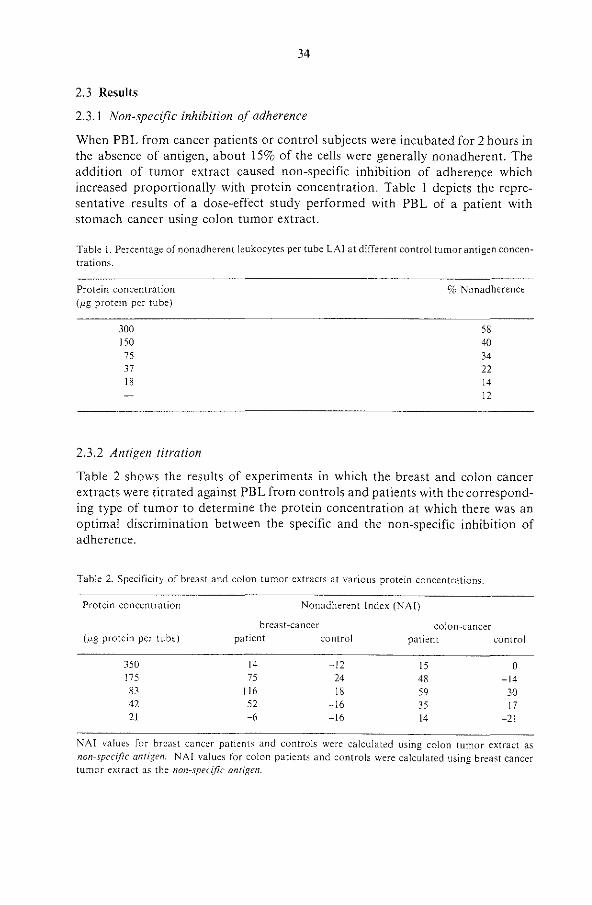
34
2.3 Results
2.3.1 Non-specific inhibition of adherence
When PBL from cancer patients or control subjects were incubated for 2 hours in the absence of antigen, about 15% of the cells were generally nonadherent. The addition of tumor extract caused non-specific inhibition of adherence which increased proportionally with protein concentration. Table 1 depicts the representative results of a dose-effect study performed with PBL of a patient with stomach cancer using colon tumor extract.
Table 1. Percentage of nonadherent leukocytes per tube LAI at different control tumor antigen concentrations.
Protein concentration (/lg protein per tube)
300
!50 75 37 18
2.3.2 Antigen titration
% Nonadherence
58 40 34 22 14 12
Table 2 shows the results of experiments in which the breast and colon cancer extracts were titrated against PBL from controls and patients with the corresponding type of tumor to determine the protein concentration at which there was an optimal discrimination between the specific and the non-specific inhibition of adherence.
Table 2. Specificity of breast and colon tumor extracts at various protein concentrations.
Protein concentration Nonadherent Index (NAI)
breast-cancer colon-cancer (,u.g protein per tube) patient control patient control
350 175 83 42 21
14 75
!16 52 -6
-12 24 18
-16 -16
15 0 48 -14 59 30 35 17 14 -21
NAI value> foe bcca't cancec patient> and contmb wm calculated using colon tumoc ntcact as non-spec!fic antigen. NAI values for colon patients and controls were calculated using breast cancer tumor extract as the non-spec((ic antigen.
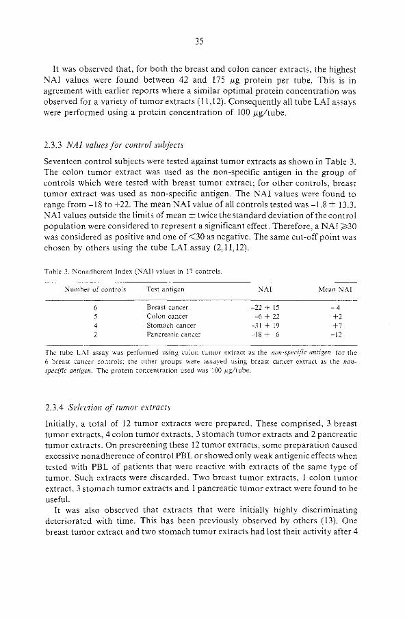
35
It was observed that, for both the breast and colon cancer extracts, the highest NAI values were found between 42 and 175 f.'g protein per tube. This is in agreement with earlier reports where a similar optimal protein concentration was observed for a variety of tumor extracts ( 11, 12). Consequently all tube LA! assays were performed using a protein concentration of 100 J.lgltube.
2.3.3 NAI values for control subjects
Seventeen control subjects were tested against tumor extracts as shown in Table 3. The colon tumor extract was used as the non-specific antigen in the group of controls which were tested with breast tumor extract; for other controls, breast tumor extract was used as non-specific antigen. The NAI values were found to range from -18 to +22. The mean NAI value of all controls tested was -1.8 ± 13.3. NAI values outside the limits of mean± twice the standard deviation of the control population were considered to represent a significant effect. Therefore, a NAI ):-30 was considered as positive and one of <30 as negative. The same cut-off point was chosen by others using the tube LA! assay (2, 11, 12).
Table 3. Nonadherent Index (NAI) values in 17 controls.
Number of controls
6 5 4 2
Test antigen
Breast cancer Colon cancer Stomach cancer Pancreatic cancer
NAI
-22 + 15 -6 + 22
-31 + 19 -18 + 6
Mean NAI
-4 +2 +7
-12
The tube LA! assay was performed using colon tumor extract as the non-specific antigen for the 6 breast cancer controls~ the other groups were assayed using breast cancer extract as the nonspec{fic antigen. The protein concentration used was 100 J.Ig/tube.
2.3.4 Selection of tumor extracts
Initially, a total of 12 tumor extracts were prepared. These comprised, 3 breast tumor extracts, 4 colon tumor extracts, 3 stomach tumor extracts and 2 pancreatic tumor extracts. On prescreening these 12 tumor extracts, some preparation caused excessive nonadherence of control PBL or showed only weak antigenic effects when tested with PBL of patients that were reactive with extracts of the same type of tumor. Such extracts were discarded. Two breast tumor extracts, I colon tumor extract, 3 stomach tumor extracts and 1 pancreatic tumor extract were found to be useful.
It was also observed that extracts that were initially highly discriminating deteriorated with time. This has been previously observed by others (13). One breast tumor extract and two stomach tumor extracts had lost their activity after 4

36
months. This observation made it imperative to renew the tumor extracts regularly and test them together with older effective preparations.
2.3.5 Clinical results af the tube LA! assay
A summary of the results obtained using the tube LAI assay in 41 cancer patients and 17 controls are shown in Table 4. The results are expressed as the number of patients which showed NAI values of ;?:30. The mean NAI values of the various groups are also dipicted. A NAI value of ~30 was accepted as positive whether or not the difference in nonadherence of the two tumor extracts used was statistically significant.
Table 4. Summary of patients tested using the tube LAI assay for reactivity to various tumor extracts.
Tumor type Number of Positive in Mean NAI patients tested LA!*
Breast cancer 15 9 ( 60%) 62
Benign breast disease 5 1 ( 20%) 21
Controls 6 0 ( Oo/c) -4
Colon cancer 11 8 ( 72(1(:.) 39 Controls 5 0 I 0%) 2
Stomach cancer 8 5 ( 6Ylr·) 47 Controls 4 1 ( 25CJr) 7
Pancreatic cancer 2 2 (100%) 56 Controls 2 0 ( 0%) 12
*A NAI value of ~30 was considered as positive. NAI values for breast cancer patients were calculated using the extract of colon cancer as the non-specific antigen. NAI values for colon. stomach and pancreatic cancer patients were calculated using breast cancer extract as the non-specific anrigen.
Positive LAI-reactions were observed in 60% of the breast cancer patients, in 72% of the colon cancer patients, and in 63% of the stomach cancer patients. The mean NAI (including the positive and negative NAI values) ranged from 39 to 72. The highest NAI values were found in the group of breast cancer patients, where NAI values of200 were sometimes observed. The lowest values were observed in the group of colon cancer patients.
From the 5 patients with benign breast disease, only one patient reacted positively in the LA! with a relatively high NAI value of77; the other4 patients had NAI values of 20, 14, 0 and -6 respectively (mean 21).
False positive reactions were recorded in 2 of 15 control patients with nonn aplastic disease (13.3%). six out of the 15 control patients were females, 4 of whom were multiparous. None of these 4 multiparous women were LAI-positive.

37
This indicated that sensitization with alloantigens was unlikely to be involved in the LAI. However, the possibility that sensitizing agents such as blood transfusions and viral infections could play a role in the LA! cannot be totally excluded.
2.4 Discussion
The peripheral blood leukocytes (PBL) from breast and gastrointestinal cancer patients showed a reduced adherence to glass in the presence of allogeneic tumor extracts. This inhibition of adherence was tumor-type specific in the majority of the cases. The reduced adherence was not due to toxicity of the tumor extracts, since adherence of control PBL was not affected. In addition, immunization by alloantigens could be excluded as the cause of adherence inhibition, since the 4 multiparous controls that were tested, showed no reduced adherence. Sensitization in vitro to histocompatibility antigens present in the tumor extracts can also be excluded, since the interaction between PBL and antigens was brief.
PBL from control subjects showed non-specific nonadherence in the presence of tumor extract; this nonadherence was related to the protein concentration of the tumor extract. It was therefore essential to titrate the various tumor extracts and use a protein concentration which allowed a clear discrimination between specific and non-specific inhibition of adherence. By interpolation, a concentration of 100 /J.g per tube was chosen. This is similar to the protein concentration used by others (2, II, 12).
In this study, about 13% of the controls were positive in LA!. This is higher than that reported by others (12,14). Generally, around 5% of the contro.ls are found to react positively in the LAI. Since, the control group in the present study is somewhat small, no definite conclusion can be drawn with regard to the significance of the observed differences.
The PBL of 70% of both breast and gastrointestinal cancer patients reacted positively with the relevant allogeneic tumor extracts. This percentage of positive tumor-specific LAI-reacti'.'ity is lower than those reported by Tataryn eta!. (13) and Halliday eta!. (15). The results from these groups indicate that under optimal conditions, positive LAI results can be observed in 80-95% of the cases. However, the highest scores were found in patients with early stages of malignancy and the percentage of positive LAI-reactions decreased dramatically in disseminated stages, with a range of 30-60%, depending on the type of cancer under investigation and the degree of dissemination (11,13). Since, the number of patients in the experimental groups in the present study is rather small, it is premature to relate the LAI results with the stage of disease. Consequently, the current results represent pooled observations of various clinical stages of cancer. When this pooling-effect is taken into account, an overall positive LAI score of 70% is promising.
Currently, it was observed that one out of 5 patients with benign breast disease reacted positively in the tube LAI assay. Similar results have been reported in earlier studies in which positive LAI-reactions were observed in 10-24% of women with benign breast disease (7,11,14,16). It has been suggested that this relatively

38
high score in false positive reactions is due to antigenic similarities between dysplastic lesions of the breast and breast cancer tissue. The biological implications of these findings may be important, as it is known that patients with a histological picture of benign mammary dysplasia have a higher incidence of breast cancer (17,18,19).
From the results presented here, it can be concluded that the tube LA! assay is a valuable test for studying various aspects of tumor immunity. However, the tube LAI assay can only be executed successfully provided it is performed routinely and tumor extracts are carefully selected and regularly renewed.
2.5 References
1. Halliday WJ. Detection of anti-tumor cell mediated immunity and serum blocking factors in cancer patients by the leukocyte adherence inhibition test. Br. J. Cancer, 1974, 29, 31-35.
2. Grosser N, Thomson DMP. Ceil mediated antitumor immunity in breast cancer patients evaluated by antigen induced leukocyte adherence inhibition in test tubes. Cancer Res .. 1975, 35, 2571-2579.
3. HellstrOm J, HellstrOm KE. Shantz G. Demonstration of tumor associated immunity with a leukocyte adherence inhibition assay. Int. J. Cancer. 1976, 18, 354-360.
4. Beek JJ van, Huuksloot CEM van, A ken M van, Bowen JR. Comparison oft he tube LAI test with the lymphocyte stimulation assay in a rat transplantation model. Cell. Immunol., 1978, 38, 84-93.
5. Kalafut F, Hung PM, Novotna L, Grofova M. Evidence of cell-mediated antitumor immunity in rats bearing syngeneic tumors by the leukocyte adherence inhibition. Neoplasma, 1978, 25, 569-
575. 6. Thomson DMP, Grosser N. Immunological mechanisms of tube leukocyte adherence inhibition.
Cancer Res., 1979, 39, 576-581. 7. O'Connor R, MacFarlane JK, Murray D, Thomson DMP. A study of false positive and negative
responses in the tube leukocyte adherence inhibition (tube LAI) assay. Br. J. Cancer, 1978, 38. 674-684.
8. Thomson DMP (ed). Assessment of immune status by the leukocyte adherence inhibition test. 1982 (Academic Press Inc., New York).
9. Lowry OR Rosebrough NJ, Farr AL, Randall RJ. Protein measurements with the folin phenol regent. J. Bioi. Chern. 1951, 193, 265-275.
10. BOyum A. Separation of blood leukocytes, granulocytes and lymphocytes. Tiss. Antig. 1974,4, 269-274.
11. Flores M, Marti JH, Grosser N, MacFarlane JK, Thomson DMP. An overview. Antitumor immunity in breast cancer assayed by tube leukocyte adherence inhibition. Cancer, 1977, 39, 494-505.
12. Marti JH, Thomson DMP. An antitumor immunity in malignant melanoma assayed by tube leukocyte adherence inhibition. Br. J. Cancer, 1976,34, 116-133.
13. Tataryn DN, MacFarlane JK, Thomson DMP. Leukocyte adherence inhibition for detecting specific tumor immunity in early pancreatic cancer. Lancet. 1978, 1, 1020-1021.
14. Fritze D, Schulte-Ucntrop C, Kaufmann K. Leukocyte adherence inhibition (LA I) tests in patients clinically suspected of having breast cancer using a panel of breast carcinoma extracts. Eur. J. Cancer, 1979, 15, 1491-1496.
!5. Halliday WJ, Koppi PA, Khan J. Davis N. Leukocyte adherence inhibition: tumor specificity of cellular and serum blocking reactions in human melanoma, breast cancer and colorectal cancer. JKCI. 1980. 65. 327-335.
16. Lopez MJ. O'Connor R, MacFarlane JK, Thomson DMP. The natural history of antitumor immunity in human breast cancer assayed by tube leukocyte adherence inhibition. Br. J. Cancer, 1978. 38,660-673.
39
17. Donelly PK, Baker KW, Carney' JA, O'Fallon WM. Benign breast lesions and subsequent breast carcinoma in Rochester, Minnesota. Mayo Clin. Proc., 1975, 50, 650-653.
18. Kovarik J, Lauerova L, Ninger E. :vlunzarova M, FeitJ, Zeman ova D. Wotke R. Evaluation of the LAI test in patients with benign and malignant breast diseases. Neoplasma, 1983, 30, 163-167.
19. Sanner T, Kotlar H, Eker P, Brennhovd L Host H, Jorgensen 0. Early detection of breast cancer by leukocyte adherence inhibition assay. Cancer detection and prevention, 1983, 6, 443-450.
40
CHAPTER 3
TUBE LEUKOCYTE ADHERENCE INHIBITION (LAI) ASSAY IN PATIENTS WITH NON-MALIGNANT DISORDERS OF THE COLON
AND COLORECTAL CANCER*
3.1. Introduction
In the preceeding chapter, the tube LAI assay was used successfully to demonstrate tumor-specific LAI-reactivity in breast and gastrointestinal cancer patients. However, the assay was found to have two main limitations. These were a) an extensive prescreening of the extracts was imperative to select extracts that discriminated well in the assay and b) crude extracts that were initially highly discriminating deteriorated on prolonged storage at -70°C. Thus, old extracts had to be regularly replaced with new ones.
The studies described in this chapter were performed in an attempt to try to overcome these two main drawbacks. The aim was to determine whether the sensitivity of the assay could be improved by extracts that contained less subcellular impurities than those used in the studies in chapter 2 and whether such extracts showed an improved "'shelf-life". Patients with colitis, various types of polyps of the large bowel and colo rectal cancer were tested. The ultimate goal was to use the tube LAI assay to follow the purification of human tumor-associated antigens (T AA) from crude tumor extracts.
3.2. Materials and Methods
3.2.1. Patients
All individuals were patients at Dijkzigt hospital, Rotterdam. Twenty nine patients with colorectal cancer, 12 patients with colitis and 21 patients with various types of polyps of the large bowel were evaluated. The control groups comprised 18 healthy volunteers and 29 patients with various non-gastrointestinal diseases.
All diagnosis were known prior to testing in the LAI assay and those of colorectal cancer were confirmed by histological examination. The clinical stage of the disease was not taken into account for the final evaluation of the results.
3.2.2. Preparation of tissue extracts
Colorectal and breast cancer tissues were obtained at surgery. Tissue extracts were prepared using the basic method described in 2.2.2, with some modiciations. After fatty and fibrous tissue was removed, a 20% (w/v) homogenate of the tumor tissue
"'LAI studies in colorectal cancer patients under recombinant leukocyte A mterferon therapy were published in Oncology, 1985, 42, 157~163.
41
was prepared in ice-cold phosphate buffered saline (PBS) using an ultra-turrax TR 18-10 homogenizer. The homogenate was centrifuged at IOOOG for 5 minutes and subsequently at 50000G for another 60 minutes. The supernatant (crude tumor antigen extract) was sterilized by filtration through 0.21' millipore filter. The protein concentration of the stock extracts was determined using the Bio-rad protein assay (Bio-rad, Holland) and ranged from 3 to 7 mg/ml. All extracts were stored at -70°C in 1 ml aliquots and were used only once.
3.2.3. Isolation of peripheral blood leukocytes (PEL)
Twenty ml heparinized blood was obtained from each patient. PBL were isolated by gradient centrifugation as described in 2.2.3. After counting and viability testing using trypan blue, the cell suspension was adjusted to a concentration of 5 x 10⁶
living cells per mi.
3.2.4. Tube LA! assay
The tube LAI assay was performed as described under 2.2.4. The results were expressed as nonadherent index (NAI). The NAI values outside the limits of mean ±twice the standard deviation of the control groups were considered to represent a significant effect.
3.3. Results
3.3.1. Selection of tumor extracts
Thirty colorectal cancer extracts and 19 breast cancer extracts were prepared. When the 49 extracts were prescreened, several extracts caused either excessive nonadherence of the PBL of healthy controls or showed only weak antigenic effects when incubated with PBL of patients that were reactive with other extracts of the same histological type of tumor. Such extracts were discarded. Twenty four colorectal and 16 breast cancer extracts were found useful.
In addition, it was observed that 20 of the colorectal and 14 of the breast cancer extracts had lost their LAI-reactivity within 4 months after preparation. The remaining 4 colorectal and 2 breast cancer extracts lost all reactivity 5 months after preparation. Similar observation have been reported earlier by others (1).
3.3.2. Nonadherent Index (NAl) values in control groups
Eighteen healthy volunteers and 29 patients with various non-gastrointestinal diseases were tested using colorectal cancer extract as specific antigen and breast cancer extract as non-specific antigen. The NAI values obtained in both the control groups are dipicted in Fig. I. The NAI values in the 18 healthy volunteers ranged from +39 to -28 with a mean of -0.2 ± 17 (lane 4). The NAI values in 29 patients with various non-gastrointestinal diseases ranged from+ 35 to -23 with a mean of+ 2 ± 16 (lane 5). NAI values outside the limits of mean± twice the standard deviation

42
'" •
" • •
" "
. • •• • " •
3 " X
w 0
" ~
" " " z w
" " w
' 0 • " z 0 z 0
_, _,
• • I • •• • • •• •• I• • • I • • • • j•
" .. j• .. I • .. • I :. • ::. • •
• I . : P.: • • I ••
• •• • •• • • • • _, •
-0·2±17 2115
2 3 4 5
Figure 1. Distribution of NAI values in 29 colo rectal cancer patients ( 1), 21 patients with polyps of the large bowel (2) and 12 patients with colitis (3).
The NAI values were calculated using colorectal cancer extracts as ~peCJfic antigen. and breast cancer extract as non-specific antigen.
The NAI values outside the limits of mean ± twice the standard deviation of the control groups consisting of healthy individuals (4) and patients with various non-gastrointestinal diseases (5) were considered to have a significant effect. Consequently, NAI values of ;;:,35 were regarded as positive.
of the control groups were considered to represent a significant effect. Consequently, a NAI value of~35 was considered as positive and one of <35 as negative. This cut off point is slightly higher than those reported by others (2-4).
3.3.3. Clinical results
The results in the 3 groups of patients with non-malignant disorders of the colon and colorectal cancer are also shown in Fig. 1. The tube LAI assay was performed using colorectal cancer extract as specific antigen and breast cancer extract as nonspecific antigen. A NAI value of ;:::35 was regarded as positive.
Positive LAI-reactions were observed in 62% (18 of 29) of the colorectal cancer patients (lane 1), in 5% (I of 21) of the patients with various types of polyps of the large bowel (lane 2) and in 8% (1 of 12) of the patients suffering from colitis (lane 3). A further subclassification of the patients with various types of polyps of the large bowel and patients suffering from colitis are shown in Table 1. The mean NAI value of the 3 groups (including positive and negative NAI values) ranged from +8 to

43
+40. The highest NAI values were observed in the colorectal cancer patients. It is seen in Table 2 that there was no correlation between the LAI results and the degree of differentiation of the tumor in the colo rectal cancer patients.
False positive reactions were observed in I of 18 (6%) healthy volunteers and in I patient of 29 (3%) with non-gastrointestinal diseases. False negative reactions were observed in II of 29 (38%) colorectal cancer patients.
Table I. Summary of LAI results in patients with non-malignant disorders of the colon.
Diagno,is of leukocyte donor Number tested Number LAI positive
I. Polyps of the large bowel: a) Hyperplastic 2 0 b) Adenoma 4 0 c) Multiple polyps 10 0 d) Familiar polyposis coli 0 c) others 4 I'
2. Colitis: a) Crohnse colitis 4 I b) Colitis ulcerosa 2 0 c) Undefined colitis 6 0
*This patient had undergone polypectomy, but no lesions were observed by colonoscopy.
Table 2. A comparison of LAI results and degree of tumor differentiation in co\orecta\ cancer patients.
Degree of colon tumor differentiation
Poorly differentiated tumors
Moderately differentiated tumors Well differentiated tumors
Number of patients LA! positive LA! negative (NAI ;;,35) (NAI <35)
5 5 3
7
A total of 22 co!orectal cancer patients were evaluable. There is no significant difference between any of
the three differentiation catagories.
3.4. Discussion
The main objectives that were persued in the investigations presented here were to increase the sensitivity of the tube LAI assay by using tumor extracts containing minimal sub-cellular impurities and to assess whether such extracts had an extended "shelf-life" than those used previously (5). A total of 49 extracts were prepared and were prescreened with PBL of healthy volunteers. Nine extracts produced excessive nonadherence and were discarded. The majority of the remaining usable extracts had lost their reactivity within 4 months of preparation. This period was similar to that observed previously (5) and would plea for a better

44
isolation technique in which the amounts of the lysozomal enzymes which could be responsible for the limited "shelf-life" of the extracts were kept at a minimum. One such approach would be the enzymatic stripping of the antigenic determinants from the tumor cell surface and/or further purification of the extracts which may yield material of an extended "shelf-life".
With regards to the specificity of the LAI assay using these crude extracts, it was observed that 6% (I of 18) of the healthy volunteers and 3% (I of29) of the patients with non-gastrointestinal diseases showed positive reactions with allogeneic colorectal cancer extracts. These values are in agreement with those observed by others ( 4, 6). In those studies, an average of 5% of the controls were found to react positively in the tube LA! assay.
Sixty two percent (18 of 29) of the colorectal cancer patients reacted positively in the LA! assay. This percentage is considerably lower than that reported by others (1, 7). In those reports, between 80 and 95% of the patients that were tested reacted positively. The positive score rate in the LAI has been shown to be related to the clinical stage of the disease (I, 3, 8). Highest percentage of positive LAI-reactions were observed in patients with an early stage malignancy. This percentage dropped considerably and ranged from 30-60% in patients with late stage malignancies according to the type of cancer. The results reported here are in agreement with our previous results where 70% of the patients with colorectal cancer showed positive LAI-reactions (9). The false negative results in 38% (II of 29) of the present colorectal cancer patients could be due to either the inherent insensitivity of the tube LAI assay, or due to a large tumor burden in these patients whereby the excess circulating tumor antigen in the serum coats the reactive leukocytes rendering them unresponsive in the LA! (10).
In the two groups of patients with non-malignant disorders of the colon, 5% (I of 21) of the patients with various kinds of polyps of the large bowel and 8% (I of 12) of the patients suffering from colitis reacted positively with allogeneic colorectal cancer extracts. Since, these percentages are comparable to those observed in the two control groups, they can be considered as false positives.
In conclusion, although the sensitivity of the tube LA! assay could not be improved by the use of extracts with minimal sub-cellular impurities, the high number of false positive reactions observed in the controls of the previous study (5) were reduced from 13% to an acceptable level of 5% by the use of such extracts. In other words, although the sensitivity of the assay was not improved, there was a tendency that its specificity was. It would therefore be worthwhile to investigate whether further purification of the crude extracts by column chromatography would ultimately result in an increase in the sensitivity while retaining the high specificity of the assay.
3.5.References.
1. Tataryn DN, MacFarlane JK, Murray D. Thomson DMP. Tube leukocyte adherence inhibition (LAI) assay in gastrointestinal (GIT) cancer. Cancer, 1979, 43, 898-912.

45
2. Grosser N, Thomson DMP. Cell mediated antitumor immunity in breast cancer patients evaluated by antigen-induced leukocyte adherence inhibition in test-tubes. Cancer Res., 1975, 35. 2571-2579.
3. Flores M. Marti JH, Grosser N, MacFarlane JK, Thomson DMP. An overview. Antitumor immunity in breast cancer assayed by tube leukocyte adherence inhibition. Cancer, 1977, 39, 494-505.
4. Marti JH, Thomson DMP. An anti-tumor immunity in malignant melanoma assayed by tube leukocyte adherence inhibition. Br. J. Cancer, 1976, 34, 116-133.
5. Marquet RL, Eggermont L Tank B. Westbroek DL, Jeekel J. Evaluation of the tube leukocyte adherence inhibition (LAI) assay in breast and gastrointestinal cancer. J. Exp. Clio. Cancer Res., 1982. I. 143-148.
6. Fritze D, Schulte-Uentrop C, Kaufmann K. Leukocyte adherence inhibition (LAI) tests in patients clinically suspected of having breast cancer using a panel of breast carcinoma extracts. Eur. J. Cancer, 1979. 15, 1491-1496.
7. Halliday WJ. Koppi PA, Khan J, Davis N. Leukocyte adherence inhibition: tumor specificity of cellular and serum blocking reactions in human melanoma, breast cancer and colorectal cancer. JNCI. !980. 65, 327-335.
8. Ayeni AO. Thomson DMP, MacFarlane JK, Daly D. A comparison of tube leukocyte adherence inhibition assay and standard physical methods for diagnosing colorectal cancer. Cancer, 198\,48, 1855-1882.
9. Tank B. Bijma AM, Marquet RL, Eggermont AMM. Weimar W, Jeekel J, Westbroek DL Leukocyte adherence inhibition (LAI), Natural Killer (NK) Cell acitivity, Gamma-interferon production capacity (GIPCA) and phytohaemagglutinin (PHA) response in patients treated with recombinant leukocyte A interferon. Oncology. 1985, 42, 157-163.
10. Thomson DMP. The LAI response to human cancer; The biology oft he LAI response and features of the antigens. In: Thomson DMP. ed. Assessment of immune status by leukocyte adherence inhibition test. (Academic Press, New York), 1982, chapter 6: 128-166.

46
CHAPTER 4
TUBE LEUKOCYTE ADHERENCE INHIBITION (LAI) ASSAY IN COLORECTAL CANCER PATIENTS USING PARTIALLY PURIFIED
TUMOR EXTRACTS
4.1. Introduction
The tube leukocyte adherence inhibition (LAI) assay was shown to have a sensitivity of 62% in colorectal cancer patients in the preceeding chapter. This percentage was considerably lower than that reported previously by others (1, 2) and therefore limits its clinical value for the routine screening of cancer patients. However, at the same time, the studies described in the previous chapter showed that the specificity of the assay was increased to 95% as compared to 87% in the earlier studies (3). This seems to indicate some reduction in the number of false positive reactions.
In the studies described in this chapter, an attempt was made to increase the sensitivity of the assay while retaining its high specificity by using partially purified extracts. Such extracts were obtained by sephacryl S-200 column chromatography of the crude tumor extracts. It was also examined whether such partially purified extracts showed an extended "shelf-life" as compared to their crude counterparts since considerable amounts of cellular lysozomal enzymes which could be responsible for the limited "shelf-life" would have been eliminated.
The tube LAI assay was performed in colotectal cancer patients using partially purified extracts of colorectal and breast cancers.
4.2. Materials and Methods
4.2. I. Patients
Eleven patients with colorectal cancer were evaluated. The control group consisted of 12 healthy volunteers and 10 patients with non-gastrointestinal diseases.
The diagnosis of colorectal cancer was known prior to testing in the LAI assay and was confirmed by histological examination. The clinical stage of the disease was not taken into account for the final evaluation of the results.
4.2.2. Preparation of tissue extracts
Colorectal and breast cancer extracts were prepared as described in 3.2.2.

47
4.2.3. Sephacryl S-200 column chromatography
After the 500000 fraction had been concentrated, a I ml sample containing approximately 30 mg of protein was applied to a calibrated K26/70 sephacryl S-200 column (Pharmacia) and eluted with PBS (pH 7.3) at a flow rate of 10 mllhr. The eluate was concentrated approximately five fold using the "on-line amicon thin channel system". Four fractions were collected from the column: A high molecular fraction designated "F 120", moving behind the excluded volume of the column; a second fraction eluting slightly ahead of where a haemoglobin/heptoglobin standard eluted, designated "F 100", a protein peak eluting ahead of the area where a bovine serum albumin (BSA) standard eluted, designated "F 68" and a low molecular weight fraction eluting in the area where an ovalbumin standard eluted, designated "F 48". All the four fractions were tested in the LA!.
4.2.4. Isolation of peripheral blood leukocytes (PEL) PBL were isolated from 20 ml heparinized blood from each patient as described in 2.2.3. After the cell suspension was counted and its viability tested using trypan blue, its concentration was adjusted to 5 X 10⁶ living cells per mi.
4.2.5. Tube LA! assay
Tube LAI assay was performed using the same conditions as described in 2.2.4. The LA! results were expressed as nonadherent index (NAI). The NAI values outside the limits of mean ± twice the standard deviation were considered to represent a significant effect.
4.3. Results
4.3.1. Selection of fraction reactive in the LA!
When crude extracts of breast or colorectal cancer were subjected to sephacryl S-200 column chromatography, four peak fractions "F 120", "F 100", "F 68" and "F 48" were obtained. The elution pattern is shown in Fig. 1. When these four fractions
5
F120
I F100
~ F68
I
0
10 20 FR.OCTION NUMBER
Figure 1. Molecular sieve chromatography of crude extracts of colorectal and breast cancers. The concentrated crude material was applied to a calibrated K26/70 Sephacryl S-200 column and eluted with PBS pH 7.3. "F 120" had the most LAI-reactivity.
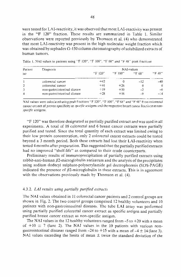
48
were tested for LAI-reactivity, it was observed that most LAI-reactivity was present in the "F 120" fraction. These results are summarized in Table 1. Similar observations were reported previously by Thomson et al. ( 4) who demonstrated that most LAI-reactivity was present in the high molecular weight fraction which was obtained by sephadex G-150 column chromatography of solubilized extracts of human tumors.
Table I. NAI values in patients using "F 120", "F 100", "F 68" and "F 48" peak fractions
Patient no
I 2 3 4
Diagnosis
colorectal cancer colorectal cancer non-gastrointestinal disease non-gastrointestinal disease
"F 120"
+42 +41 +19 +28
NAI-values "F 100" "F 68"
0 -12 +26 0 +10 -2 +16 -9
''F 48"
-40 0
-6 -14
NAI values were calculated using peak fractions "F 120", "F 100", "F 68" and "F48" from colorecta\ cancer extract of proven specificity as spec(fic antigens and the respective breast cancer fractions as nonspecific antigens.
"F 120" was therefore designated as partially purified extract and was used in all experiments. A total of 18 colorectal and 6 breast cancer extracts were partially purified and tested. Since the total quantity of each extract was limited owing to their low protein concentration, only 2 colorectal cancer extracts could be tested beyond a 3 month period. Both these extracts had lost their LAJ-reactivity when tested 4 months after preparation. This suggested that the partially purified extracts had no improved "shelf-life" as compared to their crude counterparts.
Preliminary results of immunoprecipitation of partially purified extracts using rabbit-anti-human ,82-microglobulin antiserum and the analysis of the precipitates using sodium dodecyl sulphate-polyacrylamide gel dectrophoresis (SDS-PAGE) indicated the presence of ,82-microglobulin in these extracts. This is in agreement with the observations previously made by Thomson et al. ( 4).
4.3.2. LA/ results using partially purified extracts
The NAI values obtained in 11 colorectal cancer patients and 2 control groups are shown in Fig. 2. The two control groups comprised 12 healthy volunteers and 10 patients with non-gastrointestinal diseases. The tube LAI assay was performed using partially purified colorectal cancer extract as specific antigen and partially purified breast cancer extract as non-specific antigen.
The NAI values in the 12 healthy volunteers ranged from -5 to +20 with a mean of + 10 ± 7 (lane 2). The NAI values in the 10 patients with various nongastrointestinal diseases ranged from -24 to+ 15 with a mean of -4 ± 14 (lane 3). NAI values exceeding the limits of mean ± twice the standard deviation of the

49
"' •
" " • " " •
~ E " •
• • w
0 " ~ • r z " • w " ~
" w I 0 < " z 0 z 0
• • • .. • •• • .. :· i • • • • _, • •
• _, • • _,
+10±7 -4±14
2 3
Figure 2. Distribution of NAI values in II colorectal cancer patients (1). The NAl values were calculated using partially purified colo rectal cancer extract as specific antigen. and partially purified breast cancer extract as non-specific antigen. The NAT values outside the limits of mean± twice the standard deviation of the control groups (2) and (3) were considered to have a significant effect. Consequently, NAI values of ~24 were regarded as positive.
control groups were considered to have a significant effect. Consequently, a NAI value of ~24 was regarded as positive and one of <24 as negative.
Seven out of the eleven (64%) colorectal cancer patients showed positive LAIreactivity (lane 1). The NAI values ranged from -5 to+ 100 (mean +38±35).
In neither of the two control groups false positive reactions were observed. False negative reactions were observed in 4 of 11 (36%) colorectal cancer patients.
4.3.3. Comparison of LA! results obtained using crude and partially purified extracts
Fig. 3 shows the LAI results obtained in a separate group of 6 colorectal cancer paients and the two control groups which comprised 10 healthy volunteers and 19 patients suffering from non-gastrointestinal diseases. Tube LAI assay was performed using simultaneously, crude colorectal cancer extract and its partially purified counterpart as specific antigens and the crude breast cancer extract and its partially purified counterpart as non-specific antigens respectively.

50
mo
" " 0
" • " •
a z " x 0
w Q
" ~ 0
• 0 ~ z " •• w ~
" w I Q
" ~ z 0 z 0
-U>
0 • • 00
.. • 0 •
~ • §, •• ;; • 0 0 .. :8 0 • •
• • _, • 0 _, • 0
-1±16 r:ta 7±12 0·8±14
2 3
Figure 3. Distribution of NAI values in 6 co\orectal cancer patients (1). The NAI values were calculated using crude (•) colorectal cancer extract and its partially purified (o)
counterpart as specific antigens and crude breast cancer extract and its partially purified counterpart respectively as non-~peciflc antigens. The NAI values outside the limits of mean± twice the standard deviation of the control groups (2) and (3) were considered to have a significant effect. Consequently, NAI values of ~31 using the crude extracts and ~26 using the partially purified extracts were regarded as positive.
The NAI values in the 10 healthy volunteers (lane 2) ranged from -28 to + 17 (mean -1 ± 16) using the crude colorectal cancer extract and from -6 to +20 (mean +7 ± 8) using its partially purified counterpart. The NAI values in the 19 patients with non-gastrointestinal diseases (lane 3) ranged from -15 to +29 (mean +7 ± 12) using crude colorectal cancer extract and -27 to + 14 (mean +0.8 ± 14) using its partially purified counterpart. NAI values exceeding the limits of mean± twice the standard deviation of the control groups were considered to have a significant effect. Thus, a NAI value of )::31 using crude colorectal cancer extract and )::26 using its partially purified counterpart were regarded as positive.
Three out of the 6 (50%) colorectal cancer patients reacted positively with allogeneic crude colorectal cancer extract and its partially purified counterpart. The NAI values ranged from -3 to +68 (mean+ 30 ± 30) and -9 to +80 (mean +27 ± 34) respectively.

51
No false positive reactions were observed in either of the control groups using crude colorectal cancer extracts, whereas 1 out of 19 (5%) patients suffering from non-gastrointestinal diseases reacted positively with its partially purified counterpart. False negative reactions were observed in 3 of 6 (50%) colorectal cancer patients both using the crude colorectal cancer extract and its partially purified counterpart.
4.4. Discussion
The present investigations were undertaken in order to examine whether the sensitivity of the tube LA! assay could be increased by using partially purified extracts and to assess whether these extracts had a "shelf-life" exceeding 4 months.
A total of 24 tumor extracts were partially purified using sephacryl S-200 column chromatography and tested. Since most of these extracts had low protein concentration, most of them were used up within 3 months after preparation. However, 2 partially purified extracts could be tested beyond this period, and both of them were found to have lost their LAI-reactivity within 4 months after preparation. This period was identical to that observed earlier for the crude extracts (3) and suggested that the partial purification of the crude extracts does not lead to an extended "shelf-life". One possible explanation for the limited "shelf-life" of the tumor extracts as opposed to the prolonged "shelf-life" of the intact tumor could be that once the intact tumor cells are disrupted, the LAI-reactive tumor-associated antigens on the tumor cell surface are displaced from their protective environment on the cell surface by loss of their quaternary structure. This loss results in them becoming susceptible to proteolytic enzymes leading to a gradual but permanent loss of LA!-reactivity within a finite period of time. Apparently, this rate of loss of reactivity cannot be slowed down by partial purification of the crude extracts on sephacryl S-200 columns.
None of the 12 healthy volunteers or 10 patients suffering from non-gastrointestinal diseases reacted positively with partially purified extracts of colorectal cancer (Fig. 2). One of 19 (5%) patients suffering from non-gastrointestinal diseases reacted positively with partially purified colorectal cancer extract (Fig. 3). However, the same patient showed no positive reaction with the crude extract. This high specificity of the tube LA! assay using partially purified extracts is similar to that observed previously by others (5, 6) where an average of 5% of the controls reacted positively with crude extracts.
Sixty four percent (7 out of 11) of the colorectal cancer patients reacted positively using the partially purified extract. This figure is slightly better than that observed using the crude colorectal cancer extracts where 62% of the colorectal cancer patients reacted positively indicating a slight but insignificant improvement in the sensitivity of the assay. This was confirmed in a separate series of experiments in which six colorectal cancer patients were simultaneously tested using crude colorectal cancer extract and its partially purified counterpart. In both instances 3

52
out of the 6 patients (50%) showed positive LAI-reactions confirming that the partial purification of the crude extract does not lead to a higher sensitivity of the assay. Similar results have been previously reported by others (7. 8) who observed that the use of purified antigen extracts did not result in a higher number of positive LAI-reactions.
The low protein concentrations of the partially purified extracts placed severe restrictions on the number of patients that could be tested since, considerable amounts of these extracts were used up for the determination of protein concentration for the optimum LAI-reactivity. Such limitations on the use of purified antigen extracts were previously reported by Thomson et al. (7). The amounts of tumor material available from patients undergoing surgery also imposed restriction on the amounts of crude extracts that could be chromatographed on Sephacryl S-200 columns and purified further using other physico-chemical methods.
Hence, in order to persue the initial objectives of increasing the sensitivity of the LAI assay and to improve the durability of the cancer extracts, it became essential to have a continuous source of large amounts of tumor material. Two possible approaches can be considered. The first would be a suitable animal tumor model which not only would act as a continuous source of tumor material, but could also be used to examine and improve conditions necessary for a more sensitive and reliable tube LA! assay. The second approach would be to use an appropriate human tumor cell line as a continuous source of LAI-reactive material which is either shed in the medium or obtained by solubilization of cell membrane antigens. The first approach is considered in chapters 5 and 6.
4.5. References
I. Halliday WJ, Koppi PA, Khan J, Davis N. Leukocyte adherence inhibition: tumor specificity of cellular and serum blocking reactions in human melanoma, breast cancer and colorectal cancer. JNCI. 1980. 65. 327-335.
2. Tataryn ON, MacFarlane JK, Murray D, Thomson DMP. Tube leukocyte adherence inhibition (LAI) assay in gastrointestinal (GIT) cancer. Cancer, 1979, 43, 898-912.
3. Marquet RL, Eggermont L, Tank B, Westbroek DL, Jeekel J. Evaluation of the tube leukocyte adherence inhibition (LAI) assay in breast and gastrointestinal cancer. J. Exp. Clio. Cancer Res., 1982. I. 143-148.
4. Thomson DMP, Tataryn ON, O'Connor R, Rauch, J. Friedlander, P, Gold P, Shuster J. Evidence for the expression of human tumor-specific antigens associated with /32-microglobulin in human cancer and in some colon adenomas and benign breast lesions. Cancer Res., 1979,39,604-611.
5. Marti JH, Thomson DMP. An antitumor immunity in malignant melanoma assayed by the tube leukocyte adherence inhibition. Br. J. Cancer, 1976, 34, 116-133.
6. Fritze D, Schulte-Uentrop C, Kaufmann K. Leukocyte adherence inhibition (LAI) tests in patients clinically suspected of having breast cancer using a panel of breast carcinoma extracts. Eur. J. Cancer, 1979, 15, 1491-1496.
7. Thomson DMP, Gold P, Freedman SO, Shuster J. The isolation and characterization of tumorspecific antigens of rodent and human tumors. Cancer Res., 1976, 36, 3518-3525.
8. Thomson DMP. The LAJ response to human cancer; The biology of the LAI response and features of the antigens. In: Thomson DMP, ed. Assessment of immune status by leukocyte adherence inhibition test. (Academic Press, New York), 1982, Chapter 6: 145-147.

53
CHAPTER 5
LEUKOCYTE ADHERENCE INHIBITION (LAI) IN RATS BEARING TRANSPLANTABLE SYNGENEIC TUMORS OF
DIFFERENT IMMUNOGENICITY*
5.1 Introduction
The tube leukocyte adherence inhibition (LAI) assay has been used previously with considerable success to monitor cell mediated immunity in cancer patients (1,2). However, the assay was found to have two serious limitations. These were the quality and the durability of the tumor extracts. The assay performed under optimal conditions had a sensitivity of 62% and a specificity of around 95% in colorectal cancer patients. This sensitivity of 62% could not be improved by using extracts that had been partially purified by sephacryl S-200 column chromatography. Any further purification of the extracts was severely hindered by the limited amount of tumor material obtained at surgery.
As large amounts of tumor material are necessary in order to prepare and purify the putative LAI-reactive material, it became imperative to search for a well defined animal tumor model which could be used as a constant source oflarge amounts of tumor material and which could also be used to investigate and improve conditions necessary for a more sensitive and reliable tube LAI assay.
Since the tube LA! assay had been successfully used to detect immunity in rats (3-7) and since three well defined transplantable syngeneic tumors of increasing immunogenicity in rats were available, the animal studies described here were undertaken to improve the LA! assay.
5.2 Materials and Methods
5.2.1 Rats
Male rats of inbred BN and WAG strains were used. The animals were bred under specific-pathogen-free conditions and were 8-10 weeks old.
5.2.2 Tumors
5.2.2.1 Liposarcoma (LS 175)
LS175 is a liposarcoma which originated as a spontaneous tumor in a female BN rat in the pancreatic, retroperitoneal region. The tumor was observed when the animal
*This chapter has been accepted for publication in the Eur. J. Cancer Clin. Oncol.

54
was 80 weeks old (8). Histopathological examination revealed that the infiltrating tumor was composed mainly of pale, swollen polyhedral cells with a granular cytoplasm containing fat. The tumor is palpable as early as one week after subcutaneous (s.c.) implantation. Immunization-challenge experiments performed according to the method of Prehn and Main (9), revealed that LS175 is not immunogenic i.e. the tumor growth is not inhibited or enhanced in immunized hosts. The tumor is transplantable in syngeneic animals.
5.2.2.2 Colon adenocarcinoma (CC531)
CC531 was induced by treatment of WAG rats with 1,2 dimethylhydrazine (DMH). It originated in the ascending colon 40 weeks after six weekly injections of 30 mg/kg DMH (10). The tumor is a moderately differentiated adenocarcinoma which is transplantable in syngeneic animals. When 2 mm cubes are implanted s.c., about 4 weeks are needed for the tumor to grow to a diameter of 10 mm. Classical immunization-challenge experiments (9) revealed that CC531 is weakly immunogenic i.e. a slight but significant tumor growth inhibition is observed in immunized hosts.
5.2.2.3 Skin basal cell carcinoma (1618)
1618 is a transplantable radiation induced basal cell carcinoma of the skin in the WAG rat (11). The tumor has strong immunogenic properties i.e. it does not grow in immunized hosts. Tumor 1618 has a doubling time of 2.5 days when implanted S.C.
5.2.2.4 Tumor implantation
Two mm cubes of tumor LS175, CC531 or 1618 were implanted s.c. in the right flank of the experimental animals. LA! assays were performed about 14 days after implantation when the tumors were either palpable (tumors CC531 and 1618) or had reached a diameter of between 5 and 10 mm (tumor LS175). LA! assays were also performed sequentially in rats bearing tumor CC531 on days 7, 14and 21 after implantation.
5.2.2.5 Sensitization of WAG rats with irradiated 1618 tumor cells
1618 tumor cell suspensions were prepared from s.c. tumor implants. Tumor cells were isolated according to the method of Reinhold (12). Cells were washed twice with RPM! 1640 medium (GIBCO), X-irradiated at a dose of 80 GY and mixed with Freund's incomplete adjuvant. WAG rats were immunized twice at 14 days interval by intraperitoneal injections with 108 cells. LAI assays were performed a week after the first and 2 days after the second immunization.
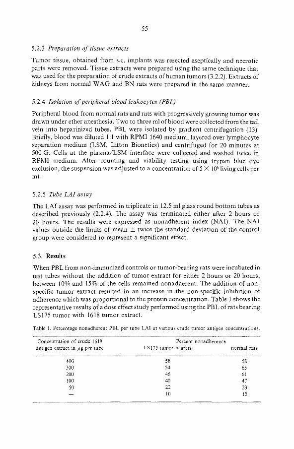
55
5.2.3 Preparation of tissue extracts
Tumor tissue, obtained from s.c. implants was resected aseptically and necrotic parts were removed. Tissue extracts were prepared using the same technique that was used for the preparation of crude extracts of human tumors (3.2.2). Extracts of kidneys from normal WAG and BN rats were prepared in the same manner.
5.2.4 Isolation of peripheral blood leukocytes (PEL)
Peripheral blood from normal rats and rats with progressively growing tumor was drawn under ether anesthesia. Two to three ml of blood were collected from the tail vein into heparinized tubes. PBL were isolated by gradient centrifugation ( 13). Briefly, blood was diluted 1:1 with RPM! 1640 medium, layered over lymphocyte separation medium (LSM, Litton Bionetics) and centrifuged for 20 minutes at 500 G. Cells at the plasma/LSM interface were collected and washed twice in RPM! medium. After counting and viability testing using trypan blue dye exclusion, the suspension was adjusted to a concentration of 5 X I06 living cells per ml.
5.2.5 Tube LAI assay
The LA! assay was performed in triplicate in 12.5 ml glass round bottom tubes as described previously (2.2.4). The assay was terminated either after 2 hours or 20 hours. The results were expressed as nonadherent index (NAI). The NAl values outside the limits of mean ± twice the standard deviation of the control group were considered to represent a significant effect.
5.3. Results
When PBL from non-immunized controls or tumor-bearing rats were incubated in test tubes without the addition of tumor extract for either 2 hours or 20 hours, between 10% and 15% of the cells remained nonadherent. The addition of nonspecific tumor extract resulted in an increase in the non-specific inhibition of adherence which was proportional to the protein concentration. Table 1 shows the representative results of a dose effect study performed using the PBL of rats bearing LS175 tumor with 1618 tumor extract.
Table I. Percentage nonadherent PBL per tube LAI at various crude tumor antigen concentrations.
Concentration of crude 1618 antigen extract in p.g per tube
400 300 200 100 50
Percent nonadherence LS 175 tumor-bearers normal rats
58 58 54 65 46 61 40 47 22 23 10 15

56
2H~ LAI 20HR lAI .. ·-"' • oo • 0
•o 0
0 • • .. • 0 0 0 ,.,. 0 0 • 0 I • 0 • • 0 0
0 • 0 i I • ,. ,. 0 • 0 0 g
lo • • 0 I 8 • 0 '0 0 • g 0 • • • I. • 0 • 0 • 0
00 • I 0 • • 0 • 0 0 0 0
'0 • • 0 0 0 • 8 • • I' ' ,. • • • 0 0 • • 30
8o 0 .. 0 0
0 0 -O·o±211 7±24 "" 11±21 13±11!1 ""'
2 3 2 3
Figure l. Distribution of NAI values in tumor-bearing BN rats (•) and control BN rats (o). The LAI assays were performed 14 days afters.c. implantation of tumor LS 175. Extract of tumor LS 175 served as specific antigen and extracts of normal BN kidneys (1), tumor CC531 (2) and tumor 1618 (3) as nonspecific antigens. The NAI values outside the limits of mean± twice the standard deviation of the control group were considered to represent a significant effect.
Fig. I shows the LA! results obtained· in the initial studies with the nonimmunogenic LS 175 tumor-bearing BN rats. The LA! assay was performed using LS 175 tumor extract as the specific antigen and extracts of normal kidney tissue and tumors CC531 and 1618 as non-specific antigens. The results were expressed as NAI. The NAI values outside the limits of mean± twice the standard deviation of the control group were considered to represent a significant effect. The results of the 2 hours LAI assay show that using kidney extract or 1618 extract as non-specific antigen none of the 10 tumor-bearing animals showed any positive LAI-reactivity (lanes I and 3 respectively). When CC531 extract was used as non-specific antigen, one tumor-bearer showed a positive LAI-reactivity (lane 2). Increasing the LA! incubation time to 20 hours resulted in no significant improvement in the LAIreactivity. Using CC531 and 1618 extracts as non-specific antigens, only one animal in each group showed a positive LAI-reactivity (lanes 2 and 3 respectively).
The results of the studies using the weakly immunogenic tumor CC531 are shown in Fig. 2. The LAI assays were performed using the same conditions as those described for LS175 tumor-bearing animals. Extract of CC53! was used as the specific antigen and extracts of normal kidneys, LS 175 and 1618 as the non-specific antigens. The results show that positive LAI-reactivity was observed in one out of
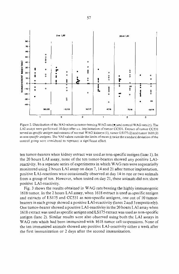
57
2H• LAI 20HR LAI
" "' "' 0
" 0
ii .. 8 0 0
" • "' ~ •o • z >O w ~ w X 0 0 ~
~ -10 z
-•o
• 0
l 0 r 8 • 0 0 • • • 0 0
I 0 0 0 • 0 • 0 0 I 0 > 0 I
0 r 0 • I i 0 0
• 0 0 • • • 0 • • 00
0 0 • 8 0 • • • 0 .. 0 0 • 0
0 0 • 0
0 .. 0 I 0
• • - 0 0
0 • 00
-•o 28±22 -3±18 14±11 •±18 3±16 -9±16
2 ' 2 ' Figure 2. Distribution of the NAI values in tumor-bea'ring WAG rats (•)and control WAG rats (o). The LAI assays were performed 14 days after s.c. implantation of tumor CC531. Extract of tumor CC531 served as specific antigen and extracts of normal WAG kidneys (I), tumor LS175 (2) and tumor 1618 (3) as non-specific antigens. The NAI values outside the limits of mean± twice the standard deviation of the control group were considered to represent a significant effect.
ten tumor-bearers when kidney extract was used as non-specific antigen (lane 1). In the 20 hours LA! assay, none of the ten tumor-bearers showed any positive LAIreactivity. In a separate series of experiments in which WAG rats were sequentially monitored using 2 hours LAI assay on days 7, 14 and 21 after tumor implantation, positive LAI-reactions were occasionally observed at day 14 in one or two animals from a group of ten. However, when tested on day 21, these animals did not show positive LAI-reactivity.
Fig. 3 shows the results obtained in WAG rats bearing the highly immunogenic 1618 tumor. In the 2 hours LA! assay, when 1618 extract is used as specific antigen and extracts of LSI75 and CC53l as non-specific antigens, one out of 10 tumorbearers in each group showed a positive LAI-reactivity (lanes 2 and 3 respectively). One tumor-bearer showed a positive LAI-reactivity in the 20 hours LAI assay when 1618 extract was used as specific antigen and LS 175 extract was used as non-specific antigen (lane 2). Similar results were also observed using both the LA! assays in WAG rats which had been immunized with 1618 tumor cell suspensions. None of the ten immunized animals showed any positive LAI-reactivity either a week after the first immunization or 2 days after the second immunization.

58
2HR LAI 20HR LAI
" " 0 • oo 0
• .. • 8
" ..
• 8 " 3 ,. • 0 • • •
~ ,. ~ •o w ~ 0 w ~ 4 -•o
~ _,.
• • 0 • 0 • 0
• 0 • • 00 00
§, 0 • 0 • 0
• 0 • • r .. 00 • 0 • 8 8 • 0 •
:1> • 0 I 0 • 8 ! I • • 0 0 0 r 0 • 0 • 0
0 8 • 88 • • -00
0 0
I 0
0
_ .. 0 0
-10:!:12 -15±14 -11:!:16 12:lo24 13:!26 13±20
2 3 2 3
Figure 3. Distribution of the NAI values in tumor-bearing WAG rats (•) and control WAG rats (o ). The LAI assays were performed 14 days after s.c. implantation of tumor 1618. Extract oftumor 1618 served as specific antigen and extracts of normal WAG kidneys ( 1), tumor LS 175 (2) and tumor CC531 (3) as non-specific anrigens . The NAI values outside the limits of mean± twice the standard deviation of the control group were considered to represent a significant effect.
5.4 Discussion
In the present studies, the 2 hours tube LA! assay which was successfully used in humans (1,2) has been used to follow the LAI-reactivity of the peripheral blood leukocytes (PBL) of rats bearing transplantable syngeneic tumors. The initial LA! studies were performed in BN rats bearing a transplantable syngeneic LS 175 tumor. The results obtained show that using the conventional 2 hours tube LA! assay, sporadic tumor-specific LAI-reactivity was observed. In previous studies in rats bearing syngeneic tumors, Kalafut et al. ( 4) and Hung et al. (5) observed that prolongation of the LA! assay incubation time to 20 hours gave better and consistent results than the 2 hours conventional assay. We therefore decided to use in addition, the 20 hours tube LA! assay. Once again, only sporadic tumor-specific LAI-reactivity was observed in the PBL of tumor-bearers. There was no qualitative difference between the results of the two LA! assays used. The possible explanation for this lack of significant tumor-specific LAI-reactivity could be that the LS175 tumor which is non-immunogenic does not or only weakly evokes an immune response which is occasionally detected in the tube LA! assay.
59
To investigate this possibility, LA! studies were performed using PBL of WAG rats bearing the weakly immunogenic CC531 tumor. Sporadic tumor-specific LAIreactivity was observed using both the LAI assays. Occasional positive LAIreactions were noted when CC531 implanted rats were sequentially monitored on days 7, 14 and 21. The results obtained using the highly immunogenic tumor 1618 were similar to those obtained with the non-immunogenic and the weakly immunogenic tumors. The results of studies using WAG rats immunized with cell suspensions of tumor 1618 also failed to demonstrate a consistent tumor-specific LAI-reactivity.
The results presented here contradict those obtained earlier by Kalafut et al. (4), Hung et al. (5) and those reported recently by Morizane and Sjogren (6,7). All these authors were able to demonstrate significant tumor-specific LAI-reactivity in PBL of tumor-bearing rats. In the studies by Kalafut et al. (4) and Hung et al. (5) the 20 hours LAI assay was performed using homologous serum and fetal calf serum (FCS)-supplemented media, respectively. FCS-supplemented medium was also used in the micro-glass-tube LA! assay by Morizane and Sjogren (6). In the current studies, when the tube LA! assays were performed using FCS-supplemented medium, no tumor-specific LAI-reactions were observed with the PBL of the rats bearing LS175, CC531 and 1618 tumors. A FCS concentration of 1% in the assay system totally inhibited the adherence of PBL from both normal and tumorbearing rats. Similar observations were reported previously (3,14,15). In those studies, normal serum or FCS inhibited the adherence of normal and "immune" leukocytes and abolished the specific LAI-reactions in the tube LA! assay.
The failure to observe any significant tumor-specific LAI-reaction in this study could be due to thefollowing reasons. Firstly, a lack of cells in the PBL that mediate the specific LAI-reaction. It has been shown previously that in the tube LA! assay, cells of the monocyte/macrophage series play a central role in triggering the cascade of events leading to nonadherence (16). Peritoneal cell suspensions (PC) contain mainly cells of the monocyte/macrophage series. In previous studies Holan et al. (17) observed specific LAI-reactions in PC of rats immunized with various antigens. However, when we performed pilot studies with the PC of LS 175, CC531 and 1618 tumor-bearing rats, no tumor-specific LAI-reactions were observed.
The sporadic tumor-specific LAI-reactivity in this study could also have been due to an insufficient amount of LAI-reactive tumor antigen in the crude tumor extracts. Increasing the concentration of these antigens by further purification of the crude extracts could possibly lead to a higher number of specific-reactions. However, the possibility that the LAI-reactive material is lost during the early stages of the preparation of the extracts has to be taken into consideration.
Finally it is possible that the site of tumor implantation in the current studies was inappropriate to evoke a specific LAI response. However, it is strange that the same tumor implantation site in the immunization-challenge experiments resulted in the tumor-specific immunity (CC531 and 1618 tumors). The unlikely possibility remains that PEL are poor effectors of tumor-associated immunity of s.c. implanted tumors. Perhaps the leukocytes isolated from regional draining lymph

60
nodes would be more suitable as tube LA! effector cells (!8). Transplantable syngeneic tumors may also induce suppressor cells or factors which impair the immune response of the host (!9). Therefore, it could be assumed that consistent tumor-specific LAI-reactions can be successfully detected only in animals with a primary autochthonous tumor.
5.5 References
I. Marquet RL, Eggermont L, Tank B, Westbroek DL, Jeekel J. Evaluation of the tube leukocyte adherence inhibition (LA I) assay in breast and gastrointestinal cancer. J. Exp. Clio. Cancer Res., 1982, 1. 143-148.
2. Tank B, Bijma AM, Marquet RL, Eggermont AMM, Weimar W, Jeekel J, Westbroek DL. Leukocyte adherence inhibition (LAI), Natural Killer (NK) cell activity, Gamma-interferon production capacity (GIPCA) and phytohaemagglutinin (PHA) response in patients treated with recombinant leukocyte A interferon. Oncology, 1985, 42, 157-163.
3. Beek JJ van, Huuksloot CEM van, Aken M van, Bowen JR. Comparison of the tube LAI test with the lymphocyte stimulation assay in a rat transplantation model. Cell. lmmunol., 1978, 38,84-93.
4, Kalafut F, Hung PM, Novotna L, Grofova M. Evidence of cell-mediated antitumor immunity in rats bearing syngeneic tumors by the leukocyte adherence inhibition test. Neoplasma, 1978,25, 569-575.
5. Hung P, Kalafut F, Novotna L. Comparison of LAI reactivity in B77 tumor bearing rats between, before and after surgical removal of tumors. Neoplasma, 1980, 27, 367-371.
6. Morizane T, SjOgren H. A leukocyte adherence inhibition (LAI) assay of antitumor immunity in rats using selective radioimmunological assessment of adherence ofT lymphocytes and monocytes. Int. J. Cancer, 1983, 31, 803-812.
7. Morizane T, SjOgren H. Detection of different states of immunity to colon cancer in rats by a leukocyte adherence inhibition (LAI) assay assessing the adherence of T lymphocytes and monocytes selectively. Int. J. Cancer, 1983, 31, 813-816.
8. Marquet RL, Schellekens H, Westbroek DL, Jee"kel J. Effect of treatment with interferon and cyclophosphamide on the growth of a spontaneous liposarcoma in rats. Int. J. Cancer, 1983, 31, 223-226.
9. Prehn RJ, Main JW. Immunity to methylcholanthrene induced sarcomas. J. Nat. Cancer Inst., 1957, 18. 769-778.
IO. Marquet RL, Westbroek DL, Jeekel J. Interferon treatment of a transplantable rat colon adenocarcinoma: Importance of tumor site. Int. J. Cancer, 1984, 33, 689-692.
II. Jeekel J, Eggermont, A, Heystek G, Marquet RL Inhibition of tumor growth by blood transfusions in rats. Eur. Sur. Res., 1982, 14, 124-125.
12. Reinhold H. A cell dispersion technique for use in quantitative transplantation studies with solid tumors. Eur. J. Cancer, 1965, 1, 67-71.
13. Boyum A. Separation of blood leukocytes, granulocytes and lymphocytes. Tiss. Antig., 1974,4, 269-274.
14. Marti JH, Thomson DMP. Antitumor immunity in malignant melanoma, assayed by tube leukocyte adherence inhibition. Br. J. Cancer, 1976, 34, 116-133.
15. Holan V, Hasek M. Specificity of cell-mediated transplantation reactions in rats detected by the macrophage adherence inhibition test. Immunogenetics, 1977, 4, l55-l6l.
16. Thomson DMP, Phelan K, Morton DG, Bach MK. Armed human monocytes challenged with a sensitizing cancer extract release substances pharmacologically similar to leukotrienes. Int. J. Cancer, 1982, 30, 299-306.
17. Holan V, Hasek M, Bubenik J, Chutna J. Antigen mediated macrophage adherence inhibition. Cell Immunol., 1974, 13, 107-116.

61
18. Holan V, Cbutna J, Hasek M. Cross-reactivity between Bacillus Calmette-Guerin and Rous virusinduced sarcoma detected in rats by tube leukocyte adherence inhibition assay. Cancer Res., 1979, 39, 593-596.
19. Isakov N, Hollander N, Segal S, Feldman M. An immunoregulatory factor associated with spleen· cells from tumor-bearing animals. III. Characterization of the factor's target cells. Int. J. Cancer, 1979, 23, 410-414.

62
CHAPTER 6
LEUKOCYTE ADHERENCE INHIBITION (LAI) IN RATS USING PARTIALLY PURIFIED TUMOR EXTRACTS*
6.1 Introduction
The sporadic tumor-specific leukocyte adherence inhibiton (LAI)-reactivity in rats bearing non-immunogenic liposarcoma (LS175), weakly immunogenic colon adenocarcinoma (CC53!) and highly immunogenic skin basal cell carcinoma (1618) tumors as presented in the preceeding chapter could have been due to several factors (I).
In the studies described here, close attention was focused on one of these factors. The possibility that the sporadic LAI-reactivity in the tumor-bearers was caused by an insufficient amount of LAI-reactive tumor antigen in the crude extracts that were used was investigated further. It was assumed that a higher number of tumorspecific LAI-reactions might be observed, if the concentration of the LAI-reactive antigens could be increased by further purification of the crude extracts.
WAG rats bearing the weakly immunogenic colon adenocarcinoma (CC531) tumor model was chosen since successful results were obtained in colorectal cancer patients using extracts that had been obtained after column chromatography (chapter 4).
Tube LA! assay was performed using peripheral blood leukocytes (PBL) and peritoneal cells (PC) of CC531 tumor-bearing animals. Crude tumor extracts were partially purified using sephacryl S-200 column chromatography. To exclude the possibility that the LAI-reactive material could have been lost during the early stages of preparation, LA! studies were also performed with extracts that had been sequentially isolated.
6.2 Materials and Methods
6.2.1 Rats
Male rats of inbred BN and WAG strains were used. The animals were bred under specific-pathogen-free conditions and were 8-10 weeks old.
*This chapter has been submitted for publication.
63
6.2.2 Tumors
6.2.2.1 Liposarcoma (LS175)
LS175 is a liposarcoma which originated as a spontaneous tumor in a female BN rat in the pancreatic, retroperitoneal region (2). Immunization-challenge experiments performed according to the method of Prehn and Main (3), revealed that LS175 is not immunogenic, i.e. the tumor growth is not inhibited or enhanced in immunized hosts. The tumor is transplantable in syngeneic animals.
6.2.2.2 Colon adenocarcinoma (CC531)
CC531 was induced by treatment ofW AG rats with 1,2 dimethylhydrazine (DMH). It originated in the ascending colon 40 weeks after six weekly injections of 30 mg/kg DMH (4). The tumor is a moderately differentiated adenocarcinoma which is transplantable in syngeneic animals. When 2 mm cubes are implanted s.c., about 4 weeks are needed for the tumor to grow to a diameter of 10 mm. Classical immunization-challenge experiments (3) revealed that CC531 is weakly immunogenic, i.e. a slight but significant tumor growth inhibition is observed in, immunized hosts.
6.2.2.3 Tumor implantation
Two mm cubes of tumor LS175 or CC531 were implanted s.c. in the right flank of BN and WAG rats, respectively. LA! assays were performed 14 days after implantation when the tumor CC531 was palpable. BN tumor-bearers served only as a source of LS175 tumor material.
6.2.3 Preparation of tissue extracts
Tumor tissue, obtained from s.c. implants was freed of necrotic, fatty and fibrous tissues. The specimen was then cut into small pieces and a 20% (w/v) homogenate was prepared in ice-cold phosphate buffered saline (PBS) using an ultra-turrax TR 18-10 homogenizer. The homogenate was centrifuged for 10 minutes at lOOOG. The supernatant was divided into 3 equal parts. The first part was left untreated and referred to as IOOOG extract. The second part was centrifuged at 20000G for another 60 minutes. The supernatant was referred to as 20000G extract. The third part was centrifuged at SOOOOG for 60 minutes. The supernatant was divided into two parts. One part was left untreated and referred to as SOOOOG extract. The second part was subjected to column chromatography after it had been concentrated four fold using a mini con concentrator B 15 (Amicon). Extracts of colon, kidneys and liver from normal WAG rats were prepared in the same manner. The protein concentration of the stock extracts was determined using the Bio-rad protein assay (Bio-rad, Holland) and ranged from 3 to 7 mg/ml.

64
All extracts were sterilized by filtration through 0.21' millipore filter. They were stored at -70°C in I ml aliquots and were used only once.
6.2.4 Sephacryl S-200 column chromatography
Sephacryl S-200 column chromatography was performed using the same conditions as those described for the human tumor extracts ( 4.2.3). All LA! studies were performed using "F 120" (partially purified extract) since it was demonstrated previously in human studies that most LAI-reactivity was present in the high molecular weight fraction (Chapter 4). Similar observations were previously reported by Thomson et al. (5).
6.2.5 Isolation of peripheral blood leukocytes (PEL)
PBL were isolated from heparinized blood as described under 5.2.4.
6.2.6 Peritoneal cells
Peritoneal cells were collected from normal and tumor-bearing rats. Rats were anaesthetized with Nembutal (Abbott Laboratories) and injected intraperitoneally with 50 ml RPM! 1640 supplemented with 10% fetal calf serum (FCS). The solution was withdrawn and collected in plastic tubes. The tubes were centrifuged at 800G for 10 minutes. The pellets were washed twice with RPM! 1640 without FCS. After viability testing, the cell suspension was adjusted to a concentration of 5 X 10⁶
living cells per ml. Suspensions containing more than 20% erythrocytes were discarded.
6.2. 7 Tube LA! assay
The LA! assay was performed in triplicate in 12.5 ml glass round bottom tubes as described previously (2.2.4). Each tube contained 0.3 ml RPM! medium, 0.1 ml of the leukocyte suspension (5 X 10⁵ cells), and 0.1 ml (100 l'g protein) of the partially purified or sequentially isolated tumor extract. The assay was terminated after 2 hours. The results were expressed as nonadherent index (NAI). The NAI values outside the limits of mean± twice the standard deviation oft he control group were considered to represent a significant effect.
6.3 Results
Figure I shows the LA! results obtained with the PBL ofW AG rats bearing CC531 tumor. The LA! assay was performed using partially purified CC531 tumor extract as the specific antigen and partially purified extracts of tumor LS 175, normal colon,

65
" • m 0
" >0 • • • <O
;; z
'0 X w 0 '0 ~
" '0 z w ~ 0 w
0 • 0 • • • .. 0 0 0 0 • 0 0
0 0
0 • 0 • 0 0 0 I 0
0 • "" 0 g, 0 • "" • • •
i 0 I : 0 • 0 0
~ 0 0 • 0 • ~ -m z 0 z _,0
• 0 0
"" 0
• 0
_,0 • -•o
8±20 20±11 13:!:27 12±15
2 3 4
Figure l. Distribution of the NAI values in tumor-bearing WAG rats (•) and control WAG rats (o). The LAI assay was performed 14 days after s.c. implantation of tumor CC531. Partially purified extract of tumor CC531 served as specific antigen and partially purified extracts of tumor LS175 (1), normal colon (2), normal kidneys (3), normal liver (4) as non-specific antigens. The NAI values outside the limits of mean ± twice the standard deviation of the control group were considered to represent a significant effect.
kidneys and liver as non-specific antigens. The results were expressed as NAI. The NAI values outside the limits of mean± twice the standard deviation of the control group were considered to represent a significant effect. The results show that using LS175 extract as non-specific antigen, none of the 10 tumor-bearing animals showed any positive LAJ-reactivity (lane l ). When normal colon, kidneys and liver extracts were used as non-specific antigens, one animal in each group showed a positive LAI-reactivity (lanes 2, 3 and 4, respectively). Similar results were also observed when PC were used instead of the PBL.
The results of the studies using the sequentially isolated extracts of tumor CC53l and normal kidneys are shown in Figure 2. The LAI assay was performed using PBL of CC53l tumor-bearing animals. Extracts of CC531 (!0000, 200000, 500000 and partially purified) were used as the specific antigen and the corresponding extracts of normal kidneys ( l 0000, 200000, 500000 and partially purified) were used as the non-specific antigens. The results show that positive LAIreactivity was observed in one out of ten tumor-bearers when 500000 kidney extract was used as non-specific antigen (lane 3).

66
" " " • 0
" 0
0
" 0
" ~ " ' w
• 0
I 0 • 0 20 • •
0 • • 0
c m z w ~
0 w I 0 ~ -m z 0 z -20
• =· 0 • 0 • • • 0 8 • 0 • 0 • 0 00 • 0 • 0
• 0 :- 0 0 • • 0
0 • 0
• •
-oo • 0
• 0 0 • -40
• • -;o 0
-3±36 15±22 3±21 -1±10
2 3 4
Figure 2. Distribution of the NAI values in tumor-bearing WAG rats (•) and control WAG rats (o). The LAI assay was performed 14 days after s.c. implantation of tumor CC531. Extracts of tumor CC531 (IOOOG, 200000. 500000 and partially purified) served as specific antigen and corresponding extracts of normal WAG kidneys; IOOOG extract (1), 200000 extract (2), 500000 extract (3) and partially purified extract (4) as non-specific antigens. The NAI values outside the limits of mean ± twice the standard deviation of the control group were considered to represent a significant effect.
Table 1. A comparison of means of percentage nonadherence in control rats and rats bearing CC531 tumor.
Mean nonadherence in percent
Type of extract control animals CC531 tumor-bearers (n=20) (n=20)
RPMI 1640 medium 23 ± 5% 22 ± 6% Normal kidney 10000 extract 71 ± 15% 66 ± 16% Normal kidney 200000 extract 55± 11% 56± 17% Normal kidney 500000 extract 57± II% 53± !5% Normal kidney sephacryl S-200
46 ± 10% 44 ± 9o/o partially purified extract Tumor CC531 IOOOG extract 68 ± 11% 65 ± 20% Tumor CC531 200000 extract 70 ± IO% 69 ± 14% Tumor CC531 500000 extract 68 ± 14% 65 ± 14% Tumor CC531 sephacry1 S-200
45 ±II% 42 ± 7% partially purilled extract
LAI assays were performed using sequentially isolated and partially purified extracts. There are no significant differences between the two groups irrespective of the type of extract used.

67
Table 1 shows a comparison of means of percent nonadherence in 20 CC531 tumor-bearers and 20 control animals. The LA! assay was performed using PBL and extracts of CC531 (I 0000, 200000, 500000 and partially purified) as specific antigen and corresponding extracts of normal kidneys as non-specific antigen. The results show that there are no significant differences in the means of percentage nonadherence between CC531 tumor-bearers and control animals regardless of the type of extract that was used.
6.4 Discussion
In our previous studies (1), the conventional tube LA! assay which proved successful in humans, was used to follow the LAI-reactivity of peripheral blood leukocytes (PBL) of rats bearing transplantable syngeneic tumors of different immunogenicity. An absence of any significant tumor-specific LAI response in that study could have been due to a number of factors. The most likely factor could have been the insufficient amount of LAI-reactive tumor-associated antigen(s) in the crude extracts that were used.
In the current study, this possibility has been investigated further using the weakly immunogenic, CC531 tumor-bearing animals. The choice of this tumor model for further investigation was based on the fact that significant tumor-specific LAI responses were observed in colon cancer patients using partially purified tumor extracts.
The results of the LA! studies performed using the partially purified CC531 extract failed to demonstrate any significant tumor-specific LAI-reactivity in the PBL of CC531 tumor-bearers. This could have been due to a lack of cells in the PBL that mediate the specific LAI-reaction. It has been reported earlier that in the tube LAI assay, cells of the monocyte/macrophage series are central in triggering the cascade of events leading to nonadherence (6). However, when peritoneal cell suspensions (PC) which contain mainly cells of the monocyte/macrophage series, were used instead of the PBL, sporadic LAI-reactivity was once again observed. A likely explanation for these results could be that instead of concentrating the LAIreactive material by partial purification of the crude extracts, a significant proportion of the LAI-reactive material was lost during the initial preparation and sephacryl S-200 column chromatography of the tumor extract.
Therefore, additional LAI studies were performed using tumor extracts that had been sequentially isolated. The results showed that irrespective of the stage of isolation of the antigen extract, only sporadic positive LAI response was noted in CC531 tumor-bearers. It is possible that since the LAI-reactivity is expressed in the form of nonadherent index (NAI), a small difference in the reactivity with the specific and non-specific extract remains indistinguishable. It would perhaps be preferable to compare the means of nonadherence in the tumor -bearers and control animals in the presence of various extracts. When such a comparison was made, it was observed that there were no significant differences in the means of nonadherence between CC531 tumor-bearers and the control animals irrespective of

68
the type of extract used. These results seem to indicate that the sporadic LAIresponses observed in our previous study (I) could have been due to a total absence of LAI-reactive material rather than its presence in low concentration in the crude tumor extract that were used.
An alternative explanation for the absence of any consistent tumor-specific LAIreactivity in this study could be that the site of implantation of the CC531 tumor was unsuitable to educe any specific LAI response. However, the same site of CC531 tumor implantation resulted in a tumor-specific immunity when immunization-challenge experiments were performed. It is known that transplantable syngeneic tumors may also induce suppressor cells or factors which inhibit the immune response of the host (7). It can therefore be assumed that consistent tumorspecific LAI-reactions can be successfully detected only in animals with a primary autochtonous tumor.
In conclusion, the results reported here indicate that consistent tumor-specific LAI-reactions (if any) in rats bearing a syngeneic transplantable tumor cannot be observed in the conventional tube LA! assay. Our original goal to use the rat model for parallel studies to improve the human tube LA! assay therefore remains unachieved.
6.5 References
1. Tank B, Marquet RL, Vennemans E, Westbroek DL. Leukocyte adherence inhibition (LAI) in rats bearing transplantable syngeneic tumors of different immunogenicity. Eur. J. Cancer Clio. Oncol., 1985 (in the press).
2. Marquet RL, Schellekens H, Westbroek DL, Jeekel J. Effect of treatment with interferon and cyclophosphamide on the growth of a spontaneous liposarcoma in rats. Int. J. Cancer, 1983, 31, 223-226.
3. Prehn RJ, Main JW. Immunity to methylcholanthrene induced sarcomas. J. Nat. Cancer lost. 1957. 18, 769-778.
4. Marquet RL, Westbroek DL, Jeekel J. Interferon treatment of a transplantable rat colon adenocarcinoma: Importance of tumor site. Int. J. Cancer, 1984, 33, 689-692.
5. Thomson DMP, Tataryn DN, O'Connor R, Rauch J, Friedlander P, Gold P, Shuster J. Evidence for the expression of human tumor-specific antigens associated with ,62-microglobulin in human cancer and in some colon adenomas and benign breast lesions. Cancer Res., 1979, 39, 604-611.
6. Thomson DMP, Phelan K, Morton DG, Bach MK. Armed human monocytes challenged with a sensitizing cancer extract release substances pharmacologically similar to leukotrienes. Int. J. Cancer, !982, 30, 299-306.
7. Isakov N, Hollander N, Segal S, Feldman M. An immunoregulatory factor associated with spleen cells from tumor-bearing animals. III. Characterization of the factor's target cells. Int. J. Cancer, 1979. 23. 410-414.

69
CHAPTER 7
GENERAL DISCUSSION
7.1 Introduction
The various types of LAI assay fall into one of the three general categories: the hemocytometer LAI (1), the microplate LAI (2) and the tube LAI (3) methods. The LAI assay measures the ability of sensitized human and animal leukocytes to recognize and react with the appropriate tumor extract(s) (and other antigens) resulting in the release of soluble immunopharmacological mediator(s) which reduce leukocyte adherence to glass or plastic surfaces.
A number of investigators have used the LAI assay in the clinical setting for several years and have found it to be reproducible~ specific to tumor extract type and a reliable in vitro monitor of cell mediated immunity (CMI)(4-8). On the other hand, some investigators have found the LAI test unreliable and of little or no clinical value (9-11).
Although, the three categories of the LAI assays have different mechanisms, they are methodologically simpler and quicker than other methods for the detection of CMI (12-16). At the same time, however, there is general consensus amongst investigators who use the LAI test that, in order to obtain consistent results, it is imperative that the LAI assay be performed routinely and by skilled laboratory personnel. The individual merits of the three categories of the LAI assay, their individual mechanisms and their applications have been extensively reviewed elsewhere ( 17, 18).
7.2 Tube LAI assay in patients
The goal of the initial studies described in chapter 2 was to confirm that the tube LAI assay was indeed a reliable assay and could be used for the detection of tumorassociated immunity in cancer patients. Peripheral blood leukocytes (PEL) of70% of both breast and gastrointestinal cancer patients reacted positively with the relevant allogeneic tumor extracts when tested in this assay. This 70% sensitivity of the assay is lower than that observed by others (8, 19) who reported a sensitivity of 80-95%. With regards to the specificity of the assay, 13% of the control individuals reacted positively. This represents a specificity of 87% which is lower than that observed by others (20,21) who reported a value of 95%. Although, neither the sensitivity nor the specificity of the assay was high, it was concluded that the assay was a useful tool for investigating tumor immunity in cancer patients.
The two main drawbacks (low sensitivity and limited "shelf-life" of the crude extracts) of the tube LAI assay formed the focus point of the studies described in

70
chapter 3. During the initial studies mentioned in chapter 2, it was observed that extensive prescreening of the crude tumor extracts was imperative in order to select extracts that discriminated well in the assay and that well discriminating extracts had a limited "shelf-life" and thus could not be used beyond a period of 4 months. Therefore, LAI assays were performed using tumor extracts containing minimal sub-cellular impurities in order to assess whether such extracts increased the sensitivity of the LA! assay and at the same time had an improved "shelf-life". Out of a total of 49 extracts that were prepared, 40 were usable for a period not exceeding 4 months. The "shelf-life" of these extracts was thus similar to that observed in the initial studies (chapter 2). The assay was found to have a sensitivity of 62% when patients with colo rectal cancer were tested. It had a specificity of95% in the two groups of patients with non-malignant disorders of the colon. It was therefore evident from these results, that although neither the sensitivity of the assay, nor the "shelf-life" of the extracts was improved, the high number of false positive reactions observed in chapter 2 were reduced from 13% (specificity of87%) to an acceptable level of 5% (specificity of95%) by the use of extracts with minimal sub-cellular impurities.
This reduction in the number of false positive reactions provided the objective for the investigations described in chapter 4, where an attempt was made to increase the sensitivity of the assay while retaining its high specificity by using tumor extracts that had been partially purified by sephacryl S-200 column chromatography. It was also examined whether such partially purified extracts showed an extended "shelflife" as compared to their crude counterparts. Seven out of eleven colorectal cancer patients (64%) reacted positively when partially purified extracts were used. Thus, there was a slight but insignificant increase in the sensitivity of the LAI assay while the 95% specificity was retained. Five percent of the patients suffering from nongastrointestinal diseases reacted positively with the partially purified colorectal cancer extracts. Out of a total of 24 extracts that were partially purified, most of them were used up within 3 months after preparation due to their low protein concentration. Only 2 partially purified extracts could be tested beyond 3 months and were found to be unreactive within 4 months suggesting that the partial purification did not lead to an extended "shelf-life". When colorectal cancer patients were tested simultaneously with crude colorectal cancer extracts and their partially purified counterparts, 50% of the patients reacted positively in both instances suggesting that the partial purification of the crude extracts did not increase the sensitivity of the assay. Similar observations were reported previously by others (22). The low protein concentration of the partially purified extracts placed severe restrictions on the number of patients that could be tested. The amount of patient tumor material also limited the amounts of crude extracts that could be chromategraphed and purified further using other physico-chemical methods.

71
7.3 Tube LA! in rats
In order to persue the initial objectives of increasing the sensitivity of the LAI assay and to improve the durability of the tumor extracts, large amounts of tumor material were necessary for the preparation and purification of the putative LAIreactive material. Therefore, it became imperative to search for a continuous source of large amounts of tumor material. One such source would be a suitable animal tumor model which could not only act as a continuous source of tumor material but could also be used to investigate and improve conditions necessary for a more sensitive and reliable tube LA! assay.
Since the tube LAI assay was previously used to detect tumor immunity in rats (23,24) and since three well defined transplantable syngeneic tumors of increasing immunogenicity in rats were available, the studies described in chapter 5 were undertaken. The initial LAI studies were performed using PBL ofBN rats bearing a transplantable syngeneic liposarcoma LS 175 tumor which is non-immunogenic. Using the PBL and crude tumor extracts in the conventional 2 hours tube LAI assay, only sporadic tumor-specific LAI-reactivity was observed. It was previously reported by Hung et al. (23) and Kalafut et al. (25), that prolongation of the assay incubation time to 20 hours gave more consistent LAI-reactivity. However, when the assay time in the present studies was increased to 20 hours, only sporadic tumorspecific LAI- reactivity was observed. There was no qualitative difference between the results of the two LA! assays. One possible explanation for this lack of significant tumor-specific LAI-reactivity could be that the LS175 tumor which is non-immunogenic does not or only weakly evokes an immune response which is occasionally detected in the tube LA! assay. This possibility was investigated using WAG rats bearing CC531 colon tumor which is weakly immunogenic and skin tumor 1618 which is highly immunogenic. In both these tumor models, only sporadic tumor-specific LAI-reactivity was observed. The results of studies in which WAG rats were immunized with cell suspensions of highly immunogenic tumor 1618 also failed to demonstrate a consistent tumor-specific LAI-reactivity. These results contradict those obtained earlier by Hung et al. (23) and Kalafut et al. (25) and recently by Morizane and Sjogren (24). All these authors observed a significant tumor-specific LAI-reactivity in PBL of rats bearing immunogenic tumors. Neither the inclusion of fetal calf serum (FCS) in the LA! assay system nor the use of peritoneal cells instead of PBL resulted in any consistent LAI-reactivity in any of the three tumor models that were investigated. Two possible explanations are: a) that the site of tumor implantation was inappropriate to evoke a specific LAI response. This is unlikely, since the same tumor implantation site in the immunization-challenge experiments resulted in the tumor-specific immunity. b) that insufficient amount of LAI-reactive material was present in the crude extracts. Increasing the concentration of the LAI-reactive material by further purification of the crude extracts could then lead to a more consistent LAI-reactivity.
This possibility was investigated in the studies described in chapter 6. Tube LA! assays were performed using PBL of the weakly immunogenic CC531 tumorbearing animals and tumor extracts that were partially purified by sephacryl S-200
72
column chromatography. No significant tumor-specific LAI-reactivity was observed. Sporadic LAI-reactivity was also observed when peritoneal cells were used instead of the PBL. An explanation for this lack of consistent tumor-specific LAIreactivity could be that instead of concentrating the LAI-reactive material by partial purification of the crude extracts, a significant proportion of the LAIreactive material was lost during the initial preparation and sephacryl S-200 column chromatography of the tumor extracts. Thus, additional studies were performed using tumor extracts that had been sequentially isolated. Once again, irrespective of the stage of isolation of the antigen extract, only sporadic LA! response was observed. When the means of the percentage nonadherence in the CC531 tumor-bearers and control animals using various sequentially isolated tumor extracts were compared, no significant differences were observed. Therefore, it could be concluded that the sporadic LA! responses observed in the studies described in chapter 5 could have been due to a total lack of LAI-reactive material rather than its presence in low concentration in the crude tumor extracts. The unlikely possibility remains that both PBL and peritoneal cells are poor effectors of tumor-associated immunity of subcutaneously implanted tumors. Perhaps the leukocytes isolated from regional draining lymph nodes would be more suitable as tube LA! effectors. Transplantable syngeneic tumors may also induce suppressor cells or factors which inhibit the immune response of the host (26). It is therefore plausible that consistent tumor-specific LAI-reactions can be successfully detected only in animals with a primary autochthonous tumor.
7.4 Concluding remarks
The effectiveness of the tube LAI assay for investigating tumor-specific immunity i~___£_~-I!~_er_patients is limited by two main factors. Firstly, the quality and limited "shelf-life" of the stimulating antigen within crude tumor extracts and secondly, the limited sensitivity of the tube assay of about 70%. Despite these two limitations, the tube LA! assay can be used with success provided it is performed routinely by skilled and dedicated laboratory personnel. The partial purification of the crude extracts neither increased the "shelf-life" of these extracts nor the sensitivity of this assay. Perhaps the use of antigen preparations of a consistent quality which can be isolated from in vitro tumor cell lines may increase the sensitivity of the assay. For the present, even though this limited sensitivity excludes it from being used on its own in the clinic for monitoring cancer pati-ents for recurrent disease or micrometastases, the sensitivity of the tube LA! assay is higher than that of other in vitro methods for the detection of cell-mediated immunity (1.3.7). One reason for this could be, that at present, although the precise nature of the LA! stimulating antigen(s) is not known, organ specific neoantigens (OSN) are the triggers of the LAI-reaction. This idea is supported by the fact that, although most spontaneous tumors are heterogeneous with numerous variations in their antigenic make-up, some common OSN determinant is preserved in the tumors of the same histological

73
origin. This common determinant present in all the allogeneic extracts of a given type of tumor is recognized by the effector cells with a greater consistency in the tube LAI assay than in the other assays in which the common determinant is recognized either with a lower frequency or not at all.
The clinical value of the LAI assay lies in its effectiveness in combination with other tests. This has been shown by Payne et al. (27), who using the tube LA! assay and carcinoembryonic antigen determinations, diagnosed colorectal carcinoma with 91% sensitivity. Ayeni et al. (28) also reported the tube LA! assay to be of considerable value in combination with standard physical methods for the evaluation of suspected cases, especially early ones, of colorectal cancer. The tube assay has also been used with success to monitor the partial purification of tumorassociated antigens (TAA) from crude tumor extracts (this study) and from tumor cell lines and spent tissue culture media of these cell lines (D.M.P. Thomson, personal communication).
The failure to observe any consistent tumor-specific LAI responses in the three rat tumor models used, indicate that consistent tumor-specific LAI-reactions (if any) in rats bearing transplantable syngeneic tumors cannot be observed in the conventional tube LA! assay. It may well be that while there is tumor antigen recognition by the effectors (PBL or peritoneal cells), the soluble immunopharmacological agents that mediate the LA! are.not released. Whatever the reason, the original goal to use the rat model for parallel studies to improve the human tube LA! assay was not achieved.
7.5 References
L Halliday WJ, Miller S. Leucocyte adherence inhibition: A simple test for cell-mediated tumor immunity and serum blocking factors. Int. J. Cancer, 1972, 9, 477-483.
2. Holt PG, Bray AE, Roberts IM, Keast D. Detection of cell-mediated immunity and associated blocking factors employing inhibition ofleucocyte adherence. J. Int. Res. Comm. (JRCS), 1974,2, 1484.
3. Holan V, Hasek M, Bubenik J, Chutna J. Antigen mediated macrophage adherence inhibition. Cell. Immunol. 1974, 13, 107-116.
4. Flores M, Marti JH, Grosser N, MacFarlane KJ, Thomson DMP. An overview: Antitumor immunity in breast cancer assayed by tube leukocyte adherence inhibition. Cancer, 1977, 39, 494-505.
5. Tataryn DN, MacFarlane JK, Murray D, Thomson DMP. Tube leukocyte adherence inhibition (LAI) assay in gastrointestinal (GIT) cancer. Cancer, 1979, 43, 898-912.
6. Thomson DMP, Ayeni AO, MacFarlane JK, Tataryn DN, Terrin M, Schraufnagel D, Wilson J, Mulder OS. A coded study of antitumor immunity to human lung cancer assayed by tube leukocyte adherence inhibition. Ann. Thorac. Surg., 1981, 31, 314-321.
7. Goldrosen MH, Dasmahapatra K, Jenkins D, Howell JH, Arbuch SG, Moore M, Douglass HO, jr. Microplate leukocyte adherence inhibition (LAI) in pancreatic cancer. Cancer, 1981, 47, 1614-1619.
8. Halliday WJ, Koppi PA, Khan J, Davis N. Leukocyte adherence inhibition: tumor specificity of cellular and serum blocking reactions in human melanoma, breast cancer, and colorectal cance.r. JNCI. 1980, 65. 327-335.
74
9. Noonan FP, Halliday WJ, Wall DR, Clunie GJA. Cell-mediated immunity and serum blocking factors in cancer patients during chemotherapy and immunotherapy. Cancer Res., 1977, 37, 2473-2480.
10. Vase BM, Hughes R, Bazill GW. Failure of leukocyte-adherence-inhibition assay to discriminate between benign and malignant breast diseases. Br. J. Cancer, 1979, 40, 954--956.
11. Derks JPA, Pearson PL. The unsuitability of the leukocyte adherence inhibition assay for diagnosis of Huntington's chorea. Clio. Immunol. Immunopath., 1982, 25, 354-361.
12. HellstrOm I. A colony inhibition (CI) technique for demonstration of tumor cell destruction by lymphoid cells in vitro. Int. J. Cancer, 1967, 2, 65-68.
13. Rosenberg SA, David JR. Inhibition ofleucocyte migration: An evaluation of this in vitro assay of delayed hypersensitivity in man to a soluble antigen. J. Immunol., 1970, 104, 1447-1452.
14. Rocklin RE, Meyers DL, Davis JR. An in vitro assay for cellular hypersensitivity in man. J. Immunol., 1970, 104, 95-102.
15. Herberman RB, Gaylord CE (Eds): Conference and workshop on cellular immune reations to human tumor-associated antigens. (Nat!. Cancer Inst), 1973.
16. Saal JG, Kieber EP, Hadam M, Reithmuller G. Lymphocytes with T-cell markers co-operate with IgG antibodies in the lysis of human tumor cells. Nature, 1977, 265, 158-160.
l7. Goldrosen MH, Howell JH (Eds). International workshop on leukocyte adherence inhibition. Cancer Res., 1979, 39, 551-662.
18. Thomson DMP (Ed). Assessment of immune status by the leukocyte adherence inhibition test. (Academic Press, New York), 1982.
19. Tataryn DN, MacFarlane JK, Thomson DMP. Leukocyte adherence inhibition for detecting specific tumor immunity in early pancreatic cancer. Lancet, 1978, 1, 1020-1021.
20. Marti JH, Thomson DMP. An antitumor immunity in malignant melanoma assayed by tube leukocyte adherence inhibition. Br. J. Cancer, 1976, 34, 116-133.
21. Fritze D, Schulte-Uentrop C, Kaufmann K. Leukocyte adherence inhibition (LAI) tests in patients clinically suspected of having breast cancer using a panel of breast carcinoma extracts. Eur. J. Cancer, 1979, 15, 1491-1496.
22. Thomson DMP, Gold P, Freedman SO, Shuster J. The isolation and characterization of tumorspecific antigens of rodent and human tumors. Cancer Res., 1976, 36, 3518-3525.
23. Hung P, Kalafut F, Novotna L. Comparison of LAI reactivity in B77 tumor bearing rats between, before and after surgical removal of tumors. NeOplasma, 1980, 27, 367-371.
24. Morizane T, SjOgren HO. Detection of different states of immunity to colon cancer in rats by a leukocyte adherence inhibition (LAI) assay assessing the adherence ofT-lymphocytes and monocytes selectively. Int. J. Cancer, !983, 31, 813-816.
25. Kalafut F, Hung PM, Novotna L, Grofova M. Evidence of cell-mediated antitumor immunity in rats bearing syngeneic tumors by the leukocyte adherence inhibition test. Neoplasma, 1978,25, 569-575.
26. Isakov H, Hollander N, Segal S, Feldman M. An immunoregulatory factor associated with spleen cells from tumor-bearing animals. Ill. Characterization of the factor's target cells. Int. J. Cancer, 1979, 23, 410-414.
27. Payne JE, Dent 0, Chapuis PH, Meyer HJ, Sutherland MA, Ruwoldt A. Leukocyte adherence inhibition and carcinoembryonic antigen in combination for diagnosis of colorectal cancer. J. Surg. Oncology, 1983, 22, 212-215.
28. Ayeni AO, Thomson DMP, MacFarlane JK, Daly D. A comparison of tube leukocyte adherence inhibition assay and standard physical methods for diagnosing colo rectal cancer. Cancer, !981, 48, 1855-1862.

75
CHAPTER 8
SUMMARY
In the first half of chapter I, some historical facts about tumor immunology are briefly considered and the general methods for the detection of tumor immunity are described. One method in particular, the leukocyte adherence inhibition (LA!) assay takes up the second half of this chapter. The LA! assay is based on the phenomenon that leukocytes from cancer patients lose their ability to adhere to glass or plastic surfaces when incubated with extracts of cancer of the same organ and histogenesis. The development of the LA! assay, its different variations (the hemocytometer LA!, the microplate LA!, the tube LA!), their individual mechanisms and their applications are reviewed in details.
In chapter 2, the applicability of one particular variation of the LA!, namely the tube LAI assay was investigated in breast and gastrointestinal cancer patients. The assay was found to detect tumor type-specific immunity in these patients with a sensitivity of around 70%. The specificity of the assay was around 87%. In order to obtain consistent results, it was necessary to perform the assay routinely. Since well discriminating tumor extracts had a limited "shelf-life", it was imperative to replace them with usable new ones.
The investigations that were persued in an attempt to increase the_sensitivity and the specificity of the tube LA! assay are described in chapters 3 and 4. The LA! assay was performed using peripheral blood leukocytes (PBL) from colorectal cancer patients and patients with non-malignant disorders of the colon. Two types of tumor extracts were used. In the studies described in chapter 3, tumor extracts that contained minimal sub-cellular impurities were used, whereas extracts that were partially purified by sephacryl S-200 column chromatography were used in the studies described in chapter 4. In both these studies, the "shelf-life" of the extracts was also assessed. The results showed that, although there was no improvement in either the sensitivity of the assay or the "shelf-life" of the extracts, the specificity of the assay was improved and maintained at 95%.
Since only a limited amount of tumor material was available from patients undergoing surgery, and since large amounts of tumor material were required for the partial purification by sephacryl S-200 column chromatography and other physico-chemical methods, it became imperative to set up parallel LAI studies in rats which would not only provide a continuous source of large amounts of tumor material, but which could also be used to investigate the conditions necessary for increasing the sensitivity of the tube LA!. In the studies described in chapter 5, sporadic tumor-specific LAI-reactions were observed using both the PBL and peritoneal cells of rats bearing the non-immunogenic liposarcoma LS 175 tumor,

76
the weakly immunogenic colon CC531 tumor and the highly immunogenic skin 1618 tumor.
Chapter 6 is devoted to the investigations performed using PBL and peritoneal cells of CC531 tumor-bearers and tumor extracts that were partially purified on sephacryl S-200 column. The results showed no consistent tumor-specific responses. In order to exclude the possibility that the lack of consistent LAI-reactivitywas due to the loss of LAI-reactive material during the initial isolation and sephacryl S-200 column chromatography, additional LAI studies were performed using extracts that were sequentially isolated. Once again, no consistent tumor-specific LAI responses were observed irrespective of the stage of isolation.
In the general discussion in chapter 7, the limitations and the clinical value of the tube LAI test is reviewed. The possible reasons for the failure of this test in the three rat tumor models are also discussed.
77
CHAPTER 9
SAMENVATTING
In hoofdstuk 1 wordt een kart overzicht gegeven van de ontwikkelingen in de tumor immunologie en worden de methodes beschreven die kunnen worden aangewend om tumor-geassocieerde immuniteit aan te tonen. Een methode in het bijzonder wordt behandeld nl. de leukocyten-adherentie-remmings (inhibitie) test of LAItest. Oeze test, die het onderwerp vormt van dit proefschrift, berust op het principe dat leukocyten van kanker patienten hun vermogen om op plastic of glas te plakken gedeeltelijk verliezen (inhibitie van adherentie) indien deze cell en in contact worden gebracht met tumor extract. Voor een specifieke LAI-reactie moet dit extract ("tumor-antigeen") afkomstig zijn van een tumor die van hetzelfde type is als de patient heeft die de leukocyten heeft afgestaan. De verschillende varianten van de LAI-test (reageerbuis-, telkamer- en microliter plaat methodes) worden gedetailleerd besproken; tevens wordt ingegaan op de immunologische mechanismen die aan deze methodes ten grondslag liggen.
In hoofdstuk 2 worden een aantal experimenten met de (buis) LAI-test bij patienten met borstkanker en gastrointestinale tumoren beschreven. Het bleek dat de LAI-test een gemiddelde gevoeligheid had van 70% terwijl de specificiteit gemiddeld 87% bedroeg. Voor het verkrijgen van deze resultaten was het nodig dat de test op routine-basis werd verricht. Bovendien bleek dat de ruwe tumorextracten slechts beperkt houdbaar waren (± 3 maanden), zodat voortdurend nieuwe extracten moesten worden geproduceerd en gescreened op hun bruikbaarheid.
In de hoofdstukken 3 en 4 worden de experimenten besproken die tot doe! hadden de gevoeligheid en specificiteit van de LAI-test te vergroten. De test werd uitgevoerd bij patienten met colonkanker en patienten met niet-kwaadaardige aandoeningen van het maagdarmkanaal. Indien gebruik werd gemaakt van tumorextracten die voornamelijk bestonden uit oplosbaar antigeen (hoofdstuk 3) of verder waren gezuiverd door passage over sephacryl S-200 kolommen (hoofdstuk 4), werd gevonden dat de specificiteit van de LAI-test kon worden opgevoerd tot ongeveer 95%. Echter, de gevoeligheid van de test bleef gelijk (±70%)terwijl ook de houdbaarheid van de extracten niet verbeterd bleek te zijn.
Omdat de voortdurende schaarste aan geschikt tumormateriaal bij de mens verhinderde dat goede vorderingen konden worden gemaakt met de verdere zuivering van tumor-geassocieerde antigenen, werden parallel-studies opgezet bij ratten. Er werd gekozen voor drie verschillende tumormodellen met toenemende immunogeniciteit: een niet-immunogeen sarcoom (LS175) bij BN ratten, een zwak immunogeen coloncarcinoom (CC531) bij WAG ratten en een sterk immunogeen huidcarcinoom (1618), eveneens bij de WAG rat. In hoofdstuk 5 worden de LA!

78
resultaten beschreven die werden waargenomen met bovengenoemde, overentbare, turnoren. Het bleek dat turnordragende ratten, ongeacht het gebruikte model, slechts incidenteel en positieve LAI-reactie te zien gaven. Oak indien in plaats van met leukocyten uit het perifere bloed, gewerkt werd met lymfoide cellen uit de peritoneaal-holte bleven de resultaten marginaal.
Om te onderzoeken of de slechte LAI resultaten bij de rat te wijten waren aan een te lage concentratie aan relevant tumor antigeen in de gebruikte tumor-extracten werden vervolg experirnenten uitgevoerd in het CC53l model (hoofdstuk 6). Turnorextract werd "gezuiverd" met behulp van Sephacryl S-200 kolommen en de LA! testen werden uitgevoerd met leukocyten uit het bloed of lyrnfocyten uit de buikholte. De resultaten bleven onbevredigend. Oak een vergelijkend onderzoek met de verschillende fracties uit de zuiveringsprocedure vermocht niet aan te geven dat er tijdens de bewerking van de tumor LAI-reactief materiaal verloren was gegaan.
In hoofdstuk 7 wordt in een eindevaluatie een oordeel gegeven over de betekenis van de LAI-test in de kliniek. Bovendien wordt ingegaan op de mogelijke redenen van het falen van de LAI-test bij de rat.

79
ACKNOWLEDGEMENTS
The author expresses his gratitude to the scientific and technical staff of the Laboratory for Experimental Surgery for their friendly co-operation. In particular he is much indebted to:
- Prof. Dr. D.L. Westbroek for his continuous interest, stimulating discussions and valuable guidance.
- Prof. Dr. R. Benner, Prof. Dr. D.W. van Bekkum and Prof. Dr. J. Jeekel for critical reading of the manuscript and valuable suggestions.
- Dr. R.L. Marque! for introducing him into the field of immunology, his continued friendship and support and for the thorough reading of the manuscript.
- Mrs. E.Q.J. Vennemans-van der Zijden for reliable technical assistance in the performance of the tube LA! assay.
- Mr. W.P. van Schalkwijk for his skilful technical assistance in performing the column chromatography of the crude extracts.
- Drs. A.M.M. Eggermont for his survey of the literature on the LA! assay. - The surgeons and the operating theater staff of the Dijkzigt hospital for
providing the patient tumor material and the nursing staff of the departments of surgery for providing the blood samples from cancer patients.
- The staff of the department of gastro-enterology for providing blood samples from patients attending the outpatient colonoscopy clinic of the Dijkzigt hospital.
- All individuals at the Laboratory for Experimental Surgery who willingly donated their blood for the LA! test.
- Mrs. M.J. Lagerman for the dedicated manner in which she typed the manuscript.
- Koningin Wilhelmina Fonds for providing extra finances necessary for a month's sabbatical at the laboratory of Dr. D.M.P. Thomson, Division of Clinical Immunology and Allergy, The Montreal General Hospital, Montreal, Canada.

80
CURRICULUM VITAE
The author was born in 1946 in Nairobi, Kenya, where he received his primary and secondary school education. In 1968 he entered the University of Strathclyde, Glasgow, Scotland and graduated in 1972 with a Bachelors of Science (B.Sc) honours degree in Biochemistry. He came to the Netherlands in 1974 and was employed at the REPGO-TNO institutes until 1978. In the latter year he obtained a doctoral degree in Biochemistry from the State University ofLeiden. From 1979 to 1980 he was employed at the Biochemistry Laboratory of the State University of Leiden. Since October 1980, he is employed at the Laboratory for Experimental Surgery, Erasmus University, Rotterdam.
The studies of this thesis were initiated in 1982 as a four year project financed by the Koningin Wilhelmina Fonds. In November 1984, the author spent one month at the laboratory of Dr. D.M.P. Thomson, Division of Clinical Immunology and Allergy, The Montreal General Hospital, Montreal, Canada.