tsang thesis fall2012 - mars.gmu.edu
TRANSCRIPT


Francisella tularensis IspD: A Target for Novel Antibiotics
A thesis submitted in partial fulfillment of the requirements for the degree of Master of Science at George Mason University
By
Arthur Tsang Bachelor of Science
George Mason University, 2009
Director: Robin Couch, Professor Department of Chemistry and Biochemistry
Fall Semester 2012 George Mason University
Fairfax, VA

ii
Copyright: 2012 Arthur Tsang All Rights Reserved

iii
Dedication
This is dedicated to all who have contributed to my education.

iv
Acknowledgements
Dr. Robin Couch, for mentoring me during this research. Dr. Timothy Born and Dr. Barney Bishop for taking the time to critique my work and serve on my committee. Dr. Weidong Zhou at the Center for Applied Proteomics and Molecular Medicine, George Mason University, Manassas, VA performed the phosphopeptide analysis work. Ms. Amy Fisher, Center for Nanophotonics Imaging, George Mason University, for guiding me through fluorescence spectroscopy. Mr. Thomas Huff and Ms. June Liu of the Shared Research Instrument Facility (SRIF) at George Mason University guided me through mass spectrometry analysis during the bioprospecting experiment. Ms. Jessica Crable, Mr. Brian Colchao, for their work on this project.

v
Table of Contents
Page List of Tables ................................................................................................................... vi List of Figures ................................................................................................................ vii Abstract ......................................................................................................................... viii Introduction ....................................................................................................................... 1 Experimental Aims ......................................................................................................... 12 Materials and Methods .................................................................................................... 15 Results and Discussion ................................................................................................... 23 Conclusion ...................................................................................................................... 41 References ....................................................................................................................... 43 Curriculum Vitae ............................................................................................................ 49

vi
List of Tables
Table Page 1: Kinetic results of IspD assays .....................................................................................27 2: Active site residues of E. coli IspD conserved in F. tularensis IspD .........................33

vii
List of Figures
Figure Page 1: Schematic of the mevalonate pathway ......................................................................…5 2: Schematic of the methylerythritol phosphate pathway .................................................6 3: Fosmidomycin and FR900098 ......................................................................................8 4: Schematic of IspD Assay ............................................................................................18 5: Vector map of F. tularensis IspD in pET101d ...........................................................24 6: Vector map of E. coli IspD cloned into pET28c .........................................................24 7: SDS-PAGE result of IspD purification .......................................................................25 8: Initial velocity measurements of F. tularensis IspD ...................................................26 9: Initial velocity measurements of E. coli IspD for MEP ..............................................26 10: Microtitre plate assay showing visibility of colorimetric assay ...............................28 11: IspD metal ion preference .........................................................................................29 12: IspD nucleotide preference in 200 µM MEP ............................................................31 13: IspD nucleotide preference in 400 µM MEP ............................................................31 14: Active site residues of E. coli IspD ...........................................................................35 15: F. tularensis IspD predicted dimer ...........................................................................36 16: Molecular masses of IspD, T141D, T141E by size exclusion ..................................37 17: Relative activity of the IspD T141D and T141E mutants ........................................38 18: Intrinsic fluorescence of wild type, T141D, and T141E IspD ..................................38 19: Activity of IspD before and after bioprospecting .....................................................40

Abstract
Francisella tularensis IspD: A Target for Novel Antibiotics
Arthur Tsang, MS
George Mason University, 2012
Thesis Director: Dr. Robin Couch
Francisella tularensis is the pathogenic bacteria responsible for causing
Tularemia, a severe disease that has been explored as a biological weapon, leading to its
classification as a Category A bioterrorist agent. To facilitate the development of novel
therapeutics against F. tularensis, we investigated the metabolic enzyme IspD, which
catalyzes an early intermediate step in the 2-C-methylerythritol-4-phosphate (MEP)
pathway (or non-mevalonate pathway) responsible for isoprenoid biosynthesis in a
variety of bacteria, including Francisella tularensis. Since the MEP pathway has no
homologs in the human genome, it is an attractive target for antibiotic development. IspD
was cloned, expressed, and purified. An assay which could be adapted to high throughput
screening was used to determine the KM = 177.9 +/- 35.2 µM, Kcat=1.0 +/- 0.10 s-1, and
Kcat/Km = 3.4×10⁵ +/- 6.7×10⁴ M-1 s-1. FtIspD was found to exclusively prefer Mg2+ for
catalytic activity, and demonstrated the highest nucleotide specificity for CTP, although

deoxy-CTP, ATP, GTP, TTP, and UTP resulted in some enzymatic activity. Site directed
mutants were used to probe whether the enzyme could be regulated by phosphorylation at
a conserved T141 site. Bioprospecting was also evaluated as an alternative to costly high
throughput screening, and although a ligand for IspD was not discovered in a tested
natural product molecular library, the bioprospecting method was validated by isolating
fosmidomycin from its known target, IspC.

1
Introduction
Francisella tularensis is a facultative intracellular bacterial pathogen known for
causing the debilitating and potentially fatal zoonotic disease Tularemia. Humans
generally acquire it naturally by bites from infected insects, drinking tainted water, direct
contact with infected animals, or by inhalation of aerosolized bacteria [1]. Like other
bacterial infections, tularemia can manifest itself in various forms depending on the mode
of exposure, such as ulceroglandular tularemia from skin exposure (the most common
form), typhoidal (septicemic) tularemia which can occur as a secondary infection, and
pneumonic tularemia from inhalation of F. tularensis which may accompany
ulceroglandular and typhoidal infections or occur on its own. Pneumonic tularemia is the
most threatening type of F. tularensis infection with a mortality rate as high as 30% in
untreated cases, and develops in 10 - 15% of ulceroglandular cases and approximately
50% of typhoidal cases [2].
While incidents of naturally acquired tularemia have declined over the years,
well-documented outbreaks have occurred that illustrate F. tularensis’ ease of
dissemination and virulence, the latest of which happened in 2000 at Martha’s Vineyard

2
in Massachusetts, United States [3, 4]. In those outbreaks, it was determined that 11 of 15
confirmed tularemia cases were primary pneumonic tularemia [4], and the significant risk
factors for confirmed cases included engaging in lawn mowing and brush cutting. This
ease of dissemination displayed by F. tularensis made it a subject of interest to several
militaries around the world that are alleged to have researched F. tularensis as a
biological weapon throughout the 20th century [5]. The Centers for Disease Control
(CDC) has recently assigned F. tularensis a Category A bioterrorism agent status to
emphasize the high threat posed by an intentional release of the organism over a highly
populated area, likely in an aerosolized form and consisting of genetically engineered,
highly virulent, drug-resistant strains.
Genetically engineered and naturally acquired antibiotic resistance can complicate
the threat posed by F. tularensis. Much attention is being given to the emergence of
bacteria resistant to multiple drug classes [6] such as methicillin-resistant Staphylococcus
aureus (MRSA) and the strains of Mycobacterium tuberculosis known as multidrug and
extremely drug resistant (MDR-TB and XDR-TB, respectively). The increasing
proliferation of drug-resistant bacteria and their ability to transfer resistance traits to other
bacterial species makes it probable that F. tularensis could acquire resistance traits in its
natural zoonotic hosts and reservoirs. Developing new drugs is an important component
of preparing the healthcare system for highly drug-resistant infectious outbreaks, whether
they are natural or manmade.

3
Isoprenoid biosynthesis has become an important target of study in pathogenic
organisms because of their essentiality and ubiquity in biological systems. The
isoprenoids are an expansive category of molecules that all derive from the repetitive
condensation of two precursor molecules, which are isopentenyl diphosphate (IPP) and
dimethylallyl diphosphate (DMAPP). The resulting molecules have wide and diverse
functions such as sterols, structural components, pigments, cell signaling, and vitamins
[7], thus cellular function is dependent on the ability to produce and regulate isoprenoid
biosynthesis. IPP and DMAPP have been known to be synthesized via the classical
mevalonate (MVA) pathway described in the 1950s [8] and was thought to be universal.
The 2-C-methyl-D-erythritol 4-phosphate (MEP) pathway was detected and reported in
the early 1990s [9] and is now known to be the primary pathway for producing IPP and
DMAPP in many organisms. For almost all organisms the MVA and MEP pathways are
exclusive. The MVA pathway is the sole producer of IPP and DMAPP in humans (all
mammals), many Gram-positive bacteria, archaea, and fungi. Some notable bacteria use
the MVA pathway, such as S. aureus and Coxiella burnetii, while a select few such as
Listeria monocytogenes harbor genes encoding enzymes from both pathways [6]. All
Gram-negative and some Gram-positive bacteria solely utilize the MEP pathway,
including F. tularensis [10], Mycobacterium tuberculosis [11, 6], Brucella abortus [12],
Streptomyces coelicolor [13], and Escherichia coli [14, 15]. The MEP pathway has also
been shown to be the operative IPP biosynthesis pathway in the Plasmodium parasites
that cause malaria, one of the most devastating human diseases in the world [16, 17]. In
4
the case of plants, which use both pathways simultaneously, the MVA and MEP
pathways are compartmentalized in the cytosol and plastids, respectively, and each
pathway contributes to different sets of isoprenoid categories with little overlap [18, 19].
However, there are no known convergent intermediate steps between the two pathways –
each has a wholly separate set of enzymes and intermediates leading to the production of
IPP and DMAPP.
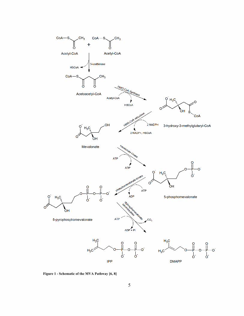
The MVA pathway is illustrated in Figure 1, and the MEP pathway is illustrated
in Figure 2.
5
Figure 1 - Schematic of the MVA Pathway [6, 8]

6
Figure 2 - Schematic of the MEP pathway [6].

7
The MEP pathway consists of seven enzymatic steps as outlined in Figure 2.
Glyceraldehyde-3-phosphate and pyruvate are condensed by DXP synthase (DXS) to 1-
deoxy-D-xylulose-5-phosphate (DXP or DOXP). Next, IspC (DXP reductoisomerase or
MEP synthase) converts it to 2-C-methyl-D-erythritol 4-phosphate (MEP). IspD (CDP-
ME synthase or MEP-cytidylyltransferase) adds CTP to MEP to yield 4-
diphosphocytidyl-2-C-methyl-D-erythritol (CDP-ME). IspE (CDP-ME kinase)
phosphorylates CDP-ME at the C2 position of the methylerythritol moiety, giving CDP-
ME2P, which is then cyclized by IspF (cMEPP synthase) to give 2-C-methyl-D-erythritol
2,4-cyclodiphosphate (cMEPP). IspG (HMBPP synthase) breaks the ring to give 1-
hydroxy-2-methyl-2-(E)-butenyl-4-diphosphate (HMBPP), and IspH (HMBPP reductase)
produces the isomers isopentenyl diphosphate (IPP) and dimethylallyl diphosphate
(DMAPP) in a 5:1 ratio [6].
The exclusive use of the MVA pathway in humans and animals for isoprenoid
biosynthesis makes the MEP pathway an attractive drug target for treating diseases
caused by bacteria or parasites that rely on the MEP pathway for survival. The MEP
pathway is vital for cellular survival as shown by transposon mutagenesis [10], where
Gallagher et al. identified the genes essential for the survival of F. novicida by randomly
disrupting its genome with antibiotic resistance trait inserts. Mutated cells grown on
selective media were sequenced to identify the genes that could be disrupted without
lethal effects, while disruption of essential genes presumably failed to produce viable
cells. After ensuring complete insertion coverage of the F. novicida genome, no cells

8
containing insertions in any of the MEP pathway genes could be isolated, indicating that
the loss of any step of the MEP pathway is lethal to the organism [10].
Fosmidomycin and FR-900098 are specific inhibitors of IspC that are lethal to the
organism [6, 11, 20]. Fosmidomycin has been shown to be effective in combination with
other antimalarial drugs for treating malaria patients by killing the parasite during its
intraerythrocytic stages, although monotherapy in humans failed to provide a complete
cure [17, 21]. A combination of fosmidomycin and clindamycin is currently in phase II
clinical trials for the treatment of malaria [21].
Figure 3 - The IspC inhibitors fosmidomycin [21] and FR-900098 [38].
Fosmidomycin has poor lipophilicity [17, 22], which makes it unlikely to diffuse
into cells and require transport. Absorption into bacterial cells occurs by transport via the
glycerol-3-phosphate transporter GlpT in E. coli and other bacteria, and it was shown that
mutants deficient in GlpT were resistant to fosmidomycin [23]. Gram positive bacteria do
not have GlpT, so uptake is especially poor in species such as M. tuberculosis and M.
smegmatis, conferring resistance [11, 24]. The development of stable fosmidomycin
resistance has also been demonstrated in Plasmodium falciparum [25].

9
IspC was discovered and identified as the target of fosmidomycin in 1998
(reviewed in [26]), and is perhaps the most studied enzyme of the MEP pathway because
it is the first committed step. Isoprenoid biosynthesis is the only known function for
MEP, while DXP can be used as a precursor for other metabolites such as pyrodoxol and
thiamine [18]. Despite its limitations, fosmidomycin has facilitated the study of IspC as a
target for improved therapeutics in F. tularensis [27]. IspC clones from E. coli [13, 14]
have served as models for study of the pathway in bacteria.
IspD is the third overall step of the MEP pathway that catalyzes the cytidylation
of MEP. It is also a target of interest because it is an early step in the pathway and could
serve as a secondary target to counter resistance to IspC-targeted drugs. Interestingly,
Zhang et al. determined recently that fosmidomycin is also a weak inhibitor of IspD.
Their studies suggest that fosmidomycin works by inhibiting IspC halfway through its
reaction, which renders it unable to fully convert DXP to MEP. The intermediate, 2-C-
methylerythrose 4-phosphate, accumulates and subsequently competitively binds IspD
along with fosmidomycin causing MEP to accumulate and the levels of metabolites
downstream from IspD decrease. 2-C-methylerythrose 4-phosphate is likely to bind IspD
with greater affinity than fosmidomycin due to a higher number of hydrogen bonds as
shown by their docking studies. They measured fosmidomycin binding to IspD with IC50
= 20.4 ± 3.3 mM in vitro, an affinity 104 times weaker than the IC50 = 0.81 ± 0.27 µM
measured for IspC [21].
The prospect of a drug with a dual inhibitory mechanism could lead to
monotherapy regimens with the same efficacy of multidrug treatments and lower risk of

10
resistance development, because an organism is much less likely to develop multiple
resistance traits simultaneously. Similarly, the development of a novel inhibitor of a
downstream MEP pathway enzyme could lead to highly effective therapy in combination
with fosmidomycin or provide protection against Gram positive bacteria. While other
compounds have been reported to inhibit IspD (L-erythritol 4-phosphate, Ki= 240 ± 17
mM; and D-erythritol 4-phosphate, IC50 = 1.36 mM) [12], these are quite weak relative to
the inhibition constants of useful drugs like fosmidomycin, with Ki = 99 nM, IC50 = 247
nM for F. tularensis IspC [27]. The challenge in developing new effective MEP pathway
inhibitors relies on optimizing for specificity, affinity, and delivery.
To date, IspE and IspF are the only MEP pathway enzymes for which inhibitor
lead identification via a high throughput screen (HTS) have been published [28, 29]. The
reactions can be measured by spectroscopic methods to expedite HTS processes [6]. IspG
and IspH catalyze more complex reactions involving several redox cofactors or require
anaerobic conditions that may severely limit the ability to perform fast analyses in a HTS
system [6].
The drug development process consists of five general phases: target
identification, target validation, lead molecule identification, lead molecule optimization,
and preclinical and clinical trials. This project focused on characterizing and validating
IspD in F. tularensis as a potential target for novel therapeutics. Enzyme kinetic
parameters were measured, the amenability of IspD to high throughput screening was
determined, a site of possible phosphoregulation was probed, and an alternative method

11
of bioprospecting for molecule leads by screening for molecules that bound to IspD was
investigated.
Relatively few IspD homologs have been isolated and characterized. Studies have
documented IspD from bacteria such as E. coli [14, 15, 30] and M. tuberculosis [31],
while some reported the bifunctional IspDF enzyme found in organisms such as
Campylobacter jejuni [32] and Helicobacter pylori [33] in which both proteins are
encoded in the same transcription unit, forming a single enzyme with dual functionality.
In E. coli, M. tuberculosis, and F. tularensis, the genes encoding IspD and IspF are in
close proximity to each other on the bacterial chromosome, but are not expressed in a
single unit. F. tularensis IspD has low homology with other species. For example, E. coli
IspD has 34% homology, while M. tuberculosis IspD is only 25% similar. This project
adds the characterization of F. tularensis IspD to the literature as a potential target for
novel antibiotics.

12
Experimental Aims
Cloning, expression, and purification.
The gene encoding IspD in the F. tularensis subsp. holarctica LVS genome was
cloned into a bacterial plasmid vector and overexpressed in a standard laboratory strain of
E. coli so that sufficient quantities of the purified protein (FtIspD) could be obtained for
study. E. coli IspD (EcIspD) was also cloned and used as a control for the FtIspD data.
This allowed validation of the data obtained to reported values of EcIspD. The protein
was then purified for downstream experiments.
Assays
IspD enzymatic activity was quantified using a spectrophotometric assay adapted
from Bernal et al [30]. This characterization allowed for analytical comparison of protein
activity levels that might be observed during molecular library and lead compound
candidate testing.
IspD’s in vitro substrate affinity was determined, as well as its preferences for
divalent metal cations and nucleotides. The assay was also adapted to a high throughput
screen in a microtitre plate. CaCl2, an inhibitor of pyrophosphatase (not IspD), was used

13
to simulate an inhibited assay which might be observed in the event of a hit during a high
throughput screen, through which the robustness of the assay could be evaluated using
the Z-factor.
Regulation of IspD
Phosphorylation of amino acid sites is a widely used intracellular mechanism for
signal transduction and regulating the activities of enzymes. The goal here was to identify
amino acid residues that might play a role in the regulation of IspD in vivo. Because the
FtIspD structure has not yet been solved, a model was constructed to show the probable
locations of phosphorylation sites on the protein. Phosphopeptide fragments were then
identified via LC-tandem-mass spectrometry by Dr. Weidong Zhou of the Center for
Applied Proteomics and Molecular Medicine at George Mason University.
After finding the T141 residue to be conserved, phosphorylated, and located in a
structurally significant location of the protein model, the mutants T141D and T141E were
cloned, expressed, purified, and assayed for activity. Intrinsic fluorescence studies of
wild type and mutant IspD were then performed to see if any changes in folding or
structure resulted from the site mutations. Ms. Amy Fisher (Center for Nanophotonics
Imaging, George Mason University) helped immensely with this work.
Bioprospecting
An alternative method for identifying lead molecules in a molecular library was
investigated as part of this study. In this bioprospecting study, the protein of interest was

14
immobilized on a purification column. A molecular library was then passed over the
protein, unbound material washed out, the protein eluted, and binding molecules were
separated from the protein and analyzed by LCMS to aid in identifying them. This
approach could expedite inhibitor lead identification and prove to be a viable lower cost
alternative to high throughput screening by selecting for molecules that bind to the
protein. Bound molecules would be subsequently identified.

15
Materials and Methods
Cloning
The gene encoding IspD (YP_514172) in the F. tularensis subsp. holarctica LVS
genome (accession number NC_007880) was cloned into a pET101/D – TOPO plasmid
(Invitrogen). The gene was constructed with a C-terminal His tag and amplified via PCR
as described in [35]. The plasmid was transformed into BL21 (DE3) CodonPlus-RIL
competent cells (Stratagene) for overexpression and cells were grown in LB media
(Fisher) with 100 µg/mL ampicillin and 50 µg/mL chloramphenicol. Expression was
induced with 0.5mM IPTG.
The E. coli ispD gene (NC_000913.2: 2869802..2870512) encoding the respective
enzyme (NP_417227) was cloned into a pET28c plasmid (EMD) with a N-terminal His
tag similar to E. coli IspD constructs used in the literature [15, 30]. E. coli K12 genomic
DNA (NC_000913) was used in the PCR process. PCR primer pairs flanking the IspD
gene consisted of:
Forward: 5’-AAAAAACATATGATGGCAACCACTCATTTG-3’.
Reverse: 5’-GGGGTCCTCGAGTTATGTATTCTCCTG-3’.

16
E. coli K12 genomic DNA was obtained by using a GenElute Miniprep Kit
(Sigma). The PCR was set up using 25 µL 2x Phusion HF, 2.5 µL each forward (44.2
µM) and reverse (39.1 µM) primers, 3 µL genomic DNA, and 17 µL ddH2O covered with
50 µL mineral oil. The PCR reaction consisted of heating to 98°C for 30 seconds, 62°C
for 30 seconds, and 72°C for 90 seconds for 24 cycles, then held at 72°C for 10 minutes
and finally kept at 4°C until use. The PCR product was purified using the Sigma
GenElute PCR Cleanup Kit and ligated into the pET28c plasmid vector (Novagen) using
Lyo-Ligase and the Xho I and Nde I restriction sites. The plasmid was then transformed
into Novagen Blue Singles cells with a 30 second heat shock at 42°C. These cells were
grown in LB media with 50 µg/mL kanamycin to select for cells that contained the
plasmid. Cultures were grown on LB agar and colonies containing the correct plasmid
and gene insert were selected using restriction mapping. The fragments were visualized
via agarose gel to verify the plasmid’s integrity. After verification of the proper size
fragments, the plasmid containing the IspD insert (pEcIspD) was sequenced.
pEcIspD was transformed into chemically competent Stratagene E. coli BL21
(DE3) codon+ cells for expression. Transformation was performed by incubating
competent cells with DNA at 4°C for 30 minutes, 30 seconds heat shock at 42°C, 4°C for
2 minutes, addition of 250 µL SOC media broth to the vial, and then incubated and
shaken for 1 hour at 37°C, 250 RPM. The successful competent cells were kept in storage
at -80°C in 20% glycerol.

17
Protein Expression and Purification
A 10 mL seed culture was grown at 37°C with shaking at 250 RPM, with E. coli
BL21 CodonPlus (DE3)-RIL cells containing pFtIspD. After 18 hours, the culture was
used to inoculate one liter of fresh LB media, grown to an OD600 of 1.1 and induced with
0.5 mM isopropyl-β-D-1-thiogalactopyranoside (IPTG). The production flask was
incubated for 18 hours at 37°C, 250 RPM, and the cells were harvested by centrifugation
at 19,200g, 4°C and stored at -80°C until use.
Cell pellets were lysed with “Buffer A” containing 0.1 M Tris pH 8.0, 0.1 M
NaCl, and 0.032% (w/v) lysozyme. 3 ml of this buffer per gram of cell pellet was used.
“Buffer B” (0.1 M CaCl2, 0.1 M MgCl2, 0.1 M NaCl, 0.02% DNase, 0.3 mL per gram
pellet) was added after. The lysate was centrifuged at 48,000g for 30 minutes at 4°C. The
clarified lysate was then passed over Talon superflow resin with immobilized Co2+ to
capture the protein via the His-tag. It was washed with 20 column volumes of the
equilibration buffer (50 mM HEPES pH 7.5, 300 mM NaCl), 20 column volumes of wash
buffer (50 mM HEPES pH 7.5, 300 mM NaCl, 10 mM imidazole), and eluted with 10
column volumes of elution buffer (50 mM HEPES pH 7.5, 300 mM NaCl, 150 mM
imidazole). The protein was buffer exchanged into 0.1 M Tris pH 8.0, 1 mM NaCl, snap-
frozen and stored at -80°C. The typical yield of IspD was 15 mg of IspD per 1 liter
culture with high purity as visualized by SDS-PAGE and coomassie staining.

18
Enzyme Assays
IspD activity at 37°C was determined by measuring the production of
pyrophosphate (PPi) in solution as described by Bernal et al [30].
CDP-ME + PPi
pyrophosphatase
O-
O-O-
O
P O-
O-O-
O
P
OOH
OH
OH
P
O
OH
O-
MEP
IspD
Treat with malachite green dye, incubate, and measure A660
Figure 4 - Schematic of the IspD assay.
The dye stock contained 0.44 g malachite green in 360 mL of 3M H2SO4 and was stored
at 4°C. A working stock of dye was made prior to each assay by mixing 2.5 mL of the
malachite green stock, 0.625 mL of 7.5% (w/v) ammonium molybdate, and 50 µL Tween
20.
For evaluating the MEP-dependent kinetic activity of IspD, a reaction mix was
made containing 100 mM Tris pH 8.0, 1 mM MgCl2, 1 mM dithiothreitol (DTT), 0.2 mM
cytidine 5'-triphosphate (CTP), 100 mU/mL pyrophosphatase, 75 µg/mL IspD, and
varying concentrations of MEP. The reaction mixture was incubated at 37°C, a 40 µL
aliquot was drawn every 30 seconds, added to 120 µL of water and 40 µL of the dye
reagent. After incubating this mixture for 10 minutes at 37°C, 22 µL of 34% sodium

19
citrate was added to the assay solution and incubated for 30 minutes at 37°C. The A660 of
this assay solution was recorded and used to determine the concentration of
pyrophosphate produced from a standard curve.
Data were fit by nonlinear regression to the Michaelis-Menten equation (below) to
determine the Vmax, Km, Kcat and Kcat/Km for MEP and CTP substrates, where Y
corresponds to the velocity of the reaction and X represents the substrate concentration.
( )XKXVY
m +=
*max
For the high throughput assay, the volume of the reaction was scaled from 1 mL to 200
µL and 1 mM CaCl2, a complete inhibitor of pyrophosphatase [37], was used as a control
for assay inhibition visualization.
Metal ion specificity was determined among Mg2+, Ca2+, Mn2+, Co2+, Cu2+, and
Zn2+. After incubating IspD with the reaction mixture containing the variable metal
cation (pyrophosphatase excluded), EDTA was added to chelate divalent cations so that it
would not interfere with pyrophosphatase activity, which is dependent on Mg2+. The
reaction was filtered through a Millipore Microcon 10K MWCO filter to remove IspD
and pyrophosphatase was added to the reaction mixture. The solution was dyed, and the
A660 was read as normal.
The Z-factor, a statistical indicator of signal error in an assay, was calculated as described
by Zhang et al [36]. This parameter is determined by the equation:

20
cs
csZµµσσ
−
+−=
)33(1
where σ is the standard deviation of the sample (s) or control (c), and µ is the mean of the
sample or control, respectively.
Size Exclusion Chromatography
The molecular mass of IspD and mutants were determined using size exclusion
chromatography on a sephacryl 200 column (Sigma) and measured with a gel filtration
protein standard (BioRad 151-1901). The mobile phase contained 0.1M tris pH 8.0, 1
mM NaCl at 2 mL/min at room temperature (22°C). A 100 µL load sample was used
containing protein at 8.9 mg/mL. The sample was centrifuged at 16,110g rpm for 10
minutes prior to loading.
Phosphopeptide Analysis
IspD expression was induced with 0.01 mM IPTG (instead of the normal 0.5
mM). After purification, the enzyme was reduced with 10mM DTT, alkylated by 50 mM
iodoacetamide, and digested with trypsin (Promega) in ammonium bicarbonate buffer (50
mM, pH 9.0), and urea (2 M). The mixture was desalted using a SepPak column (Waters,
Milford, MA). A TiO2 column was used to separate phosphopeptides from the digestion
mixture and the phosphopeptides were analyzed via reverse-phase liquid chromatography
nanospray tandem mass spectrometry (LC-MS/MS) with an LTQ-Orbitrap mass
spectrometer (ThermoFisher). SEQUEST was used to identify phosphopeptides.

21
Mutagenesis
T141D and T141E mutants were cloned by GenScript Corporation and expressed in
Stratagene E. coli BL21 CodonPlus (DE3)-RIL cells. Expression, purification and assays
were performed as described for wild type IspD.
Fluorescence Spectroscopy
The intrinsic fluorescence was measured with a Fluoromax-3 fluorometer (Horiba Jobin
Yvon) using a 5 µM protein concentration against a buffer blank (0.1 M Tris pH 8.0, 1
mM NaCl). An excitation wavelength of 290 nm was emitted and emissions were
measured between 310-400 nm using a 5 nm slit.
Molecular Modeling
Sequences containing phosphorylated residues were identified and mapped onto a
structural model of FtIspD built with the I-TASSER server. Model quality was assessed
using ProQRes on the ExPASy server. Models were viewed and edited using the Deep
View, and graphics were generated in PyMOL.
Bioprospecting
A model library made using V8 vegetable juice was filtered through a 3000
MWCO filter to remove macromolecules and particulates. The filtrate was centrifuged
and the supernatant was passed over immobilized IspD protein on a TALON column as
an extra step in the purification protocol. It was washed with the equilibration and wash
buffers, and the protein was eluted with the elution buffer.

22
The eluate was heated at 75°C for 10 minutes to denature the enzyme and filtered
through a 3000 MWCO filter to remove IspD. The filtrate was analyzed via LCMS on a
Waters Micromass ZQ liquid chromatography system with quadrupole detector with
electrospray ionization. The same procedure was applied to IspC using fosmidomycin.
Analysis of fosmidomycin standards was performed on the LCMS with a reverse phase
C18 column using a 0-50% acetonitrile gradient over 30 minutes and ionized using the
following parameters: ESI negative, capillary 4.0 kV, cone: 18V, source temperature
150°C, desolvation 250°C, 250 L/hr, with the data published in [38] as a starting point.

23
Results and Discussion
The need for novel antibiotics has grown due to the extensive spread of natural
and engineered bacterial resistance to current drugs. Many pathogenic bacteria are known
to solely utilize the MEP pathway for isoprenoid biosynthesis, which humans do not use,
thus making it a highly attractive target. Although fosmidomycin is currently considered
an effective inhibitor of the MEP pathway, resistance by way of limited uptake was
identified early in its history [23]. However, no evidence has been found in the literature
that mechanistic resistance to fosmidomycin caused by mutation of IspC in target
organisms is responsible for resistance. Other commonly used antibiotics presently used
in the treatment of F. tularensis infections are seeing increasing resistance in the world as
well.
Francisella tularensis IspD is a 229 amino acid protein with a calculated
molecular mass of 25.9 kDa encoded by the gene ispD (also known as ygbP).
Recombinant FtIspD expressed with high copy number in the laboratory E. coli host with
IPTG induction and was isolated with the polyhistidine tag with more than 90% purity.
SDS-PAGE visualization shows the high purity after coomassie blue staining (Figure 7).

24
Figure 5 - F. tularensis IspD in pET101 directional vector with topoisomerase (Invitrogen). The open reading frames for the ampicillin resistance gene and lac operon are also shown indicated as arrows
Figure 6 - Vector map showing E. coli IspD in pET28c (pEc-IspD). The arrow beneath the IspD open reading frame indicates the IspF open reading frame on the complementary strand.

25
Figure 7 - FtIspD visualized by SDS-PAGE. The most intense band corresponds to a molecular mass of approximately 26 kDa with yield in excess of 15 mg purified protein.
Kinetic Assay Results
For the kinetic curves shown in Figure 8, assays were carried out at MEP
concentrations ranging from 10 – 400 µM while the CTP concentration was held at a
saturating concentration of 200 µM for the kinetic characterization. The activity of IspD
was measured over a range of CTP concentrations from 10 – 400 µM at a constant
saturating MEP concentration of 200 µM.

26
Figure 8 - F. tularensis IspD kinetic activity.*
Figure 9 - E. coli IspD kinetic assay, MEP dependent.

27
Table 1 - MEP cytidylyltransferase Apparent Kinetic Parameters. N/A entries were not determined.
MEP cytidylyltransferase
(µM)
(µM)
(s-1)
(s-1)
(M-
1min-1)
Source
F. tularensis
177.9
73.0
1.0
0.8
3.4×10⁵
[35]
E. coli (test clone)
221.9
N/A
57.1
N/A
1.5×10⁷
Exp Data
E. coli
range of kinetic parameters from
two sources
61
58
0.7
54.1
6.6×10⁵
[30]
370 760 25.9 N/A 7.86×10⁶ [15]
The observed kinetic values for FtIspD were !!"#!"# = 1.0 +/- 0.10 s-1 and
!!"#!"#/!!!"# = 3.4×10⁵ +/- 6.7×10⁴ M-1 s-1 (Table 1). The F. tularensis data in this study
were measured with the colorimetric assay and are comparable to the literature datasets,
accommodating for differences in kinetics due to potential structural variations arising
from the low sequence homology that FtIspD has to most other homologs. The kinetic
values for EcIspD control in Table 1 are within the spread of literature values, although
the malachite green assay indicated a !!!"# nearly four times higher for the in-house
control clone than its literature counterparts. Bernal et al. obtained !!!"#= 61 +/- 14 µM
[30] for E. coli via the malachite green assay, while radiological assays from other
sources found the !!!"#ranging between a similar value of 32 µM [13], and 370 µM [15]
all for the same enzyme. This wide spread of values may be attributed to the sensitivity of

28
the assays to handling or construct variations. Bernal et al. also suggest pyrophosphatase
grade and condition may play a role in assay quality [30]. Regardless, the assay behaved
predictably, and showed a strong measure of robustness with a high Z-factor of 0.8.
Values over 0.5 are indicative of a robust assay for high throughput screening that has
good signal response and low background interference [36]. This assay was scaled for a
96-well plate with MEP concentration increasing by row in Figure 10 from 0 µM to 100
µM. Columns 1-3 are replicate uninhibited assays, and columns 4-6 are replicate assays
containing 1 mM CaCl2, simulating an inhibitor candidate hit.
Figure 10 - The robustness of the IspD assay can be visually perceived by this microtitre test. The absorbance value of each well is shown. Rows A-D contain 0, 25, 50, and 100 µM MEP respectively, while columns 4-6 have CaCl2 added to them.

29
Metal Ion Preference
IspD’s metal ion specificity was evaluated among MgCl2, CaCl2, MnCl2, CoCl2,
CuCl2, and ZnCl2. The reaction was performed as usual except that EDTA was added
after incubating IspD with substrate, the solution was filtered to remove IspD, and
pyrophosphatase was then added to the filtrate to continue the assay as normal. Like other
IspD homologs, FtIspD natively prefers Mg2+ to catalyze the reaction (Fig. 14). Mg2+ is
essential for cytidylyltransferase activity and is believed to aid in coordinating the
phosphate groups of CTP and providing them complementary charge so that MEP can be
cytidylated, displacing the PPi group [39].
During this experiment, Mg2+ was replaced with an alternative divalent metal
chloride salt in the assay. Only Mg2+ supported assay activity with FtIspD, which
contrasts from other studies on the E. coli and M. tuberculosis homologs that supported
activity with at least one other divalent cation besides Mg2+.
Figure 11 - MgCl2 was the only divalent metal salt tested that conferred measurable activity to FtIspD.

30
E. coli IspD was found to be catalytically active in the presence of Mg2+, Mn2+,
and Co2+ using a 32PPi liquid scintillation spectrometry assay [14]. Eoh et al., also using
the 32PPi assay, reported that M. tuberculosis IspD maintained comparable activity with
Mn2+, lower activity with Zn2+, and Ca2+ was ineffective [31]. Shi et al., using the
coupled pyrophosphatase assay, detected activity in the same enzyme with Mg2+, Mn2+,
Fe2+, and Co2+, while Zn2+ was ineffective [40]. Despite the difficulty in directly
comparing these sets of data, the various studies agree overall that there is a clear
specificity for Mg2+ and it is the preferential cation for IspD. There is significant
interplay between the enzyme active site residues, Mg2+, substrates, and solvent [39], and
the arrangement of these components in FtIspD appears to be especially sensitive to the
nature of the divalent cation in this study.
Nucleotide Specificity
The activity of IspD in the presence of various nucleotides was measured at 200
µM and 400 µM. FtIspD displayed optimal activity with CTP as expected, while much
lower levels of activity were detected in the presence of deoxy-CTP (dCTP), ATP, GTP,
TTP, and UTP (Fig. 15). These results are consistent with the data reported for
homologous enzymes. A 400 µM concentration of nucleotide appeared to support higher
relative activity in all nucleotides than 200 µM, but at most only approximately one third
of the activity demonstrated with CTP, indicating that FtIspD is very specific for CTP.

31
Figure 12 - IspD activity was greatest with CTP, and significantly less active with other nucleotides.
Figure 13 - At 400 µM, the relative amount of activity increased slightly for other nucleotides, but was still much lower than activity with CTP, showing a clear preference.

32
Phosphoregulation
The IspD active site involves a number of key residues that form a complex
network with CTP, Mg2+, and MEP in order to catalyze the reaction. In the EcIspD
crystal structure, residues Asp106, Arg109, Thr165, and Lys213 form polar contacts with
the MEP-derived moiety of CDP-ME within the active site cleft. Thr140’and Arg157’ are
contributed from the other chain in the dimer and also contact the MEP moiety of CDP-
ME near the interface of the β arm domains, suggesting they may help guide MEP into
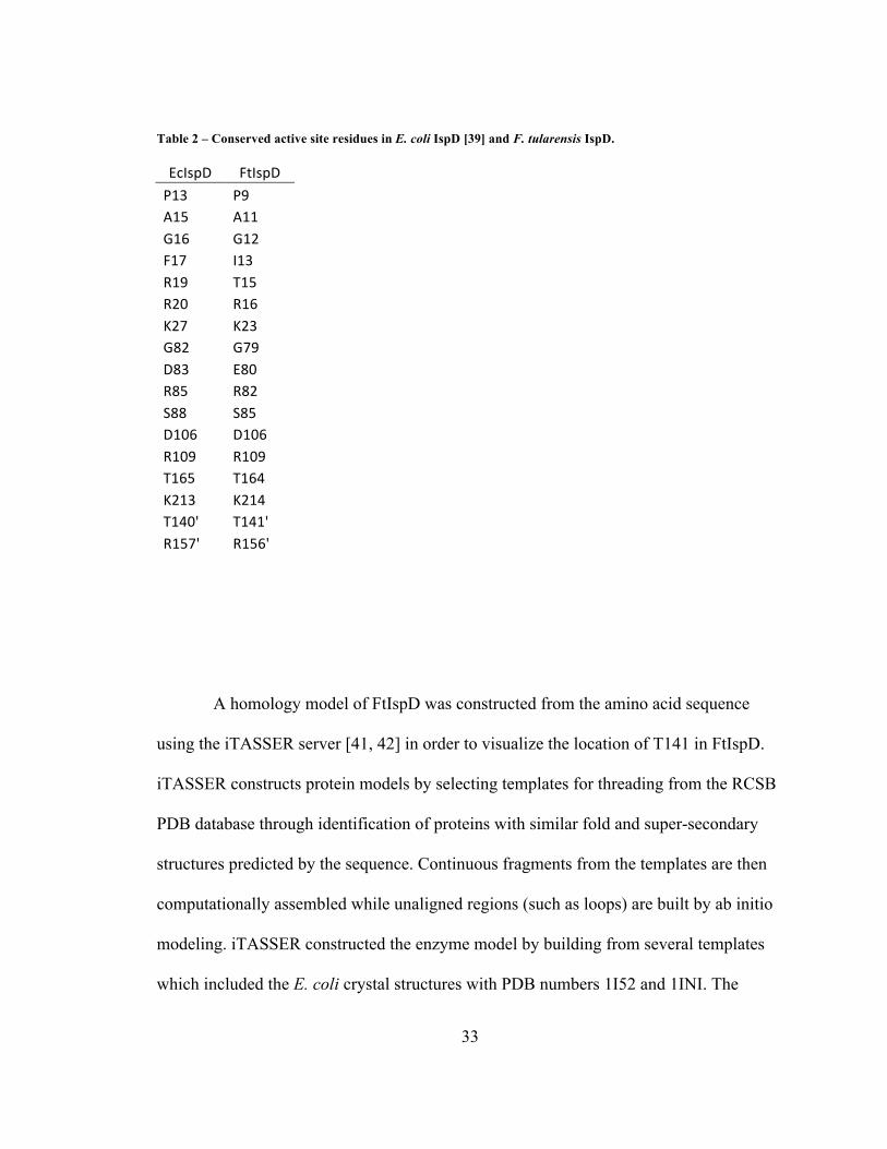
the active site [39]. These residues are well conserved in FtIspD, as can be seen in Table
2. In the FtIspD model, these conserved residues all line the active site cleft and are
expected to play similar roles. Regulation of IspD may occur through phosphorylation of
one or more of these key residues. LCMS/MS analysis of FtIspD for phosphorylated
amino acids indicated a phosphorylated threonine corresponding to the 141 position in
the enzyme sequence. A sequence alignment showed that Thr141 in FtIspD is equivalent
to Thr140 in EcIspD, and Richard et al predicted that this residue plays a key role in
guiding MEP toward the active site for cytidylation during the reaction [39].
33
Table 2 – Conserved active site residues in E. coli IspD [39] and F. tularensis IspD.
EcIspD' FtIspD'P13' P9'A15' A11'G16' G12'F17' I13'R19' T15'R20' R16'K27' K23'G82' G79'D83' E80'R85' R82'S88' S85'D106' D106'R109' R109'T165' T164'K213' K214'T140'' T141''R157'' R156''
A homology model of FtIspD was constructed from the amino acid sequence
using the iTASSER server [41, 42] in order to visualize the location of T141 in FtIspD.
iTASSER constructs protein models by selecting templates for threading from the RCSB
PDB database through identification of proteins with similar fold and super-secondary
structures predicted by the sequence. Continuous fragments from the templates are then
computationally assembled while unaligned regions (such as loops) are built by ab initio
modeling. iTASSER constructed the enzyme model by building from several templates
which included the E. coli crystal structures with PDB numbers 1I52 and 1INI. The
34
model output was then analyzed with ProQRes which rates the quality of each residue
prediction as a score ranging from 0 (unreliable) to 1 (reliable) by measuring structural
features within a sliding window including atom-atom contacts, residue-residue contacts,
solvent accessibility surfaces, and secondary structure information [43].
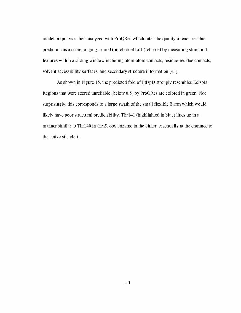
As shown in Figure 15, the predicted fold of FtIspD strongly resembles EcIspD.
Regions that were scored unreliable (below 0.5) by ProQRes are colored in green. Not
surprisingly, this corresponds to a large swath of the small flexible β arm which would
likely have poor structural predictability. Thr141 (highlighted in blue) lines up in a
manner similar to Thr140 in the E. coli enzyme in the dimer, essentially at the entrance to
the active site cleft.

35
Figure 14 - The active site network of E. coli IspD with the residues that contribute to the cytidylation of MEP labeled [39]. All of the labeled residues are highly conserved in F. tularensis IspD.

36
Figure 15 - FtIspD model predicted by iTASSER. The region in green scored below 0.5 in ProQRes. Thr141 is shown in stick form and colored dark blue.

37
To determine if phosphorylation of Thr141 could play a role in inhibiting FtIspD
and act as a regulatory mechanism, two mutants with Thr141 replaced by Asp (T141D)
and Glu (T141E) were constructed to simulate a phosphate group attached to the residue.
The wild type and mutant enzyme quaternary states were determined to be dimers using
size exclusion chromatography (Figure 16). Assays of the enzymes showed that the
T141D mutant lost approximately 75% of activity, while T141E lost all activity,
indicating that Thr141 plays a crucial role in the reaction, and FtIspD would likely be
inhibited by phosphorylation. There was also no shift in intrinsic fluorescence (Figure
18), indicating that there was no major conformational change between wild type active
IspD and the T141D and T141E mutants. This suggests that the loss of activity is due to
mechanistic blockage at the reaction site, not a loss of enzyme conformation.
Figure 16 - Molecular masses of IspD wild type, T141D, and T141E mutants on a standard curve determined by size exclusion chromatography.

38
Figure 17 - Mutants constructed to simulate phosphorylation at the T141 site demonstrated markedly reduced activity, suggesting a T141 may be a site of regulation.
Figure 18 - Fluorescence of wild type and mutant IspD indicated no conformational changes of the enzyme after residue modification.
0"
500000"
1000000"
1500000"
2000000"
2500000"
3000000"
3500000"
4000000"
300" 310" 320" 330" 340" 350" 360" 370" 380" 390" 400" 410"
Fluo
rescen
ce*units*
Emission*Wavelength*(nm)*
Intrinsic*Fluorescence*of*FtIspD*
WT"
T141D"
T141E"

39
Bioprospecting
An alternative approach to high throughput screening for lead molecule
identification is bioprospecting, a process in which molecules that bind the target enzyme
such as IspD would be separated from a mixture by their interaction with the enzyme.
Traditional high throughput screening is a very resource intensive process and works
essentially through brute force, by screening many thousands of purified compounds
within molecular libraries and assaying the enzyme for each compound. In contrast,
bioprospecting could be applied to mixed molecular libraries, such as raw extracts. This
could allow much more rapid collection of molecular libraries without the need to
identify and purify each compound within. In this case, the target protein is immobilized
on a substrate, the molecular mixture is passed over it, washed, and the bound enzyme is
analyzed for ligands.
To illustrate the use of bioprospecting as an alternative to an HTS for developing
inhibitors of the MEP pathway, a proof of concept was established using IspC and its
known inhibitor, fosmidomycin. Approximately 5 mg of IspC was immobilized on Talon
cobalt resin via its polyhistidine tag following cell lysis, washed, and a solution of 200
µg/mL fosmidomycin in water was passed over it. After elution, denaturation of the
protein by heat, and filtration to separate it, the filtrate was compared to a fosmidomycin
standard curve via LCMS in ESI negative mode. Ion peaks corresponding to
fosmidomycin were identified (a molecular ion of 182 m/z and a daughter peak of 136
m/z). The LCMS standard curve was calibrated to detect fosmidomycin concentrations

40
from 0.05 µg/ml to 7.5 µg/ml, and fosmidomycin was detected in highly diluted (1:1000)
eluate at a concentration of 0.16 µg/ml, which shows that most of the fosmidomycin was
recovered.
To screen for natural product inhibitors of IspD, a molecular library was
constructed by filtering V8 juice (a product of concentrate from vegetable extracts
suspended in tomato juice) through a 3000 MWCO filter and centrifuging the filtrate,
then passing the supernatant over immobilized IspD as described for IspC above. While
IspD displayed slightly inhibited catalytic activity after this treatment (Figure 19), LCMS
analysis could not successfully identify an associated ligand. Thus, while a natural
inhibitor of IspD may indeed be present in the library (possibly in low abundance), it is
plausible that enzymatic denaturation of IspD may also account for the observed decline
in activity. None-the-less, the quantitative binding of fosmidomycin to IspC illustrates the
potential of bioprospecting as an alternative to HTS and encourages further study and
screening with IspD.
Figure 19 - The IspD assay reported approximately 25% less specific activity after the enzyme was exposed to the test library.

41
Conclusion
The MEP pathway is an attractive target for novel drugs because of its prevalence
in many pathogenic organisms but not in humans. This study characterized and validated
IspD of Francisella tularensis as a strong target candidate for drug development. The
first steps in drug discovery are target identification and validation, followed by lead
compound identification and optimization, and lastly clinical trials. F. tularensis IspD
was identified as a target based on the observation that MEP pathway knockouts are
lethal [10], and this project characterized the enzyme by a number of criteria to validate it
as an attractive target for antibiotics. Some of the most well studied homologs of IspD are
from E. coli and M. tuberculosis, with which FtIspD has 34% and 25% homology to,
respectively. Few other homologs have been characterized in the literature, and this was
the first study of F. tularensis IspD to be published. FtIspD kinetic values are similar to
the values reported for other homologs in the literature, and furthermore the assay could
be scaled down to a high throughput screen format.
The regulation of metabolic flux through the MEP pathway is poorly understood.
The MEP pathway appears to be regulated by several enzymes, with evidence indicating
that the early and late steps of the pathway are most regulated [18]. Phosphorylation as a
regulatory mechanism of the pathway has not been reported. Threonine 141 is an

42
accessible residue that is a key component of the cytidylation reaction in FtIspD and the
observation that it may be phosphorylated may suggest a mechanism by which the
regulation of IspD contributes to overall rate limitation of the MEP pathway. Finally, lead
compounds for IspD inhibitors may be discovered through bioprospecting and may
reduce the costs associated with high throughput. This method was validated by capturing
fosmidomycin with IspC, while further experiments may uncover a source of an inhibitor
for IspD.

43
References

44
References
1. M. Friend (2006). Tularemia. United States Geological Survey Circular 1297,
Reston, VA. 2. B. Cunha (2009). Tularemia. The Merck Manual (Online).
http://www.merckmanuals.com/professional/sec14/ch173/ch173r.html 3. K. Feldman, R. Enscore, S. Lathrop, B. Matyas, M. McGuill, M. Schriefer, D.
Stiles-Enos, D. Dennis, L. Petersen, E. Hayes (2001). An Outbreak of Primary Pneumonic Tularemia on Martha’s Vineyard. N. Engl. J. Med. 345(22): 1601-1606.
4. K. Feldman, D. Stiles-Enos, K. Julian, B. Matyas, S. Telford III, M. Chu, L.
Petersen, E. Hayes (2003). Tularemia on Martha’s Vineyard: Seroprevalence and Occupational Risk. Emerg. Infect. Dis. 9(3): 350-354.
5. J. Hudspeth (2005). Francisella tularensis. In G. Zubay (Ed.) Agents of
Bioterrorism, (p 44). New York: Columbia. 6. H. Eoh, P. Brennan, D. Crick (2009). The Mycobacterium tuberculosis MEP (2C-
methyl-D-erythritol 4-phosphate) pathway as a new drug target. Tuberculosis. 89: 1-11.
7. J. Gershenzon, N. Dudareva (2007) The function of terpene natural products in
the natural world. Nat. Chem. Biol. 3: 408-414. 8. B. Agranoff, H. Eggerer, U. Henning, F. Lynen (1959) Isopentenol
Pyrophosphate Isomerase. Journal of the American Chemical Society 81: 1254-1255.
9. M. Rohmer, M. Knani, P. Simonin, B. Sutter, H.Sahm (1993). Isoprenoid
biosynthesis in bacteria: a novel pathway for the early steps leading to isopentenyl diphosphate. Biochem. J. 295: 517–524.

45
10. L. Gallagher, E. Ramage, M. Jacobs, R. Kaul, M. Brittnacher, C. Manoil (2006) A
comprehensive transposon library of Francisella novicida, a bioweapon surrogate. PNAS 104(3): 1009-1014.
11. R. Dhiman, M. Schaeffer, A. Bailey, C. Testa, H. Scherman, D. Crick. (2005) 1-
Deoxy-D-Xylulose 5-Phosphate Reductoisomerase (IspC) from Mycobacterium tuberculosis: towards Understanding Mycobacterial Resistance to Fosmidomycin. J. Bacteriol. 187(24): 8395-8402.
12. A. Lillo, C. Tetzlaff, F. Sangari, D. Cane (2003). Functional Expression and
Characterization of EryA, the Erythritol Kinase of Brucella abortus, and Enzymatic Synthesis of L-Erythritol-4-phosphate. Bioorg. & Med. Chem. Lett. 13: 737-739.
13. D.E. Cane, C. Chow, A. Lillo, I. Kang (2001). Molecular cloning, expression and
characterization of the first three clones in the mevalonate-independent isoprenoid pathway in Streptomyces coelicolor. Bioorg. Med. Chem. 9: 1467-1477.
14. F. Rohdich, J. Wungsintaweekul, M. Fellermeier, S. Sagner, S. Herz, K. Kis, W.
Eisenreich, A. Bacher, M. Zenk (1999) Cytidine 5’-triphosphate-dependent biosynthesis of isoprenoids: YgbP protein of Escherichia coli catalyzes the formation of 4-diphosphocytidyl-2-C-methylerythritol. PNAS 96(21): 11758-11763.
15. S. Richard, A. Lillo, C. Tetzlaff, M. Bowman, J. Noel, D. Cane (2004). Kinetic
Analysis of Escherichia coli 2-C-Methyl-D-erythritol-4-phosphate Cytidyltransferase, Wild Type and Mutants, Reveals Roles of Active Site Amino Acids. Biochemistry, 43: 12918 -12197.
16. M. Cassera, F. Gozzo, F. D’Alexandri, E. Merino, H. del Portillo, V. Peres, I.
Almeida, M. Eberlin, G. Wunderlich, J. Wiesner, H. Jomaa, E. Kimura, A. Katzin (2004). The Methylerythritol Phosphate Pathway is Functionally Active in All Intraerythrocytic Stages of Plasmodium falciparum. J. Biol. Chem. 279(50): 51749-51759.
17. C. Botté, F. Dubar, G. McFadden, E. Maréchal, C. Biot (2011) Plasmodium
falciparum Apicoplast Drugs: Targets or Off-Targets? Chemical Reviews. Article ASAP: 10.1021/cr200258w.
18. M. Rodriguez-Concepcion (2006). Early steps in isoprenoid biosynthesis:
Multilevel regulation of the supply of common precursors in plant cells. Phytochem. Reviews 5: 1-15.

46
19. M. Rohmer. (2008). From Molecular Fossils of Bacterial Hopanoids to the Formation of Isoprene Units: Discovery and Elucidation of the Methylerythritol Phosphate Pathway. Lipids. 43: 1095-1107.
20. H. Kojo, Y. Shigi, M. Nishida (1980). FR-31564, a New Phosphonic Acid
Antibiotic: Bacterial Resistance and Membrane Permeability. J. Antibiot. (Tokyo). 33: 44-48.
21. B. Zhang, K. Watts, D. Hodge, L. Kemp, D. Hunstad, L. Hicks, A. Odom (2011).
A Second Target of the Antimalarial and Antibacterial Agent Fosmidomycin Revealed by Cellular Metabolic Profiling. Biochemistry. 50: 3570-3577.
22. L. Deng, K. Endo, M. Kato, G. Cheng, S. Yajima, Y. Song (2011) Structures of 1-
deoxy-D-xylulose-5-phosphate reductoisomerase/lipophilic phosphonate complexes. ACS Med. Chem. Lett. 2: 165-170.
23. Y. Sakamoto, S. Furukawa, H. Ogihara, M. Yamasaki (2003) Fosmidomycin
resistance in adenylate cyclase deficient (cya) mutants of Escherchia coli. Biosci. Biotechnol. Biochem. 67(9): 2030-2033.
24. A. Brown, T. Parish (2008) Dxr is essential in Mycobacterium tuberculosis and
fosmidomycin resistance is due to lack of uptake. 25. N. Dharia, A. Sidhu, M. Cassera, S. Westenberger, S. Bopp, R. Eastman, D.
Plouffe, S. Batalov, D. Park, S. Volkman, D. Wirth, Y. Zhou, D. Fidock, E. Winzeler (2009). Use of high-density tiling microarrays to identify mutations globally and elucidate mechanisms of drug resistance in Plasmodium falciparum. Genome Biol., 10(2): R21.
26. P. Proteau (2004) 1-Deoxy-D-xylulose 5-phosphate reductoisomerase: an
overview. Bioorg. Chem. 32: 483-493. 27. S. Jawaid, H. Seidle, W. Zhou, H. Abdirahman, M. Abadeer, J. Hix, M. van Hoek,
R.D. Couch (2009) Kinetic Characterization and Phosphoregulation of the Francisella tularensis 1-Deoxy-D-Xylulose 5-Phosphate Reductoisomerase (MEP Synthase). PLoS One 4(12): e8280.
28. M. Tang, S.I. Odejinmi, Y.M. Allette, H. Vankayalapati, K. Lai (2011)
Identification of novel small molecule inhibitors of 4-diphosphocytidyl-2-C-methyl-D-erythritol (CDP-ME) kinase of Gram-negative bacteria. Bioorg. Med. Chem. 19: 5886-5895.
29. J. Geist, S. Lauw, V. Illarionova, B. Illarionov, M. Fischer, T. Gräwert, F.
Rohdich, W. Eisenreich, J. Kaiser, M. Groll, C. Scheurer, S. Wittlin, J. Alonso-
47
Gómez, W.B. Schweizer, A. Bacher, F. Diederich (2010) Thiazolopyrimidine inhibitors of 2-methylerythritol 2,4-cyclodiphosphate synthase (IspF) from Mycobacterium tuberculosis and Plasmodium falciparum. Chem. Med. Chem. 5(7): 1092-1101.
30. C. Bernal, C. Palacin, A. Boronat, S. Imperial (2004). A colorimetric assay for the
determination of 4-diphosphocytidyl-2-C-methyl-D-erythritol-4-phosphate synthase activity. Anal. Biochem. 337: 55-61.
31. H. Eoh, A. Brown, L. Buetow, W. Hunter, T. Parish, D. Kaur, P. Brennan, D.
Crick (2007) Characterization of the Mycobacterium tuberculosis 4-diphosphocytidyl-2-C-methyl-D-erythritol synthase: Potential for drug development. J. Bacteriol. 189(24): 8922-8927.
32. M. Gabrielsen, C. Bond, I. Hallyburton, S. Hecht, A. Bacher, W. Eisenreich, F.
Rohdich, W. Hunter (2004) Hexameric assembly of the bifunctional methylerythritol 2,4-cylcodiphosphate synthase and protein-protein associations in the deoxy-xylulose-dependent pathway of isoprenoid precursor biosynthesis. J. Biol. Chem. 279(50): 52753-52761.
33. J. Perez-Gil, M. Bergua, A. Boronat, S. Imperial (2010) Cloning and functional
characterization of an enzyme from Helicobacter pylori that catalyzes two steps of the methylerythritol phosphate pathway for isoprenoid biosynthesis. Biochim. Biophys. Acta 1800(9): 919-928.
34. RCSB Protein Data Bank. 1i52, 1ini, 1inj
Richard, S., Bowman, M., Kwiatkowski, W., Kang, I., Chow, C., Lillo, A., Cane, D., Noel, J. (2001). Structure of 4-diphospho-2-C-methylerythritol synthase involved in mevalonate-independent isoprenoid biosynthesis. Nature Struct. Biol. 8: 641-648. http://www.rcsb.org
35. A. Tsang, H. Seidle, S. Jawaid, W. Zhou, C. Smith, R.D. Couch (2011).
Francisella tularensis 2-C-Methyl-D-Erythritol 4-Phosphate Cytidylyltransferase: Kinetic Characterization and Phosphoregulation. PLoS One 6(6): e20884.
36. J. Zhang, T. Chung, K. Oldenburg (1999). A Simple Statistical Parameter for Use in Evaluation and Validation of High Throughput Screening Assays. J Biomol Screen. 4(2): 67–73.
37. V. Samygina, A. Popov, E. Rodina, N. Vorobyeva, V. Lamzin, K. Polyakov, S. Kurilova, T. Nazarova, S. Aveava (2001). The Structures of Escherichia coli Inorganic Pyrophosphatase Complexed with Ca2 or CaPPi at Atomic Resolution and their Mechanistic Implications. J. Mol. Biol. 314: 633-645.

48
38. T. Johannes, M. DeSieno, B. Griffin, P. Thomas, N. Kelleher, W. Metcalf, H. Zhao (2010) Deciphering the late biosynthetic steps of antimalarial compound FR-900098. Chem. Biol. 17: 57-64.
39. S. Richard, M. Bowman, W. Kwiatkowski, I. Kang, C. Chow, A. Lillo, D. Cane,
J. Noel (2001). Structure of 4-diphospho-2-C-methylerythritol synthase involved in mevalonate-independent isoprenoid biosynthesis. Nature Struct. Biol. 8: 641-648.
40. W. Shi, J. Feng, M. Zhang, X. Lai, S. Xu, X. Zhang, H. Wang (2007)
Biosynthesis of isoprenoids: characterization of a functionally active recombinant 2-C-methyl-D-erythritol 4-phosphate cytidylyltransferase (IspD) from Mycobacterium tuberculosis H37Rv. J. Biochem. Mol. Biol. 40(6): 911-920.
41. A. Roy, A. Kucukural, Y. Zhang (2010) I-TASSER: a unified platform for
automated protein structure and function prediction. Nature Protocols, 5: 725-738.
42. Y. Zhang (2008) I-TASSER server for protein 3D structure prediction. BMC
Bioinformatics, 9(40). 43. B. Wallner, A. Elofsson (2005) Identification of correct regions in protein models
using structural, alignment and consensus information. Protein Sci., 15(94): 900-913.

49
Curriculum Vitae
Arthur K. Tsang received his Bachelor of Science in Chemistry with a concentration in Biochemistry from George Mason University in 2009.