trilogy stereotactic system stereotactic, imrt, and 3d crt in …€¦ · trilogy stereotactic...
TRANSCRIPT
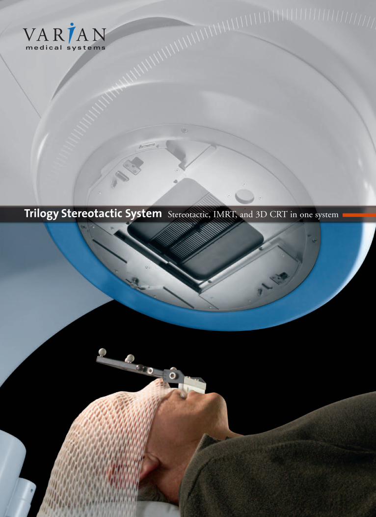
Trilogy Stereotactic System Stereotactic, IMRT, and 3D CRT in one system

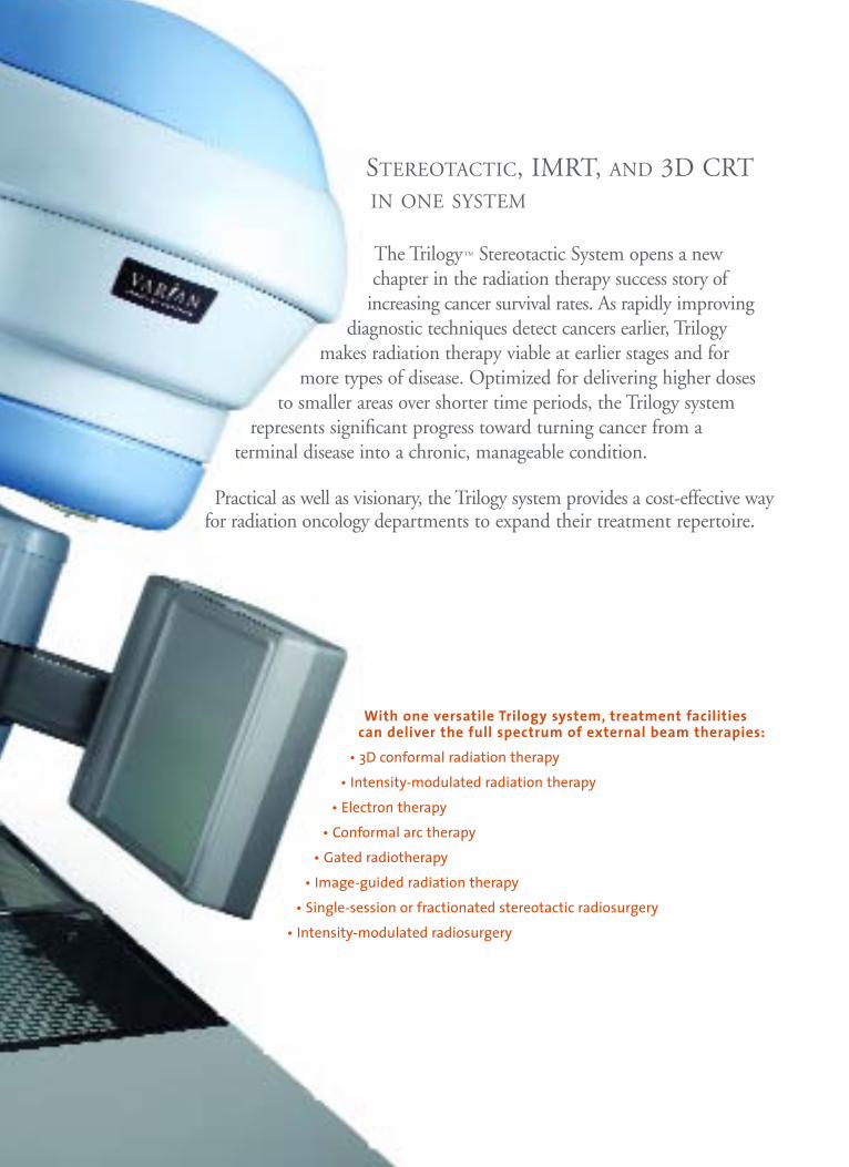
STEREOTACTIC, IMRT, AND 3D CRT IN ONE SYSTEM
The Trilogy™ Stereotactic System opens a new chapter in the radiation therapy success story of
increasing cancer survival rates. As rapidly improvingdiagnostic techniques detect cancers earlier, Trilogy
makes radiation therapy viable at earlier stages and for more types of disease. Optimized for delivering higher doses
to smaller areas over shorter time periods, the Trilogy system represents significant progress toward turning cancer from a
terminal disease into a chronic, manageable condition.
Practical as well as visionary, the Trilogy system provides a cost-effective wayfor radiation oncology departments to expand their treatment repertoire.
With one versatile Trilogy system, treatment facilities can deliver the full spectrum of external beam therapies:
• 3D conformal radiation therapy
• Intensity-modulated radiation therapy
• Electron therapy
• Conformal arc therapy
• Gated radiotherapy
• Image-guided radiation therapy
• Single-session or fractionated stereotactic radiosurgery
• Intensity-modulated radiosurgery

Highest dose rateAt 1,000 monitor units (MU) per minute, the Trilogy sys-tem delivers a stereotactic dose rate that is 20 percent higherthan any comparable system and twice as high as mostlinacs. A higher dose rate translates into shorter sessions,enabling the treatment of more patients per day.
Tight isocenter alignment—on all three axesTrilogy leads in beam accuracy for treating increasinglysmaller tumors. On two axes—the gantry and the collimator—Trilogy’s isocenter radius measures 0.5 mm or less. Add the third rotational axis—the table—and the isocenter radius is 0.75 mm or less, the tightest alignment available in a dual-energy system.
Rapid on-board imagingWith Trilogy’s On-Board Imager™ accessory, it’s fast andeasy to acquire kilovoltage anterior/posterior and lateralimages for high-precision repositioning of most patients.Daily online setup corrections typically can be achieved injust a few minutes.
Independently movable robotic armsTrilogy’s unique On-Board Imager features two independentrobotic arms, enabling many image acquisition geometries.
Potential to correct for motion during treatmentMonitoring and correcting for patient motion during treat-ment is possible using the fluoroscopic mode of Trilogy’sOn-Board Imager plus the Real-Time Position Management(RPM™) gating system.
Clear, easy-to-read kV images Because it’s a kilovoltage imaging system, the On-BoardImager produces clear, easy-to-analyze images that result invery low patient doses. And Trilogy’s patented carbon-fibercouch top minimizes distracting artifacts.
2
All in one. Best in one. The Trilogy system is optimized for both conventional and stereotactic treatment approaches.

Cone-beam CTWith Trilogy’s On-Board Imager accessory, cone- beam CT scans (3D CT volumes) can be acquired and used to correct patient positioning based onsoft-tissue landmarks. Using sophisticated imageanalysis tools, the cone-beam CT scans can be compared with reference CT scans to determinehow the couch should be moved to fine-tune thesetup of the patient.
Treatment planning and delivery withoutmoving the patient
Varian’s technology makes it possible to use the Trilogysystem to acquire a treatment planning CT scan, create atreatment plan, and deliver the treatment without moving thepatient, yielding high accuracy in treatment delivery.
Automated image registrationPatient positioning is fast and precise with Trilogy.Automated image registration algorithms for both anatomyand radiopaque markers instantly compare daily imageswith reference images.
Remote-controlled couch Trilogy’s remotely controlled couch makes treatment deliveryfaster and more accurate. Patient setups can be finely tunedfrom outside the treatment room, with user-definable couchrotation and translation limits. Therapists can also controlplanned rotations of the couch from outside the treatmentroom, speeding the delivery of multiple noncoplanar arcs.
Integrated imaging and delivery processTrilogy integrates imaging and delivery into a single processso well designed that radiation therapists can be proficientfrom the first day of clinical operation.
3
Improving treatment precision
Trilogy is the most precise dual-energydelivery system in the world, enabling the treatment of very small lesions in the head and neck as well as deep-seatedlesions in the abdomen and pelvis. Thebeam focal point is a sphere less than0.75 mm radius, and integrated imagingtechnology provides precise patientpositioning and tumor localization.
Streamlining the stereotactic session
Trilogy streamlines lengthy stereotacticsessions. The high dose rate shortensbeam-on time. The remote-controlledcouch speeds up treatment delivery andmaximizes the number of treatmentfields and arcs that can be used.
Maximizing system utilization
Because it’s a multipurpose system,Trilogy can pay for itself quickly—often within a few years. While yourstereotactic practice builds up, theTrilogy system can be fully utilized delivering IMRT treatments.
4
With a multipurpose Trilogy system, you can enhance the flexibility of treatment processes and shorten delivery timeswhile improving the patient’s overall comfort level. The following example describes the clinical process for intracranialstereotactic radiosurgery using a Trilogy system with Varianstereotactic components.
STEP 1 | Immobilization Create a custom bite block andhead-and-neck immobilization system. The bite block, attached to a fiducial array, enables optical guidance of
patient positioning in the treatment room. Because the immobilization system is frameless, imaging and treatmentdelivery can be performed on different days, providing scheduling flexibility and improving the patient experience.Frame-based immobilization is also included.
STEP 2 | MR imaging Acquire an MR scan, which is used todefine the target volume.
STEP 3 | CT simulation Acquire a treatment planning CTscan, which is used to establish the stereotactic coordinate system.
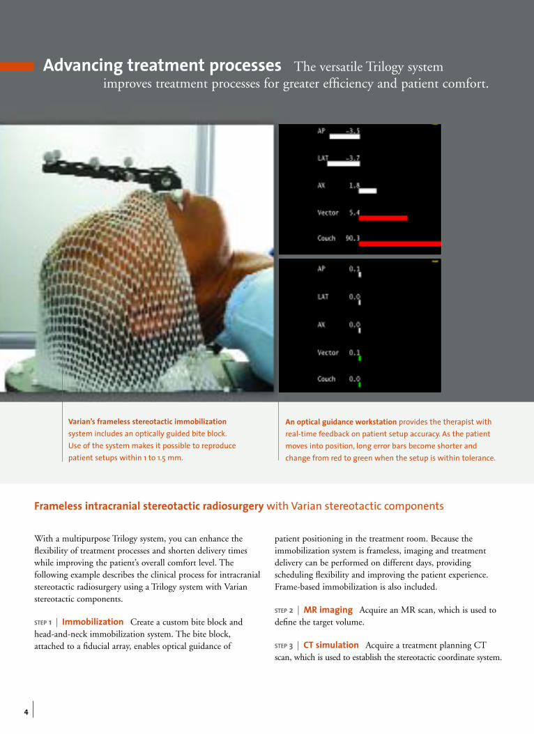
Advancing treatment processes The versatile Trilogy systemimproves treatment processes for greater efficiency and patient comfort.
Frameless intracranial stereotactic radiosurgery with Varian stereotactic components
An optical guidance workstation provides the therapist withreal-time feedback on patient setup accuracy. As the patientmoves into position, long error bars become shorter andchange from red to green when the setup is within tolerance.
Varian’s frameless stereotactic immobilizationsystem includes an optically guided bite block.Use of the system makes it possible to reproducepatient setups within 1 to 1.5 mm.
5
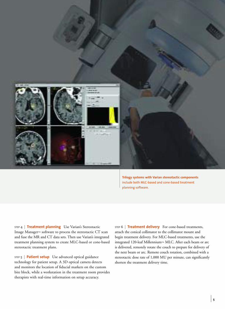
STEP 4 | Treatment planning Use Varian’s StereotacticImage Manager™ software to process the stereotactic CT scanand fuse the MR and CT data sets. Then use Varian’s integratedtreatment planning system to create MLC-based or cone-basedstereotactic treatment plans.
STEP 5 | Patient setup Use advanced optical guidance technology for patient setup. A 3D optical camera detects and monitors the location of fiducial markers on the custombite block, while a workstation in the treatment room providestherapists with real-time information on setup accuracy.
STEP 6 | Treatment delivery For cone-based treatments,attach the conical collimator to the collimator mount and begin treatment delivery. For MLC-based treatments, use theintegrated 120-leaf Millennium™ MLC. After each beam or arcis delivered, remotely rotate the couch to prepare for delivery ofthe next beam or arc. Remote couch rotation, combined with astereotactic dose rate of 1,000 MU per minute, can significantlyshorten the treatment delivery time.
Trilogy systems with Varian stereotactic components include both MLC-based and cone-based treatmentplanning software.
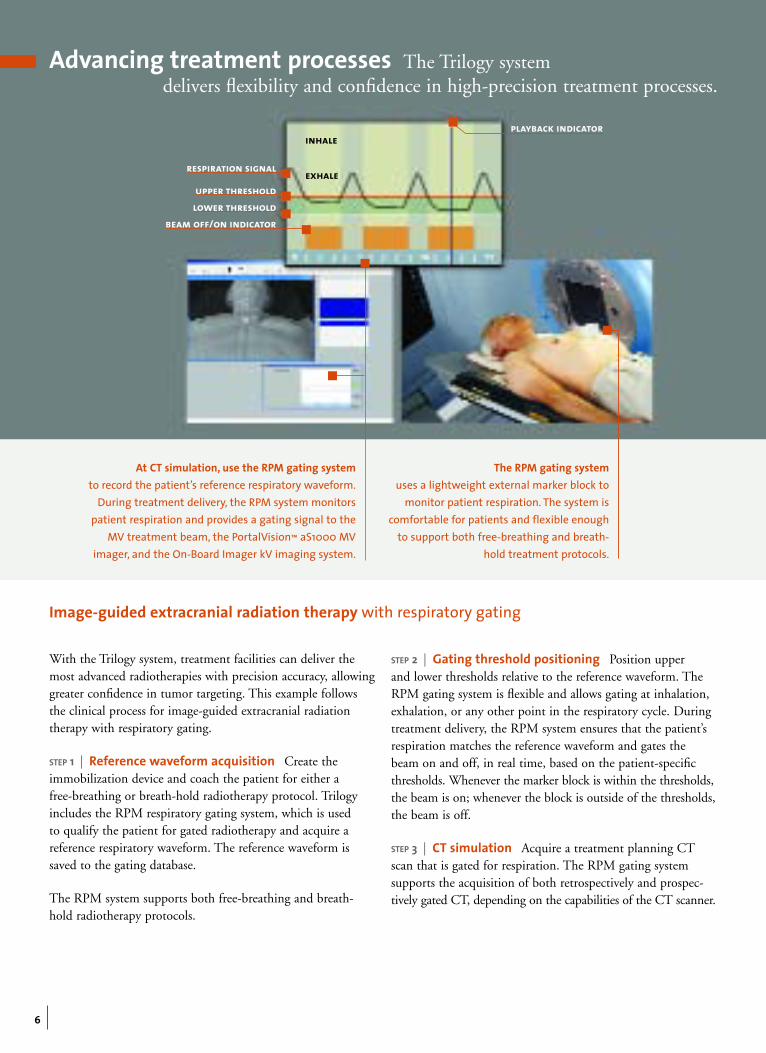
respiration signal
upper thresholdlower threshold
beam off/on indicator
playback indicator
6
With the Trilogy system, treatment facilities can deliver themost advanced radiotherapies with precision accuracy, allowinggreater confidence in tumor targeting. This example followsthe clinical process for image-guided extracranial radiationtherapy with respiratory gating.
STEP 1 | Reference waveform acquisition Create theimmobilization device and coach the patient for either a free-breathing or breath-hold radiotherapy protocol. Trilogyincludes the RPM respiratory gating system, which is used to qualify the patient for gated radiotherapy and acquire a reference respiratory waveform. The reference waveform issaved to the gating database.
The RPM system supports both free-breathing and breath-hold radiotherapy protocols.
STEP 2 | Gating threshold positioning Position upper and lower thresholds relative to the reference waveform. TheRPM gating system is flexible and allows gating at inhalation,exhalation, or any other point in the respiratory cycle. Duringtreatment delivery, the RPM system ensures that the patient’srespiration matches the reference waveform and gates thebeam on and off, in real time, based on the patient-specificthresholds. Whenever the marker block is within the thresholds,the beam is on; whenever the block is outside of the thresholds,the beam is off.
STEP 3 | CT simulation Acquire a treatment planning CTscan that is gated for respiration. The RPM gating system supports the acquisition of both retrospectively and prospec-tively gated CT, depending on the capabilities of the CT scanner.
Image-guided extracranial radiation therapy with respiratory gating
The RPM gating system uses a lightweight external marker block to
monitor patient respiration. The system iscomfortable for patients and flexible enough
to support both free-breathing and breath-hold treatment protocols.
At CT simulation, use the RPM gating systemto record the patient’s reference respiratory waveform.
During treatment delivery, the RPM system monitorspatient respiration and provides a gating signal to the
MV treatment beam, the PortalVision™ aS1000 MV imager, and the On-Board Imager kV imaging system.
inhale
exhale
Advancing treatment processes The Trilogy system delivers flexibility and confidence in high-precision treatment processes.
7
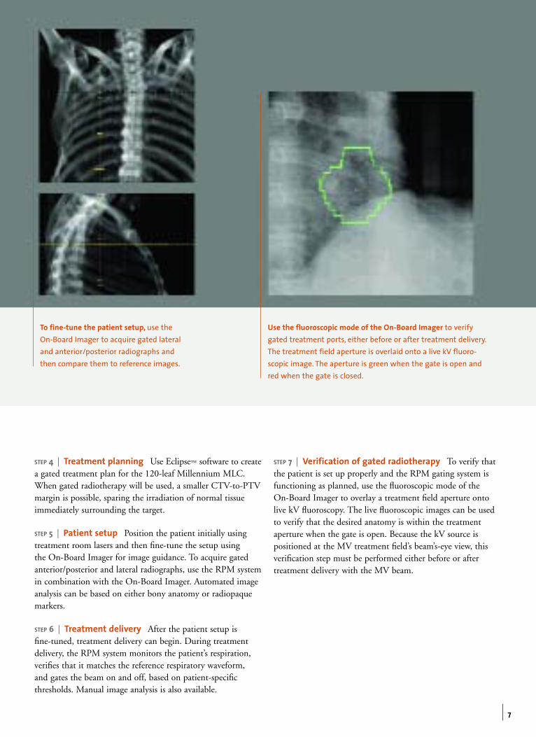
STEP 4 | Treatment planning Use Eclipse™ software to createa gated treatment plan for the 120-leaf Millennium MLC. When gated radiotherapy will be used, a smaller CTV-to-PTVmargin is possible, sparing the irradiation of normal tissueimmediately surrounding the target.
STEP 5 | Patient setup Position the patient initially usingtreatment room lasers and then fine-tune the setup using the On-Board Imager for image guidance. To acquire gatedanterior/posterior and lateral radiographs, use the RPM systemin combination with the On-Board Imager. Automated imageanalysis can be based on either bony anatomy or radiopaquemarkers.
STEP 6 | Treatment delivery After the patient setup is fine-tuned, treatment delivery can begin. During treatmentdelivery, the RPM system monitors the patient’s respiration,verifies that it matches the reference respiratory waveform, and gates the beam on and off, based on patient-specificthresholds. Manual image analysis is also available.
STEP 7 | Verification of gated radiotherapy To verify thatthe patient is set up properly and the RPM gating system isfunctioning as planned, use the fluoroscopic mode of the On-Board Imager to overlay a treatment field aperture ontolive kV fluoroscopy. The live fluoroscopic images can be usedto verify that the desired anatomy is within the treatmentaperture when the gate is open. Because the kV source is positioned at the MV treatment field’s beam’s-eye view, thisverification step must be performed either before or after treatment delivery with the MV beam.
To fine-tune the patient setup, use the On-Board Imager to acquire gated lateraland anterior/posterior radiographs andthen compare them to reference images.
Use the fluoroscopic mode of the On-Board Imager to verifygated treatment ports, either before or after treatment delivery.The treatment field aperture is overlaid onto a live kV fluoro-scopic image. The aperture is green when the gate is open andred when the gate is closed.
8
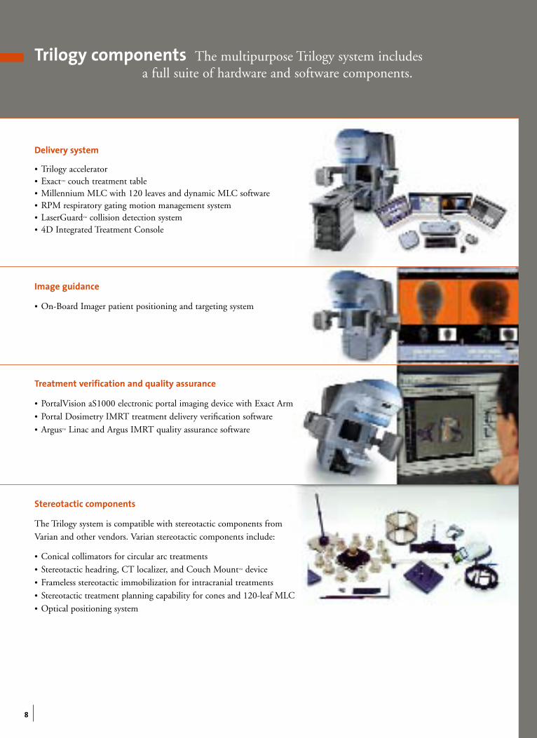
Trilogy components The multipurpose Trilogy system includes a full suite of hardware and software components.
Delivery system
• Trilogy accelerator• Exact™ couch treatment table• Millennium MLC with 120 leaves and dynamic MLC software• RPM respiratory gating motion management system• LaserGuard™ collision detection system• 4D Integrated Treatment Console
Image guidance
• On-Board Imager patient positioning and targeting system
Treatment verification and quality assurance
• PortalVision aS1000 electronic portal imaging device with Exact Arm • Portal Dosimetry IMRT treatment delivery verification software • Argus™ Linac and Argus IMRT quality assurance software
Stereotactic components
The Trilogy system is compatible with stereotactic components from
Varian and other vendors. Varian stereotactic components include:
• Conical collimators for circular arc treatments• Stereotactic headring, CT localizer, and Couch Mount™ device• Frameless stereotactic immobilization for intracranial treatments
• Stereotactic treatment planning capability for cones and 120-leaf MLC • Optical positioning system
T R I LO GY A CC E L E R ATO R H I G H L I G H T S
2 photon beams for 3D CRT and IMRTEnergy * 6/10, 6/15, 6/18, or 6/20 MV
Maximum dose rate 600 MU/minMaximum field size 40 cm x 40 cm
1 photon beam for SRS and SRTEnergy * 6 MV
Maximum dose rate 1,000 MU/minMaximum field size 15 cm x 15 cm
6 electron beamsEnergy * 4/6/9/12/15/18, 6/9/12/15/18/22, 4/6/9/12/16/20 MeV
Maximum dose rate 1,000 MU/min
IsocenterM 0.5 mm radius for gantry and collimator axesM 0.75 mm radius for gantry, couch, and collimator axes
Remote couch motionCorrective, small translations and rotations to fine-tune patient setupsPlanned, large rotations to sequence between noncoplanar arcs
Millennium MLCNumber of leaves 120 leaves
Leaf width 0.5 cm, centralmost 40 pairs1.0 cm, outermost 20 pairs
On-Board ImagerRadiographic mode Daily online setup correction based on bony anatomy or radiopaque markersFluoroscopic mode Pretreatment verification of gated treatment ports
Cone-beam CT mode Daily online setup correction based on soft-tissue anatomyAdaptive radiotherapy
* Energy designations are given according to British Journal of Radiology Supplement 11 (BJR 11) definitions.
Inspiration, the Varian advantageThe Trilogy Stereotactic System is part of the Inspiration™ integrated oncology environment.
Varian and Varian Medical Systems are registered trademarks and Argus, Couch Mount, Eclipse, Exact, Inspiration, LaserGuard, Millennium, On-Board Imager, PortalVision, RPM, Stereotactic Image Manager, and Trilogy are trademarks of Varian Medical Systems, Inc. The names of other companies and products mentioned herein
are used for identification purposes only and may be trademarks or registered trademarks of their respective owners.
RAD 9506A © 2004 Varian Medical Systems, Inc. Printed in USA. 11/04 (7k)
USA HeadquartersCaliforniaVarian Medical SystemsPalo Alto, CA Tel: 650.424.5700
800.544.4636Fax: 650.493.5637
USA Regional OfficesCaliforniaVarian Medical SystemsCorona, CATel: 909.280.4401Fax: 909.280.4300
GeorgiaVarian Medical SystemsMarietta, GATel: 770.955.1367Fax: 678.255.3850
IllinoisVarian Medical SystemsDes Plaines, ILTel: 847.296.5533Fax: 847.296.0043
New JerseyVarian Medical SystemsClark, NJTel: 732.340.9346Fax: 732.381.1060
European HeadquartersSwitzerlandVarian Medical SystemsInternational AGZug, SwitzerlandTel: 41.41.749.8844Fax: 41.41.740.3340
AustriaVarian Medical Systems Gesellschaft m.b.H.Voesendorf, AustriaTel: 43.1.698.56.56Fax: 43.1.698.56.59
BelgiumVarian Medical Systems Belgium N.V.Sint-Katelijne, BelgiumTel: 0800.903.23Fax: 31.30.636.2466
FinlandVarian Medical Systems Finland OyHelsinki, FinlandTel: 358.9.430.771Fax: 358.9.455.4585
FranceVarian Medical Systems FranceBuc, FranceTel: 33.1.30.83.83.83Fax: 33.1.30.83.83.00
GermanyVarian Medical SystemsDeutschland GmbHDarmstadt, GermanyTel: 49.61.51.73130Fax: 49.61.51.731313
IndiaVarian Medical SystemsIndia Pvt Ltd.Mumbai, IndiaTel: 91.22.26162301/04Fax: 91.22.26162277
ItalyVarian Medical SystemsItalia, S.p.A.Cernusco s/N (MI), ItalyTel: 39.02.921.351Fax: 39.02.921.35240
NetherlandsVarian Medical Systems Netherlands B.V.Houten, NetherlandsTel: 31.30.634.0506Fax: 31.30.636.2466
ScandinaviaVarian Medical SystemsScandinavia ASHerlev, DenmarkTel: 45.44.500.100Fax: 45.44.500.190
Spain/PortugalVarian Medical Systems Ibérica, S.L.Madrid, SpainTel: 34.91.799.4530Fax: 34.91.799.4541
UK/IrelandVarian Medical Systems UK Ltd.Crawley, West Sussex, UKTel: 44.1293.531.244Fax: 44.1293.510.260
Asian HeadquartersHong KongVarian Medical Systems Pacific, Inc.Kowloon, Hong KongTel: 85.22.724.2836Fax: 85.22.369.4280
ChinaVarian Medical Systems China Ltd.Beijing, P.R. ChinaTel: 8610.6512.7169Fax: 8610.6523.2039
JapanVarian Medical Systems K.K.Chuo-ku, Tokyo, JapanTel: 81.3.3639.9700Fax: 81.3.3639.9623
Latin American HeadquartersFloridaVarian Medical SystemsMiami, FL USATel: 305.929.1970Fax: 305.929.1971
BrazilVarian Medical Systems do Brasil Ltda.São Paulo, BrazilTel: 55.11.3457.2655Fax: 55.11.3286.0034
Australian HeadquartersAustraliaVarian Medical Systems (Australasia) Pty Ltd.Frenchs ForestNSW 2086Sydney, AustraliaTel: 612.9975.8800Fax: 612.9452.5737
Oncology Systems3100 Hansen WayPalo Alto, CA 94304-1038Tel: 650.424.5700Tel: 800.544.4636www.varian.com