triiodothyronine and thyroxine the serum€¦ · triiodothyronine and thyroxine in the serum and...
TRANSCRIPT
Triiodothyronine and Thyroxine in the Serum
and Thyroid Glands of Iodine-Deficient Rats
G. M. ABRAMSand P. R. LARSEN
From the Division of Endocrinology and Metabolism, Department of Medicine,
School of Medicine, University of Pittsburgh, Pittsburgh, Pennsylvania 15261
A B S T R A C T Triiodothyronine (T3) and thyroxine(T4) were measured by immunoassay in the serum andthyroid hydrolysates of control (group A), mildly io-dine-deficient (group B), and severely iodine-deficientrats (group C). These results were correlated withchanges in thyroidal weight, 'I uptake and "2I contentas well as with the distribution of 131I in Pronase digestsof the thyroid. There was a progressive increase inthyroid weight and '1I uptake at 24 h with decrease iniodine intake. The PI content of the thyroids of thegroup B animals was 44% and that of the group C ani-mals 2% of that in group A. The mean labeled mono-
iodotyrosine/diiodotyrosine (MIT/DIT) and T3/T4 ra-
tios in group A were 0.42±0.07 (SD) and 0.12±0.01,0.59±0.06 and 0.11±0.03 in group B, and 2.0±0.3 and1.8±0.9 in the group thyroid digests.
Mean serum T4 concentration in the control rats was
4.2±0.6 (SD) Ag T4/100 ml, 4.5+0.3 Ag/100 ml ingroup B animals, and undectectable (<0.5 tL/100ml) in group C animals. There was no effect of iodinedeficiency on serum T3 concentrations, which were 44+9 (Mean±SD) ng/100 ml in A animals, 48±6 ng/100ml .n B animals, and 43+6 ng/100 ml in the C group.Thyroidal digest Ts and To concentrations were 39 and400 ng/mg in group A animals and were reduced to 5and 1% of this, respectively, in group C. The molarratio of Ta/Ti in the thyroid digests of the groups A andB animals was identical to the ratio of labeled Ts/T4and was slightly less (1.0±0.9) than the labeled T3/Tratio in the group C animals.
The mean ratio of labeled T4 to labeled Ts in the se-
rum of the severely iodine-deficient animals 24 h afterisotope injection was 11+1 (SEM). With previouslypublished values, it was possible to correlate the ratioof labeled Tt/Ts in the thyroid digest with the labeled
P. R. Larsen is a Career Development Awardee of theU. S. Public Health Service, Award #5 K04 AM70401.
Received for publication 13 April 1973 and in revised form4 June 1973.
T4/T3 ratio in the serum of each iodine-deficient animal.This analysis suggested that the labeled thyroid hor-mones in the severely iodine-deficient rat were secretedin the ratio in which they are present in the gland.
Kinetic analysis of total iodothyronine turnover indi-cated that two-thirds of the Ts utilized per day by theiodine-sufficient rat arises from T4. If the Tr-Ts con-version ratio remains the same in iodine deficiency, thenthe analysis suggests that about 90% of the T3 arises di-rectly from the thyroid. Therefore, it would appearthat absolute Ts secretion by the thyroid increases sev-eralfold during iodine deficiency. The fact that serumT3 remains constant and T4 decreases to extremely lowlevels, combined with previous observations that iodine-deficient animals appear to be euthyroid, is compatiblewith the hypothesis that T4 in the normal rat serves pri-marily as a precursor of T3.
INTRODUCTIONThe thyroidal response to iodine deficiency has been anarea of active investigation for many years. Previousstudies in the rat reviewed by Studer and Greer indicatethat when iodine intake is severely restricted there is anincrease in thyroid weight, a decrease in protein-boundiodine (PBI)1 and an altered pattern of tracer iodinedistribution in the thyroid gland (1). The last-mentionedchanges include an increase in labeled monoiodotyrosine(MIT) and decrease in labeled diiodotyrosine (DIT)as well as a progressive increase in the ratio of labeledtriiodothyronine (T3) to labeled thyroxine (T4). Al-though Studer and Greer, and Greer, Grimm, andStuder have demonstrated that serum PBI decreases tovery low levels within 1 mo of initiation of a low-iodinediet, rats maintained on this regimen for even 1 yr re-
'Abbreviations used in this paper: DIT, diiodotyrosine;MIT, monoiodotyrosine; PBI, protein-bound iodine; T8,triiodothyronine; T4, thyroxine; TAA, tertiary-amyl-alcohol/hexane/ammonia; TSH, thyroid-stimulating hormone.
The Journal of Clinical Investigation Volume 52 October 1973 -2522-25312522

main healthy and grow at normal rates (2, 3). Recentstudies by Silva have shown that 02 con1Sumnption andbody temperature regulation of rats under severe iodinerestriction for 120 days is not different from that of con-
trol animals (4).The shift of the l)redom11inaLnt intrathyroi(lal-labele(l
iodotlhvronine from Tl; to T3 in response to iodine re-
striction has led to speculation that the iodine-deficientrat maintains its apl)arent euthyroid status by preferen-tial syinthesis and release of T3 (3). Since it has not beenpossible previously to measure serum T. directly underthese circumstances, this hypothesis has not been sub-stantiated. While this study was in progress, Volpertand Werner presented data suggesting that there was a
decrease in the immunoassayable serum T3 in the iodine-deficient rat though the ratio of T3 to PBI was increased(5). However, the small group of animals examinednecessarily limited the scope of their conclusions. Inthe following study data are presented that correlate thechanges in serum immunoassayable T3 and T4 withchanges in thyroidal T3, T4, and iodine content in mildlyand severely iodine-deficient rats.
METHODSAnimals and diets. Sprague-Dawley male rats weighing
150-200 g were maintained on Remington low-iodine testdiet (Diet I, Nutritional Biochemicals Corporation, Cleve-land, Ohio) for a period of 3 mo. Half of this group re-ceived distilled water and the other half distilled water con-taining 1.3 yg iodide/ml (estimated intake 20-30 ml H20/day). After 3 mo the supplier of the low-iodine diet waschanged (Diet II, General Biochemicals Div., Mogul Corp.,Chagrin Falls, Ohio) since the anticipated elevation inMIT/DIT ratios was not observed up to this time. Thelevel of iodine supplementation in the control animals re-mained the same. However, the substitution of a differentcommercial low-iodine diet after 3 mo of this study made itimpossible to examine the longitudinal effects of sustainediodine restriction under constant conditions. Therefore, wehave not attempted an analysis of the data as related toduration of iodine deprivation. The diets were analyzed bythe Boston Medical Laboratory, and both contained non-detectable amounts of iodine (<0.165 zg/g diet [6]). Thefact that the thyroidal changes in animals receiving diet IIwvere substantially greater than those in animals receivingdiet I is best explained by the assumption that it containedless iodine. However, without verification of this by actualmeasurement, the theoretical possibility that diet II con-tains a goitrogen that accelerated the appearance of thechanges in the low-iodine animals cannot be entirely dis-counted.
Admninistration. of ...I and calculation of uptake. 5-40,uCi of carrier-free 13"I was injected intraperitoneally 24 hbefore sacrifice. Groups of control and iodine deficient-ratswere killed at various intervals after starting the diets andthyroids were dissected from the trachea and weighed.Radioactivity in thyroids and suitable dilutions of theinjected dose were measured with an end window Geigercounter under conditions of constant geometry. Blood wasobtained from each animal by cardiac puncture.
Digestion and extraction. One or two thyroid glall(Iswere digested with Pronase (Calbiochem, Los Angeles,Calif.), by the method of Inoue and Taurog (7). This pro-cedure was slightly modified as we have previously de-scribed (8). Labeled [1"JI]T3 and T4 (Abbott Laboratories,North Chicago, Ill.) were added at the start of digestion tomonitor recovery and identify ['"'I]iodothyronines. The la-beled iodothyronines contained less than 3% I- and no othersignificant contaminants. In addition, after digestion thehydrolysate was extracted twice with 0.4 ml of methanolin 7.4 N ammonia (1:1, vol: vol). Residual 'I and '5I inthe pellet after this procedure were 6% or less of the total.Analysis of the pellet 127 Iin the animals on the high-iodineintake showed similar losses of "I. Digestion of extractswas nearly complete as estimated by chromatography ofthe extracts in collidine-2 N ammonia (3:1, vol: vol).The amount of 127I and "I remaining at the origin was lessthan 4%. The identification of "'I-labeled I-*,2 MIT* andDIT* was carried out as previously described by chromatog-raphy in butanol/acetic acid (8). Separation of T4* andTs* was obtained in tertiary-amyl-alcohol hexane/ammonia(TAA) also as previously described (8). Briefly, 25-75 1.1of the extract was chromatographed in both systems, and1-2 cm segments of the strips were subsequently countedfor both "I and .3I with suitable correction. In the TAAsystem, the percent T3* was corrected for the 0.34%o arti-factual deiodination of T4* during the chromatography aswe have previously described (9). Ts* and T4* were cor-rected for losses during the procedure by reference to theoriginal amounts of [HI] Ts and T4 added. Results wereexpressed as a fraction of the total 131I in the extract.
'7I content in 0.1 ml of the extract (about 0.8 ml totalvolume) was determined by Boston Medical Laboratory(6). This was then corrected for the total extraction vol-ume as well as the small losses occurring during extractionof the Pronase hydrolysate. Results were expressed asug "I per mg wet weight of thyroid.
Serum and thyroidal Ts and T4 content. T, and T4content in the thyroid hydrolyates and serum were deter-mined by radioimmunoassays as previously described (10,11). All assays included samples from paired control andiodine-deficient rats and were performed in duplicate attwo dilutions. In analyzing the extract for iodothyroninessuitable dilutions were made in MeOH/NH4OH (99: 1,vol: vol). 5 or 10 1(d of this dilution was then added totubes containing either T3-free or T4-free human serumin 1 ml total volume. This concentration of MeOH/NH4OHdoes not affect the assays. In measuring T, and T3 in thy-roid hydrolysates from iodine sufficient rats, the MeOH/NH4OHdilutions used were 1/100 to 1/500. In both assays,curves parallel to the standard were obtained with increas-ing quantities of extract. Thus, the duplicate determinationsat two dilutions were in excellent agreement. In the ex-tracts of thyroids from the group C animals, dilutions aslow as i and i were sometimes necessary for accurate quan-titation, indicating that there was no artifactual contribu-tion to either the calculated T3 or T4 value from non-iodine-containing substances in the thyroid extracts. Again,excellent agreement was obtained in the estimated iodothy-ronine content with two different dilutions. There is nosignificant displacement of either labeled iodothyronine fromits antibody by 1000-fold excesses (by weight) of I-, MIT,or DIT. Since there is essentially no cross-reaction of theTs antibody with T4, prior separation of T3 and T4 is
'An asterisk will be used to denote MI-labeled compoundspresent in thyroid glands or in serum.
Triiodothyronine and Thyroxine in Iodine-Deficient Rats 2523
not required for this method. Total thyroid T8 and T. werecorrected for losses in extraction and expressed as ng T3or T4 per mg wet weight of thyroid.
Analysis of serum T. was performed by using a modi-fication of the T8-free human serum system that employssodium salicylate to block T3-protein binding (10). Pre-liminary studies indicated that as much as 100 ,ul of ratserum could be substituted for human serum in the assaywithout disturbing the parallelism of the rat serum curvewith the standard curve or changing the percentage oftracer bound in the absence of antibody. Thus, unknownsamples contained either 100 or 50 Il unknown rat serumplus 100 or 150 Ml of T3-free human serum added to makethe serum concentration 20% in all tubes. Duplicates of thetwo dilutions were in excellent agreement. T3-free rat serumprepared as we have previously described for human serumcontained no Ts (10). Recovery of T3 from pooled rat serawas 96±8% (mean+-SD) for 100 ng Ts/100 ml and 99±8% (mean+SD) for 200 ng Ts/100 ml. Similar recoverieswere obtained using serum from iodine-deficient rats in asmaller series of studies. Likewise, the T4 determinationscould be performed in the system containing T4-free humanserum as long as the quantity of rat serum per tube didnot exceed 5 Au (total serum per tube, 10 ,ul).
Distribution of labeled compounds in serum 24 h after"'I injection. In the severely iodine-deficient animals the
3.01
2.
.8<
*'H
it
1.21
0.6
-
I
I
0 0.2
I0 SINGLE TH* TWOTHYR
POOLED
.0o * I
* 0 C0
B I
I
0.4 0.6 0.82I(pFg/mg THYROID)
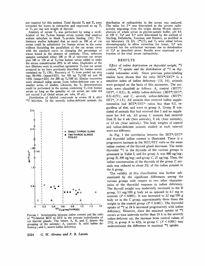
FIGURE 1 Relationship between iodine coof "I-labeled MIT to DIT in the pronarat thyroid glands. The letters A, B,grouping of the animals: A, control; Ificiency; and C, severe iodine deficiency.
iYROIC
OID GLANDDGLANDS
distribution of radioactivity in the serum was analyzed.The value for I-* was determined as the percent radio-activity migrating from the origin during 90-min electro-phoresis of whole serum in glycine-acetate buffer, pH 8.6,at 150 V. Ts* and T4* were determined by the method ofSterling, Dellabarba, Newman, and Brenner, as modified inour laboratory (9, 12). [JI]TT and T4 were added beforechromatography to allow correction for losses. T3* wascorrected for the artifactual increases due to deiodinationof TA* as described above. Results were expressed as afraction of the total serum radioactivity.
RESULTSEffect of iodine deprivation on thyroidal weight, "I7J
content, "'I uptake and the distribution 'of .S.I in thy-roidal iodoamino acids. Since previous pulse-labelingstudies have shown that the ratio MIT*/DIT* is asensitive index of iodine deficiency (13, 14), animalswere grouped on the basis of this parameter. The ani-mals were classified as follows: A, control (MIT*/DIT*, < 0.5); B, mildly iodine-deficient (MIT*/DIT*,0.51-0.75); and C, severely iodine-deficient (MIT*/DIT*, > 1.5). All animals that received iodine supple-mentation had MIT*/DIT* ratios less than 0.5, re-gardless of diet, and were in group A. Group B con-sisted of animals that had received diet I and no supple-ment for 4-8 wk. All group C animals had receivedDiet II for 4 wk (two animals), 8 wk (four animals),or 12 wk (four animals). The body weights of controland iodine-deficient animals studied at each intervalwere not different.
In Fig. 1 the correlation between the MIT*/DIT*and thyroidal iodine content is illustrated. There is aprogressive increase in the MIT/DIT ratio as the meaniodine content of the thyroid gland decreases. The meanthyroidal "I in the thyroids of the various groups ispresented in Table I, and for group A was 880 ng/mg;group B, 390 ng/mg; and group C, 21 ng/mg. Thus, theiodine concentration of the thyroids of the group C ani-mals was reduced to about 2% of the iodine present inthe A group.
The validity of this classification was further sub-stantiated by the significant differences among thevarious groups with respect to two other character-istics of the thyroidal response to iodine deficiency.The thyroid weight was moderately increased in the B
0 0 group, 5.1 mg/100 g body wt as opposed to 4.1 mg incontrols (P < 0.005). It was increased to 12 mg/100 g
A body wt in the C group, approximately three times the
1.0 1.2 1.4weight in the control group (P < 0.001). The thyroidal
1.0 1.2 .4 uptake of "1I at 24 h increased progressively with iodinedeflciency. However, since the maximal uptake of 'I
intent and the ratio occurs at time intervals earlier than 24 h in the severelyase hydrolysates of iodine-deficient rat, the increase from control values ofand C denote theB, mild iodine de- 15% in group A to 42% in group C (P < 0.001) may
underestimate the difference in maximal l'I uptake.
2524 G. M. Abrams and P. R. Larsen
.4p

TABLE I'I7I Content, Gland lIeight, and Uptake and Distribution of 'l~I in Pronase Hydrolysates of
from Control and Iodine-Deficient Rats (Meanl±SD)Thyroid Glands
Group n 127I Thyroid weight RAIUt I-* MIT* DIT* T3* T4* MIT*/DIT* T3*/T4*
ng/mg thyroid mg/100 g body wt
A 12 880±180 4.1±0.7 15+10 4.8±1.0 1943 46±:3 2.6±0.3 21±1 0.42±0.07 0.12±0.01B 9 390±100 5.140.6 27±11 3.8±1.1 26±3 44±t3 2.5±0.8 23±3 0.59±0.06 0.11±40.03CHj 1 7.7 10 55 5.0 44 17 20 7.3 2.7 2.7
2 4.4 13 49 3.5 41 16 19 5.2 2.5 3.53 6.9 12 36 3.3 38 18 17 6.7 2.1 2.44 6.4 14 43 5.5 41 20 16 12 2.1 1.35 6.1 13 37 6.0 38 20 16 14 1.9 1.16 9.5 7 48 5.1 36 21 23 17 1.8 1.47 7.7 12 37 4.3 33 19 22 17 1.7 1.38 7.2 16 54 2.6 32 21 23 17 1.5 1.49§ 55 11 26 3.6 37 22 18 12 1.7 1.5
10(§ 97 7.1 33 2.7 38 23 18 14 1.7 1.3Mean
±SD 21±30 12±3 42±9 4.2±1.2 38±3 20±3 19±t3 12±3 2.0±-0.3 1.8±0.9t Uptake of 1311 in thyroid 24 h after isotope injection.§ Animals on iodine restriction for 4 wk.JJ In group C, data are provided for individual animals.
The distribution of radioactivity in the Pronasehydrolysates of the thyroids is presented in Table I.The I-* percentage was not different in the threegroups. The percent of MIT* increases progressivelyas the iodine supply is further restricted, the mean being19 in group A, 26 in group B, and 38 in group C.There is no difference in the DIT* percentage ingroups A and B, but the value of 20% in group C issignificantly lower than that observed in the controlgroup (P <0.001). In addition, there is also no sig-nificant difference between the A and the B animalswith regard to the relative distribution of isotope inT3 and T4, while in group C there is a significant in-crease in the percent Ts* from a mean of 2.6 in groupsA and B to 19 in group C (P <0.001). In addition,there is a substantial decrease in the percent T4* froma mean of 22 in groups A and B, to 12 in group C(P < 0.001). Approximately 90-100% of the labelpresent in the thyroid hydrolysate is accounted for bythese five components. The remainder of the labeledmaterial appeared to be distributed evenly along chro-matographic paper strips between the identifiable peaks.
As previously mentioned, there is a progressive in-crease in the ratio of MIT*/DIT* with increasingseverity of iodine restriction. The ratio of T8*/T4* is0.12 in group A, and not significantly different (0.11)in group B. However in group C the ratio is reversed,T2* being 1.8 times T4*. The distribution of the isotopein these normal and iodine-deficient glands is substan-
tially the same as that observed by previous investigators(1).
Serum Ts and T4 levels in control and iodine-deficientrats. Mean levels of serum Ts and T4 are presented inTable II.-Where thyroid glands from two animals werepooled, the mean of the values for serum Ts and T4were used. Serum T3 levels in the control animalsranged from 29 to 54 ng/100 ml with a mean of 44.Ts levels were not significantly different in group Bwith a range of from 42 to 61 ng/ml with the mean48 ng/100 ml. In group C, the mean serum Ts concen-tration was unchanged from the group A animals, being43 ng/100 ml. Thus there was no change in the serumTs concentration in the iodine-deficient rat.
In the second column are given the concentrations ofimmunoassayable T4. In control animals, the mean was4.2 Ag/100 with a range of 3.6-5.3 Ag/100 ml. Againthere was no difference in the values obtained in thegroup B animals, where the mean was 4.5±0.1 (mean+SD). However, in the group C animals, T4 was un-
detectable. With current methods, we were able toquantitate T4 in rat serum to a minimum of 0.5 ig/100 ml. In parenthesis the values for PBI are shown,as determined by Boston Medical Laboratory in theserum of most of the group C animals. While it is notknown precisely what quantities of noniodothyronineiodine are present in severely iodine-deficient rats, theselow PBI values confirm the low serum T4 estimatesobtained by immunoassay. Inspection of the group C
Triiodothyronine and Thyroxine in Iodine-Deficient Rats 2525

animals shows that in animals 9 and 10, which had beenon the diet for a period of only 4 wk, T4 had alreadylisappeared. These animals were examined at 10 days
after institution of the low-iodine diet and still hadnormal T4 levels at that time.
Ts and T1 in the pronase hydrolysates of control andiodine-deficient thyroid glands. The T3 and T4 con-centrations of the rat thyroid hydrolysates are alsoshown in Table II. In the thyroid glands of the controlrats, the T3 concentration varied from 27 to 58 ng/mgwet wt with a mean of 39 ng/mg. In mildly deficientanimals, Ts content was moderately reduced to a meanof 26 ng/mg (P < 0.005). An even more substantialreduction, to less than 5% of the control group, wasobserved in the severely iodine-deficient thyroids. Themean Ts content in this group was 1.7 ng/mg. Whilethe T3 concentration was markedly reduced in the groupC animals, the larger size of the thyroid gland resultedin an overall decrease of the gland Ts content of ap-proximately 85%, from a mean of 0.64 itg to about 0.10Ag/total gland.
T4 content of the normal rat thyroid gland was 400ng/mg wet wt. Like Ts, the concentration of this iodo-thyronine was reduced in mild iodine deficiency to amean of 270 ng/mg, significantly less than that of theA group (P <0.001). The mean thyroidal T4 contentin the C animals was 3.2 ng/mg wet wt, less than 1%of the control T4 concentration. This is similar inmagnitude to the re(luction in the "=I content of thegland. The total T4 content of thle goitronis glainds was
only 3% of the control valuie.
Of considerable interest is the fact that the molarratio of T, to T4 in these thyroid glands is similar tothe ratio of T3*/T4* given in Table I. This ratio isprecisely the same for groups A and B. However, ingroup C there appears to be a slight difference, themean molar ratio of T../T4 of 1.0 being less than theratio of 1.8 of the labeled hormones. However, the ratiois markedly increased over the control value by bothmethods. Thus, it would appear that the T3*/T4* ratioeven as soon as 24 h after labeling is an accurate re-flection of the absolute ratio of T8 to T4 present in theiodine-sufficient gland. This agreement occurs despite thefact that prolonged periods are required for completeequilibration of labeled iodine in the thyroid gland, andit will be discussed at greater length below.
Distribution of ...I in the serum of severely iodine-deficient rats. Analysis of the labeled components inthe serum in iodine-deficient rats was performed 24 hafter isotope injection. Accurate estimates of T:* andT4* were possible only in this group, due to the highratio of T4* to T3* present in normal animals. Sincethere is artifactual deiodination of a variable fractionof T4* during chromatographic separation, it is difficultto determine accurately how much T3* present in nor-mal serum arises in vivo.
Direct chromatography of serum in collidine/NH40Hindicated that the only labeled components in serumof iodine-deficient rats there I-*, T3*, and T4*, as hasbeen previously reported (3). As can be seen in Table111. the quantity of 1-* l)resent at 24 h was quite small.in all cases 6%/i, or less of the total. The percentage of
TABLE II
T3 and T4 Concentrations in Serum and Thyroids of Control and Iodine-l>ficient Rats (Mean±SD)
Serum
Ta T4
ng/100 ml
44±948±6
41454048413335584053
pg/100 111l4.2 ±t0.64.5 ±0.3
< 0. 50 (0.4)+I (0.6). .
" (0.6). .
" (0.6)" (0.6)
" (0.8)" (0.6)
43±t6 <0.50 (0.6±0.1)
lg/mg
39±-1026±6
0.790.550.880.970.892.92.02.91.83.6
Thyroid
T3
p.g/gland0.64±0.270.44±0.14
0.0420.0390.0650.0810.0790.140.160.300.0700.14
1.7±t0.9 0.099i0.078
ng mg
400± 100270+85
0.820.181.92.21.13.72.82.56.49.9
T4
jig/gland6.7 ±3.04.4±0.8
0.0440.0130.140.190.0970.180.220.250.380.38
3.2 ±2.8 0.19±0.12
$ PBI, jsg I/100 ml.§ In Group c, data are provided for individual animals.11 Animals on iodine restriction for 4 wk.
2526 G. M. Abrams and P. R. Larsen
Group n
A 12B 9C§ 1
2345678911
1011Mean±SD
Molar ratioTai T4
0.124±0.030.12 ±0.03
1.13.60.550.530.970.930.850.830.330.43
1.01 ±0.90

label as T3 varied from 5 up to 17% at this time, where-as To* ranged from 70 to 102% of the total. Since T3*and T4* content of the original serum was determinedindependently by "'5I-labeled recovery standards, it waspossible that the total percent recovery could be greaterthan 100. The mean recovery of labeled compoundstotaled 96.5%, quite close to the anticipated figure of100%. The ratio of T4*/Ts* in the serum of the iodine-deficient animals varied from a low of 4.1 to as highas 18 with a mean of 11. In the last column, the pre-dicted ratio of T4*/T3* is presented based on the glandT4*/T,*. The derivation of these ratios is discussedsubsequently.
Estimation of the respective contributions of the thy-roid and peripheral T4-to-Ts conversion to the periph-eral Ts pool in the rat. It has been previously shownthat peripheral T4-to-T3 conversion contributes sub-stantially to the circulating Ts pool in man (15). Asimilar pathway for T4 metabolism has been shown inthe rat by Schwartz, Surks, and Oppenheimer (16).If the clearances of T3 and T4 do not change in theiodine-deficient rat, it should be possible to calculatethe relative contributions of the thyroid and peripheralT4-to-T3 conversion to the serum T3 pool in this situa-tion. If plasma T3 or T4 clearance did change in iodinedeficiency, then the knowledge of the serum concen-trations alone would not suffice to calculate the totalproduction rate of each hormone. Silva has recentlydemonstrated that there are no changes in the clearanceof labeled Ts and T4 in rats deprived of iodine for 3mo (4) Using the kinetic data for Ts and T4 clearancein the rat, previously published by Oppenheimer et al.(17), and Silva's conclusions (4), it is possible tocalculate total T3 and T4 utilization in the control andiodine-deficient rats by using our values for serum T3and T4. It is then possible to estimate the relative pro-portion of T8 coming from T4 and that coming fromthe thyroid gland with the T4-to-T3 conversion ratioof 0.17 determined by Schwartz et al. in the rat (16).The estimates of the quantities of Ts and T4 metabo-lized by these rats are presented in Table IV (see
TABLE IIILabeled Compounds in the Serum of Severely Iodine-Deficient
Rats 24 h After 'l~I Administration
%70 TotalPredictedl
I-* T3* T4* T4*/ T3* T4*/T3
1 4.5 10 74 7.4 7.02 6.3 17 70 4.1 5.53 4.4 10 74 7.4 7.74 2.6 5.6 84 15 135 2.4 5.0 92 18 166 4.1 5.6 89 16 137 3.2 7.6 83 11 148 4.0 7.5 90 12 129 4.4 8.6 83 9.7 12
10 4.6 7.4 102 14 13
Mean 4.1 8.4 84 1 1 1 1SEMI 0.4 1.1 3 1 1
PPredicted on the basis of the gland T4* /Tl:,* (sec Discuission).
Appendix for formulae). Total T4 degradation is 710and 760 ng/100 g body wt/day in groups A and B.Total T3 metabolism is 150 and 160 ng/100 g/day withapproximately 67% of this Ts arising from T4 to T3deiodination. In group C, the total T4 metabolism isreduced to approximately 11% of that present in theA and B groups even if the maximum T4 value of 0.5tg/100 ml is used as the estimate of the T4 level.Nevertheless, Ts utilization is essentially unchanged.Since there is little T3 arising from T4, over 90% ofthis T3 must originate from the thyroid gland if theconversion rate of 0.17 remains constant. While thesedata are obviously approximations, it would appearthat a two-to-threefold increase in the absolute thyroidalTs secretion rate must occur in the iodine-deficient rat.Even if the T4 to T3 conversion rate increased to 100%in these animals, a maximum of 71 ng of Ts/100 gbody wt/day could be generated. This is still less thanone-half of the T3 utilized per day.
TABLE IVAnalysis of the Quantity and Origin of T3 and T4 Utilized by the Rat+
Source of T3
Grouip T4 T3 T4 Thyroid
g 100 g day-l gXgI00 g dzy-1 lig %7c Total Bag % Total
A\ 71() 150 101 67 49 3313 760 160 108 68 52 32C 85§ 150 12 8 138 92
$ Values based on mean serum iodothyronine concentrations in the three groups.§ Calculated if maximum serum T4 (0.5 jug/100 ml) is assumed.
Triiodothyronine and Thyroxine in Iodine-Deficient Rats 2527
I.2r
* 0
0 4 8
0
12
* 0
16 20SERUMT4*SERUMT.*
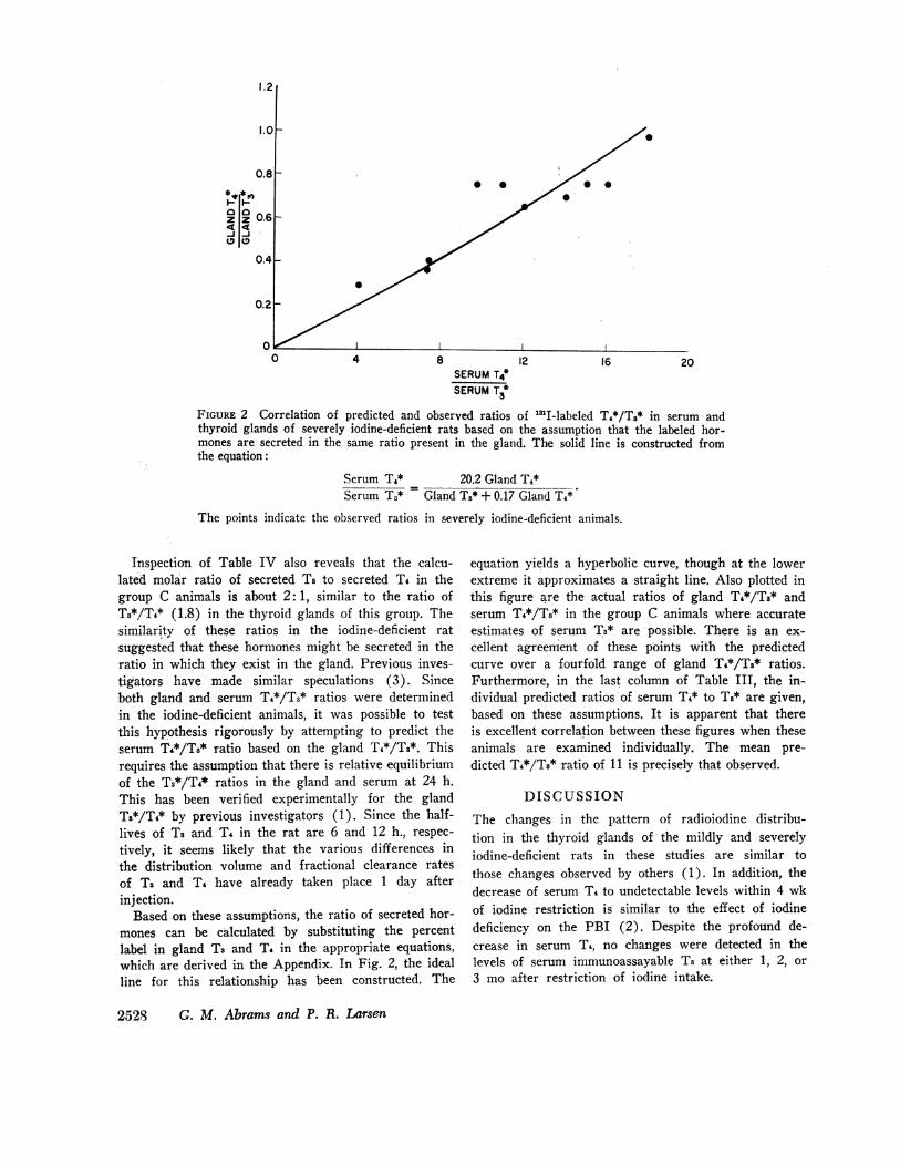
FIGURE 2 Correlation of predicted and observed ratios of 'I-labeled T4*/T3* in serum andthyroid glands of severely iodine-deficient rats based on the assumption that the labeled hor-mones are secreted in the same ratio present in the gland. The solid line is constructed fromthe equation:
Serum T4* 20.2 Gland T4*Serum T3* = Gland Ts* + 0.17 Gland T4*
The points indicate the observed ratios in severely iodine-deficient animals.
Inspection of Table IV also reveals that the calcu-lated molar ratio of secreted Ts to secreted T4 in thegroup C animals is about 2: 1, similar to the ratio ofT3*/T4* (1.8) in the thyroid glands of this group. Thesimilarity of these ratios in the iodine-deficient ratsuggested that these hormones might be secreted in theratio in which they exist in the gland. Previous inves-tigators have made similar speculations (3). Sinceboth gland and serum T4*/T3* ratios were determinedin the iodine-deficient animals, it was possible to testthis hypothesis rigorously by attempting to predict theserum T4*/Ti* ratio based on the gland T4*/Ts*. Thisrequires the assumption that there is relative equilibriumof the Ts*/T4* ratios in the gland and serum at 24 h.This has been verified experimentally for the glandTs*/T4* by previous investigators (1). Since the half-lives of Ts and T4 in the rat are 6 and 12 h., respec-tively, it seems likely that the various differences inthe distribution volume and fractional clearance ratesof Ts and T4 have already taken place 1 day afterinjection.
Based on these assumptions, the ratio of secreted hor-mones can be calculated by substituting the percentlabel in gland T3 and T. in the appropriate equations,which are derived in the Appendix. In Fig. 2, the idealline for this relationship has been constructed. The
equation yields a hyperbolic curve, though at the lowerextreme it approximates a straight line. Also plotted inthis figure are the actual ratios of gland T4*/Ts* andserum T4*/T8* in the group C animals where accurateestimates of serum T3* are possible. There is an ex-cellent agreement of these points with the predictedcurve over a fourfold range of gland T4*/T8* ratios.Furthermore, in the last column of Table III, the in-dividual predicted ratios of serum T4* to Ts* are given,based on these assumptions. It is apparent that thereis excellent correlation between these figures when theseanimals are examined individually. The mean pre-dicted T4*/Ts* ratio of 11 is precisely that observed.
DISCUSSIONThe changes in the pattern of radioiodine distribu-tion in the thyroid glands of the mildly and severelyiodine-deficient rats in these studies are similar tothose changes observed by others (1). In addition, thedecrease of serum T4 to undetectable levels within 4 wkof iodine restriction is similar to the effect of iodinedeficiency on the PBI (2). Despite the profound de-crease in serum T4, no changes were detected in thelevels of serum immunoassayable T3 at either 1, 2, or3 mo after restriction of iodine intake.
2528 G. M. Abrams and P. R. Larsen

These data appear to be at variance with the resultsof a previous systematic study of this problem. Hen-inger and Albright calculated serum T3 and T4 valuesin control and iodine-deficient rats using equilibriumlabeling and iodine specific activity (18). In normalrats, the serum Ts was estimated to be 43 ng/100 ml,similar to our control values. In animals receivingiodine-deficient diet for 2 mo, a serum T3 concentrationof 101 ng/100 ml was calculated. However, determi-nation of the iodine specific activity in the low-iodineanimals depended on accurate estimates of iodine at verylow levels in the diet. Whether or not the differentresults obtained might be due to an overestimation ofthe dietary iodine remains to be determined.
Volpert and Werner reported values of 19-41 ng/100ml in seven samples of serum from rats on low iodinediet for 4-5 wk as opposed to levels of 55 to 60 ng/100ml in animals receiving a normal diet (5). Nejad,Bollinger, Mitnick, and Reichlin have recently reportedno significant change in T3 values in rats fed a lowiodine diet for an unspecified period of time while T4levels decreased to less than 25% of control (19). How-ever, the estimates of normal T3 concentrations in ratserum in these studies (16 ng/100 ml) were substan-tially lower than those we have obtained, for reasonsnot immediately apparent.
It has been previously speculated by Gross and Pitt-Rivers that T4 might have to undergo deiodination toT3 in order to be metabolically active and that, there-fore, T4 might act primarily as a precursor of theactive thyroid hormone, Ts (20). Oppenheimer,Schwartz, and Surks have demonstrated that T4-to-T3conversion occurs in the rat and that this processcan be inhibited by propylthiouracil (16, 21). In orderto maintain hepatic mitochondrial alpha glycerophos-phate dehydrogenase at normal levels in the propylthi-ouracil-treated animals, 2.5 times the usual replace-ment dose of T4 had to be given. This also restored thenet T3 production from T4 to normal levels. If, as thesestudies suggest, T4 must be converted to Ts to exert itsmetabolic effect, feedback regulation of the thyroid-pituitary-hypothalamic axis would most likely be de-pendent on tissue (pituitary) T3 levels. The presenceof specific binding sites for T3 but not for T4 in thenuclei of pituitary cells provides further support forthis concept (22). The T3 present in the pituitary couldbe derived either from T4 or from the thyroid glandvia direct secretion. One would anticipate under thesecircumstances that the normal homeostatic mechanismin the rat would then operate to maintain Ts produc-tion constant regardless of the source of the hormone.
Analysis of data in Tables III and IV suggests thatthese animals compensate for low iodine uptake by ashift from T4 as the primary source of T3, to the thyroid
gland itself. Our data further indicate that this markedchange in the ratio of secreted hormone results fromincreases in the molar ratio of T3/T4 in iodine-deficientthyroglobulin. Whether this change in thyroglobulincomposition is a result of increased thyroid-stimulatinghormone (TSH) stimulation or decreased iodination,or both, remains to be determined. However, our analy-sis of the labeled hormone ratios in the serum is con-sistent with the simple assumption that they are secretedin the proportion in which they exist in the gland. Asimilar process may also occur in the normal rat,though the calculated molar ratio of secreted T4 to T3is 12: 1 (Table IV) and the ratio in the thyroid glandis 8-9/1.
As indicated above, iodine-deficient rats have elevatedTSH levels that lead to the goiter and increased radio-active iodine uptake characteristic of this condition. Itmay be argued that such animals are, by definition, hy-pothyroid. Nevertheless, as previously pointed out, theyappear to be "clinically" euthyroid (2-4). Whether, infact, these animals are euthyroid in all respects awaitsthe results of more careful in vitro studies of tissuethyroid status. If it can be demonstrated that suchanimals are euthyroid and T3 levels remain normal withmarkedly decreased serum T4, the results would be con-sistent with the interpretation that the TSH elevationin these animals is a compensatory response designedto maintain the animal in a euthyroid state. In addi-tion, it would support the above-mentioned hypothesisthat T4, in the normal rat, is primarily a precursor ofT3. Other investigators, as well as ourselves, haveobserved that normal serum T3 concentrations may beassociated with substantial TSH elevations and lowserum T4 levels in patients with primary thyroid dis-ease or after radioactive iodine treatment for hyper-thyroidism (23-25). It is possible that some of thesepatients may have "intrathyroidal" iodine deficiency dueto acquired defects in iodide trapping or organificationwith a similar compensatory response.
Loewenstein and Wollman have shown that prolongedperiods (>99 days) are required for complete equili-bration of isotopic iodine into the total body iodine poolin the rat (26). For this reason accurate measurementof thyroidal and serum T3 and T4 by equilibrium label-ing techniques is tedious. Inspection of Tables I andII shows that in groups A and B, multiplication of thefraction of T4* by the 12I content results in calculatedT4 concentrations that are considerably less than theimmunoassayable estimates. This presumably resultslargely from the incomplete equilibration of the isotopewith the iodine pool and is in agreement with earlierstudies (14). Nevertheless, immunoassayable thyroidT4 content in the control group agrees well with thevalues of about 350 ng T4/mg wet wt in rat thyroid
Triiodothyronine and Thyroxine in Iodine-Deficient Rats 2529

that can be calculated from recent ['I] T4 measure-ments (14). The fact that acute labeling with 'Iresults in the same Ts/T4 ratios as the immunoassaydetermination indicates that, in spite of the functionalheterogeneity of thyroid gland iodine previously shownby many investigators, the Ts/T4 ratio of acutely formedhormone and total thyroidal iodothyronine are essen-tially the same. Since the immunoassay does not requireseparation of T8 and T4 with its attendant artifacts, ithas obvious advantages in studies of this type andeven more in the study of human thyroid tissue thatcannot be chronically labeled.
Whether or not the rat has a mechanism for con-servation of iodine efficient enough to allow a normalTs to be maintained indefinitely is not known. Our pre-liminary evidence suggests that after prolonged restric-tion of iodine intake, T3 levels begin to decrease. Moreextensive studies are currently under way to confirmthis observation.
APPENDIX
The T4 or T3 metabolized in 24 h can be expressed as follows:
Total Tx = [Tx] X lrx X KTX
where [Tx] is the concentration of iodothyronine (molesper milliliter), VT. is the volume of distribution per 100 gbody wt and KTZ is the mean daily fractional removal rate ofT3 or T4.
Under steady-state conditions, the T4 cleared daily equalsthe T4 secreted by the thyroid. It has been shown in the ratthat approximately 17% of the T4 is metabolized via conver-sion to T3 (16). Thus, thyroidal T3 secretion can be expressedas the difference between the metabolized T3 and the quantityderived from T4 or:
Secreted T3
= ([T3] X VT3 X KT3) -0.17([T4] X VT4XK T4)Substitution of experimentally measured values for kineticconstants gives (17):
Secreted T4/100 g/day= [T4] X 16.4 X 1.032
Secreted T3/100 g/day= ([T3] X 165 X 2.07) -0.17([T4] X 16.4 X 1.032)
If T4* and T3* are secreted in the same ratio as they existwithin the gland and the peripheral pool is subsequentlylabeled with 1311, then when the ratios of T4* to T3* in thegland and in the serum are in relative equilibrium:
Secreted T4* Gland T4*Secreted T3* Gland T{*
(T4* X 16.4 X 1.032)T3* X 165 X 2.07 - 0.17
X (1T4* X 16.4 X 1.032). (1)
Eq. (1) reduces tco
Gland f4* 16.9 T4*Gland T3* 340.96 T3* - 2.88 T4*- (2)
Eq. (2) can be solved for the ratio of labeled iodothyroninesin the serum
T4* 20.2 Gland T4*T,* Gland T3* + 0.17 Gland T*- (3)
ACKNOWLEDGMENTSThe authors would like to acknowledge the careful techni-cal assistance of Ms. Darina Sipula and Jitka Dockalovaand the expert secretarial help of Ms. Barbara Brenneman.
This work was supported by Grant AM14283 from theU. S. Public Health Service and Grant 0-20 from theHealth Research and Services Foundation of Pittsburgh.
REFERENCES1. Studer, H., and M. A. Greer. 1966. The Regulation of
Thyroid Function in Iodine Deficiency. Hans Huber,Bern, Switzerland.
2. Studer, H., and M. A. Greer. 1965. A study of themechanisms involved in the production of iodine-cle-ficiency goiter. Acta Endocrinol. 49: 610.
3. Greer, M. A., Y. Grimm, and H. Studer. 1968. Quali-tative changes in the secretion of thyroid hormones in-duced by iodine deficiency. Endocrinology. 83: 1193.
4. Silva, E. 1972. Disposal rates of thyroxine and triiodo-thyronine in iodine-deficient rats. Endocrinology. 91:1430.
5. Volpert, E. M., and S. C. Werner. 1972. Serum tri-iodothyronine concentration in the iodine-deficient rat.Am. J. Anat. 135: 187.
6. Benotti, J., N. Benotti, S. Pino, and J. Gardyna. 1965.Determination of total iodine in urine, stool, diets, andtissue. Clin. Chem. 11: 932.
7. Inoue, K., and A. Taurog. 1967. Digestion of 'lI-labeledlthyroid tissue with maximum recovery of 1"I-iodothyro-nines. Endocrinology. 81: 319.
8. Larsen, P. R., K. Yamashita, A. Dekker, and J. B.Field. 1973. Biochemical observations in functioninghuman thyroid adenomas. J. Clin. Endocrinol. Metab.36: 1109.
9. Larsen, P. R. 1971. Technical aspects of the estimationof triiodothyronine in human serum: evidence of con-version of thyroxine to triiodothyronine during assay.Metabolism. 20: 609.
10. Larsen, P. R. 1972. Direct immunoassay of triiodothy-ronine in human serum. J. Clin. Invest. 51: 1939.
11. Larsen, P. R., J. Dockalova, D. Sipula, and F. M. WVu.1973. Immunoassay of thyroxine in unextracted humanserum. J. Clin. Endocrinol. Metab. In press.
12. Sterling, K., D. Bellabarba, E. S. Newman, and M. A.Brenner. 1969. Determination of triiodothyronine con-centration in human serum. J. Clin. Invest. 48: 1150.
13. Inoue, K., and Taurog, A. 1968. Acute and chroniceffects of iodide on thyroid radioiodine metabolism iniodine-deficient rats. Endocrinology. 83: 279.
14. Lamas, L., and G. M. de Escobar. 1972. Iodoamino aciddistribution in the thyroids of rats on different iodineintakes and with normal plasma protein-bound iodine.Acta Endocrinol. 69: 473.
2530 G. M. Abrams and P. R. Larsen
15. Braverman, L. E., S. H. Ingbar, and K. Sterling. 1970.Conversion of thyroxine (T4) to triiodothyronine (Ta)in athyreotic human subjects. J. Clin. Invest. 49: 855.
16. Schwartz, H. L., M. I. Surks, and J. H. Oppenheimer.1971. Quantitation of extrathyroidal conversion of L-thyroxine to 3,5,3'-triiodo-L-thyronine in the rat. J.( lii. Invest. 50: 1124.
17. Oppeniheimer, J. 11., H1. L. Schwartz, I1. Shapiro, G.IBernsteiin, M. Martinez, and M. I. Surks. 1970. I)iffer-ences in primary cellular factors in influencing themetabolism and distribution of 3,5,3'-triiodothyronineand L-thyroxine. J. Clin. Invest. 49: 1016.
18. Heninger, R. WV., and E. C. Albright. 1966. Effect ofiodine deficiency on iodine-containing compounds of rattissues. Endocrinology. 79: 309.
19. Nejad, I. F., J. A. Bollinger, M. Mitnick, and S.Reichlin. 1973. Importance of T3 secretion in alteredstates of thyroid function in the rat: cold exposure,subtotal thyroidectomy, and hypophysectomy. Trans.1lssoc. .4Am1. Physicians Phila. 85: 295.
20. Gross, J., and R. Pitt-Rivers. 1954. Thyroid hormonephysiology and biochemistry. II. Triiodothyronine in re-
lation to thyroid physiology. Recent Prog. Hormn. Rcs.10: 109.
21. Oppenheimer, J. H., H. L. Schwartz, and M. I. Surks.1973. Propylthiouracil inhibits the conversion of L-thy-roxine to L-triiodothyronine. J. Clin. Invest. 51: 2493.
22. Schadlow, A. R., M. I Surks, H. L. Schwartz, andJ.. 11. Oppenheimer. 1972. Specific trii(eothyroninic-Ibi(lindn- sites ill the anterior pituitary of thl rat. Sci-ence (UVash. 1). C.). 176: 1252.
23. Sterling, K., M. A. Brenner, E. S. Newman, Xv. 1).Odell, and D. Bellabarba. 1971. The significance of tri-iodothyronine (T3) in maintenance of euthyroid statusafter treatment of hyperthyroidism. J. Clin. Eindocrinol.33: 729.
24. Lieblich, J., and R. D. Utiger. 1972. Triiodothyronineradioimmunoassay. J. Clint. Invest. 51: 157.
25. Larsen, P. R. 1972. Triiodothyronine: reviewe of recentstudies of its physiology and pathoplhysiology in man.Metab. (Clin. Exp.). 21: 1073.
26. Loewenstein, J. E., and S. H. Wollman. 1967. Kinetics ofequilibrium-labeling of the rat thlyroid gland With 12a-l.Endocrinology. 81: 1063.
Triiodothyronine and Thyroxine in Iodine-Deficient Rats 2531