treatment strategies in colorectal cancer patients with ... protocol v.7.2 11-01-2018_ clean.pdf ·...
TRANSCRIPT

Treatment strategies in colorectal cancer patients with initially unresectable
liver-only metastases CAIRO5
a randomised phase 3 study of the Dutch Colorectal Cancer Group
(DCCG)
EudraCT number: 2013-005435-24 Principal Investigators: Prof. dr. C.J.A. Punt - dept. of Medical Oncology, AMC Amsterdam Prof. dr. T.M. van Gulik - dept. of Surgery, AMC Amsterdam Coordinating Pathologist: dr. N.C.T. van Grieken, dept. of Pathology, VUmc Amsterdam Coordinating Radiologists: Dr. K. van Lienden, Dr. M Engelbrecht, dept. Radiology, AMC Amsterdam Trial coordinator: Drs. J. Huiskens, AMC Amsterdam (until May 2017) Drs. K. Bolhuis, AMC Amsterdam (from May 2017) CAIRO5 phone: +31 6 20581694 Data management, randomisation and logistics: IKNL clinical research department [email protected]; phone (+31) 088 - 234 6500 Statistician: Dr. H. van Tinteren, dept. of Biometrics, NKI/AvL Amsterdam Translational research coordinators: Prof. dr. C.J.A. Punt, Prof.dr. T.M. van Gulik, Prof.dr. G.A. Meijer Study sponsor: Dutch Colorectal Cancer Group (DCCG). The DCCG is financially supported by Roche and Amgen for the financing of the study.
Version 7.1, December 1th 2017

2
DCCG CAIRO5 study, protocol version 7.2 (11-01-2018)
Index Synopsis ................................................................................................................................... 41 Introduction and rationale .................................................................................................. 7
Secondary liver resections after induction systemic treatment .................................. 71.1
Induction treatment with chemotherapy plus either anti-EGFR antibodies or 1.2
bevacizumab ......................................................................................................................... 9 Selection of patients for anti-EGFR therapy ............................................................ 101.3
Choice of chemotherapy regimen in induction treatment ......................................... 101.4
Hepatotoxicity of induction treatment ....................................................................... 121.5
Conclusions ............................................................................................................. 121.6
2 Objectives ....................................................................................................................... 153 Study design ................................................................................................................... 154 Study population ............................................................................................................. 16
Inclusion criteria ....................................................................................................... 164.1
Exclusion criteria ...................................................................................................... 164.2
5 Evaluations at baseline and follow-up ............................................................................. 17 Definition of sidedness primary tumor: ..................................................................... 175.1
Evaluations at baseline ............................................................................................ 175.2
Evaluations at follow up ........................................................................................... 175.3
6 Assessment of RAS and BRAF mutation status ............................................................. 187 Patient registration, randomisation procedure ................................................................ 188 Panel procedure and evaluation ..................................................................................... 18
Panel procedure ....................................................................................................... 188.1
Radiologist review, including specifications of radiological assessment .................. 198.2
Surgical review, including specifications of surgical assessment ............................ 198.3
9 Study treatment: systemic therapy .................................................................................. 20 Doses and schedules ............................................................................................... 209.1
Change of treatment for reasons of toxicity or patient refusal within 6 months ....... 219.2
Treatment duration after 6 months without disease progression ............................. 229.3
Treatment after first progression of disease ............................................................ 229.4
Dose modifications for toxicity, reporting of SAE ..................................................... 239.5
Evaluation of treatment ............................................................................................ 239.6
Continuation of treatment after liver surgery ............................................................ 249.7
10 Study treatment: surgery ................................................................................................. 25 Criteria for resectability, surgical procedures ........................................................... 2510.1
Definitions of R0 and R1 resection .......................................................................... 2610.2
Use of repeat hepatectomy, resection of new extrahepatic metastases .................. 2610.3
Local treatment (surgery, radiotherapy) for primary tumor in patients with 10.4
synchronous metastases .................................................................................................... 26 Complications after liver surgery .............................................................................. 2610.5
11 Follow-up and disease evaluation during and after protocol treatment ........................... 2612 Study flow charts ............................................................................................................. 2713 Informed consent procedure ........................................................................................... 29

3
DCCG CAIRO5 study, protocol version 7.2 (11-01-2018)
14 Reporting of SAE ............................................................................................................ 29 Section 10 WMO event ............................................................................................ 2914.1
AEs, SAEs and SUSARs ......................................................................................... 2914.2
Annual safety report ............................................................................................. 313214.3
Data Safety Monitoring Board .................................................................................. 3214.4
15 Study statistics, sample size, planned analyses ............................................................. 32 Sample size considerations - primary endpoint ....................................................... 3215.1
Interim analysis ........................................................................................................ 3315.2
Subgroup analysis ................................................................................................... 3515.3
Secondary endpoints ............................................................................................... 3615.4
16 Translational research ..................................................................................................... 36 Central pathology review ......................................................................................... 3616.1
General aim ............................................................................................................. 3616.2
Materials and Methods ............................................................................................. 3716.3
Biomarker validation ................................................................................................ 3716.4
17 Ethical and legal aspects ................................................................................................ 38 Independent physician ............................................................................................. 3917.1
Insurance ................................................................................................................. 3917.2
18 Quality ............................................................................................................................. 39 Monitoring ................................................................................................................ 3918.1
Quality assurance .................................................................................................... 3918.2
19 Public disclosure and publication policy .......................................................................... 4020 References ...................................................................................................................... 4121 Appendix I: CAIRO5 study design ................................................................................... 4322 Appendix II: Flow chart for registration, randomisation, pathology and central panel
review ..................................................................................................................................... 44

4
DCCG CAIRO5 study, protocol version 7.2 (11-01-2018)
Synopsis
Study title Treatment strategies in colorectal cancer patients with initially
unresectable liver-only metastases: CAIRO5, a randomised phase 3
study of the Dutch Colorectal Cancer Group (DCCG)
Study phase Randomised phase 3
Background Colorectal cancer patients with initially unresectable liver-only
metastases may be cured after downsizing of metastases by induction
systemic therapy. However, the optimal induction regimen has not been
defined, and no consensus exist on criteria for resectability.
Objectives To determine the median progression-free survival (PFS) upon
induction systemic treatment in colorectal cancer patients with initially
unresectable liver-only metastases, stratified by RAS and BRAF tumor
mutation status and primary tumor location.
Study design Colorectal cancer patients with initially unresectable liver-only
metastases, as prospectively confirmed by an expert panel according to
predefined criteria, and tested for RAS (KRAS exon 2, 3 en 4 and
NRAS exon 2 and 3) and BRAF tumor mutation status. Patients with
RAS and BRAF wildtype left-sided primary colorectal tumors will be
randomised between doublet chemotherapy (FOLFOX or FOLFIRI)
plus either bevacizumab or panitumumab, and patients with RAS or
BRAF mutant tumors and/or right sided primary colon tumors will be
randomised between doublet chemotherapy (FOLFOX or FOLFIRI)
plus bevacizumab and triple chemotherapy (FOLFOXIRI) plus
bevacizumab.
Patient imaging will be reviewed for resectability by a central panel,
consisting of at least one radiologist and three surgeons every
assessment. Central panel review will be performed prior to
randomisation as well as during treatment, as described in the protocol.
For study design see also appendix I
Stratification
parameters
Patients will be stratified for resectability of liver metastases (potentially
resectable versus permanently unresectable), serum LDH obtained ≤ 4
weeks (normal vs abnormal), use of irinotecan- versus oxaliplatin-
containing regimen and reporting institute.
Study endpoints Primary endpoints: median progression-free survival (PFS)
Secondary endpoints: R0/1 resection rates, median overall survival,
response rate, toxicity, pathological complete response rate (pCR) of
resected lesions, postoperative morbidity, and correlation of baseline
and follow-up evaluation by the panel with outcome.
Main criteria for
inclusion
- Histologically proven colorectal cancer
- Unresectable metastases confined to the liver according to CT scan,
obtained ≤ 4 weeks prior to randomisation. Unresectability is

5
DCCG CAIRO5 study, protocol version 7.2 (11-01-2018)
confirmed by the panel. Patients with small (≤ 1 cm) extrahepatic
lesions that are not clearly suspicious of metastases are eligible.
- RAS and BRAF mutation status known
- Status WHO performance status 0-1 (Karnofsky performance status
≥ 70)
- Age ≥ 18 years
- No contraindications for liver surgery
- In case of primary tumor in situ: tumor should be resectable
- In case of resected primary tumor: adequate recovery from surgery
- Adequate organ functions, as determined by normal bone marrow
function (Hb 6.0 mmol/L, absolute neutrophil count 1.5 x 10⁹/L,
platelets 100 x 109/L), renal function (serum creatinine ≤ 1.5x ULN
and creatinine clearance, Cockroft formula, 30 ml/min), liver
function (serum bilirubin ≤ 2 x ULN, serum transaminases ≤ 5x ULN)
- Life expectancy > 12 weeks
- Expected adequacy of follow-up
- Written informed consent
Main criteria for
exclusion
- Extrahepatic metastases (extrahepatic lesions of ≤ 1 cm that are not
clearly suspicious for metastases not included)
- Unresectable primary tumor
- Serious comorbidity or any other condition preventing the safe
administration of study treatment (including both systemic treatment
and surgery)
- Major cardiovascular events (myocardial infarction, severe/unstable
angina, congestive heart failure, CVA) within 12 months before
registration
- Uncontrolled hypertension, or unsatisfactory blood pressure control
with ≥3 antihypertensive drugs
- Previous systemic treatment for metastatic disease; previous
adjuvant treatment is allowed if completed ≥ 6 months prior to
registration
- Previous surgery for metastatic disease
- Previous intolerance of study drugs in the adjuvant setting
- Pregnant or lactating women
- Second primary malignancy within the past 5 years with the
exception of adequately treated in situ carcinoma of any organ or
basal cell carcinoma of the skin
- Any concomitant experimental treatment.
Treatment Patients with RAS and BRAF wildtype left-sided primary tumors will be
randomised between doublet chemotherapy (FOLFOX or FOLFIRI)
plus either bevacizumab or panitumumab. The choice between
FOLFOX or FOLFIRI is to the discretion of the local investigator,
however, the treatment is restricted to regimens that are specified in the
6
DCCG CAIRO5 study, protocol version 7.2 (11-01-2018)
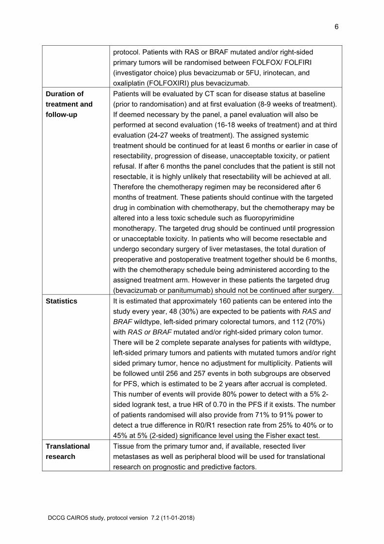
protocol. Patients with RAS or BRAF mutated and/or right-sided
primary tumors will be randomised between FOLFOX/ FOLFIRI
(investigator choice) plus bevacizumab or 5FU, irinotecan, and
oxaliplatin (FOLFOXIRI) plus bevacizumab.
Duration of
treatment and
follow-up
Patients will be evaluated by CT scan for disease status at baseline
(prior to randomisation) and at first evaluation (8-9 weeks of treatment).
If deemed necessary by the panel, a panel evaluation will also be
performed at second evaluation (16-18 weeks of treatment) and at third
evaluation (24-27 weeks of treatment). The assigned systemic
treatment should be continued for at least 6 months or earlier in case of
resectability, progression of disease, unacceptable toxicity, or patient
refusal. If after 6 months the panel concludes that the patient is still not
resectable, it is highly unlikely that resectability will be achieved at all.
Therefore the chemotherapy regimen may be reconsidered after 6
months of treatment. These patients should continue with the targeted
drug in combination with chemotherapy, but the chemotherapy may be
altered into a less toxic schedule such as fluoropyrimidine
monotherapy. The targeted drug should be continued until progression
or unacceptable toxicity. In patients who will become resectable and
undergo secondary surgery of liver metastases, the total duration of
preoperative and postoperative treatment together should be 6 months,
with the chemotherapy schedule being administered according to the
assigned treatment arm. However in these patients the targeted drug
(bevacizumab or panitumumab) should not be continued after surgery.
Statistics It is estimated that approximately 160 patients can be entered into the
study every year, 48 (30%) are expected to be patients with RAS and
BRAF wildtype, left-sided primary colorectal tumors, and 112 (70%)
with RAS or BRAF mutated and/or right-sided primary colon tumor.
There will be 2 complete separate analyses for patients with wildtype,
left-sided primary tumors and patients with mutated tumors and/or right
sided primary tumor, hence no adjustment for multiplicity. Patients will
be followed until 256 and 257 events in both subgroups are observed
for PFS, which is estimated to be 2 years after accrual is completed.
This number of events will provide 80% power to detect with a 5% 2-
sided logrank test, a true HR of 0.70 in the PFS if it exists. The number
of patients randomised will also provide from 71% to 91% power to
detect a true difference in R0/R1 resection rate from 25% to 40% or to
45% at 5% (2-sided) significance level using the Fisher exact test.
Translational
research
Tissue from the primary tumor and, if available, resected liver
metastases as well as peripheral blood will be used for translational
research on prognostic and predictive factors.

7
DCCG CAIRO5 study, protocol version 7.2 (11-01-2018)
1 Introduction and rationale
Approximately 50% of patients with colorectal cancer (CRC) will develop metastases,
and approximately 25% will present with distant metastases at diagnosis. Colorectal cancer
disseminates predominantly to the liver. The 5-year overall survival rates in patients with
metastatic CRC who participated to clinical trials is currently around 20%. [1] This result has
clearly improved during the past 2 decades, and is predominantly due to the increased use of
surgical resections of metastases and the increased efficacy of systemic drugs.
Fluoropyrimidine-containing chemotherapy plus the anti-VEGF antibody bevacizumab is
currently considered to be the standard 1st-line treatment in metastatic CRC. [2-5] In patients
with RAS and BRAF wildtype left-sided primary colorectal tumors, chemotherapy plus
cetuximab or panitumumab (anti-EGFR) is a useful alternative. [6-8] Randomised phase 3
studies on a direct comparison between chemotherapy plus either bevacizumab or anti-
EGFR antibodies in unselected (i.e. not restricted to liver-only disease) metastatic CRC
patients are either ongoing (CALGB 80405) or preliminary results have been presented
(FIRE-3 and PEAK trials, to be discussed later).
In patients with resectable liver metastases at diagnosis, a radical resection of these
metastases is the first choice of treatment which results in 5-year survival rates in the order
of 25-40%. [9] However, only a minority of patients present with resectable metastases. The
main reasons for unresectability are liver involvement which is too extensive, or involvement
of non-resectable structures. Resection of liver metastases in patients who also have
extrahepatic metastases is a matter of debate, and, if done at all, is usually restricted to
patients with limited extrahepatic metastases. Evidence for the benefit of induction
chemotherapy with the objective to improve resectability rates was already established in
1996, when it was shown that initially unresectable metastases could become resectable
(further defined as secondary surgery) after downsizing by chemotherapy. [10] Currently,
most phase 2 and 3 studies in metastatic CRC present data on the rate of secondary
resections in the subgroup of patients with metastases confined to the liver. However, this
almost invariably concerns unplanned retrospective analyses. The rate of secondary surgery
in phase 3 studies with unselected metastatic CRC patients (i.e. with metastases not
confined to the liver) is usually less than 10%. However, results on this outcome are greatly
confounded by the variable use of liver resections among different hospitals and countries.
Furthermore, these patients were not specifically screened for extrahepatic disease since the
outcome in patients with liver-only metastases usually was not a prospectively defined
objective of the study.
Secondary liver resections after induction systemic treatment 1.1
Data from a single institution by Adam et al. [11] have shown that of 1104 patients with
metastases confined to the liver, 12.5% of patients became eligible for secondary surgery,
and that these patients had a 5-year survival rate of 33%. In another retrospective analysis of
184 patients who underwent radical secondary resection of initially unresectable CRC liver
metastases after downsizing by chemotherapy, the 5- and 10-year survival rates were 33%
and 27%, respectively. After a follow-up of ≥5 years, 16% of 148 patients was considered to

8
DCCG CAIRO5 study, protocol version 7.2 (11-01-2018)
be cured [12]. However, as in most if not all retrospective series, the relative contribution of
systemic treatments to survival was not evaluated.
As is the case for primary surgery for resectable liver metastases, the benefit of
secondary surgery has not been evaluated in prospective randomised studies. However,
given the consistent data from large series, there is little doubt that a radical resection of liver
metastases (primary or secondary) prolongs survival. Indeed, in the liver survey database
the survival benefits of primary and secondary surgery were shown to be in the same range.
[13] The 3, 5, and 10-year survival rates for primary and secondary resections were 63% and
53%, 45% and 33%, and 28% and 19%, respectively. These outcomes are better than have
been reported for systemic therapy alone. Again, formal randomised trials on this topic have
not been performed, and most likely never will be performed given these results.
Although a radical (R0) resection with tumor-free margins should be attempted in all
patients, long-term survival results have been shown for R1 resections as well. [14]
Therefore, R1 resections appear to be a relevant surgical outcome as well. An important issue regarding the optimal strategy for this group of patients is that the published series differ in their selection of patients as well as in their definition of resectability. This refers to the maximal size and number of metastases, the use of more extensive surgical procedures such as portal vein embolization and 2-stage resections, and the number of organs involved since some series also include patients with resections of metastases in more than one site. This implies that cross-study comparisons are not valid. Multivariate analyses have identified the size, the number, and the pathological response to induction systemic treatment as independent predictors for survival [12]. A complicating factor is the lack of general consensus on the criteria for resectability, which is nicely illustrated by the recent CELIM study [15]. In this randomised phase 2 study with CRC patients with unresectable liver-only metastases, the CT scans of the liver before and after systemic therapy were retrospectively reviewed in a blinded way by a panel of 7 surgical experts. The panel was asked to vote for one of the following options: initial resection or surgical exploration, initial systemic therapy with the possibility of secondary resection, or permanently unresectable. There was considerable variation in the voting of this panel, with even completely opposing views in 7% of cases. Moreover, one-third of the patients were considered by the reviewers to be resectable at baseline and therefore in retrospect ineligible for the study. A second important issue is that in the discussions on the optimal strategy that should lead to the downsizing of metastases to allow secondary resections, the assumption is usually made that response rate is a surrogate marker for resection rate. Indeed this hypothesis was supported by a retrospective analysis. [16] However, a downsizing in the number of metastases leading to a technically feasible secondary resection may translate into a worse outcome compared to a downsizing in the size of metastases. This is supported by the observation that more than 80% of liver metastases in complete radiological remission by systemic treatment still contain viable tumor cells. [17] These data strongly suggest that response rate is unlikely to accurately predict the clinical outcome in this patient category, and support the timing of a resection as soon as this appears to be feasible (i.e. not to wait until metastases may have completely regressed). These data also support to resect all sites where lesions in complete remission were located. Lastly, the issue of response rate is further complicated by the observations that the addition of bevacizumab to induction chemotherapy may rather increase the pathological than the objective (RECIST) response of
9
DCCG CAIRO5 study, protocol version 7.2 (11-01-2018)
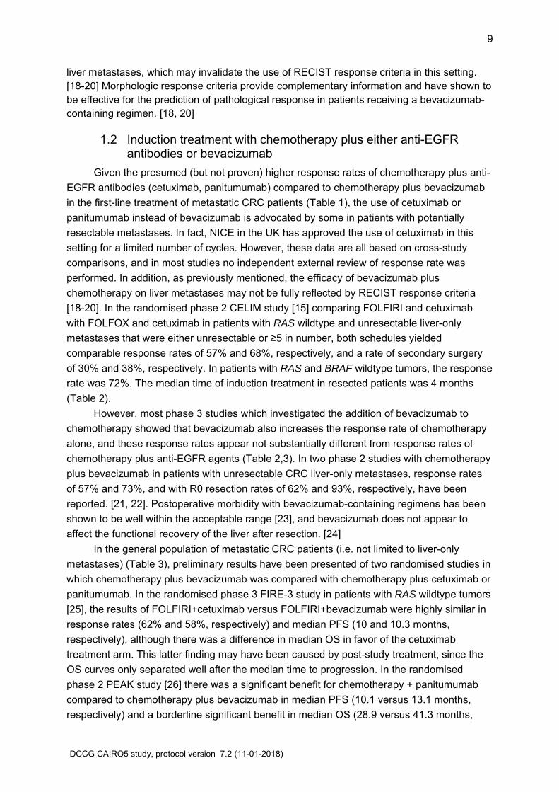
liver metastases, which may invalidate the use of RECIST response criteria in this setting. [18-20] Morphologic response criteria provide complementary information and have shown to be effective for the prediction of pathological response in patients receiving a bevacizumab-containing regimen. [18, 20]
Induction treatment with chemotherapy plus either anti-EGFR 1.2antibodies or bevacizumab
Given the presumed (but not proven) higher response rates of chemotherapy plus anti-
EGFR antibodies (cetuximab, panitumumab) compared to chemotherapy plus bevacizumab
in the first-line treatment of metastatic CRC patients (Table 1), the use of cetuximab or
panitumumab instead of bevacizumab is advocated by some in patients with potentially
resectable metastases. In fact, NICE in the UK has approved the use of cetuximab in this
setting for a limited number of cycles. However, these data are all based on cross-study
comparisons, and in most studies no independent external review of response rate was
performed. In addition, as previously mentioned, the efficacy of bevacizumab plus
chemotherapy on liver metastases may not be fully reflected by RECIST response criteria
[18-20]. In the randomised phase 2 CELIM study [15] comparing FOLFIRI and cetuximab
with FOLFOX and cetuximab in patients with RAS wildtype and unresectable liver-only
metastases that were either unresectable or ≥5 in number, both schedules yielded
comparable response rates of 57% and 68%, respectively, and a rate of secondary surgery
of 30% and 38%, respectively. In patients with RAS and BRAF wildtype tumors, the response
rate was 72%. The median time of induction treatment in resected patients was 4 months
(Table 2).
However, most phase 3 studies which investigated the addition of bevacizumab to
chemotherapy showed that bevacizumab also increases the response rate of chemotherapy
alone, and these response rates appear not substantially different from response rates of
chemotherapy plus anti-EGFR agents (Table 2,3). In two phase 2 studies with chemotherapy
plus bevacizumab in patients with unresectable CRC liver-only metastases, response rates
of 57% and 73%, and with R0 resection rates of 62% and 93%, respectively, have been
reported. [21, 22]. Postoperative morbidity with bevacizumab-containing regimens has been
shown to be well within the acceptable range [23], and bevacizumab does not appear to
affect the functional recovery of the liver after resection. [24]
In the general population of metastatic CRC patients (i.e. not limited to liver-only
metastases) (Table 3), preliminary results have been presented of two randomised studies in
which chemotherapy plus bevacizumab was compared with chemotherapy plus cetuximab or
panitumumab. In the randomised phase 3 FIRE-3 study in patients with RAS wildtype tumors
[25], the results of FOLFIRI+cetuximab versus FOLFIRI+bevacizumab were highly similar in
response rates (62% and 58%, respectively) and median PFS (10 and 10.3 months,
respectively), although there was a difference in median OS in favor of the cetuximab
treatment arm. This latter finding may have been caused by post-study treatment, since the
OS curves only separated well after the median time to progression. In the randomised
phase 2 PEAK study [26] there was a significant benefit for chemotherapy + panitumumab
compared to chemotherapy plus bevacizumab in median PFS (10.1 versus 13.1 months,
respectively) and a borderline significant benefit in median OS (28.9 versus 41.3 months,
10
DCCG CAIRO5 study, protocol version 7.2 (11-01-2018)
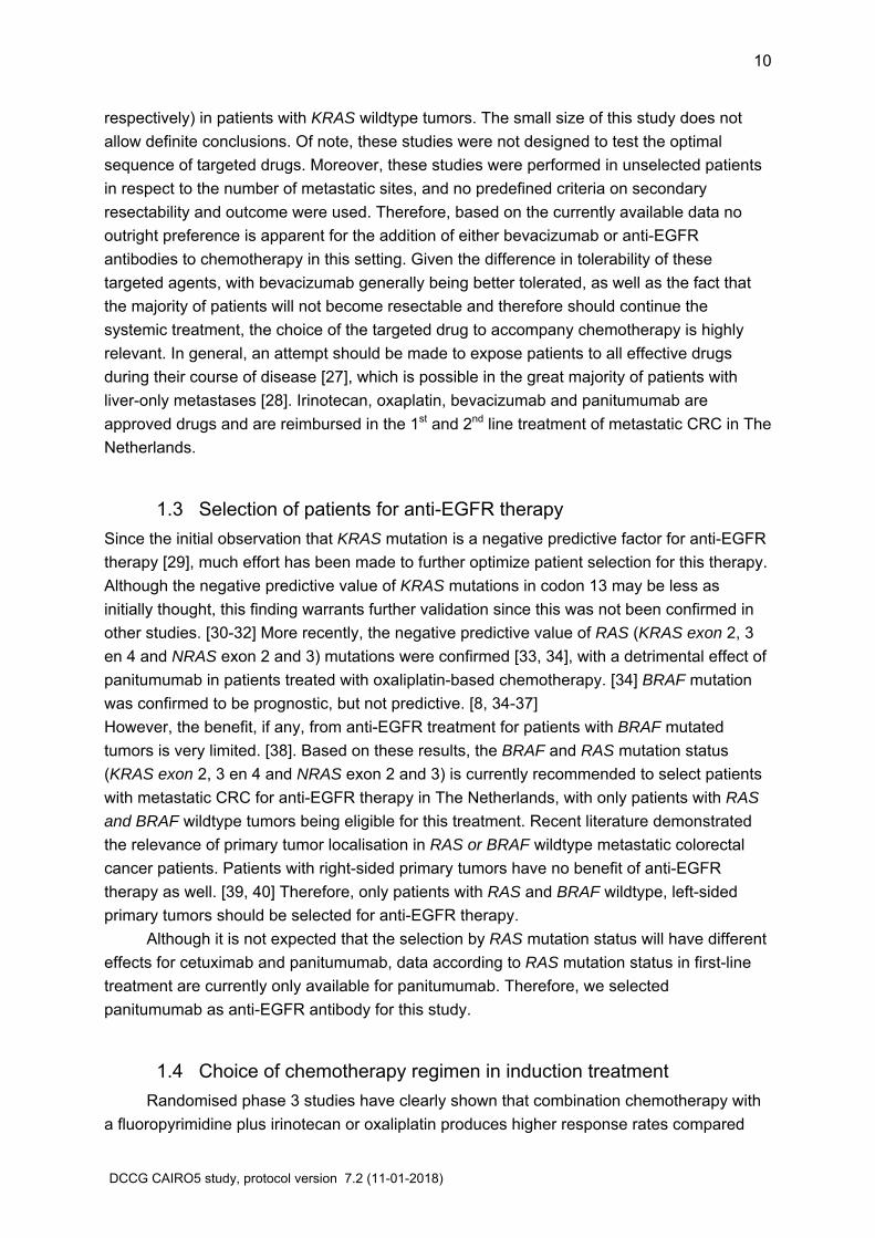
respectively) in patients with KRAS wildtype tumors. The small size of this study does not
allow definite conclusions. Of note, these studies were not designed to test the optimal
sequence of targeted drugs. Moreover, these studies were performed in unselected patients
in respect to the number of metastatic sites, and no predefined criteria on secondary
resectability and outcome were used. Therefore, based on the currently available data no
outright preference is apparent for the addition of either bevacizumab or anti-EGFR
antibodies to chemotherapy in this setting. Given the difference in tolerability of these
targeted agents, with bevacizumab generally being better tolerated, as well as the fact that
the majority of patients will not become resectable and therefore should continue the
systemic treatment, the choice of the targeted drug to accompany chemotherapy is highly
relevant. In general, an attempt should be made to expose patients to all effective drugs
during their course of disease [27], which is possible in the great majority of patients with
liver-only metastases [28]. Irinotecan, oxaplatin, bevacizumab and panitumumab are
approved drugs and are reimbursed in the 1st and 2nd line treatment of metastatic CRC in The
Netherlands.
Selection of patients for anti-EGFR therapy 1.3
Since the initial observation that KRAS mutation is a negative predictive factor for anti-EGFR
therapy [29], much effort has been made to further optimize patient selection for this therapy.
Although the negative predictive value of KRAS mutations in codon 13 may be less as
initially thought, this finding warrants further validation since this was not been confirmed in
other studies. [30-32] More recently, the negative predictive value of RAS (KRAS exon 2, 3
en 4 and NRAS exon 2 and 3) mutations were confirmed [33, 34], with a detrimental effect of
panitumumab in patients treated with oxaliplatin-based chemotherapy. [34] BRAF mutation
was confirmed to be prognostic, but not predictive. [8, 34-37]
However, the benefit, if any, from anti-EGFR treatment for patients with BRAF mutated
tumors is very limited. [38]. Based on these results, the BRAF and RAS mutation status
(KRAS exon 2, 3 en 4 and NRAS exon 2 and 3) is currently recommended to select patients
with metastatic CRC for anti-EGFR therapy in The Netherlands, with only patients with RAS
and BRAF wildtype tumors being eligible for this treatment. Recent literature demonstrated
the relevance of primary tumor localisation in RAS or BRAF wildtype metastatic colorectal
cancer patients. Patients with right-sided primary tumors have no benefit of anti-EGFR
therapy as well. [39, 40] Therefore, only patients with RAS and BRAF wildtype, left-sided
primary tumors should be selected for anti-EGFR therapy.
Although it is not expected that the selection by RAS mutation status will have different
effects for cetuximab and panitumumab, data according to RAS mutation status in first-line
treatment are currently only available for panitumumab. Therefore, we selected
panitumumab as anti-EGFR antibody for this study.
Choice of chemotherapy regimen in induction treatment 1.4
Randomised phase 3 studies have clearly shown that combination chemotherapy with
a fluoropyrimidine plus irinotecan or oxaliplatin produces higher response rates compared

11
DCCG CAIRO5 study, protocol version 7.2 (11-01-2018)
with fluoropyrimidine monotherapy. Therefore combination chemotherapy is the backbone of
systemic treatment when downsizing of metastases is the primary objective. Studies on triple
chemotherapy (5FU+oxaliplatin+irinotecan, FOLFOXIRI) have shown high response rate in
phase 2 studies, but conflicting results on its survival benefit have been demonstrated in two
phase 3 studies [41-43]. However, retrospective analysis of both phase 3 studies showed
that the rate of secondary resections was increased, from 12% to 36% and from 7 to 11
patients, respectively. Again, secondary resections were not a prospective or standardized
part of the study, and given the inconsistency of the data the use of triple chemotherapy is
considered to be promising but not yet standard when downsizing of metastases is the
objective. In a phase 2 trial with 57 metastatic CRC patients, FOLFOXIRI plus bevacizumab
was shown to be feasible and to result in a high response rate of 77%, with the remaining
23% of patients achieving stabilization of disease as best response. [44] Recently the
preliminary results have been presented of a randomised phase 3 study (TRIBE), in which
FOLFOXIRI+bevacizumab showed significantly higher response rates (65% versus 53%,
respectively), median PFS (12.1 versus 9.7 months, respectively), and median OS (31.0
versus 25.8 months, respectively) when compared to FOLFIRI + bevacizumab [45]. Again,
this study also included patients with extrahepatic disease, and did not prospectively
investigate the outcome in patients with liver-only metastases according to uniform and
predefined criteria. However, these results show that the combination of
FOLFOXIRI+bevacizumab is feasible, and encourage further testing in the setting of
induction treatment.
With more and more data becoming available on chemotherapy plus targeted agents in
metastatic CRC, it becomes also clear that the choice of chemotherapy to accompany the
targeted drug may matter. The most convincing argument to date is the observation that the
detrimental effect of anti-EGFR antibodies in patients with RAS mutated tumors has more
often been observed in combination with oxaliplatin- compared with irinotecan-based
schedules. [6, 7, 46-48] However, in patients with RAS wildtype tumors no outright
preference for either irinotecan-based or oxaliplatin-based schedules has been
demonstrated, although data from direct comparisons are only available from randomised
phase 2 [15] and not yet from phase 3 studies. Although the largest experience with
induction chemotherapy in patients with potentially resectable liver metastases has been with
oxaliplatin-based schedules, data with irinotecan-based schedules show comparable
outcomes [49].
As to the choice of fluoropyrimidine in the treatment of metastatic CRC patients, there
is controversy in the literature about the equivalence of capecitabine and 5-fluorouracil in
combination chemotherapy regimens. Published data show increased toxicity and decreased
efficacy for capecitabine plus irinotecan (CAPIRI) versus 5-fluorouracil plus irinotecan
(FOLFIRI) [50]. Capecitabine plus oxaliplatin (CAPOX) results in lower response rates
compared to 5-fluorouracil plus oxaliplatin (FOLFOX), with different toxicity profiles for these
regimens [51]. A phase 3 study investigating the added value of cetuximab to chemotherapy
consisting of either FOLFOX or CAPOX [52] showed inferior results for CAPOX compared to
FOLFOX as chemotherapy backbone in terms of efficacy and safety. The authors concluded
that the use of CAPOX plus cetuximab cannot be recommended. No phase 3 data are

12
DCCG CAIRO5 study, protocol version 7.2 (11-01-2018)
available on CAPIRI or with irinotecan+oxaliplatin in combination with targeted therapy, and
therefore we restrict the chemotherapy regimens in this study to FOLFOX, FOLFIRI, and
FOLFOXIRI.
Hepatotoxicity of induction treatment 1.5
Several studies have shown the feasibility and safety of induction chemotherapy prior
to liver resection. The most important message from these studies is that chemotherapy
should be of limited duration, since the morbidity of liver resections increases with the
prolonged use (i.e. more than 4 months) of chemotherapy [49]. Since the maximal response
is usually achieved within this timeframe, there is no clinical need to prolong chemotherapy
beyond such a period. [15] Different types of hepatotoxicity have been observed with
cytotoxic drugs, with fluoropyrimidines being associated with steatosis, irinotecan with
steatohepatitis, and oxaliplatin with sinusoidal obstruction [49]. However, these toxicities
have not been shown to result in different safety outcomes, and therefore no regimen of
choice can be identified based on the incidence or type of hepatotoxicity.
While the use of anti-EGFR agents prior to liver surgery does not provide reasons for
concern, the use of induction bevacizumab may in theory cause excessive bleeding and
impaired wound healing and liver regeneration in case of surgery. However, it was shown
that bevacizumab can be safely administered up to 5 weeks before liver surgery without
affecting wound healing and liver regeneration or causing any excess in morbidity after
surgery. [23]
Conclusions 1.6
Secondary resection of livermetastases offers the only chance for cure in patients with
initially unresectable liver-only metastases. There are no data from prospective studies with
transparent and standardized criteria for staging and resectability in patients with initially
unresectable liver-only metastases which may serve as a reference for clinical practice and
future studies. The CAIRO5 study is designed to provide clinically relevant data on the
optimal strategy that is to be used in these patients. Given the lack of a predictive model that
allows the selection of patients in whom secondary resections may be achieved, we propose
to include all patients with unresectable liver-only metastases.
The standard induction treatment of patients with initially unresectable liver-only
metastases currently consists of combination chemotherapy of a fluoropyrimidine plus either
oxaliplatin or irinotecan, with triple chemotherapy (fluoropyrimidine+oxaliplatin+irinotecan)
showing promising results. The addition of a targeted drug to chemotherapy has been shown
to increase response rates, which provides a clear rationale for use in this setting, but a clear
preference for either the anti-VEGF antibody bevacizumab or one of the anti-EGFR
antibodies cetuximab or panitumumab has not been demonstrated in this setting.
Given the lack of good prospective data on bevacizumab versus anti-EGFR antibodies
in CRC patients with potentially resectable livermetastases, and the clinical relevance of this
topic, we propose to randomise patients with RAS and BRAF wildtype, left-sided primary
tumors between these two targeted therapies in combination with a two-drug combination

13
DCCG CAIRO5 study, protocol version 7.2 (11-01-2018)
chemotherapy (5-fluorouracil plus either irinotecan, FOLFIRI, or oxaliplatin, FOLFOX, choice
of investigator). Given the superior data for panitumumab compared to cetuximab in patients
with RAS (as opposed to KRAS) wildtype tumors in the first-line treatment setting, the former
antibody is selected for use in this study. Since anti-EGFR antibodies are not indicated in
patients with RAS mutated tumors, we propose to randomise patients with RAS mutated
tumors between FOLFOX/FOLFIRI (choice of investigator) plus bevacizumab and triple
chemotherapy (FOLFOXIRI) plus bevacizumab. Given the negative prognostic and predictive
value of a BRAF mutation (which only occurs in RAS wildtype tumors), patients with a BRAF
mutated tumor will be randomised accordingly to patients with RAS mutated tumors [37].
Finally, given the lack of benefit of anti-EGFR therapy in patients with right-sided primary
colon tumors, these patients will be randomised accordingly to RAS mutated tumors. [39, 40]
A further innovative aspect of CAIRO5 is the prospective planning and evaluation of
treatment based on mutation status of the tumor and resectability status of metastases.
Table 1. Cross-study comparison of response rates in phase 3 studies with combination
chemotherapy plus bevacizumab or anti-EGFR antibody in metastatic CRC patients
chemotherapy
+ bevacizumab
Response rate
chemotherapy +
cetuximab/panitumumab (ref)
Hurwitz (IFL) [2] 45% 57% CRYSTAL (FOLFIRI) [7]
NO16966 (FOLFOX/CAPOX) [4] 38% 57% OPUS (FOLFOX) [46]
CAIRO2 (control CAPOX) [48] 50% 59% COIN (FOLFOX/CAPOX) [52]
PACCE [47] 56% 55% PRIME (FOLFOX) [6]
Table 2. Studies on induction systemic treatments in patients with initially unresectable liver-
only CRC metastases
Author schedule N response
rate
R0
resection
rate
PFS
median
OS
median
Folprecht et
al. [15]
FOLFIRI +
cetuximab
FOLFOX +
cetuximab
111 57%
68%
30%
38%
N.S. N.S.
Gruenberger
et al. [22]
CAPOX +
bevacizumab
56 73% 93% NS NS
Bertolini et
al. [21]
FOLFOX +
bevacizumab
21 57% 62% 12.9 m 22.5 m
NS = not stated

14
DCCG CAIRO5 study, protocol version 7.2 (11-01-2018)
Table 3. Randomised phase 2/3 studies with combination chemotherapy plus targeted
therapy in previously untreated patients with metastatic CRC not restricted to the liver
Author Schedule# N Response
rate
R0 resection
rate
PFS median OS median
Moosmann et
al. [53]
CAPOX + cetuximab
CAPIRI + cetuximab
89 45%
50%
N.S. 7.1 m
6.2 m
23.5 m
21.1 m
van Cutsem et
al. [7]
FOLFIRI + cetuximab
FOLFIRI
316
350
57%*
40%
5.1%*
2.0%
9.9 m*
8.4 m
23.5 m*
20.0 m
Bokemeyer et
al. [46]
FOLFOX+ cetuximab
FOLFOX
82
97
57%*
34%
7.3%
3.1%
8.3 m*
7.2
22.8 m
18.5 m
Maughan et al.
[52]
FOLFOX/CAPOX +
cetuximab
FOLFOX/CAPOX
362
367
59%*
50%
N.S. 8.6 m
8.6 m
17.0 m
17.9 m
Douillard et al.
[6]
FOLFOX+ panitumumab
FOLFOX
325
331
55%
48%
8.3%
7.0%
9.6 (10.1)1 m*
8.0 (7.9)1 m
23.9 (25.8)1 m*
19.7 (20.2)1 m
Hurwitz et al.
[2]
IFL + bevacizumab
IFL
402
401
45%*
35%
< 2% 10.6 m*
6.2 m
20.3 m*
15.6 m
Saltz et al. [4] FOLFOX/CAPOX +
bevacizumab
FOLFOX/CAPOX
699
701
38%
38%
8.4%
6.1%
9.4 m*
8.0 m
21.3 m
19.9 m
Fuchs et al. [50,
54]
FOLFIRI + bevacizumab
IFL + bevacizumab
FOLFIRI
IFL
CAPIRI
57
60
144
141
145
58%
53%
47%
43%
39%
N.S.
11.2 m
8.3 m
7.6 m
5.9 m
5.8 m
28.0 m*
19.2 m
23.1 m
17.6 m
18.9 m
Falcone et al.
[45]
FOLFOXIRI +
bevacizumab
FOLFIRI + bevacizumab
252
256
65%*
53%
15%
12%
12.1 m*
9.7 m
31.0 m
25.8 m
Heinemann et
al. [25]
FOLFIRI + bevacizumab
FOLFIRI + cetuximab
295
297
58%
62%
N.S. 10.3 m
10.0 m
25.0 m
28.7 m*
Schwartzberg
et al. [26]
FOLFOX + bevacizumab
FOLFOX + panitumumab
82
88
49%
56%
N.S. 10.1 m
13.0 m*
28.9 m
41.3 m
Ye et al. [55]2 FOLFOX/FOLFIRI +
cetuximab
FOLFOX/FOLFIRI
70
68
57%*
29%
26%*
7%
10.2*
5.8
30.9*
21.0
# in patients treated with anti-EGFR antibodies: subset of RAS wildtype patients only
1 in patients with RAS wildtype,tumors.
2 in patients with liver-only metastases
N.S. = not stated
* statistically significant difference

15
DCCG CAIRO5 study, protocol version 7.2 (11-01-2018)
2 Objectives
The primary objective of this study in metastatic CRC patients with initially unresectable
liver-only metastases is to determine the median progression-free survival (PFS) in each of
the 4 study arms upon induction treatment with chemotherapy plus targeted therapy.
Secondary objectives are to assess the R0/1 secondary resection rate, the median
overall survival, response rate, toxicity, pathological complete response rate (pCR) of
resected lesions, postoperative morbidity, and correlation of baseline and follow-up
evaluation by the panel with outcome. Translational research will be performed on
predictive/prognostic biomarkers and imaging methods.
3 Study design
The study is designed as a randomised phase 3 trial. Tumor tissue from all eligible
patients will be tested for RAS (KRAS exon 2, 3 en 4 and NRAS exon 2 and 3) and BRAF
mutation status prior to randomisation. Patients with RAS and BRAF wildtype, left-sided
tumors (approx. 30%) are being randomised between doublet chemotherapy (5-fluorouracil +
irinotecan or oxaliplatin) plus either bevacizumab or panitumumab. Patients with RAS or
BRAF mutated and/or right-sided tumors (approx. 70%) are being randomised between
doublet chemotherapy (5-fluorouracil + irinotecan or oxaliplatin) plus bevacizumab or triple
chemotherapy (5-fluorouracil + oxaliplatin + irinotecan) plus bevacizumab.
Stratification will be done on the following parameters: resectability of liver metastases
(potentially resectable versus permanently unresectable, as decided by the central liver
expert panel), serum LDH (normal versus abnormal) and treatment centre. Patients with RAS
wild type tumours will also be stratified for use of irinotecan- versus oxaliplatin-containing
regimen. For each candidate patient, a liver expert panel of at least 3 surgeons and one
radiologist will evaluate the imaging scans for resectability status (potentially resectable
versus permanently unresectable) at baseline (prior to randomisation) and, if patient was
randomised for trial treatment, at first evaluation (8-9 weeks of treatment). If deemed
necessary by the panel, a panel evaluation will also be performed at second evaluation (16-
18 weeks of treatment) and at third evaluation (24-27 weeks of treatment).
Patients with resectable metastases at baseline are ineligible for the study. By general
consensus among Dutch hepatic surgeons, resectability at baseline for this study is defined
as a radical (R0) resection being feasible in a single procedure with surgery alone (see
paragraph 11.1). Patients with small (≤ 1 cm) extrahepatic lesions that are not clearly
suspicious of metastases are eligible.

16
DCCG CAIRO5 study, protocol version 7.2 (11-01-2018)
4 Study population
Inclusion criteria 4.1
- Histological proof of colorectal cancer
- Initially unresectable metastases confined to the liver according to CT scan, obtained
< 4 weeks prior to randomisation. Unresectability should be confirmed by the liver
expertpanelpanel. Patients with small (≤ 1 cm) extrahepatic lesions that are not
clearly suspicious of metastases are eligible
- Know mutation status of RAS and BRAF
- WHO performance status 0-1 (Karnofsky performance status ≥ 70)
- Age ≥ 18 years
- No contraindications for liver surgery In case of primary tumor in situ: tumor should be
resectable
- In case of resected primary tumor: adequate recovery from surgery
- Adequate organ functions, as determined by normal bone marrow function (Hb 6.0
mmol/L, absolute neutrophil count 1.5 x 10⁹/L, platelets 100 x 109/L), renal
function (serum creatinine ≤ 1.5x ULN and creatinine clearance, Cockroft formula,
30 ml/min), liver function (serum bilirubin ≤ 2 x ULN, serum transaminases ≤ 5x ULN)
- Life expectancy > 12 weeks
- Expected adequacy of follow-up
- Written informed consent
Exclusion criteria 4.2
- Extrahepatic metastases, with the exception of small (≤ 1 cm) extrahepatic lesions
that are not clearly suspicious of metastases
- Unresectable primary tumor
- Serious comorbidity or any other condition preventing the safe administration of study
treatment (including both systemic treatment and surgery)
- Major cardiovascular events (myocardial infarction, severe/unstable angina,
congestive heart failure, CVA) within 12 months before randomisation
- Uncontrolled hypertension, or unsatisfactory blood pressure control with ≥3
antihypertensive drugs
- Previous systemic treatment for metastatic disease; previous adjuvant treatment is
allowed if completed ≥ 6 months prior to randomisation
- Previous surgery for metastatic disease
- Previous intolerance of study drugs in the adjuvant setting
- Pregnant or lactating women
- Second primary malignancy within the past 5 years with the exception of adequately
treated in situ carcinoma of any organ or basal cell carcinoma of the skin
- Any concomitant experimental treatment.

17
DCCG CAIRO5 study, protocol version 7.2 (11-01-2018)
5 Evaluations at baseline and follow-up
Definition of sidedness primary tumor: 5.1
Right sided primary tumors: coecum, ascending colon, hepatic flexure, transverse colon.
Left sided primary tumors: splenic flexure, descending colon, sigmoid, rectum.
Evaluations at baseline 5.2
Prior to randomisation, the following test results should be available:
History, physical examination: cardiac/vascular history, other comorbidities, baseline signs
and symptoms, concomitant medication, previous history/prior malignancies, WHO
performance status, weight, length, blood pressure.
Laboratory test results obtained within 2 weeks before registration: full blood count (Hb,
WBC, differential, platelets), serum creatinin, urea, Na, K, Ca, P, Mg, albumin, bilirubin,
alkaline phosphatase, ASAT, ALAT, LDH, CEA; urine protein (dipstick, in case ≥2, then
protein concentration in 24 hours urine). Creatinine clearance Cockroft Gault calculation.
Imaging test results: CT scan thorax + abdomen prior to randomisation (see below for
specifications); a PET scan to confirm the absence of extrahepatic disease is recommended
but not mandatory; a MRI scan of liver to assess resectability is recommended but not
mandatory.
Study treatment is recommended to start within 4 weeks after baseline CT scan, this period
should not exceed 5 weeks.
Evaluations at follow up 5.3
Prior to each cycle until end of treatment or progression (whichever comes first):
Physical examination: evaluation of adverse events (CTCAE criteria),WHO performance
status, blood pressure (in bevacizumab-treated patients).
Laboratory test results: full blood count (Hb, WBC, differential, platelets), serum creatinin, Mg
(in panitumumb-treated patients), albumin, bilirubin, alkaline phosphatase, ASAT, ALAT,
LDH, urine protein (in bevacizumab-treated patients; dipstick, in case ≥2, then protein
concentration in 24 hours urine). Serum CEA should be determined at the time of each
radiological evaluation (every 8 weeks).
Every 4 cycles (8 weeks) until progression:
Imaging test results: A CT scan of thorax and abdomen is performed every 8 weeks. Patients
who have undergone resection of livermetastases will be followed according to the current
national guideline: ultrasound or CT scan of liver every 6 months for 2 years, then every 12
months up to 5 years after surgery; imaging in case of rectal cancer may include a chest X-
ray or CT scan of thorax; assessment of serum CEA every 3-6 months for 3 years, then
every 6 months up to 5 years after surgery.
Laboratory test results: serum CEA.

18
DCCG CAIRO5 study, protocol version 7.2 (11-01-2018)
Extra bloodsamples (Streck tubes); optional; only if patient has given informed
consent for extra bloodsamples: Blood will be collected at the following timepoints from
patient who have given informed consent for extra blood sample collecting: baseline, 2 tubes;
first evaluation (2 months), 1 tube. In case of surgery: 1 tube <1 week before surgery + 1
tube 1 day after surgery + 1 tube every three months until progression for a maximum of
three years. In case of no surgery: 1 tube every two months until progression.
6 Assessment of RAS and BRAF mutation status
RAS and BRAF mutation status will be assessed before randomisation. Adequate tumor
tissue for this assessment should be available, which may be from either the primary tumor
or liver metastases. A high concordance of RAS mutation status between the primary tumor
and corresponding liver metastases has been shown [55].
Mutation analyses should be carried out according to national pathologist guidelines in a
laboratory that is CCKL accredited. An assessment in a large central pathology laboratory is
indicated to perform mutation analyses.
In addition, an adequate tissues sample should be made available for post hoc quality
control of RAS and BRAF mutation analysis as well as additional translational research,
including TMA construction. Translational research will be addressed in the informed consent
of the study. For Pathology flow chart see appendix III.
Patient material will be returned to original hospital after analyses in the central
pathology laboratory. Patient material will be returned upmost six months after submitting it,
or earlier in case the material is required immediately for diagnostic purposes.
7 Patient registration, randomisation procedure
Patients will be registered by the IKNL clinical research department, tel +31 (0)20 346 25
44, fax +31 (0)88 2346011 or email [email protected] . Only after completion of central
panel review and receipt of RAS/BRAF mutation status, patients will be randomised. Patients
will be stratified for resectability of liver metastases (potentially resectable versus
permanently unresectable), serum LDH obtained ≤ 4 weeks (normal versus abnormal), use
of irinotecan- versus oxaliplatin-containing regimen and reporting institute. The result of
randomisation/ treatment assignment will be communicated to the local investigator by email
or fax.
For randomisation flow chart see appendix I.
8 Panel procedure and evaluation
Panel procedure 8.1
A central panel is formed of surgeons (recruited from the Dutch Study Group for Liver
Surgery in The Netherlands and, if applicable, from participating liver centres outside The

19
DCCG CAIRO5 study, protocol version 7.2 (11-01-2018)
Netherlands) and radiologists, who will evaluate the CT scans for resectability status
(potentially resectable versus permanently unresectable, see paragraph 11.1). The central
panel will review patient imaging at baseline (prior to randomisation) and at first evaluation
(8-9 weeks of treatment). If deemed necessary by the panel, a panel evaluation will also be
performed at second evaluation (16-18 weeks of treatment) and at third evaluation (24-27
weeks of treatment). Each evaluation will be done by at least 3 surgeons and 1 radiologist
from the panel. Patient images will be uploaded in a program specially designed to share
patient imaging on a save and privacy-respecting manner. For quality and privacy assurance,
see chapter 17.
All registered patients will be evaluated by the panel before randomisation.
For panel flow chart see appendix II.
Radiologist review, including specifications of radiological 8.2assessment
A radiologist from the central panel will review all imaging prior to the surgical review.
The scans will be reviewed for quality of the imaging and absence of extrahepatic
metastases. In case of poor quality or suspicion of extrahepatic metastases, this result will be
returned to the local investigator with a recommendation for further analysis, and no surgical
panel evaluation will be performed. Patients with small lung lesions < 10 mm without a typical
aspect of metastases are eligible for the study. In the absence of extrahepatic metastases
the panel radiologist reviews patient imaging according to predefined criteria, among which
number, size and segmental location of liver metastases, involvement of major vascular
structure and morphological response criteria [18, 20].
Surgical review, including specifications of surgical assessment 8.3
At least 3 surgeons of the panel will evaluate the scans and vote for either of the
following 3 categories: 1) resectable liver metastases (in which case the patient is ineligible
for the study), 2) potentially resectable liver metastases, or 3) permanently unresectable liver
metastases. The chairman of the panel will coordinate the voting and determine the final
conclusion
Patients are considered resectable when a R0 resection can be achieved of all lesions
with preservation of >25% of total liver volume in one single procedure. The type of resection
required is also specified. Patients with marginally resectable liver metastases for whom
initial systemic treatment is preferred are to be categorized as potentially resectable liver
metastases. These patients are possibly resectable after portal vein embolization, in
combination with local ablative techniques such as RFA, or in the setting of a two-stage
resection.
If 3 panel surgeons obtain no consensus 2 other panel surgeonswill be consulted.
Then, the majority vote is accepted as the final vote. In case the vote is 2 vs 2 vs 1,the panel
chairman will determine the final conclusion.. The IKNL clinical research department is
immediately informed on the result of this vote and type of surgery.

20
DCCG CAIRO5 study, protocol version 7.2 (11-01-2018)
9 Study treatment: systemic therapy
Systemic therapy can be administered at each participating hospital. Until the primary
endpoint (PFS) has been reached, systemic treatment should be administered according to
protocol, and no other experimental systemic treatment should be administered.
Doses and schedules 9.1
Patients with RAS and BRAF wildtype, left-sided primary tumors are being randomised
between bevacizumab or panitumumab, both in combination with FOLFOX or FOLFIRI
(investigator choice). Patients with RAS or BRAF mutant, and/or right sided primary tumors
are being randomised between FOLFOX/FOLFIRI (investigator choice) plus bevacizumab or
FOLFOXIRI plus bevacizumab. Other fluoropyrimidines (i.e. capecitabine) are not allowed in
these combinations. The choice between FOLFIRI and FOLFOX is to the discretion of the
local investigator, and may be selected on a per patient basis (see 9.2).
Based on RAS and BRAF mutation status or outcome of randomisation, the following
schedules:
9.1.1 Systemic treatment schedules:
FOLFIRI + bevacizumab
Bevacizumab 5 mg/kg in 15-30 minutes i.v., followed by irinotecan 180 mg/m2 i.v. in
60 minutes together with leucovorin 400 mg/m2 i.v. in 120 minutes, followed by bolus 5-
fluorouracil 400 mg/m2 within 4 minutes, all on day 1, followed by continuous infusion of 5-
fluorouracil 2400 mg/m2 in 46 hours, every 2 weeks
FOLFIRI + panitumumab
Panitumumab 6 mg/kg i.v. (1st dose in 60 minutes, if well tolerated subsequent doses
in 30 minutes), followed by irinotecan 180 mg/m2 i.v. in 60 minutes together with leucovorin
400 mg/m2 i.v. in 120 minutes, followed by bolus 5-fluorouracil 400 mg/m2 within 4 minutes,
all on day 1, followed by continuous infusion of 5-fluorouracil 2400 mg/m2 in 46 hours, every
2 weeks
FOLFOX6 + bevacizumab
Bevacizumab 5 mg/kg in 15-30 minutes i.v., followed by oxaliplatin 85 mg/m2 i.v.
together with leucovorin 400 mg/m2 i.v. in 120 minutes, followed by bolus 5FU 400 mg/m2
within 4 minutes, all on day 1, followed by continuous infusion of 5-fluorouracil 2400 mg/m2
in 46 hours, every 2 weeks
FOLFOX6 + panitumumab
Panitumumab 6 mg/kg i.v. (1st dose in 60 minutes, if well tolerated subsequent doses
in 30 minutes), followed by oxaliplatin 85 mg/m2 i.v. together with leucovorin 400 mg/m2 i.v.

21
DCCG CAIRO5 study, protocol version 7.2 (11-01-2018)
in 120 minutes, and bolus 5FU 400 mg/m2 within 4 minutes, all on day 1, followed by
continuous infusion of 5-fluorouracil 2400 mg/m2 in 46 hours, every 2 weeks
FOLFOXIRI + bevacizumab
Bevacizumab 5 mg/kg in 15-30 minutes i.v., followed by irinotecan 165 mg/m2 i.v. in
60 minutes, followed by oxaliplatin 85 mg/m2 i.v. together with leucovorin 400 mg/m2 i.v. in
120 minutes, all on day 1, followed by continuous infusion of 5-fluorouracil 3200 mg/m2 in 46
hours, every 2 weeks
In patients who are planned for liver surgery, bevacizumab should be discontinued at
least 5-6 weeks prior to surgery. During this period patients may receive an additional cycle
of chemotherapy without bevacizumab.
For treatment duration, see paragraph 9.4
9.1.2 Maintenance treatment after 6 months:
Chemotherapy: discontinue irinotecan (FOLFIRI schedule) or oxaliplatin (FOLFOX
schedule) or both (FOLFOXIRI schedule) and continue with 5FU/LV + targeted drug
according to the following schedule:
5FU/LV + targeted drug:
Bevacizumab/panitumumab, followed by leucovorin 400 mg/m2 i.v. in 120 minutes,
followed by bolus 5-fluorouracil 400 mg/m2 within 4 minutes, all on day 1, followed by
continuous infusion of 5-fluorouracil 2400 mg/m2 in 46 hours, every 2 weeks.
The assigned targeted drug (bevacizumab or panitumumab) should be continued during
maintenance treatment with 5FU/LV at the previous dose and schedule.
Change of treatment for reasons of toxicity or patient refusal within 9.26 months
In case unacceptable toxicity despite dose reductions or patient refusal occurs within
the first 6 months of treatment that can be attributed to irinotecan of oxaliplatin, the local
investigator is free to switch the chemotherapy regimen from FOLFIRI to FOLFOX6 or vice-
versa, which is the preferred option in patients that may become resectable, or to 5FU/LV if
resectability is not a realistic goal, or when this is considered to be in the interest of the
patient. In all these situations the assigned targeted drug should be continued.
In case unacceptable toxicity or patient refusal occurs during treatment with
FOLFOXIRI, that can be attributed to a particular chemotherapy drug, the local investigator is
free to adapt the schedule accordingly (i.e. to FOLFOX, FOLFIRI, or 5FU/LV according to the
abovementioned guidelines), while continuing bevacizumab.
The use of targeted drugs should be continued according to protocol, or should be
discontinued in case of unacceptable toxicity or patient refusal. The targeted drug should not

22
DCCG CAIRO5 study, protocol version 7.2 (11-01-2018)
be replaced by any other targeted drug during first-line treatment prior to disease
progression.
Treatment duration after 6 months without disease progression 9.3
The assigned treatment will be continued for at least 6 months (12 cycles) unless there
is earlier progression of disease, unacceptable toxicity, or patient refusal. If after 6 months
the panel concludes that the liver metastases are still not resectable, it is highly unlikely that
resectability will be achieved at all. Therefore, the chemotherapy regimen should be
reconsidered after 6 months of treatment. These patients should continue with the targeted
drug in combination with chemotherapy, but the chemotherapy should be continued as
maintenance treatment with 5FU/LV (see 9.1).
Treatment after first progression of disease 9.4
Treatment after first progression is not part of the study. However, the following
strategies are strongly recommended:
Patients with first progression of disease after 6 months, i.e. who are on maintenance
treatment with 5FU/LV + the assigned targeted drug and who have discontinued irinotecan
and/or oxaliplatin for other reasons than disease progression (see 10.3), oxaliplatin or
irinotecan should be re-introduced upon first progression of disease together with the
continued administration of the assigned targeted drug, provided that irinotecan or oxaliplatin
were well tolerated. Patients initially treated with FOLFOXIRI may receive FOLFOX or
FOLFIRI as re-introduction (to the discretion of the local investigator) with bevacizumab. In
patients who did not tolerate the previous administration of irinotecan and/or oxaliplatin, a
different second-line regimen should be offered and the assigned targeted drug should be
discontinued (see below), although bevacizumab may be continued beyond first progression
as this was shown to have a survival benefit [56].
Patients with first progression of disease in any other situation, i.e. occurring during
treatment with FOLFOX, FOLFIRI or FOLFOXIRI plus the assigned targeted drug, or after 6
months of treatment while on maintenance therapy but unable to receive re-introduction of
irionotecan and/or oxaliplatin, or in patients in whom a specific drug was discontinued for
reasons of toxicity or patient refusal, is to the discretion of the local investigator. However,
an attempt should be made to expose all patients to all available effective drugs. This
is based on the experience with the use of cytotoxic drugs [27] , as well as the benefits of
second-line treatments including bevacizumab [56, 57] and panitumumab [58] in combination
with chemotherapy. The potential value of the use of bevacizumab after progression on first
line treatment with chemotherapy plus panitumumab is further supported by a posthoc
analysis of the PRIME study [6], which showed superior median OS for patients treated with
a bevacizumab-containing regimen (40 versus 26 months, respectively, HR 0.64, descriptive
p value p 0.04) [59].

23
DCCG CAIRO5 study, protocol version 7.2 (11-01-2018)
Therefore, the following second-line regimens are strongly recommended:
Patients with RAS and BRAF wildtype tumors:
After FOLFOX + bevacizumab: switch to FOLFIRI + panitumumab*
After FOLFOX + panitumumab: switch to FOLFIRI + bevacizumab
After FOLFIRI + bevacizumab: switch to FOLFOX + panitumumab*
After FOLFIRI + panitumumab: switch to FOLFOX + bevacizumab
* continuation of bevacizumab beyond first progression is also an option [56], in that case panitumumab should be an option in 3rd line in order to expose patients to all available effective drugs.
Patients with RAS or BRAF mutated and/or right-sided tumors:
After FOLFOX + bevacizumab or FOLFIRI + bevacizumab: switch the chemotherapy to
FOLFIRI and FOLFOX, respectively. Given the benefit of continuation of bevacizumab
beyond progression [56], bevacizumab may be continued in combination with either schedule
in second line.
After FOLFOXIRI + bevacizumab: there is no preferred chemotherapy regimen.
Patients progressing during maintenance therapy (5FU/LV+bevacizumab) may be treated
with an irinotecan-based regimen in the second line and an oxaliplatin-based regimen in the
third line, or vice versa. Given the benefit of continuation of bevacizumab beyond
progression [56], bevacizumab may be continued in combination with either schedule in
second line. There is no preferred or recommended therapy for patients who experience
disease progression during FOLFOXIRI + bevacizumab. Anti-EGFR therapy should not be
administered to these patients given the RAS or BRAF mutation status and location of their
tumor.
Dose modifications for toxicity, reporting of SAE 9.5
Toxicity will be scored according to Common Terminology Criteria for Adverse Events
4.0. In particular grade 3 and 4 will be tabulated (both related- and all reported toxicities) and
aggregated as worst toxicity in particular relevant time intervals with respect to the
intervention (neo-adjuvant, post-surgery, follow-up). Toxicities will be compared between the
different randomised groups with either Chi-square tests or Fishers’ exact tests whenever
appropriate. Dose modifications and dose delays should be administered and applied
according to standard practice.
Evaluation of treatment 9.6
Tumor response will be evaluated according to RECIST 1.1 criteria as well as
morphological criteria [18].
A central panel (see chapter 8) of at least 3 surgeons and one radiologist will evaluate
CT scans of thorax and abdomen of all patients at baseline prior to randomisation, at first
evaluation after 8 weeks (usually after four 2-weekly cycles of
FOLFOX/FOLFIRI/FOLFOXIRI) and at second evaluation at 16 weeks (usually after eight 2-
weekly cycles of FOLFOX/FOLFIRI/FOLFOXIRI). At second evaluation the panel will decide

24
DCCG CAIRO5 study, protocol version 7.2 (11-01-2018)
whether it is useful (in terms of resectability) or not to have a third panel evaluation after 24
weeks (usually after twelve 2-weekly cycles). The objective of the evaluation at baseline is to
exclude patients with initially resectable metastases, to assess potentially resectable
metastases versus permanently unresectable metastases, and at subsequent evaluations to
assess resectability. Criteria for resectability are the possibility of achieving R0 resection of
all metastases with preservation of >25% of total liver volume. If 3 panel surgeons obtain no
consensus, 2 other panel surgeons will be consulted, then the majority vote is accepted as
the final vote. The chairman of the panel will coordinate the voting and determine the final
conclusion. In case the vote is 2 vs 2 vs 1, the panel chairman will determine the final
conclusion.In case of a non-unanimous decision on (potential) resectability, the vote of the
majority of the surgeons in the panel will be final. Of note: the criteria for resectability during
treatment are different from baseline!
When there is doubt whether the future remnant liver is sufficient (i.e. >25%), the panel
will recommend to perform CT-volumetry of total liver and future liver remnant.
In case the liver metastases of any patient will become resectable according to the
panel based on CT scans, a FDG-PET scan is recommended but not mandatory to exclude
extrahepatic metastases or liver metastases that were not demonstrated by CT scan [60],
and a MRI of liver to exclude liver metastases not visible on CT scan [61]. The final decision
to perform resection will be made based on the outcome of the available imaging. Resection
may be planned after portal vein embolization, in combination with local ablative techniques
such as RFA, or as a two-stage resection. When lesions have disappeared under treatment,
resection should include all original sites if possible.
Patients will be evaluated at their own site for tumor response according to RECIST 1.1
criteria. The panel radiologists will perform the measurements for the RECIST 1.1 for as long
as a patient is evaluated by the panel. Progression-free survival is calculated from the date of
randomisation to first progression, and overall survival from the date of randomisation to
death. Patients will be evaluated at the start of each treatment cycle for toxicity according to
CTCAE version 4.0.
Continuation of treatment after liver surgery 9.7
In patients who become resectable and undergo secondary surgery for livermetastases,
the total duration of preoperative and postoperative treatment together should be 6 months,
with the chemotherapy schedule being continued postoperatively according to the
preoperative schedule. However, given the lack of benefit of adding a targeted drug to
chemotherapy in the adjuvant setting of stage III colon cancer (C-08, AVANT, and NO147
trials) as well as of resected liver metastases (EPOC trial), the targeted drug should not be
continued after surgery. For postoperative treatment, the same recommendations for
continuation of treatment in case of toxicity or patient refusal are applicable as mentioned
before.

25
DCCG CAIRO5 study, protocol version 7.2 (11-01-2018)
10 Study treatment: surgery
Liver surgery should only be performed in designated centers that meet the national
criteria by the NVvH (Netherlands Association for Surgery). These criteria can be found on
the NVvH website (www.heelkunde.nl, normering). Until the primary endpoint (PFS) has
been reached, no experimental local treatment of livermetastases should be administered.
Criteria for resectability, surgical procedures 10.1
Patients with resectable metastases at baseline are ineligible for the study. Although
there is no formal international consensus on resectability of liver metastases, the panel will
adhere to the following guidelines:
Resectability at baseline:
- based on preoperative imaging, all lesions are resectable with a tumor-free margin of
at least 3 mm, leaving a minimum remnant liver volume of 25-30% in normal livers,
and 35-40% in compromised livers (fibrosis/cirrhosis, steatosis), in a single procedure
employing resection(s) only.
Resectability during study:
- based on preoperative imaging, all lesions appear resectable with a tumor-free
margin of at least 3 mm, leaving a minimum remnant liver volume of 25-30% in
normal livers, and 35-40% in compromised livers. Assessment of remnant liver
volume may be required using CT-volumetry.
- in case a tumor-free margin of < 3 mm can not be achieved for all lesions with
preservation of sufficient remnant liver volume,, liver resection may be combined with
a local ablative technique such as radiofrequency ablation (RFA).
- in case a minimum remnant liver volume of 25-30% in normal livers, and 35-40% in
compromised livers (fibrosis/cirrhosis, steatosis) is not feasilble, preoperative portal
vein embolization (PVE) should be performed [62] in designated centers. Increase in
remnant liver volume is assessed 3 weeks after PVE using CT-volumetry. According
to currently available evidence, chemotherapy will be continued following PVE to
prevent tumor progression during liver hypertrophy [63]. Alternatively, a two-stage
resection may be performed. During the first stage usually involving the lesser
resection, concomitant embolization or ligation is performed of the portal vein branch
to the liver lobe that will be resected in the second stage.
- in case of complete radiological response, an attempt should be made to resect all
original sites of the liver in which the lesions were previously detected. When this is
not feasible, it is acceptable to leave disappeared metastases in situ with the intention
of subsequent approaches for recurrent metastases [64] .
- during all surgical procedures, fresh frozen tumor tissue should be collected.
In case the primary tumor is still in situ, this tumor should be resected at a time which is
considered to be medically appropriate.

26
DCCG CAIRO5 study, protocol version 7.2 (11-01-2018)
Definitions of R0 and R1 resection 10.2
R0 resection indicates a microscopically margin-negative resection, in which no gross
or microscopic tumor remains in the tumor bed. R1 resection indicates the removal of all
macroscopic disease, but microscopic margins are positive for tumor.
Use of repeat hepatectomy, resection of new extrahepatic 10.3metastases
The use of repeat-hepatectomies, local ablative techniques and resection of new
extrahepatic metastases should be performed according to standard or local practice.
Local treatment (surgery, radiotherapy) for primary tumor in 10.4patients with synchronous metastases
Patients with a resectable primary tumor in situ are eligible for the study, provided that this tumor does not require immediate surgery for symptoms such as obstruction, bleeding, etc. If immediate surgery is required, patients may be eligible for study participation after succesfull recovery from surgery, and if all eligibility criteria are met. Patients with minor or no symptoms of their primary tumor may be included in the study, and should receive subsequent surgical treatment for their primary tumor in case of the development of local symptoms, or in case liver metastases become eligible for radical surgery. Patients with synchronous metastases of rectal cancer are generally treated with local radiotherapy 5x5 Gy [59].
Complications after liver surgery 10.5Surgical complications will be collected separately. Complications will be registered
using the Clavien-Dindo grading system for the classification of surgical complications. [65] Complications will be compared between the different randomised groups with either Chi-square tests or Fishers’ exact tests whenever appropriate
11 Follow-up and disease evaluation during and after protocol treatment
During systemic treatment, patients will be evaluated until disease progression by CT
scan of thorax and abdomen every 8 weeks. In case of R0/1 resection of the liver
metastases, CT scan evaluation should be performed every 6 months until disease
progression. After disease progression, patients will be followed for overall survival. The trial
will be considered completed when all patients have reached the primary endpoint of the
study.
Toxicity of systemic treatment will be assessed prior to each treatment cycle. After resection
of liver metastases, patients will be evaluated for surgical morbidity during 2 months.

27
DCCG CAIRO5 study, protocol version 7.2 (11-01-2018)
12 Study flow charts
See table 4 for flow chart of the CAIRO5.

28
DCCG CAIRO5 study, protocol version 7.2 (11-01-2018)
Table 4: Flow chart CAIRO5 Registration Baseline/
randomisation First 6 months In case of
surgery ** After 6 months After end of treatment
Prior to each cycle
During cycle 1 and
2
After every cycle
Every 8 weeks
Prior to each cycle
Every 8 weeks
Informed
consent
X
In/ exclusion
criteria
X Request for
confirmation
Tissue for
pathology
X X
Upload imaging
for review 1
X X
Imaging and
measurement
X* X X
History and
Physical
examination,
incl WHO PS
X X X
Concomitant
medication
X X X X
Clinical
Chemistry and
hematology 2
X X X X
PK sample *** X
AE/ toxicity 3 X X X X
Follow up X
CTCAE 4.0 and RECIST 1.1 are used
* Study treatment is recommended to start within 4 weeks after baseline CT scan, this period should not exceed 5 weeks
** In patients who become resectable and undergo secondary surgery for livermetastases, the total duration of preoperative and postoperative treatment together should be 6
months, with the chemotherapy schedule being continued postoperatively according to the preoperative schedule.
*** Pharmacokinetic (PK) side study – see chapter 16.2 - participation is optional
1 upload of imaging (e.g. CT-scan of abdomen and thorax) for panel review until panel agrees that panel review for a patient can be terminated
2 see paragraph 5 for detailed instructions
3SAEs to be reported from moment of registration until 30 days after protocol treatment

29
DCCG CAIRO5 study, protocol version 7.2 (11-01-2018)
13 Informed consent procedure
All patients will be informed of the aims of the study, the possible adverse events, the
procedures and possible hazards to which they will be exposed, and the mechanism of
treatment allocation. They will be informed about the strict confidentiality of their patient data,
but that their medical records may be reviewed for trial purposes by authorized individuals
other than their treating physician. Patients will also be asked to agree to the use of patient
tissues for trial purposes.
It will be emphasized that the participation is voluntary and that the patient is allowed to
refuse further participation in the protocol whenever she/ he wants. This will not prejudice the
patient’s subsequent care. Documented informed consent must be obtained for all patients
included in the study before they are registered in the study. The written informed consent
form should be signed and personally dated by both the patient and the physician.
14 Reporting of SAE
Section 10 WMO event 14.1
In accordance to section 10, subsection 1, of the WMO, the investigator will inform the
subjects and the reviewing accredited METC if anything occurs, on the basis of which it
appears that the disadvantages of participation may be significantly greater than was
foreseen in the research proposal. The study will be suspended pending further review by
the accredited METC, except insofar as suspension would jeopardise the subjects’ health.
The investigator will take care that all subjects are kept informed.
AEs, SAEs and SUSARs 14.2
14.2.1 Adverse events (AEs)
Adverse events are defined as any undesirable experience occurring to a subject during the
study, whether or not considered related to protocol treatment.
All adverse events reported spontaneously by the subject or observed by the
investigator or his staff will be recorded. AEs will be scored according to CTCAE version 4.0.
CTCAE version 4.0 can be found on the EORTC website:
http://www.eortc.be/services/doc/ctc/
14.2.2 Serious adverse events (SAEs)
An adverse event (AE) is any symptom, sign, illness or experience, which develops or
worsens in severity from informed consent to up to 30 days following the last administration
of any of the study drugs or up to PFS following surgery (liver or primary tumor). Intercurrent
illnesses or injuries should be regarded as adverse events. Progression of the malignancy is
not considered a SAE unless the outcome is fatal within the safety reporting period.

30
DCCG CAIRO5 study, protocol version 7.2 (11-01-2018)
Hospitalization due to signs and symptoms of disease progression should always be
reported as an SAE. Adverse events are classified as either serious or non-serious.
A serious adverse event (SAE) is any event that is:
- fatal
- life-threatening
- requires or prolongs hospitalisation
- results in persistent or significant disability or incapacity
- a congenital anomaly or birth defect
- an important medical event.
Important medical events are those which may not be immediately life threatening, but
are clearly of major clinical significance. They may jeopardize the patient, and may require
intervention to prevent one of the other serious outcomes. Any SAE, which occurs after the
study period and is considered to be possibly related to study treatment or study participation
should be recorded and reported immediately.
14.2.3 Recording of adverse events (AE)
Information on all AEs should be recorded at each contact on the AE-module of the
CRF. Grading will be done according to the NCI-CTCAEAE version 4.0 or if not applicable,
the event will be graded as 1 = mild, 2 = moderate, 3 = severe, 4 = life-threatening.
14.2.4 Reporting of serious adverse events (SAE)
The local investigators are responsible for reporting SAEs. All SAEs, whether or not
considered to be related to the study treatment, must be reported by fax or e-mail to the
central data management (IKNL Clinical research department; fax: +31(0)88 2346011; e-
mail: [email protected] ) within 24 hours, using the completed SAE report form.
The DCCG as the initiator (“verrichter” in the terminology of the Dutch law) is
responsible for SAE assessment and reporting to the authorities in accordance with all
requirements of the Dutch law. The DCCG has delegated these responsibilities to the
principal investigator of this study.
All SAEs will be reported by the IKNL clinical research department to the medical
ethical committee . Half yearly a line listing of all reported SAEs will be send to the METC via
ToetsingOnline. In case of fatal cases or cases compromising patient safety, the SAE will be
reported via ToetingOnline. within 7 days after the case was reported to IKNL
14.2.5 Suspected Unexpected Serious Adverse Reaction (SUSARs)
Unexpected adverse reactions are SUSARs if the following three conditions are met:
- the event must be serious (see chapter 9.2.2);
- there must be a certain degree of probability that the event is a harmful and an
undesirable reaction to the medicinal product under investigation, regardless of the
administered dose;

31
DCCG CAIRO5 study, protocol version 7.2 (11-01-2018)
- the adverse reaction must be unexpected, that is to say, the nature and severity of
the adverse reaction are not in agreement with the product information as recorded
in: Summary of Product Characteristics (SPC) for an authorised medicinal product;
Investigator’s Brochure for an unauthorised medicinal product.
The sponsor will report expedited the following SUSARs through the web portal
ToetsingOnline to the METC:
- SUSARs that have arisen in the clinical trial that was assessed by the METC;
- SUSARs that have arisen in other clinical trials of the same sponsor and with the
same medicinal product, and that could have consequences for the safety of the
subjects involved in the clinical trial that was assessed by the METC.
The remaining SUSARs are recorded in an overview list (line-listing) that will be submitted
once every half year to the METC. This line-listing provides an overview of all SUSARs from
the study medicine, accompanied by a brief report highlighting the main points of concern.
The expedited reporting of SUSARs through the web portal ToetsingOnline is sufficient as
notification to the competent authority.
The sponsor will report expedited all SUSARs to the competent authorities in other
Member States, according to the requirements of the Member States.
The expedited reporting will occur not later than 15 days after the sponsor has first
knowledge of the adverse reactions. For fatal or life threatening cases the term will be
maximal 7 days for a preliminary report with another 8 days for completion of the report.
Adverse reactions are all untoward and unintended responses to an investigational
product related to any dose administered. Unexpected adverse reactions are adverse
reactions, of which the nature, or severity, is not consistent with the applicable product
information (e.g. IB for an unapproved IMP or Summary of Product Characteristics (SPC) for
an authorized medicinal product).
The IKNL clinical research department is responsible for the expedited reporting of the
SUSARs through the web portal Toetsing Online to the METC:
The expedited reporting will occur not later than 15 days after the sponsor has first
knowledge of the adverse reactions. For fatal or life threatening cases the term will be
maximal 7 days for a preliminary report with another 8 days for completion of the report.
Annual safety report 14.3
In addition to the expedited reporting of SUSARs, the sponsor will submit, once a year
throughout the clinical trial, a safety report to the accredited METC, competent authority, and
competent authorities of the concerned Member States.
This safety report consists of:

32
DCCG CAIRO5 study, protocol version 7.2 (11-01-2018)
- a list of all suspected (unexpected or expected) serious adverse reactions, along with
an aggregated summary table of all reported serious adverse reactions, ordered by
organ system, per study;
- a report concerning the safety of the subjects, consisting of a complete safety
analysis and an evaluation of the balance between the efficacy and the harmfulness
of the medicine under investigation.
Data Safety Monitoring Board 14.4
In the CAIRO5 a DSMB is established to perform ongoing safety surveillance and to
perform interim analyses on the safety data. This committee is an independent committee.
The composition of the DSMB will be described in the DSMB charter and it should be clear
that each member has no conflict of interest with the sponsor of the study.
The tasks and responsibility of the DSMB are described in the DSMB charter.
The advice(s) of the DSMB will only be sent to the sponsor of the study. Should the
sponsor decide not to fully implement the advice of the DSMB, the sponsor will send the
advice to the reviewing METC, including a note to substantiate why (part of) the advice of the
DSMB will not be followed.
15 Study statistics, sample size, planned analyses
Sample size considerations - primary endpoint 15.1The study is designed as a randomised phase 3 trial with progression free survival (PFS)
as primary endpoint. Two hypotheses will be tested simultaneously:
- In patients with (K or N)RAS and BRAF wildtype (WT) with a left-sided primary
colorectal tumor it is hypothesized that FOLFOX or FOLFIRI + panitumumab will
improve PFS as compared to FOLFOX or FOLFIRI + bevacizumab.
- In patients with(K or N)RAS or BRAF mutant and/or right-sided primary colon tumor it
is hypothesized that FOLFOXIRI + bevacizumab will improve PFS as compared to
FOLFOX or FOLFIRI + bevacizumab.
Given recent literature, it is expected that about 30% of the patients will have RAS (KRAS
exon 2, 3 en 4 and NRAS exon 2 and 3) and BRAF wildtype, left-sided primary colorectal
tumors. While 70% will have RAS or BRAF mutated and/or right-sided primary colon tumors
[66]. Patients with RAS and BRAF wildtype left-sided primary colorectal tumors will be
randomised between doublet chemotherapy (FOLFOX or FOLFIRI) plus bevazicumab or
panitumumab and patients with RAS or BRAF mutated and/or right sided colon tumors will
be randomised between doublet chemotherapy (FOLFOX or FOLFIRI) + bevacizumab or
triple chemotherapy (FOLFOXIRI) + bevacizumab.

33
DCCG CAIRO5 study, protocol version 7.2 (11-01-2018)
(K or N)RAS and BRAF wildtype
The median PFS in RAS and BRAF wildtype patients is 12.8 months, for RAS mutant
patients it is 10.9 months and for BRAF mutant patients it is 7 months. The median PFS in
RAS wildtype patients (BRAF mutation included) with a right-sided primary tumor is 7.6 - 9
months. [67] The median PFS of RAS and BRAF wildtype patients with a left-sided primary
colorectal tumor is 11.6 months.
The treatment is assumed to reduce the hazard rate for PFS by 30%. To detect such
an improvement in PFS (hazard ratio of 0.7, e.g. to a median of approximately 14.3 months),
with 80% power and a two-sided logrank test at 5%, 256 events need to be observed.
Assuming an accrual period of 4 years and 3 months and an analysis at 2 years follow-up
after inclusion of the last patient (total study duration 75 months) a total of 290 RAS and
BRAF WT patients with left-sided primary tumors (15 in each arm) should be enrolled
including a planned interim analysis after 128 events. (see chapter 15.2 interim analysis).
Interim analysis 15.2For the primary endpoint of PFS one interim analysis and a final analysis will be
performed, equally spaced based on the number of events (approximately at 50%) of the
way through the trial. At the interim analysis both futility and efficacy will be considered. The
trial may be discontinued in either subgroup (RAS/BRAF wildtype, RAS/BRAF mutated and
left or right-sided primary tumor patients) when the treatment is very efficacious, but the trial
may also be discontinued early in either subgroup if the new treatment is unlikely to show
superiority to control based on the interim analysis.

34
DCCG CAIRO5 study, protocol version 7.2 (11-01-2018)
(K or N)RAS and BRAF wildtype and left-sided primary tumor
Statistical details:
Analysis Value Futility Efficacy
IA1:50% Z‐value 0.41 2.75
N:226 HR 0.93 0.61
Events:128 p(1‐sided) 0.3397 0.003
39.9months P{Cross}ifHR=1 0.6603 0.003
P{Cross}ifHR=0.7 0.0538 0.2335
Finalanalysis Z‐value 1.98 1.98
N:290 HR 0.78 0.78
Events:256 p(1‐sided) 0.0238 0.0238
75months P{Cross}ifHR=1 0.9761 0.0239
P{Cross}ifHR=0.7 0.2 0.8
As a consequence of the interim analysis, if the trial continues as planned, a total of
256 events need to be observed and 290 patients need to be enrolled.
It is estimated that 160 patients per year could be entered into the study; 48 patients
(30%) will have RAS and BRAF wildtype tumors and a left-sided primary colorectal cancer,
290 in 6 years.
(K or N)RAS or BRAF mutation and/or right-sided primary tumor It is expected that about 70% will have RAS or BRAF mutation and/or a right-sided
primary colon cancer. For this group the following parameters are assumed, a median PFS
of about 8,7 months and a HR of about 0.7. The inclusion of patients with a RAS or BRAF
mutation and/or a right-sided primary colon tumor will be closed after 274 patients have been
randomised to guarantee at least 80% power.

35
DCCG CAIRO5 study, protocol version 7.2 (11-01-2018)
Analysis Value Futility Efficacy
IA1:50\% Z‐value 0.41 2.75
N:204 HR 0.93 0.62
Events:129 p(1‐sided) 0.3397 0.003
37.8months P{Cross}ifHR=1 0.6603 0.003
P{Cross}ifHR=0.7 0.0538 0.2335
Finalanalysis Z‐value 1.98 1.98
N:274 HR 0.78 0.78
Events:257 p(1‐sided) 0.0238 0.0238
75months P{Cross}ifHR=1 0.9761 0.0239
P{Cross}ifHR=0.7 0.2 0.8
It is estimated that 160 patients per year could be entered into the study; 112 patients
(70%) will have RAS or BRAF mutated and/or a a right-sided primary colon cancer; 274 in 2
years and 6 months.
Statististical analysis – survival endpoints
Analysis of the primary endpoint will be based on the ‘intention-to-treat’ population.
PFS curves by treatment arm will be calculated and depicted by means of the Kaplan Meier
technique and will be compared using the (stratified) logrank test. Hazard ratios and 95%
confidence intervals will be calculated with a (stratified) cox-proportional hazard analysis.
Subgroup analysis 15.3Planned subset analyses will be performed in the following subgroups:
- patients with unresectable but potentially resectable versus permanently unresectable
metastases (panel decision),
- RAS and BRAF mutation status,
- R0 versus R1 resected patients,
- outcome of FOLFIRI versus FOLFOX-treated patients,
- prognostic role of RAS and BRAF mutation status.

36
DCCG CAIRO5 study, protocol version 7.2 (11-01-2018)
Subgroups analysis will be presented by means of forest plots with 99% confidence intervals
for the comparison of treatment versus control within the subcategories. Tests for interaction
of treatment by subgroups will be performed using cox-proportional hazard model.
Secondary endpoints 15.4R0/1 resection rate will be a secondary endpoint. The number of patients randomised
will provide from 71% to 91% power for the comparison of the secondary endpoint, the
resection rate, to detect a true difference from 25% to 40% or to 45% at 5% (2-sided)
significance level using the Fisher exact test, in favour of chemotherapy plus panitumumab in
RAS and BRAF wildtype patients and in favor of FOLFOXIRI plus bevacizumab in RAS or
BRAF mutated patients.
Secondary study endpoints also include the median overall survival, 3- and 5-year
overall survival rates, tumor response rate, toxicity, pathological complete response rate
(pCR) of resected lesions, postoperative morbidity, and correlation of baseline and follow-up
evaluation by the panel with outcome.
16 Translational research
Central pathology review 16.1
Central review of the pathology will be performed at the dept. of Pathology, VUmc
Amsterdam and will include tumor typing, grading and assessment of histological prognostic
factors, including proliferation and apoptosis. Participating pathologists will be requested to
submit when possible, tumor and normal tissue for studies related to the research questions
of the trial. Preferably also fresh tissue will be collected when possible. All studies will be
performed on tissue that has already been obtained from patients for diagnostic purposes.
No tissue will be collected with the sole purpose of research. Written informed consent will be
obtained from patients prior to tissue and peripheral blood collection.
General aim 16.2
The general aim of translational research in the CAIRO5 study is to improve the clinical
outcome of CRC patients. We aim to validate molecular biomarkers to improve the clinical
management of metastatic CRC patients through translational research coupled to the
DCCG CAIRO5 clinical trial, thereby specifically addressing the following unmet clinical
needs:
1. Identify the subgroup of metastatic CRC patients with unresectable liver-only
metastases that will achieve sufficient downstaging to benefit from secondary liver
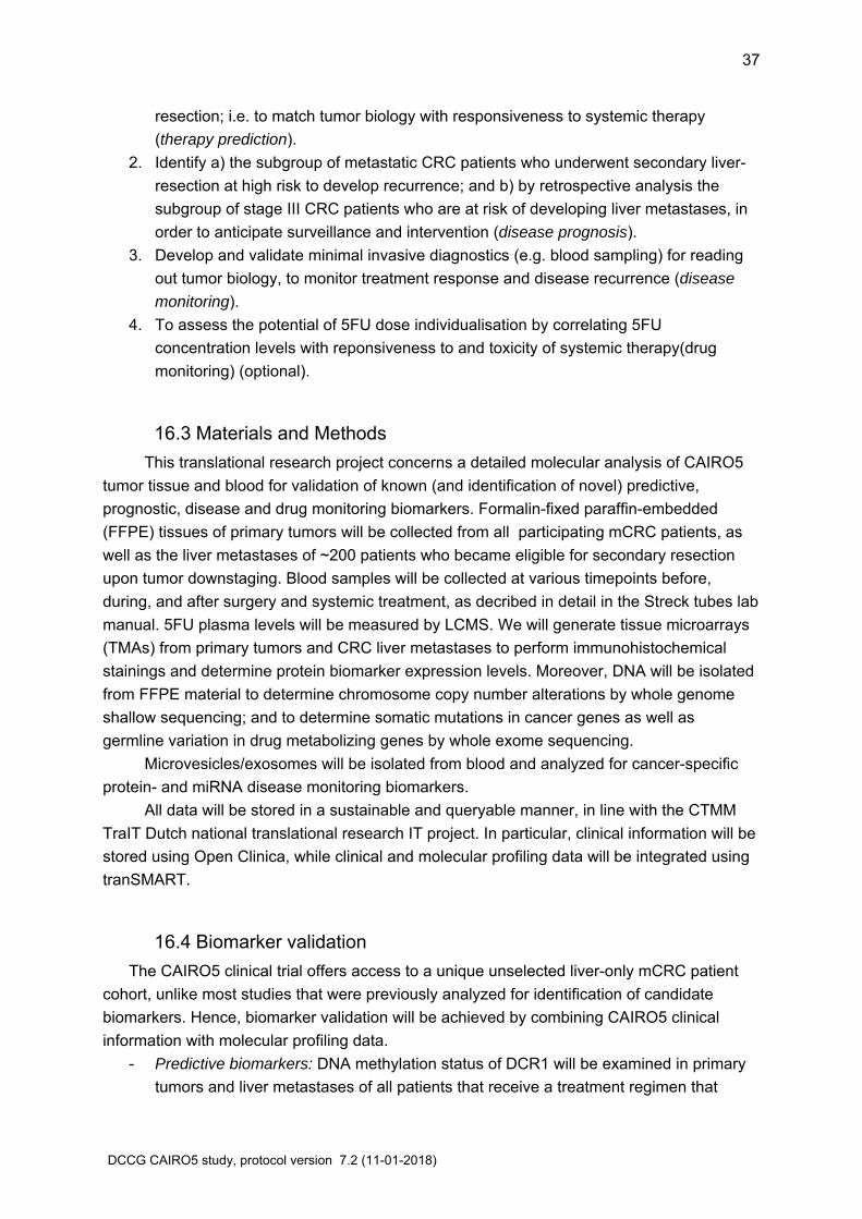
37
DCCG CAIRO5 study, protocol version 7.2 (11-01-2018)
resection; i.e. to match tumor biology with responsiveness to systemic therapy
(therapy prediction).
2. Identify a) the subgroup of metastatic CRC patients who underwent secondary liver-
resection at high risk to develop recurrence; and b) by retrospective analysis the
subgroup of stage III CRC patients who are at risk of developing liver metastases, in
order to anticipate surveillance and intervention (disease prognosis).
3. Develop and validate minimal invasive diagnostics (e.g. blood sampling) for reading
out tumor biology, to monitor treatment response and disease recurrence (disease
monitoring).
4. To assess the potential of 5FU dose individualisation by correlating 5FU
concentration levels with reponsiveness to and toxicity of systemic therapy(drug
monitoring) (optional).
Materials and Methods 16.3
This translational research project concerns a detailed molecular analysis of CAIRO5
tumor tissue and blood for validation of known (and identification of novel) predictive,
prognostic, disease and drug monitoring biomarkers. Formalin-fixed paraffin-embedded
(FFPE) tissues of primary tumors will be collected from all participating mCRC patients, as
well as the liver metastases of ~200 patients who became eligible for secondary resection
upon tumor downstaging. Blood samples will be collected at various timepoints before,
during, and after surgery and systemic treatment, as decribed in detail in the Streck tubes lab
manual. 5FU plasma levels will be measured by LCMS. We will generate tissue microarrays
(TMAs) from primary tumors and CRC liver metastases to perform immunohistochemical
stainings and determine protein biomarker expression levels. Moreover, DNA will be isolated
from FFPE material to determine chromosome copy number alterations by whole genome
shallow sequencing; and to determine somatic mutations in cancer genes as well as
germline variation in drug metabolizing genes by whole exome sequencing.
Microvesicles/exosomes will be isolated from blood and analyzed for cancer-specific
protein- and miRNA disease monitoring biomarkers.
All data will be stored in a sustainable and queryable manner, in line with the CTMM
TraIT Dutch national translational research IT project. In particular, clinical information will be
stored using Open Clinica, while clinical and molecular profiling data will be integrated using
tranSMART.
Biomarker validation 16.4
The CAIRO5 clinical trial offers access to a unique unselected liver-only mCRC patient
cohort, unlike most studies that were previously analyzed for identification of candidate
biomarkers. Hence, biomarker validation will be achieved by combining CAIRO5 clinical
information with molecular profiling data.
- Predictive biomarkers: DNA methylation status of DCR1 will be examined in primary
tumors and liver metastases of all patients that receive a treatment regimen that

38
DCCG CAIRO5 study, protocol version 7.2 (11-01-2018)
includes irinotecan (FOLFOXIRI and FOLFIRI; n=~435) and associated with
progression free survival (PFS) and OS. Likewise, gain of 6q will be examined in the
same population for responsiveness to 5FU-based and irinotecan combination
therapy. Loss of 5q12.1-12.3 will be associated with PFS and OS upon 5FU-based
treatment (all patients), while loss of 5q34 will be associated with PFS and OS of
patients treated with bevacizumab in combination with FOLFOXIRI or FOLFOX
(n=~350).
- Prognostic biomarkers: Protein expression of VEGFA, EGFR, PTGS2 (COX2),
AURKA, SERPINB5, KCNQ1 and other potentially prognostic markers of interest will
be examined in primary tumors and metastases of mCRC patients who receive
secondary resection (n = ~200), and will be associated with recurrence and OS.
Protein expression of these markers will also be examined in a separate cohort of
stage III CRC patients who did not develop recurrence and liver metastases.
- Disease monitoring biomarkers: Microvesicles/exosomes will be isolated from blood
taken before, during, and after surgery and drug treatment. Microvesicle-associated
abundance of approximately 40 proteins, among which MCM5, SERPINB5, AGRN,
and IPO4, will be measured by targeted mass spectrometry and correlated to disease
status. Meanwhile, CRC- and metastases-associated miRNA candidate biomarkers
will be identified and validated in the same population by miRNA sequencing
analysis.
- Ciculating tumor DNA (ctDNA) and other potentially prognostic, predictive and
disease monitoring markers of interest will be isolated from all taken blood samples.
- Data analyses: For all comparisons described, multivariate analyses will be
performed that take various known prognostic clinical parameters as well as the
spectrum of most common somatic mutations in cancer genes into account. In
addition, we will analyze novel parameters such as recurrent structural genomic
variations and effects of germline variation.
- Drug monitoring biomarkers: the continuous infusion of 5FU allows to calculate
exposure to 5FU (area under the curve- AUC) by the concentration of 5FU during
infusion (Css) times the time of infusion (t): AUC = C steady state x t.
A 5FU sample will be taken during 2 cycles to evaluate intrapatient variability in 5FU
exposure. The mean AUC will be correlated with responsiveness and toxicity of 5FU
treatment. Based on previous studies we expect to find a target level for 5FU between
20 and 25 ng.L/h. We will need about 90 patients to establish these cut-offs.
17 Ethical and legal aspects
This study will be conducted in accordance to the standards of Good Clinical Practice, in
agreement with the Declaration of Helsinki (latest amendment) and with Dutch law in general
and with the W.M.O. (Wet Medisch-wetenschappelijk Onderzoek met mensen) in particular.
Before they agree to participation in this trial, all patients will be provided with written
information in the form of a Patient Information Sheet (Appendix IV).

39
DCCG CAIRO5 study, protocol version 7.2 (11-01-2018)
The formal written consent of a patient must be obtained before initiation of any study-
specific procedure.
Independent physician 17.1
In accordance with Dutch law and the WMO, an independent physician has been
assigned to this study.Dr. M.J. Kersten, department of Hematology, AMC Amsterdam, tel.
020-5665955, who is not otherwise involved in this study, has agreed to act as the
independent physician.
Insurance 17.2In accordance with Dutch law and the W.M.O., an insurance policy, covering all
participating patients, has been effected with AON Risk Solutions.
18 Quality
Monitoring 18.1This study will be monitored based on the recommendations as described in the brochure
“Kwaliteitsborging mensgebonden onderzoek 2.0” published October 2012 by the Dutch
Federation of University Medical Centres (NFU). The monitor plan is based on the judgment
of the principle investigators that the study treatment takes negligible risk for the participating
patients.
The trial will be monitored by independent qualified monitors, local and central oncology
data managers of IKNL clinical research department. A comprehensive description of the
aspects and frequency of monitoring can be found in a separate monitoring plan filed in the
Trial Master File and the Investigator Site Files.
Quality assurance 18.2
Quality is assured in this trial by the follow aspects:
- Only panel members and study coordinators have access to the program used by the
panel. The statisticians and employees from the central IKNL clinical research
departement will not have access to the program used by the panel.
- All information stored in the study database will be anonymised according to laws and
regulations.
- Accounts for the panel program or the study database will be handed out according to
role-description as noted on the signature logs.
- A plan about the panel processes, including privacy statements, is part of the Trial
Master File
- A data management plan, including validation plan, is part of the Trial Master File

40
DCCG CAIRO5 study, protocol version 7.2 (11-01-2018)
- All staff working on this trial is competent, able and qualified, as shown by their
curriculum vitae, which will be stored in the Trial Master File.
19 Public disclosure and publication policy
Authorship will include at least the principal investigators, all members of the liver panel, the
statistician, the principal datamanager, the trial coordinator, the top ten local investigators
(one medical oncologist and liver surgeon per site) regarding to inclusion numbers, and any
other person that made an significant contribution to CAIRO5 which latter is decided by the
principal investigators.

41
DCCG CAIRO5 study, protocol version 7.2 (11-01-2018)
20 References
1. Kopetz S et al, J Clin Oncol, 2009. 27: p. 3677-83.
2. Hurwitz H et al, N Engl J Med, 2004. 350: p. 2335-42.
3. Kabbinavar F et al, J Clin Oncol, 2005. 23: p. 3706-12.
4. Saltz L et al, J Clin Oncol, 2008. 26: p. 2013-19.
5. Tebbutt N et al, J Clin Oncol, 2010. 28: p. 3191-8.
6. Douillard JY et al, J Clin Oncol, 2010. 28: p. 4697-705.
7. Van Cutsem et al, N Engl J Med, 2009. 360: p. 1408-17.
8. Van Cutsem et al, J Clin Oncol, 2011. 29: p. 2011-19.
9. Ruers R et al, Eur J Cancer, 2002. 38: p. 1023-33.
10. al, B.H.e., Ann Surg, 1996. 224: p. 509-20.
11. Adam R et al, Ann Surg, 2004. 240: p. 644-57.
12. Adam R et al, J Clin Oncol, 2009. 27: p. 1829-35.
13. LiverMetSurvey: http://www.livermetsurvey.org.
14. Wicherts DA et al, Ann Surg, 2008. 248: p. 627-37.
15. Folprecht G et al, Lancet Oncol, 2010. 11: p. 38-47.
16. Folprecht G et al, Ann Oncol, 2005. 16: p. 1311-19.
17. Benoist S et al, J Clin Oncol, 2006. 24: p. 3939-45.
18. Chun YS et al, JAMA, 2009. 302: p. 2338-44.
19. Ribero D et al, Cancer, 2007. 110: p. 2761-7.
20. Shindoh J et al, J Clin Oncol, 2012. 30: p. 4566-72.
21. Bertolini F et al, Br J Canceer, 2011. 104: p. 1079-84.
22. Gruenberger B et al, J Clin Oncol, 2008. 26: p. 1830-5.
23. Kesmodel SB et al, J Clin Oncol, 2008. 26: p. 5254-60.
24. Wicherts DA et al, Br J Surg, 2011. 98: p. 399-407.
25. Heinemann V et al, ASCO 2013. abstract #LBA3506.
26. Schwarzberg LS et al, ASCO 2013. abstract #3631.
27. Grothey A et al, J Clin Oncol, 2005. 23: p. 9441-2.
28. Grothey A, J Clin Oncol, 2010. 28: p. 4668-70.
29. Lièvre A et al, J Clin Oncol, 2008. 26: p. 374-9.
30. De Roock W et al, JAMA, 2010. 304: p. 1812-20.
31. Peeters M et al, J Clin Oncol, 2013. 31: p. 759-65.
32. Tejpar S et al, J Clin Oncol, 2012. 30: p. 3570-77.
33. De Roock W et al, Lancet Oncol, 2010. 11: p. 753-62.
34. Oliner KS et al, ASCO 2013. abstract # 3511.
35. Ince WL et al, J Natl Cancer Inst, 2005. 97: p. 981-9.
36. Richman SD et al, J Clin Oncol, 2009. 27: p. 5931-7.
37. Tol J et al, N Engl J Med, 2009. 361: p. 98-9.
38. Pietrantonio F et al, Eur J Cancer, 2015. 51: p. 587-94.
39. Holch et al, Eur J Cancer, 2017. 70: p. 87-98.
40. Tejpar S et al, JAMA Oncol, 2016. 3: p. 194-201.
41. Falcone A et al, J Clin Oncol, 2007. 25: p. 1670-76.

42
DCCG CAIRO5 study, protocol version 7.2 (11-01-2018)
42. Masi G et al, J Natl Cancer Inst, 2011. 103: p. 21-30.
43. Souglakos et al, Br J Canceer, 2006. 94: p. 798-805.
44. Masi G et al, Lancet Oncol, 2010. 11: p. 845-52.
45. Falcone A et al, ASCO 2013. abstract #3505.
46. Bokemeyer C et al, Ann Oncol, 2011. 22: p. 1536-46.
47. Hecht JR et al, J Clin Oncol, 2009. 27: p. 672-80.
48. Tol J et al, N Engl J Med, 2009. 360: p. 563-72.
49. Saif MW et al, World J Gastroenterol, 2009. 15: p. 3855-64.
50. Fuchs CS et al, J Clin Oncol, 2007. 25: p. 4779-86.
51. Arkenau T et al, J Clin Oncol, 2008. 26: p. 5910-17.
52. Maughan T et al, Lancet, 2011. 377: p. 2103-14.
53. Moosmann N et al, J Clin Oncol, 2011. 29: p. 1050-8.
54. Fuchs CS et al, J Clin Oncol, 2008. 26: p. 689-90.
55. Ye LC et al, J Clin Oncol, 2013. 31: p. 1931-8.
56. Bennouna J et al, Lancet Oncol, 2013. 14: p. 29-37.
57. Giantonio B et al, J Clin Oncol, 2007. 25: p. 1539-44.
58. Peeters M et al, J Clin Oncol, 2010. 28: p. 4706-13.
59. Peeters M et al, Proc. Markers in Cancer, joint meeting of ASCO, EORTC and NCI
2013 (abstract).
60. Brush J et al, Health Technol Assess, 2011. 15: p. i-192.
61. Lee KH et al, Korean J Radiol, 2013. 14: p. 894-904.
62. Van Lienden KP et al, Cardiovasc Intervent Radiol, 2013. 36: p. 25-34.
63. Fischer C et al, JAMA Surg, 2013. 148: p. 1103-8.
64. Bischof DA et al, Br J Surg, 2013. 100: p. 1414-20.
65. Dindo D et al, Ann Surg, 2004. 240: p. 205-13.
66. Arnold D et al, Ann Oncol, 2017. DOI 10.1093/annonc/mdx175.
67. Cremolini CF, Lancet Oncol, 2015. 16: p. 1306-15.

43
DCCG CAIRO5 study, protocol version 7.2 (11-01-2018)
21 Appendix I: CAIRO5 study design
Previously untreated unresectable
colorectal cancer liver‐only metastases
Registration
Panel decision on potentially resectable vs
permanently unresectable metastases
RAS, BRAF mutation status
RAS or
BRAF mutation and/or
right‐sided primary tumor
RAS & BRAF wildtype
+ left‐sided primary tumor
(all criteria must be met)
FOLFOX or FOLFIRI
+ bevacizumab
FOLFOXIRI
+ bevacizumab
FOLFOX or FOLFIRI
+ bevacizumab
FOLFOX or FOLFIRI
+ panitumumab
Panel decision on resectability status
Randomisation Randomisation

44
DCCG CAIRO5 study, protocol version 7.2 (11-01-2018)
22 Appendix II: Flow chart for registration, randomisation, pathology and central panel review
Flow chart for registration, randomisation, pathology, radiology, central panel
Local Investigator (day 0):
-registration at IKNL clinical research department
-submit CT/ MRI of thorax & abdomen
-send patient material for translational research
Panel radiologist (day 0-2):
-evaluation and measurement by radiologist
-in case of poor imaging or extrahepatic metastases,
notify local investigator
-fill in radiology form
-alert coordinator that review is ready for surgeons
Panel surgeons (day 2-8):
-evaluation about resectability by three surgeons
-fill in individual forms
-draw conclusion by panel chair
-inform local investigator
Pathologist (day 1-8):
-assess RAS/ BRAF mutation status
IKNL research department (day 5-9):
-ask local investigator for confirmation of registration
-randomisation of RAS&BRAF wt left-sided tumors and
RAS/BRAF mut and/or right sided tumors
Local investigator (day 8-10):
-start systemic treatment within 14 days after
randomisation