treatment options, timing and sequencing: … appliances with elastic chains and a consolidation...
TRANSCRIPT

141© Springer International Publishing Switzerland 2016U. Erdemir, E. Yildiz (eds.), Esthetic and Functional Management of Diastema: A Multidisciplinary Approach, DOI 10.1007/978-3-319-24361-0_11
D. Germec Cakan , DDS, PhD (*) Orthodontics , Yeditepe University, Dental Faculty , Istanbul , Turkey e-mail: [email protected]
K. Sayınsu , DDS, PhD Orthodontics , Private Practice , Istanbul , Turkey
11 Treatment Options, Timing and Sequencing: Orthodontics
Derya Germec Cakan and Korkmaz Sayınsu
Abstract The treatment of diastema either by orthodontics, restorative dentistry, periodon-tology or a combination of these disciplines should be focused on the aetiologi-cal factors, patient needs, aesthetics and stable results. Identifi cation of the aetiological factors and consideration of the dentofacial characteristics of the patient are essential for the appropriate therapy. Following diagnosis and indi-vidualised treatment planning, satisfactory treatment outcomes can be achieved with different orthodontic mechanics and approaches. This chapter will present management of spacing with orthodontics alone or as a part of interdisciplinary treatment depending on the causative factors. The timing and sequencing of the orthodontic treatment and retention protocol at the end of therapy will also be explained.
11.1 Introduction
Diastema can occur in deciduous, mixed or permanent dentition. According to epi-demiological studies, the prevalence of midline diastema is high in the children of 6–9 years old, ranging between 43 and 97 % [ 1 – 3 ]. As mentioned in Chap. 2, the physiologic diastema of early dentitional stages generally does not necessitate orth-odontic intervention because it spontaneously closes during development of the dentition. However, approximately 10 % of the orthodontic patients have been reported to have a midline diastema larger than 0.5 mm after the mixed dentition
142
Clin
ical
pra
ctic
e gu
idel
ines
for
corr
ectio
n of
dia
stem
a
Dec
iduo
us d
entit
ion
Mix
ed d
entit
ion
Per
man
ent
dent
ition
Eva
luat
ion
of th
e ca
use
No
trea
tmen
t(p
hysi
olog
ic)
No
trea
tmen
t with
follo
w-u
p un
til th
epe
rman
ent c
anin
eser
upt
Mid
line
path
olog
y(m
esio
dens
, odo
ntom
a, e
tc.)
Trea
tmen
t of t
he u
nder
lyin
g ca
use
(sur
gica
l rem
oval
)
No
trea
tmen
t with
follo
w-u
p
Ort
hodo
ntic
spa
ce c
losu
re if
dia
stem
ape
rsis
ts a
t per
man
ent d
entit
ion
Eva
luat
ion
of th
e ca
use
Fren
umat
tach
men
t
Ort
hodo
ntic
s+fr
enec
tom
y
Inte
rdis
cipl
inar
ytr
eatm
ent
Per
iodo
ntal
loss
Mac
rogl
ossi
a
Res
tora
tive
oror
thod
ontic
or
inte
rdis
cipl
inar
ytr
eatm
ent
Toot
h si
ze d
iscr
epan
cy
Unf
avou
rabl
epo
sitio
n,m
aloc
clus
ion
Inte
rdis
cipl
inar
ytr
eatm
ent
(ort
hodo
ntic
+re
stor
ativ
e)
Res
tora
tive
trea
tmen
t
Favo
urab
le p
ositi
on,
no m
aloc
clus
ion
Ret
ract
ion
with
orth
odon
tictr
eatm
ent
Elim
inat
ion
of th
eha
bit(
+or
thod
ontic
sif
requ
ired)
Pem
icio
us h
abits
Pro
clin
ed te
eth
Mid
line
dias
tem
a <
4 m
mM
idlin
e di
aste
ma
≥4 m
m
Fig
. 11
.1
Clin
ical
pra
ctic
e gu
idel
ines
for
cor
rect
ion
of d
iast
ema
D. Germec Cakan and K. Sayınsu

143
Fig. 11.2 ( a – c ) Diagrammatic illustration of anterior diastema closure with retraction of the fl ared maxillary incisors
a b
c
period [ 4 ]. If the spacings in the dental arches persist after the eruption of the per-manent canines, orthodontic treatment may be required.
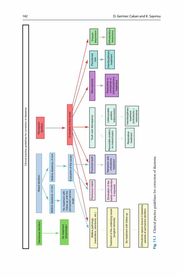
Clinical practice guidelines of midline diastema treatment are summarised in Fig. 11.1 . However, the clinician should also consider that these general guidelines may not apply in every case.
11.2 Orthodontic Closure of Diastema
Orthodontic closure of an anterior diastema can be accomplished either by mesio-distal and/or anteroposterior movement of the teeth. If the teeth are proclined and the overjet is increased, retraction of the incisors will automatically close the ante-rior spacing (Fig. 11.2 ). If the teeth are not protruded but laterally migrated (e.g. when the maxillary lateral incisors are congenitally missing), mesially directed forces will bring the teeth together to close a median diastema. In cases where the buccolingual position of the teeth and lips should be maintained, anterior movement of the posterior teeth would be preferred (Fig. 11.3 ). The decision of which orth-odontic mechanics to use in order to obtain these forces and moments largely depends on the amount and localization of the diastema, the age and dentitional
11 Treatment Options, Timing and Sequencing: Orthodontics

144
stage of the patient, inclinations and angulations of the teeth and the presence of an adequate overjet.
Minor diastema (less than 2 mm) caused by distal crown angulation of the teeth can easily be corrected by tipping movement with removable appliances in an ado-lescent patient (Fig. 11.4 ). The fi nger springs of a removable Hawley appliance cre-ate mesially directed tipping forces to bring the teeth together. As an alternative, clear plastic appliances and rubber bands can also be used to close median diastema [ 5 ]. It is important to note though whenever possible, mechanics enabling three- dimensional tooth control should be chosen especially in complex cases (e.g. large median diastema, generalised spacings, deepbite, skeletal problems, microdontia, hypodontia). Therefore, active orthodontic treatment to close a diastema is prefera-bly achieved by bodily tooth movement generated by fi xed appliances (Fig. 11.5 ). For this purpose, anterior segmental archwires, 2 × 4 appliances (extending from fi rst molars to incisors) or continuous archwires can be used (Fig. 11.6 ). Stiff and rectan-gular archwires provide good control of tooth movement during space closure.
Key Note The evaluation of overjet is important in diastema cases. If the overjet is reduced, diastema cannot be closed by palatal tipping of the maxillary incisors.
Key Note Never place elastics around the teeth to close a diastema without the use of orthodontic appliances. Uncontrolled subgingival dislocation of the elastics may cause severe periodontal breakdown.
Fig. 11.3 Diagrammatic illustration of generalised diastema closure with mesialisation of the posterior teeth when the retraction of the incisors is contraindicated
D. Germec Cakan and K. Sayınsu

145
11.2.1 Case 1: Maxillary Midline Diastema
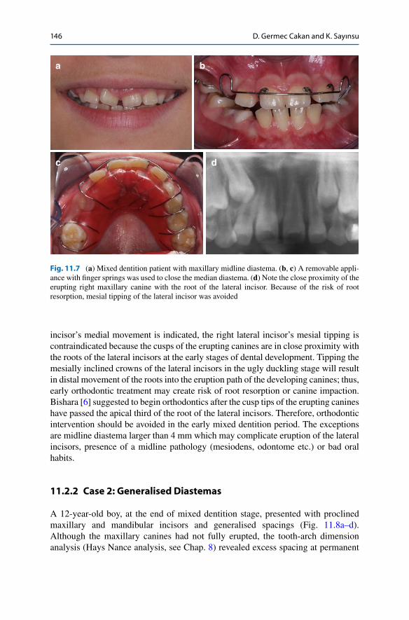
The patient in Fig. 11.7 had a maxillary midline diastema in mixed dentition. She was complaining about the unpleasant appearance of the spacings and irregularities. Although the physiological process was explained, she preferred not to wait for the eruption of the canines for psychological reasons. A removable appliance with fi nger springs was used to close the median diastema. Once the median diastema is closed, the fi nger spring on the left lateral incisor will be activated. Although the left lateral
a b
Fig. 11.4 ( a ) Distally tipped maxillary central incisors and minor median diastema. ( b ) Closure of the median diastema by mesial tipping of the incisors (note the fi nal upright position of the teeth)
a b
Fig. 11.5 ( a ) Generalised anterior diastemas. ( b ) Closure of the diastemas by mesially directed bodily tooth movement using fi xed orthodontic appliances
a b
Fig. 11.6 The use of anterior segmental archwire ( a ) and 2 × 4 mechanics ( b ) for orthodontic treatment of maxillary diastemas
11 Treatment Options, Timing and Sequencing: Orthodontics
146
incisor’s medial movement is indicated, the right lateral incisor’s mesial tipping is contraindicated because the cusps of the erupting canines are in close proximity with the roots of the lateral incisors at the early stages of dental development. Tipping the mesially inclined crowns of the lateral incisors in the ugly duckling stage will result in distal movement of the roots into the eruption path of the developing canines; thus, early orthodontic treatment may create risk of root resorption or canine impaction. Bishara [ 6 ] suggested to begin orthodontics after the cusp tips of the erupting canines have passed the apical third of the root of the lateral incisors. Therefore, orthodontic intervention should be avoided in the early mixed dentition period. The exceptions are midline diastema larger than 4 mm which may complicate eruption of the lateral incisors, presence of a midline pathology (mesiodens, odontome etc.) or bad oral habits.
11.2.2 Case 2: Generalised Diastemas
A 12-year-old boy, at the end of mixed dentition stage, presented with proclined maxillary and mandibular incisors and generalised spacings (Fig. 11.8a–d ). Although the maxillary canines had not fully erupted, the tooth-arch dimension analysis (Hays Nance analysis, see Chap. 8 ) revealed excess spacing at permanent
a b
c d
Fig. 11.7 ( a ) Mixed dentition patient with maxillary midline diastema. ( b , c ) A removable appli-ance with fi nger springs was used to close the median diastema. ( d ) Note the close proximity of the erupting right maxillary canine with the root of the lateral incisor. Because of the risk of root resorption, mesial tipping of the lateral incisor was avoided
D. Germec Cakan and K. Sayınsu

147
a b
c d
e f
g
Fig. 11.8 ( a – c ) Frontal and occlusal intraoral views of generalised spacing before orthodontic treatment. ( d ) Closing the spaces with a consolidation arch in the maxillary dental arch and elastic chains in the mandibular dental arch during fi xed orthodontic treatment. Headgear and intermaxil-lary elastics were used to reinforce the posterior anchorage. ( e – g ) Frontal and occlusal intraoral views after orthodontic space closure. Fixed lingual retainers were bonded to each tooth from right fi rst premolar to left fi rst premolar in the maxillary and mandibular dental arches following orth-odontic treatment of polydiastema to prevent relapse
11 Treatment Options, Timing and Sequencing: Orthodontics
148
dentition. The maxillary and mandibular spacings were closed using fi xed orth-odontic appliances with elastic chains and a consolidation arch aiming to control root divergence and torque (Fig. 11.8e–g ).
11.3 Management of Diastema Due to Abnormal Oral Habits: Breaking the Habit
The teeth are in equilibrium between forces generated by muscles, mastication and stabilisation of the periodontium [ 7 ]. If there is an alteration in the equilibrium due to an abnormal oral habit such as fi nger sucking, dental movement is likely to occur resulting in spacing in the dental arches in addition to other malocclusions. If the abnormal habit is stopped before the eruption of permanent teeth, normal cheek and lip pressures can establish the equilibrium and self-correction of the displaced teeth. However, if it persists in mixed dentition, orthodontic treatment may be required. In general, when the parafunctional cause is eliminated, a spontaneous correction will be observed [ 8 ]. There are several methods to stop abnormal oral habits. In ortho-dontics, myofunctional therapy and the use of habit breakers are very effective as a complementary to a psychological approach [ 8 , 9 ]. After the patient has stopped the pernicious habit, wearing of the appliance is recommended for an additional several months.
11.3.1 Case 3: Lower Lip Sucking and Maxillary Spacing
An 11-year-old female patient presented with generalised maxillary spacing of 7 mm (Fig. 11.9a, b ). Her history revealed that she was sucking her lower lip in her sleep throughout the night (Fig. 11.9c ). Lower lip sucking habit caused proclina-tion of the maxillary incisors which could not have been opposed by the forces of the upper lip. The aim was to normalise the extraoral muscle force and establish the equilibrium. A lip bumper appliance was used to break the sucking habit (Fig. 11.9d ). This prefabricated appliance was placed at the level of the gingiva 2–3 mm in front of the lower incisors and 4–5 mm away from the buccal segments and was fi xed to the molar tubes. It was reactivated when necessary. After 3 months of lip bumper therapy (full-time wear), the lower lip sucking habit was stopped, maxillary incisors spontaneously retroclined due to elimination of increased forces generated by sucking, and thus, maxillary diastemas were reduced (Fig. 11.9e, f ). The residual diastemas were closed with fi xed orthodontic therapy (Fig. 11.9g, h ).
Key Note In general, excessive proclination of the teeth generates spacing, whereas retraction of the teeth to their neutral position helps to close the diastemas. However, elimination of the aetiological factor is essential.
D. Germec Cakan and K. Sayınsu

149
11.3.2 Case 4: Finger Sucking and Maxillary Spacing
A female patient in early transitional dentition having a fi nger sucking habit showed increased overjet, labial inclination of the incisors and maxillary spacing (beyond physiologic diastema of mixed dentition) particularly on the right side where she inserted her right fi nger to suck (Fig. 11.10a–d ). A myofunctional appliance was
a b
c d
e f
g h
Fig. 11.9 ( a , b ) Generalised maxillary spacing and increased overjet. ( c ) Lower lip sucking exert-ing abnormal and unopposed force causing fl aring of the upper teeth. ( d ) Use of lip bumper appli-ance to break the sucking habit. ( e , f ) Spontaneous reduction of maxillary spacing due to normalised muscle forces shows retroclination of the maxillary incisors after 3 months of lip bumper therapy. ( g ) Fixed orthodontic therapy phase. ( h ) Correction of the malocclusion at the end of orthodontic treatment (From Germeç and Taner [ 8 ]. Reprinted with permission from Angle Orthodontist)
11 Treatment Options, Timing and Sequencing: Orthodontics

150
a b
c
d e
f g
h
Fig. 11.10 ( a – d ) A 7.5-year-old patient with fi nger sucking habit, placing her fi nger on the right side of the dentition, leading to proclination of the incisor, increased diastema and overjet. ( e ) Soft myofunctional appliance was used to stop the habit and worn during sleep (when she used to suck her fi nger). ( f – h ) Intraoral photographs showing decrease in spacing, correction of overjet and retroclination of the incisors after 3 months of myofunctional therapy (From Tozlu and Germeç [ 9 ]. Reprint permission from 7 Tepe Klinik)
D. Germec Cakan and K. Sayınsu

151
used for 3 months (only at night) to stop fi nger sucking habit (Fig. 11.10e ). This soft appliance with vestibular screens broke the fi nger sucking habit and thus eliminated the abnormal pressure caused by the fi nger sucking and established a balance between intraoral and extraoral muscles. In addition, it generated palatally directed forces to the overproclined right maxillary incisors. The fi nal result was decreased maxillary spacing. At the end of myofunctional therapy, the maxillary spacing was reduced but not totally eliminated (ideal for a transitional period, see Chap. 2 ), and the overproclined incisors and the increased overjet were corrected (Fig. 11.10f–h ).
11.4 The Role of Orthodontics in the Interdisciplinary Management of Diastema Due to Tooth Size Discrepancies and Missing Teeth
Diastemas caused by tooth size discrepancies can generally be treated by restora-tions. This treatment approach offers a quick solution and is readily accepted by patients. However, sometimes it is not possible to correct tooth size discrepancy with restorations alone due to unfavourable tooth positions and malocclusions (Fig. 11.11 ). Orthodontics helps to solve these problems by tooth movement, prop-erly positioning the teeth and enabling an infrastructure for aesthetic restorations.
Interdisciplinary treatment starts with collecting accurate data, analysing it and composing a list of problems. Subsequently a treatment with alternatives is planned and discussed in the interdisciplinary team considering both treatment objectives and the patient’s needs, demands and expectations. Once the fi nal treatment plan is agreed and upon approval of the patient, every discipline works in collaboration to achieve the stated goals. This collaborative team work has some important steps. A crucial step of interdisciplinary treatment in diastema cases is to determine the fi nal positions of the teeth and redistribute the spaces with orthodontics, which is guided by the principles of proportion and occlusion. The tooth size should be in harmony with the adjacent teeth and dental arch. The clinician can use the tooth proportions
Fig. 11.11 A maxillary and mandibular generalised spacing case due to small teeth and large alveolar base. Restorative approach without orthodontics for this case presenting a Class III maloc-clusion with anterior crossbite, a midline diastema of 3 mm, disproportionate localisation of the maxillary and mandibular spacings between teeth may result in compromised aesthetics and impede the survival of the restorations
11 Treatment Options, Timing and Sequencing: Orthodontics

152
(e.g. width/length ratio, relationship between the sizes of the adjacent teeth) and tooth size analysis (see Chap. 8 ) as a guide when redistributing the spaces between the teeth. A diagnostic wax set-up is also very helpful to visualise the fi nal result (see Chap. 4 ), which will provide information about the fi nal position of the teeth, the number of teeth to be restored, the position and dimensions of the restorations and the timing, sequencing and progress of the treatment.
When the discrepancy involves several teeth, the ideal treatment is to redistribute the spacings between these undersized teeth (Fig. 11.12 ). Otherwise, if the spacings in the maxillary arch are collected between lateral incisors and canines, this will result in very large, disproportionate lateral incisors which might also impede the gingival health as well as aesthetic outcomes.
The size and shape anomalies of the teeth are mostly seen in upper lateral inci-sors [ 10 ]. When there are small- or peg-shaped lateral incisors, the tooth can be positioned slightly closer to the central incisor, because the distal curvature of the lateral incisor is more convex than the mesial curvature [ 11 ]. In other words, the orthodontist should leave more space on the distal than the mesial side of the upper lateral incisor to enhance the restorative results (Fig. 11.13 ).
Key Note Dental proportional norms and occlusal requirements guide the clinician when redistributing the spaces and repositioning the teeth in a complicated generalised diastema case.
Key Note Redistribution of the spaces and positioning of the teeth with orthodontics require close collaboration and communication between orthodontist and restorative dentist not only in planning but also during treatment. Therefore, the orthodontist should refer the patient to the restorative dentist for the evalu-ation of the fi nal tooth position prior to debonding of the braces.
Fig. 11.12 Redistribution of the spaces in a case with maxillary anterior tooth size discrepancy. ( a ) When the discrepancy involves several teeth, collecting spaces distal to lateral incisors will ruin the tooth form and proportions. ( b ) Redistribution of the spaces between anterior teeth using push coils. Note the right side is appropriate, whereas the left side requires distal movement of the lat-eral incisor. ( c ) Proper positioning of the anterior teeth prior to restorations. Note that the spacings were generated on the mesial and distal side of the undersized teeth. ( d ) Spacing on the mesial and distal side of the lateral incisor. ( e ) Avoid building up only one tooth and creating a large tooth as shown in the illustration. ( f ) Illustration of the build-ups guided by width/length ratios and propor-tions between anterior adjacent teeth. ( g – i ) Intraoral views showing proportional anterior tooth size and good occlusal relationships at the end of interdisciplinary treatment accomplished with direct composite restorations of the six anterior teeth (Restorations by Dr. Esra Can Say)
D. Germec Cakan and K. Sayınsu

153
a b
c d
e f
g h
i
11 Treatment Options, Timing and Sequencing: Orthodontics

154
Another important aspect of interdisciplinary management of diastema is the tim-ing and sequencing of the treatments. Treatment timing and sequencing depends on the initial position of the teeth, the presence of a malocclusion and demands of the patient. In general orthodontics is becoming easier when there are contact points between teeth, which prevent jiggling. Therefore, whenever possible, it is wise to begin interdisciplinary treatment with provisional restorations before orthodontic therapy. The key point is to restore teeth with respect to its axial inclination meaning the radiographic evaluation is essential. When the tooth positions and occlusion are not suitable for build-ups, restoration of the teeth is generally recommended after the correction of tooth positions and during distribution of the spaces in the last stages of orthodontic treatment. The fi nal restorations should be postponed to the end of active orthodontic treatment for several reasons. Patients experiencing gingivitis with swol-len and bleeding gingiva are not uncommon during orthodontic treatment due to poor oral hygiene. A healthy and properly levelled gingival tissue is required before any kind of restorations are made. Therefore, gingival status should be optimised by peri-odontal care after the removal of the braces. In addition, if bleaching is necessary, the sequencing is fi rst orthodontics, followed by bleaching and fi nally restorations.
The interdisciplinary approach is the gold standard for most patients with dia-stema due to undersized incisors. For patients who reject this approach, increasing the root divergence with orthodontics may be an alternative method to treat gener-alised spacing. When the positive angulation of the teeth (the root divergence) is increased, the crown occupies more space because the mesiodistal width of the tooth is also increased [ 12 ]. However, distal tipping of the roots of the anterior teeth results in canted incisal edges that should be reshaped for aesthetic reasons. This treatment option should only be preserved for patients with minor discrepancies who do not accept restorative approaches.
11.4.1 Case 5: Tooth Size-Arch Size Discrepancy and Bimaxillary Generalised Spacing; Redistribution of the Spaces
The chief complaint of this adult female patient was the unaesthetic appearance of her smile. Her clinical examination and cephalometric analysis revealed that the inclination of the maxillary and mandibular incisors should be maintained to pre-serve the position of the lips in her orthognathic profi le (Fig. 11.14a–f ). Her model
a b
Fig. 11.13 ( a ) Slightly mesial positioning of the microdontic upper lateral incisor. ( b ) After direct composite restoration (both mesial and distal sides of the lateral incisor were built up) (Restoration by Dr. Umut Cakan)
D. Germec Cakan and K. Sayınsu

155
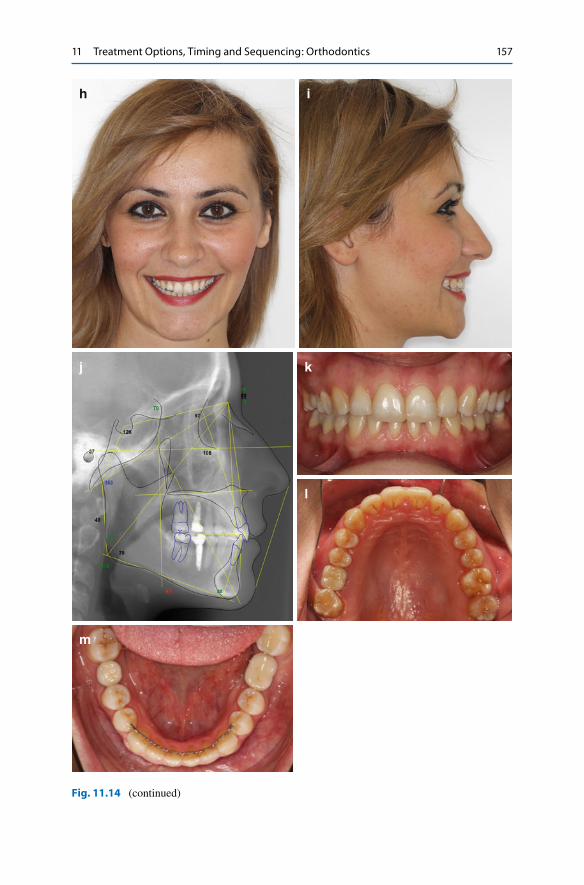
analysis showed a tooth size-arch size discrepancy caused by small mesiodistal widths of the entire dentition compared to large arch size. Furthermore, the presence of previously extracted right maxillary fi rst molar contributed to the creation of diastema due to distal migration of the premolars. Maxillary midline diastema was 3.6 mm. Bolton analysis revealed mandibular anterior excess of 2.8 mm. A restor-ative approach alone would lead to disproportionate, oversized central incisors with distorted crown shape and compromised periodontal health. An interdisciplinary treatment was planned. The role of orthodontics was to redistribute the spacing to facilitate restorative and prosthetic dentistry and treat the malocclusion character-ised by deepbite and posterior scissors bite (Fig. 11.14g ). At the end of the orth-odontic treatment, teeth were aligned and properly positioned enabling fabrication of proportional restorations and prosthetic rehabilitation. Maxillary central and lat-eral incisors were built up by direct composite restorations (Fig. 11.14h–m ).
11.4.2 Case 6: Tooth Size -Arch Size Discrepancy and Bimaxillary Generalised Spacing; Orthodontic Closure of the Diastemas and Periodontal Surgery
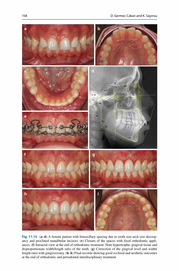
A 30-year-old female patient’s chief complaint was the spacing between her teeth. She had maxillary and mandibular polydiastema, deepbite and Class II skeletal and Class I molar relationships (Fig. 11.15 ). The generalised spacing was mainly due to tooth size-arch size discrepancy and proclined mandibular incisor. Therefore, the treatment plan included closure of the spacing by incisor retraction. At the end of 15 months of orthodontic treatment, the inclination of the incisors was corrected, and bimaxillary spacing was closed. However, the gingival tissue was hypertrophic and the tooth width/length ratio was compromised. To obtain satisfactory aesthetic out-comes, periodontal surgery was carried out. Gingival margins were levelled and proper width/length ratio was achieved after periodontal surgery.
11.4.3 Case 7: Tooth Size-Arch Size Discrepancy and Bimaxillary Generalised Spacing; Generating Space for Extra Teeth
A 35-year-old male patient was complaining about the spacings between his upper teeth. He had bimaxillary generalised spacing due to tooth size-arch size
Key Note The essential of diastema closure is to establish proper tooth proportions that are as close to the ideal as possible. From an orthodontic perspective, the orthodon-tist should not try to close all the spaces if there are tooth size anomalies (e.g. peg-shaped lateral incisors). From a restorative perspective, the clinician should not restore teeth when it is impossible to achieve proportional aesthetic results. In either situation, one must seek for interdisciplinary treatment because no mat-ter how well treated, unaesthetic results will lead to patient dissatisfaction.
11 Treatment Options, Timing and Sequencing: Orthodontics

156
a b
c d
e
f g
Fig. 11.14 ( a – f ) A female patient with a large median diastema, generalised bimaxillary spacing due to small tooth size and previously extracted teeth. ( g ) Redistribution of the maxillary spaces and correction of scissors bite. ( h – m ) At the end of interdisciplinary treatment (maxillary incisors were built up by direct composite restorations. Note that abraded incisal edges of the maxillary central incisors were also restored. The incisor and lip positions were maintained during orthodon-tic treatment)
D. Germec Cakan and K. Sayınsu
157
h i
j k
l
m
Fig. 11.14 (continued)
11 Treatment Options, Timing and Sequencing: Orthodontics
158
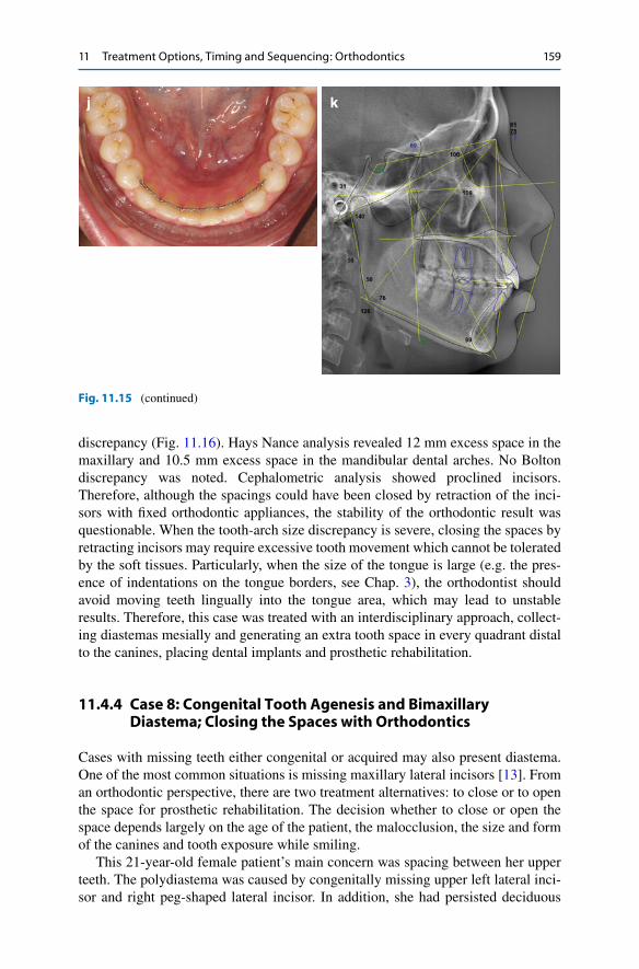
Fig. 11.15 ( a – d ) A female patient with bimaxillary spacing due to tooth size-arch size discrep-ancy and proclined mandibular incisors. ( e ) Closure of the spaces with fi xed orthodontic appli-ances. ( f ) Intraoral view at the end of orthodontic treatment. Note hypertrophic gingival tissue and disproportionate width/length ratio of the teeth. ( g ) Correction of the gingival level and width/length ratio with gingivectomy. ( h – k ) Final records showing good occlusal and aesthetic outcomes at the end of orthodontic and periodontal interdisciplinary treatment
a b
c d
e
f g
h i
D. Germec Cakan and K. Sayınsu
159
discrepancy (Fig. 11.16 ). Hays Nance analysis revealed 12 mm excess space in the maxillary and 10.5 mm excess space in the mandibular dental arches. No Bolton discrepancy was noted. Cephalometric analysis showed proclined incisors. Therefore, although the spacings could have been closed by retraction of the inci-sors with fi xed orthodontic appliances, the stability of the orthodontic result was questionable. When the tooth-arch size discrepancy is severe, closing the spaces by retracting incisors may require excessive tooth movement which cannot be tolerated by the soft tissues. Particularly, when the size of the tongue is large (e.g. the pres-ence of indentations on the tongue borders, see Chap. 3 ), the orthodontist should avoid moving teeth lingually into the tongue area, which may lead to unstable results. Therefore, this case was treated with an interdisciplinary approach, collect-ing diastemas mesially and generating an extra tooth space in every quadrant distal to the canines, placing dental implants and prosthetic rehabilitation.
11.4.4 Case 8: Congenital Tooth Agenesis and Bimaxillary Diastema; Closing the Spaces with Orthodontics
Cases with missing teeth either congenital or acquired may also present diastema. One of the most common situations is missing maxillary lateral incisors [ 13 ]. From an orthodontic perspective, there are two treatment alternatives: to close or to open the space for prosthetic rehabilitation. The decision whether to close or open the space depends largely on the age of the patient, the malocclusion, the size and form of the canines and tooth exposure while smiling.
This 21-year-old female patient’s main concern was spacing between her upper teeth. The polydiastema was caused by congenitally missing upper left lateral inci-sor and right peg-shaped lateral incisor. In addition, she had persisted deciduous
j k
Fig. 11.15 (continued)
11 Treatment Options, Timing and Sequencing: Orthodontics

160
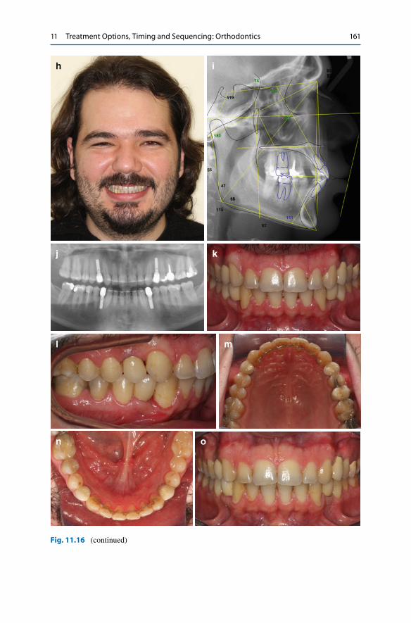
Fig. 11.16 ( a – g ) A male patient with severe bimaxillary generalised spacing due to tooth size-arch size discrepancy. ( h – n ) Interdisciplinary treatment comprised of generating spaces for a third premolar in every quadrant, placement of dental implants and prosthetic rehabilitation. Cephalometric tracing shows that maxillary and mandibular incisors were slightly upright. ( o – q ) Five years postretention photographs showing stable results (Restorations by Dr. Arzu Aykor)
a b
c d
e f
g
D. Germec Cakan and K. Sayınsu
161
h i
j k
l m
n o
Fig. 11.16 (continued)
11 Treatment Options, Timing and Sequencing: Orthodontics

162
maxillary left and mandibular right canine, congenitally missing mandibular incisor and left second premolar (Fig. 11.17 ). Cephalometric evaluation revealed labially inclined incisors, Class II skeletal pattern and a straight facial profi le.
Two different interdisciplinary treatment alternatives were proposed: (1) space opening for congenitally missing teeth with orthodontic treatment, prosthetic reha-bilitation with implants and ceramic crowns and restorative treatment of the peg- shaped upper right lateral incisor and (2) closing the spaces caused by missing teeth with orthodontic treatment, canine substitution, restorative treatment of the canines and gingival surgery. The patient rejected the fi rst treatment plan because she did not want to have implants at the end of the treatment. The second treatment option was chosen. In order to achieve symmetrical maxillary arch form, left peg-shaped lateral incisor was extracted. Maxillary canines and right mandibular canine were substituted for lateral incisors. All the spaces caused by tooth agenesis were closed with orthodontic treatment. Gingival levelling was achieved with gingivectomy, and canines and fi rst premolars were reshaped with restorative treatment.
11.5 Orthodontic Management of Spacing in Periodontal Loss
Periodontal bone loss is considered as one of the aetiological factors of diastema. When the teeth lost their periodontal support due to periodontal disease, the stabili-sation of the periodontal ligament is decreased leading to changes in the equilibrium between the teeth, soft tissues and forces generated at rest or function. Thus, patho-logical migrations may be observed after severe periodontal breakdown, which may cause diastema. Generally, these patients with no previous history of diastema in younger ages are complaining about the recently developed spacings. Orthodontics can help to bring the teeth together in alveolar bone, level the alveolar crest and eliminate the occlusal trauma (Fig. 11.18 ). The use of fi xed retainers following orth-odontic treatment also serves as splints in a periodontally compromised patient. The prerequisite to orthodontics in these cases is to control infl ammation by periodontal therapy.
p q
Fig. 11.16 (continued)
D. Germec Cakan and K. Sayınsu

163
a b
c d
e f
g h
i j
Fig. 11.17 ( a – e ) Extraoral, intraoral photographs and panoramic radiograph of a patient with maxillary and mandibular spacing caused by congenital tooth agenesis and size anomaly. ( f , g ) Intraoral and radiographic views after spaces were closed by orthodontic treatment. ( h ) Gingival levelling with gingivectomy. ( i – l ) Final extraoral and intraoral photographs after interdisciplinary treatment. Note maxillary canines and fi rst premolars were reshaped with direct composite restora-tions (Restorations by Dr. Murat Ozarslan)
11 Treatment Options, Timing and Sequencing: Orthodontics
164
11.6 Timing of Frenectomy
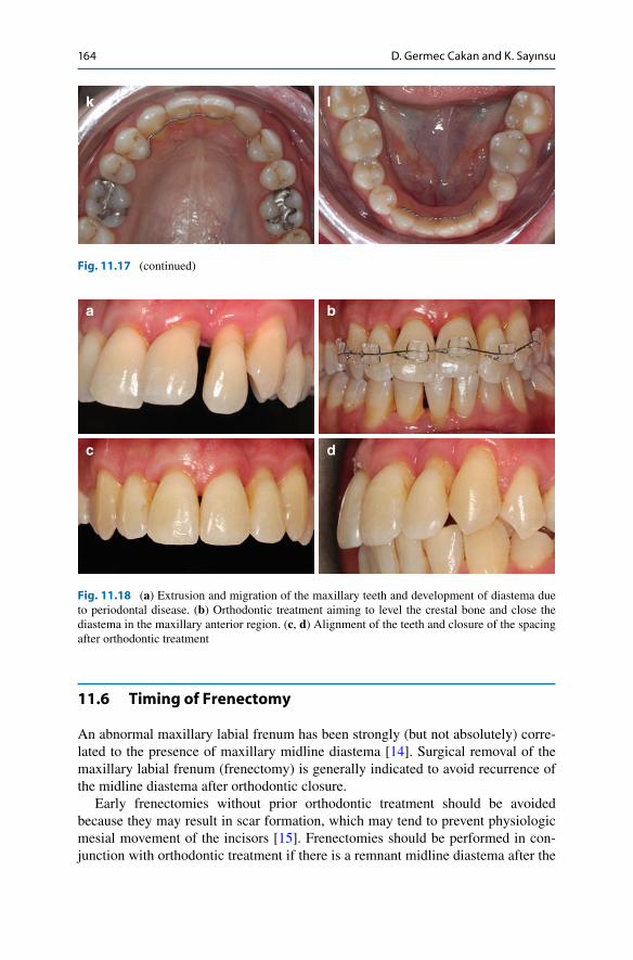
An abnormal maxillary labial frenum has been strongly (but not absolutely) corre-lated to the presence of maxillary midline diastema [ 14 ]. Surgical removal of the maxillary labial frenum (frenectomy) is generally indicated to avoid recurrence of the midline diastema after orthodontic closure.
Early frenectomies without prior orthodontic treatment should be avoided because they may result in scar formation, which may tend to prevent physiologic mesial movement of the incisors [ 15 ]. Frenectomies should be performed in con-junction with orthodontic treatment if there is a remnant midline diastema after the
a b
c d
Fig. 11.18 ( a ) Extrusion and migration of the maxillary teeth and development of diastema due to periodontal disease. ( b ) Orthodontic treatment aiming to level the crestal bone and close the diastema in the maxillary anterior region. ( c , d ) Alignment of the teeth and closure of the spacing after orthodontic treatment
k l
Fig. 11.17 (continued)
D. Germec Cakan and K. Sayınsu
165
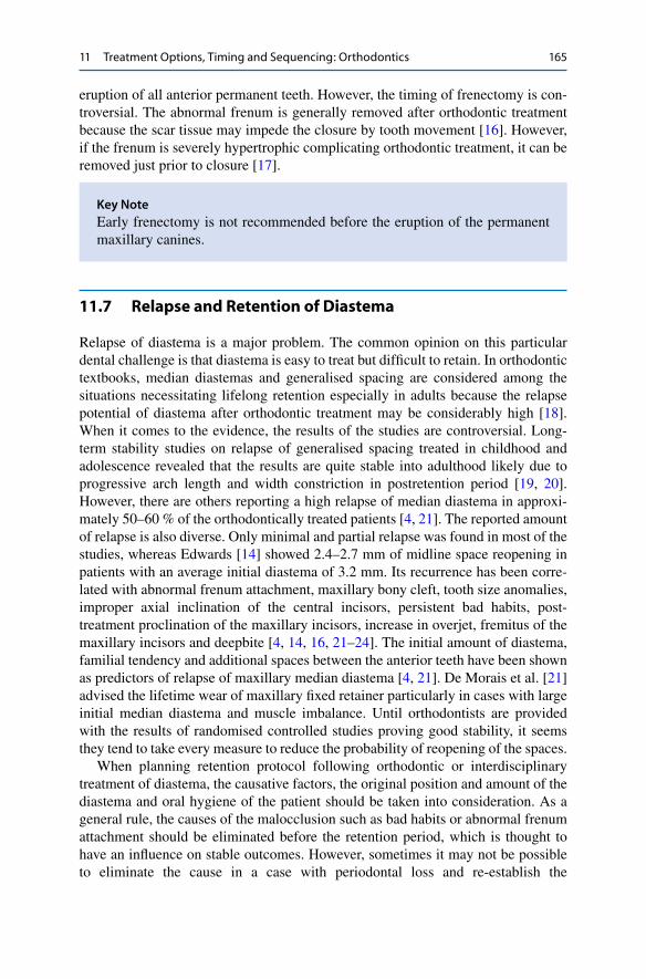
eruption of all anterior permanent teeth. However, the timing of frenectomy is con-troversial. The abnormal frenum is generally removed after orthodontic treatment because the scar tissue may impede the closure by tooth movement [ 16 ]. However, if the frenum is severely hypertrophic complicating orthodontic treatment, it can be removed just prior to closure [ 17 ].
11.7 Relapse and Retention of Diastema
Relapse of diastema is a major problem. The common opinion on this particular dental challenge is that diastema is easy to treat but diffi cult to retain. In orthodontic textbooks, median diastemas and generalised spacing are considered among the situations necessitating lifelong retention especially in adults because the relapse potential of diastema after orthodontic treatment may be considerably high [ 18 ]. When it comes to the evidence, the results of the studies are controversial. Long- term stability studies on relapse of generalised spacing treated in childhood and adolescence revealed that the results are quite stable into adulthood likely due to progressive arch length and width constriction in postretention period [ 19 , 20 ]. However, there are others reporting a high relapse of median diastema in approxi-mately 50–60 % of the orthodontically treated patients [ 4 , 21 ]. The reported amount of relapse is also diverse. Only minimal and partial relapse was found in most of the studies, whereas Edwards [ 14 ] showed 2.4–2.7 mm of midline space reopening in patients with an average initial diastema of 3.2 mm. Its recurrence has been corre-lated with abnormal frenum attachment, maxillary bony cleft, tooth size anomalies, improper axial inclination of the central incisors, persistent bad habits, post- treatment proclination of the maxillary incisors, increase in overjet, fremitus of the maxillary incisors and deepbite [ 4 , 14 , 16 , 21 – 24 ]. The initial amount of diastema, familial tendency and additional spaces between the anterior teeth have been shown as predictors of relapse of maxillary median diastema [ 4 , 21 ]. De Morais et al. [ 21 ] advised the lifetime wear of maxillary fi xed retainer particularly in cases with large initial median diastema and muscle imbalance. Until orthodontists are provided with the results of randomised controlled studies proving good stability, it seems they tend to take every measure to reduce the probability of reopening of the spaces.
When planning retention protocol following orthodontic or interdisciplinary treatment of diastema, the causative factors, the original position and amount of the diastema and oral hygiene of the patient should be taken into consideration. As a general rule, the causes of the malocclusion such as bad habits or abnormal frenum attachment should be eliminated before the retention period, which is thought to have an infl uence on stable outcomes. However, sometimes it may not be possible to eliminate the cause in a case with periodontal loss and re-establish the
Key Note Early frenectomy is not recommended before the eruption of the permanent maxillary canines.
11 Treatment Options, Timing and Sequencing: Orthodontics
166
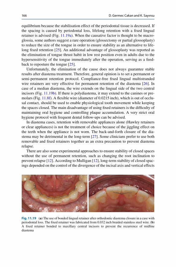
equilibrium because the stabilisation effect of the periodontal tissue is decreased. If the spacing is caused by periodontal loss, lifelong retention with a fi xed lingual retainer is advised (Fig. 11.19a ). When the causative factor is thought to be macro-glossia, some authors suggest a rare operation (glossectomy or partial glossoplasty) to reduce the size of the tongue in order to ensure stability as an alternative to life-long fi xed retention [ 25 ]. An additional advantage of glossoplasty was reported as the elimination of tongue thrust habit in low rest position even in adults due to the hypersensitivity of the tongue immediately after the operation, serving as a feed-back to reposture the tongue [ 25 ].
Unfortunately, the elimination of the cause does not always guarantee stable results after diastema treatment. Therefore, general opinion is to set a permanent or semi-permanent retention protocol. Compliance-free fi xed lingual multistranded wire retainers are very effective for permanent retention of the diastema [ 26 ]. In case of a median diastema, the wire extends on the lingual side of the two central incisors (Fig. 11.19b ). If there is polydiastema, it may extend to the canines or pre-molars (Fig. 11.8f ). A fl exible wire (diameter of 0.0215 inch), which is out of occlu-sal contact, should be used to enable physiological tooth movement while keeping the spaces closed. The main disadvantage of using fi xed retainers is the diffi culty of maintaining oral hygiene and controlling plaque accumulation. A very strict oral hygiene protocol with frequent dental follow-ups can be advised.
In diastema cases, retention with removable appliances alone (Hawley retainers or clear appliances) is not the treatment of choice because of the jiggling effect on the teeth when the appliance is not worn. The back-and-forth closure of the dia-stema may be detrimental in the long-term [ 27 ]. Some clinicians prefer to use both removable and fi xed retainers together as an extra precaution to prevent diastema relapse.
There are also some experimental approaches to ensure stability of closed spaces without the use of permanent retention, such as changing the root inclination to prevent relapse [ 12 ]. According to Mulligan [ 12 ], long-term stability of closed spac-ings depended on the control of the divergence of the incisal axis and vertical effects
a b
Fig. 11.19 ( a ) The use of bonded lingual retainer after orthodontic diastema closure in a case with periodontal loss. The fi xed retainer was fabricated from 0.032 inch braided stainless steel wire. ( b ) A fi xed retainer bonded to maxillary central incisors to prevent the recurrence of midline diastema
D. Germec Cakan and K. Sayınsu

167
of occlusal forces. But this approach does not seem to be widely used among orthodontists.
When diastema is relapsed, what can be done? If there is minimal relapse, dia-stema can be closed with removable appliances (retraction of the incisors by activat-ing the vestibular arch), clear aligners with or without set-up, segmental fi xed appliances or restorations (if the tooth proportions allow). When there is a major relapse, the clinician should reconsider the causes and replan comprehensive treat-ment either orthodontic, restorative or interdisciplinary.
References
1. Taylor JE. Clinical observations relating to the normal and abnormal frenum labii superioris. Am J Orthod Oral Surg. 1939;25:255–9.
2. Gardiner JH. Midline spaces. Dent Pract. 1987;17:287–98. 3. Weyman J. The incidence of median diastema during the eruption of the permanent teeth. Dent
Pract. 1987;17:276–98. 4. Shashua D, Artun J. Relapse after orthodontic correction of maxillary median diastema: a
follow-up evaluation of consecutive cases. Angle Orthod. 1999;69:257–63. 5. Sheridan JJ, Armbruster P. Clear plastic appliances for retention and tooth movement. In:
Graber TM, Vanarsdall Jr RL, Vig KWL, editors. Orthodontics: current principles and tech-niques. 4th ed. St. Louis: Elsevier Mosby; 2005. p. 1169.
6. Bishara SE. Development of dental occlusion. In: Bishara SE, editor. Textbook of orthodon-tics. Pennsylvania: W.B. Saunders Company; 2001. p. 56.
7. Proffi t WR, Fields Jr HW, Sarver DM. Contemporary orthodontics. 4th ed. St. Louis: Mosby Elsevier; 2007. p. 145–8.
8. Germeç D, Taner TU. Lower lip sucking habit treated with a lip bumper appliance. Angle Orthod. 2005;75:1071–6.
9. Tozlu M, Germeç D. Ortodontide Myofonksiyonel Tedavi Seçenekleri. 7 Tepe Klinik. 2008;2:34–42.
10. Smith SS, Buschang PH, Watanabe E. Interarch tooth size relationships of 3 populations: “does Bolton’s apply?”. Am J Orthod Dentofacial Orthop. 2000;117:169–74.
11. Kokich VG, Kokich VO. Interrelationship of orthodontics with periodontics and restorative dentistry. In: Nanda R, editor. Biomechanics and esthetic strategies in clinical orthodontics. St. Louis: Elsevier Saunders; 2005. p. 361.
12. Mulligan TF. Diastemas: is permanent retention really necessary? In: Nanda R, Kapila S, edi-tors. Current therapy in orthodontics. 1st ed. St. Louis: Mosby Elsevier; 2010. p. 215–27.
13. Polder BJ, Van’t Hof MA, Van der Linden FP, Kuijpers-Jagtman AM. A meta-analysis of the prevalence of dental agenesis of permanent teeth. Community Dent Oral Epidemiol. 2004;32:217–26.
14. Edwards JG. The diastema, the frenum, the frenectomy: a clinical study. Am J Orthod. 1977;71:489–508.
15. Dewel BF. The normal and abnormal labial frenum: clinical differentiation. J Am Dent Assoc. 1946;33:318–29.
16. Bishara SE. Management of diastemas in orthodontics. Am J Orthod. 1972;61:55–63. 17. Meister Jr F, Van Swol RL, Rank DF. The maxillary anterior frenectomy. J Wis Dent Assoc.
1981;57:205–10. 18. Joondeph DR. Stability, retention and relapse. In: Graber TM, Vanarsdall Jr RL, Vig KWL,
editors. Orthodontics: current principles and techniques. 5th ed. St. Louis: Elsevier Mosby; 2012. p. 1011.
11 Treatment Options, Timing and Sequencing: Orthodontics

168
19. Little RM, Reidel RA. Postretention evaluation of stability and relapse – mandibular arches with generalized spacing. Am J Orthod Dentofacial Orthop. 1989;95:37–41.
20. Jonsson T, Magnusson TE. Crowding and spacing in the dental arches: long-term development in treated and untreated subjects. Am J Orthod Dentofacial Orthop. 2010;138:384.e1–7.
21. de Morais JF, de Freitas MR, de Freitas KM, Janson G, Castello Branco N. Postretention sta-bility after orthodontic closure of maxillary interincisor diastemas. J Appl Oral Sci. 2014;22:409–15.
22. Becker A. The median diastema: a review of its aetiology. Israel J Dent Med. 1977;26:21–7. 23. Sullivan TC, Turpin DL, Artun J. A postretention study of patients presenting with a maxillary
median diastema. Angle Orthod. 1996;66:131–8. 24. Nanda R, Kapila S. Current therapy in orthodontics. 1st ed. St. Louis: Mosby Elsevier; 2010.
p. 215–27. 25. Attia Y. Midline diastemas: closure and stability. Angle Orthod. 1993;63:209–12. 26. Zachrisson BU. Clinical experience with direct-bonded orthodontic retainers. Am J Orthod.
1977;71:440–8. 27. Proffi t WR, Fields Jr HW, Sarver DM. Contemporary orthodontics. 4th ed. St. Louis: Mosby
Elsevier; 2007. p. 626–8.
D. Germec Cakan and K. Sayınsu