treatment of cutaneous lichen planus (part 1): a review of ... · despite the strong evidence of...
TRANSCRIPT

DERMATOLOGY | REVIEW ARTICLE
Treatment of cutaneous lichen planus (Part 1):A review of topical therapies and phototherapyYasmeen Thandar1*, Rivesh Maharajh1, Firoza Haffejee1 and Anisa Mosam2
Abstract: Background: Lichen planus is a chronic inflammatory immune-mediateddisease that more frequently affects the skin and oral mucosa. Various treatmentmodalities are available for the condition. The aim of this review is to provideclinicians with consolidated evidence of the various treatments of cutaneous lichenplanus (CLP). This first part focuses on topical therapies and phototherapy. Methods:Various Databases were searched for all studies up until January 2018, whichreported on topical therapies and phototherapy for CLP. There were no exclusionsbased on study design. Results: We identified four systemic reviews and fourreviews. We found additional single studies that contributed to this review. Evidencesupporting the use of topical corticosteroids, as a first-line therapy, is absent.Conclusion: Narrowband UVB is the preferred phototherapeutic treatment option forcutaneous lichen planus and should be considered before commencing systemictreatment. Topical calcineurin inhibitors show promising results despite evidenceonly available from case reports. Vitamin D3 is not recommended for the treatmentof cutaneous lichen planus due to poor patient outcomes. The second part of this
ABOUT THE AUTHORSYasmeen Thandar is a senior pharmacology lec-turer at Durban University of Technology (DUT)in South Africa. She is a clinical pharmacologistwith a PhD in Pharmacology from University ofKwaZulu-Natal (UKZN). Her research interests liein the evidence-based role of treating diseases,particularly in inflammatory skin diseases.
Rivesh Maharajh has a Master of MedicalSciences in Public Health from UKZN, with aninterest in communicable diseases. His researchfocus is on epidemiological studies involvingMalaria, Tuberculosis and HIV/AIDS.
Firoza Haffejee holds a PhD in Women’sHealth (UKZN) and is Associate Professor at DUT.Her current research centres on epidemiologyand public health issues. She lectures Physiologyand Epidemiology and supervises postgraduate.
Anisa Mosam is an Asssociate Professor in theDepartment of Dermatology at UKZN andPrincipal specialist dermatologist at King EdwardVIII and Inkosi Albert Luthuli Central Hospitals.Her area of expertise: HIV psoriasis, epidemiol-ogy of HIV skin disease; quality of life of HIVrelated dermatoses and drug reactions in HIV/AIDS
PUBLIC INTEREST STATEMENTLichen planus is a skin condition which has beennamed as it resembles the “lichens” in the plantworld. It is an itchy, chronic problem which can goon for years as it can be difficult to control andthe itching can be distressing for those affected.In some patients, it can cause significant scarring.Although various treatment options have beenused in the treatment of lichen planus, it is stillchallenging to choose the most effective one. Inthis investigation, all topical treatments usedwere evaluated so that both doctors and patientscould be better informed about their choices. Itwas found that topical steroids, the first line oftreatment for lichen planus, have not beeninvestigated enough to prove that they work.Light therapy in the form of ultraviolet light hasbeen shown to be effective and should be usedbefore embarking on oral treatments for lichenplanus.
Thandar et al., Cogent Medicine (2019), 6: 1582467https://doi.org/10.1080/2331205X.2019.1582467
© 2019 The Author(s). This open access article is distributed under a Creative CommonsAttribution (CC-BY) 4.0 license.
Received: 17 August 2018Accepted: 17 January 2019First Published: 15 February 2019
*Corresponding author: YasmeenThandar, Department of BasicMedical Sciences, Faculty of HeathSciences, Durban University ofTechnology, 29 Saltfleet road, Durban3630, South AfricaE-mail: [email protected]
Reviewing editor:Udo Schumacher, University MedicalCenter Hamburg-Eppendorf,Germany
Additional information is available atthe end of the article
Page 1 of 21

review will investigate the efficacy of systemic treatments for cutaneous lichenplanus in the current literature.
Subjects: Pharmaceutical Medicine; Dermatology; Pharmacy & Dispensing
Keywords: cutaneous lichen planus; topical treatments; phototherapy; review
1. IntroductionLichen Planus (LP) is a chronic inflammatory immune-mediated disease thatmore frequently affects theskin and oral mucosa (Le Cleach and Chosidow, 2012; Gorouhi, Davari, & Fazel, 2014). Other areas thatmay be affected include the scalp, hair, nails andmucousmembranes of the genitalia, oesophagus andconjunctiva (Le Cleach and Chosidow, 2012). The global prevalence of LP is estimated to be in the rangeof 0.22–5% of the population (Gorouhi et al., 2014). LP occurs in all age groups but affects adultssignificantly more than children (Gorouhi et al., 2014; Payette, Weston, Humphrey, Yu, & Holland,2015). The disease, although not gender specific has been reported to affect more women than men(Payette et al., 2015). Cutaneous lichen planus (CLP) presents as the traditional 6 “P’s” of LP—pruritic,purple, polygonal, planar, papules and plaques, frequently affecting the flexures of the extremities(Gorouhi et al., 2009;Usatine&Tinitigan, 2011). Variants of CLP are site specific and includehypertrophic,pigmentosus, annular, atrophic, follicular, linear or actinic forms on skin surfaces (Weston & Payette,2015). Generally, CLP is largely managed based on clinical experience, location and severity of thelesions, most of which resolve spontaneously within a few years (Weston & Payette, 2015). Despitetreatment, recurrence is common (Usatine & Tinitigan, 2011). Generalised eruptions have reported toheal faster than limited cutaneous disease. Hypertrophic LP is typically unrelenting (Gorouhi et al., 2009).
CLP is associated with intense itching and often pigmentation, which affects the patients qualityof life due to discomfort and cosmetic problems (Gorouhi et al., 2009). Despite numerous medi-cines available for the treatment of CLP, there exists a gap in the knowledge of recommendeddrugs as many of the prescribed treatments lack conclusive evidence for efficacy, accompanied byside effects and often produce disappointing results.
There have been four systematic reviews (SRs) (Antiga, Caproni, Parodi, Cianchini, & Fabbri, 2014;Atzmony, Reiter, Hodak, Gdalevich, & Mimouni, 2016; Cribier, Frances, & Chosidow, 1998; Fazel,2014) and four review articles (Asch & Goldenberg, 2011; Lehman, Tollefson, & Gibson, 2009;I. Manousaridis, Manousaridis, Peitsch, & Schneider, 2013; Puza & Cardones, 2017) published overthe past three decades which help in ascertaining evidence of efficacy of many treatmentmodalities. However, due to the varying inclusion and exclusion criteria amongst these reviews,and the advent of newer treatments being tested, no consolidated publication exists whichprovides reports of evidence of all types of studies carried out for CLP. This two-part overviewpresents the findings from all previously published SRs and reviews, including studies that havebeen omitted in these publications for unidentified reasons and novel studies that have becomeevident subsequent to these publications.
Part 1 addresses the current literature focusing on topical therapies and phototherapy for thetreatment of CLP. Part 2 encompasses all systemic treatments for CLP. The aim is to provideclinicians with a summarised and consolidated evidence of the various treatments of CLP; hencemost of the treatments have been tabulated providing information on the type of study, dosageused, study sizes, outcome, as well as the category of evidence.
2. Methodology
2.1. Data sources and search strategyRelated literature published up until January 2018 were obtained from the following electronicdatabase searches: Cochrane Library, Google Scholar, Medline, PubMed, EBSCOHost andScienceDirect. The following search terms were transcribed to yield articles of relevance:
Thandar et al., Cogent Medicine (2019), 6: 1582467https://doi.org/10.1080/2331205X.2019.1582467
Page 2 of 21

“cutaneous lichen planus”, “treatment”, “systematic review” and “review” in combination with;“topical treatment”, “systemic treatment”, “UV light/phototherapy”; “low molecular weightheparin”, “alternative/complementary medicine”, “calcineurin inhibitors”. Screening of the litera-ture was performed independently by two authors (YT and RM) in order to validate the reliability ofthe information and prevent author bias. Reference lists of included papers were scanned, andfurther relevant publications were retrieved. This review presents findings of all studies includingthe most recent current literature available.
2.2. Inclusion and exclusion criteriaWe included all English studies that have been previously published in peer-reviewed journals upto January 2018. There was no restriction for the type of study and hence we included rando-mised controlled trials (RCTs), non-randomised control studies, cohort studies, case series, casereports and anecdotal studies. We included the following clinical subtypes of CLP—hypertrophic,pigmentosus, annular, atrophic, follicular, linear or actinic. Studies of oral lichen planus withcutaneous involvement were also included. We excluded those studies that focused on solelyoral involvement without cutaneous lesions, lichen planopilaris, palmoplantar and lichenpemphigoids.
2.3. Formulation of study strategyUpon retrieval of all published literature, CLP studies were collated according to the treat-ment modalities. We found four systematic reviews (SRs) (Antiga et al., 2014; Atzmony et al.,2016; Cribier et al., 1998; Fazel, 2014) and four review articles (Asch & Goldenberg, 2011;Lehman et al., 2009; Manousaridis et al., 2013; Puza & Cardones, 2017) published on CLPincluding additional studies not mentioned by the current reviews. To date, the most recentpublished SR by Atzmony et al. (2016) was used as a benchmark to compare data frompreviously published SRs and reviews, in addition of any relevant old or current literatureobtained. Table 1 provides a summary of the criteria of selection of scientific publications foreach SR and review previously published.
2.4. Level of evidence gradingThree different grading systems were used to describe the level of evidence in the various SRs.These included the Grading of Recommendation, Development and Evaluation (GRADE) system,American College of Physicians (ACP) guidelines grading system and Sackett’s system of grad-ing (Table 1). For the purpose of standardisation for evidence-based medicine, we used theGRADE system (Guyatt et al., 2008) to categorise each study. The GRADE system offers anexplicit and comprehensive grading criterion and provides clear characterisation of the levelsof evidence and strength of recommendation for clinicians (Guyatt et al., 2008). Where studieswere already graded (as in the respective SRs), we represented it using the GRADE format forconsistency. The level of evidence is based on the quality of the study i.e. study design,consistency and degree of specificity. These are graded as High, Moderate, Low and VeryLow. The strength of recommendation is graded as either strong or conditional (weak),based on the treatment outcome in individual studies (Guyatt et al., 2008; Schunemann, Hill,Guyatt, Akl, & Ahmed, 2011).
● High level of evidence is allocated to studies that have a controlled trial study design whichminimises the risk of bias and have a high confidence that the true effect may coincide withthe estimated effect.
● Moderate level studies are in line with high level of evidence but there may be a possibility ofchange in the estimate.
● Low level of evidence may have limited confidence on the estimate of effect, while Very lowlevels of evidence in studies that have minimal confidence and that the true effect may besubstantially different from the estimate of effect.
Thandar et al., Cogent Medicine (2019), 6: 1582467https://doi.org/10.1080/2331205X.2019.1582467
Page 3 of 21

Table1.
Summaryof
publishe
dsy
stem
atic
review
san
dreview
sforCL
P
System
atic
review
s
Referenc
ecitation
Databa
sesus
edDateof
search
Num
berof
stud
ies
includ
edIn
clus
ioncriteria
Grading
system
used
toas
sess
streng
thof
eviden
ce
Atzmon
yet
al.,20
16Pu
bMed
,CEN
TRAL,
ClinicalTrials.gov
registry
TillMay
2014
16To
tal:
12-RC
Ts4-no
nRC
Ts
Allrand
omised
controlle
dtrials,
Non
-ran
domised
case-
controls
tudies,
Coho
rtstud
ieswith
more
than
onetrea
tmen
tarm
GRA
DE(Rai,K
aur,&Ku
mar,2
002)
system
(4leve
ls:h
igh,
mod
erate,
low,v
erylow)
mod
erateto
high
=RC
Tswith
strong
eviden
ceve
rylow
tolow
=co
hort
stud
iesthat
prov
ideob
servationa
levide
nce
Fazel,20
14;T
uran
,Ba
skan
,Tun
ali,Ya
zici,a
ndSa
ricao
glu,
2009
PubM
edEM
BASE
Coch
rane
Datab
aseof
SRs
Coch
rane
CentralR
egister
ofCo
ntrolle
dTrials
Datab
aseof
Abs
trac
tsof
Review
sof
Effects
Hea
lthTe
chno
logy
Assessm
entDatab
ase
Till20
122SR
s9RC
TsAllSR
san
dRC
Tsof
any
design
RCTs
that
compa
redat
leas
ton
etrea
tmen
tarm
with
control,plac
ebo,
alternatetherap
yor
notrea
tmen
t
ACP
guidelines
grad
ingsystem
(Ans
ari,
Hen
derson
,Stott,&
Parr,2
017)
(2leve
ls;
high
,mod
erate)
High=allR
CTsthat
was
equa
lorha
lfthe
inclus
ioncrite
riamod
erate=mee
tstheminim
uminclus
ion
crite
riaof
RCTs
Antigaet
al.,20
14Med
line
Janu
ary19
99–
Nov
embe
r20
1221
Total:
3-Le
velB
stud
ies
18—Le
velC
stud
ies
Allpa
pers
publishe
dbe
twee
n19
99–20
12ab
outtrea
tmen
tof
CLP
Sackett’s
(Cha
uhan
,De,
Han
da,N
aran
g,&
Saikia,2
017)
system
(3leve
ls;A
,B,C
)A=
largeRC
Twith
defin
edco
nclusion
s,B=RC
Tswith
unce
rtainresu
lts,
C=trials
with
out
rand
omised
controls
Cribieret
al.,19
98MED
LINE
BIOSIS
Till19
9827
Total:
1—Le
velB
stud
y26
—Le
velC
stud
ies
Allpa
pers
publishe
dfrom
ince
ptionof
trea
tmen
ttill
1998
forCL
P
Sackett’s
(Cha
uhan
etal.,20
17)system
(3leve
ls;A
,B,C
)A=largeRC
Twith
defin
edco
nclusion
s,B=RC
Tswith
unce
rtainresu
lts,
C=trials
with
outrand
omised
controls
Review
s
Puza
andCa
rdon
es,2
017
PubM
ed,C
ENTR
AL,
EBSC
Oho
st19
72–April20
1721
Total:
9RC
Ts6op
entrials
6ca
seserie
s/co
hort/cas
erepo
rts
AllRC
Tsan
dtherap
eutic
stud
iesrelatin
gto
CLP
trea
tmen
ts
Non
e
(Con
tinue
d)
Thandar et al., Cogent Medicine (2019), 6: 1582467https://doi.org/10.1080/2331205X.2019.1582467
Page 4 of 21
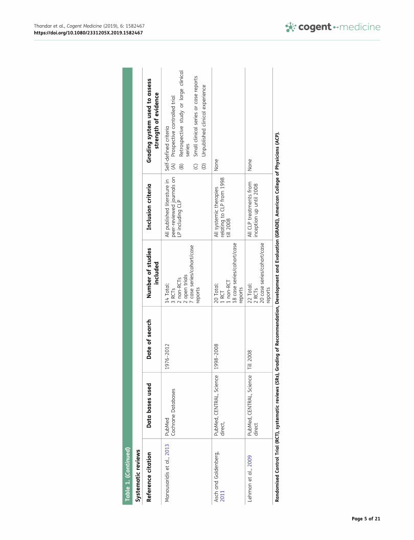
Table1.
(Con
tinu
ed)
System
atic
review
s
Referenc
ecitation
Databa
sesus
edDateof
search
Num
berof
stud
ies
includ
edIn
clus
ioncriteria
Grading
system
used
toas
sess
streng
thof
eviden
ce
Man
ousa
ridiset
al.,20
13Pu
bMed
Coch
rane
Datab
ases
1976
–20
1214
Total:
3RC
Ts2no
n-RC
Ts2op
entrials
7ca
seserie
s/co
hort/cas
erepo
rts
Allpu
blishe
dliteraturein
peer-rev
iewed
journa
lson
LPinclud
ingCL
P
Self-de
fined
crite
ria(A)
Pros
pectiveco
ntrolle
dtrial
(B)
Retros
pective
stud
yor
large
clinical
serie
s
(C)
Smallc
linical
serie
sor
case
repo
rts
(D)
Unp
ublishe
dclinical
expe
rienc
e
Aschan
dGolde
nberg,
2011
PubM
ed,C
ENTR
AL,Scienc
edirect,
1998
–20
0820
Total:
1RC
T1no
n-RC
T18
case
serie
s/co
hort/cas
erepo
rts
Allsystem
ictherap
ies
relatin
gto
CLPfrom
1998
till2
008
Non
e
Lehm
anet
al.,20
09Pu
bMed
,CEN
TRAL,Scienc
edirect
Till20
0822
Total:
2RC
Ts20
case
serie
s/co
hort/cas
erepo
rts
AllCL
Ptrea
tmen
tsfrom
ince
ptionup
until
2008
Non
e
Rand
omised
ControlTrial(RCT
),sy
stem
atic
review
s(SRs
),Grading
ofRe
commen
dation
,Dev
elop
men
tan
dEv
alua
tion
(GRA
DE),A
merican
Colle
geof
Phys
icians
(ACP
).
Thandar et al., Cogent Medicine (2019), 6: 1582467https://doi.org/10.1080/2331205X.2019.1582467
Page 5 of 21

3. Treatment regimens
3.1. Topical interventions for cutaneous lichen planusTopical treatments for CLP include corticosteroids, calcineurin inhibitors e.g. tacrolimus andVitamin D3 analogues, e.g. calcipotriol. Also included in this review are phototherapy treatments.
3.2. Topical corticosteroidsClass 1 (ultra-high potency) and Class II (high potency) topical corticosteroids are still consideredthe first line of treatment for CLP due to their anti-inflammatory properties that focus on localisedlesions and reduce pruritus (Ramachandran, 2014). Six studies on topical corticosteroids wereidentified in the literature. Only one RCT was reported by Atzmony et al. (2016) which includedbetamethasone valerate 0.1% vs calcipotriol. In addition to that, four studies reported in theAtzmony et al. SR were non-randomised case-control trials (Atzmony et al., 2016). Two studiesconducted before 1970 were reported by Cribier et al. (1998) but were excluded by Atzmony et al.(2016). A preceding study conducted in 1976 by Björnberg and Hellpen (1976) was only reported ina review by Manousaridis et al. (Hazra et al., 2013).
Upon evaluations of studies that explore the effectiveness of topical corticosteroids, it is apparentthat majority of these studies have low patient sample size with the largest study group of 25 patients.Only a single RCT was conducted (Theng et al., 2004), following two non-RCTs (Chopra, Mittal, & Kaur,1999; Sharma & Mishra, 2003), two open, non-comparative trials (Björnberg & Hellpen, 1976; Marsden,1968) and one case-controlled study (Brock & Cullen, 1967). Results varied across studies withdifferent corticosteroid formulations used. Response across each of these studies demonstrated nosignificant difference with a lower response between the topical corticosteroid betamethasone vale-rate 0.1% and topical calcipotriol (Theng et al., 2004). Similarly, with betamethasone diproprionate0.05%, no significant difference was observed in a comparison against PUVAsol (Sharma & Mishra,2003). Furthermore, in an open trial by Björnberg and Hellpen (1976), a large effect (73.7% improve-ment) with betamethasone-17,21-diproprionate 0.05% was demonstrated, although there was nocomparative measure (Björnberg & Hellpen, 1976). Other studies with topical fluocinonide acetonidereported a low (28.6%) complete response (Marsden, 1968) and with triamcinolone 0.5%, a lowerpercentage of patients had a better response with the topical steroid (Brock & Cullen, 1967). TopicalClobetasol propionate lotion together with a hydrocolloid occlusive dressing was used in patients withchronic skin diseases including CLP. It was reported that in those patients with CLP, 2.8 weeks was theaverage time to remission (Volden, 1992).
Grading of the studies showed that none of the studies published reported a high level of evidence.Apart from the one RCT conducted by Theng et al. (Theng et al., 2004), all other studies demonstratedeither a low or very low level of evidence. Considering that no high-level evidence exists for the use oftopical corticosteroids in CLP, its routine use by clinicians as first-line treatment for CLP is primarilybased on their own experiences. The use of topical corticosteroids under occlusion and intra-lesionalcorticosteroid injections are primarily anecdotal and there are no published trials demonstrating theirefficacy. The strength of recommendation for topical corticosteroids is discretionary, and its role asfirst-line therapy is therefore arguable. Table 2 provides a summary of all published topical corticos-teroid studies to date.
3.3. PhototherapyPhototherapy is often used in the treatment of various inflammatory skin disorders (Vangipuram &Feldman, 2016). It is a specialised technique that can act as an alternative treatment to assist inclearing of lesions observed in CLP. The mechanism of its action is controversial since sunlight isknown to aggravate certain variants of CLP (Taneja & Taylor, 2002), although at different wave-lengths, treatment using phototherapy is widely explored. Following the very first SR in 1998 byCribier et al. (1998), only one RCT on phototherapy was conducted by Iraji et al. (2011) andreported in the three subsequent SRs (Antiga et al., 2014; Atzmony et al., 2016; Turan et al.,2009). Since then, there have been no additional RCTs on phototherapy. Atzmony et al. (2016) also
Thandar et al., Cogent Medicine (2019), 6: 1582467https://doi.org/10.1080/2331205X.2019.1582467
Page 6 of 21

Table2.
Summaryof
topica
lco
rticos
teroid
publishe
dstud
ies
Corticos
teroids(Top
ical)
Stud
yde
sign
Autho
r/ye
arTy
peof
Lich
enplan
usTrea
tmen
t(n)
Compa
rative
Trea
tmen
t(n)
Leve
lof
Eviden
ceRe
sult
RCT
Then
get
al.,20
04Gen
eralised
Betametha
sone
valerate
0.1%
bdsfor
12wee
ks(n
=16
)
Topica
lCalcipo
triol
0.05
%bd
sfor12
wee
ks(n
=15
)
Mod
erate
50%
lesion
flatten
ingwith
betametha
sone
.How
ever,n
odifferen
cebe
twee
ngrou
pswere
repo
rted
after12
wee
ks.
Non
-RCT
Sharmaan
dMishra,
2003
Hyp
ertrop
hic,
Guttate,
atroph
icBe
tametha
sone
diprop
iona
te0.05
%da
ilyfor12
wee
ks(tog
ethe
rwith
cetirizine
10mgda
ily)
(n=24
)
PUVA
sol4
mg/kg
onalternateda
ys3Xwee
kfor12
wee
ks(n
=23
)vs Metronida
zole
200m
gtdsfor3wee
ks(n
=23
)
Low
Goo
dan
dex
celle
ntresp
onse
was
notedwith
betametha
sone
in54
.2%
ofpa
tients.
This
was
compa
rableto
PUVA
sola
ndabe
tter
resp
onse
compa
redto
metronida
zole.
Non
-RCT
Chop
raet
al.,19
99Va
rious
type
sinclud
ing
Clas
sic,
liche
nac
tinicus
,lin
earLP
,LPP
,muc
osal
LP
Betametha
sone
0.1%
bdsfor3mon
ths
(n=25
)
OralD
apso
ne50
mgtds
for3mon
ths(tog
ethe
rwith
chlorphe
niramine
malea
te4m
gbd
san
dco
conu
toil)
(n=50
)
Low
Resp
onse
tobe
tametha
sone
was
less
compa
redto
daps
one(40%
good
resp
onse
with
betametha
sone
vs58
%da
pson
e).
Ope
ntrial
Björnb
ergan
dHellpen
,19
76NS
0.05
%be
tametha
sone
-17
,21-diprop
iona
teointmen
ton
ceor
twice
daily
for2–
3wee
ks(n
=19
)
Non
eVe
ryLo
wPa
tientstrea
tedwerethos
ewho
mprev
ious
lyde
mon
strated
resistan
ceto
prolon
ged
trea
tmen
twith
0.05
%be
tametha
sone
−17
-valerate
ointmen
t.73
.7%
improv
emen
twas
noted.
Theremaining
26.3%
hadno
resp
onse.
Ope
ntrial
Marsd
en,1
968
NS
Fluo
cino
nide
aceton
ide
0.2%
tds(n
=7)
Non
eVe
ryLo
w28
.6%
hadco
mpleteresp
onse
(after
unkn
ownde
lay)
Case
controlle
d(dou
bleblind)
Broc
kan
dCu
llen,
1967
NS
Triamcino
lone
0.5%
infle
xibleco
llodion
aceton
ide.
One
halfof
body
(n=7)
Excipien
tOther
halfof
body
Very
Low
42.8%
hadabe
tter
resp
onse
ontheco
rticos
teroid
side
Rand
omised
controltrial(RCT
),Lich
enplan
usPe
mph
igoids
(LPP
),Tw
iceda
ily(bds
),Th
reetimes
daily
(tds
),Not
stated
(NS)
Thandar et al., Cogent Medicine (2019), 6: 1582467https://doi.org/10.1080/2331205X.2019.1582467
Page 7 of 21
reported on three non-RCTs, and although part of the inclusion date, this SR did not include theGonzalez, Momtaz-T, and Freedman (1984) study in 1984 which was reported by Cribier et al.(1998). Further studies which were predominately case series, non-comparative open trials andretrospective studies were reported in SRs by Cribier et al. (1998) and Antiga et al. (2014). Thesestudies did not meet the Atzmony et al. (2016) SR inclusion criteria.
Additionally, we found the other two studies (case series), which explored the efficacy ofphototherapy, that were not mentioned in any of the SRs and reviews despite meeting theirinclusion criteria. One older study by Gamil, Nassar, Saadawi, El-Qashishi, and Ahmed (2009) anda novel treatment by Fan et al. (2015). A four-year retrospective study by Solak, Sevimli Dikicier,and Erdem (2016) which demonstrated a significant positive response with NBUVB for generalisedlichen planus was only reported in the Puza and Cardones review (Samycia & Lin, 2012). Table 3summarises all documented phototherapy studies.
3.3.1. Ultraviolet B (UVB) therapyCommonly used in the treatment of CLP, Narrow-band UVB (NBUVB) and Broad-band UVB (BBUVB)radiation exposure are known to reduce skin lesions by causing apoptosis of the affected cells andinterfering with immunological functions that inhibit the expression of related inflammatoryfactors (Berneburg, Röcken, & Benedix, 2005). Post 2002 and to date, there were seven publishedstudies conducted with NBUVB with sample sizes up to a maximum of 43 patients. Results fromthe only RCT conducted with phototherapy demonstrated that NBUVB had a better response tosystemic prednisolone therapy (Iraji et al., 2011). This evidence was considered moderate. NBUVBwas compared to oral PUVA in one non-RCT where it was reported to be comparable to oral PUVA(Wackernagel et al., 2007). The evidence of efficacy was considered low. Three case seriesdemonstrated a positive response to NBUVB. Evidence of efficacy from these was very low(Gamil et al., 2009; Saricaoglu, Karadogan, Baskan, & Tunali, 2003; Taneja & Taylor, 2002). Threeretrospective studies, although regarded as low evidence studies, reported a favourable outcomefor NBUVB treatment; two of which were non-comparative studies by Solak et al. (Majid, 2017) andHabib et al. (2005), with one comparative to BBUVB reported by Pavlotsky, Nathansohn, Kriger,Shpiro, and Trau (2008).
Despite the level of evidence in majority of studies with NBUVB being considered low, animproved outcome with NBUVB was reported in six of the seven studies. In only one study,NBUVB was comparable to oral PUVA in the long term in terms of efficacy and relapse rates,with oral PUVA demonstrating an initial superior response. Oral PUVA may have a better responsein patients with hypertrophic LP who do not respond to NBUVB. The recommendation based onthese results is that NBUVB should be considered as an effective treatment option for CLP beforeproceeding to use systemic corticosteroids or systemic immunosuppressants. However, the highcosts associated with outpatient phototherapy and the frequency of sessions required, need to beconsidered.
3.3.2. Psoralen plus Ultraviolet A (PUVA) therapyPhotochemotherapy using Ultraviolet A light in conjunction with psoralen (as a photosensitizer)enhances the efficacy of UVA in the treatment of CLP (Vangipuram & Feldman, 2016). There are noRCTs with psoralen plus ultraviolet A (PUVA) reported, with either oral or bath therapy. Although,one non-RCT conducted in 1987 by Helander, Jansen, and Meurman (1987) established that thereis no significant difference between oral PUVA and bath PUVA with no significant difference in thelong-term outcomes between PUVA and no treatment. In contrast to NBUVB, while oral PUVAinitially showed a clinically better response, there was a very similar overall response in long termobservation (Wackernagel et al., 2007). Sharma and Mishra (2003) reported that PUVA is compar-able to treatment with topical betamethasone and cetirizine combined. In other studies,a favourable response to PUVA was evident in one non-RCT (Gonzalez et al., 1984) and four caseseries (Karvonen & Hannuksela, 1985; Kerscher, Volkenandt, Lehmann, Plewig, & Röcken, 1995;Ortonne, Thivolet, & Sannwald, 1978; Väätäinen, Hannuksela, & Karvonen, 1981) between 1978
Thandar et al., Cogent Medicine (2019), 6: 1582467https://doi.org/10.1080/2331205X.2019.1582467
Page 8 of 21

Table3.
Summaryof
photothe
rapy
publishe
dstud
ies.
Photothe
rapy
Narrow/Broad
Band
UVB(N
BUVB/BB
UVB)
Stud
yde
sign
Autho
r/ye
arTy
peof
Lich
enplan
usTrea
tmen
tregime(n)
Compa
rative
trea
tmen
t(n)
Leve
lof
Eviden
ceRe
sult
RCT
Irajie
tal.,
2011
Gen
eralised
NBU
VB3tim
esawee
kat
70%
MED
9j/cm
2
6wee
ks(n
=23
)
Pred
niso
lone
0.3m
g/kg
6wee
ks(n
=23
)Mod
erate
NBU
VBha
d52
.2%
completeresp
onse
and
47.8%
partialrespo
nse.
This
was
better
than
pred
niso
lone
trea
tmen
t.
non-RC
TWac
kernag
elet
al.,20
07Gen
eralised
;Hyp
ertrop
hic(2
patie
nts)
NBU
VB(22.5ex
posu
res)
0.34
j/cm
2
(n=13
)8.2wee
ks
Oralo
xsoralen
1.2m
g/kg
+UVA
1j/cm
2
(n=15
)10
.5wee
ks
Low
67%
completeresp
onse
with
PUVA
and
33%
partialrespo
nse.
30.1%
complete
resp
onse
with
NBU
VBan
d46
.2%
partial
resp
onse.L
ongterm
follo
w-upsh
owed
that
theeffectiven
essof
oral
PUVA
isco
mpa
rableto
NBU
VB.
Case
serie
sGam
ilet
al.,
2009
Gen
eralised
NBU
VB3tim
eswee
kly
40sessions
(0.411
–0.70
7J/cm
2)(n
=16
)
Non
eVe
rylow
Completeresp
onse
was
observed
in69
%of
patie
nts,pa
rtialrespo
nsein
12%
ofpa
tients
and19
%ha
dno
resp
onse.
Case
serie
sSa
ricao
glu
etal.,20
03Lo
calised
(eith
ertrun
kor
extrem
ities)
Narrow
band
UVB
3–4tim
eswee
kly
(30sessions
)(n
=10
)mea
ncu
mulativedo
se=17
.7J/cm
2
Non
eVe
rylow
50%
patie
ntsresp
onde
dco
mpletely
40%
werepa
rtially
resp
onsive
,while
10%
show
edno
improv
emen
t.
Case
serie
sTa
neja
and
Taylor,2
002
Loca
lised
(mos
tlytrun
kan
dex
trem
ities);with
oral
lesion
s(2
patie
nts)
Narrow
band
UVB
2–3tim
eswee
kly
(mea
n=40
sessions
)(n
=5)
mea
ncu
mulativedo
se=87
.2J/cm
2
Non
eVe
rylow
Pruritu
sresp
onde
dea
rlyin
allp
atients.
Flattening
oflesion
swas
achiev
edin
loca
lcu
tane
ouslesion
sbu
tno
tin
oral
lesion
s.
Retros
pective
Solaket
al.,
2016
Gen
eralised
NBU
VB(durationva
ried)
(n=24
)
Non
eVe
rylow
45.8%
completeresp
onse
toNBU
VB,2
0.5%
partialrespo
nsean
d33
.7%
noresp
onse.
Retros
pective
Pavlotsky
etal.,20
08Gen
eralised
NBU
VBthreetim
eswee
kly
Mea
ncu
mulativedo
se31
.5joules/
cm2
(n=43
)
BBUVB
threetim
eswee
kly
Mea
ncu
mulativedo
se11
joules/cm
2
(n=7)
Very
low
NBU
VBha
d85
%co
mpleteresp
onse
and
BBUVB
had70
%co
mpe
teresp
onse
after
34.7
mon
thsof
remission
.
(Con
tinue
d)
Thandar et al., Cogent Medicine (2019), 6: 1582467https://doi.org/10.1080/2331205X.2019.1582467
Page 9 of 21
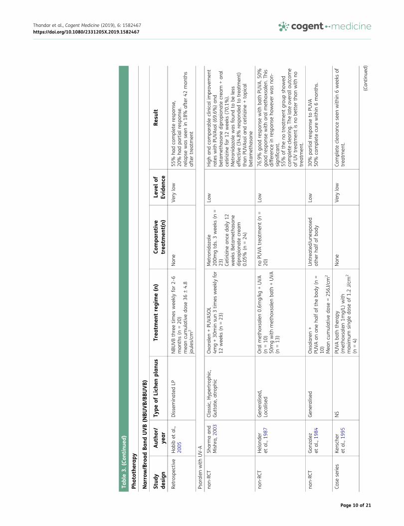
Table3.
(Con
tinu
ed)
Photothe
rapy
Narrow/Broad
Band
UVB(N
BUVB/BB
UVB)
Stud
yde
sign
Autho
r/ye
arTy
peof
Lich
enplan
usTrea
tmen
tregime(n)
Compa
rative
trea
tmen
t(n)
Leve
lof
Eviden
ceRe
sult
Retros
pective
Hab
ibet
al.,
2005
Disseminated
LPNBU
VBthreetim
eswee
klyfor2–
6mon
ths(n
=20
)mea
ncu
mulativedo
se36
±4.8
joules/cm
2
Non
eVe
rylow
55%
hadco
mpleteresp
onse,
20%
hadpa
rtialrespo
nse.
relaps
ewas
seen
in18
%after42
mon
ths
aftertrea
tmen
t
Psoralen
with
UV-A
non-RC
TSh
armaan
dMishra,
2003
Clas
sic,
Hyp
ertrop
hic,
Guttate,a
trop
hic
Oxo
ralen+PU
VASO
L4m
g+30
min
sun3tim
eswee
klyfor
12wee
ks(n
=23
)
Metronida
zole
200m
gtds.
3wee
ks(n
=23
)Ce
tirizineon
ceda
ily12
wee
ksBe
tametha
sone
diprop
iona
tecrea
m0.05
%(n
=24
)
Low
Highan
dco
mpa
rableclinical
improv
emen
trateswith
PUVA
sol(69
.6%)an
dbe
tametha
sone
diprop
iona
tecrea
m+oral
cetirizinefor12
wee
ks(70.1%
).Metronida
zole
was
foun
dto
beless
effective(34.8%
resp
onde
dto
trea
tmen
t)than
PUVA
sola
ndce
tirizine+topica
lbe
tametha
sone
non-RC
THelan
der
etal.,19
87Gen
eralised
,Lo
calised
Oralm
etho
xsalen
0.6m
g/kg
+UVA
(n=10
)50
mgwith
metho
xsalen
bath
+UVA
(n=13
)
noPU
VAtrea
tmen
t(n
=20
)Lo
w76
.9%
good
resp
onse
with
bath
PUVA
.50%
good
resp
onse
with
oral
metho
xsalen
.This
differen
cein
resp
onse
howev
erwas
non-
sign
ifica
nt.
55%
oftheno
trea
tmen
tgrou
psh
owed
completeclea
ring.
Thelate
overallo
utco
me
ofUVtrea
tmen
tis
nobe
tter
than
with
notrea
tmen
t.
non-RC
TGon
zalez
etal.,19
84Gen
eralised
Oxsolaren
+PU
VAon
oneha
lfof
thebo
dy(n
=10
)Mea
ncu
mulativedo
se=25
6J/cm
2
Untreated
/une
xpos
edothe
rha
lfof
body
Low
30%
partialrespo
nseto
PUVA
50%
completecu
rewith
in6mon
ths.
Case
serie
sKe
rsch
eret
al.,19
95NS
PUVA
bath
therap
y(m
etho
xsalen
1mg/L)
with
max
imum
sing
ledo
seof
1.2J/cm
2
(n=4)
Non
eVe
rylow
Completeclea
ranc
eseen
with
in6wee
ksof
trea
tmen
t.
(Con
tinue
d)
Thandar et al., Cogent Medicine (2019), 6: 1582467https://doi.org/10.1080/2331205X.2019.1582467
Page 10 of 21

Table3.
(Con
tinu
ed)
Photothe
rapy
Narrow/Broad
Band
UVB(N
BUVB/BB
UVB)
Stud
yde
sign
Autho
r/ye
arTy
peof
Lich
enplan
usTrea
tmen
tregime(n)
Compa
rative
trea
tmen
t(n)
Leve
lof
Eviden
ceRe
sult
Case
serie
sKa
rvon
enan
dHan
nuksela,
1985
NS
PUVA
bath
therap
y(trio
xsalen
+UVA
)(n
=75
)
Non
eVe
rylow
65%
cure
rate
(after
2cycles)
15%
improv
emen
trate
25%
relaps
erate
Case
serie
sVä
ätäine
net
al.,19
81pa
pular/
hype
rtroph
icPU
VAba
ththerap
y(trio
xsalen
3mg/L)
(n=19
)
Non
eVe
rylow
Completereco
very
from
papu
larLP
notedin
all1
6pa
tients
67%
completeresp
onse
and33
%pa
rtial
resp
onse
seen
in33pa
tientswith
hype
rtroph
icLP
Case
serie
sOrton
neet
al.,19
78NS
PUVA
(0.4mg/kg
metho
xsalen
+UVA
)mea
ncu
mulativedo
se=10
7J/cm
2
(n=7)
Non
eVe
rylow
85.7%
hadco
mpleteresp
onse.
Nofurthe
rde
tails
repo
rted
.
Laserdiod
etherap
y
Case
serie
sFa
net
al.,
2015
Loca
lised
(5males
loca
lised
onpe
nis,
1femaleloca
lised
forehe
ad,n
ose,
mou
than
dothe
rloca
lised
onwris
t)
10%
5-am
inolev
ulinicac
id+63
5nm
laserdiod
e(ALA
-med
iated
photod
ynam
ictherap
y)ap
plied
topica
lly(n
=7)
Non
eVe
rylow
71.4%
completeresp
onse
28.6%
partialrespo
nse
overallp
atientsresp
onde
dwellto
trea
tmen
t.
Rand
omised
controltrial(RCT
),minim
alerythe
mado
se(M
ED),Th
reetimes
daily
(tds
),Not
stated
(NS)
Thandar et al., Cogent Medicine (2019), 6: 1582467https://doi.org/10.1080/2331205X.2019.1582467
Page 11 of 21
and 1995. The level of evidence in these studies with PUVA is considered low. PUVA is notconsidered the ideal choice of treatment for CLP. NBUVB is a preferred phototherapeutic optionhowever patients that do not respond well to NBUVB may be susceptible to PUVA treatment.
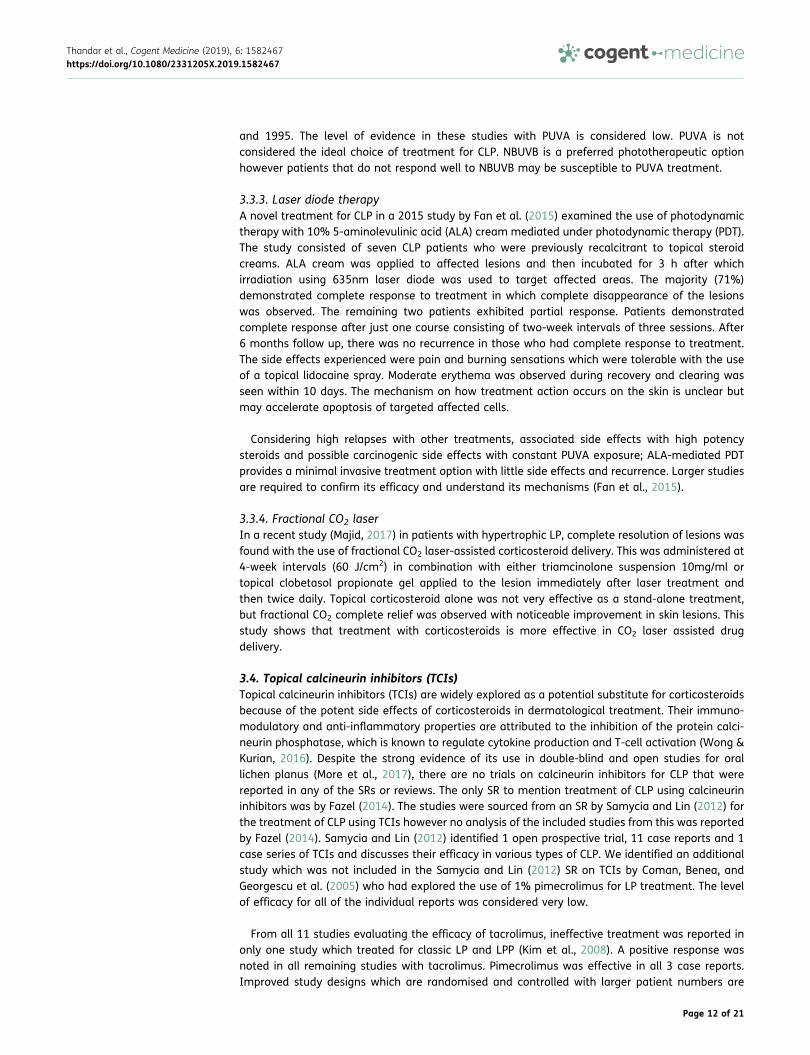
3.3.3. Laser diode therapyA novel treatment for CLP in a 2015 study by Fan et al. (2015) examined the use of photodynamictherapy with 10% 5-aminolevulinic acid (ALA) cream mediated under photodynamic therapy (PDT).The study consisted of seven CLP patients who were previously recalcitrant to topical steroidcreams. ALA cream was applied to affected lesions and then incubated for 3 h after whichirradiation using 635nm laser diode was used to target affected areas. The majority (71%)demonstrated complete response to treatment in which complete disappearance of the lesionswas observed. The remaining two patients exhibited partial response. Patients demonstratedcomplete response after just one course consisting of two-week intervals of three sessions. After6 months follow up, there was no recurrence in those who had complete response to treatment.The side effects experienced were pain and burning sensations which were tolerable with the useof a topical lidocaine spray. Moderate erythema was observed during recovery and clearing wasseen within 10 days. The mechanism on how treatment action occurs on the skin is unclear butmay accelerate apoptosis of targeted affected cells.
Considering high relapses with other treatments, associated side effects with high potencysteroids and possible carcinogenic side effects with constant PUVA exposure; ALA-mediated PDTprovides a minimal invasive treatment option with little side effects and recurrence. Larger studiesare required to confirm its efficacy and understand its mechanisms (Fan et al., 2015).
3.3.4. Fractional CO2 laserIn a recent study (Majid, 2017) in patients with hypertrophic LP, complete resolution of lesions wasfound with the use of fractional CO2 laser-assisted corticosteroid delivery. This was administered at4-week intervals (60 J/cm2) in combination with either triamcinolone suspension 10mg/ml ortopical clobetasol propionate gel applied to the lesion immediately after laser treatment andthen twice daily. Topical corticosteroid alone was not very effective as a stand-alone treatment,but fractional CO2 complete relief was observed with noticeable improvement in skin lesions. Thisstudy shows that treatment with corticosteroids is more effective in CO2 laser assisted drugdelivery.
3.4. Topical calcineurin inhibitors (TCIs)Topical calcineurin inhibitors (TCIs) are widely explored as a potential substitute for corticosteroidsbecause of the potent side effects of corticosteroids in dermatological treatment. Their immuno-modulatory and anti-inflammatory properties are attributed to the inhibition of the protein calci-neurin phosphatase, which is known to regulate cytokine production and T-cell activation (Wong &Kurian, 2016). Despite the strong evidence of its use in double-blind and open studies for orallichen planus (More et al., 2017), there are no trials on calcineurin inhibitors for CLP that werereported in any of the SRs or reviews. The only SR to mention treatment of CLP using calcineurininhibitors was by Fazel (2014). The studies were sourced from an SR by Samycia and Lin (2012) forthe treatment of CLP using TCIs however no analysis of the included studies from this was reportedby Fazel (2014). Samycia and Lin (2012) identified 1 open prospective trial, 11 case reports and 1case series of TCIs and discusses their efficacy in various types of CLP. We identified an additionalstudy which was not included in the Samycia and Lin (2012) SR on TCIs by Coman, Benea, andGeorgescu et al. (2005) who had explored the use of 1% pimecrolimus for LP treatment. The levelof efficacy for all of the individual reports was considered very low.
From all 11 studies evaluating the efficacy of tacrolimus, ineffective treatment was reported inonly one study which treated for classic LP and LPP (Kim et al., 2008). A positive response wasnoted in all remaining studies with tacrolimus. Pimecrolimus was effective in all 3 case reports.Improved study designs which are randomised and controlled with larger patient numbers are
Thandar et al., Cogent Medicine (2019), 6: 1582467https://doi.org/10.1080/2331205X.2019.1582467
Page 12 of 21

required to define and formalise the role of calcineurin inhibitors in CLP. Based on the findings fromnumerous reports and its mechanism of inhibition of cytokine production and proliferation which inturn limits T-cell propagation, it may be advisable to use topical calcineurin inhibitors in CLP inconjunction with topical steroids and thereby possibly reducing the need for long term topicalsteroids. Table 4 summarises all reported topical calcineurin inhibitor studies.
3.5. Topical cyclosporinTopical cyclosporin acts as an immunomodulatory drug which suppresses the direct effects ofT-lymphocytes that are associated with immunoregulatory dysfunctions associated with CLP(Faulds, Goa, & Benfield, 1993). Only one study of 5% w/v intravenous cyclosporin (Grattan,Boon, & Gregory, 1989) used topically under occlusion was reported in the study by Cribier et al.(1998). This was a case series of four chronic hypertrophic LP patients. Improvement was noted inall patients. No further studies were conducted using intravenous cyclosporin topically since 1989.Evidence for its use is thus very low. Long term use of cyclosporin is not advised as renal toxicityand arterial hypertension may occur. Minimising the dosage may reduce the risk of adverse effects(Dehesa, Abuchar, Nuno-Gonzalez, Vitiello, & Kerdel, 2012). Results are summarised in Table 5.
3.6. Vitamin D3 analoguesVitamin D3 analogues have shown to have immunomodulatory properties affecting cell growthand have been used in clinical trials for the treatment of CLP (Turan et al., 2009). We identifieda total of three RCTs using topical Vitamin D3 analogues, specifically, calcipotriol and KH1060(Vitamin D3 analogue) of the treatment of CLP. Only one RCT by Theng (Theng et al., 2004) usingcalcipotriol treatment was reported by Atzmony et al. (2016). The remaining two RCTs thatexplored the efficacy of KH1060 were included in the SR by Fazel (Turan et al., 2009).Furthermore, one open study using calcipotriol by Bayramgürler, Apaydın, and Bilen (2002) wasreported in a review by Puza (Ansari et al., 2017).
All three RCTs with moderate to high level of evidence showed no significant difference betweentopical Vitamin D3 to placebo or betamethasone valerate. Vitamin D3 analogues are therefore nota strongly recommended treatment for CLP. It is further suggested by Berneburg et al. (2005) thatthe combination treatment of Vitamin D3 in conjunction with phototherapy may enhance efficacyof treatment outcomes. Table 6 summarises all studies with topical Vitamin D3.
4. ConclusionDespite various treatment modalities available, CLP remains a therapeutic challenge. On analysesfrom previously published literature, we identified studies not reported on and included newlypublished evidence. For topical treatment options, we report on all available clinical trials, however,the quality of evidence of many of the treatments remain low. Attributable to the fact that largerandomised prospective controlled trials with rigorous methods are insufficient, we rely on evi-dence from single RCTs if conducted, smaller trials, non-randomised trials, retrospective studies aswell as case series and reports. Although the level of evidence is low in the majority of studies, ourrecommendation for use is based on a combination of factors including side effects, patientsatisfaction and cost-effectiveness.
While routinely used as first-line treatment for CLP by clinicians, strong evidence that supportsthe use of topical corticosteroids is absent and its role as first-line therapy is controversial. NBUVBis the preferred phototherapeutic treatment option for CLP and should be considered beforecommencing with systemic treatment. Vitamin D3 is not recommended for the treatment of CLPdue to poor patient outcomes. TCIs show promising results despite evidence only available fromcase reports. Once-off studies with novel treatments like ALA-mediated PDT (Fan et al., 2015) forlocalised lesions, fractional CO2 laser (Majid, 2017) and intravenous cyclosporine (Grattan et al.,1989) used topically for hypertrophic LP have demonstrated favourable results and further inves-tigation into the use of these is recommended. Complementary and alternative treatments forconcomitant skin diseases have been explored (Thandar, Gray, Botha, & Mosam, 2017) but there is
Thandar et al., Cogent Medicine (2019), 6: 1582467https://doi.org/10.1080/2331205X.2019.1582467
Page 13 of 21

Table4.
Summaryof
topica
lca
lcineu
rininhibitors
publishe
dstud
ies
Tacrolim
us
Stud
yde
sign
Autho
r/ye
arTy
peof
Lich
enplan
usTrea
tmen
tregime(n)
Leve
lof
eviden
ceRe
sult
Ope
ntrial
Al-Mutairian
dEl-Kha
lawan
y,20
10LP
PTa
crolim
us0.03
%ointmen
tbd
s(n
=13
)16
wee
ks
Low
54%
show
edim
prov
emen
tin
pigm
entatio
nof
lesion
s
Case
repo
rtSă
lăvă
stru
andTiplica20
10Ulcerativeplan
tar
tacrolim
us0.1%
bds(n
=1)
6mon
ths
Very
low
Sign
ifica
ntim
prov
emen
tin
4wee
ks
Case
serie
sUjiie,
Shibak
i,Akiya
ma,
and
Shim
izu,
2010
Nail
Tacrolim
us0.1%
bds
ointmen
t(n
=5)
15–71
mon
ths
Very
low
Goo
deffect
inallp
atients.
Improv
emen
tin
1–6mon
ths
Case
repo
rtFo
rtina,
Giulio
ni,a
ndTo
nin,
2008
Lower
leg
Tacrolim
us0.03
%bd
s(n
=1)
3wee
ksVe
rylow
Sign
ifica
ntim
prov
emen
tin
2mon
ths
Case
repo
rtAl-Kh
enaiza
nan
dAlM
ubarak
,20
08Ulcerativeplan
tar
Tacrolim
us0.1%
bds(n
=1)
2ye
ars
Very
low
Completereso
lutio
nin
4wee
ks
Case
repo
rtKim
etal.,20
08LP
Pinve
rsus
,groin
Tacrolim
us0.1%
bds(n
=1)
4wee
ksVe
rylow
Noresp
onse
totacrolim
usor
Clob
etas
ol
Case
repo
rtDom
ingu
ez,M
ateu
,and
Vieira
etal.,20
06Trun
kTa
crolim
us0.1%
bds(n
=1)
DurationNS
Very
low
Completeclea
ringwith
tacrolim
us
Case
repo
rtMey
eret
al.,20
05Plan
tar,pa
lmer
Tacrolim
us0.1%
bds(n
=1)
1mon
thVe
rylow
Lich
enPlan
usclea
redwith
tacrolim
us.
Reoc
curren
ceoc
curred
dueto
metop
rolol
Case
repo
rtEism
anan
dOrteu
,200
4Ulcerated
Flex
ural
Tacrolim
us0.1%
bds(n
=1)
5mon
ths
Very
low
Someim
prov
emen
tin
8wee
ks.W
ithad
ded
thalidom
ideclea
redin
3mon
ths
Case
repo
rtWatsky,
2003
Peria
nal
Tacrolim
us0.1%
bds(n
=1)
1mon
th
Very
low
Completeclea
ring.
Case
repo
rtNaz
zaro
andCe
stari,20
02Ulcerativeplan
tar
Tacrolim
us0.1%
bds(n
=1)
4wee
ksVe
rylow
Completehe
alingof
ulce
ratio
nin
4wee
ks.
Still
inremission
at8mon
ths
Pimec
rolim
us
Case
repo
rtEzzedine
,Sim
onart,
Vereec
ken,
andHee
nen,
2009
Facial
actin
icPimec
rolim
us0.1%
bds(n
=1)
2ye
ars
Very
low
Improv
emen
tafter2wee
ks.N
orelaps
ein
2ye
ars
(Con
tinue
d)
Thandar et al., Cogent Medicine (2019), 6: 1582467https://doi.org/10.1080/2331205X.2019.1582467
Page 14 of 21

Table4.
(Con
tinu
ed)
Tacrolim
us
Stud
yde
sign
Autho
r/ye
arTy
peof
Lich
enplan
usTrea
tmen
tregime(n)
Leve
lof
eviden
ceRe
sult
Case
repo
rtLim
andLo
ve,2
004
Plan
tarpa
lmar
Pimec
rolim
us0.1%
bds(n
=1)
1mon
thVe
rylow
Greatly
improv
edin
onemon
th.N
oim
prov
emen
twith
clob
etas
ol.
Case
repo
rtCo
man
etal.,20
05NS
Pimec
rolim
us1%
bds(n
=3)
6mon
ths
Very
low
Initial
worsening
oflesion
sin
first
3da
ys(in
2pa
tients).Improv
emen
twas
seen
after2
wee
ksan
dco
mpleteresp
onse
with
in8–
10wee
ks.N
orelaps
eafter3mon
thsof
discon
tinue
dus
e
Lich
enplan
usPe
mph
igoids
(LPP
),Tw
iceda
ily(bds
),Th
reetimes
daily
(tds
),Not
stated
(NS)
Thandar et al., Cogent Medicine (2019), 6: 1582467https://doi.org/10.1080/2331205X.2019.1582467
Page 15 of 21

Table5.
Summaryof
topica
lcy
clos
porinpu
blishe
dstud
ies
Cyclos
porin
Stud
yde
sign
Autho
r/ye
arTy
peof
Lich
enplan
usTrea
tmen
tregime(n)
Leve
lof
eviden
ceRe
sult
Case
series
Grattan
etal.,19
89Ch
ronichy
pertroph
icLP
5%w/v
intrav
enou
scyclos
porin
used
topica
llyun
derpo
lythen
eoc
clus
ion
with
in4wee
ks(n
=4)
Very
low
Redu
ctionin
scalingwas
notedin
allp
atients.
Thinne
rplaq
uesin
75%
ofpa
tients
andirrita
tionredu
cedin
50%.
Thandar et al., Cogent Medicine (2019), 6: 1582467https://doi.org/10.1080/2331205X.2019.1582467
Page 16 of 21

Table6.
Summaryof
topica
lVitam
inDpu
blishe
dstud
ies
Vitam
inD
Stud
yde
sign
Autho
r/ye
arTy
peof
Lich
enplan
usTrea
tmen
tregime
(n)
Compa
rative
trea
tmen
t(n)
Leve
lof
Eviden
ceRe
sult
RCT
Then
get
al.,20
04Gen
eralised
Topica
lCalcipo
triol
0.05
%bd
sfor12
wee
ks(n
=15
)
Betametha
sone
valerate
0.1%
bdsfor
12wee
ks(n
=16
)
Mod
erate
46.7%
lesion
flatten
ingwith
calcipotrio
l.How
ever,n
odifferen
cebe
twee
ngrou
pswererepo
rted
after12
wee
ks.A
dverse
even
tswerehigh
erforca
lcipotrio
l(irritationan
dincrea
sedpruritis)
RCT
Bouloc
,Rev
uz,B
agot,
Wec
hsler,an
dNatta,
2000
NS
Topica
lKH10
601u
g/g
(Vita
min
D3
analog
ue)bd
sfor8
wee
ks(n
=38
)
Plac
ebo
(n=36
)High
37%
clea
ranc
ewith
topica
lKH10
60co
mpa
redto
42%
clea
ranc
ewith
plac
ebo.
Nosign
ifica
ntdifferen
cebe
twee
ntrea
tmen
tan
dplac
ebogrou
pswas
noted.
RCT
Glade
,Van
Der
Vleu
ten,
vanErp,
De
Jong
,and
vande
Kerkho
f,19
98
NS
Topica
lKH10
601u
g/g
(Vita
min
D3
analog
ue)bd
sfor8
wee
ks(n
=5)
Plac
ebo
(n=5)
Mod
erate
Noclinically
sign
ifica
ntdifferen
cebe
twee
ntrea
tmen
tan
dplac
ebogrou
ps.
Onace
llularleve
l,thetrea
tmen
tmay
inhibitep
idermal
grow
than
dredu
cemesen
chym
alce
lls
Ope
ntrial
Bayram
gurle
r,20
02Differen
tclinical
subtyp
esTo
pica
lcalcipo
triol
ointmen
tbd
sfor2–
3mon
ths
(n=18
)
Non
eLo
w31
.25%
hadco
mpleteresp
onse,2
5%pa
rtialrespo
nse.
43.75%
hadno
resp
onse.
Rand
omised
controltrial(RCT
),Not
stated
(NS),b
ds(twiceda
ily).
Thandar et al., Cogent Medicine (2019), 6: 1582467https://doi.org/10.1080/2331205X.2019.1582467
Page 17 of 21
no available evidence exploring the efficacy of these treatments in CLP. The second part of thisreview will investigate the efficacy of systemic treatments for CLP in the current literature.
FundingThe authors received no direct funding for this research.
Disclosure StatementThe authors report no conflict of interest. The authorsalone are responsible for the content and presentation ofthe review paper.
Author detailsYasmeen Thandar1
E-mail: [email protected] ID: http://orcid.org/0000-0003-4169-2296Rivesh Maharajh1
E-mail: [email protected] ID: http://orcid.org/0000-0003-0546-5918Firoza Haffejee1
E-mail: [email protected] ID: http://orcid.org/0000-0002-3908-8949Anisa Mosam2
E-mail: [email protected] ID: http://orcid.org/0000-0003-2942-65421 Department of Basic Medical Sciences, Faculty of HeathSciences, Durban University of Technology, Durban,South Africa.
2 Department of Dermatology, University of KwaZulu-Natal & Nelson Mandela School of Medicine, Durban,South Africa.
Citation informationCite this article as: Treatment of cutaneous lichen planus(Part 1): A review of topical therapies and phototherapy,Yasmeen Thandar, Rivesh Maharajh, Firoza Haffejee &Anisa Mosam, Cogent Medicine (2019), 6: 1582467.
ReferencesAl-Khenaizan, S., & Al Mubarak, L. (2008). Ulcerative
lichen planus of the sole: Excellent response to topi-cal tacrolimus. International Journal of Dermatology,47(6), 626–628. doi:10.1111/j.1365-4632.2008.03545.x
Al-Mutairi, N., & El-Khalawany, M. (2010).Clinicopathological characteristics of lichen planuspigmentosus and its response to tacrolimus oint-ment: An open label, non-randomized, prospectivestudy. Journal of the European Academy ofDermatology and Venereology, 24(5), 535–540.doi:10.1111/j.1468-3083.2009.03460.x
Ansari, U., Henderson, L. I., Stott, G., & Parr, K. (2017).Treatment with ledipasvir-sofosbuvir for hepatitisC resulting in improvement of lichen planus. JAADCase Reports, 3(1), 67. doi:10.1016/j.jdcr.2017.04.009
Antiga, E., Caproni, M., Parodi, A., Cianchini, G., & Fabbri, P.(2014, Dec). Treatment of cutaneous lichen planus:An evidence based analysis of efficacy by the Italiangroup for cutaneous immunopathology. Journal ofItalian Dermatology and Venereology, 149(6),719–726.
Asch, S., & Goldenberg, G. (2011). Systemic treatment ofcutaneous lichen planus: An update. Cutis, 87(3),129–134.
Atzmony, L., Reiter, O., Hodak, E., Gdalevich, M., &Mimouni, D. (2016). Treatments for cutaneous lichenplanus: A systematic review and meta-analysis.American Journal of Clinical Dermatology, 17(1),11–22. doi:10.1007/s40257-015-0160-6
Bayramgurler, D., Apaydın, R., & Bilen, N. (2002). Limitedbenefit of topical calcipotriol in lichen planus
treatment: A preliminary study. Journal ofDermatological Treatment, 13(3), 129-132.
Bayramgürler, D., Apaydın, R., & Bilen, N. (2002). Limitedbenefit of topical calcipotriol in lichen planus treat-ment: A preliminary study. Journal of DermatologicalTreatment, 13(3), 129–132. doi:10.1080/09546630260199497
Berneburg, M., Röcken, M., & Benedix, F. (2005).Phototherapy with narrowband vs broadband UVB.Acta Dermato-Venereologica, 85(2), 98-108.
Björnberg, A., & Hellpen, L. (1976). Betamethasone-17,21-dipropionate ointment: An effective topical pre-paration in lichen ruber planus. Current MedicalResearch and Opinion, 4(3), 212–213. doi:10.1185/03007997609109305
Bouloc, A., Revuz, J., Bagot, M., Wechsler, J., & Natta, P.(2000). KH 1060 for the treatment of lichen planus:A multicenter, randomized, double-blind,vehicle-controlled study. Archives of Dermatology,136(10), 1272.
Brock, W., & Cullen, S. I. (1967). Triamcinolone acetonidein flexible collodion for dermatologic therapy.Archives of Dermatology, 96(2), 193–194.
Chauhan, P., De, D., Handa, S., Narang, T., & Saikia, U. N.(2017). A prospective observational study to compareefficacy of topical triamcinolone acetonide 0.1% oralpaste, oral methotrexate, and a combination oftopical triamcinolone acetonide 0.1% and oralmethotrexate in moderate to severe oral lichenplanus. Dermatologic Therapy, 31, e12563.
Chopra, A., Mittal, R., & Kaur, B. (1999). Dapsone versuscorticosteroids in lichen planus. Indian Journal ofDermatology, Venereology and Leprology, 65(2), 66.
Coman, O., Benea, V., Georgescu, S., & Naumescu, E. (2005).Pimecrolimus 1% cream in the treatment of cutaneouslichen planus: P07. 64. Journal of the EuropeanAcademy of Dermatology and Venereology, 19, 211.
Cribier, B., Frances, C., & Chosidow, O. (1998). Treatmentof lichen planus: An evidence-based medicine ana-lysis of efficacy. Archives of Dermatology, 134(12),1521–1530.
Dehesa, L., Abuchar, A., Nuno-Gonzalez, A., Vitiello, M., &Kerdel, F. A. (2012). The use of cyclosporine indermatology. Journal of Drugs in Dermatology, 11(8),979–987.
Dominguez, M., Mateu, A. V., Vieira, R., Solano, J. L., Sintes,R. N. & Salmeron, M. (2006). Linear lichen planus andhepatitis C. Dermatology Online Journal, 12(2), 17.
Eisman, S., & Orteu, C. (2004). Recalcitrant erosive flexurallichen planus: Successful treatment witha combination of thalidomide and 0.1% tacrolimusointment. Clinical and Experimental Dermatology, 29(3), 268–270. doi:10.1111/j.1365-2230.2004.01500.x
Ezzedine, K., Simonart, T., Vereecken, P., & Heenen, M.(2009). Facial actinic lichen planus following theBlaschko’s lines: Successful treatment with topical0.1% pimecrolimus cream. Journal of the EuropeanAcademy of Dermatology and Venereology, 23(4),458–459. doi:10.1111/j.1468-3083.2008.02903.x
Fan, Z.-X., Zhang, -L.-L., Wang, H.-W., Wang, P.-R.,Huang, Z., & Wang, X.-L. (2015). Treatment of cuta-neous lichen planus with ALA-mediated topicalphotodynamic therapy. Journal of Innovative OpticalHealth Sciences, 8(1), 1540004. doi:10.1142/S1793545815400040
Faulds, D., Goa, K. L., & Benfield, P. (1993, Jun).Cyclosporin. A review of its pharmacodynamic and
Thandar et al., Cogent Medicine (2019), 6: 1582467https://doi.org/10.1080/2331205X.2019.1582467
Page 18 of 21

pharmacokinetic properties, and therapeutic use inimmunoregulatory disorders. Drugs, 45(6), 953–1040.PubMed PMID: 7691501; eng. doi:10.2165/00003495-199345060-00007
Fazel, N. (2014, Jun). Cutaneous lichen planus:A systematic review of treatments. The Journal ofDermatological Treatment, 26(3), 280–283.doi:10.3109/09546634.2014.933167
Fortina, A. B., Giulioni, E., & Tonin, E. (2008). Topicaltacrolimus in the treatment of lichen planus in achild. Pediatric Dermatology, 25(5), 570–571.doi:10.1111/j.1525-1470.2008.00736.x
Gamil, H., Nassar, A., Saadawi, A., El-Qashishi, K., &Ahmed, F. (2009). Narrow-band ultravioletB phototherapy in lichen planus. Journal of theEuropean Academy of Dermatology and Venereology,23(5), 589–590. doi:10.1111/j.1468-3083.2008.02970.x
Glade, C. P., Van Der Vleuten, C. J., van Erp, P. E., DeJong, E. M., & van de Kerkhof, P. C. (1998, Jan). Theepidermis of chronic idiopathic lichen planus duringtopical treatment with the vitamin D3 analogue KH1060. Clinical and Experimental Dermatology, 23(1),14–18. PubMed PMID: 9667102.
Gonzalez, E., Momtaz-T, K., & Freedman, S. (1984).Bilateral comparison of generalized lichen planustreated with psoralens and ultraviolet A. Journal ofthe American Academy of Dermatology, 10(6),958–961.
Gorouhi, F., Davari, P., & Fazel, N. (2014). Cutaneous andmucosal lichen planus: A comprehensive review ofclinical subtypes, risk factors, diagnosis, andprognosis. The Scientific World Journal, 2014, 1–22.doi:10.1155/2014/742826
Gorouhi, F., Firooz, A., Khatami, A., Ladoyanni, E.,Bouzari, N., Kamangar, F., & Gill, J. K. 2009.Interventions for cutaneous lichen planus. CochraneDatabase of Systematic Reviews, (4). PubMed PMID:CD008038. doi:10.1002/14651858.CD008038
Grattan, C., Boon, A., & Gregory, J. (1989). A preliminaryopen study of topical cyclosporin for hypertrophiclichen planus. Journal of Dermatological Treatment, 1(1), 39–41. doi:10.3109/09546638909086688
Guyatt, G. H., Oxman, A. D., Vist, G. E., Kunz, R., Falck-Ytter, Y., Alonso-Coello, P., & Schünemann, H. J.(2008). GRADE: An emerging consensus on ratingquality of evidence and strength ofrecommendations. BMJ, 336(7650), 924–926.doi:10.1136/bmj.39489.470347.AD
Habib, F., Stoebner, P., Picot, E., Peyron, J. L., Meynadier, J.,& Meunier, L. (2005). Narrow band UVB phototherapyin the treatment of widespread lichen planus.Annales Dermatol Veneréol, 17–20. doi:10.1016/S0151-9638(05)79189-4
Hazra, S., Choudhury, A., Asaduzzaman, A., & Paul, H. K.(2013). Adverse outcome of methotrexate and minipulse betamethasone in the treatment of lichenplanus. Bangladesh Medical Research Council Bulletin,39(1), 22–27.
Helander, I., Jansen, C., & Meurman, L. (1987). Long-termefficacy of PUVA treatment in lichen planus:Comparison of oral and external methoxsalenregimens. Photodermatology, Photoimmunology &Photomedicine, 4(5), 265–268.
Iraji, F., Faghihi, G., Asilian, A., Siadat, A. H., Larijani, F. T.,& Akbari, M. (2011). Comparison of the narrow bandUVB versus systemic corticosteroids in the treatmentof lichen planus: A randomized clinical trial. Journalof Research in Medical Sciences, 16(12), 1578.
Karvonen, J., & Hannuksela, M. (1985). Long term resultsof topical trioxsalen PUVA in lichen planus and nod-ular prurigo. Acta Dermato-Venereologica, 120,53–55.
Kerscher, M., Volkenandt, M., Lehmann, P., Plewig, G., &Röcken, M. (1995). PUVA-bath photochemotherapy oflichen planus. Archives of Dermatology, 131(10),1210–1211.
Kim, B., Aum, J., Kim, H., Kim, S. J., Kim, M. B., Oh, C. K., …Kwon, K. S. (2008). Coexistence of classic lichen pla-nus and lichen planus pigmentosus-inversus:Resistant to both tacrolimus and clobetasol propio-nate ointments. Journal of the European Academy ofDermatology and Venereology, 22(1), 106–107.doi:10.1111/j.1468-3083.2007.02257.x
Le Cleach, L., & Chosidow, O. (2012). Lichen planus. TheNew England Journal of Medicine, 366(8), 723–732.doi:10.1056/NEJMcp1103641
Lehman, J. S., Tollefson, M. M., & Gibson, L. E. (2009).Lichen planus. International Journal of Dermatology,48(7), 682–694. doi:10.1111/j.1365-4632.2009.04062.x
Lim, S., & Love, E. (2004). Steroid-free pimecrolimus(Elidel) for monotherapy of lichen planus. Journal ofDrug Dermatology, 3(5), 563–564.
Majid, I. (2017). Fractional carbon dioxide laser in com-bination with topical corticosteroid: An innovativetreatment for hypertrophic lichen planus. Journal ofthe American Academy of Dermatology, 77(3), e67–e68. doi:10.1016/j.jaad.2017.05.005
Manousaridis, I., Manousaridis, K., Peitsch, W. K., &Schneider, S. W. (2013). Individualizing treatmentand choice of medication in lichen planus: A step bystep approach. Journal Der DeutschenDermatologischen Gesellschaft = Journal of theGerman Society of Dermatology, 11(10), 981–991.
Marsden, C. (1968). Fluocinolone acetonide 0.2% cream—
A co-operative clinical trial. British Journal ofDermatology, 80(9), 614–617.
Meyer, S., Burgdorff, T., Szeimies, R., Vogt, T., Landthaler, M.,& Karrer, S. (2005). Management of erosive lichen pla-nus with topical tacrolimus and recurrence secondaryto metoprolol. Journal of the European Academy ofDermatology and Venereology, 19(2), 236–239.doi:10.1111/j.1468-3083.2004.01116.x
More, Y. E., Khatu, S. S., Chavan, D. C., Mahajan, P.,Pawar, S., & Gokhale, N. (2017). Evaluation of safetyand efficacy of low-dose methotrexate as an alter-native treatment option to systemic corticosteroidsin generalized lichen planus. Medical Journal of Dr. D.Y. Patil University, 10(2), 149. doi:10.4103/0975-2870.202094
Nazzaro, G., & Cestari, R. (2002). Topical tacrolimus oint-ment in ulcerative lichen planus: An alternativetherapeutic approach. European Journal ofDermatology, 12(4), 321.
Ortonne, J., Thivolet, J., & Sannwald, C. (1978). Oralphotochemotherapy in the treatment of lichen pla-nus (LP). The British Journal of Dermatology, 99(1),77–88.
Pavlotsky, F., Nathansohn, N., Kriger, G., Shpiro, D., &Trau, H. (2008). Ultraviolet-B treatment for cuta-neous lichen planus: Our experience with 50 patients.Photodermatology, Photoimmunology &Photomedicine, 24(2), 83–86. doi:10.1111/j.1600-0781.2008.00344.x
Payette, M. J., Weston, G., Humphrey, S., Yu, J., &Holland, K. E. (2015). Lichen planus and other liche-noid dermatoses: Kids are not just little people.
Thandar et al., Cogent Medicine (2019), 6: 1582467https://doi.org/10.1080/2331205X.2019.1582467
Page 19 of 21

Clinics in Dermatology, 33(6), 631–643. doi:10.1016/j.clindermatol.2015.09.006
Puza, C., & Cardones, A. (2017). Concepts and controver-sies in the treatment of cutaneous lichen planus.G Ital Dematol Venereol, 152(6), 607–614.
Rai, R., Kaur, I., & Kumar, B. (2002). Low-doselow-molecular-weight heparin in lichen planus.Journal of the American Academy of Dermatology, 46(1), 141–143. doi:10.1067/mjd.2002.117389
Ramachandran, S. (2014). Lichen planus. Encyclopedia ofmedical immunology (pp. 633–637). New York, NY:Springer.
Sălăvăstru, C., & Tiplica, G. S. (2010). Therapeutic Hotline:Ulcerative lichen planus—Treatment challenges.Dermatologic Therapy, 23(2), 203–205. doi:10.1111/j.1529-8019.2010.01316.x
Samycia, M., & Lin, A. N. (2012). Efficacy of topical calci-neurin inhibitors in lichen planus. Journal ofCutaneous Medicine and Surgery, 16(4), 221–229.doi:10.1177/120347541201600403
Saricaoglu, H., Karadogan, S. K., Baskan, E. B., & Tunali, S.(2003). Narrowband UVB therapy in the treatment oflichen planus. Photodermatology, Photoimmunologyand Photomedicine, 19(5), 265–267. doi:10.1034/j.1600-0781.2003.00051.x
Schunemann, H., Hill, S., Guyatt, G., Akl, E. A., & Ahmed, F.(2011). The GRADE approach and Bradford Hill’s criteriafor causation. Journal of Epidemiology & CommunityHealth, 65(5), 392–395. doi:10.1136/jech.2010.119933
Sharma, L., & Mishra, M. (2003). A comparative study ofPUVASOL therapy in lichen planus. Indian Journal ofDermatology, Venereology and Leprology, 69(3), 212.
Solak, B., Sevimli Dikicier, B., & Erdem, T. (2016). Narrowband ultraviolet B for the treatment of generalizedlichen planus. Cutaneous and Ocular Toxicology, 35(3), 190–193. doi:10.3109/15569527.2015.1074587
Taneja, A., & Taylor, C. R. (2002). Narrow-band UVB forlichen planus treatment. International Journal ofDermatology, 41(5), 282–283.
Thandar, Y., Gray, A., Botha, J., & Mosam, A. (2017).Topical herbal medicines for atopic eczema:A systematic review of randomized controlled trials.The British Journal of Dermatology, 176(2), 330–343.doi:10.1111/bjd.14840
Theng, C., Tan, S. H., Goh, C. L., Suresh, S., Wong, H. B., &Machin, D. (2004). A randomized controlled trial tocompare calcipotriol with betamethasone valeratefor the treatment of cutaneous lichen planus. Journalof Dermatological Treatment, 15, 141–145.doi:10.1080/09546630410031891
Turan, H., Baskan, E. B., Tunali, S., Yazici, S., &Saricaoglu, H. (2009). Methotrexate for the treatmentof generalized lichen planus. Journal of the AmericanAcademy of Dermatology, 60(1), 164–166.doi:10.1016/j.jaad.2008.09.054
Ujiie, H., Shibaki, A., Akiyama, M., & Shimizu, H. (2010).Successful treatment of nail lichen planus with topi-cal tacrolimus. Acta Dermato-Venereologica, 90(2),218–219. doi:10.2340/00015555-0814
Usatine, R. P., & Tinitigan, M. (2011). Diagnosis andtreatment of lichen planus. American FamilyPhysician, 84(1), 53-60.
Väätäinen, N., Hannuksela, M., & Karvonen, J. (1981).Trioxsalen baths plus UV-A in the treatment of lichenplanus and urticaria pigmentosa. Clinical andExperimental Dermatology, 6(2), 133–138.
Vangipuram, R., & Feldman, S. (2016). Ultraviolet photo-therapy for cutaneous diseases: A concise review.Oral Diseases, 22(4), 253–259. doi:10.1111/odi.12366
Volden, G. (1992). Successful treatment of chronic skindiseases with clobetasol propionate anda hydrocolloid occlusive dressing. Acta Dermato-Venereologica, 72(1), 69–71.
Wackernagel, A., Legat, F. J., Hofer, A., Quehenberger, F.,Kerl, H., & Wolf, P. (2007). Psoralen plus UVA vs. UVB-311 nm for the treatment of lichen planus.Photodermal Photo, 23(1), 15–19. doi:10.1111/ppp.2007.23.issue-1
Watsky, K. L. (2003). Erosive perianal lichen planusresponsive to tacrolimus. International Journal ofDermatology, 42(3), 217–218.
Weston, G., & Payette, M. (2015). Update on lichen planusand its clinical variants. International Journal ofWomen’s Dermatology, 1(3), 140–149. doi:10.1016/j.ijwd.2015.04.001
Wong, E., & Kurian, A. (2016). Off-label uses of topicalcalcineurin inhibitors. Skin Therapy Letter, 21(1),8–10.
Thandar et al., Cogent Medicine (2019), 6: 1582467https://doi.org/10.1080/2331205X.2019.1582467
Page 20 of 21

©2019 The Author(s). This open access article is distributed under a Creative Commons Attribution (CC-BY) 4.0 license.
You are free to:Share — copy and redistribute the material in any medium or format.Adapt — remix, transform, and build upon the material for any purpose, even commercially.The licensor cannot revoke these freedoms as long as you follow the license terms.
Under the following terms:Attribution — You must give appropriate credit, provide a link to the license, and indicate if changes were made.You may do so in any reasonable manner, but not in any way that suggests the licensor endorses you or your use.No additional restrictions
Youmay not apply legal terms or technological measures that legally restrict others from doing anything the license permits.
Cogent Medicine (ISSN: 2331-205X) is published by Cogent OA, part of Taylor & Francis Group.
Publishing with Cogent OA ensures:
• Immediate, universal access to your article on publication
• High visibility and discoverability via the Cogent OA website as well as Taylor & Francis Online
• Download and citation statistics for your article
• Rapid online publication
• Input from, and dialog with, expert editors and editorial boards
• Retention of full copyright of your article
• Guaranteed legacy preservation of your article
• Discounts and waivers for authors in developing regions
Submit your manuscript to a Cogent OA journal at www.CogentOA.com
Thandar et al., Cogent Medicine (2019), 6: 1582467https://doi.org/10.1080/2331205X.2019.1582467
Page 21 of 21