treatment effects of sleep apnoea: where are we now?
DESCRIPTION
The present article summarises some of the topics of discussion held during one ofthe workshops in preparation for the 7th International Symposium of the Katholieke UniversiteitLeuven on ‘‘Respiratory somnology: a clinical update; March 2006’’. Participants discussed theeffectiveness of treatment in obstructive sleep apnoea/hypopnoea syndrome (OSAHS).TRANSCRIPT

Treatment effects of sleep apnoea: where
are we now?B. Buyse* and the participants of working group 2#
ABSTRACT: The present article summarises some of the topics of discussion held during one of
the workshops in preparation for the 7th International Symposium of the Katholieke Universiteit
Leuven on ‘‘Respiratory somnology: a clinical update; March 2006’’. Participants discussed the
effectiveness of treatment in obstructive sleep apnoea/hypopnoea syndrome (OSAHS).
Of the topics discussed, the following are considered in the present article. 1) Sleepiness and
attention deficit, as well as higher cognitive/executive defects in OSAHS, and the closely related
clinical dilemma of ‘‘how to deal with the car-driving-ability problem in OSAHS’’. 2) Continuous
positive airway pressure (CPAP) in post-stroke patients. The most important data discussed
during the workshop for 1) and 2) are presented in the present article. 3) The effects of CPAP on
metabolic outcome. One metabolic dysfunction of OSAHS is the change in leptin and ghrelin
levels, which represent the ‘‘yin and yang’’ of an appetite regulatoion system that has developed
to inform the brain about the current energy balance state. Data on the impact of sleep loss, either
behavioural or OSAHS-related, on this neuroendocrine regulation of appetite are also presented.
The participants ended the workshop with a discussion session on the results of more
‘‘controversial’’ treatment strategies for obstructive sleep apnoea/hypopnoea syndrome, such as
cardiac pacing, hyoid bone expansion (a preliminary surgical technique), drug treatment for
obstructive sleep apnoea/hypopnoea syndrome, female hormone replacement therapy and the
role of stimulants for refractory sleepiness in already treated obstructive sleep apnoea/
hypopnoea syndrome patients.
KEYWORDS: Cardiac pacing, drug treatment, insulin resistance, sleep apnoea, sleepiness, stroke
The present article is a summary of thediscussion held by experts during one ofthe workshops in preparation of the 7th
International Symposium of Katholieke Univer-siteit Leuven on ‘‘Respiratory somnology: aclinical update’’. The participants discussed indetail the effectiveness of continuous positiveairway pressure (CPAP) on different outcomeparameters of obstructive sleep apnoea/hypop-noea syndrome (OSAHS), which are as follows:sleepiness and cognitive performance, cardiovas-cular outcome, metabolic outcome and mortality.Preliminary and/or controversial results of othertreatment strategies were also discussed, includ-ing cardiac pacing, surgical hyoid expansion andhormone/drug treatment.
EFFECTS OF CPAP ON SLEEPINESS ANDCOGNITIVE PERFORMANCE IN OSAHS
Effects on excessive daytime sleepiness andcognitive behavioural dysfunctionsExcessive daytime sleepinessDaytime sleepiness is the most common com-plaint of patients with OSAHS. At first glance,
there are two mechanisms by which OSAHS mayproduce sleepiness [1]. 1) Breathing disturbancesare accompanied by sleep arousals resulting insleep fragmentation; and 2) hypoxaemia occursduring apnoeic events and might result inrepetitive brain oxygen desaturations. The impor-tance of these mechanisms, their link withsleepiness and the impact of CPAP treatmentneed to be clarified.
Arousal versus hypoxia
ROEHRS et al. [2] demonstrated that in patientswith OSAHS, based on multiple regressionanalysis using arousal index and measures ofhypoxaemia as predictors, the arousal index wasthe best predictor of multiple sleep latency test(MSLT) score. Their finding was supported byCOLT et al. [3], who treated OSAHS patients whohad sleep fragmentation and hypoxaemia atbaseline with either an optimal CPAP pressure(leading to no further sleep fragmentation orhypoxaemia) or CPAP with episodic exposure to100% nitrogen (resulting in no sleep fragmenta-tion but regular episodes of hypoxaemia). Under
AFFILIATIONS
*Dept of Pulmonology, Centre for
Sleep Monitoring and Home
Ventilation, University Hospital
Gasthuisberg Leuven, Belgium.#Participants of working group 2 are
listed in the Appendix.
CORRESPONDENCE
B. Buyse
Dept of Pulmonology
University Hospital Gasthuisberg
Herestraat 49
3000 Leuven, Belgium.
Fax: 32 16346803
E-mail: Bertien.Buyse@
uz.kuleuven.ac.be
STATEMENT OF INTEREST
See Appendix
European Respiratory Review
Print ISSN 0905-9180
Online ISSN 1600-0617
146 VOLUME 16 NUMBER 106 EUROPEAN RESPIRATORY REVIEW
Eur Respir Rev 2007; 16: 106, 146–168
DOI: 10.1183/09059180.00010605
Copyright�ERSJ Ltd 2007

both sets of experimental conditions, mean MSLT scores wereimproved compared with baseline. Consequently, the authorsconcluded that hypoxaemia alone is not a cause of daytimesleepiness. Conversely, KINGSHOTT et al. [4] could not demon-strate any relationship between the arousal index and objectiveparameters of sleepiness (either the MSLT or the maintenanceof wakefulness test (MWT); fig. 1). In addition, KINGSHOTT et al.[4] were unable to demonstrate any significant relationshipbetween the apnoea/hypopnoea index (AHI) and objectiveparameters of sleepiness (fig. 1). The only significant relation-ship between nocturnal variables and objective sleepiness wasa very weak correlation between the lowest oxygen saturationand the mean MWT result. CHERVIN and ALDRICH [5]demonstrated that the AHI was useful in explaining variationin measured levels of sleepiness in their study population, andthat the minimum arterial oxygen saturation was as importantas the AHI to the level of sleepiness.
Various CPAP interventional studies have been published.BENNETT et al. [6] found that sleep fragmentation indices areuseful for identifying OSAHS patients with sleepiness who arelikely to respond to CPAP. However, KINGSHOTT et al. [7]demonstrated that the baseline arousal frequency was nopredictor of improvement of sleepiness after CPAP treatment.They found statistically significant relationships between
measures of baseline nocturnal hypoxaemia and improve-ments of sleepiness; however, these correlations were weak: inno case did it explain .22% of the variance.
It is important to know why contradictory studies can befound, and why the relationships found are only weak. Thismight be due to the confoundingly high prevalence ofexcessive daytime sleepiness (EDS) in the general population.In the Sleep Heart Health Study, GOTTLIEB et al. [8] demon-strated that even 21% of community-dwelling subjects with anAHI ,5 demonstrated an Epworth sleepiness scale score .10,compatible with subjective hypersomnolence. In addition,BIXLER et al. [9], who assessed the association between EDSand sleep apnoea considering a wide range of possible riskfactors for sleepiness in a population sample of 16,583 malesand females, demonstrated that depression was the mostsignificant risk factor for EDS, followed by body mass index(BMI), age, sleep duration, diabetes, smoking and, finally,sleep apnoea. However, sleep apnoea did not make asignificant contribution to the model (p50.266).
OSAHS is an inflammatory disease (a subject that will bediscussed in more detail by BUYSE et al. [10] in the present issueof the European Respiratory Review (ERR)); consequently thequestion of whether and how much inflammation contributes
��
��
��
�
�
��
��
��
��
�
�
��
���
��
�����
��
� ��� ���
���
�
��
����
�
�
�
� ��
��
�
��
� �
��
��
�
�
� ��
�
��
�
� �
�
�
�
��
��
��
�
�
�
��
����
���
�
����
��
�� ��
��
��
���
��
��� �
�� �
��������
��
���
�
�
�����
���
��
������
��� �
�����
���
��
� �
���
��� ���
��
���
�
�� �
�
��� ��
���
�
�
� �
�� �
���
�
��
���
���
���
��
�
��
��
��
�
��
�
��
�
���
�
�� �
��
����
�
��
��
�� �� �
��� � �
�
��
�
�
��
�
���
� �
� �
�
�
�
�
��
�
�
��
�
�
�
�
�
�
��
�
�
����
�
��
���
��
���
�
�
��
��
��
��
�
��
�
��
��
�
�
��
�� ��
�
�
� ����
�
���
��� �� �
� �
�
�
�
��
��
����
�
��
��
��
�
��
�����
�
� �
��
�������������
���������
��� �� �
���
�
�
������
�
����
�
�� � �
�����
� ��
��
� ���
�
�
��
��
��
��
���
�
�
�
���
���
� �� ��� ����� ��������� ������ ����� ���!�
"�
� �� ��� ����#�$�� ���� ���!�
�
�
� ��
� �
�
�
�
�
�
��
��
������������������������������ �
��
���
��
��
��
�
� � �� ��
��
��
� ��� � �
��
��� �
��
��
�
�
�
�
�
���
���
��
��� ���
��
� �
��
�
�
�
������
�
�
���
�
�
����
��
�
�
� ��
��
�
�
FIGURE 1. There is no relationship between the objective multiple sleep latency test (MSLT) and the maintenance of wakefulness test (MWT) during daytime and the
apnoea/hypopnoea index (a and c) or arousal index during sleep (b and d). SOL: sleep onset latency. a) rho50.02, p50.85; b) rho50.12; p50.14; c) rho50.05, p50.55; and
d) rho50.08, p50.36. Reproduced from [4] with permission from the publishers.
B. BUYSE ET AL. TREATMENT EFFECTS OF SLEEP APNOEA
cEUROPEAN RESPIRATORY REVIEW VOLUME 16 NUMBER 106 147

to EDS is intriguing. The inflammatory cytokines, tumournecrosis factor (TNF)-a and interleukin (IL)-6, play a role inmediating sleepiness in disorders of EDS in humans (fig. 2) [11,12]. TNF-a and IL-6 are elevated in patients with sleep apnoea,and this elevation is compounded with, but independent of,obesity (fig. 2) [12]. Moreover, in a pilot, placebo-controlled,double-blind study, VGONTZAS et al. [13] demonstrated a markeddecrease in sleepiness. They prescribed etanercept (a TNF-aantagonist) in obese patients with OSAHS and demonstrated anincrease of mean sleep latency on MSLT of 3.1 min, an effectabout three-fold higher than the reported effects of CPAP onMSLT in patients with OSAHS in general [14].
Other cognitive behavioural dysfunction
Sleepiness and attentional capacity deficit due to sleepiness areonly one aspect of cognitive behavioural dysfunction inpatients with OSAHS [15]; higher cognitive/executive leveldeficits are also present [16–18]. Executive functions allowindividuals to use their basic skills (e.g. core language skills)adaptively in a complex and changing external environment.Thus, persons of high intellect but poor executive functioningmay experience occupational and social failure because theirverbal discourse is disorganised, disjointed and marked by‘‘losing track’’ of what was being said, and may at times be
redundant, irrelevant or tangential, rigid or lacking inappropriate emotional overtones.
In 2002, a theoretical OSAHS model proposed that sleepdisruption and blood gas abnormalities prevent sleep restora-tive processes and induce chemical and structural centralnervous system cellular injuries that are linked to prefrontalsystem executive deficits (fig. 3) [16]. Consequently, cognitiveexecutive system deficits in patients with OSAHS drew theattention of the research community to specific regions of thebrain. THOMAS et al. [19] performed magnetic resonanceimaging (MRI) during executive cognitive testing before andafter treatment with CPAP. Executive cognitive test resultswere significantly lower in OSAHS patients than in healthysubjects and the MRI showed absence of prefrontal activation.After treatment with CPAP, resolution of subjective sleepinessoccurred but with no significant change in cognitive-beha-vioural executive performance and a persistent lack ofprefrontal activation (fig. 4) [19].
Based on current scientific knowledge, it is impossible todifferentiate the impact of chronic intermittent hypoxaemiafrom the impact of sleep fragmentation and sleep deprivationon the impairment of function in the prefrontal cortex.However, it seems that executive function deficits are notimproved to the same extent by OSAHS treatment as excessivedaytime sleepiness (e.g. as shown in the experiment by THOMAS
et al. [19]). In addition, long-term intermittent hypoxaemia hasbeen proven to have detrimental effects on, for example, theactivation of pro-inflammatory pathways and induction ofapoptotic events, in the rodent brain, particularly in theregions mediating sleep–wake regulations and cognitivefunctions [20].
In conclusion, OSAHS results in sleepiness and attentiondeficits, as well as higher cognitive/executive defects related tothe frontal brain. It is not clear whether these disturbances aresecondary to sleep fragmentation, to long-term intermittenthypoxaemia or to both. Recent observations emphasising theimportance of intermittent hypoxia resulting in pro-inflamma-tory pathway activation are intriguing [20], and warrantfurther investigation.
Effects on driving ability: a matter of weight in clinicalpracticePerformance impairment in OSAHS is characterised by beingsleepy, with micro-sleep attacks, attention lapses and increasedreaction times interfering with driving ability. There is a largebody of evidence linking sleep apnoea and daytime sleepinesswith an increased risk of vehicle accidents, with enoughevidence to suggest that CPAP treatment can counter theaccident risk [21]. This raises the question as to whetherscreening of drivers, and especially occupational drivers,should take place for sleep apnoea and sleepiness, in orderto treat the subjects and prevent traffic accidents. Thefollowing two main aspects were discussed.
The cost-effectiveness of a screening programme
The first point to be taken into account, when discussing cost-effectiveness of a screening programme, is the fact that thereare no exact data regarding the size of the risk. Studies fromthe USA, evaluating accidents during day- and night-time,
�
�
�
�
�
�
�%
&!�
�'�
�!�
��
((
�
�
�
�
�
�
)�!*
�'�
�!�
��
(
(
���� ������� �#� �
%�#� !+��'���� �#� �
FIGURE 2. Inflammatory cytokines a) tumour necrosis factor (TNF)-a and b)
interleukin (IL)-6 and sleepiness in obstructive apnoea/hypopnoea syndrome
(OSAHS). &: before 12 noon; &: after 12 noon. *: p,0.05; **: p,0.01.
Reproduced from [12] with permission from the publisher.
TREATMENT EFFECTS OF SLEEP APNOEA B. BUYSE ET AL.
148 VOLUME 16 NUMBER 106 EUROPEAN RESPIRATORY REVIEW

demonstrated that sleepiness was the cause in 2.5–41.6% ofaccidents [22, 23]. This broad range can be explained by theapplication of different models for the calculation of the
percentage: validity of police reports, density of road traffic,modifying factors, such as alcohol, and distractions from roadtraffic were considered to differing extents.
�������������
������������������������������
,��#$���� �-#����#�����-���$#���-� ���
,��#$���� �-�� $ �#�#������ ����������
���������������������������
�����������������������������������
.������$#� � �������
������-�� '
�� -!#�'$ ���� �-�--���� "�#�$��
��#/� '��#�
� � ������� ������
0� ��1�$� ��#�
��#����� �$#2+�#") ��#-�#� �����/��3���� ����
4�#����#���� � "�1� �������� �� 0�#"��#�� '�������� "�$������-
����#�� �/� '����
0� �� $�$���#-�#� �������
,�'����#� '�5��$� ��6$� ���
& $� �����/� 7����#� '+�� #����#��� +���
���/�
���������������������
4#�� ��� � �� �� ��$ ��� '� -�#���� 4��#� � � '� "�����3�#"�1��$��� �-� � �,���#'� ������ 4��#8$"'�� ��"������ !�/� '7�'�"��� /� ',�--��$ ��� �� ��� � '���� ��� � "�������� 9���� � ��� ���:;��"�+� '�;����#�����������$ ������:������� �� ��� "#� �
FIGURE 3. The generally accepted prefrontal obstructive sleep apnoea/hypopnea syndrome executive dysfunction model. Adapted from [16] with permission from the
publisher.
�� �� ��
FIGURE 4. Functional magnetic resonance imaging of brain activation during a working memory task in healthy subjects versus patients with obstructive sleep apnoea/
hypopnea syndrome (OSAHS) before and after continuous positive airway pressure treatment. Group activation map comparing a) 10 healthy subjects, where activation is
more extensive and bilateral, with b) 16 patients with OSAHS without treatment and c) 16 patients with OSAHS on treatment, where there is recovery of activation in the
posterior parietal, but not the prefrontal areas. Reproduced from [19] with permission from the publisher.
B. BUYSE ET AL. TREATMENT EFFECTS OF SLEEP APNOEA
cEUROPEAN RESPIRATORY REVIEW VOLUME 16 NUMBER 106 149

The second point concerns the costs of such an exercise. Thecost of testing should be balanced against the accuracy(sensitivity, specificity) of the tests used; indeed, the greaterthe reduction in parameters (or the more simple and cheaperthe screening device), the higher the risk becomes of missingthe diagnosis. In addition, treatment efficacy and effectivenessmust also be considered. In order to evaluate the cost-effectiveness of a screening programme, all costs for diagnosisand treatment must be set against the costs for those missed bythe programme. At the present time, data on cost-effectiveprogrammes are scarce. However, pilot studies have beenstarted. GURUBHAGAVATULA et al. [24] applied a two-stageapproach to a large cohort of commercial drivers, whichincluded apnoea-related symptoms combined with BMI at thefirst stage, and oximetry at the second; polysomnography wasperformed in parallel. Using optimised cut-off points, specifi-city was 95% and sensitivity 91%. In another pilot study, stillongoing, MOSKOWITCH [25] employed a similar design, usingthe ambulatory Edentrace unit, instead of oximetry, and theSleepstrip to detect apnoeas. Results from this study [25] arepresently awaited.
How should we proceed?
Both pilot studies [24, 25] focused their attention on thedetection of sleep apnoea. However, although the risk of trafficaccidents appears to increase with the severity of sleep apnoea,the number of respiratory events, and even the arousal index,does not accurately predict daytime vigilance impairment orthe risk of a traffic accident [26]. Consequently, the questionarises: should testing or screening for the presence of sleepapnoea and performance of a night test be carried out, orwould it be better to test whether the subject is sleepy andperform a sleepiness or attention test during the daytime? Bothsleepiness and attention tests are possible, but whether thesetests can adequately predict an increased risk of a road trafficaccident is unknown.
Subjective sleepiness can be minimised or underestimated bythe patient; thus, objective daytime testing is required. Testingunder real driving conditions is theoretically possible inOSAHS patients and appears to be the gold standard, andthis is likely to be more relevant to on-road driving risks thanin-laboratory testing [27]. However, such tests are impracticaland very laborious.
Procedure to test sleepiness
Rather than using MSLT, which measures a variable that maynot be relevant to driving fitness (MSLT tests the ability to fallasleep), the MWT (electrophysiological monitoring of theability to maintain wakefulness) has been advocated as thebest tool to evaluate the fitness to safely perform potentiallydangerous tasks in sleepy patients [28]; however, suchelectrophysiological testing is expensive. A modified, simplertest of maintenance of wakefulness, the Oxford Sleep resistancetest [29], correlates well with MWT and may be useful forroutine evaluation of OSAHS patients [30].
Procedure to test attention
Several tests have been used to test attention, such as theContinuous Performance Test, which measures selective andsustained attention, and the Symbol Digit Modalities test,
which measures information processing speed [18]. In addi-tion, different driving simulators exist that generally assessreaction time and/or sustained or divided attention perfor-mance [31]. Although driving simulators do not truly simulatedriving, these tools attempt to test ‘‘all’’ neuropsychologicalresources that are necessary to drive safely. Driving simulatorperformances in OSAHS patients are indeed lower than incontrol subjects, and improve with effective treatment [32–36].
However, none of the results of the daytime sleepiness tests orattention tests mentioned above, including the results on thedriving simulator tests, correlate with on-road conditions orpredict a given individual’s risk of having an accident [26]. Thecombination of several tests may yield more meaningfulresults than a single test [37]. Indeed, MAZZA et al. [37]demonstrated that the magnitude of sleepiness and attentiondeficits vary among OSAHS patients: some patients presentedwith specific difficulties in maintaining wakefulness insoporific situations without any other attentional abnormal-ities, whereas others exhibited attention difficulties onlywhen the information was presented in a more stimulatingenvironment.
OSAHS can be eliminated by CPAP treatment. Therefore itappears important to try to evaluate fitness to drive in OSAHSpatients not only at the time of diagnosis, but also aftertreatment has been implemented. However, no guidelines existconcerning when re-evaluations should be performed, and thefollowing questions arise: 1) could re-evaluation take place1 month after treatment was started or sooner; and 2) howoften should a patient be re-evaluated.
As it is not clear how driving ability should be evaluated, andas there are no guidelines stating when to evaluate and re-evaluate patients, emphasising regular CPAP use and propersleep hygiene becomes an important aspect that should not beforgotten. The problem of poor CPAP compliance in OSAHS iswell known, and depends on the following factors: 1) theseverity of potential side-effects; 2) the severity of apnoea andsleepiness; and 3) on other variables, including psychologicalones. For instance, in 11–28% of patients, anxiety (namelyclaustrophobia) prevents CPAP use and, in this setting, themerits of CPAP desensitisation become apparent [38]. WEAVER
et al. [39] used a self-efficacy measure for sleep apnoea-instrument (SEMSA); their findings indicated that the SEMSAhas strong psychometric properties and has the potential toidentify patient perceptions that may indicate those most likelynot to adhere to treatment. STEPNOWSKY et al. [40] found thatCPAP compliance was significantly associated with a measureof coping-strategy variables, especially planful problem sol-ving and confronting coping (which refers to the effort/abilityof the subject to alter a situation, suggesting some degree ofrisk-taking and antagonism). Consequently, encouragingpatients to use coping techniques might help to improvecompliance.
In conclusion, there is ample evidence that untreated OSAHSpatients are at risk of traffic accidents and being unfit to drive.Therefore they should be evaluated at the time of diagnosis,after treatment initiation and during regular follow-up visits. Itis not yet clear whether in-laboratory testing results trulyreflect a patient’s fitness to drive and can thus be used to
TREATMENT EFFECTS OF SLEEP APNOEA B. BUYSE ET AL.
150 VOLUME 16 NUMBER 106 EUROPEAN RESPIRATORY REVIEW

recommend that subjects who perform poorly should not driveuntil results normalise. This issue deserves to be carefullyexamined in future studies. In addition, due to (sometimesintermittent) nonadherence to CPAP and/or bad sleephygiene, the subject can demonstrate dangerous sleepinessbetween the test situations proposed. Psychometric tests, whichdetect the patient prone to nonadherence, can be worthwhileand help patients to cope with the adjustment to use CPAP.Alternatively, the development of new sensors for cars, such asposture (seat sensors) or camera-based sensors for the detectionof eye blinks, with feedback of sleepiness to the driver, could bethe ultimate solution for determining fitness to drive.
EFFECTS OF CPAP ON CARDIOVASCULAR OUTCOMESIN PATIENTS WITH OSAHSCPAP treatment in OSAHS patients might result in thereduction of blood pressure, and data on the positive effectsof CPAP treatment on cardiac ischaemic disease or cardiacfailure also appear quite convincing. Consequently, the presentworking group decided to address these issues in twosupplementary articles also published in the current issue ofthe ERR [10, 41].
Several studies have demonstrated a very high prevalence ofsleep-disordered breathing (SDB) following stroke, and havesuggested that obstructive apnoea predominates [42].Establishing whether SDB in the post-stroke period pre-dated,or was caused or worsened by the stroke is difficult. Indeed, itmay not actually matter; what matters is whether the presenceof OSAHS in patients with stroke has an important effect onfunctional outcome, recovery from stroke, and if OSAHS instroke is amenable to intervention. Consequently, the presentworking group focused the discussion on the question ‘‘is ituseful to treat SDB after stroke?’’, and posed the followingquestions.
Who should be treated?It is obvious that attempts should be made to treat the patientas soon as possible, in the acute, immediate post-stroke phase.At this time, areas of the brain are thought to be criticallyischaemic, with areas in boundary zones and those supplied byterminal arteries most susceptible (the so called ischaemicpenumbra); any fluctuation in cerebral blood flow or bloodoxygen saturation at this time may be critical.
Who is at risk of post-stroke SDB?The majority of studies have demonstrated that typicalOSAHS-type risk factors (BMI, neck circumference, snoringand pre-stroke sleepiness) are the best predictors of post-strokeSDB [43–46]. Post-stroke SDB cannot be predicted from strokecharacteristics; although there is evidence that patients withlacunar strokes demonstrate worse SDB than those withanterior circulation cortical strokes [46, 47].
Should SDB be treated?Impact on mortalityIn the study by PARRA et al. [48], the number of SDB eventspresent within 48–72 h after admission for stroke or transientischaemic attack was a powerful predictor for the 2-yrmortality (fig. 5). In their multivariate model, only SDB (AHI,5% risk increase per unit), age, infarct localisation with
involvement of the middle cerebral artery and concomitantischaemic disease were independent predictors of mortality.
In the study by TURKINGTON et al. [49], patients demonstratingan AHI .10 events?h-1 within 24 h of admission for acutestroke had a higher mortality rate 6 months later (fig. 5).Logistic regression analysis was used to predict mortality andconfounders, such as different stroke characteristics, age, BMI,neck circumference, hypertension, diabetes and oxygen satura-tion indexes, were entered into the equation. Stroke severity,but also severity of upper airway collapse during sleep wereindependently associated with death. Using receiver operatingcharacteristic curve analysis, the authors demonstrated that themean length of the respiratory event most significantlyassociated with death was 15 s, suggesting that longerrespiratory events appeared to have a greater effect.
Impact on functional outcome
IRANZO et al. [50] demonstrated that the presence of SDB duringthe first night after cerebral infarction is correlated with earlyneurological deterioration, but not with functional outcome at
�<��
�<=�
�<��
�<��
�$#
����
��
������������� ���
0$
$ ��
���
�$#�
���
��
����-�$#���� "���
�<�
�<�
�<>
�<?
�<=
�<*
�<��������
�<��
FIGURE 5. Differences in the survival of post-stroke patients demonstrating
obstructive sleep apnoea/hypopnoea syndrome (OSAHS) versus no OSAHS in the
immediate, acute post-stroke period with a) apnoea/hypopnoea index (AHI) o30 or
,30 events?h-1, and b) AHI .10 or ,10 events?h-1. a) –––––: AHI o30 events?h-1;
------: AHI ,30 events?h-1; b) ––––: AHI .10 events?h-1; -----: AHI ,10 events?h-1;
p,0.04. Reproduced from [48, 49] with permission from the publishers.
B. BUYSE ET AL. TREATMENT EFFECTS OF SLEEP APNOEA
cEUROPEAN RESPIRATORY REVIEW VOLUME 16 NUMBER 106 151

6 months. Conversely, in the study by TURKINGTON et al. [49],logistic regression analysis taking into account differentpossible confounders demonstrated that both stroke severityand severity of upper airway collapse were independentlyassociated with dependency in the long term. In addition, theinvestigators demonstrated that patients with AHI.10 events?h-1 who survived at 6 months spent a longer timein hospital than those with AHI ,10 events?h-1.
Can sleep-disordered breathing be treated?Very few studies have evaluated the role of CPAP in therecovery of stroke patients presenting OSAHS, and most ofthese CPAP intervention studies started CPAP in a late post-stroke rehabilitation period. These studies (only) demonstratedimprovement in depressive symptoms and well-being scores[51, 52]; however, in the study by MARTINEZ-GARCIA et al. [53], itwas demonstrated that long-term CPAP treatment reduced thenumber of new vascular events (fig. 6). Based on their data[53], it could be calculated that the appearance of new vascularevents could be avoided in one out of every four patientsadequately treated with CPAP.
However, in the study by MARTINEZ-GARCIA et al. [53], only29.4% of the patients who began CPAP in the late post-strokerehabilitation period tolerated and still used the device2 months later. Several other studies have shown that mostpatients with SDB diagnosed after stroke do not tolerate orcontinue to use CPAP in the long term. Patients unable toadapt to CPAP typically had less hypersomnia, fewerrespiratory events and a greater number of neurologicalrepercussions of stroke [43, 51–53]. Obviously, the problemof CPAP nontolerance might especially be present when CPAPhas to be started in the immediate post-stroke phase. Indeed, arecent study [43], which aimed to implement CPAP for stroke
patients with sleep apnoea in the acute phase, has reported avery low compliance and was only able to maintain four out of34 ischaemic stroke patients on long-term domiciliary CPAP.In addition, another point should be taken into account whenstarting CPAP in stroke patients in the acute phase: cautionmust be employed as it is possible that CPAP in thispopulation could do harm. There is some controversyregarding the impact of CPAP on cerebral blood flow velocity,and even a clear fall in cerebral blood flow velocity, associatedwith hypocapnia and anxiety due to breathing against positivepressure, has been described [54].
In conclusion, CPAP should theoretically be started in acutepost-stroke patients with OSAHS; however, compliance is lowand there might be some small deleterious effects of CPAP onthe cerebral blood flow velocity, although the beneficial effectsmay well outweigh these negative aspects. Further studies areneeded to determine a formula to improve adhesion to CPAPtherapy. CPAP therapy might be easier to implement in caseswhere it is aimed simply at prevention of more severe upperairway collapse, possibly with lower and, therefore, better-tolerated pressures. In addition, other ‘‘simple’’ treatmentstrategies should not be forgotten. TURKINGTON et al. [55]demonstrated higher levels of SDB when stroke patients werein the supine or near-supine position, suggesting that carefulpositioning of patients in the immediate period after the strokemay be important.
EFFECTS OF CPAP ON METABOLIC OUTCOMES INPATIENTS WITH OSAHSThe first aspect of the effects of CPAP on metabolic outcomes isinsulin resistance, a very important topic discussed later in thecurrent issue of the ERR [56].
A second intriguing metabolic dysfunction of OSAHS is thechange in leptin and ghrelin levels. Leptin and ghrelin representthe ‘‘yin and yang’’ of a regulatory system that has developed toinform the brain about the current energy balance state [57]; inaddition, leptin and ghrelin levels play a role in cardiovascularphysiology and pathophysiology [58]. The impact of sleep losson this regulatory system is quite intriguing.
The impact of sleep loss due to voluntary bedtimerestriction of leptin and ghrelin levelsChronic sleep loss due to voluntary bedtime restriction isincreasingly common in industrialised societies. Over the past40 yrs, in the USA, self-reported sleep duration has decreasedby nearly 2 h [59, 60]. Intriguingly, the dramatic increase in theincidence of obesity seems to have developed over the sameperiod of time as the progressive decrease in sleep duration[61, 62].
The putative impact of recurrent sleep curtailment on the riskfor obesity has only been investigated recently. In one study[63], healthy young males were submitted to sleep curtailment(six nights of 4 h in bed) versus sleep extension (six nights of12 h in bed; fig. 7). Mean 24-h levels of the anorexigenichormone leptin were 19% lower in the case of sleepcurtailment. In a second study [64], two nights of sleeprestriction to 4 h of sleep compared with two nights of 10 h inbed, under controlled conditions of caloric intake (glucoseinfusion at a constant rate of 5 g?kg-1 of body weight every 24 h
�<�
�?������
�<>
�<?
�<=
�<*>*
4��
�� �
�+
����$
����
�$ �
##�
���
�@
���������$ �##� ����� ���
FIGURE 6. The impact of continuous positive airway pressure (CPAP) on
relapse of cerebrovascular ischaemic events (VEs) in stroke patients presenting
with an apnoea/hypopnoea index (AHI) of o20 events?h-1 in the rehabilitation
period. In this study, CPAP was prescribed in 51 patients with stroke demonstrating
an AHI o20 events?h-1 2 months after the acute phase; the incidence of new VEs
during a follow-up period of 18 months was greater in the group who did not
tolerate CPAP (group II: ------; n536) versus the CPAP users (group I: –––––; n515).
New VEs occurred in 36.1 and 6.7% of patients, respectively, in group I and II.
Reproduced from [53] with permission from the publisher.
TREATMENT EFFECTS OF SLEEP APNOEA B. BUYSE ET AL.
152 VOLUME 16 NUMBER 106 EUROPEAN RESPIRATORY REVIEW

without other sources of calories), resulted in an 18% decreasein leptin, a 28% increase in the orexigenic hormone ghrelin,and a 23–24% increase in hunger and appetite, particularly forcalorie-dense foods (33–45%) with high carbohydrate content,so-called ‘‘junk food’’ (fig. 8a). Importantly, these neuroendo-crine changes were strongly correlated with the increase inhunger (fig. 8b). If the 23–24% increase of hunger and appetiteratings during sleep restriction translates into an increase offood intake, this would correspond to a caloric excess of,500 kcal?day-1 for a young, normal-weight sedentary adultand result in a high risk of weight gain.
Increased sympathetic activity and/or decreased parasympa-thetic activity are likely to be one of the pathways involved inthe adverse impact of sleep loss on the neuroendocrineregulation of appetite. Six nights of 4 h in bed were indeedassociated with lower levels of heart rate variability, indicatinga shift towards higher sympathovagal balance (fig. 7) [63].Since leptin release is inhibited by sympathetic activity [65], itis possible that decreased leptin levels during sleep loss resultfrom the inhibitory effect of increased sympathetic outflow.Increased cardiac sympathovagal balance could also reflect
decreased vagal activity, which could explain increasedghrelin levels, as the vagus has been demonstrated to exert anegative influence in ghrelin secretion [66].
The impact of sleep loss on appetite regulation seems to besimilar under acute and chronic conditions, as both short-termlaboratory studies [63, 64] and studies of habitual shortsleepers show similar results: recent findings from a popula-tion study involving 1,024 subjects showed that restricted sleepduration was found to be associated with reduced leptin levels,increased ghrelin levels and elevated BMI [67, 68].
The impact of sleep loss as a consequence of SDBChronic sleep loss may also be the consequence of SDB. SDBinvolves sleep fragmentation; however, reduced total sleeptime is also a major component of this condition [69].Furthermore, the severity of SDB is exacerbated by restrictedbedtimes [70], leading to even greater sleep loss.
Obesity is associated with an increased prevalence of SDB andit is an interesting point that patients with SDB have difficultylosing weight and are more predisposed to weight gain in
*<�
�<�
�<�
�<�
�<�
�<�
����
�
'�
�!�
�� ��
�<�
�<�
�<�
�<�
*<�
=<�
�<*�
�<*�
�<=�
�<=�
�<?�
�<?�
��
����
���'
� �
� �
��#
77
��
> �� �= �� � � >0 ��/���
"�
> �� �= �� � � >0 ��/���
FIGURE 7. a) and b) Levels of leptin and c) and d) sympathovagal balance after six nights of a) and c) 4 h in bed with 3 h 49 min sleep and b) and d) 12 h in bed with 9 h
3 min of sleep. The dark box on the horizontal axes represents the time spent in bed. Decreased leptin levels after sleep curtailment appear to be due to increased
sympathovagal balance. rRR: autocorrelation coefficient of consecutive interbeat interval analysis of heart rate variability. Reproduced from [63] with permission from
the publisher.
B. BUYSE ET AL. TREATMENT EFFECTS OF SLEEP APNOEA
cEUROPEAN RESPIRATORY REVIEW VOLUME 16 NUMBER 106 153

comparison with similarly obese control subjects without SDB[71, 72].
Consistent with the upregulation of ghrelin observed duringshort sleep in subjects without OSAHS, patients with OSAHShave higher ghrelin levels that decrease to levels only slightlyhigher than BMI-matched controls after only 2 days of CPAPtreatment (fig. 9) [73].
In contrast, while leptin levels are reduced in short sleepers(without OSAHS), patients with OSAHS display higher leptinlevels than BMI-matched controls [12, 72, 73]; CPAP partlycorrects this abnormality, regardless of whether weight lossoccurs (fig. 9) [73–78]. The hyperleptinaemia found in obesepatients, and the even more pronounced hyperleptinaemia inobese OSAHS patients is thought to reflect leptin resistance.The mechanisms involved are not fully understood. As leptinrelease is inhibited by sympathetic activity through stimulationof adipocyte b-3 receptors [65], leptin levels would have been
�<�
�<�
�<�
�<�
�<�
��
�<=
�<�
�<�
�<?
�<�
�<�
����
�
'�
�!�
��
A�#
� �
'�
�!�
?<�
=<�
*<�
�<�
�<�
�<�
��
B$
'�#
���>�=������>0 ��/���
�<�
�<�
�<�
�<�
!�<�
) �#
����
� �
$ '�
#�-
��#
�"�
���
-�!
���
"��
��
"�
�
�
�
� �
��
�
�
�<��<��<�!�<�) �#���� '� '�#� � �� ���� #�����-��#�"����-�!�
��"����
FIGURE 8. a)–c) Sleep restriction (–––––) compared with sleep extension (-----)
under controlled conditions of caloric intake (arrow: continuous glucose infusion of
5 mg?kg-1 per 24 h) results in decrease in daytime leptin (a) and increase in daytime
ghrelin levels (b) accompanied by increase in hunger (c) strongly correlated with the
changes in leptin/ghrelin ratio (d). d) Spearman coefficient50.867, p50.014.
Reproduced from [64] with permission from the publisher.
���
���
���
?�
*�
��
��
�
A�#
� �
�'�
��!�
��
�� �� �� ��.�)/'�!�
�
�
�
�� �
�
�
� �
��
�
���
�
�
�
��
�
�
�
�
�
�
��
��
�
�
��#
$ �
���
'�
�!�
��
���B� 0� �#� 4#�!04�4
4���!04�4
FIGURE 9. a) Ghrelin and b) leptin levels in obstructive sleep apnoea/
hypopnoea syndrome (OSAHS) and the impact of continuous positive airway
pressure (CPAP) treatment. a) Ghrelin levels are higher in untreated OSAHS
patients ($) than in body mass index (BMI)-matched controls (&), and decrease to
levels only slightly higher than BMI-matched controls after only 2 days of CPAP (#),
n59. b) Leptin levels are higher in OSAHS patients than in controls matched for
BMI, sex and menopausal status, and decrease after 6 months of CPAP use.
Reproduced from [73, 74] with permission from the publishers.
TREATMENT EFFECTS OF SLEEP APNOEA B. BUYSE ET AL.
154 VOLUME 16 NUMBER 106 EUROPEAN RESPIRATORY REVIEW

expected to be reduced in OSAHS patients; the reason forhigher leptin levels in OSAHS patients than in BMI-matchedcontrols is unclear. Adipocyte b-3 receptor downregulation, asa result of apnoea-induced sympathetic activation, has beenproposed as one possible explanation [72]. It has also beenpostulated that ‘‘tissue and/or arterial oxygen tension mayinterfere with the feedback control of leptin secretion’’ [78], i.e.hypoxia resulting in raised leptin concentrations. Finally,insights into the functions of leptin suggest that elevatedleptin levels may reflect an adaptive response to thepathophysiological picture associated with OSAHS, includingnocturnal hypoventilation and impaired glucose metabolism[79]. Indeed, leptin has been shown to be a powerfulrespiratory stimulant that displays its maximum efficacyduring sleep [79], as well as to abolish the increase of insulinsecretion after intermittent hypoxia [80].
In conclusion, chronic sleep loss, either behavioural or disease-related, affects millions of individuals in modern society andrecent studies have provided evidence in support of itsdeleterious impact on the neuro-endocrine regulation ofappetite. In the increasingly prevalent OSAHS syndrome, afeed-forward cascade of negative events generated by sleeploss (sleep fragmentation and hypoxia) are likely to exacerbatethe severity of the metabolic disturbances.
EFFECTS OF CPAP ON THE ULTIMATE OUTCOMEPARAMETER: MORTALITYOver recent years, several important studies appeared that fo-cused on the effect-size of CPAP on mortality. This subject is dis-cussed in the article by LAVIE [81] in the current issue of the ERR.
RESULTS OF MORE ‘‘CONTROVERSIAL’’ TREATMENTSTRATEGIES FOR OSAHSFinally, the results of therapeutic modalities, which appearcontroversial at a first glance, but do possibly have a certaintherapeutic potential for the future, will be discussed.
Cardiac pacing: still a viable treatment modality for OSAHS?There was considerable excitement when, in 2002, GARRIGUE
et al. [82] reported that atrial overdrive pacing reduced theseverity of sleep apnoea. GARRIGUE et al. [82] investigated 15patients with implanted dual-chamber pacemakers for symp-tomatic bradycardia, presenting sleep apnoea (seven withobstructive and eight with central sleep apnoea), and reporteda 50% reduction of the number of central as well as obstructivesleep apnoea episodes. Overdrive pacing, first improvescardiac output by increasing the heart rate during sleep and,secondly, might counteract nocturnal hypervagotonia byinfluencing cardiac vagal or sympathetic afferent neurones;two key underlying mechanisms were postulated [83, 84], asfollows.
1) A low cardiac output results in an increase of circulationtime, pulmonary congestion and elevated right heart pressure.In turn, a long circulation time causes an increase of circulationtime between the lung and the chemoreceptor, therebydestabilising breathing by increasing loop gain. Pulmonarycongestion causes activation of pulmonary J receptors, therebyinducing hyperventilation and hypocapnia with impact on thecarbon dioxide tension–apnoeic threshold favouring theoccurrence of central sleep apnoea. Increase of right heart
pressure results in an increase in venous blood volume in thepharyngeal and neck tissues, which might decrease upperairway size [85]. Consequently, improvement of cardiac outputby overdrive pacing might reduce the occurrence of centralapnoeas. Moreover, depending on the anatomical propensityof the airway to collapse and/or appearance of differentialactivation of the various respiratory/pharyngeal musclegroups, not only central but also obstructive apnoeas candevelop [86, 87]. Consequently, in theory, overdrive pacingmight also result in a decrease of obstructive events.
2) Given the demonstrated efficacy of atrial overdrive pacingin preventing vagally mediated atrial arrhythmias and syncopein some patients, GARRIGUE et al. [82] concluded that atrialpacing is itself vagolytic, although nothing in the citedliterature justifies this conclusion [83]. It remains an intriguinghypothesis, however, that atrial pacing might directly influ-ence signalling from cardiac vagal or sympathetic afferents.Although their effect on respiration is unknown, cardiacsynapses do form synapses in the nucleus of the tractussolitarius [88], an important component of the medullaryrespiratory centre [89]. Pulmonary vagal afferents to this areainhibit inspiration, and speculation that cardiac vagal afferentsalso inhibit respiration presents a testable hypothesis. Cardiacsympathetic afferents may also be relevant, since norepinephr-ine, like serotonin, has an excitatory effect on respiratorymotoneurons, including those that innervate the pharyngealmuscles [89].
Subsequently, several studies have been conducted evaluatingthe clinical value of cardiac pacing for the treatment of sleepapnoea in patients with predominantly obstructive or centralsleep apnoea.
Obstructive sleep apnoea
Three studies [90–92] now exist on the effect of atrial overdrivepacing in patients with a dual-chamber pacemaker implantedfor symptomatic bradycardia. In these three study populations,presenting mainly obstructive sleep apnoea, overdrive pacingdid not significantly affect either the AHI or the oxygendesaturation or sleep structure/fragmentation, even afterperforming pacing during 1 month (table 1). In addition, twoother studies that aimed to evaluate the effect of atrialoverdrive pacing via an externalised pacemaker in patientswith obstructive sleep apnoea, but without pacemaker indica-tion, were unable to demonstrate any significant change ofsleep (breathing) parameters (table 1) [93, 94].
In summary, the striking positive effect of atrial overdrivepacing found by GARRIGUE et al. [82] was not replicated bysubsequent studies. A possible reason for this discrepancymight be differences in the patient populations investigated.While obstructive and central sleep apnoea were of equalprevalence in the study by GARRIGUE et al. [82], the ensuinginvestigators focused specifically on the obstructive type of thesyndrome [90–94]; furthermore, the patients in the first studysuffered from pronounced sinus bradycardia. Moreover, noneof the studies addressed patients with (obstructive) sleepapnoea and severe lowered ejection fraction: in such patients,the potential role of respiratory control system instabilityrelated to circulatory delay and hyperventilation is greater
B. BUYSE ET AL. TREATMENT EFFECTS OF SLEEP APNOEA
cEUROPEAN RESPIRATORY REVIEW VOLUME 16 NUMBER 106 155

than in the subjects included in these studies, available atthis moment.
Central sleep apnoeaTo date, two studies have investigated the influence of cardiacpacing on central sleep apnoea [95, 96]. However, these studiesdid not focus on atrial overdrive pacing, but on cardiacresynchronisation therapy (CRT) by atrially triggered left- orbi-ventricular pacing. Indeed, CRT may be therapeuticallymore effective in treating central sleep apnoea, as CRT appearsto have more pronounced haemodynamic benefits than atrialoverdrive pacing [97]. Both studies [95, 96] focused on patientswith severely impaired left ventricular function and indicationfor CRT according to current guidelines (table 2). In the studyby SINHA et al. [95], CRT resulted in a significant reduction ofthe AHI by ,75%, and similar results were observed in thestudy by GABOR et al. [96]: CRT significantly reduced thecentral apnoea events by ,50%, whereas obstructive eventswere not affected.
In conclusion, current data do not support cardiac pacing as analternative therapeutic strategy for the majority of patientswith OSAHS and no signs of chronic heart failure. Promisingevidence exists of an improvement to central sleep apnoea byCRT therapy. However, none of the studies so far haveaddressed the question whether atrial overdrive pacing aloneor in addition to CRT can further improve the severity ofcentral sleep apnoea.
New surgical treatment possibilities: is hyoid expansionstrategy an option?Expansion hyoidplasty was first described in dogs by PATTON
et al. [98]. The investigators trisected the hyoid bone just medialto each lesser cornu; the trisected hyoid bone was then held inan expanded position by a permanent brace. The greater
cornua with attached middle constrictor and hyoglossus weremoved laterally, while the body of the hyoid with attachedgeniohyoid and genioglossus shifted the base of the tongueanteriorly. Consequently, a significant decrease of closingpressure in the canine upper airway was observed (fig. 10).These results support the continued experimentation towardsimplementation of the expansion hyoidplasty in humans.
TABLE 1 Short-term impact of atrial overdrive pacing (+15 beats above baseline rate, with pacemaker set on a back-up pacing of40 beats?h-1)
Garrigue et al. [82] Pepin et al. [90] Luthje et al. [91] Simantirakis et al. [92] Unterberg et al.
[93]
Krahn et al. [94]
Age yrs 69¡9 71¡2 63¡2 60¡11 61¡2 60¡13
M/F 11/4 11/4 19/1 12/4 9/1 12/3
BMI kg?m-2 27¡1 28¡3 30¡1 33¡2 30
LVEF % 54¡11 64¡13 49¡3 59¡4 54¡3
DLVEF %
Nocturnal heart rate beats?min-1 51¡8 64¡6 50¡3 55¡6 63¡2 65¡7
D nocturnal heart rate
beats?min-1
18# 11# 11# 17# 13# 12#
OAI without pacing events?h-1 7¡4 5¡11 5¡1 11¡3
DOAI with pacing events?h-1 -3# -1
CAI without pacing events?h-1 12¡14 1¡1 2¡1 3¡1
DCAI with pacing events?h-1 -7# 1
AHI without pacing events?h-1 27¡16 43¡27 21¡2 56 41¡7 39¡21
DAHI with pacing events?h-1 -16# 7 -3 0 No change 3
Data are presented as n or mean¡SD, unless otherwise indicated. M: male; F: female; LVEF: left ventricular ejection fraction; D: change; OAI: obstructive apnoea index;
CAI: central apnoea index; AHI: apnoea/hypopnoea index. #: statistically significant.
TABLE 2 Impact of .4-month cardiac resynchronisationtherapy in patients with severe cardiac failure andcentral sleep apnoea
Sinha et al. [95] Gabor et al. [96]
Age yrs 65¡11 65¡14
M/F 12/2 12/0
BMI kg?m-2 25¡3 29¡4
LVEF % 25¡5 19¡4
DLVEF % 35¡9# 24¡8#
AHI without pacing events?h-1 19¡10" 43¡9
DAHI with pacing events?h-1 -14# -12
CSR events without pacing events?h-1 31¡14
DCSR events with pacing events?h-1 -16#
OAHI without pacing events?h-1 10¡12
DOAHI with pacing events?h-1 2
Data are presented as n or mean¡SD, unless otherwise indicated. M: male; F:
female; BMI: body mass index; LVEF: left ventricular ejection fraction; D:
change; AHI: apnoea/hypopnoea index; CSR events: repetitive episodes of
central apnoea or hypopnoea, with a characteristic crescendo–decrescendo
pattern; OAHI: obstructive apnoea/hypopnoea index. #: statistically significant;": all patients demonstrated central sleep apnoea on polygraphy.
TREATMENT EFFECTS OF SLEEP APNOEA B. BUYSE ET AL.
156 VOLUME 16 NUMBER 106 EUROPEAN RESPIRATORY REVIEW

Recently, surgeons of the University Clinics of Antwerp(Belgium) and Mannheim (Germany) performed a humanpilot study on expansion hyoidoplasty. They simplified thesurgical technique used by PATTON et al. [98]: the hyoid bonewas split into two parts and a titanium implant (Air FrameTM
system; Aspire Medical, Palo Alto, CA, USA) was attached toboth split parts of the hyoid bone and screwed together in sucha way that 9–11 mm lateral expansion was reached (fig. 10).Theoretically, by broadening the hyoid bone, the hypophar-yngeal space enlarges and should stabilise [99].
The primary aim of this pilot study was to evaluate the safetyand efficacy of hyoid expansion in a group of 17 selectedpatients with OSAHS presenting a clear hypopharyngeal wallcollapse on sleep endoscopy [100]. The study comprised 15males and two females, with a mean (range) age of 45 (29–54) yrs, BMI 26 (22–30) kg?m-2 and AHI 28 (20–48) events?h-1.All patients underwent pre- and post-operative sleep endo-scopy under induced sleep [100], polysomnography, MRI and athree-dimensional computed tomography (CT) scan. Hyoidexpansion appeared to be feasible and safe: all procedures werecompleted without complications or unforeseen difficulties and
patients could be discharged after 3 days in good general health.However, even in this study population consisting of selectedpatients with a clear hypopharyngeal/lateral collapse on sleependoscopy, the overall result on AHI, and three-dimensional CTand MRI measurement of change of upper airway size were notsignificant. Nonetheless, there were three responders, andfurther exploration is warranted.
Is there a place for medication in patients with OSAHS?CPAP is usually effective in the treatment of OSAHS, but somepeople cannot tolerate it and many would prefer to takemedication. A variety of drugs has been proposed, but untilnow, most drugs have proved to be ineffective [101]. However,some areas of pharmacological intervention still hold somepromise of broader utility.
Is there a role for manipulation of the serotonergic or cholinergicsystems?What is the potential of medication influencing the serotonergicsystem?The hypoglossal nerve, which is depolarised by serotonin (5-hydroxytryptamine; 5-HT), innervates the genioglossal muscle(GG), the major upper airway dilator muscle [102].Serotonergic input from the medullary raphe to the hypoglos-sal motoneurons decreases from wakefulness to non-rapid eyemovement (REM), to a minimum in REM sleep (fig. 11) [103];this parallels the increase of OSAHS from non-REM to REMsleep, present in many patients.
The pharmacology of 5-HT modulation of upper airway dilatormotoneurons is very complex. The following factors add to thecomplexity: 1) several 5-HT receptor subtypes exist; 2) 5-HTexcitation not only enhances oropharyngeal opening but, in thecase of excitation of some 5-HT subtypes, there is also aninhibitory hypoglossal action, presumably through autoregu-latory mechanisms acting at pre-synaptic receptors; 3) more-over, 5-HT receptors are not only present in the hypoglossalmotoneuron, but also elsewhere in the brain and brainstem,with possible influences on sleep generation and respirationwell beyond the upper airway motoneuron activity: 5-HTaffects, amongst other things, respiratory rate, and inspiratory
FIGURE 10. Expansion hyoidoplasty. a) Trisection of the hyoid bone held by an expansion brace, increasing the pharyngeal area in lateral and sagittal direction, a
technique demonstrated to be effective in animal (dog) models. A safe procedure, as tested in humans in a pilot study involves b) the hyoid bone being split into two parts
and (c) a titanium implant (Air FrameTM system; Aspire Medical, Palo Alto, CA, USA) being placed in-between. Reproduced from [98] with permission from the publisher.
��"$ �#�#����
�� ���!B�� �$�
,�� � � '-#�+�/�-$ ����� � !79���79�
B���' ���� �#��
�$���#� ��#������# %����� ��#������#
�$��$ $�'� ��' ���$�C
C!
FIGURE 11. Serotonergic and cholinergic innervation of the hypoglossal
nerve. 5-HT: 5-hydroxytryptamine (serotonin); REM: rapid eye movement.
B. BUYSE ET AL. TREATMENT EFFECTS OF SLEEP APNOEA
cEUROPEAN RESPIRATORY REVIEW VOLUME 16 NUMBER 106 157

and expiratory neurons; and 4) 5-HT receptors are not onlypresent in the brain, but also in the myocardium, urinarybladder, adrenal glands, and nodose ganglion, a source ofpossible ‘‘adverse’’ events [101, 104–106].
Studies on 5-HT medication in human beings aiming to reducepharyngeal collapse are limited and, at a first glance,disappointing.
One of the oldest 5-HT medications used is protriptyline.Protryptiline is a tricyclic antidepressant that also reduces re-uptake of 5-HT in the brain. The drug reduces REM sleep and,in a cat model, preferentially activates the hypoglossal nerve[107]. However, in humans, three randomised controlled trials(RCTs) did not find a reduction in apnoea frequency overnight[108–110].
It has also been demonstrated that the selective serotonin re-uptake inhibitor (SSRI) paroxetine increases GG activity inawake healthy volunteers [111]. However, during sleep, and inparticular REM sleep, the serotonergic medullary raphe nucleiare less active than during wakefulness and lower levels of 5-HT are released; consequently, inhibiting re-uptake will be lesslikely to have any affect on airway tone. Indeed, in OSAHSpatients, according to the only study performed to date [112],paroxetine showed only a modest improvement of the apnoeafrequency with a fall from 25–18 events?h-1 without anyreduction of symptoms. Fluoxetine (another SSRI) has beencompared to protriptyline, and was better tolerated, but nomore effective [113].
Excitation of some 5-HT subtype receptors results in a decreaserather than increase of the GG activity [105]. It is possible that,by blocking this action, the 5-HT antagonist (and anti-emetic)ondansetron decreases the frequency of apnoeas in adultbulldogs [114]. However, no effect was seen in humans,possibly because the dose of ondansetron used in the studywas too low, or perhaps because the mechanism of apnoeas isdifferent in bulldogs [115].
However, promising publications still exist, especially on thedrug mirtazapine, another antidepressant that has been shownto improve sleep quality (in contrast to the other SSRIs that areassociated with reduced sleep efficiency and increasedarousals from sleep) [116]. Mirtazapine is an agonist at the 5-HT1 receptor, and antagonist at 5-HT2 and 5-HT3 receptors[116, 117]. The drug is promising, on a theoretical basis for thefollowing reasons. 1) Current theories of sleep apnoeapathogenesis suggest a vicious cycle between apnoea andarousal, with each behaviour perpetuating the other [118]. Thecycle may be broken by blocking either apnoea or arousal. 2) A5-HT agonist offers the possibility of affecting airway tone insleep by raising 5-HT levels, even if the resting tone is low (asis the case during sleep). 3) 5-HT2 and 5-HT3 antagonisticeffects appear to be a positive point: systemic injection ofserotonin in rats has shown to produce apnoea, an effectmediated by 5-HT2 and 5-HT3 receptors in or on the nodoseganglia [119]; moreover, administration of mirtazapine (pos-sessing both 5-HT2 and 5-HT3 antagonistic potential) in the ratsignificantly reduced central apnoea [120].
However, the implications for the management of OSAHSmust be verified by appropriate clinical trials in humans. There
exists one RCT on mirtazapine in OSAHS in which CARLEY et al.[121] demonstrated that the drug significantly reduced theapnoea severity compared with placebo: a reduction of ,50%in AHI. However, this RCT only included a small number ofpatients (12 in total) and their promising result has not beencorroborated by other larger RCTs untill now. Nevertheless, acase report exists describing a male subject unable to tolerateCPAP who was treated with mirtazapine for 3 months; a fall ofAHI from 40.4–9.3 events?h-1 was observed, with a subjectiveimprovement in daytime sleepiness [122].
What is the potential of medication influencing the cholinergicsystem?Recent work in rats has shown that cholinergic agonistsdirectly applied to the hypoglossal motoneurone decrease tonein the GG [123]. The suppression is muscarinic and outweighsan excitatory nicotinic drive (fig. 11).
In human beings, tricyclic antidepressants and to a lesserextent SSRIs, in particular paroxetine, have anti-muscarinicactivity; however, to date, this action has not been shown to bebeneficial [107–110, 112].
Nicotine not only has a direct stimulating action on thehypoglossal motoneuron output: the molecule also increasesventilatory drive via respiratory neurones in the medulla [123,124]. In a small study of patients with OSAHS, nicotine gum wasgiven at bedtime [125]. In view of the short half-life of nicotine,GOTHE et al. [125] analysed the beginning of the night separatelyand found a reduction in AHI from 85–49 in the first hour. In aRCT, nonsmoking snorers were given transdermal nicotine atnight [126]. Snoring intensity was reduced, but the number ofapnoeas was not reduced and sleep quality deteriorated.
Inhibition of anticholinesterase by physostigmine, an anti-cholinesterase that increases both muscarinic and nicotinicactivity, has been shown to elevate brain and peripheralacetylcholine content in humans with Alzheimer’s disease[127]. In an RCT, physostigmine was administered intra-venously overnight to a group of patients with OSAHS [128].The AHI was reduced by 23% compared with placebo (AHI 41versus 54). The greatest fall in AHI was even present in REMsleep (AHI 30 versus 54). However, only one-half of subjectscould be considered as responders and found that non-responders had a higher weight and that their sleep tendedto deteriorate when on medication. HEDNER et al. [128]suggested the following possible mechanisms for the reductionin AHI: 1) effects on ventilatory drive, as acetylcholinetransmission is involved in the central regulatory control ofrespiration in the medulla oblongata [129, 130] as well as inchemosensory signalling in the carotid body [131]; 2) increasedsalivation changing airway mechanics by altering upperairway surface tension and thereby increasing airway stability[132, 133]; and 3) increased heart rate reducing the loop gain inthe ventilatory control [134].
In conclusion, regarding the accumulated data, paroxetine mayhave a role in a minority of patients while protriptyline andondansetron have no place in the treatment of OSAHS. Therole of mirtazapine is not clear and use of this drug isassociated with weight gain - a negative side-effect, especiallyin patients with OSAHS. Cholinergic drugs remain of interest,with benefits shown in REM sleep when OSAHS may be most
TREATMENT EFFECTS OF SLEEP APNOEA B. BUYSE ET AL.
158 VOLUME 16 NUMBER 106 EUROPEAN RESPIRATORY REVIEW

pronounced; SSRIs are least effective. The observation thatdifferent drugs have benefits in different sleep stages (non-REM versus REM) may indicate that future considerationshould be given to studying drug combinations.
What is the role of oestrogen and/or progestin hormone therapyfor OSAHS in post-menopausal females?Female hormones are thought to protect from SDB
Community-based studies show increased prevalence of sleepapnoea after the menopause and demonstrate that an increasedAHI is rare in pre-menopausal females (in the pre-menopausalfemales, the presence of OSAHS appeared to be associatedexclusively with severe obesity) [135–137]. Consequently, onecan expect that hormone therapy is able to reduce apnoeaseverity in post-menopausal females.
Different mechanisms to explain how female hormones protectfrom SDB have been postulated, as follows. 1) GAMBACCIANNI
et al. [138] demonstrated that the menopause is associated withan accelerated increase of body weight and body fat, with a
prevalent central, android distribution predisposing toobstructive sleep apnoea; this fat redistribution could becounteracted, at least in part, by hormone therapy. 2)Progestin could exert its beneficial effects by increasing GGmuscle tone. POPOVIC et al. [139] demonstrated that GG activityduring wakefulness was highest in the luteal phase of themenstrual cycle, followed by the follicular phase, and waslowest in post-menopausal females. There was a positivecorrelation between progesterone levels and the GG-electro-myogram (EMG) and a significant increase of EMG activitywas found in the post-menopausal females re-studied afterhormone therapy. 3) Progesterone is also a respiratorystimulant; it is well known that females experience increaseof ventilatory drive during the luteal phase of the menstrualcycle when progesterone is increased, as well as duringpregnancy. In contrast, it has been shown that oophorectomy,with subsequent sex-steroid deficiency, is associated withdecreased hypoxic ventilatory response [140]. The role ofoestrogen in the control of breathing is not that clear, althoughit has been suggested that oestrogen may work in a synergistic
�
��
��
��
��
����
�9
�D0
��
B
'
�
�
�
�
*
=����
9�D
0�
�!��
B' �
�
(
(
4#�!� ���$��
-�� ��
4���!� ���$��
-�� ��
�� ��4#�!� ���$��
-�� ��
4���!� ���$��
-�� ��
�� ��
�*
�?
��
��
��
�*
�?��
�9
�D0
��
B
'
.��� � � � B7�
�
��
�
��
�
�
�
���
��
��
�*
�?
��
��
��
�*"�
�9
�D0
��!�
�
B
'
.��� � � � B7�
�
�
�
��
�
�
�
�
�
�
�
E
�<�
�<�
�<�
�<�
�<�
�<�
�<���
��
9�D
0�
�!��
B'
.��� � � � B7�
�<� �
�
E
����
�
��
���
�
�
FIGURE 12. End-tidal carbon dioxide tension (PET,CO2), PET,CO2 apnoeic thresholds (PET,CO2-AT) and differences between baseline PET,CO2 and the PET,CO2-AT
(DPET,CO2-AT) in pre- and post-menopausal females compared with post-menopausal females and males. a) There was no significant difference in the PET,CO2-AT between
groups. b) DPET,CO2-AT was increased in pre-menopausal females compared with both post-menopausal females and males, with no difference between the latter two
groups. Comparison of c) PET,CO2, d) PET,CO2-AT, and e) DPET,CO2-AT at baseline and after 1 month of hormone replacement therapy (HRT) in post-menopausal females
showed that PET,CO2-AT was significantly decreased and DPET,CO2-AT significantly increased with HRT treatment. #: PET,CO2-AT was significantly decreased and DPET,CO2-AT
significantly increased with HRT. *: p50.05. 1 mmHg 5 0.133 kPa. Reproduced from [148] with permission from the publisher.
B. BUYSE ET AL. TREATMENT EFFECTS OF SLEEP APNOEA
cEUROPEAN RESPIRATORY REVIEW VOLUME 16 NUMBER 106 159
manner because it increases the sensitivity for progesterone byinducing the formation of progesterone receptors [141–144].Declining levels of these hormones might predispose somefemales to SDB, especially by lowering the ventilatory drive tothe upper airway, which could lead to imbalance between forceswith impact on upper airway patency and counteracting forces.4) It has been demonstrated that pre-menopausal females areless susceptible than males to the development of hypocapniccentral apnoea during non-REM sleep, an observation suggest-ing that males have a more robust ventilatory response toarousal [145–147]; in addition, the change in end-tidal carbondioxide tension (PET,CO2) necessary to induce a central apnoeadiffers between pre- and post-menopausal females, and couldbe influenced by hormone therapy (fig. 12) [148].
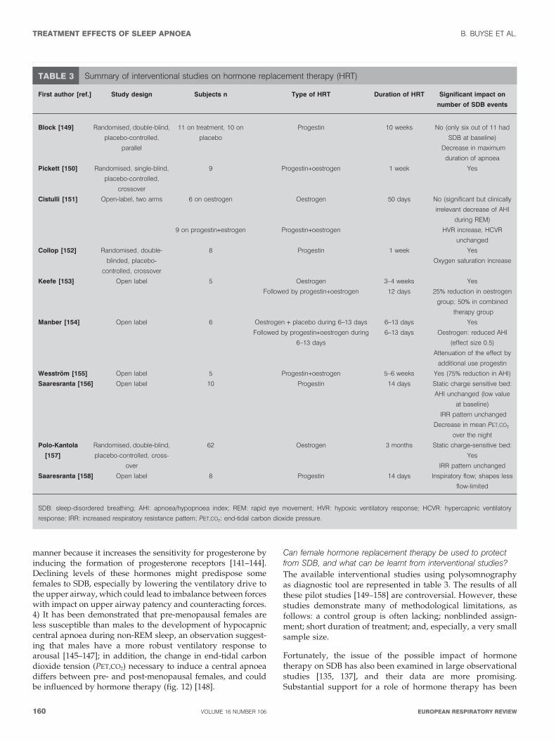
Can female hormone replacement therapy be used to protectfrom SDB, and what can be learnt from interventional studies?
The available interventional studies using polysomnographyas diagnostic tool are represented in table 3. The results of allthese pilot studies [149–158] are controversial. However, thesestudies demonstrate many of methodological limitations, asfollows: a control group is often lacking; nonblinded assign-ment; short duration of treatment; and, especially, a very smallsample size.
Fortunately, the issue of the possible impact of hormonetherapy on SDB has also been examined in large observationalstudies [135, 137], and their data are more promising.Substantial support for a role of hormone therapy has been
TABLE 3 Summary of interventional studies on hormone replacement therapy (HRT)
First author [ref.] Study design Subjects n Type of HRT Duration of HRT Significant impact on
number of SDB events
Block [149] Randomised, double-blind,
placebo-controlled,
parallel
11 on treatment, 10 on
placebo
Progestin 10 weeks No (only six out of 11 had
SDB at baseline)
Decrease in maximum
duration of apnoea
Pickett [150] Randomised, single-blind,
placebo-controlled,
crossover
9 Progestin+oestrogen 1 week Yes
Cistulli [151] Open-label, two arms 6 on oestrogen Oestrogen 50 days No (significant but clinically
irrelevant decrease of AHI
during REM)
9 on progestin+estrogen Progestin+oestrogen HVR increase, HCVR
unchanged
Collop [152] Randomised, double-
blinded, placebo-
controlled, crossover
8 Progestin 1 week Yes
Oxygen saturation increase
Keefe [153] Open label 5 Oestrogen 3–4 weeks Yes
Followed by progestin+oestrogen 12 days 25% reduction in oestrogen
group; 50% in combined
therapy group
Manber [154] Open label 6 Oestrogen + placebo during 6–13 days 6–13 days Yes
Followed by progestin+oestrogen during
6–13 days
6–13 days Oestrogen: reduced AHI
(effect size 0.5)
Attenuation of the effect by
additional use progestin
Wesstrom [155] Open label 5 Progestin+oestrogen 5–6 weeks Yes (75% reduction in AHI)
Saaresranta [156] Open label 10 Progestin 14 days Static charge sensitive bed:
AHI unchanged (low value
at baseline)
IRR pattern unchanged
Decrease in mean PET,CO2
over the night
Polo-Kantola
[157]
Randomised, double-blind,
placebo-controlled, cross-
over
62 Oestrogen 3 months Static charge-sensitive bed:
Yes
IRR pattern unchanged
Saaresranta [158] Open label 8 Progestin 14 days Inspiratory flow; shapes less
flow-limited
SDB: sleep-disordered breathing; AHI: apnoea/hypopnoea index; REM: rapid eye movement; HVR: hypoxic ventilatory response; HCVR: hypercapnic ventilatory
response; IRR: increased respiratory resistance pattern; PET,CO2: end-tidal carbon dioxide pressure.
TREATMENT EFFECTS OF SLEEP APNOEA B. BUYSE ET AL.
160 VOLUME 16 NUMBER 106 EUROPEAN RESPIRATORY REVIEW

provided by the population-based epidemiology study byBIXLER et al. [135]: the prevalence of sleep apnoea in post-menopausal females without hormone therapy (2.7%) wasalmost similar to males (3.9%), whereas in post-menopausalhormone therapy-users, the prevalence of sleep apnoea (0.5%)was compatible with that in pre-menopausal females (0.6%)[135]. In the study by SHAHAR et al. [137], the prevalence ofsleep apnoea in post-menopausal females was higher than inthe study by BIXLER et al. [135], but, again, the prevalence inpost-menopausal females on hormone therapy was 2–3 timeslower than in nonusers. The power of the study by SHAHAR et al.[137] is that they performed a multivariable adjustment forknown determinants of obstructive sleep apnoea, includingage, BMI and neck circumference. The adjustment attenuatedthe inverse association between hormone use and SDB, butonly moderately.
However, when considering hormone therapy, the increasedrisk for thromboembolic events should be noted in alloestrogen and/or progestin therapy and also the increasedrisk of breast cancer in patients receiving long-term oestrogenand progestin [159]. However, unopposed oestrogen therapymay not be linked to increased risk of breast cancer and thereare no data on the risk of breast cancer when using progestinmonotherapy [159].
In conclusion, although the results of experimental studies arecontroversial, based on epidemiological data showing lowerprevalence of SDB in hormone therapy-users than in nonusers,post-menopausal hormone therapy (preferably progestin oroestrogen monotherapy) may be considered as a second-linetreatment in selected patient groups (perhaps especially inpost-menopausal females presenting OSAHS with obesityhypoventilation syndrome, i.e. patients with decreased respira-tory drive) when risks of untreated SDB are likely to overcomethose related to hormone therapy. Indeed, progesterone raisesresting ventilation and augments chemosensitivity to a greaterextent in patients with obesity-hypoventilation syndrome thanin normal subjects or in patients with chronic obstructivepulmonary disease [160].
Stimulants in refractory sleepiness in sleep apnoea
The chief symptom of patients suffering from OSAHS isexcessive daytime sleepiness. Long-term use of CPAP com-monly improves or eliminates sleepiness; however, anunknown subset of patients remain somnolent, despite anappropriate compliance and efficacy of CPAP.
Possible explanations for this retention of somnolence are asfollows. 1) ‘‘CPAP-induced sleep deprivation’’: patients com-monly sleep less after CPAP treatment than before. This can becaused by the positive impact on sleep by the CPAP resultingin a reduction of light, unstable non-REM sleep in favour ofstable, restorative slow-wave and REM sleep; alternatively, onemay imagine that some side-effects of CPAP (e.g. uncomfor-table mask and mask leaks, nasal/oral dryness and nasalobstruction resulting in mouth leaks) would prolong nocturnalawakenings, and would make it difficult for the patient toresume sleep and lead to chronic sleep deprivation. 2)Refractory sleepiness can be due to alcohol and sedative drugabuse. 3) Sometimes depression, also a symptom of obstructivesleep apnoea, is not totally reversible on CPAP [161].
4) Additional sleep disorders can be present. The patient candemonstrate narcolepsy; aside from primary narcolepsy,secondary cases of narcolepsy have been observed in neuro-logical conditions, including dystrophic myopathy,Parkinsonism and multiple sclerosis [162]. Periodic leg move-ments without restless legs can be present, a condition morefrequent in aged patients that may be unmasked after relief ofapnoea. The patient can suffer from hypersomnia, aside fromidiopathic hypersomnia, a rare disease priming adolescents oryoung adults; there are several neurological conditions leadingto secondary hypersomnia in middle-aged adults [162].
In addition, it is possible that some patients with OSAHSdevelop hypoxic lesions of central wake-active systems,leading to long-term CPAP-resistant secondary hypersomnia.Oxidative neural injury of wake-promoting neural groups hasindeed been demonstrated in animal models of OSAHS(fig. 13) [163, 164].
FIGURE 13. Impact of intermittent hypoxia on the brains and sleepiness of
mice. a) and b) Staining with 2,4-dinitrophenyl hydrazone (a marker of protein
oxidation indicating brain injury) in sleep-wake brain regions in the basal forebrain of
mice. The staining is more pronounced in mice exposed to intermittent hypoxia (a)
than in sham mice (b). c) The mean sleep latency is shorter in mice exposed to
intermediate hypoxia, and the shortening is especially obvious after sleep
curtailment. #: not significant. **: p,0.01; ***: p,0.001. Reproduced from [163]
with permission from the publishers.
B. BUYSE ET AL. TREATMENT EFFECTS OF SLEEP APNOEA
cEUROPEAN RESPIRATORY REVIEW VOLUME 16 NUMBER 106 161

Stimulants may improve refractory sleepiness. Some anti-depressants, such as fluoxetine, might even have a mildalerting effect [165], making them a good choice in CPAP-treated OSAHS patients who are still depressed and somno-lent. Caffeine is used by OSAHS patients at daily doses twotimes higher than in controls that do not decrease after CPAPtreatment [166]. Caffeine blocks adenosine receptors thatcontrol sleep; these rebound after sleep deprivation [167].However, both caffeine and antidepressants have not beenspecifically studied in this condition.
Modafinil is a well-studied nonamphetaminic alerting agentthat was first developed in patients with narcolepsy andidiopathic hypersomnia [168–170]. Its mechanism of action isnot fully understood, although modafinil-induced potentiationof norepinephrine transmission is observed in animals [171,172], and the benefit of modafinil on alertness is largelyreduced in animals already co-treated with norepinephrine a-or b-blocker agents (a condition quite frequent in OSAHSpatients with hypertension). The drug has an excellent benefit/risk ratio; it provides a long-lasting alerting effect with a low(but still possible) risk of irritability, anxiety, restlessness andrare sympathetic effects, such as tachycardia, headache andincreased blood pressure. It is the unique stimulant investi-gated in OSAHS patients using randomised, placebo-controlled, double-blind trials. It was previously shown toimprove daytime sleepiness and memory in untreated OSAHSpatients [173]. Later, it was shown to significantly reduceresidual sleepiness in larger groups of CPAP-treated OSAHSpatients [174–176], with rare nervousness and headaches, and
no [175, 177] or marginal [174, 176] reduction of CPAP use andno increased blood pressure (table 4). The trial by KINGSHOTT etal. [174], the first RCT on this topic, observed a significantimprovement of the objective measure of MWT (table 4).However, the mean AHI of the study sample while on CPAPtreatment was 8 and the arousal index was 28. Although themean pre-treatment AHI and arousal index for this group wereboth 45, one may speculate that CPAP therapy in this groupwas suboptimal. In the following studies [175–177], subjectivesleepiness as measured by the Epworth sleepiness scale andobjective sleepiness determined by MSLT and MWT andquality of life, were statistically significantly improved(table 4). In addition, it has been shown that modafinilimproved attention performance in these patients [178].However, none of these studies has evaluated the safety ofmodafinil in OSAHS patients on a long-term basis, especiallyregarding the risk for hypertension and decreased CPAP use.
Clinicians sometimes consider the use of dexamphetamine ormethylphenidate (second choice stimulant drugs used to treatnarcolepsy, products blocking norepinephrine and dopaminere-uptake) for refractory sleepiness in OSAHS. However, thepresent authors could only find one study showing cases oftreatment of OSAHS refractory sleepiness with dexampheta-mine [179]. In addition, the potential benefit of these drugsmay be limited by their frequent deleterious cardiovasculareffects.
When taken during the night, c-hydroxybutyrate (GHB;renamed sodium oxybate or Xyrem1) improves sleepiness innarcolepsy patients via incompletely known mechanisms (but
TABLE 4 Overview of the modafinil trials in obstructive sleep apnoea/hypopnoea patients on continuous positive airway pressure(CPAP) with refractory sleepiness
First author [ref.] Study design n Dose mg Benefit Adverse events
Kingshott [174] Randomised,
double-blind,
placebo-controlled,
cross-over
30 400/2 weeks ESS unchanged
qalertness on MWT
MSLT unchanged
Cognitive performance
unchanged
QoL unchanged
Nausea
Dry mouth
Q CPAP use (from 6.5–6.3 h?night-1)
Pack [175] Randomised,
double-blind,
placebo-controlled,
parallel
77 on modafinil
80 on placebo
200 (week 1)
400 (weeks 2–4)
Q sleepiness on ESS
Q sleepiness on MSLT
Headache
CPAP use unchanged
Schwartz [176] Open label 125 200–400 mg (individually
titrated) during 12 weeks
Q sleepiness on ESS
q QOL on FOSQ
Anxiety
Headache
Nausea
Insomnia
Q CPAP use (from 6.3–5.9 h?night-1)
Black [177] Randomised,
double-blind,
placebo-controlled,
parallel (three arms)
101 using 400 mg,
104 using 200 mg
400 mg
200 mg during 12 weeks
Q sleepiness on ESS
q alertness on MWT
q QOL on FOSQ
Headache
Nausea
CPAP use unchanged
ESS: Epworth sleepiness scale; q: increase; MWT: multiple wakefulness test; MSLT: multiple sleep latency test; Q: decrease; QoL: quality-of-life questionnaire; FOSQ:
functional outcomes of sleep questionnaire.
TREATMENT EFFECTS OF SLEEP APNOEA B. BUYSE ET AL.
162 VOLUME 16 NUMBER 106 EUROPEAN RESPIRATORY REVIEW

possibly night-time blockade/daytime release of dopamine,and increased restorative sleep). GHB had no clear effect onsleep apnoea in one study [180], but has not, so far, been triedin CPAP-treated OSAHS patients.
Pre-optic histamine neurones are part of an importantvigilance system that modulate reactivity to novel environ-ments [181, 182]. A new class of stimulants prolong histaminestimulation through partial blockade of pre-synaptichistamine-3 receptors (H3 receptors are present mainly in thebrain, with a few in the stomach). They showed promisingalerting effect in animal studies [183].
In conclusion, refractory sleepiness in patients with obstructivesleep apnoea/hypopnoea syndrome adequately treated withcontinuous positive airway pressure exists, yet the percentageis unknown. The mechanism of residual sleepiness is possiblyoxidative injury in wake-active neurones. Symptomatic treat-ment with modafinil in these patients shows consistent benefiton vigilance with low adverse effects, although there is someconcern about possible decrease of continuous positive airwaypressure use. To date, there are no other valuable drugs; someproducts, such as sodium oxybate or products blocking thepre-synaptic histamine-3 receptors, appear promising andfurther tests are warranted.
APPENDIXWorking group 2. France: I. Arnulf (Paris), S. Launois-Rollinat,P. Levy (Grenoble); Germany: H. Becker (Marburg), T. Penzel,(Berlin), I. Harsch (Erlangen), L. Luthje (Gottingen); Sweden: L.Grote, J. Hedner, (Gothenburg); Belgium: E. Hamans(Antwerp), R. Poirrier (Liege), K. Spiegel, (Brussels); USA: S.Javaheri (Mason, OH); Israel: L. Lavie, P. Lavie (Haıfa);Finland: T. Saaresranta (Turku); UK: I. Smith (Cambridge), E.Verstraeten (Swansea), P. Turkington (Salford).
The present article was presented as part of the internationalsymposium ‘‘Respiratory somnology: a clinical update’’,sponsored by GlaxoSmithKline, Belgium. T. Penzel hasreceived reimbursement for conference attendance andspeaker fees from Respironics and Weinmann. L. Grote hasreceived research funding from Breas Medical AB, ResMed,Respironics, Weinmann, Itamar Medical Ltd and Pfizer.L. Grote has also received speaker and educational fees fromResMed, Respironics, Weinmann and Pfizer. S. Javaheri hasacted as a consultant for, and received honoraria for lecturesand research from, Respironics and ResMed. P. Lavie is afounder and board member of Itamar Medical Ltd, SLP Ltd,Sleep Health Centers US and Sleep Disorders Center Israel;he owns options and shares in these companies. I. Smith hasreceived travel grants and honoraria from Cephalon, UK.His research staff have been funded by unrestricted educa-tional grants from ResMed and Friday Medical Ltd. I. Arnulfhas received speaker fees from Cephalon Ltd.
REFERENCES1 Chugh DK, Dinges DF. Mechanisms of sleepiness in
obstructive sleep apnea. In: Pack AI, ed. Sleep Apnea.Pathogenesis, Diagnosis and Treatment. New York,Marcel Dekker, 2002; pp. 265–285.
2 Roehrs T, Zorick F, Wittig R, Conway W, Roth T.Predictors of objective level of daytime sleepiness inpatients with sleep-related breathing disorders. Chest1989; 95: 1202–1206.
3 Colt HG, Haas H, Rich GB. Hypoxemia vs sleepfragmentation as cause of excessive daytime sleepinessin obstructive sleep apnea. Chest 1991; 100: 1542–1548.
4 Kingshott RN, Engleman HM, Deary IJ, Douglas NJ. Doesarousal frequency predict daytime function? Eur Respir J1998; 12: 1264–1270.
5 Chervin RD, Aldrich MS. Characteristics of apneas andhypopneas during sleep and relation to excessive daytimesleepiness. Sleep 1998; 21: 799–806.
6 Bennett LS, Langford BA, Stradling JR, Davies RJ. Sleepfragmentation indices as predictors of daytime sleepinessand nCPAP response in obstructive sleep apnea. Am JRespir Crit Care Med 1998; 158: 778–786.
7 Kingshott RN, Vennelle M, Hoy CJ, Engleman HM,Deary IJ, Douglas NJ. Predictors of improvements indaytime function outcomes with CPAP therapy. Am JRespir Crit Care Med 2000; 161: 866–871.
8 Gottlieb DJ, Whitney CW, Bonekat WH, et al. Relation ofsleepiness to respiratory disturbance index. The SleepHeart Health Study. Am J Respir Crit Care Med 1999; 159:502–507.
9 Bixler EO, Vgontzas AN, Lin HM, Calhoun SL, Vela-BuenoA, Kales A. Excessive daytime sleepiness in a generalpopulation sample: the role of sleep apnea, age, obesity,diabetes, and depression. J Clin Endocrinol Metab 2005;90: 4510–4515.
10 Buyse B, Hedner J, the participants of working group 2.Sleep apnoea, hypertension and vascular disease: whereare we now? Eur Respir Rev 2007; 16: 169–182.
11 Vgontzas AN, Papanicolaou DA, Bixler EO, Kales A,Tyson K, Chrousos GP. Elevation of plasma cytokines indisorders of excessive sleepiness: role of sleep distur-bance and obesity. J Clin Endocrinol Metab 1997; 82: 1313–1316.
12 Vgontzas AN, Papanicolaou DA, Bixler EO, et al. Sleepapnea and daytime sleepiness and fatigue: relation tovisceral obesity, insulin resistance, and hypercytokinemia.J Clin Endocrinol Metab 2000; 85: 1151–1158.
13 Vgontzas AN, Zoumakis E, Lin HM, Bixler EO,Trakade G, Chrousos GP. Marked decrease in sleepinessin patients with sleep apnea by etanercept, a tumornecrosis factor-a antagonist. J Clin Endocrinol Metab 2004;89: 4409–4413.
14 Patel SR, White DP, Malhotra A, Stanchina ML, Ayas NT.Continuous positive airway pressure therapy for treatingsleepiness in a diverse population with obstructive sleepapnea: results of a meta-analysis. Arch Intern Med 2003;163: 565–571.
15 Fulda S, Shulz H. Cognitive dysfunction in sleepdisorders. Sleep Med Rev 2001; 5: 423–445.
16 Beebe DW, Gozal D. Obstructive sleep apnea and theprefrontal cortex: towards a comprehensive model linkingnocturnal upper airway obstruction to daytime cognitiveand behavioral deficits. J Sleep Res 2002; 11: 1–16.
17 Verstraeten E, Cluydts R. Executive control of attention insleep apnea patients: theoretical concepts and methodo-logical considerations. Sleep Med Rev 2004; 8: 257–267.
B. BUYSE ET AL. TREATMENT EFFECTS OF SLEEP APNOEA
cEUROPEAN RESPIRATORY REVIEW VOLUME 16 NUMBER 106 163
18 Verstraeten E, Cluydts R, Pevernagie D, Hoffmann G.Executive function in sleep apnea: controlling for atten-tional capacity in assessing executive attention. Sleep2004; 27: 685–693.
19 Thomas RJ, Rosen BR, Stern CHE, Weiss JW, Kwong KK.Functional imaging of working memory in obstructivesleep-disordered breathing. J Appl Physiol 2005; 98: 2226–2234.
20 Gozal D, Kheirandish L. Sleepiness and neurodegenara-tion in sleep-disordered breathing: convergence ofsignaling cascades. Am J Respir Crit Care Med 2005; 171:1325–1327.
21 Krieger J. Sleep apnoea and driving: how can this be dealtwith? Eur Respir Rev 2007; 16: 189–195.
22 Leger D. The cost of sleep-related accidents: a report forthe National Commission on Sleep Disorders Research.Sleep 1994; 17: 84–93.
23 Webb WB. The cost of sleep-related accidents: areanalysis. Sleep 1995; 18: 276–80.
24 Gurubhagavatula I, Maislin G, Nkwuo JE, Pack AI.Occupational screening for obstructive sleep apnea incommercial drivers. Am J Respir Crit Care Med 2004; 170:371–376.
25 Moskowitch A. Pilot development and testing of costeffective approach to screening, diagnosis, treatment ofsleep apnea in commercial motor carriers. Sleep Medicine2005; 6: Suppl. 2, S58 (abstract 026).
26 Turkington PM, Sircar M, Allgar V, Elliott MW.Relationship between obstructive sleep apnoea, drivingsimulator performance, and risk of road traffic accidents.Thorax 2001; 56; 800–805.
27 Philip P, Sagaspe P, Taillard J, et al. Fatigue, sleepiness,and performance in simulated versus real driving condi-tions. Sleep 2005; 28; 1511–1516.
28 Wise MS. Objective measures of sleepiness and wakeful-ness: application to the real world? J Clin Neurophysiol2006; 23; 39–49.
29 Bennett LS, Stradling JR, Davies RJ. A behavioural test toassess daytime sleepiness in obstructive sleep apnoea. JSleep Res 1997; 6; 142–145.
30 Krieger AC, Ayappa I, Norman RG, Rapoport DM,Walsleben J. Comparison of the maintenance of wakeful-ness test (MWT) to a modified behavioral test (OSLER) inthe evaluation of daytime sleepiness. J Sleep Res 2004; 13;407–411.
31 George CF. Driving simulators in clinical practice. SleepMed Rev 2003; 7; 311–320.
32 Findley LJ, Fabrizio MJ, Knight H, Norcross BB, LaForteAJ, Suratt PM. Driving simulator performance in patientswith sleep apnea. Am Rev Respir Dis 1989; 140; 529–530.
33 Engleman HM, Martin SE, Deary IJ, Douglas NJ. Effect ofcontinuous positive airway pressure treatment on day-time function in sleep apnoea/hypopnoea syndrome.Lancet 1994; 343; 572–575.
34 Hack M, Davies RJ, Mullins R, et al. Randomisedprospective parallel trial of therapeutic versus subther-apeutic nasal continuous positive airway pressure onsimulated steering performance in patients with obstruc-tive sleep apnoea. Thorax 2000; 55; 224–231.
35 Turkington PM, Sircar M, Saralaya D, Elliott MW. Timecourse of changes in driving simulator performance with
and without treatment in patients with sleep apnoeahypopnoea syndrome. Thorax 2004; 59; 56–59.
36 Orth M, Duchna HW, Leidag M, et al. Driving simulatorand neuropsychological testing in OSAS before andunder CPAP therapy. Eur Respir J 2005; 26; 898–903.
37 Mazza S, Pepin JL, Naegele B, Plante J, Deschaux C,Levy P. Most obstructive sleep apnoea patients exhibitvigilance and attention deficits on an extended battery oftests. Eur Respir J 2005; 25: 75–80.
38 Edinger JD, Radke RA. Use of in vivo desensitization totreat a patient’s claustrophobic response to nasal CPAP.Sleep 1993; 16: 678–680.
39 Weaver TE, Maislin G, Dinges DF, et al. Self-efficacy insleep apnea: instrument development and patient per-ceptions of obstructive sleep apnea risk, treatmentbenefit, and volition to use continuous positive airwaypressure. Sleep 2003; 26: 727–732.
40 Stepnowsky CJ Jr, Bardwell WA, Moore PJ, Ancoli-Israel S, Dimsdale JE. Psychologic correlates of compli-ance with continuous positive airway pressure. Sleep2002; 25: 758–762.
41 Javaheri S. Treatment of obstructive and central sleepapnoea in heart failure: practical options. Eur Respir Rev2007; 16: 183–188.
42 Yaggi H, Mohsenin V. Obstructive sleep apnoea andstroke. Lancet Neurol 2004; 3: 333–342.
43 Hui D, Choy D, Wong L, et al. Prevalence of sleep-disordered breathing and continuous positive airwaypressure compliance. Chest 2002; 122: 852–860.
44 Bassetti C, Aldrich MS. Sleep apnoea in acute cerebrova-sular diseases: final report on 128 patients. Sleep 1999; 22:217–223.
45 Wessendorf TE, Teschler H, Wang YM, Konietzko N,Thilmann AF. Sleep-disordered breathing amongpatients with first-ever stroke. J Neurol 2000; 247: 41–47.
46 Lawrence E, Dundas R, Higgens S, et al. The naturalhistory and associations of sleep disordered breathing infirst ever stroke. Int J Clin Pract 2001; 55: 584–588.
47 Harbison J, Ford GA, James OFW, Gibson GJ. Sleep-disordered breathing following acute stroke. Q J Med2002; 95: 741–742.
48 Parra O, Arboix A, Montserrat JM, Quinto L, Bechich S,Garcia-Eroles L. Sleep-related breathing disorders:impact on mortality of cerebrovascular disease. EurRespir J 2004; 24: 267–272.
49 Turkington PM, Allgar V, Bamford J, Wanklyn P,Elliott MW. Effect of upper airway obstruction in acutestroke on functional outcome at 6 months. Thorax 2004;59: 367–371.
50 Iranzo A, Santamaria J, Berenguer J, Sanchez M,Chamorro A. Prevalence and clinical importance of sleepapnea in the first night after cerebral infarction. Neurology2002; 58: 911–916.
51 Sandberg O, Franklin KA, Bucht G, Eriksson S,Gustafson Y. Nasal continuous positive airway pressurein stroke patients with sleep apnoea: a randomizedtreatment study. Eur Respir J 2001; 18: 630–634.
52 Wessendorf TE, Wang YM, Thilmann AF, Sorgenfrei U,Konietzko N, Teschler H. Treatment of obstructive sleepapnoea with nasal continuous positive airway pressure instroke. Eur Respir J 2001; 18: 623–629.
TREATMENT EFFECTS OF SLEEP APNOEA B. BUYSE ET AL.
164 VOLUME 16 NUMBER 106 EUROPEAN RESPIRATORY REVIEW

53 Martinez-Garcia MA, Galiano-Blancart R, Roman-Sanchez P, Soler-Cataluna JJ, Cabero-Salt L, Salcedo-Maiques E. Continuous positive airway pressure treat-ment in sleep apnea prevents new vascular events afterischemic stroke. Chest 2005; 128: 2123–2129.
54 Scala R, Turkington PM, Wanklyn P, Bamford J,Elliott MW. Effects of incremental levels of continuouspositive airway pressure on cerebral blood flow velocityin healthy adult humans. Clin Sci (Lond) 2003; 104: 633–639.
55 Turkington PM, Bamford J, Wanklyn P, Elliott MW.Prevalence and predictors of upper airway obstruction inthe first 24 hours after acute stroke. Stroke 2002; 33: 2037–2042.
56 Harsch IA. Metabolic disturbances in patients withobstructive sleep apnoea syndrome. Eur Respir Rev2007; 16: 196–202.
57 Broberger C. Brain regulation of food intake and appetite:molecules and networks. J Intern Med 2005; 258: 301–327.
58 Sharma V, McNeill JH. The emerging roles of leptin andghrelin in cardiovascular physiology and pathophysiology.Curr Vasc Pharmacol 2005; 3: 169–180.
59 Kripke DF, Simons RN, Garfinkel L, Hammond EC. Shortand long sleep and sleeping pills. Is increased mortalityassociated? Arch Gen Psychiatry 1979; 36: 103–116.
60 National Sleep Foundation, ‘‘Sleep in America’’ Poll.Washington, DC, National Sleep Foundation, 2000, 2001,2002.
61 Flegal KM, Carroll MD, Kuczmarski RJ, Johnson CL.Overweight and obesity in the United States: prevalenceand trends, 1960–1994. Int J Obes Relat Metab Disord 1998;22: 39–47.
62 Flegal KM, Carroll MD, Ogden CL, Johnson CL.Prevalence and trends in obesity among US adults,1999–2000. JAMA 2002; 288: 1723–1727.
63 Spiegel K, Leproult R, L’hermite-Baleriaux M,Copinschi G, Penev PD, Van Cauter E. Leptin levels aredependent on sleep duration: relationships with sym-pathovagal balance, carbohydrate regulation, cortisol,and thyrotropin. J Clin Endocrinol Metab 2004; 89: 5762–5771.
64 Spiegel K, Tasali E, Penev P, Van Cauter E. Briefcommunication: sleep curtailment in healthy youngmen is associated with decreased leptin levels, elevatedghrelin levels, and increased hunger and appetite. AnnIntern Med 2004; 141: 846–850.
65 Rayner DV, Trayhurn P. Regulation of leptin production:sympathetic nervous system interactions. J Mol Med 2001;79: 8–20.
66 Sugino T, Yamaura J, Yamagishi M, et al. Involvement ofcholinergic neurons in the regulation of the ghrelinsecretory response to feeding in sheep. Biochem BiophysRes Commun 2003; 304: 308–312.
67 Taheri S, Lin L, Austin D, Young T, Mignot E. Short sleepduration is associated with reduced leptin, elevatedghrelin, and increased body mass index. PLoS Med2004; 1: e62.
68 Taheri S, Lin L, Austin D, Young T, Mignot E. Short sleepduration is associated with reduced leptin, elevatedghrelin, and increased body mass index. Sleep 2004; 27:Suppl. A, 146–147.
69 Bassiri AG, Guilleminault C. Clinical features andevaluation of obstructive sleep apnea–hypopnea syn-drome. In: Kryger MH, Roth T, Dement WC, eds.Principles and Practice of Sleep Medicine. 3rd Edn.Philadelphia, PA, W.B. Saunders, 2000; pp. 869–878.
70 Persson HE, Svanborg E. Sleep deprivation worsensobstructive sleep apnea. Comparison between diurnaland nocturnal polysomnography. Chest 1996; 109: 645–650.
71 Phillips BG, Hisel TM, Kato M, et al. Recent weight gainin patients with newly diagnosed obstructive sleepapnea. J Hypertens 1999; 17: 1297–300.
72 Phillips BG, Kato M, Narkiewicz K, Choe I, Somers VK.Increases in leptin levels, sympathetic drive, and weightgain in obstructive sleep apnea. Am J Physiol Heart CircPhysiol 2000; 279: H234–H237.
73 Harsch IA, Konturek PC, Koebnick C, et al. Leptin andghrelin levels in patients with obstructive sleep apnoea:effect of CPAP treatment. Eur Respir J 2003; 22: 251–257.
74 Ip S, Lam K, Ho C, Tsang K, Lam W. Serum leptin andvascular risk factors in obstructive sleep apnea. Chest2000; 118: 580–586.
75 Saarelainen S, Lahtela J, Kallonen E. Effect of nasal CPAPtreatment on insulin sensitivity and plasma leptin. J SleepRes 1997; 6: 146–147.
76 Chin K, Shimizu K, Nakamura T, et al. Changes in intra-abdominal visceral fat and serum leptin levels in patientswith obstructive sleep apnea syndrome following nasalcontinuous positive airway pressure therapy. Circulation1999; 100: 706–712.
77 Shimizu K, Chin K, Nakamura T, et al. Plasma leptinlevels and cardiac sympathetic function in patients withobstructive sleep apnoea–hypopnoea syndrome. Thorax2002; 57: 429–434.
78 Sanner BM, Kollhosser P, Buechner N, Zidek W, Tepel M.Influence of treatment on leptin levels in patients withobstructive sleep apnoea. Eur Respir J 2004; 23: 601–604.
79 O’Donnell CP, Tankersley CG, Polotsky VP, Schwartz AR,Smith PL. Leptin, obesity, and respiratory function. RespirPhysiol 2000; 119: 163–170.
80 Polotsky VY, Li J, Punjabi NM, et al. Intermittent hypoxiaincreases insulin resistance in genetically obese mice. JPhysiol 2003; 552: 253–264.
81 Lavie P. Mortality in sleep apnoea syndrome: a review ofthe evidence. Eur Respir Rev 2007; 16: 203–210.
82 Garrigue S, Bordier P, Jais P, et al. Benefit of atrial pacingin sleep apnea syndrome. N Engl J Med 2002; 346: 404–412.
83 Gottlieb DJ. Cardiac pacing – a novel therapy for sleepapnea? N Engl J Med 2002; 346: 444–445.
84 Garrigue S, Bordier P, Barold SS, Clementy J. Sleepapnea: a new indication for cardiac pacing? Pacing ClinElectrophysiol 2004; 27: 204–211.
85 Shepard JW Jr, Pevernagie DA, Stanson AW, Daniels BK,Sheedy PF. Effects of changes in central venous pressureon upper airway size in patients with obstructive sleepapnea. Am J Respir Crit Care Med 1996; 153: 250–254.
86 Alex CG, Onal E, Lopata M. Upper airway occlusionduring sleep in patients with Cheyne–Stokes respiration.Am Rev Respir Dis 1986; 133: 42–45.
B. BUYSE ET AL. TREATMENT EFFECTS OF SLEEP APNOEA
cEUROPEAN RESPIRATORY REVIEW VOLUME 16 NUMBER 106 165

87 Badr MS, Kawak A, Skatrud JB, Morrell MJ, Zahn BR,Babcock MA. Effect of induced hypocapnic hypopnea onupper airway patency in humans during NREM sleep.Respir Physiol 1997; 110: 33–45.
88 Crick SJ, Sheppard MN, Anderson RH. Neural supply ofthe heart. In: Ter Horst GJ, ed. The Nervous System andthe Heart. Totowa, NJ, Humana Press, 2000; pp. 3–54.
89 Orem J, Kubin L. Respiratory physiology: central neuralcontrol. In: Kryger MH, Roth T, Dement WC, eds.Principles and Practice of Sleep Medicine. 3rd Edn.Philadelphia, PA, W.B. Saunders, 2000; pp. 205–241.
90 Pepin JL, Defaye P, Garrigue S, Poezevara Y, Levy P.Overdrive atrial pacing does not improve obstructivesleep apnoea syndrome. Eur Respir J 2005; 25: 343–347.
91 Luthje L, Unterberg-Buchwald C, Dajani D, Vollmann D,Hasenfuss G, Andreas S. Atrial overdrive pacing inpatients with sleep apnea with implanted pacemaker. AmJ Respir Crit Care Med 2005; 172: 118–122.
92 Simantirakis EN, Schiza SE, Chrysostomakis SI, et al.Atrial overdrive pacing for the obstructive sleep apnea–hypopnea syndrome. N Engl J Med 2005; 353: 2568–2577.
93 Unterberg C, Luthje L, Szych J, Vollmann D, Hasenfuss G,Andreas S. Atrial overdrive pacing compared to CPAP inpatients with obstructive sleep apnoea syndrome. EurHeart J 2005; 26: 2568–2575.
94 Krahn AD, Yee R, Erickson MK, et al. Physiologic pacingin patients with obstructive sleep apnea: a prospective,randomized crossover trial. J Am Coll Cardiol 2006; 47:379–383.
95 Sinha AM, Skobel EC, Breithardt OA, et al. Cardiacresynchronization therapy improves central sleep apneaand Cheyne–Stokes respiration in patients with chronicheart failure. J Am Coll Cardiol 2004; 44: 68–71.
96 Gabor JY, Newman DA, Barnard-Roberts V, et al.Improvement in Cheyne–Stokes respiration followingcardiac resynchronisation therapy. Eur Respir J 2005; 26:95–100.
97 Floras JS, Bradley TD. Atrial overdrive pacing for sleepapnea. A door now closed? Am J Respir Crit Care Med2005; 172: 1–5.
98 Patton TJ, Thawley SE, Waters RC, Vandermeer PJ,Ogura JH. Expansion hyoidplasty: a potential surgicalprocedure designed for selected patients with obstructivesleep apnea syndrome. Experimental canine results.Laryngoscope 1983; 93: 1387–1396.
99 Pevernagie DA, Stanson AW, Sheedy PF 2nd, Daniels BK,Shepard JW Jr, Effects of body position on the upperairway of patients with obstructive sleep apnea. Am JRespir Crit Care Med 1995; 152: 179–185.
100 den Herder C, van Tinteren H, de Vries N. Sleependoscopy versus modified Mallampati score in sleepapnea and snoring. Laryngoscope 2005; 115: 735–739.
101 Smith IE, Quinnell TG. Pharmacotherapies for obstruc-tive sleep apnoea: where are we now? Drugs 2004; 64:1385–1399.
102 Kubin L, Tojima H, Davies RO, Pack AI. Serotonergicexcitatory drive to hypoglossal motorneurons in thedecerabrate cat. Neurosci Lett 1992; 139: 243–248.
103 Jacobs BL, Azmitia EC. Structure and function of thebrain serotonin system. Physiol Rev 1992; 72; 165–229.
104 Richter DW, Manzke T, Wilken B, Ponimaskin E.Serotonin receptors: guardians of stable breathing.Trends Mol Med 2003; 9: 542–548.
105 Veasey SC. Serotonin agonists and antagonists inobstructive sleep apnea: therapeutic potential. Am JRespir Med 2003; 2: 21–29.
106 Veasey S. Pharmacotherapies for obstructive sleep apnea:how close are we? Curr Opin Pulm Med 2001; 7: 399–403.
107 Bonora M, St John WM, Bledsoe TA. Differentialelevation by protriptyline and depression by diazepamof upper airway respiratory motor activity. Am Rev RespirDis 1985; 131: 41–45.
108 Brownell LGP, West P, Sweatman P, Acres JC,Kryger MH. Protriptyline in obstructive sleep apnea: adouble blind trial. N Engl J Med 1982; 307: 1037–1042.
109 Stepanski E, Conway W, Young D, Zorick FJ, Wittig RM,Roth T. A double blind trial of protriptyline in thetreatment of sleep apnea syndrome. Henry Ford Hosp MedJ 1988; 36: 5–8.
110 Whyte K, Gould G, Airlie A, Shapiro CM, Douglas NJ.Role of protriptyline and acetazolamide in the sleepapnoea/hypopnoea syndrome. Sleep 1988; 11: 463–472.
111 Sunderram J, Parisi RA, Strobel RJ. Serotonergic stimula-tion of the genioglossus and the response to nasalcontinuous positive airway pressure. Am J Respir CritCare Med 2000; 162: 925–929.
112 Kraiczi H, Hedner J, Dahlof P, Ejnell H, Carlson J. Effectof serotonin uptake inhibition on breathing during sleepand daytime symptoms in obstructive sleep apnea. Sleep1999; 22: 61–67.
113 Hanzel DA, Proia NG, Hudgel DW. Response ofobstructive sleep apnea to fluoxetine and protriptyline.Chest 1991; 100: 416–421.
114 Veasey SC, Chachkes J Fenik P, Hendricks JC. The effectsof ondansetron on sleep- disordered breathing in theEnglish Bulldog. Sleep 2001; 24: 155–160.
115 Stradling J, Smith D, Radulovacki M, Carley C. Effect ofondansetron on moderate obstructive sleep apnoea, asingle night, placebo-controlled trial. J Sleep Res 2003; 12:169–170.
116 De Vane C. Differential pharmacology of newer anti-depressants. J Clin Psychiatry 1998; 59: 85–93.
117 de Boer T. The pharmacologic profile of mirtazapine. JClin Psychiatry 1996; 57: 19–25.
118 McNicholas W. Implications of pathophysiology formanagement of the obstructive sleep apnoea syndrome.Monaldi Arch Chest Dis 1998; 53: 524–529.
119 Yoshioka M, Goda Y, Togashi H, Matsumoto M, Saito H.Pharmacological characterization of 5-hydroxytryptamine-induced apnea in the rat. J Pharmacol Exp Ther 1992; 260:917–924.
120 Carley DW, Radulovacki M. Mirtazapine, a mixed-profileserotonin agonist/antagonist, suppresses sleep apnea inthe rat. Am J Respir Crit Car Med 1999; 160: 1824–1829.
121 Carley DW, Olopade C, Ruigt GS, Radulovacki M.Efficacy of mirtazapine in obstructive sleep apnoeasyndrome. Sleep 2007; 30: 35–41.
122 Castillo JL, Menendez P, Segovia L, Guilleminault C.Effectiveness of mirtazapine in the treatment of sleepapnea/hypopnea syndrome (SAHS). Sleep Med 2004; 5:507–508.
TREATMENT EFFECTS OF SLEEP APNOEA B. BUYSE ET AL.
166 VOLUME 16 NUMBER 106 EUROPEAN RESPIRATORY REVIEW

123 Liu X, Sood S, Liu H, Horner RL. Opposing muscarinicand nicotinic modulation of hypoglossal motor output togenioglossus muscle in rats in vivo. J Physiol 2005; 565:965–980.
124 Shao XM, Feldman JL. Pharmacology of nicotinicreceptors in preBotzinger complex that mediate modula-tion of respiratory pattern. J Neurophysiol 2002; 88:1851–1858.
125 Gothe B, Strohl KP, Levin S, Cherniack NS. Nicotine: adifferent approach to treatment of obstructive sleepapnea. Chest 1985; 87: 11–17.
126 Davila DG, Hurt RD, Offord KP, Harris CD, Shepard JWJr. Acute effects of transdermal nicotine on sleep archi-tecture, snoring, and sleep-disordered breathing in non-smokers. Am J Respir Crit Care Med. 1994; 150: 469–474.
127 Nilsson L, Nordberg A, Hardy J, Wester P, Winblad B.Physostigmine restores (3H)acetylcholine efflux fromAlzheimer brain slices to normal level. J Neural Transm1986; 67: 275–285.
128 Hedner J, Craiczi H, Peker Y, Murphy P. Reduction ofsleep-disordered breathing after physostigmine. Am JRespir Crit Care Med 2003; 168: 1246–1251.
129 Bellingham MC, Ireland MF. Contribution of cholinergicsystems to state-dependent modulation of respiratorycontrol. Respir Physiol Neurobiol 2002; 131: 135–144.
130 Burton MD, Kazemi H. Neurotransmitters in centralrespiratory control. Respir Physiol 2000; 122: 111–121.
131 Fitzgerald RS, Shirahata M, Wang HY. Acetylcholine isreleased from in vitro cat carotid bodies during hypoxicstimulation. Adv Exp Med Biol 2000; 475: 485–494.
132 Hedner E, Birkhed D, Hedner J, Ekstrom J, Helander H.Stimulation of minor salivary glands by intraoral treat-ment with the cholinesterase inhibitor physostigmine inman. Eur J Oral Sci 2001; 109: 371–374.
133 Jokic R, Klimaszewski A, Mink J, Fitzpatrick MF. Surfacetension forces in sleep apnea: the role of a soft tissuelubricant: a randomized double-blind, placebo-controlledtrial. Am J Respir Crit Care Med 1998; 157: 1522–1525.
134 Younes M. The physiologic basis of central apnea andperiodic breathing. Curr Pulmonol 1989; 10: 265–326.
135 Bixler EO, Vgontzas AN, Lin H-M, et al. Prevalence ofsleep-disordered breathing in women: effects of gender.Am J Respir Crit Care Med 2001; 163: 608–613.
136 Young T, Finn L, Austin D, Peterson A. Menopausalstatus and sleep-disordered breathing in the WisconsinSleep Cohort Study. Am J Respir Crit Care 2003; 167: 1181–1185.
137 Shahar E, Redline S, Young T, et al. Hormone replace-ment therapy and sleep-disordered breathing. Am JRespir Crit Care Med 2003; 167: 1186–1192.
138 Gambacciani M, Ciapono M, Cappagli B, De Simone L,Orlandi R, Genazzani AR. Prospective evaluation of bodyweight and body fat distribution in early postmenopau-sal women with an without hormonal replacementtherapy. Maturitas 2001; 39: 125–132.
139 Popovic RM, White DP. Upper airway muscle activity innormal women: influence of hormonal status. J ApplPhysiol 1998; 84: 1055–1062.
140 Robinson RW, Zwillich CW. The effects of drugs onbreathing during sleep. Clin Chest Med 1985; 6: 603–614.
141 Regensteiner JG, Woodard WD, Hagerman DD, et al.Combined effects of female hormones and metabolic rateon ventilatory drives in women. J Appl Physiol 1989; 66:808–813.
142 Bayliss DA, Millhorn DE. Central neural mechanismsof progesterone action: application to the respiratorysystem. J Appl Physiol 1992; 73: 393–404.
143 Moore LG, McCullough RE, Weil JV. Increased HVR inpregnancy: relationship to hormonal end metabolicchanges. J Appl Physiol 1987; 62: 158–163.
144 Hannhart B, Pickett CK, Weil JV, Moore LG. Influence ofpregnancy on ventilatory and carodtid body neuraloutput responsiveness to hypoxia in cats. J Appl Physiol1989; 67: 797–803.
145 Zhou XS, Shahabuddin S, Zahn BR, Babdock MA,Badr MS. Effect of gender on the development ofhypocapnic apnea/hypopnea during NREM sleep. JAppl Physiol 2000; 89: 192–199.
146 Jordan AS, Eckert DJ, Catcheside PG, McEvoy RD.Ventilatory response to brief arousal from non-rapideye movement sleep is greater in men than in women.AJRCCM 2003; 168: 1512–1519.
147 Jordan AS, McEvoy RD, Edwards JK, et al. The influenceof gender and upper airway resistance on the ventilatoryresponse to arousal in obstructive sleep apnoea inhumans. J Physiol 2004; 558: 993–1004.
148 Rowley JA, Zhou XS, Diamond MP, Badr MS. Thedeterminants of the apnea threshold during NREM sleepin normal subjects. Sleep 2006; 29: 16–18.
149 Block AJ, Wynne JW, Boysen PG, Lindsey S, Martin C,Cantor B. Menopause, medroxyprogesterone and breath-ing during sleep. Am J Med 1981; 70: 506–510.
150 Pickett CK, Regensteiner JG, Woodard WD,Hagerman DD, Weil JV, Moore LG. Progestin andestrogen reduce sleep-disordered breating in postmeno-pausal women. J Appl Physiol 1989; 66: 1656–1661.
151 Cistulli PA, Barnes DJ, Grunstein RR, Sullivan CE. Effectof short term hormone replacement in the treatment ofobstructive sleep apnoea in postmenopausal women.Thorax 1994; 49: 699–702.
152 Collop NA. Medroxyprogesterone acetate and ethanol-induced exacerbation of obstructive sleep apnea. Chest1994; 106: 792–799.
153 Keefe DL, Watson R, Naftolin F. Hormone replacementtherapy may alleviate sleep apnea in menopausalwomen: a pilot study. Menopause 1999; 6: 196–200.
154 Manber R, Kuo TF, Cataldo N, Colrain IM. The effects ofhormone replacement therapy on sleep-disorderedbreathing in postmenopausal women: a pilot study.Sleep 2003; 26: 163–168.
155 Wesstrom J, Ulfberg J, Nilsson S. Sleep apnea andhormone replacement therapy: a pilot study and aliterature review. Acta Obstet Gynecol Scand 2005; 84: 54–57.
156 Saaresranta T, Polo-Kantola P, Rauhala E, Polo O.Medroxyprogesterone in postmenopausal females withpartial upper airway obstruction during sleep. Eur RespirJ 2001; 18: 989–995.
157 Polo-Kantola P, Rauhala E, Helenius H, Erkkala R,Irjala K, Polo O. Breathing during sleep in menopause:a randomized, controlled, cross-over trial with estrogentherapy. Obstet Gynecol 2003; 102: 68–75.
B. BUYSE ET AL. TREATMENT EFFECTS OF SLEEP APNOEA
cEUROPEAN RESPIRATORY REVIEW VOLUME 16 NUMBER 106 167

158 Saaresranta T, Aittokallio T, Polo-Kantola P, Helenius H,Polo O. Effect of medroxyprogesterone on inspiratoryflow shapes during sleep in postmenopausal women.Respir Physiol Neurobiol 2003; 134: 131–143.
159 Greiser CM, Greiser EM, Doren M. Menopausal hormonetherapy and risk of breast cancer: a meta-analysis ofepidemiological studies and randomized controlledtrials. Hum Reprod Update 2005; 11: 561–573.
160 Weil JV, Zwillich CW. Control of breathing in endocrineand metabolic disorders and in obesity. In: Altose MD,Kawakami Y, eds. Control of Breathing in Health andDisease. New York, NY, Marcel Dekker, 1999; pp. 581–607.
161 Bardwell WA, Moore P, Ancoli-Israel S, Dimsdale JE.Fatigue in obstructive sleep apnea: driven by depressivesymptoms instead of apnea severity? Am J Psychiatry2003; 160: 350–355.
162 Black JA, Brooks SN, Nishino S. Narcolepsy andsyndromes of primary excessive daytime somnolence.Semin Neurol 2004; 24: 271–282.
163 Veasey SC, Davis CW, Fenik P, et al. Long-termintermittent hypoxia in mice: protracted hypersomno-lence with oxidative injury to sleep-wake brain regions.Sleep 2004; 27: 194–201.
164 Zhan G, Serrano F, Fenik P, et al. NADPH oxidasemediated hypersomnolence and brain oxidative injury ina murine model of sleep apnea. AJRCCM 2005; 172: 921–929.
165 Wilson SJ, Bailey JE, Alford C, Weinstein A, Nutt DJ.Effects of 5 weeks of administration of fluoxetine anddothiepin in normal volunteers on sleep, daytimesedation, psychomotor performance and mood. JPsychopharmacol 2002; 16: 321–331.
166 Robinson GV, Pepperell JC, Davies RJ, Stradling JR.Caffeine levels following treatment of obstructive sleepapnoea. Thorax 2003; 58: 801–802.
167 Boutrel B, Koob GF. What keeps us awake: theneuropharmacology of stimulants and wakefulness-pro-moting medications. Sleep 2004; 27: 1181–1194.
168 Bastuji H, Jouvet M. Successful treatment of idiopathichypersomnia and narcolepsy with modafinil. Prog Neuro-Psychopharmacol Biol Psychiat 1988; 12: 695–700.
169 Billiard M, Besset A, Montplaisir J, et al. Modafinil: adouble-blind multicentric study. Sleep 1994; 17: S107–S112.
170 Randomized trial of modafinil for the treatment ofpathological somnolence in narcolepsy. US Modafinil inNarcolepsy Multicenter Study Group. Ann Neurol 1998;43: 88–97.
171 Lin JS, Roussel B, Akaoka H, Fort P, Debilly G, Jouvet M.Role of catecholamines in the modafinil and amphetamineinduced wakefulness, a comparative pharmacologicalstudy in the cat. Brain Res 1992; 591: 319–326.
172 Lin JS, Hou Y, Jouvet M. Potential brain neuronal targetsfor amphetamine-, methylphenidate-, and modafinil-induced wakefulness, evidenced by c-fos immunocyto-chemistry in the cat. Proc Natl Acad Sci USA 1996; 93:14128–14133.
173 Arnulf I, Homeyer P, Garma L, Whitelaw WA, DerenneJP. Modafinil in obstructive sleep apnea–hypopneasyndrome: a pilot study in 6 patients. Respiration 1997;64: 159–161.
174 Kingshott RN, Vennelle M, Coleman EL, Engleman HM,Mackay TW, Douglas NJ. Randomized, double-blind,placebo-controlled crossover trial of modafinil in thetreatment of residual excessive daytime sleepiness in thesleep apnea/hypopnea syndrome. Am J Respir Crit CareMed 2001; 163: 918–923.
175 Pack AI, Black JE, Schwartz JR, Matheson JK. Modafinilas adjunct therapy for daytime sleepiness in obstructivesleep apnea. Am J Respir Crit Care Med 2001; 164: 1675–1681.
176 Schwartz JR, Hirshkowitz M, Erman MK, Schmidt-Nowara W. Modafinil as adjunct therapy for daytimesleepiness in obstructive sleep apnea: a 12-week, open-label study. Chest 2003; 124: 2192–2199.
177 Black JE, Hirshkowitz M. Modafinil for treatment ofresidual excessive sleepiness in nasal continuous positiveairway pressure-treated obstructive sleep apnea/hypop-nea syndrome. Sleep 2005; 28: 464–471.
178 Dinges DF, Weaver TE. Effects of modafinil on sustainedattention performance and quality of life in OSA patientswith residual sleepiness while being treated with nCPAP.Sleep Med 2003; 4: 393–402.
179 Guilleminault C, Philip P. Tiredness and somnolencedespite initial treatment of obstructive sleep apneasyndrome (what to do when an OSAS patient stayshypersomnolent despite treatment). Sleep 1996; 19:Suppl. 9, S117–S122.
180 Series F, Series I, Cormier Y. Effects of enhancing slow-wave sleep by gamma-hydroxybutyrate on obstructivesleep apnea. Am Rev Respir Dis 1992; 145: 1378–1383.
181 Lin JS, Sakai K, Jouvet M. Hypothalamo-preoptichistaminergic projections in sleep-wake control in thecat. Eur J Neurosci 1994; 6: 618–625.
182 Parmentier R, Ohtsu H, Djebbara-Hannas Z, Valatx JL,Watanabe T, Lin JS. Anatomical, physiological, andpharmacological characteristics of histidine decarboxy-lase knock-out mice: evidence for the role of brainhistamine in behavioral and sleep–wake control. JNeurosci 2002; 22: 7695–7711.
183 Hancock AA. The challenge of drug discovery of a GPCTtarget: analysis of preclinical pharmacology of histamineH3 antagonists/inverse agonists. Biochem Pharmacol 2006;71: 1103–1113.
TREATMENT EFFECTS OF SLEEP APNOEA B. BUYSE ET AL.
168 VOLUME 16 NUMBER 106 EUROPEAN RESPIRATORY REVIEW