treatment and quality of life of patients with varicose veins anke adriana maria.pdf · treatment...
TRANSCRIPT
Treatment and Quality of Lifeof Patients with Varicose Veins
Anke A.M. Biemans
Voor Opa

Financial support for the printing of this thesis was generously provided by
Major sponsors were
Minor sponsors
LEO Pharma BV, Galderma Benelux BV, Roche Nederland BV, Fagron BV, AbbVie BV, Mediq
i.s.m. Pierre Fabre Dermo-Cosmétique, Beiersdorf NV, BAP Medical Nederland BV, Oldekamp
Medisch BV, biolitec biomedical technology GmbH, ChipSoft BV, Bo Medical Technologies BV,
NV Varitex, Juzo, Esaote Benelux BV, Dalton Medical BV and La Roche-Posay
ISBN: 978-94-6191-838-3
Lay-out: Legatron Electronic Publishing, Rotterdam
Printing: Ipskamp Drukkers, Enschede
Cover: Joris Biemans, Boekel
Copyright © A.A.M. Biemans
No part of this book may be reproduced or transmitted in any form of by any means, electronic
or mechanic, including photocopying, recording and any information storage and retrieval
system, without the permission in writing of the author, or when appropriate, of the publishers
of the publications.
THE COMPRESSION COMPANY

Treatment and Quality of Lifeof Patients with Varicose Veins
Behandeling en kwaliteit van levenbij patiënten met varices
Proefschrift
ter verkrijging van de graad van doctor aan deErasmus Universiteit Rotterdam
op gezag van derector magnificus
Prof.dr. H.G. Schmidt
en volgens besluit van het College voor Promoties.De openbare verdediging zal plaatsvinden op
woensdag 25 september 2013 om 11.00 uur
door
Anke Adriana Maria Biemans
geboren 26 december 1980 te Boekel

ProMoTiecoMMissie
Promotoren: Prof.dr. T. Nijsten
Prof.dr. H.A.M. Neumann
overige leden: Prof.dr. P.J.E. Bindels
Prof.dr. H.J.M. Verhagen
Prof.dr. M.G.R. de Maeseneer
copromotor: Dr. R.R. van den Bos

LisT of freQuenTLy used ABBreViATions
AASV, anterior accessory saphenous vein
ASVAL, ambulatory selective varicose ablation under local anesthesia
AVVQ, Aberdeen Varicose Vein Questionnaire
CEAP, Clinical Etiology Anatomy Pathophysiology classification
CIVIQ, Chronic Veines Insufficiency Quality of Life Questionnaire
CS, conventional surgery (ligation with stripping)
CVD, chronic venous disorder
CVI, chronic venous insufficiency
DUS, duplex ultrasound
DVT, deep vein thrombosis
EQ5D, EuroQoL 5D
EVLA, endovenous laser ablation
EVTA, endovenous thermal ablation
GSV, great saphenous vein
HRQoL, health related quality of life
LMWH, low molecular weight heparin
PRO, patient reported outcome
RCT, randomized controlled trial
RFA, radiofrequency ablation
SF-36, Short Form 36
SFJ, saphenofemoral junction
SPJ, saphenopopliteal junction
SSV, small saphenous vein
UGFS, ultrasound guided foam sclerotherapy
US, ultrasound examination
VCSS, venous clinical severity score
VEINES, VEnous INsufficiency Epidemiological and Economic Studies


contents
chapter 1 9
General Introduction
chapter 2 27
Endovenous Therapies of Varicose Veins: Indications, Procedures, Efficacy and Safety.
G Ital Dermatol Venereol 2010 Apr;145(2):161-73.
chapter 3 51
Validation of the Chronic Venous Insufficiency Quality of Life Questionnaire in Dutch
Patients Treated for Varicose Veins. Eur J Vasc Endovasc Surg 2011 Aug;42(2):246-53.
Translation and Validation of the Dutch VEINES-QOL/Sym in Varicose Vein Patients.
Phlebology 2013 Apr 4; Epub before print
chapter 4 79
Comparing Endovenous Laser Ablation, Foam Sclerotherapy and Conventional
Surgery for Great Saphenous Varicose Veins. J Vasc Surg. 2013 Jun 12 (Epub ahead of
print)
chapter 5 97
The Effect of Single Phlebectomies of Tributary on Great Saphenous Vein Reflux.
Journal of Vascular Surgery 2013 (submitted)
chapter 6 115
New Concepts on Recurrence of Varicose Veins According to the Different Treatment
Techniques. Phlebology 2013 (submitted).
chapter 7 127
General Discussion
chapter 8 135
Summary / Samenvatting

chapter 9 141
Dankwoord 142
List of co-authors 145
List of publications 147
Curriculum Vitae 149
PhD Portfolio 151

Chapter 1
General Introduction
Chapter 1

10 Chapter 1
GenerAL inTroducTionChronic venous disorders (CVD) are defined as the full spectrum of morphological and functional
abnormalities of the venous system, from telangiectasia to venous ulceration. Different forms
of CVD may have a great impact on patients’ quality of life and therefore CVD represents an
important social as well as economic burden. The incidence of CVD increases with age, except
for congenital venous malformations.
CVD may start with minor symptoms and/or appearance of varicose veins. Long-standing
CVD may slowly progress over time, leading to oedema and skin changes such as pigmentation,
“atrophy blanche” (white atrophy), lipodermatosclerosis and finally leg ulceration. Chronic
venous insufficiency (CVI) is a part of CVD where the function of the venous system is disturbed
and leads to clinical complications. These more advanced stages are classified in the CEAP
classification as C3 to C6 (see page 15). Other clinical manifestations of advanced CVD are
varicose veins, blow outs, nail changes, subcutaneous calcifications, induration, pachyderma
and eczema.
PhysioLoGy And PAThoPhysioLoGyVeins return the deoxygenated blood from the tissues to the heart. In the upright position,
especially when standing still, gravitation has to be overcome, to ensure venous flow. Among
different mechanisms for the venous return, the muscle pump function is the most important.
Deep intramuscular veins are being compressed during muscle contraction. In addition,
valves play a major role in maintaining the right flow direction, from the extremities towards
the heart. After compression of the muscle the blood flows though the perforator veins to
the deep system. The most important pump in the lower extremity is the calf muscle pump,
followed by the compression of the plantar plexus during walking.
In standing position, with open valves, the pressure in the veins is around 90 mmHg.
After activation of the muscle pumps this pressure decreases to 20 mmHg. However, when
there is valve incompetence or severe venous insufficiency, due to deep venous thrombosis,
the pressure will decrease less. This condition is called increased ambulant venous pressure
or venous hypertension.1,2 The high venous pressure will be transferred, to the venular side
of the skin microcirculation.3,4 Capillary hypertension causes capillary leakage of fluid which
is responsible for oedema, and erythrocytes migrating from the capillaries leading to iron
deposition and -hyper pigmentation.5 Leakage of plasmaproteines induces an inflammatory
reaction resulting in lipodermatosclerosis.3,6 Capillary hypertension also leads to dilation
of capillaries that cause decrease of the blood flow velocity with deposition of fibrin and
thrombocytes and leukocyte adhesion, leading to microthombosis, with atrophy blanche as a
result. 7, 8 These skin changes lead to skin that is vulnerable to venous ulcerations.4 (Figure 1)
The pathogenesis of primary venous reflux and the etiologic mechanism of morphologic
changes in the vein wall are largely unknown. For a long period the hypothesis was that varicose
veins develop due to inborn unstable elastic layer in the vein wall and that widening takes

General Introduction 11
Chapter 1
place mainly under influence of gravitation.9,10 The increase of vein diameter makes the valves
incompetent (leakage) and this results in reflux. There are two possibly etiologic explanations.
In the first concept varicose dilation will develop first in the cranial part of a vein and extend
distally along with the effect of gravitation, progressively affecting the more distal valves
(descending varicosity, Figure 2a).11-13 Contrarily, there is increasing evidence that superficial
venous disease has a multifocal origin and can be ‘ascending’ from the tributaries towards the
saphenous trunk, and further to the junction.11,14-19 Other studies also found evidence for the
ascending concept (Figure 2b) with disappearance of GSV reflux after phlebectomy or ablation
of an incompetent tributary, as well as the reduction in GSV diameter after ablation of refluxing
collaterals.19-23 Probably both theories play a role in the etiology of varicose veins.
figure 1: Rotterdam Model of CVD.
ePideMioLoGyThere is a lot of epidemiological data about CVD available in the literature.24-27 CVD is not
only a European problem. It affects a significant part of the population worldwide. Recently
the findings of the Vein Consult Program, an international observational, prospective study
collecting global epidemiologic data on chronic venous disorders, based on the CEAP
classification (Table 1, tekst page 15), have been published. In a total cohort of 91545 adults,
the world wide prevalence of these disorders was 83.6% (19.7% subjects with C0 and 63.9%
with C1-C6 according to the CEAP classification.28

12 Chapter 1
figure 2: Descending and ascending theory.
2a. Progression of varicose changes (from A to D) according to the descending theory.Ciaggiati, J Vasc Surg 2006;44:1291-5. Copyright 2006 Elsevier Inc. AII rights reserved.
2b. Progression of varicose changes (from A tot D) according to the ascending theory. Ciaggiati, J Vasc Surg 2006;44:1291-5. Copyright 2006 Elsevier Inc. AII rights reserved.
Varicose veins are a common manifestation of CVD. General population studies reported a
prevalence of varicose veins in 10-40% in men and 26-32% in women.24,29-31 The incidence
of varicose veins increases with age in a linear manner. The overall prevalence of saphenous
varicose veins in the Edinburgh Vein Study, which is a general population study, increased
from 12% in those aged 18-24 years to 56% in those aged 55-64 years.24 These findings are
concordant with the Framingham Study results in which the prevalence of varicose veins in
women younger than 30 years was less than 10% while in women aged 70 years and older
it increased to 77%.32 Multiple studies have shown that varicose veins are more common in

General Introduction 13
Chapter 1
women than in men. Selection bias may be a problem, as women consider varicose veins more
often a cosmetic problem than men, present more frequently at a varicose vein clinic and
are therefore more likely to participate in studies. Most general population studies could not
demonstrate a sex difference. Moreover, more advanced CVD occurs equally in both sexes.33 It
is estimated that venous leg ulceration, the end stage of CVI, affects 1% of the population.34 It
is assumed that approximately 50% of venous leg ulcers are the result of superficial varicose
veins.35 Therefore, the treatment of varicose veins, which may reduce the incidence of leg ulcers
by 50%, is likely to be cost-effective. The prevalence of venous ulcerations decreases slightly
the last years. Possibly this is due to early and more effective treatments of venous disorders.
(2.7% in 197030 en 0.6% in the Edinburgh Vein Study24 en 0.7% in the Bonn Vein Study I.33,36
cLinicAL chArAcTerisTicsCVD is associated with multiple and generally subjective symptoms consisting of discomfort,
aching, tingling, heaviness, burning, itching, muscle cramps and leg tiredness. The clinical
characteristics of CVD appear when the mechanisms that compensate for insufficient venous
return fails. The clinical features increase almost linearly in time and consist of teleangiectasias,
varicose veins, oedema, hyperpigmentation, eczema, atrophy blanche, lipodermatosclerosis
and ulceration. (Figure 3)
The CEAP classification (Table I) has been described to classify patients with CVD based
on clinical characteristics and duplex ultrasound findings.37,38 The CEAP classification is used for
the description of Clinical signs of CVD, Etiology (congenital, primary or secondary), Anatomy
(superficial, deep and perforating veins) and Pathophysiologic (reflux, obstruction or both).
The CEAP classification serves as an orderly documentation system and forms a synthesis of the
phlebological status. Eklöf et al. revised and refined the original classification, also introducing
the nowadays most frequently used basic CEAP as a simpler alternative to the advanced CEAP
classification.37
diagnostics At the end of the last century duplex ultrasound (a combined use of echography and Doppler)
has been widely introduced as diagnostic tool for CVD, and has now replaced many of the
previously available tests. Duplex ultrasound (DUS) allows detailed visualization of the
anatomy (veins, arteries, nerves, muscles etc) and provides all useful information about blood
flow hemodynamics. Duration of reflux time, peak reflux velocity and other parameters can be
measured by means of pulsed wave Doppler technology. Flow direction can also be visualized
directly by using colour coding technology, which is integrated in all modern duplex devices.
Reflux in superficial veins is defined as duration of reverse flow during >0.5 s. In addition the
diameter of the vein can be measured. Duplex ultrasound is now the gold standard diagnostic
technique,39 which should be available in each phlebologic practice. Excellent guidelines have

14 Chapter 1
been published, which are helpful to unravel the duplex anatomy40 in patients presenting with
varicose veins and other venous disorders, as well as to investigate the veins after treatment. 41
figure 3: Clinical characteristics of chronic venous disorders.
A. Telangiectases and reticular veins; B. Varicose veins; C. Edema (right leg); D. Eczema; E. Hyper pigmentations; F. Lipodermatosclerosis (with small ulcer pretibial); G. Atrophy blanche; H. Healed ulcer; I. Active ulcer

General Introduction 15
Chapter 1
Table i: Revision of CEAP Classification of chronic venous disease: summary.37
clinical classification
C0 No visible or palpable signs of venous disease
C1 Telangiectasies or reticular veins
C2 Varicose veins
C3 Edema
C4a Pigmentation or eczema
C4b Lipodermatosclerosis or atrophie blanche
C5 Healed venous ulcer
C6 Active venous ulcer
S Symptomatic, including ache, pain, tightness, skin irritation, heaviness, and muscle cramps, and other complaints attributable to venous dysfunction
A Asymptomatic
etiologic classification
Ec Congenital
Ep Primary
Es Secondary (postthrombotic)
En No venous cause identified
Anatomic classification
As Superficial veins
Ap Perforator veins
Ad Deep veins
An No venous location identified
Pathophysiologic classification
Basic ceAP
Pr Reflux
Po Obstruction
Pr,o Reflux and obstruction
Pn No venous pathophysiology identifiable
Advanced ceAP
Same as basic CEAP, with addition that any of 18 named venous segments can be used as locators for venous pathology.
DUS has largely contributed to clarify venous anatomy. For instance the saphenous
compartment in which the saphenous trunks run. In a transverse scan this compartment
resembles an, ‘Egyptian eye’. (Figure 4a) The ‘eye’ sign is always present and allows clear
identification of the saphenous vein. In this way, the main trunk of the great saphenous vein

16 Chapter 1
(GSV), small saphenous vein (SSV), anterior accessory saphenous vein (AASV) and posterior
accessory saphenous vein (PASV) can be clearly distinguished from tributaries running in
the subcutaneous tissue, outside the saphenous compartment. (Figure 4b) Knowledge of
the anatomy of this compartment is also essential when performing all types of endovenous
procedures. Before ablation tumescent anaesthesia is injected, exactly in the saphenous
compartment, under ultrasound guidance.
figure 4: Duplex ultrasound characteristics.
4a: Transverse ultrasound image of the great saphenous vein (GSV) in the saphenous compartment
of the thigh. Cavezzi, Eur J Vasc Endovasc Surg 2006;31:288-299. Copyright 2006 Elsevier Inc. AII rights reserved.
4b: Relationship between the great saphenous vein and a tributary in the mid thigh area (A) diagram
showing the position of the GSV and of its (incompetent) tributary. (B) transverse colour duplex
image: Left: GSV within the saphous eye. Right: tributary above the saphenous fascia and GSV with
the saphenous eye (right). Cavezzi, Eur J Vasc Endovasc Surg 2006;31:288-299. Copyright 2006 Elsevier Inc. AII rights
reserved.
General Introduction 17
Chapter 1
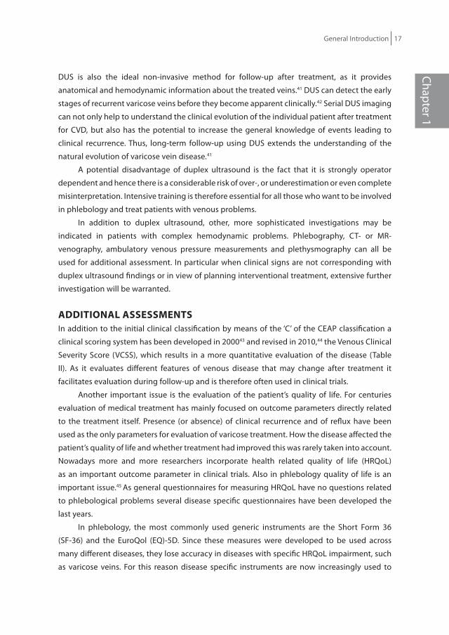
DUS is also the ideal non-invasive method for follow-up after treatment, as it provides
anatomical and hemodynamic information about the treated veins.41 DUS can detect the early
stages of recurrent varicose veins before they become apparent clinically.42 Serial DUS imaging
can not only help to understand the clinical evolution of the individual patient after treatment
for CVD, but also has the potential to increase the general knowledge of events leading to
clinical recurrence. Thus, long-term follow-up using DUS extends the understanding of the
natural evolution of varicose vein disease.41
A potential disadvantage of duplex ultrasound is the fact that it is strongly operator
dependent and hence there is a considerable risk of over-, or underestimation or even complete
misinterpretation. Intensive training is therefore essential for all those who want to be involved
in phlebology and treat patients with venous problems.
In addition to duplex ultrasound, other, more sophisticated investigations may be
indicated in patients with complex hemodynamic problems. Phlebography, CT- or MR-
venography, ambulatory venous pressure measurements and plethysmography can all be
used for additional assessment. In particular when clinical signs are not corresponding with
duplex ultrasound findings or in view of planning interventional treatment, extensive further
investigation will be warranted.
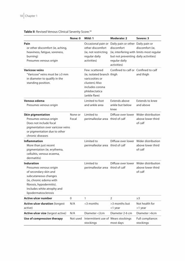
AddiTionAL AssessMenTsIn addition to the initial clinical classification by means of the ‘C’ of the CEAP classification a
clinical scoring system has been developed in 200043 and revised in 2010,44 the Venous Clinical
Severity Score (VCSS), which results in a more quantitative evaluation of the disease (Table
II). As it evaluates different features of venous disease that may change after treatment it
facilitates evaluation during follow-up and is therefore often used in clinical trials.
Another important issue is the evaluation of the patient’s quality of life. For centuries
evaluation of medical treatment has mainly focused on outcome parameters directly related
to the treatment itself. Presence (or absence) of clinical recurrence and of reflux have been
used as the only parameters for evaluation of varicose treatment. How the disease affected the
patient’s quality of life and whether treatment had improved this was rarely taken into account.
Nowadays more and more researchers incorporate health related quality of life (HRQoL)
as an important outcome parameter in clinical trials. Also in phlebology quality of life is an
important issue.45 As general questionnaires for measuring HRQoL have no questions related
to phlebological problems several disease specific questionnaires have been developed the
last years.
In phlebology, the most commonly used generic instruments are the Short Form 36
(SF-36) and the EuroQol (EQ)-5D. Since these measures were developed to be used across
many different diseases, they lose accuracy in diseases with specific HRQoL impairment, such
as varicose veins. For this reason disease specific instruments are now increasingly used to
18 Chapter 1
Table ii: Revised Venous Clinical Severity Score.44
none: 0 Mild: 1 Moderate: 2 severe: 3
Painor other discomfort (ie, aching, heaviness, fatigue, soreness, burning)Presumes venous origin
Occasional pain or other discomfort (ie, not restricting regular daily activities)
Daily pain or other discomfort (ie, interfering with but not preventing regular daily activities)
Daily pain or discomfort (ie, limits most regular daily activities)
Varicose veins“Varicose” veins must be ≥3 mm in diameter to qualify in the standing position.
Few: scattered(ie, isolated branch varicosities or clusters) Also includes corona phlebectatica (ankle flare)
Confined to calf or thigh
Confined to calf and thigh
Venous edemaPresumes venous origin
Limited to foot and ankle area
Extends above ankle but below knee
Extends to knee and above
skin pigmentationPresumes venous originDoes not include focal pigmentation over varicose veins or pigmentation due to other chronic diseases
None orFocal
Limited to perimalleolar area
Diffuse over lower third of calf
Wider distributionabove lower third of calf
inflammationMore than just recent pigmentation (ie, erythema, cellulitis, venous eczema, dermatitis)
Limited to perimalleolar area
Diffuse over lower third of calf
Wider distributionabove lower third of calf
indurationPresumes venous origin of secondary skin and subcutaneous changes (ie, chronic edema with fibrosis, hypodermitis). Includes white atrophy and lipodermatosclerosis
Limited to perimalleolar area
Diffuse over lower third of calf
Wider distributionabove lower third of calf
Active ulcer number 0 1 2 ≥3
Active ulcer duration (longest active)
N/A <3 months >3 months but <1 year
Not health for >1 year
Active ulcer size (largest active) N/A Diameter <2cm Diameter 2-6 cm Diameter >6cm
use of compression therapy Not used Intermittent use of stockings
Wears stockings most days
Full compliance: stockings
General Introduction 19
Chapter 1
evaluate the effects of specific treatments in patients with varicose veins, in combination with
generic instruments.46-49 The available disease specific HRQoL tools focusing on chronic venous
insufficiency and/or varicose veins are: Chronic Lower Limb venous Insufficiency (CIVIQ),
Aberdeen Varicose Vein Questionnaire (AVVQ) and VEINES-QOL/Sym.47-49 The CIVIQ focuses
on HRQoL impairment and includes only one symptom related item. The CIVIQ results in a
global score and four separate domain scores (physical, psychological, social impairments and
level of pain)47. The AVVQ calculates one global HRQoL score summing symptom and clinical
class related items.48 The VEnous INsufficiency Epidemiological and Economic Studies (VEINES)
questionnaire is positioned in between these two instruments because it balances symptom
(VEINES-SYM) and quality of life (VEINES-QOL) items resulting in two separate scores.49
Most of these questionnaires do not exist in the mother tongue of the patients. For use in
the Netherlands these questionnaires should be translated to Dutch and validated.
Although there are neither guidelines nor any evidence that this HRQoL investigation
has to be done in every patient undergoing treatment, it may be helpful to evaluate different
techniques and to determine optimal treatment strategies.
TreATMenTThere are three main reasons to treat patients with varicose veins. First of all, treatment aims
at preventing acute complications, such as bleeding and superficial vein thrombosis and
chronic deterioration consisting of all clinical features of chronic venous insufficiency (C3-C6).
All these have a major impact on patients’ HRQoL. Secondly, treatment relieves symptoms
caused by varicose veins, such as heaviness, tired legs, cramps etc. Thirdly, patients may seek
for treatment mainly for cosmetic reasons which also affect patient’s HRQoL.
Treatment of varicose veins can roughly be divided into four groups: compression
therapy, endovenous thermal ablation, sclerotherapy and surgical treatment. In this thesis only
treatment of the great saphenous vein (GSV) and tributaries will be considered.
Treatment of the GsV In 1905 Keller described a surgical technique to remove the GSV by stripping and invagination.50
For nearly 100 years high ligation and stripping was the gold standard for the treatment of
incompetence of the GSV.
Although sclerotherapy had already been introduced in the mid 19th century this
technique was reintroduced after the Second World War.51 It gained more and more interest
when a technique was developed to make foam with the detergent sclerosing agents.52-54
Nowadays the Tessari method is widely accepted for routine foam sclerotherapy of refluxing
saphenous trunks and tributaries.55
The true ‘endovenous revolution’ started with the beginning of the 3rd millennium.
Endovenous thermal ablation (EVTA) techniques were introduced. The first EVTA procedures

20 Chapter 1
were performed with radiofrequency ablation (RFA) with the VNUS Closure Plus System.56
Immediately thereafter endovenous laser ablation (EVLA) was developed. The radiologist
Min introduced a minimally invasive endovenous laser treatment for varicose veins, aiming at
elimination of incompetence at the saphenofemoral junction (SFJ) and closure of the GSV.57,58
The first ELVA procedures were with 810nm diode laser, of which hemoglobin is the main target.
Laser light absorption is followed by heat production. This heat is transmitted to the vein wall
leading to destruction. The precise mechanisms of EVTA and the relation with wavelength,
chromophores and carbonization are not yet completely understood. A meta-analysis showed
that the above described minimally invasive techniques appeared to be at least as effective as
surgery for the treatment of varicose veins.59
Nowadays EVLA has turned into a generally accepted, easy to perform and patient friendly
technique.60 The actual tendency is to use lasers with higher wavelengths up to 1470nm61 and
to move to modified laser tips such as the radial tip, tulip-tip and others replacing the initial
bare tip laserfibers. The newest thermal technique is steam ablation, which works by heating
the vein wall with hyperheated steam.62
Treatment failure and varicose vein recurrence remains a problem occurring after
all treatment modalities. Different etiologic factors may play a role in the development of
recurrence: tactical and technical failure, neovascularisation (mainly after surgery, at the site
of high ligation), recanalisation of a previously obliterated trunk (after endovenous ablation)
and finally, progression of the disease. Prevention of recurrence should try to interfere with
these factors. In the first place, a thoroughly performed duplex ultrasound should lead
to a correct diagnosis of the varicose disease, to avoid tactical failure. Further, the planned
procedure should be performed correctly. Training in duplex scanning and in ultrasound
guided procedures is therefore the cornerstone of good clinical practice in phlebology.
Neovascularisation, mainly at SFJ and saphenopopliteal junction (SPJ) remains a concern after
surgical treatment of varicose veins. It can be easily detected by means of duplex ultrasound,
which shows the presence of multiple tortuous veins at the site of the previous high ligation
or in the strip track.41 Some barrier techniques to mitigate the effect of neovascularisation
at the SFJ or SPJ have been tried out successfully.63,64 However, the best way to avoid
neovascularisation seems to be not to operate at the SFJ or SPJ. Nowadays, in many countries
like the Netherlands, surgical high ligation and stripping has been replaced by endovenous
ablation techniques. After these endovenous interventions indeed neovascularisation is a very
exceptional phenomenon. However recurrence remains a problem, also after endovenous
techniques. It may be due to recanalisation of the obliterated trunk, with or without recurrent
reflux at the junction. Prevention of recanalisation after thermal ablation is mainly a matter of
using enough tumescence and a correct amount of energy, dependent of vein diameter. The
only factor we cannot really influence, unfortunately, is progression of the disease. To further
unravel the problem of recurrent varicose veins after all kinds of interventions for varicose

General Introduction 21
Chapter 1
veins, more and better performed randomized studies with long-term follow-up (of at least 5
years) are certainly needed.
Treatment of tributariesPittaluga showed that incompetent tributaries may render the GSV incompetence, based
on the ascending pathophysiologic theory. Under certain circumstances single treatment of
such incompetent tributaries – without treatment of the refluxing GSV trunk – may abolish
truncal reflux completely. If this is the case, unnecessary ablation of a refluxing trunk could be
avoided in certain cases. Properly selected patients could benefit from a treatment of refluxing
tributaries only. This finding certainly needs more research.
The gold standard for treating incompetent tributaries is ambulatory phlebectomy,
based on the findings of a randomised controlled trial comparing liquid sclerotherapy with
phlebectomies for tributary treatment.65 So far no randomised controlled trial is available in
which foam sclerotherapy and ambulatory phlebectomy are compared.
Research in medicine is done to improve the quality of diagnosis and treatment. The
translation from pure research to daily practice is the aim of every clinical investigator. In the
last two decades translation of evidence based research has changed phlebological practice
considerably. Duplex ultrasound has become the gold standard for diagnosis and treatment
guidance. Major surgery has largely been abandoned and more patient friendly, less expensive
and minimally invasive endovenous techniques and surgical procedures have been introduced.
Diagnosis and treatment of CVD, and especially of varicose veins, can be realised in nearly
100% in day care, often in special clinics dedicated to phlebology. Compared to the situation
25 years ago, there is now growing interest in integrating HRQoL investigation in our clinical
research. Not only will the individual patient benefit from this, but also society as a whole, as
it may lead to more responsible choices in treatment strategy for patients with varicose veins.
22 Chapter 1
AiMs of The Thesis To contribute to evidence-based medicine and identify gaps in the knowledge concerning the
treatment of saphenous varicose veins we performed a systematic literature research. (Chapter
2).
There are a few questionnaires available to assess patient reported outcome or HRQoL,
but so far only one of these questionnaires was available in Dutch. Patient reported outcome
instruments are necessary to answer several questions in scientific investigations. We therefore
translated and validated the CIVIQ and the VEINES Sym/Qol for Dutch patients with varicose
veins. (Chapter 3)
The third aim was to investigate the best treatment options for patients with GSV
incompetence. In a randomized controlled trial we evaluated the effect of three commonly
used treatment methods for insufficiency of the GSV; endovenous laser ablation, ultrasound-
guided foam sclerotherapy and conventional surgery. (Chapter 4) In a subsequent prospective
study we wanted to evaluate the effect of single phlebectomies on the reflux of the GSV and
to describe predictors for success of this approach. (Chapter 5) After a literature search we
reviewed the existing evidence on recurrence after different treatment techniques. (Chapter 6)
In summary, first we tried to comprehend the current literature on varicose vein treatments,
and find evidence for what could be the most successful therapy with the least complications.
Then we translated and validated two disease specific HRQoL questionnaires, that can be used
in future studies. In a randomized controlled trial we compared the three most used treatments
for GSV incompetence. We further evaluated the effect of phlebectomy of tributaries on GSV
reflux and made a prediction model by means of a score chart. Finally, we reviewed the literature
on the subject of recurrent varicose veins after treatment. In the discussion we highlighted the
importance of the patients’ voice, which might be more important than clinical recurrence and
duplex ultrasound results. The studies in this thesis will contribute to the translations of our
findings to a more rational treatment approach in daily practice fore varicose patients.

General Introduction 23
Chapter 1
references
1. Hosoi Y, Zukowski A, Kakkos SK, Nicolaides AN. Ambulatory venous pressure measurements: new parameters derived from a mathematic hemodynamic model. J Vasc Surg. 2002;36:137-42.
2. Recek C. Conception of the venous hemodynamics in the lower extremity. Angiology 2006;57:556-63.
3. Belcaro G, Laurora G, Cesarone MR, De Sanctis MT, Incandela L. Microcirculation in high perfusion microangiopathy. J Cardiovasc Surg. (Torino) 1995;36:393-8.
4. Junger M, Steins A, Hahn M, Hafner HM. Microcirculatory dysfunction in chronic venous insufficiency (CVI). Microcirculation 2000;7:S3-12.
5. Ackerman Z, Seidenbaum M, Loewenthal E, Rubinow A. Overload of iron in the skin of patients with varicose ulcers. Possible contributing role of iron accumulation in progression of the disease. Arch Dermatol. 1988;124:1376-8.
6. Neumann HAM. Measurement of microcirculation. In: Altmeyer, ed. Wound healing and skin physiology. Heidelberg: Springer Verlag Berlin; 1995:115-26.
7. Maessen-Visch MB, Koedam MI, Hamulyak K, Neumann HA. Atrophie blanche. Int J Dermatol .1999;38:161-72.
8. Mwaura B, Mahendran B, Hynes N, et al. The impact of differential expression of extracellular matrix metalloproteinase inducer, matrix metalloproteinase-2, tissue inhibitor of matrix metalloproteinase-2 and PDGF-AA on the chronicity of venous leg ulcers. Eur J Vasc Endovasc Surg. 2006;31:306-10.
9. Zsoter T, Cronin RF. Venous distensibility in patients with varicose veins. Can Med Assoc J. 1966;94:1293-7.
10. Sansilvestri-Morel P, Nonotte I, Fournet-Bourguignon MP, et al. Abnormal deposition of extracellular matrix proteins by cultured smooth muscle cells from human varicose veins. J Vasc Res. 1998;35:115-23.
11. Caggiati A, Rosi C, Heyn R, Franceschini M, Acconcia MC. Age-related variations of varicose veins anatomy. J Vasc Surg 2006;44:1291-5.
12. Ludbrook J, Beale G. Femoral venous valves in relation to varicose veins. Lancet 1962;1:79-81.13. Trendelemburg. Uber die Tunderbindung der Vena Saphena Magnabie unterschenkel varicen. Beitr
Z Clin Chir. 1890;7:195.14. Labropoulos N, Giannoukas AD, Delis K, et al. Where does venous reflux start? J Vasc Surg.
1997;26:736-42.15. Labropoulos N, Kang SS, Mansour MA, Giannoukas AD, Buckman J, Baker WH. Primary superficial
vein reflux with competent saphenous trunk. Eur J Vasc Endovasc Surg. 1999;18:201-6.16. Engelhorn CA, Engelhorn AL, Cassou MF, Salles-Cunha SX. Patterns of saphenous reflux in women
with primary varicose veins. J Vasc Surg. 2005;41:645-51.17. Pittaluga P, Chastane S, Rea B, Barbe R. Classification of saphenous refluxes: implications for
treatment. Phlebology 2008;23:2-9.18. Pittaluga P, Chastanet S, Guex JJ. Great saphenous vein stripping with preservation of sapheno-
femoral confluence: hemodynamic and clinical results. J Vasc Surg. 2008;47:1300-4; discussion 4-5.19. Pittaluga P, Chastanet S, Rea B, Barbe R. Midterm results of the surgical treatment of varices by
phlebectomy with conservation of a refluxing saphenous vein. J Vasc Surg. 2009;50:107-18.20. Pittaluga P, Chastanet S, Locret T, Barbe R. The effect of isolated phlebectomy on reflux and diameter
of the great saphenous vein: a prospective study. Eur J Vasc Endovasc Surg. 2010;40:122-8.21. Creton D. Diameter reduction of the proximal long saphenous vein after ablation of a distal
incompetent tributary. Dermatol Surg. 1999;25:394-7.

24 Chapter 1
22. Theivacumar NS, Darwood RJ, Dellegrammaticas D, Mavor AI, Gough MJ. The clinical significance of below-knee great saphenous vein reflux following endovenous laser ablation of above-knee great saphenous vein. Phlebology 2009;24:17-20.
23. Zamboni P, Cisno C, Marchetti F, Quaglio D, Mazza P, Liboni A. Reflux elimination without any ablation or disconnection of the saphenous vein. A haemodynamic model for venous surgery. Eur J Vasc Endovasc Surg. 2001;21:361-9.
24. Evans CJ, Fowkes FG, Ruckley CV, Lee AJ. Prevalence of varicose veins and chronic venous insufficiency in men and women in the general population: Edinburgh Vein Study. J Epidemiol Community Health 1999;53:149-53.
25. Beebe-Dimmer JL, Pfeifer JR, Engle JS, Schottenfeld D. The epidemiology of chronic venous insufficiency and varicose veins. Ann Epidemiol. 2005;15:175-84.
26. Ruckley CV, Evans CJ, Allan PL, Lee AJ, Fowkes FG. Chronic venous insufficiency: clinical and duplex correlations. The Edinburgh Vein Study of venous disorders in the general population. J Vasc Sur. 2002;36:520-5.
27. Maurins U, Hoffmann BH, Losch C, Jockel KH, Rabe E, Pannier F. Distribution and prevalence of reflux in the superficial and deep venous system in the general population--results from the Bonn Vein Study, Germany. J Vasc Surg. 2008;48:680-7.
28. Rabe E, Guex JJ, Puskas A, Scuderi A, Fernandez Quesada F, Coordinators VCP. Epidemiology of chronic venous disorders in geographically diverse populations: results from the Vein Consult Program. Int Angiol. 2012;31:105-15.
29. Abramson JH, Hopp C, Epstein LM. The epidemiology of varicose veins. A survey in western Jerusalem. J Epidemiol Community Health 1981;35:213-7.
30. Coon WW, Willis PW, 3rd, Keller JB. Venous thromboembolism and other venous disease in the Tecumseh community health study. Circulation 1973;48:839-46.
31. Criqui MH, Jamosmos M, Fronek A, et al. Chronic venous disease in an ethnically diverse population: the San Diego Population Study. Am J Epidemiol. 2003;158:448-56.
32. Brand FN, Dannenberg AL, Abbott RD, Kannel WB. The epidemiology of varicose veins: the Framingham Study. Am J Prev Med. 1988;4:96-101.
33. Rabe E, Pannier-Fischer F, Bromen K, et al. Bonner Venenstudie der Deutschen Gesellschaft für Phlebologie – Epidemiologische Untersuchung zur Frage der Häufigkeit und Ausprägung von chronischen Venenkrankheiten in der städtischen und ländlichen Wohnbevölkerung. Phlebologie 2003;32:1-14.
34. Callam MJ. Epidemiology of varicose veins. Br J Surg. 1994;81:167-73.35. Dwerryhouse S, Davies B, Harradine K, Earnshaw JJ. Stripping the long saphenous vein reduces the
rate of reoperation for recurrent varicose veins: five-year results of a randomized trial. J Vasc Surg. 1999;29:589-92.
36. Pannier F RE. Progression of chronic venous disorders: results from the Bonn Vein Study II. Paper presented at the 23d Annual Meeting of the American Venous Forum; 23-26 Feb 2011; San Diego, USA J Vasc Surg. 2011;53(1):254-255.
37. Eklof B, Rutherford RB, Bergan JJ, et al. Revision of the CEAP classification for chronic venous disorders: consensus statement. J Vasc Surg. 2004;40:1248-52.
38. Porter JM, Moneta GL. Reporting standards in venous disease: an update. International Consensus Committee on Chronic Venous Disease. J Vasc Surg. 1995;21:635-45.
39. Coleridge-Smith P, Labropoulos N, Partsch H, Myers K, Nicolaides A, Cavezzi A. Duplex ultrasound investigation of the veins in chronic venous disease of the lower limbs--UIP consensus document. Part I. Basic principles. Eur J Vasc Endovasc Surg. 2006;31:83-92.
40. Cavezzi A, Labropoulos N, Partsch H, et al. Duplex ultrasound investigation of the veins in chronic venous disease of the lower limbs--UIP consensus document. Part II. Anatomy. Vasa 2007;36:62-71.

General Introduction 25
Chapter 1
41. De Maeseneer M, Pichot O, Cavezzi A, et al. Duplex ultrasound investigation of the veins of the lower limbs after treatment for varicose veins - UIP consensus document. Eur J Vasc Endovasc Surg. 2011;42:89-102.
42. De Maeseneer MG, Vandenbroeck CP, Hendriks JM, Lauwers PR, Van Schil PE. Accuracy of duplex evaluation one year after varicose vein surgery to predict recurrence at the sapheno-femoral junction after five years. Eur J Vasc Endovasc Surg. 2005;29:308-12.
43. Rutherford RB, Padberg FT, Jr., Comerota AJ, Kistner RL, Meissner MH, Moneta GL. Venous severity scoring: An adjunct to venous outcome assessment. J Vasc Surg. 2000;31:1307-12.
44. Vasquez MA, Rabe E, McLafferty RB, et al. Revision of the venous clinical severity score: venous outcomes consensus statement: special communication of the American Venous Forum Ad Hoc Outcomes Working Group. J Vasc Surg. 2010;52:1387-96.
45. Carradice D, Mazari FA, Samuel N, Allgar V, Hatfield J, Chetter IC. Modelling the effect of venous disease on quality of life. Br J Surg. 2011;98:1089-98.
46. Wiebe S, Guyatt G, Weaver B, Matijevic S, Sidwell C. Comparative responsiveness of generic and specific quality-of-life instruments. J Clin Epidemiol. 2003;56:52-60.
47. Launois R, Reboul-Marty J, Henry B. Construction and validation of a quality of life questionnaire in chronic lower limb venous insufficiency (CIVIQ). Qual Life Res. 1996;5:539-54.
48. Garratt AM, Macdonald LM, Ruta DA, Russell IT, Buckingham JK, Krukowski ZH. Towards measurement of outcome for patients with varicose veins. Qual Health Care 1993;2:5-10.
49. Lamping DL. Measuring health-related quality of life in venous disease: practical and scientific considerations. Angiology 1997;48:51-7.
50. Keller W. A new method of extirpating the internal saphenous and similar veins in varicose conditions: a preliminary report. N Y Med J. 1905;82:385-6.
51. Orbach EJ. Clinical evaluation of a new technic in the sclerotherapy of varicose veins. J Int Coll Surg. 1948;11:396-402.
52. Cabrera J, Cabrera J, Jr., Garcia-Olmedo MA, Redondo P. Treatment of venous malformations with sclerosant in microfoam form. Arch Dermatol. 2003;139:1409-16.
53. Frullini A, Cavezzi A. Sclerosing foam in the treatment of varicose veins and telangiectases: history and analysis of safety and complications. Dermatol Surg. 2002;28:11-5.
54. Smith PC. Chronic venous disease treated by ultrasound guided foam sclerotherapy. Eur J Vasc Endovasc Surg. 2006;32:577-83.
55. Tessari L, Cavezzi A, Frullini A. Preliminary experience with a new sclerosing foam in the treatment of varicose veins. Dermatol Surg. 2001;27:58-60.
56. Goldman MP. Closure of the greater saphenous vein with endoluminal radiofrequency thermal heating of the vein wall in combination with ambulatory phlebectomy: preliminary 6-month follow-up. Dermatol Surg. 2000;26:452-6.
57. Navarro L, Min RJ, Bone C. Endovenous laser: a new minimally invasive method of treatment for varicose veins--preliminary observations using an 810 nm diode laser. Dermatol Surg. 2001;27:117-22.
58. Min RJ, Zimmet SE, Isaacs MN, Forrestal MD. Endovenous laser treatment of the incompetent greater saphenous vein. J Vasc Interv Radiol. 2001;12:1167-71.
59. van den Bos R, Arends L, Kockaert M, Neumann M, Nijsten T. Endovenous therapies of lower extremity varicosities: a meta-analysis. J Vasc Surg. 2009;49:230-9.
60. Nijsten T, van den Bos RR, Goldman MP, et al. Minimally invasive techniques in the treatment of saphenous varicose veins. J Am Acad Dermatol. 2009;60:110-9.
61. Pannier F, Rabe E, Maurins U. First results with a new 1470-nm diode laser for endovenous ablation of incompetent saphenous veins. Phlebology 2009;24:26-30.
62. van den Bos RR, Milleret R, Neumann M, Nijsten T. Proof-of-principle study of steam ablation as novel thermal therapy for saphenous varicose veins. J Vasc Surg. 2010;53:181-6.

26 Chapter 1
63. De Maeseneer MG, Van Schil PE, Philippe MM, Vanmaele RG, Eyskens EJ. Is recurrence of varicose veins after surgery unavoidable? Acta Chir Belg. 1995;95:21-6.
64. van Rij AM, Jiang P, Solomon C, Christie RA, Hill GB. Recurrence after varicose vein surgery: a prospective long-term clinical study with duplex ultrasound scanning and air plethysmography. J Vasc Surg. 2003;38:935-43.
65. de Roos KP, Nieman FH, Neumann HA. Ambulatory phlebectomy versus compression sclerotherapy: results of a randomized controlled trial. Dermatol Surg. 2003;29:221-6.

Chapter 2
Endovenous Therapies of Varicose Veins: Indications, Procedures, Efficacy and Safety
Anke A. Biemans Renate R. van den BosTamar Nijsten
G Ital Dermatol Venereol 2010 Apr;145(2):161-73.
Chapter 2
28 Chapter 2
ABsTrAcT Venous insufficiency of the lower-extremity is common and the prevalence increases with age.
Chronic venous insufficiency has a high impact on patients’ health related quality of life and is
associated with considerable health care costs. In addition to classical symptoms, it may result
in skin changes and venous ulcers. Since more than hundred years, surgical ligation of the
junction with or without stripping has been the standard of care in the treatment of insufficient
great and small saphenous veins. However, the recurrence rates are relatively high and surgery
may be associated with serious adverse events, considerable down time and is cosmetically
suboptimal. In the last decade several minimally invasive techniques have been introduced, to
improve efficacy, patients’ health related quality of life and treatment satisfaction, and to reduce
serious side effects, costs and post-operative pain. Ultrasound guided foam sclerotherapy,
endovenous laser and radiofrequency ablation are the most commonly used therapies, and
challenge surgery as the gold standard of care in patients with varicose veins. The objective of
this review is to inform clinicians about these three therapeutic options for saphenous varicose
veins and to describe and compare the indications, procedures, efficacy and safety profile.

Endovenous therapies for varicose veins 29
Chapter 2
inTroducTionSymptomatic varicose veins of the lower extremities represent one of the most common
conditions in the adult population. About 25% of the population has lower-extremity varicose
veins1 and half of the adult population has stigmata of minor venous disease.2 Since the
prevalence of varicose veins increases with age in a linear manner, the prevalence of venous
insufficiency will increase considerably in the next decades. Associated symptoms range
from mild complaints such as fatigue, heaviness, and itching to more serious conditions
such as edema, skin hyperpigmentation, eczema and leg ulceration. Venous ulcers have a
prevalence of 1-2% in people over 65 years of age.3 Chronic venous insufficiency has, because
of its complications, a great impact on patients’ health related quality of life (HRQoL), and
it is associated with considerable health care costs.4 In the Netherlands, chronic venous
insufficiency and its related venous ulcers, costs approximately 1% of the national health care
budget. It has been estimated that about half the venous ulcers can be prevented by treating
the varicose veins.
The mean goal in the treatment of varicose veins is to reduce the symptoms and
complications of chronic venous insufficiency, and to improve health-related quality of life
(HRQoL) of patients. Surgery has been the standard of care in the treatment of saphenous
varicose veins for more than a century. More than a quarter of all cases of chronic venous
insufficiency is caused by reflux of the great saphenous vein (GSV), and is traditionally treated
with surgical ligation at the saphenofemoral junction and stripping of the incompetent
saphenous vein. Usually, surgery of the small saphenous vein (SSV) consists of ligation at the
saphenopopliteal junction (SPJ) with or without a short strip until the mid-calf of the SSV. It
is well-known that stripping is related to a high recurrence rate and neovascularization. The
recurrence rate of surgery is about 25% for the GSV and 50% for the SSV after 5 years.5 After a
mean follow-up of 34 years, Fischer showed a recurrence of varicose veins in 60% in 125 limbs
after SFJ ligation and GSV stripping.6 Neovascularization, a double saphenous vein system,
technical failure (up to 30%)7 and/or incomplete procedure5,8 are several reasons of failure after
surgery. Other disadvantages of surgical therapy are the common use of general or epidural
anesthesia, presence of at least two fairly-long scars, post-operative down time and risk of
adverse events such as femoral artery or femoral vein damage, wound infection, neurological
injury (about 7% in short to 40% in long stripping of GSV)9 and lymphatic complications.
New minimally invasive techniques such as ultrasound guided foam sclerotherapy (UGFS),
endovenous laser ablation (EVLA) and radiofrequency ablation (RFA) have been introduced
in the last decade, to improve the efficacy, patients’ HRQoL and treatment satisfaction, and
to reduce serious side effects, costs and post-operative pain.10 These new methods can
be performed in outpatient settings or ambulant day-care facilities. This has an additional
advantage compared to surgery, which is usually performed in the operating theatre under
general or spinal anesthesia. Dermatologic surgeons were among the first in developing and

30 Chapter 2
reporting on these minimal invasive techniques for saphenous varicose veins such as RFA,
EVLA and UGFS. The objective of this review is to inform physicians about the most commonly
used minimal invasive therapies used for saphenous varicose veins, to describe the procedures
and to review their efficacy and safety.
uLTrAsound Guided foAM scLeroTherAPy (uGfs)The use of foamed sclerosants has been described since 1944, when Orbach injected a small
amount of air into the venous segment targeted for treatment in order to displace blood and
intensify the contact time between sclerosant and the endothelium of the vein (so called, ‘air
block’ technique).11 Liquid sclerotherapy is an established method of causing venous occlusion
by the injection of sclerosing liquid into affected veins. Direct contact of sclerosant with the
venous endothelium initiates endothelial and mural injury by an irritative reaction. As a result,
a local, wall-adherent thrombus is formed and subsequent sclerosis transforms the treated
vein into a fibrous cord.12,13 The success of foam, which is more effective for the treatment
of saphenous veins than liquid, is mostly caused by its qualities. Foam displaces blood and
creates an increased effective contact area between the sclerosant and the endothelium and
induces venous spasm.14 Various methods and procedures have been described for creating a
foamed sclerosant, but is in essence very straightforward. Foam is created by forcibly mixing
liquid sclerosant with air, oxygen or carbon dioxide.15,16,17
indicationsPrimary insufficient GSVs and SSVs as well as previously treated varicose veins and recurrences
after surgery (for example due to neovascularization) can be treated with ultrasound-guided
foam sclerotherapy. All sizes of GSVs, independent of CEAP class and type (linear and tortuous),
can be treated safely and effectively (see Table I).21 Small and large diameters, are successfully
treated, but saphenous veins with diameters of 10 mm or more may require multiple
treatments and relatively large volumes of foam (up to 3 sessions and 15 cc of foam).23 UGFS
can be used in patients with severe chronic venous insufficiency (CVI) and may enhance ulcer
healing.19 Also perforator veins and congenital venous malformations have been treated with
this technique.24-26
Suitability of a patient for foam sclerotherapy depends on the aims of treatment as
well as the venous anatomy. The main advantage of foam sclerotherapy is that it can be
carried out successfully in almost any patient with clinically significant venous disease,
although morbid obesity, old age or frailty and severe concomitant diseases may entangle
the intervention. Moreover, the procedure is swift and takes a couple of minutes. The only
absolute contraindications are severe allergy to aethoxysclerol and, as for all venous therapies,
obliteration of the deep veins. Patients should be fully informed about the method of treatment
as well as the complications that may arise. They should be warned about the (inflamed)

Endovenous therapies for varicose veins 31
Chapter 2
lumps caused by thrombophlebitis and the chance of skin hyperpigmentation. Deep venous
trombosis (DVT) is also an alleged serious complication that should be mentioned as well as
severe allergy, although both are very uncommon.
Table i: Indications for the different minimal invasive treatments of varicose veins.
indications uGfs eVLA rfA
GSV + + +
SSV + + +
Accessory veins + +/- +/-
Perforator veins + +/- +/-a
Diameter <0.5 cm + - +/-
Diameter 0.5-1.0 cm + + +
Diameter >1.0 cm +/- + +b
Tortuous vein + - -
Neovascularizationc + - -
Partial intraluminal obstruction(s)d + - -a mini-RFA can be usedb maximum vein diameter equals 12 mm in conventional RFA, not in fast versionc may occur after surgical strippingd after thromboflebitis, UGFS, EVLA, or RFA treatment
ProcedureThe most common method of producing foam is the Tessari-method. In this method, two
syringes are connected by a three-way valve, and the liquid sclerosant (1 cc aethoxysclerol or
sodium tetradecyl sulfate 1-3% in Europe and the USA), is forcibly mixed with air (3-4cc) and
frothed into foam by a pumping action (Figure 1a). The liquid sclerosing solution, which is used
in classic sclerotherapy, is mixed with air to create foam. This foam of fine bubbles is injected
intravenously with ultrasound (US) guidance. In classic sclerotherapy, the air block technique
(i.e., first inject an air bubble before injecting the sclerosant) has been used to prolong contact
time with the venous wall and to reduce the ‘wash out’ of the agent that is injected in the vein.16
Foam is usually administered at one or more points of the saphenous varicose vein while
the patient is in horizontal position. Ideally, the first bolus of foam (3-5cc) is administered in the
proximal part of the treated vein and subsequent injections more distally because most of the
foam moves in the direction of venous flow. In the 2nd European Consensus it is recommended
to inject at the proximal thigh ten centimeters below the saphenofemoral junction in order to
achieve optimal occlusion of the proximal part of the vein.27 The vein is visualized longitudinally
by ultrasound to guide and control venous access (Figure 1b). The foam can be injected directly
or through a cannula, catheter or butterfly needle.17,28 The echodens foam is clearly visible,

32 Chapter 2
confirming proper injection (Figure 1c and d). The volume of injected foam depends on the
length and diameter of the vessel and may range between 3-50 ml per session. There is no
high level of evidence on how much the maximum volume of foam should be per treatment
session; this is highly variable between physicians. The suggestion not to exceed 10 ml per
session in the 2nd European Consensus is based on expert opinion. For saphenous varicose
veins, a foam that is made of 3% aethoxysclerol appears to be modestly more effective than
a foam made of 1% aethoxysclerol but it is more frequently associated with adverse events
such as hyperpigmentation and phlebitis.29 After injection, the patient remains horizontally
or in reverse Trendelenburg position for at least 5 minutes, to enhance contact time of the
foam with the venous wall. After therapy, cotton wool or foam pads can be applied over the
tract of the veins and compression therapy (bandages, anti-thromboembolism stockings and/
or medical elastic compression stockings class II) are recommended for a period that varies
between physicians from one to six weeks.
figure 1: Creation of foam (A); gaining access to varicose vein (B); injection of foam in cross-sectional
view before (c) and after injection (d).

Endovenous therapies for varicose veins 33
Chapter 2
efficacyFoam sclerotherapy is about four times more effective than the classic liquid sclerotherapy,
because of the increased contact time with the venous wall, increased surface area, and the
induction of venous spasm.22 In several studies, about two thirds of the saphenous varicose
veins were occluded after one UGFS session and more than 90% of treatments were successful
after two or three sessions.19,20,29 Several large case series 18,29 and one multicentre study30 have
been published but very few comparative studies have been performed.
Ceulen and colleagues showed occlusion in 88% of GSVs and 82% of SSVs after treatment
with UGFS in 1411 limbs after a mean follow-up of 11 months.29 Smaller series showed 69%
complete sclerosis in 99 limbs after 24 months of follow-up,23 44% occlusion in 211 limbs after
5 years of follow-up31 and 88% occlusion in 143 limbs after 6 weeks of follow-up.19 Cabrera
et al.26 published a case series of 500 legs treated with foam sclerotherapy. He reported that
after three or more years 81% of treated GSVs remained occluded and 97% of superficial
varicose veins had disappeared. This required one session of sclerotherapy in 86% of patients,
two in 11% and three sessions in 3% of patients. Subsequently, a number of authors have
published clinical series based on this technique including Frullini and Cavezzi,18 who reported
a series of 453 patients, and Barrett et al.,23 who reported a series of 100 limbs. Cavezzi et al.32
has subsequently published a detailed analysis of the efficacy of foam sclerotherapy in 194
patients, reporting a good outcome in 93% of patients. UGFS was an effective treatment for the
SSV, with abolition of reflux and visible varicose veins, and improvement in HRQoL for at least
12 months.33 Obliteration rate of the GSV one day post-procedure was high with 94% of treated
limbs. Ultrasound examination showed complete thrombosis of these veins with complete
elimination of retrograde flow in the GSV. This efficacy is similar to, or even better than the
reported obliteration rates of 60%-99% in the English literature.17 Foam sclerotherapy has
shown to be an effective treatment for primary varicose veins in the lower limbs by obliteration
of the GSV. Unlike surgery, this treatment does not require general, spinal, or local anaesthesia.
Furthermore, the treatment time is shorter and the recovery faster. In comparison with other
endovascular techniques such as EVLA and RFA, the use of foam as obliteration method is
significantly more cost-effective.
safetyThe most common adverse events (Table II) associated with foam sclerotherapy are
thrombophlebitis, matting, hyperpigmentation, and pain provoked by injection or pain
persisting at the sclerosed area.34 The minor complications in the immediate post-procedure
period include hyperpigmentation (6.1%), superficial thrombophlebitis (7.6%) and cellulitis
(1.5%). These local effects are mostly mild and may represent the spectrum of the inflammatory
effect of the sclerosant. In several trials and case series, the rate of deep venous thrombosis
varied from 0%-6%.34

34 Chapter 2
Table ii: Likelihood of specific adverse events associated with each of the three minimal invasive
techniques.
uGfs eVLA rfA
Allergic reaction + -* -*
Skin necrosis / burns +/- + +/-
Ecchymosis +/- + -
Pain (‘pulling chord’) due to venous contraction - + +/-
Thrombophlebitis + + +
Deep venous thrombosis and emboli + +/- +/-
Nerve damage (peripheral) +/- +/- +
Pigmentation + +/- +/-
Matting + - -
Scotoma (transient) + - -
Central neurological symptoms (transient) + - -
UGFS ultrasound guided foam sclerotherapyEVLA endovenous laser ablationRFA radiofrequency ablation* Allergic reaction to local anesthesia can occur
Local cutaneous side effects such as hyperpigmentation and, very rarely, skin necrosis can
result from after extravenous injection of foam. Foam sclerotherapy is, in comparison to classic
liquid sclerotherapy, more likely to induce post inflammatory hyperpigmentation but less likely
to induce skin necrosis because it has a much higher sclerosing power at a 3-4 time dilution.
A few weeks after therapy, patients may complain of a strand-like induration of the injected
vein due to venous obliteration. Most of the adverse events are comparable with those after
liquid sclerotherapy and include rare events such as migraine-like neurological symptoms and
scotomas, especially in people with an open foramen ovale. Although the moving sclerosing
foam enters the systemic circulation and is detected in the right ventricle of the heart seconds
after administration,29 very few DVT34 and emboli have been reported.35 The likelihood of these
serious side effects may depend on the volume of injected foam.16 Some authors recommend
the use of low molecular weight heparins (LMWH) for 5 days to prevent DVT, especially in
patients with a higher risk for thromboembolic complications.27,28
summaryUltrasound-guided foam sclerotherapy is a safe and effective treatment for superficial
saphenous insufficiency. This treatment is swift, inexpensive and is indicated for the treatment
of both primary and recurrent varicose veins. However, UGFS of large subcutaneous branches
may induce thrombophlebitis.
Endovenous therapies for varicose veins 35
Chapter 2
endoVenous LAser ABLATion (eVLA)In 2001, Navarro and Min published the first case series of a novel way to use laser energy
through an endoluminal laser fiber for the treatment of saphenous varicose veins, with the
objective to eliminate the highest point of reflux and to obliterate the incompetent segment
and with the secondary aim to increase patient’s comfort, and to reduce procedure-related
costs and risks.36,37
Despite consensus on the requirement of a thermally damaged venous wall, the
uncertainty relates to the mechanism or mechanisms that are responsible for the thermal
injury. Laser energy is delivered endovenously from the fiber tip and is highly focused, and the
temperature close to the fiber tip can rise to 1000°C.38,39 The high temperature that is caused
by the laser energy may induce multiple (micro)perforations of the venous wall.40,41 Three other
mechanisms, besides the direct contact that results in micro perforations, have been proposed:
(1) direct laser light absorption by the vein wall,38,42 (2) steam bubble generation at the fiber
tip43,44 and (3) heat conduction from the fiber tip.45
indicationsEVLA can be used for the treatment of a selected type of insufficient GSVs and SSVs (Table
I). Linear primary saphenous varicose veins with a diameter of at least 4-5 mm are ideal for
EVLA, because of the rigidity and size of the disposables. For more tortuous veins such as the
accessory veins and perforator veins, thinner fibers can be used.46,47 Caution is indicated, in
the treatment of large parts of recurrent varicose veins, because introducing the laser fiber
may be difficult and there might be more risk of inducing embolic events or perforation of
the vein. However, EVLA can be used for smaller proximal segments (5-10 cm in length) of
recurrent saphenous veins, which is in accordance with a surgical re-ligation, in combination
with phlebectomy and/or UGFS of the distal parts of the recurrent varicose vein.
ProcedureOne of the benefits of EVLA, in comparison to stripping, is that it can be performed under
local tumescent anesthesia in an outpatient setting. Venous access is obtained by puncturing
the vein with a 16 or 18 French needle under US guidance and only in a minority of cases by
direct exposure through a phlebectomy incision (Figure 2a). Most commonly, the insufficient
GSV is entered at knee level because of ease of access (i.e., large diameter and linear course)
and the smaller risk of nerve injury. If possible, identified causes of venous insufficiency, such
as insufficient perforator veins (e.g., Boyd’s, Dodd’s or May’s perforator) should be treated
concurrently. After entrance to the varicose vein is established, a guide wire is passed through
the hollow needle into the vein until beyond the junction. If the varicose vein is too tortuous,
has a small diameter (due to spasm), large side branches, or contains thrombotic or sclerotic
fragments (after a phlebitis or prior treatment, respectively), advancing the wire can be difficult

36 Chapter 2
and caution is indicated because of the enhanced risk of perforation and embolic events. After
the guide wire is in place, the needle is removed and a small cutaneous incision of 3 mm is made,
an introducer sheath will pass over the guide wire and will be positioned a few centimeters
below the junction (Figure 2b). Subsequently, the laser fiber (diameter ranges between 200
to 600 micrometer) can be introduced after removing the guide wire (Figure 2c). There are
disposable EVLA sets that no longer use the guide wire and/or have the EVLA fiber already
inserted in the sheath. The most pivotal step in the EVLA procedure is positioning the echo
dense tip of the sheath 1 to 2 cm distally from the junction under longitudinal US visualization
(Figure 2b). The wavelengths that are used in EVLA target (deoxygenated) hemoglobin and/
or water and range between 810 and 1500 nm). About 250-500 ml (depending on the length
of treated vein) of tumescent anesthesia (5 ml epinephrine, [5 ml bicarbonate] and 35 ml
lidocaine 1% diluted in 500 ml saline or ringers lactate) is administered into the perivenous
space under US guidance (Figure 2d) using a syringe or mechanical infusion pump. Tumescent
anesthesia is warranted because it reduces pain, cools perivenous tissue and decreases the
venous diameter. After activation, the laser is pulled back continuously (about 2-5 mm/s,
depending on the power and wavelength that is used) (with 1320 nm laser, a pull-back speed
of 1 mm/s is commonly used)48 or in a pulsed fashion with the objective to administer about
50-70 Joule/cm.
efficacyMultiple case-series (number of treated limbs ranging from 6 to 1250) have been presented and
systematic reviews have been published.49,50,51 Although EVLA’s success rate decreases slightly
in time, it remains at least 90% in the majority of the studies.50 In a prospective study, 93% of
499 GSVs were occluded 2 years after therapy; an Italian workgroup reported a success rate of
97% in 1000 patients with a follow-up of 3 years and another large study with more than 1250
limbs treated showed a success rate of approximately 95%.52-54 In a combined 4 year follow up
study looking at endovascular laser ablation combined with ambulatory phlebectomies for
the treatment of superficial venous incompetence using an 815 nm diode laser, a recurrence
rate of 4.3% at 4-years, 3.6% at 2 years and 5.9% at 1 year was found with the majority of
recurrences after 1 year of follow-up.55
Myers and Jolley recently reported a successrate of 76% in 509 GVSs at 4 years of follow-
up using life-table analysis. After secondary treatment by ultrasound guided sclerotherapy of
the recurred varicose veins, the successrate increased to 97%.56 In the recent study by Ravi and
colleagues, in which 2841 saphenous varicose veins were treated with EVLA, US examination
after therapy showed a success rate of 98% for the GSV and 93% for the SSV. Of this large group
of patients, 105 patients were participating in an annual follow-up study with a mean follow-
up of 6.7 years.57
Endovenous therapies for varicose veins 37
Chapter 2
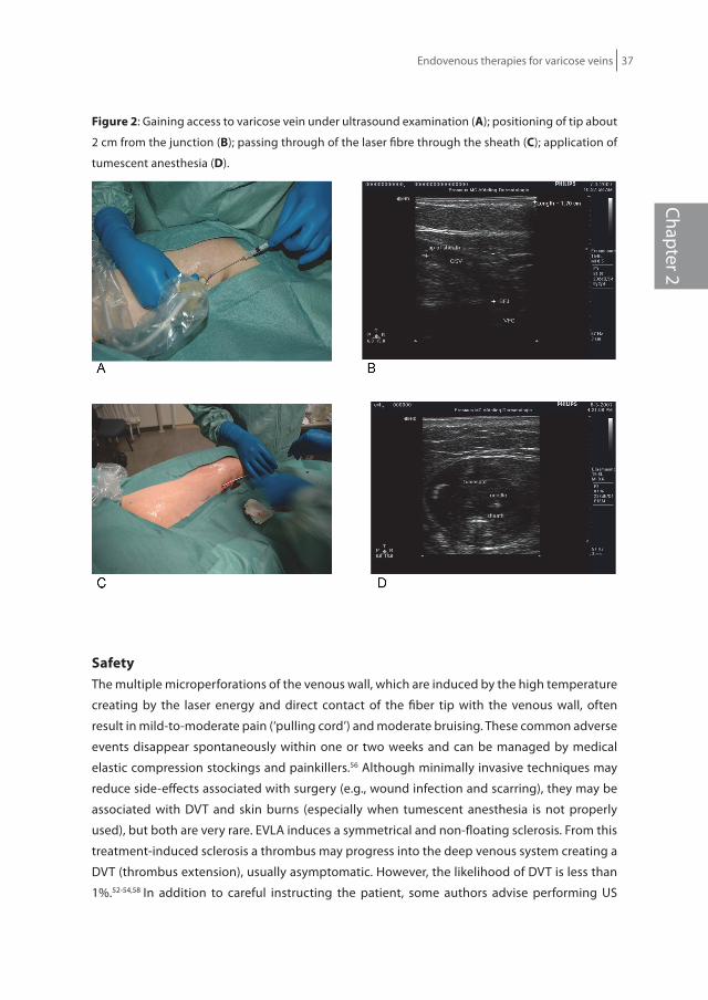
figure 2: Gaining access to varicose vein under ultrasound examination (A); positioning of tip about
2 cm from the junction (B); passing through of the laser fibre through the sheath (c); application of
tumescent anesthesia (d).
safetyThe multiple microperforations of the venous wall, which are induced by the high temperature
creating by the laser energy and direct contact of the fiber tip with the venous wall, often
result in mild-to-moderate pain (‘pulling cord’) and moderate bruising. These common adverse
events disappear spontaneously within one or two weeks and can be managed by medical
elastic compression stockings and painkillers.56 Although minimally invasive techniques may
reduce side-effects associated with surgery (e.g., wound infection and scarring), they may be
associated with DVT and skin burns (especially when tumescent anesthesia is not properly
used), but both are very rare. EVLA induces a symmetrical and non-floating sclerosis. From this
treatment-induced sclerosis a thrombus may progress into the deep venous system creating a
DVT (thrombus extension), usually asymptomatic. However, the likelihood of DVT is less than
1%.52-54,58 In addition to careful instructing the patient, some authors advise performing US

38 Chapter 2
examination one week after EVLA to exclude DVT and others prescribe LMWH for 5-7 days after
the procedure to prevent the development of DVT.59 Skin burns are also rare and may occur
when the distributed amount of energy is too high, when superficial veins are treated, or when
the cooling effect of the tumescent anesthesia is insufficient. Extra caution is needed when
treating the extrafascial part of the saphenous varicose vein and at the cutaneous exit site
of the laser fiber. Superficial thrombosis, dysesthesia, hematoma, cellulitis and arteriovenous
fistula have been reported after EVLA (Table II).52,56,59,60
summaryEndovenous laser ablation has shown to be one of the most effective treatments for primary
insufficient GSV and SSV: the overall rate of satisfaction, symptoms relief and absence
of varicose veins was 86%54 and a success rate of at least 90%.50 Also, EVLA procedures are
considered to be safe, well-tolerated, and are associated with minimal complications.57,58
endoVenous rAdiofreQuency ABLATion (rfA)Since 2000, several case series have been published about the use of RFA in the treatment of
lower extremity varicose veins.61-65
The temperature-controlled endovenous radiofrequency ablation is accomplished by
endoluminal application of radiofrequency energy directed into the vein wall with specially
designed configurations of bipolar electrodes. Additionally, control of radiofrequency energy
delivery using a temperature feedback loop allows the intima of the vein to be maintained at
or near a predetermined setpoint temperature during catheter pullback through the vein. The
length of time a section of vein wall is exposed to the setpoint temperature is determined by
the speed at which the catheter is withdrawn along the vein segment to be treated.66 RFA works
by heating the vein wall in its whole circumference. Endovenous temperatures are controlled
between 85° to 90°C,38 and causes collagen shrinking in state of destruction and carbonisation
which is seen in treatment with EVLA.
Recently, a new segmental catheter (VNUSClosure Fast® VNUS Medical Technologies, Inc,
Sunnyvale CA) has been introduced, which has a segment of 7 cm that heats to 120 degrees
Celsius.69 This technique is much faster than the previous radiofrequency catheters. The special
operating parameters to provide sufficient energy to heat the vein wall and cause collagen
contraction and destruction of the vein wall, while limiting the degree of perivascular heating.
Microscopic examination of the vein wall after treatment does not show carbonisation, in
contrast to veins treated with EVLA. Procedural advances, such as ultrasound-guided tumescent
infiltration along the course of the vein to be treated, have provided an added level of thermal
protection to the perivenous tissue during the application of radiofrequency energy.66

Endovenous therapies for varicose veins 39
Chapter 2
indicationsThe indications for RFA are comparable to EVLA, except that RFA cannot be used to treat veins
with diameters greater than 12 mm (Table I). A 5-F catheter (1.7 mm) is used for veins with a
diameter of 2-8 mm and an 8-F (2.7 mm) catheter can be used for veins as large as 12 mm. The
new segmental fiber is one size and can be used independent of vein diameter. Because of the
rigidity and size of the catheter, caution is indicated in tortuous and relatively small varicose
veins to avoid perforation.
ProcedureIn accordance with EVLA, RFA can be performed in an ambulatory setting. Access to the
varicose vein is obtained with a 16 Gauge needle under US guidance usually at or below knee
level or distally from the point of reflux. The RFA catheter is positioned in the close proximity
of the saphenous junction. The pods of the catheter are expanded in the common femoral
vein and, with US guidance, withdrawn into the orifice of the junction. A cuff or bandage
can be used to compress the blood out of the vein. The small electrodes at the end of the
‘umbrella’ catheter are in direct contact with the venous wall and omit high radiofrequency
energy (regulated by power, impedance and time) that is generated by a radiofrequency
generator. The radiofrequency waves heat the local tissue at the site of direct contact to 85
or 90 degrees Celsius, causing collagen shrinkage, denudation of endothelial and obliteration
of the venous lumen.66 A thermocouple monitors the temperature during treatment. Similar
to EVLA, perivenous tumescent anesthesia is applied (Figure 2d) to increase contact surface
and to decrease the pain sensation and the risk of dysesthesia.67 Also, manual compression
is recommended during the treatment in order to enhance contact of the catheter with the
venous wall. The catheter is slowly pulled back with a speed of approximately 3 cm/min (total
pull back time is about 20 minutes on average for the GSV between SFJ and knee level) but
can be faster at higher temperatures.68 Compressive bandages or medical elastic compression
stockings are indicated for one or two weeks after treatment.
The segmental RFA catheter is much faster than the previous radiofrequency catheters
and the first case series of 252 treated GSVs showed an occlusion rate of 99.6%.70
efficacySince 2000, several case series have been published showing that RFA can be successfully used
in the treatment of lower extremity varicose veins.61-65 The first long-term, large, single centre
case series showed that RFA was effective in about 90% of 140 limbs after two years of follow-
up.67 This study also showed that 98% of patients were satisfied with the treatment and would
recommend it to a friend. A multicenter study that included 1006 persons (1222 limbs) showed
good anatomical success rates of 88,8% occlusion and patient satisfaction in more than 85% of
the people after 4 years of follow-up.71

40 Chapter 2
safetyBecause important procedural changes have been made after the earliest case series of RFA
serious side effects such paresthesia, skin burns and DVT, are barely reported. One study found
16% DVT in 73 limbs treated with RFA, and presented this number with ‘a word of caution’.72
However, this study may be considered as an exception, because most studies report DVT
in less than 1% of treated limbs.57,73,74 Initially, paresthesia was reported relatively frequent,
but the incidence decreased significantly after the use of tumescent anesthesia.67, 75 Possibly
because the temperature in RFA is lower and the energy in RFA is distributed in a different way
than the laser energy,38,39 the local RFA induced adverse events, such as pain and ecchymosis,
are milder compared to EVLA. Skin burns and phlebitis are reported in about 2-5% of cases
(Table II).75
summaryThere is sufficient evidence to conclude that endovenous RFA is a safe and effective treatment
for saphenous insufficiency. The RFA catheters are more expensive than most EVLA disposables.
This treatment is best suitable for the management of primary uncomplicated varicose veins
with a diameter of at least 5 mm but less then 12mm. More studies assessing the segmental
RFA catheters are warranted.
coMPArison of TherAPiesThe minimally invasive therapies that are described above have shown to be very effective
and safe. Unfortunately, there is a lack of well-performed randomised comparative studies.
Some studies are currently ongoing. Recently a meta-analysis was published that showed a
pooled success rate for stripping, foam sclerotherapy, radiofrequency ablation, and laser
therapy of about 78%, 77%, 84%, and 94%, respectively. After adjustment for follow-up, EVLA
was significantly more effective compared with the other three therapies.76
uGfs versus surgeryHobbs and Rutgers have both demonstrated that liquid classic sclerotherapy is not as effective
as surgical stripping.77,78 Though a small prospective randomized trial suggested that SFJ
ligation and one session of UGFS was less effective in the short term, but significantly less
costly and time consuming than SFJ ligation, stripping and multiple avulsions.79 Recently,
Darvall80 published in a nonrandomized observational study that patients who had surgery
were more likely to have significant bruising (44 versus 7.2 per cent; p<.001) and pain (17 versus
5.5 per cent; p=.001) compared with UGFS. UGFS was associated with less time off work (43.2%
of patients returned to work within 24 h, whereas none of the patients who had surgery did
so (p<.001)) and the UGFS group was able to return to driving earlier (within 4 days, p=.014).
In comparative studies, the risk of adverse events compared between foam sclerotherapy and
Endovenous therapies for varicose veins 41
Chapter 2
other treatments was not significantly different. However, in the French registry35 the risk of
visual disturbance was significantly higher for foam compared with liquid sclerotherapy.
eVLA versus strippingThree small short-term studies that compared EVLA with surgical stripping suggested that
the clinical efficacy parameters were comparable, but EVLA had a significantly better effect
on HRQoL.81,82,83 A recent meta-analysis showed that EVLA was significantly more effective
compared to stripping, UGFS and RFA.76 Darwood concluded that abolition of reflux and
improvement of disease-specific quality of life was comparable between EVLA and surgery.
Patients return earlier to their normal activities after EVLA, this may have an important socio-
economic advantage.84 After 2 years the recurrence of varicose veins was similar after surgery
and EVLA, although the neovascularisation, a predictor of future recurrence, was less common
after EVLA.85 Although cost-effectiveness studies are lacking, EVLA is likely to reduce costs
because there is less down-time after EVLA and it can be performed in an outpatient setting.86
rfA versus eVLARFA and EVLA differ in the delivery of thermal energy to the vein wall as is described before.
Because of the high temperature and the direct contact of the focused laser beam with the vein
wall, EVLA, is generally associated with vein wall perforations whereas RFA seems to re-model
and/or shrink the veins wall.40,41 This is one of the reasons why EVLA seems to be associated
with more treatment-related pain and indurations one or two weeks after therapy than RFA.
The RECOVERY study observed that segmental RFA was significantly superior to EVLA in terms
of HRQoL. The group that was treated with RFA had less complains of pain, ecchymosis and
tenderness and had better HRQoL parameters than the group that was treated with EVLA.87
Older retrospective studies showed an equal effect between RFA and EVLA,88 and a significant
higher closure rate for EVLA compared to RFA at 500 days (92% vs. 85%).75
rfA versus strippingThree small randomized clinical trials that compared RFA with stripping, demonstrated that
the two therapies were about equally effective on the relatively short term, however patients
treated with RFA reported less postoperative pain and physical limitations, faster recovery,
fewer adverse events and superior HRQoL parameters compared to patients treated with
surgical stripping.89,90,91
A recent meta-analysis on stripping and endovenous therapies showed that EVLA was
significantly more effective than stripping (p<.0001), UGFS (p<.0001) and RFA (p=.01). No
significant difference in effectiveness was observed between RFA versus stripping (p=.14) and
RFA versus UGFS (p=.13).76

42 Chapter 2
In clinical trial registries can be found that several RCTs such as RFA versus stripping and UGFS
versus surgery are currently ongoing. Our study group is involved in a multicenter randomised
controlled trial (MAGNA-trial) that compares stripping, EVLA, and UGFS.
discussionProcedureEach of the minimally invasive therapies can be performed in an outpatient setting. EVLA and
RFA can be done using local tumescent anesthesia and UGFS does not require anesthesia.
In contrast to UGFS, EVLA and RFA should be performed in a sterile environment. RFA is a
patented and standardized procedure. Although the characteristics of the laser treatment,
such as wavelength, mode of pulling back the fiber, power and pullback speed may vary, the
procedure is relatively well standardized and it is widely accepted that 60 Joule/cm or more
should be administered (but this may be less as well). Compared to RFA and EVLA, UGFS seems
to vary more between physicians (for example, type and concentration of sclerosant used,
the creation of foam, volume of foam needed, localization of injection, and one vs. multiple
injections, type of compression therapy). Each of the minimal invasive techniques requires
US experience, preferably performed by the physician but technicians may assist during the
procedure. UGFS takes about 15 minutes whereas the endovenous therapies may take about
45 minutes. Of the three therapies, UGFS may be the most cost effective because it is fast,
cheap, and easily repeated, but unfortunately, good studies are lacking. Although the period of
using medical elastic compression stockings after UGFS is controversial (between 0-6 weeks), it
seems to be essential in achieving an optimal result.
efficacyEVLA and RFA are equally effective compared to vein stripping but are more appreciated
by patients.87,88 However, one meta-analysis showed highest occlusion rates for EVLA.76 The
frequency of recurrence of varicose veins was similar in a comparitive study after two years
of follow-up after surgery and EVLA, though there was slightly less neovascularization, which
may be a predictor of recurrences, in the EVLA group.85 UGFS is very effective for treating
saphenous recurrences after vein stripping, because these recurrent veins are usually tortuous,
are not located outside the fascial blades and often have a relatively small diameter. In veins
with very large diameter EVLA may be the best option, in comparison with the other minimal
invasive treatments, because in these cases, UGFS often requires 3 or more treatment sessions
and the RFA catheters are designed for a maximum diameter of 12 mm, except for the new
segmental RFA catheter. UGFS is indicated, in small and symptomatic insufficient saphenous
veins (diameter <4 mm) and recurrence of tortuous veins.

Endovenous therapies for varicose veins 43
Chapter 2
safetyWith each of the three minimal invasive treatments, the development of DVT is the main
concern. This is because of the destruction and/or irritation of endothelial cells and the
formation of a sclerosis (which is different from a thrombus, because it is symmetrical and
adheres to the venous wall and has no free floating component). There are no comparative
safety studies, but a comparison of the largest case series of each of the treatments suggests
that the risk may be highest for RFA (up to 1%), which is comparable to the risk of DVT after
vein stripping.73,74,92 However, symptomatic and asymptomatic thromboembolic events and
methods of diagnosis should be differentiated when comparing thromboembolic outcomes
of these treatments. There are several opportunities mentioned to avoid the prevalence
of DVT, such as hypercoagulability screen (for high-risk individuals selected by history and
physical examination), low molecular weight heparin preoperative and/or postoperative (for
about a week), US control of the junction following treatment after 1 week, localization of the
catheter distal from the inferior epigastric vein, ample administration of tumescent anesthesia,
avoidance of perforation, and for UGFS limb elevation, mobilization and applying pressure on
the junction after injecting the foam, have been suggested to reduce the risk of DVT. However,
these measures are controversial and there is no consensus on DVT prevention.
Among the most common serious side affects of saphenous stripping is neurological
damage. In about 7% in short and 40% in long stripping of the GSV, nerve damage occurs.93,94
Although the incidence is significantly lower compared to surgery, paresthesia has been
reported for UGFS, EVLA and RFA due to extravenous injection of foam or increased perivenous
temperature, respectively. In accordance with surgery, the likelihood of nerve injury may
increase when EVLA and RFA procedures are started at the ankle and extra tumescent in the
popliteal fossa should be administered in the EVLA and RFA treatment of the SSV to avoid sural
nerve damage.
With endovenous techniques no or minimal scars are created and the risk of wound
infection is very low in contrast to surgery. However, each of the minimally invasive techniques
may be associated with local cutaneous side effects. Skin burns have been reported in less
than 1% after EVLA and RFA procedures and can be easily avoided by applying a sufficient
volume of tumescent anesthesia. Hyperpigmentation and to a lesser extent skin necrosis has
been reported after UGFS. Postoperative pain and ecchymoses along the treated vein are very
common after EVLA (for about 2 weeks and are controlled by NSAIDs) and to a lesser extent
after RFA and UGFS.
Recurrence of varicose veins after stripping is thought to be due to neovascularization at
the SFJ and occurs in about 25% of the patients 5 years after therapy.95 This has been studied
using US but has not been documented for UGFS, EVLA and RFA. This may be because these
techniques, in contrast to surgery, do not disrupt the endothelial lining and do not eliminate
other veins connected with the SFJ. This difference may explain the high long-term success
44 Chapter 2
rates of the three minimally invasive procedures. Endovenous therapies show anatomical
failure in about 10%, with (partial) recanalization of the treated vein but these failures are not
always clinically important failures.
Steam ablation is a new and promising endovenous thermal therapy, but there are no
data on steam ablation of varicose veins available yet. A randomized controlled trial is going
on in our centrum, which compares EVLA with steam ablation.
conclusionAdditional comparative studies between minimally invasive treatments and surgery are
needed to assess the difference in efficacy, patient reported outcomes including HRQoL and to
determine the most cost-effective therapy. EVLA seems to be the endovenous treatment that
is most commonly used, because it is less expensive and faster than standard RFA. However,
UGFS may be the most cost-effective therapy, but several treatment sessions may be needed.
Endovenous therapies for varicose veins 45
Chapter 2
references
1. Rabe E, Pannier-Fischer F, Bromen K, Schuldt K, Stang A, Poncar C, et al. Bonner Venenstudie der Deutschen Gesellschaft für Phlebologie – Epidemiologische Untersuchung zur Frage der Häufigkeit und Ausprägung von chronischen Venenkrankheiten in der städtischen und ländlichen Wohnbevölkerung. Phlebologie. 2003;32:1-14.
2. Callam MJ. Epidemiology of varicose veins. Br J Surg. 1994;81(2):167-173.3. Margolis DJ, Bilker W, Santanna J, Baumgarten M. Venous leg ulcer: incidence and prevalence in the
elderly. J Am Acad Dermatol. 2002;46(3):381-386.4. Kurz X, Kahn SR, Abenhaim L, Clement D, Norgren L, Baccaglini U, et al. Chronic venous disorders
of the leg: epidemiology, outcomes, diagnosis and management. Summary of an evidence-based report of the VEINES task force. Venous Insufficiency Epidemiologic and Economic Studies. Int Angiol. 1999;18(2):83-102.
5. van Rij AM, Jiang P, Solomon C, Christie RA, Hill GB. Recurrence after varicose vein surgery: a prospective long-term clinical study with duplex ultrasound scanning and air plethysmography. J Vasc Surg. 2003; 38(5): 935-943.
6. Fischer R, Linde N, Duff C, Jeanneret C, Chandler JG, Seeber P. Late recurrent saphenofemoral junction reflux after ligation and stripping of the greater saphenous vein. J Vasc Surg. 2001;34(2):236-240.
7. Perrin MR, Labropoulos N, Leon LR Jr. Presentation of the patient with recurrent varices after surgery (REVAS). J Vasc Surg. 2006;43(2):327-334.
8. Dwerryhouse S, Davies B, Harradine K, Earnshaw JJ. Stripping the long saphenous vein reduces the rate of reoperation for recurrent varicose veins: five-year results of a randomized trial. J Vasc Surg. 1999;29(4):589-592.
9. Sharif MA, Soong CV, Lau LL, Corvan R, Lee B, Hannon RJ. Endovenous laser treatment for long saphenous vein incompetence. Br J Surg. 2006;93(7):831-835.
10. Stirling M, Shortell CK. Endovascular treatment of varicose veins. Semin Vasc Surg. 2006; 19(2): 109-115.
11. Orbach EJ. Sclerotherapy of varicose veins – utilization of an intravenous air-block. Am J Surg. 1944; LXVI: 362-6.
12. Wollmann JC. The history of sclerosing foams. Dermatol Surg. 2004;30(5):694-703.13. Rabe E, Pannier-Fischer F, Gerlach H, et al. Guidelines for sclerotherapy of varicose veins. Dermatol
Surg. 2004; 30:687-93.14. Yamaki T, Nozaki M, Iwasaka S. Comparative study of duplex-guided foam sclerotherapy and duplex-
guided liquid sclerotherapy for the treatment of superficial venous insufficiency. Dermatol Surg. 2004;30(5):718-22.
15. Frullini A. New technique in producing sclerosing foam in a disposable syringe. Dermatol Surg. 2000;26(7):705-706.
16. Tessari L, Cavezzi A, Frullini A. Preliminary experience with a new sclerosing foam in the treatment of varicose veins. Dermatol Surg. 2001;27(1):58-60.
17. Tan VK, Tan SG. Technique and early results of ultrasound-guided foam sclerotherapy of the long saphenous vein for treatment of varicose veins. Singapore Med J. 2009 Mar;50(3):284-7.
18. Frullini A, Cavezzi A. Sclerosing foam in the treatment of varicose veins and telangiectases: history and analysis of safety and complications. Dermatol Surg. 2002;28(1):11-15.
19. Darke SG, Baker SJ. Ultrasound-guided foam sclerotherapy for the treatment of varicose veins. Br J Surg. 2006;93(8):969-974.
20. Pascarella L, Bergan JJ, Mekenas LV. Severe chronic venous insufficiency treated by foamed sclerosant. Ann Vasc Surg. 2006;20(1):83-91.

46 Chapter 2
21. Myers KA, Jolley D, Clough A, Kirwan J. Outcome of ultrasound-guided sclerotherapy for varicose veins: medium-term results assessed by ultrasound surveillance. Eur J Endovasc Surg. 2007; 33:116-21.
22. Chapman-Smith P, Browne A. Prospective five-year study of ultrasound-guided foam sclerotherapy in the treatment of great saphenous vein reflux. Phlebology 2009;24:183-188.
23. Barrett JM, Allen B, Ockelford A, Goldman MP. Microfoam ultrasound-guided sclerotherapy treatment for varicose veins in a subgroup with diameters at the junction of 10 mm or greater compared with a subgroup of less than 10 mm. Dermatol Surg. 2004;30(11):1386-1390.
24. Masuda EM, Kessler DM, Lurie F, Puggioni A, Kistner RL, Eklof B. The effect of ultrasound-guided sclerotherapy of incompetent perforator veins on venous clinical severity and disability scores. J Vasc Surg. 2006;43(3):551-556.
25. Nitecki S, Bass A. Ultrasound-guided foam sclerotherapy in patients with Klippel-Trenaunay syndrome. Isr Med Assoc J. 2007;9(2):72-75.
26. Cabrera J, Cabrera J, Garcia-Olmedo MA, Redondo P. Treatment of venous malformations with sclerosant in microfoam form. Arch Dermatol. 2003;139(11):1409-1416.
27. Breu FX, Guggenbichler, Wollmann JC. 2nd European Consensus Meeting on Foam Sclerotherapy, 28 – 30 April 2006, Tegernsee, Germany. Vasa 2008;37 Supplement 71:1-32.
28. Smith PC. Chronic venous disease treated by ultrasound guided foam sclerotherapy. Eur J Vasc Endovasc Surg. 2006;32(5):577-583.
29. Ceulen RP, Bullens-Goessens YI, Pi-Van De Venne SJ, Nelemans PJ, Veraart JC, Sommer A. Outcomes and side effects of duplex-guided sclerotherapy in the treatment of great saphenous veins with 1% versus 3% polidocanol foam: results of a randomized controlled trial with 1-year follow-up. Dermatol Surg. 2007;33(3):276-281.
30. Rabe E, Otto J, Schliephake D, Pannier F. Efficacy and Safety of Great Saphenous Vein Sclerotherapy Using Standardised Polidocanol Foam (ESAF): A Randomised Controlled Multicentre Clinical Trial. Eur J Endovasc Vasc Surg. 2008;35:238-45.
31. Belcaro G, Cesarone MR, Di Renzo A, Brandolini R, Coen L, Acerbi G, et al. Foam-sclerotherapy, surgery, sclerotherapy, and combined treatment for varicose veins: a 10-year, prospective, randomized, controlled, trial (VEDICO trial). Angiology 2003;54(3):307-315.
32. Cavezzi A, Frullini A, Ricci S, Tessari L. Treatment of varicose veins by foam sclerotherapy: two clinical series. Phlebology 2002;17:13-18
33. Darvall KAL, Bate GR, Silverman SH, Adam SJ and Bradbury AW. Medium-term results of ultrasound-guided foam sclerotherapy for small saphenous varicose veins. British Journal of Surgery 2009; 96: 1268-1273.
34. Jia X, Mowatt G, Ho V, et al. Systematic review of the safety and efficacy of foam sclerotherapy for venous disease of the lower limbs. Br J Surg. 2007; 94: 925-936
35. Guex JJ, Allaert FA, Gillet JL, Chleir F. Immediate and midterm complications of sclerotherapy: Report of a prospective multicenter registry of 12,173 sclerotherapy sessions. Dermatol Surg. 2005;31(2):123-128.
36. Navarro L, Min RJ, Bone C. Endovenous laser: a new minimally invasive method of treatment for varicose veins--preliminary observations using an 810 nm diode laser. Dermatol Surg. 2001;27(2):117-122.
37. Min RJ, Zimmet SE, Isaacs MN, Forrestal MD. Endovenous laser treatment of the incompetent greater saphenous vein. J Vasc Interv Radiol. 2001;12(10):1167-1171.
38. Disselhoff BCVM, Rem AI, Verdaasdonk RM, der Kinderen DJ, Moll FL. Endovenous laser ablation: an experimental study on the mechanism of action. Phlebology 2008;23:69-76.
39. Fan C-M, Anderson RR (2008) Endovenous laser ablation: mechanism of action. Phlebology 23:206-213.

Endovenous therapies for varicose veins 47
Chapter 2
40. Weiss RA. Comparison of endovenous radiofrequency versus 810 nm diode laser occlusion of large veins in an animal model. Dermatol Surg. 2002;28:56-61.
41. Proebstle TM, Lehr HA, Kargl A, Espinola-Klein C, Rother W, Bethge S, et al. Endovenous treatment of the greater saphenous vein with a 940-nm diode laser: thrombotic occlusion after endoluminal thermal damage by laser-generated steam bubbles. J Vasc Surg. 2002;35:729-36.
42. Mordon SR, Wassmer B, Zemmouri J (2007) Mathematical modeling of 980-nm and 1320-nm endovenous laser treatment. Lasers Surg Med. 39:256-265.
43. Meissner OA, Schmedt C-G, Hunger K, Hetterich H, Sroka R, Rieber J, et al. Endovascular optical coherence tomography ex vivo: venous wall anatomy and tissue alterations after endovenous therapy. Eur Radiol. 2007; 17:2384-2393.
44. Proebstle TM, Lehr HA, Kargl A, Espinola-Klein C, Rother W, Bethge S, Knop J. Endovenous treatment of the greater saphenous vein with a 940 nm diode laser: thrombotic occlusion after endoluminal thermal damage by laser-generated steam bubbles. J Vasc Surg. 2002; 35:729-736.
45. van den Bos RR, Kockaert MA, Neumann HAM, Bremmer RH, Nijsten T, van Gemert MJC. Heat conduction from the exceedingly hot fiber tip contributes to the endovenous laser ablation of varicose veins. Lasers Med Sci. 2009; 24: 247-251
46. Proebstle TM, Herdemann S. Early results and feasibility of incompetent perforator vein ablation by endovenous laser treatment. Dermatol Surg. 2007; 33(2): 162-168.
47. Bush RG, Hammond K. Treatment of incompetent vein of Giacomini (thigh extension branch). Ann Vasc Surg. 2007; 21(2): 245-248.
48. Goldman MP, Mauricio M, Rao J. Intravascular 1320-nm laser closure of the great saphenous vein: a 6- to 12-month follow-up study. Dermatol Surg. 2004; 30(11): 1380-1385.
49. Mundy L, Merlin TL, Fitridge RA, Hiller JE. Systematic review of endovenous laser treatment for varicose veins. Br J Surg. 2005; 92(10): 1189-1194.
50. Pannier F, Rabe E. Endovenous laser therapy and radiofrequency ablation of saphenous varicose veins. J Cardiovasc Surg. 2006; 47(1): 3-8.
51. Nootheti PK, Cadag KM, Goldman MP. Review of intravascular approaches to the treatment of varicose veins. Dermatol Surg. 2007; 33(10): 1149-1157.
52. Min RJ, Khilnani N, Zimmet SE. Endovenous laser treatment of saphenous vein reflux: long-term results. J Vasc Interv Radiol. 2003; 14(8): 991-996.
53. Agus GB, Mancini S, Magi G; IEWG. The first 1000 cases of Italian Endovenous-laser Working Group (IEWG). Rationale, and long-term outcomes for the 1999-2003 period. Int Angiol. 2006; 25(2): 209-215.
54. Ravi R, Rodriguez-Lopez JA, Trayler EA, Barrett DA, Ramaiah V, Diethrich EB. Endovenous ablation of incompetent saphenous veins: a large single-center experience. J Endovasc Ther. 2006; 13(2): 244-248.
55. Sadick NS, Wasser S. Combined endovascular laser plus ambulatory phlebectomy for the treatment of superficial venous incompentence: A 4-year perspective. J Cosmet Laser Ther. 2007; 9(1): 9-13.
56. Myers KA, Jolley D. Outcome of endovenous laser therapy for saphenous reflux and varicose veins: medium-term results assessed by ultrasound surveillance. Eur J Vasc Endovasc Surg. 2009; 37(2): 239-45.
57. Ravi R, Trayler EA, Barrett DA, Diethrich EB. Endovenous thermal ablation of superficial venous insufficiency of the lower extremity: single-center experience with 3000 limbs treated in a 7-year period. J Endovasc Ther. 2009 Aug; 16(4): 500-5.
58. van den Bos RR, Neumann HAM, de Roos K-P, Nijsten T. Endovenous laser ablation induced complications: review of the literature and new cases. Dermatol Surg. 2009; Aug 35(8): 1206-14.
59. Puggioni A, Kalra M, Carmo M, Mozes G, Gloviczki P. Endovenous laser therapy and radiofrequency ablation of the great saphenous vein: analysis of early efficacy and complications. J Vasc Surg. 2005;42(3):488-493.
48 Chapter 2
60. Timperman PE. Arteriovenous fistula after endovenous laser treatment of the short saphenous vein. J Vasc Interv Radiol. 2004; 15(6): 625-627.
61. Goldman MP. Closure of the greater saphenous vein with endoluminal radiofrequency thermal heating of the vein wall in combination with ambulatory phlebectomy: preliminary 6-month follow-up. Dermatol Surg. 2000; 26(5): 452-456.
62. Chandler JG, Pichot O, Sessa C, Schuller-Petrović S, Osse FJ, Bergan JJ. Defining the role of extended saphenofemoral junction ligation: a prospective comparative study. J Vasc Surg. 2000; 32(5): 941-953.
63. Manfrini S, Gasbarro V, Danielsson G, Norgren L, Chandler JG, Lennox AF, et al. Endovenous management of saphenous vein reflux. Endovenous Reflux Management Study Group. J Vasc Surg. 2000; 32: 330-342.
64. Goldman MP, Amiry S. Closure of the greater saphenous vein with endoluminal radiofrequency thermal heating of the vein wall in combination with ambulatory phlebectomy: 50 patients with more than 6-month follow-up. Dermatol Surg. 2002; 28(1): 29-31.
65. Sybrandy JE, Wittens CH. Initial experiences in endovenous treatment of saphenous vein reflux. J Vasc Surg. 2002; 36(6): 1207-1212.
66. Schmedt CG, Sroka R, Steckmeier S, Meissner OA, Babaryka G, Hunger K, et al. Investigation on radiofrequency and laser (980 nm) effects after endoluminal treatment of saphenous vein insufficiency in an ex-vivo model. Eur J Vasc Endovasc Surg. 2006; 32(3): 318-325.
67. Weiss RA, Weiss MA. Controlled radiofrequency endovenous occlusion using a unique radiofrequency catheter under duplex guidance to eliminate saphenous varicose vein reflux: a 2-year follow-up. Dermatol Surg. 2002; 28(1): 38-42.
68. Zikorus AW, Mirizzi MS. Evaluation of setpoint temperature and pullback speed on vein adventitial temperature during endovenous radiofrequency energy delivery in an in-vitro model. Vasc Endovascular Surg. 2004; 38(2): 167-74.
69. www.closurefast.com70. Proebstle TM, Vago B, Alm J, Göckeritz O, Lebard C, Pichot O. Treatment of the incompetent great
saphenous vein by endovenous radiofrequency powered segmental thermal ablation: first clinical experience. J Vasc Surg. 2008; 47: 151-6.
71. Merchant RF, Pichot O, Myers KA. Four-year follow-up on endovascular radiofrequency obliteration of great saphenous reflux. Dermatol Surg. 2005; 31(2): 129-134.
72. Hingorani AP, Ascher E, Markevich N, Schutzer RW, Kallakuri S, Hou A, et al. Deep venous thrombosis after radiofrequency ablation of greater saphenous vein: a word of caution. J Vasc Surg. 2004; 40(3): 500-504.
73. Merchant RF, DePalma RG, Kabnick LS. Endovascular obliteration of saphenous reflux: a multicenter study. J Vasc Surg. 2002; 35(6): 1190-1196.
74. Gale SS, Dosick SM, Seiwert AJ, Comerota AJ. Regarding “Deep venous thrombosis after radiofrequency ablation of greater saphenous vein”. J Vasc Surg. 2005; 41(2): 374.
75. Almeida JI, Raines JK. Radiofrequency ablation and laser ablation in the treatment of varicose veins. Ann Vasc Surg. 2006; 20(4): 547-552.
76. Van den Bos RR, Kockaert MA, Neumann HAM, Nijsten T. Endovenous therapies of lower extremity varicosities: a meta-analysis. J Vasc Surg. 2009; 49(1): 230-9
77. Hobbs JT. Surgery and sclerotherapy in the treatment of varicose veins. A random trial. Arch Surg. 1974; 109(6): 793-796.
78. Rutgers PH, Kitslaar PJ. Randomized trial of stripping versus high ligation combined with sclerotherapy in the treatment of the incompetent greater saphenous vein. Am J Surg. 1994; 168(4): 311-315.
Endovenous therapies for varicose veins 49
Chapter 2
79. Bountouroglou DG, Azzam M, Kakkos SK, Pathmarajah M, Young P, Geroulakos G. Ultrasound-guided foam sclerotherapy combined with sapheno-femoral ligation compared to surgical treatment of varicose veins: early results of a randomised controlled trial. Eur J Vasc Endovasc Surg. 2006; 31(1): 93-100.
80. Darvall KA, Bate GR, Adam DJ, Bradbury AW. Recovery after ultrasound-guided foam sclerotherapy compared with conventional surgery for varicose veins. Br J Surg. 2009 Nov; 96(11): 1262-7.
81. De Medeiros CA, Luccas GC. Comparison of endovenous treatment with an 810 nm laser versus conventional stripping of the great saphenous vein in patients with primary varicose veins. Dermatol Surg. 2005; 31(12): 1685-1694.
82. Mekako AI, Hatfield J, Bryce J, Lee D, McCollum PT, Chetter I. A nonrandomised controlled trial of endovenous laser therapy and surgery in the treatment of varicose veins. Ann Vasc Surg. 2006; 20(4): 451-457.
83. Rasmussen LH, Bjoern L, Lawaetz M, Blemings A, Lawaetz B, Eklof B. Randomized trial comparing endovenous laser ablation of the great saphenous vein with high ligation and stripping in patients with varicose veins: short-term results. J Vasc Surg. 2007; 46: 308-315.
84. Darwood RJ, Theivacumar N, Dellagrammaticas D, Mavor AID and Gough MJ. Randomized clinical trial comparing endovenous laser ablation with surgery for the treatment of primary great saphenous varicose veins. Br J of Surg. 2008; 95: 294–301.
85. Theivacumar NS, Darwood R, Gough MJ. Neovascularisation and Recurrence 2 Years After Varicose Vein Treatment for Sapheno-Femoral and Great Saphenous Vein Reflux: A Comparison of Surgery and Endovenous Laser Ablation. Eur J Vasc Endovasc Surg. 2009; 38: 203-207.
86. Rautio T, Ohinmaa A, Perala J, Ohtonen P, Heikkinen T, Wiik H, Karjalainen P, Haukipuro K, Juvonen T. Endovenous obliteration versus conventional stripping operation in the treatment of primary varicose veins: a randomized controlled trial with comparison of the costs. J Vasc Surg. 2002; 35(5): 958-965.
87. Almeida JI, Kaufman J, Göckeritz O, Chopra P, Evans MT, Hoheim DF, Makhoul RG, Richards T, Wenzel C, Raines JK. Radiofrequency endovenous ClosureFAST versus laser ablation for the treatment of great saphenous reflux: a multicenter, single-blinded, randomized study (RECOVERY study). J Vasc Interv Radiol. 2009 Jun;20(6): 752-9.
88. Puggioni A, Kalra M, Carmo M, Mozes G, Gloviczki P. Endovenous laser therapy and radiofrequency ablation of the great saphenous vein: analysis of early efficacy and complications. J Vasc Surg. 2005; 42(3): 488-493.
89. Lurie F, Creton D, Eklof B, Kabnick LS, Kistner RL, Pichot O, et al. Prospective randomized study of endovenous radiofrequency obliteration (Closure) versus ligation and vein stripping (EVOLVeS): two-year follow-up. Eur J Vasc Endovasc Surg. 2005; 29: 67-73.
90. Perala J, Rautio T, Biancari F, Ohtonen P, Wiik H, Heikkinen T, Juvonen T. Radiofrequency endovenous obliteration versus stripping of the long saphenous vein in the management of primary varicose veins: 3-year outcome of a randomized study. Ann Vasc Surg. 2005; 19(5): 669-672.
91. Hinchliffe RJ, Ubhi J, Beech A, Ellison J, Braithwaite BD. A prospective randomised controlled trial of VNUS closure versus surgery for the treatment of recurrent long saphenous varicose veins. Eur J Vasc Endovasc Surg. 2006 Feb; 31(2): 212-8.
92. Bohler K, Baldt M, Schuller-Petrovic S, Grunwald C, Sellner W, Watzke H, Wolff K. Varicose vein stripping--a prospective study of the thrombotic risk and the diagnostic significance of preoperative color coded duplex sonography. Thromb Haemost. 1995; 73(4): 597-600.
93. Morrison C, Dalsing MC. Signs and symptoms of saphenous nerve injury after greater saphenous vein stripping: prevalence, severity, and relevance for modern practice. J Vasc Surg. 2003; 38(5): 886-890.

50 Chapter 2
94. Holme JB, Skajaa K, Holme K. Incidence of lesions of the saphenous nerve after partial or complete stripping of the long saphenous vein. Acta Chir Scand. 1990; 156(2): 145-148.
95. De Maeseneer MG, Vandenbroeck CP, Hendriks JM, Lauwers PR, Van Schil PE. Accuracy of duplex evaluation one year after varicose vein surgery to predict recurrence at the sapheno-femoral junction after five years. Eur J Vasc Endovasc Surg. 2005; 29(3): 308-312.

Chapter 3
Validation of the Chronic Venous Insufficiency Quality of Life Questionnaire in Dutch Patients Treated for Varicose VeinsAnke A. BiemansSimone K. van der VeldenCornelis M. BruijninckxJaap ButhTamar Nijsten
Eur J Vasc Endovasc Surg 2011 Aug;42(2):246-53.
Chapter 3

52 Chapter 3
ABsTrAcTBackground: The Chronic Venous Insufficiency Quality of Life Questionnaire (CIVIQ) is a
disease specific instrument, to measure the impact of chronic venous insufficiency (CVI) on
patients’ lives. The objective of this study is to test the psychometric properties of the CIVIQ,
and to validate the use of the questionnaire translated in Dutch.
Methods: A standardized questionnaire, including CIVIQ and SF-36 was obtained before
and 1 month after treatment to al new patients with varicose veins. The feasibility was tested
by missing responses and response distribution. CIVIQ’s scores were compared to the SF-36
scores and between different levels of severity of varicose veins. The CIVIQ’s reliability was
assessed using Cronbach’s alpha and test-retest reliability. The structure was studied using
factor analysis. The scores before and after therapy were compared to assess responsiveness.
results: There was a response rate of 93.5%. None of 20 items missed less than 10% of responses,
but 3 showed ceiling effect. The CIVIQ correlated well with the physical and moderately with
the mental MCS of the SF-36 suggesting a good construct validity of the CIVIQ. The median
CIVIQ scores increased significantly with the severity of varicose veins. The CIVIQ showed an
excellent internal consistency and an excellent test-retest reliability. The CIVIQ score decreased
in 75.65% after treatment. The results were in accordance with the Norman’s rule and showed
a median effect size.
conclusion: This study confirms the feasibility, validity, reliability and responsiveness of the
CIVIQ in patients with varicose veins. The psychometric properties of the Dutch CIVIQ were
comparable to the original French version.
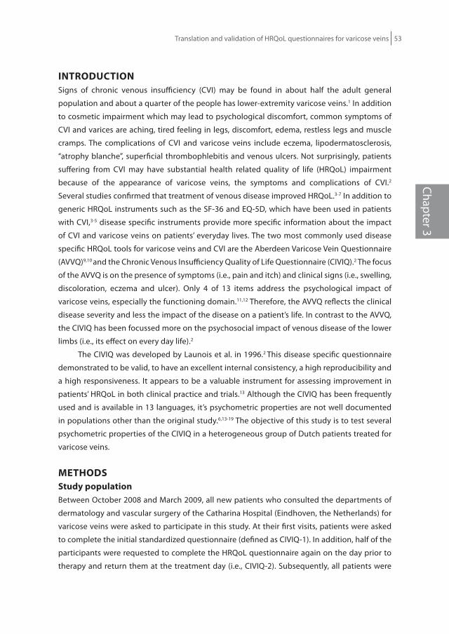
Translation and validation of HRQoL questionnaires for varicose veins 53
Chapter 3
inTroducTionSigns of chronic venous insufficiency (CVI) may be found in about half the adult general
population and about a quarter of the people has lower-extremity varicose veins.1 In addition
to cosmetic impairment which may lead to psychological discomfort, common symptoms of
CVI and varices are aching, tired feeling in legs, discomfort, edema, restless legs and muscle
cramps. The complications of CVI and varicose veins include eczema, lipodermatosclerosis,
“atrophy blanche”, superficial thrombophlebitis and venous ulcers. Not surprisingly, patients
suffering from CVI may have substantial health related quality of life (HRQoL) impairment
because of the appearance of varicose veins, the symptoms and complications of CVI.2
Several studies confirmed that treatment of venous disease improved HRQoL.3-7 In addition to
generic HRQoL instruments such as the SF-36 and EQ-5D, which have been used in patients
with CVI,3-5 disease specific instruments provide more specific information about the impact
of CVI and varicose veins on patients’ everyday lives. The two most commonly used disease
specific HRQoL tools for varicose veins and CVI are the Aberdeen Varicose Vein Questionnaire
(AVVQ)9,10 and the Chronic Venous Insufficiency Quality of Life Questionnaire (CIVIQ).2 The focus
of the AVVQ is on the presence of symptoms (i.e., pain and itch) and clinical signs (i.e., swelling,
discoloration, eczema and ulcer). Only 4 of 13 items address the psychological impact of
varicose veins, especially the functioning domain.11,12 Therefore, the AVVQ reflects the clinical
disease severity and less the impact of the disease on a patient’s life. In contrast to the AVVQ,
the CIVIQ has been focussed more on the psychosocial impact of venous disease of the lower
limbs (i.e., its effect on every day life).2
The CIVIQ was developed by Launois et al. in 1996.2 This disease specific questionnaire
demonstrated to be valid, to have an excellent internal consistency, a high reproducibility and
a high responsiveness. It appears to be a valuable instrument for assessing improvement in
patients’ HRQoL in both clinical practice and trials.13 Although the CIVIQ has been frequently
used and is available in 13 languages, it’s psychometric properties are not well documented
in populations other than the original study.6,13-19 The objective of this study is to test several
psychometric properties of the CIVIQ in a heterogeneous group of Dutch patients treated for
varicose veins.
MeThodsstudy populationBetween October 2008 and March 2009, all new patients who consulted the departments of
dermatology and vascular surgery of the Catharina Hospital (Eindhoven, the Netherlands) for
varicose veins were asked to participate in this study. At their first visits, patients were asked
to complete the initial standardized questionnaire (defined as CIVIQ-1). In addition, half of the
participants were requested to complete the HRQoL questionnaire again on the day prior to
therapy and return them at the treatment day (i.e., CIVIQ-2). Subsequently, all patients were

54 Chapter 3
asked to complete a third questionnaire at their follow up visit, which was at least four weeks
after therapy (i.e., CIVIQ-3). Therapy consisted of surgical, non-surgical or a combination of
these. The responsible physician was asked to record the ‘C’ of the CEAP classification and all
performed therapies for each study patient.
QuestionnaireTogether with a standardized questionnaire, which was self-administered and included
questions about demographic and disease characteristics, the SF-36 and the CIVIQ (Appendix
I) were included at all visits. The 20 questions of the CIVIQ (Table I) result in a global score and
four separate domain scores: physical (items 5, 6, 7 and 9), psychological (items 12-20) and
social impairment (items 8, 10 and 11) and level of pain (items 1, 2, 3 and 4). All questions
have a 5-point response category with higher scores reflecting more severe impairment. Three
separate scores can be calculated: a score per item (1 to 5), a score of each of the 4 dimensions
(0 to 100) and a global score (value 0 to 100). Higher scores represent lower HRQoL due to CVI
or varicose veins.2
The translation of the CIVIQ into Dutch was based on forward-backward translation
as recommended.20 In brief, three translators, all native speakers in Dutch, independently
translated the questions and the response options of the English CIVIQ into Dutch. They
were instructed to pay attention to conceptual rather than literal equivalence, and to choose
words and language constructions that were as simple as possible. The translators were
two employees of a registered translation office and a dermatologist. The three resulting
independent forward translations were compared and discussed in a group meeting of the
three translators. Differences were documented and discussed until consensus was reached
about the optimal phrasing of the Dutch CIVIQ. This common forward translation was then
given to two translators who were native speakers in English and fluent in Dutch. They each
produced a backward translation that was both compared to the English CIVIQ for conceptual
equivalence with the original source version. The analysis was documented and necessary
adaptations to the Dutch CIVIQ version were made. The resulting Dutch CIVIQ was then
administrated to twenty patients with venous disease of the lower limbs to provide qualitative
testing of readability and comprehension. Because this qualitative testing revealed no problem
with the Dutch CIVIQ, it was subsequently administered in the study population to collect data
for psychometric analysis (Appendix 1).
The SF-36 is a generic HRQoL instrument and has proven applicability in several areas of
disease including varicose veins.3-5,7,22-24 The 36 items can be grouped in a mental and physical
component scale (MCS and PCS, respectively). We included the SF-36 to test construct validity.
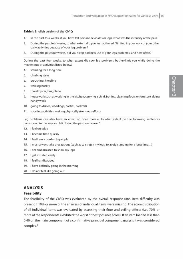
Translation and validation of HRQoL questionnaires for varicose veins 55
Chapter 3
Table i: English version of the CIVIQ.
1. In the past four weeks, if you have felt pain in the ankles or legs, what was the intensity of the pain?
2. During the past four weeks, to what extent did you feel bothered / limited in your work or your other daily activities because of your leg problem?
3. During the past four weeks, did you sleep bad because of your legs problems, and how often?
During the past four weeks, to what extent dit your leg problems bother/limit you while doing the movements or activities listed below?
4. standing for a long time
5. climbing stairs
6. crouching, kneeling
7. walking briskly
8. travel by car, bus, plane
9. housework such as working in the kitchen, carrying a child, ironing, cleaning floors or furniture, doing handy work
10. going to discos, weddings, parties, cocktails
11. sporting activities, making physically strenuous efforts
Leg problems can also have an effect on one’s morale. To what extent do the following sentences correspond to the way you felt during the past four weeks?
12. I feel on edge
13. I become tired quickly
14. I feel I am a burden to people
15. I must always take precautions (such as to stretch my legs, to avoid standing for a long time…)
16. I am embarrassed to show my legs
17. I get irritated easily
18. I feel handicapped
19. I have difficulty going in the morning
20. I do not feel like going out
AnALysisfeasibility The feasibility of the CIVIQ was evaluated by the overall response rate. Item difficulty was
present if 10% or more of the answers of individual items were missing. The score distribution
of all individual items was evaluated by assessing their floor and ceiling effects (i.e., 70% or
more of the respondents exhibited the worst or best possible score). If an item loaded less than
0.40 on the main component of a confirmative principal component analysis it was considered
complex.8

56 Chapter 3
Patients with 3 or more missing scores were excluded from the analysis, except from the
feasibility assessment. Missing values were replaced by the median of the completed items
reported by an individual.
structureBefore analysis, a confirmative principal axis factoring (PAF) followed by promax rotation
was performed to test the structure of the CIVIQ with four dimensions.19 This PAF analysis
reflected 57.18% of the variance of the CIVIQ, but 9 of the 20 items did not load considerably
(>0.40) on their original factor suggesting that the proposed structure of four dimensions was
suboptimal. Therefore, in this study we have focussed on the global score of the CIVIQ and
excluded its four subscales.
reliabilityCronbach’s alpha (reflecting the internal consistency of an instrument) was tested using the
data from the first pre-therapy assessment and was considered good if between 0.7-0.9. The
degree of test-retest reliability was estimated by Spearman correlation coefficients (Ω) of two
assessments in the pre-treatment period about 4 weeks apart and was considered excellent if
>0.80.
ValidityThe construct validity of the CIVIQ (i.e., how it relates to other HRQoL measures) was tested
calculating Spearman correlation coefficients (Ω) between patients’ CIVIQ scores and the MCS
and PCS of the SF-36. To test the convergent validity of the CIVIQ, we assumed that patients
with higher level of clinical severity (‘C’ from the CEAP classification; C1 vs C2 vs C3-6) should
have a significantly higher impact on HRQoL, which was tested using an ANOVA.
responsivenessWilcoxon’s signed ranks test was used to compare the CIVIQ scores prior to and after therapy,
to estimate CIVIQ’s sensitivity to changes brought about by treatment. To gauge whether the
treatment related difference in CIVIQ scores was clinically relevant we used Norman’s rule of
thumb: if the change in score was more than half a standard deviation (SD) of the distribution
of the CIVIQ score prior to therapy the change was considered clinically meaningful.24 Effect
size was used to measure the strength of the relationship between two variables in a statistical
population. Effect sizes (d) were interpreted as follows: d=0.2-0.5 is considered a small effect
size, d=0.5-0.8 is considered a medium effect size and d >0.8 is a large effect size.25 Because C1
varicose veins can be considered a cosmetic condition, Norman’s rule of thumb and the effect
size were calculated for all patients and those with grades of C2 or more, separately.
Translation and validation of HRQoL questionnaires for varicose veins 57
Chapter 3
The distribution of the global CIVIQ scores was nonparametric, therefore, it was represented by
the median and its interquartile range (IQR). The other continuous variables will be presented
by a mean and standard deviation (SD). Two sided p-values of 0.05 or less were considered
statistically significant. All analysis were conducted using SPSS version 15.0. The Medical
ethical committee of the Catharina Hospital (Eindhoven, The Netherlands) granted exempt
status for this observational study. All participants provided written informed consent.
resuLTsstudy populationOf the eligible 170 patients with varicose veins who were invited to participate in this study,
159 patients (response rate 93.5%) completed the initial questionnaire (CIVIQ-1). After four
weeks 80 participants were requested to complete a second questionnaire before treatment,
to assess reproducibility, of which 73 (response rate 91%) returned the CIVIQ-2. The CIVIQ-3
(i.e., assessment at least 4 weeks after therapy) was completed by 93.5% of the 115 treated
patients.
Of the 159 participants, 70.4% were women and the mean age of the study population
was 53 years (SD 13.13, range 17-84; Table II). Classified according to C-component of CEAP
classification, 14.5% of the participants were classified in ‘C1’, 37.1% in ‘C2’ and almost half in
‘C3’ levels or more. About half of the patients were considered ‘treatment naïve’ patients, those
without previous treatment for varicose veins. The treatment of varicose veins during the study
period was predominantly EVLA (and some had surgical ligation and stripping) in combination
with phlebectomy and/or sclerotherapy (SCT).
feasibilityOf the 159 patients who returned the CIVIQ-1, 10 patients (6.2 %) did not respond to 3 or
more items. None of the individual items were considered suboptimal since missing responses
varied between 0-9.4%. However, item 10 (‘going out’) and 11 (‘sports/heavy work’) were
missing in 8.8% and 9.4% of the participants suggesting borderline feasibility of these items.
Interestingly, 11 patients whose age ranged between 59-84 years did not respond to both
these two items suggesting they were closely related.
Of the 20 items, three items demonstrated a substantial ceiling effect with 70% or more
of the respondents indicating the lowest score (item 14 [74.8%]; item 18 [79.9%] and item 20
[84.9%]). This observation suggests that only few patients had the perception to ‘constitute
a burden to others’, to ‘feel disabled’ or to ‘be anxious to meet other people’ because of the
complaints of their legs. This suggests a poor discriminative effect of these three items for the
presence of varicose veins. None of the items showed floor effects.

58 Chapter 3
Table ii. Demographic, disease and therapy characteristics of study population (159 patients).
characteristics no. of patients (%)
sex 112 Women (70.4%)35 Men (22%)12 unknown
Age, mean (SD, range) 53 years (SD 13.13, range 17-84 year)
‘c’ of the ceAP classification C1 C2 C3 C4 C5 C6 Unknown
14,5% 37.1% 30.2% 11.9%3.1% 2.5 %0.6%
Previous therapies for varicose veins Sclerocompression therapy Phlebectomy Proximal GSV ligation (with or without stripping) Endovenous ablation Foam sclerotherapy Unknown
78 (49.1%)49 (30.8%)8 (5%) 31 (19.5%)3 (1.9%)1 (0.6%)10 (6.3%)
Treated in study periodNot treated in study period
125 34
Therapy in study period (n=) Sclerocompression therapy Phlebectomy Proximal GSV ligation (with or without stripping) Endovenous ablation Foam sclerotherapy Combination
Phlebectomy and GSV ligation (n=14) Phlebectomy and EVLA (n=10) Other
12543 (27.0%)47 (29.6%)20 (12.6%)35 (22%)22 (13.8%)57 (46.4%)
3 (2.4%)
currently affected leg Left Right Both Missing
38 (23.9%)33 (20.8%)78 (49.1%)10 (6.3%)
Questionnaire 1. before treatment2. before treatment, after 4 weeks3. after treatment
159 73115
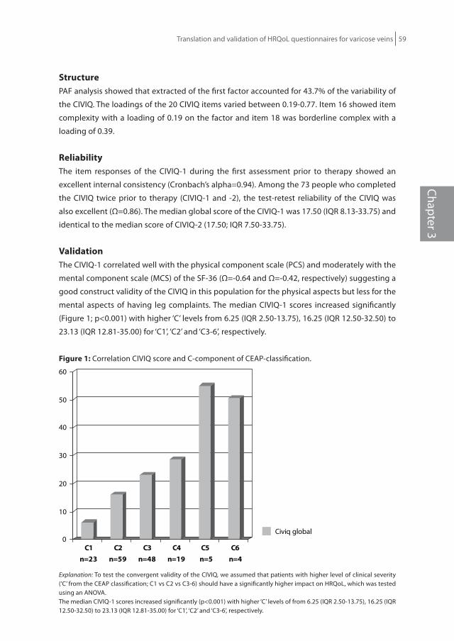
Translation and validation of HRQoL questionnaires for varicose veins 59
Chapter 3
structurePAF analysis showed that extracted of the first factor accounted for 43.7% of the variability of
the CIVIQ. The loadings of the 20 CIVIQ items varied between 0.19-0.77. Item 16 showed item
complexity with a loading of 0.19 on the factor and item 18 was borderline complex with a
loading of 0.39.
reliabilityThe item responses of the CIVIQ-1 during the first assessment prior to therapy showed an
excellent internal consistency (Cronbach’s alpha=0.94). Among the 73 people who completed
the CIVIQ twice prior to therapy (CIVIQ-1 and -2), the test-retest reliability of the CIVIQ was
also excellent (Ω=0.86). The median global score of the CIVIQ-1 was 17.50 (IQR 8.13-33.75) and
identical to the median score of CIVIQ-2 (17.50; IQR 7.50-33.75).
ValidationThe CIVIQ-1 correlated well with the physical component scale (PCS) and moderately with the
mental component scale (MCS) of the SF-36 (Ω=-0.64 and Ω=-0.42, respectively) suggesting a
good construct validity of the CIVIQ in this population for the physical aspects but less for the
mental aspects of having leg complaints. The median CIVIQ-1 scores increased significantly
(Figure 1; p<0.001) with higher ‘C’ levels from 6.25 (IQR 2.50-13.75), 16.25 (IQR 12.50-32.50) to
23.13 (IQR 12.81-35.00) for ‘C1’, ‘C2’ and ‘C3-6’, respectively.
figure 1: Correlation CIVIQ score and C-component of CEAP-classification.
0
10
20
30
40
50
60
C1n=23
C2n=59
C3n=48
C4n=19
C5 n=5
C6n=4
Civiq global
Explanation: To test the convergent validity of the CIVIQ, we assumed that patients with higher level of clinical severity (‘C’ from the CEAP classification; C1 vs C2 vs C3-6) should have a significantly higher impact on HRQoL, which was tested using an ANOVA.The median CIVIQ-1 scores increased significantly (p<0.001) with higher ‘C’ levels of from 6.25 (IQR 2.50-13.75), 16.25 (IQR 12.50-32.50) to 23.13 (IQR 12.81-35.00) for ‘C1’, ‘C2’ and ‘C3-6’, respectively.

60 Chapter 3
responsivenessOf the 115 patients who were assessed prior to and at least four weeks after therapy (CIVIQ-1
and CIVIQ-3), the CIVIQ score decreased in 75.65% and increased in 16.52% of participants. The
median CIVIQ score prior to therapy was 18.75 (IQR 11.25-33.75) and significantly decreased
to 12.50 (IQR 5.00-22.50) after therapy. The mean global score decreased from 23.68 (SD 16.04)
to 15.66 (SD 13.20). For all patients, the decrease in the mean CIVIQ score after therapy was
8.02 which was exactly half the SD of CIVIQ prior to therapy and is, therefore, in agreement
with Norman’s rule of thumb clinically significant. In accordance with Norman’s rule of thumb,
effect size of the CIVIQ was 0.50, which is considered a medium effect size suggesting that the
treatment effect on patients HRQoL was clinically meaningful. Restricting the analyses to pre-
and post treatment outcomes in 99 patients who were graded C2 or more showed that both
Norman’s rule of thumb was satisfied (8.42 vs 7.92) and the effect size increased to 0.53.
To specify between a treatment or a combination of treatments further analysis were
made. The analysis showed that with EVLA alone (n=19) the median CIVIQ score prior to
therapy was 25.00 (IQR 16.25-36.25) and decreased to 18.75 (IQR 12.50-28.75) after therapy.
Significantly according norman’s rule. Compared to treatment with EVLA in combination of
phlebectomy (n=10) the median CIVIQ score prior to therapy was 20.63 (IQR 8.1-34.06) and
decreased to 11.25 (IQR 1.25-15.93) after therapy. Significantly according norman’s rule. Only
few patients received EVLA in combination with Foam (n=3), sclerocompression (n=3), and/
or crossectomy (n=3), although respectively the last two showed a significant decrease after
therapy, no conclusions can be made because of the low sample size.
discussionIn this study, we have demonstrated that the CIVIQ is a feasible, valid, reliable and responsive
tool in the assessment of HRQoL in patients with varicose disease. However, because the
CIVIQ’s four dimensional structure could not be confirmed, we recommend to use the global
score of the CIVIQ only. It appears to be a reliable instrument but the extremely high internal
consistency (>0.90) suggest some item redundancy, which was also observed in the initial
validation study.18
The change in CIVIQ score after therapy for varicose veins was statistically significant
and clinically meaningful, especially among patients with clinical relevant disease (C2 or
more, according to CEAP classification). The tests analyzing the responsiveness of the CIVIQ
confirm the importance of the concept of minimal clinical important difference (MCID), which
implies that it is insufficient to only compare the HRQoL scores before and after therapy
but also estimate the size of the effect and whether it is clinically relevant to the patient.7
Categorization of CIVIQ scores using anchor-banding techniques would also be very useful
for the interpretation of the scores because it would allow physicians to categorize patients’
degree of HRQoL impairment.8 Moreover, the responsiveness findings suggest that the CIVIQ
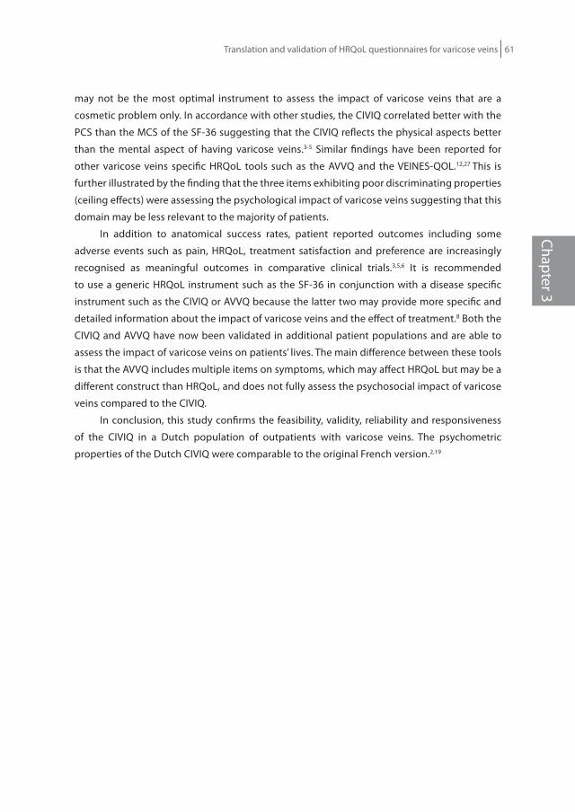
Translation and validation of HRQoL questionnaires for varicose veins 61
Chapter 3
may not be the most optimal instrument to assess the impact of varicose veins that are a
cosmetic problem only. In accordance with other studies, the CIVIQ correlated better with the
PCS than the MCS of the SF-36 suggesting that the CIVIQ reflects the physical aspects better
than the mental aspect of having varicose veins.3-5 Similar findings have been reported for
other varicose veins specific HRQoL tools such as the AVVQ and the VEINES-QOL.12,27 This is
further illustrated by the finding that the three items exhibiting poor discriminating properties
(ceiling effects) were assessing the psychological impact of varicose veins suggesting that this
domain may be less relevant to the majority of patients.
In addition to anatomical success rates, patient reported outcomes including some
adverse events such as pain, HRQoL, treatment satisfaction and preference are increasingly
recognised as meaningful outcomes in comparative clinical trials.3,5,6 It is recommended
to use a generic HRQoL instrument such as the SF-36 in conjunction with a disease specific
instrument such as the CIVIQ or AVVQ because the latter two may provide more specific and
detailed information about the impact of varicose veins and the effect of treatment.8 Both the
CIVIQ and AVVQ have now been validated in additional patient populations and are able to
assess the impact of varicose veins on patients’ lives. The main difference between these tools
is that the AVVQ includes multiple items on symptoms, which may affect HRQoL but may be a
different construct than HRQoL, and does not fully assess the psychosocial impact of varicose
veins compared to the CIVIQ.
In conclusion, this study confirms the feasibility, validity, reliability and responsiveness
of the CIVIQ in a Dutch population of outpatients with varicose veins. The psychometric
properties of the Dutch CIVIQ were comparable to the original French version.2,19
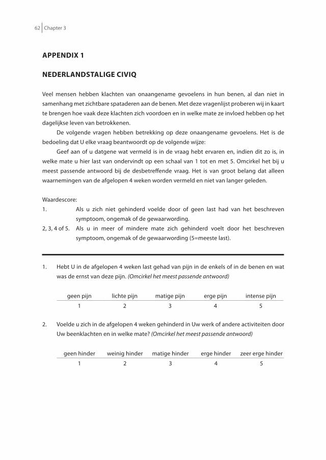
62 Chapter 3
APPendix 1
nederLAndsTALiGe ciViQ
Veel mensen hebben klachten van onaangename gevoelens in hun benen, al dan niet in
samenhang met zichtbare spataderen aan de benen. Met deze vragenlijst proberen wij in kaart
te brengen hoe vaak deze klachten zich voordoen en in welke mate ze invloed hebben op het
dagelijkse leven van betrokkenen.
De volgende vragen hebben betrekking op deze onaangename gevoelens. Het is de
bedoeling dat U elke vraag beantwoordt op de volgende wijze:
Geef aan of u datgene wat vermeld is in de vraag hebt ervaren en, indien dit zo is, in
welke mate u hier last van ondervindt op een schaal van 1 tot en met 5. Omcirkel het bij u
meest passende antwoord bij de desbetreffende vraag. Het is van groot belang dat alleen
waarnemingen van de afgelopen 4 weken worden vermeld en niet van langer geleden.
Waardescore:
1. Als u zich niet gehinderd voelde door of geen last had van het beschreven
symptoom, ongemak of de gewaarwording.
2, 3, 4 of 5. Als u in meer of mindere mate zich gehinderd voelt door het beschreven
symptoom, ongemak of de gewaarwording (5=meeste last).
1. Hebt U in de afgelopen 4 weken last gehad van pijn in de enkels of in de benen en wat
was de ernst van deze pijn. (Omcirkel het meest passende antwoord)
geen pijn lichte pijn matige pijn erge pijn intense pijn
1 2 3 4 5
2. Voelde u zich in de afgelopen 4 weken gehinderd in Uw werk of andere activiteiten door
Uw beenklachten en in welke mate? (Omcirkel het meest passende antwoord)
geen hinder weinig hinder matige hinder erge hinder zeer erge hinder
1 2 3 4 5

Translation and validation of HRQoL questionnaires for varicose veins 63
Chapter 3
3. Sliep U de afgelopen 4 weken slecht door Uw beenklachten en hoe vaak?
(Omcirkel het meest passende antwoord)
nooit zelden vrij vaak zeer vaak elke nacht
1 2 3 4 5
In welke mate hebben Uw beenklachten U de afgelopen 4 weken gehinderd bij de onderstaande
activiteiten?(Omcirkel achter ieder item het meest passende antwoord)
Geen last
Beetje last
Vrij veel last
Zeer veel last
Gaat gewoon niet
4. Lang staan 1 2 3 4 5
5. Traplopen 1 2 3 4 5
6. Knielen of hurken 1 2 3 4 5
7. Snel wandelen 1 2 3 4 5
8. Reizen met tram/bus/trein/auto/vliegtuig
1 2 3 4 5
9. Huishoudelijk werk zoals (koken, voor kind zorgen, strijken, schoonmaken)
1 2 3 4 5
10. Uitgaan naar discotheek, feesten, recepties, e.d.
1 2 3 4 5
11. Sport en/of zwaar werk 1 2 3 4 5
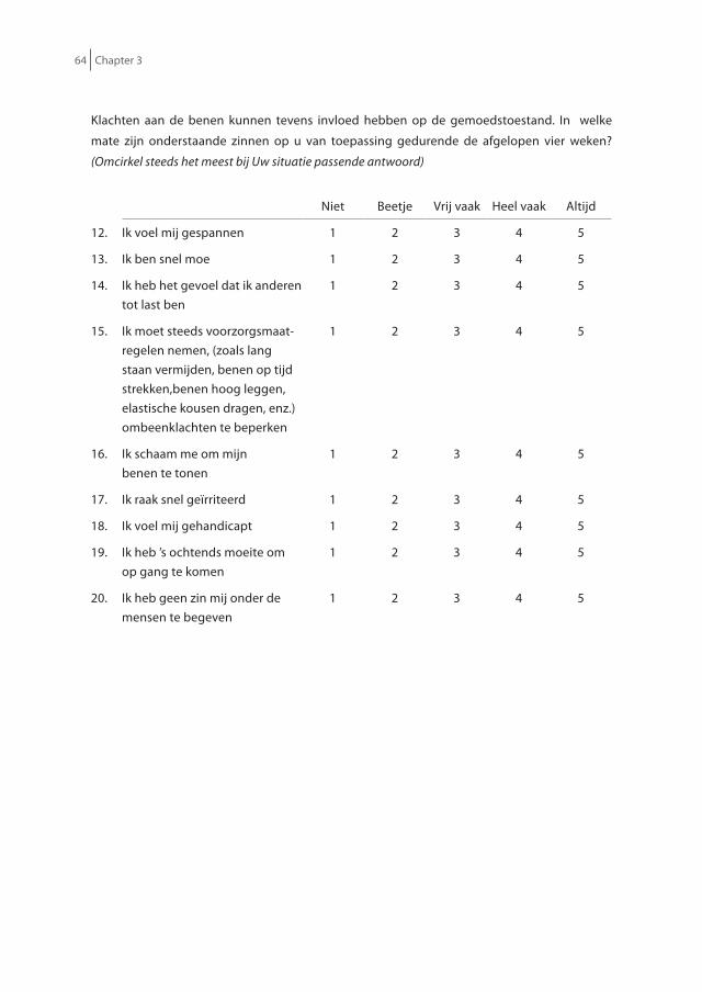
64 Chapter 3
Klachten aan de benen kunnen tevens invloed hebben op de gemoedstoestand. In welke
mate zijn onderstaande zinnen op u van toepassing gedurende de afgelopen vier weken?
(Omcirkel steeds het meest bij Uw situatie passende antwoord)
Niet Beetje Vrij vaak Heel vaak Altijd
12. Ik voel mij gespannen 1 2 3 4 5
13. Ik ben snel moe 1 2 3 4 5
14. Ik heb het gevoel dat ik anderen tot last ben
1 2 3 4 5
15. Ik moet steeds voorzorgsmaat-regelen nemen, (zoals lang staan vermijden, benen op tijd strekken,benen hoog leggen, elastische kousen dragen, enz.) ombeenklachten te beperken
1 2 3 4 5
16. Ik schaam me om mijnbenen te tonen
1 2 3 4 5
17. Ik raak snel geïrriteerd 1 2 3 4 5
18. Ik voel mij gehandicapt 1 2 3 4 5
19. Ik heb ’s ochtends moeite om op gang te komen
1 2 3 4 5
20. Ik heb geen zin mij onder de mensen te begeven
1 2 3 4 5

Translation and validation of HRQoL questionnaires for varicose veins 65
Chapter 3
references
1. Evans CJ, Fowkes FG, Ruckley CV, Lee AJ. Prevalence of varicose veins and chronic venous insufficiency in men and women in the general population: Edinburgh Vein Study. J Epidemiol Community Health 1999;53:149-53.
2. Launois R., Reboul-Marty J., Henry B. Construction and validation of a quality of life questionnaire in Chronic Lower Limb Venous Insufficiency (CIVIQ). Quality of Life Research 1996;5:539-554.
3. Kaplan RM., Criqui MH., Denenberg JO., Bergan J., Fronek A. Quality of life in patients with chronic venous disease: San Diego population study. J Vasc Surg. 2003 May;37(5):1047-53
4. Baker DM., Turnbull NB., Pearson JC., Makin GS. How successful is varicose vein surgery? A patient outcome study following varicose vein surgery using the SF-36 Health Assessment Questionnaire. Eur J Vasc Endovasc Surg. 1995 Apr;9(3):299-304.
5. Durkin MT., Turton EP., Wijesinghe LD., Scott DJ., Berridge DC. Long saphenous vein stripping and quality of life--a randomised trial. Eur J Vasc Endovasc Surg. 2001 Jun;21(6):545-9.
6. Guex JJ., Myon E., Didier L., Nguyen Le C., Taieb C. Chronic venous disease: health status of a population and care impact on this health status through quality of life questionnaires. Int Angiol. 2005 Sep;24(3):258-64.
7. MacKenzie RK., Paisley A., Allan PL., Lee AJ., Ruckley CV., Bradbury AW. The effect of long saphenous vein stripping on quality of life. J Vasc Surg. 2002 Jun;35(6):1197-203.
8. Both H., Essink-Bot ML., Busschbach J., Nijsten T. Critical review of Generic and Dermatolgy-Specific Health-Related Quality of Life Instruments. Journal of investigative Dermatology (2007)127:2726-2739.
9. Smith JJ., Garratt AM., Guest M., Greenhalgh RM. Davies AH. Evaluating and improving health-related quality of life in patients with varicose veins. J Vasc Surg. 1999;30:710-9.
10. Garratt AM., Macdonald LM., Ruta DA., Russell IT. Towards measurement of outcome for patients with varicose veins. Quality in Health Care 1993;2:5-10.
11. Klem TMAL., Sybrandy JEM., Wittens CHA., Essink-Bot ML. Reliability and Validity of the Dutch Translated Aberdeen Varicose Vein Questionnaire. Eur J Vasc Endovasc Surg. (2009);37:232-238.
12. Klem TMAL., Sybrandy JEM., Wittens CHA. Measurement of Health-related Quality of Life with the Dutch Translated Aberdeen Varicose Vein Questionnaire before and after Treatment. Eur J Vasc Endovasc Surg. (2009); 37(4):470-6.
13. Lozano F.S., Launois R. Quality of life (Spain and France): Validation of the Chronic Venous Insufficiency Questionnaire (CIVIQ) Methods Find Exp Clin Pharmacol. 2002 Sep;24(7):425-9.
14. Neglén P., Hollis KC., Raju S. Combined saphenous ablation and iliac stent placement for complex severe chronic venous disease. J Vasc Surg. 2006 Oct;44(4):828-33
15. Kalteis M., Berger I., Messie-Werndl S., Pistrich R., Schimetta W., Pölz W., Hieller F. High ligation combined with stripping and endovenous laser ablation of the great saphenous vein: early results of a randomized controlled study. J Vasc Surg. 2008 Apr;47(4):822-9
16. Brizzio E., Amsler F., Lun B., Blättler W. Comparison of low-strength compression stockings with bandages for the treatment of recalcitrant venous ulcers. J Vasc Surg. 2010 Feb;51(2):410-6.
17. Lurie F., Creton D., Eklof B., Kabnick LS., Kistner RL., Pichot O., Schuller-Petrovic S., Sessa C. Prospective randomized study of endovenous radiofrequency obliteration (closure procedure) versus ligation and stripping in a selected patient population (EVOLVeS Study). J Vasc Surg. 2003 Aug;38(2):207-14.
18. Erevnidou K., Launois R., Katsamouris A., Lionis C. Translation and validation of a quality of life questionnaire for chronic lower limb venous insufficiency into greek. Int Angiol. 2004 Dec;23(4):394-9.
19. Arnould B., Jantet G., Marquis P., Aussage P., Launois R. International Psychometric Validation of the CIVIQ, a specific Quality of Life.

66 Chapter 3
20. Questionnaire in Patients suffering from Chronic Venous Insufficiency: results of the RELIEF study, a Large International Quality of Life Investigation. www.rees-france.com
21. Bullinger M., Alonso J., Apolone G., Leplège A., Sullivan M., Wood-Dauphinee S., Gandek B., Wagner A., Aaronson N., Bech P., Fukuhara S., Kaasa S., Ware JE jr. Translating health status questionnaires and evaluating the quality: the IQOLA project approach. J Clin Epidemiol. 1998;51(11):913-23.
22. Ware JE. Jr, Sherbourne CD. The MOS 36-item short-form health survey (SF-36). I. Conceptual framework and item selection. Med Care 1992 Jun;30(6):473-83.
23. Blomgren L., Johansson G., Bergqvist D. Quality of life after Surgery for Varicose veins and the Impact of Preoperative Duplex Results Based on a Randomized Trial. Ann of Vascular Surg. 2006;20(1):30-34.
24. Assadian A., Wickenhauser G., Hübl W., Wiltos B., Sami A., Senekowitsch C., Hagmüller GW. Traditional versus endoscopic saphenous vein stripping: a prospective randomized pilot trial. Eur J Vasc Endovasc Surg. 2008 Nov;36(5):611-5. Epub 2008 Aug 20.
25. Norman GR., Sloan JA., Wyrwich KW. Interpretation of changes in Health-related Quality of Life. The Remarkable universality of Half a Standard Deviation. Medical Care 2003 ; 41(5), 582-592.
26. Cohen J. Statistical power analysis for the behavorial sciences. New York: Academic Press; 1977. 27. Van Korlaar I., Vossen C., Rosendaal F., Cameron L., Bovill E., Kaptein A. Quality of life in venous
disease. Thromb Heamost. (2003);90:27-35.28. Lamping DL, Schroter S, Kurz X, Kahn SR, Abenhaim L. Evaluation of outcomes in chronic venous
disorders of the leg: Development of a scientifically rigorous, patient-reported measure of symptoms and quality of life. J Vasc Surg. 2003;37:410-9.

Translation and Validation of the Dutch VEINES-QOL/Sym in Varicose Vein Patients
Simone K van der VeldenAnke A. BiemansTamar NijstenAnja Sommer
Phlebology 2013 Apr 4; Epub before print
Chapter 3

68 Chapter 3
ABsTrAcTobjectives: To translate from English to Dutch and evaluate the psychometric properties
of the VEnous INsufficiency Epidemiological and Economic Studies (VEINES) questionnaire,
divided in symptom (VEINES-Sym) and quality of life (VEINES-QOL) subscales.
Methods: Standard forward–backward translation method was used to translate the 26 items
of the VEINES-QOL/Sym. Eligible patients had to complete a standardized questionnaire.
Demographic, venous disease characteristics, clinical venous signs, CEAP (clinical, aetiological,
anatomical and pathological elements) classification and ultrasound findings were also noted.
If item’s scores were in an extreme category in more than 70% of patients a floor or ceiling
effect was present. Feasibility of the individual items was considered poor if 10% or more of the
responses were missing. The validity was tested by comparing the VEINES-QOL/Sym scores to
the Short Form 36 (SF-36) scores and across the different ‘CEAP’ categories. Confirmatory factor
analysis was used to assess the underlying structure of the VEINES-QOL/Sym.
results: Sixty-six patients were included (response rate of 72%). None of the 26 items
missed,10% of responses, but two showed ceiling effect. Both the VEINES-QOL and VEINES-
Sym showed an excellent internal consistency (Cronbach’s alpha of 0.88 and 0.81, respectively).
The VEINES-QOL demonstrated a good construct validity for the physical component of the
SF-36, but not for the mental component (rho ¼ 0.62 and 0.22, respectively), as expected.
The VEINES-Sym correlated poorly to both SF-36’s components. According to the confirmatory
principle axis factoring, only three out of 25 items did not load sufficiently on the factor.
conclusions: The Dutch VEINES-QOL/Sym can be used for health-related quality of life research
in varicose veins patients and the evaluation of therapies.

Translation and validation of HRQoL questionnaires for varicose veins 69
Chapter 3
inTroducTionChronic venous disorders (CVD) are defined as the morphological and functional abnormalities
of the venous system of long duration manifested either by symptoms and/ or signs indicating
the need for investigation and/or care.1 It is a common disease and about 62.1% of females and
49.1% of males in the Western countries are affected with venous symptoms.2 Most patients
affected by CVD experience discomfort and disability in their daily lives which is caused by
symptoms such as tingling, aching, burning, pain, muscle cramps, swelling, sensation of
throbbing or heaviness, itching skin, restless legs, leg-tiredness and/or fatigue.
Over the last decades much research has been done on measuring health related
quality of life (HRQoL) in CVD patients, using generic or disease specific questionnaires. The
most commonly used generic instruments in phlebology are the Short Form 36 (SF-36) and
the EuroQol (EQ)-5D. Since these measures were developed to be used across many different
diseases, it loses precision and sensitivity to change in diseases with specific HRQoL impairment
such as varicose veins. Subsequently, disease specific instruments are increasingly used to
evaluate the effects of specific treatments in patients with varicose veins.3-6 To complement the
shortcomings of the generic instruments, it is recommended to combine generic and disease
specific HRQoL instruments. The available disease specific HRQoL tools focusing on chronic
venous insufficiency and/or varicose veins are: Chronic Lower Limb venous Insufficiency
(CIVIQ), Aberdeen Varicose Vein Questionnaire (AVVQ) and VEINES-QOL/Sym.4-6 The AVVQ
calculates one global HRQoL score summing symptom and clinical class related items and to
a lesser extent HRQoL items.4 The CIVIQ focuses on HRQoL impairment and includes only one
symptom related item. The CIVIQ results in a global score and four separate domain scores
(physical, psychological, social impairments and level of pain).5 The VEnous INsufficiency
Epidemiological and Economic Studies (VEINES) questionnaire is positioned in between these
two instruments because it balances symptom (VEINES-SYM) and quality of life (VEINES-QOL)
items resulting in two separate scores.6 In our opinion, it is recommended to separate the
evaluation of symptoms and HRQoL impairment. The presence and extent of symptoms are
likely to correlate well with HRQoL impairment because it is a proxy for disease severity (i.e.,
construct validity), but symptoms are a different construct than HRQoL and should therefore
not be summed in a total score.7
Symptom measurement is important and is part of the domain of patient reported
outcomes (PRO), but not within the HRQoL construct. Increasingly more clinical trials assessing
treatments of varicose veins include improvement of symptoms as an important endpoint in
addition to anatomical and HRQoL measures.8
The study’s objective is to translate and evaluate the psychometric properties of the
Dutch translated VEINES-QOL/Sym for patients with varicose veins.

70 Chapter 3
MeThodsTranslation and pilot testingThe VEINES-QOL/Sym questionnaire was translated into Dutch by using the recommended for-
ward-backward translation.9 All translators were asked to place emphasis on conceptual rather
than literal equivalence and taking the reading level of a 14years-old in consideration. The
forward translations were performed by the principal investigator (SV) and two independent
academic dermatologists with special interest in phlebology. The three forward translations
were compared, and after consensus was reached, a preliminary common forward-translation
was developed. The common forward translation was sent to two English native speakers, who
each translated the questionnaire back into English. The backward translations were reviewed
by those responsible for the forward translation and an independent dermatologist for
conceptual equivalence with the original English version. If necessary, adaption’s to the Dutch
version were made. Subsequently, the Dutch questionnaire was pilot tested in a heterogeneous
group of 20 patients with symptoms and signs of chronic venous disease. Respondents were
asked about item difficulty by using a brief questionnaire, but none were encountered during
the pilot testing. Therefore, the Dutch VEINES-QOL/Sym was not revised and administered to
the study population.
study subjectsAll consecutive patients presenting to the outpatient department of Dermatology with
symptoms and objective signs of venous disease, were asked to participate in the study.
Patients were excluded if cosmetic complaints were reported only or if there was no sign
of venous disease during physical examination or if no reflux (longer than 0.5s) was found
during venous ultrasound examination. Furthermore, patients were excluded if edema was the
only clinical sign during physical examination because these patients were more likely to have
non-venous causes of edema.
A trained physician examined patient’s affected legs that were classified according to
the clinical component of the international accepted CEAP classification.10 Venous signs, such
as telangiectasia, reticular veins, varicose veins, edema, skin changes (hyperpigmentation,
lipodermatosclerosis) or ulcerations were recorded. Subsequently, ultrasound examination
of both the superficial and deep venous systems was performed in standing position.11
Reflux duration of more than 0.5 seconds was considered as evidence for superficial venous
insufficiency.
QuestionnaireAfter participants provided written informed consent, the Short Form Health Survey (SF-
36) and the VEINES- QOL/Sym were completed by the participants. Patients were requested
to complete the questionnaires at home and return it by prepaid mail. In case of missing

Translation and validation of HRQoL questionnaires for varicose veins 71
Chapter 3
questionnaire items, patients were contacted by phone in order to retrieve the missing data. If
we were not able to reach a patient by phone, missing items were replaced by the median of
the completed items reported by an individual for that (sub)scale.
The SF-36 consists of 36 items, divided into 8 different dimension which are summarized
in physical (Physical Component Summary [PCS]) and mental (Mental Component Summary
[MCS] QOL scores. The MCS and PCS SF-36 scales are scored from 0 to 100, with 0 indicating the
least favorable possible health state.12
The VEINES-QOL/Sym is a 26-item questionnaire that measures venous symptoms (10
items), time of day leg problems are most intense (1 item), change over the past year (1 item),
limitations in daily activities as a result of CVD (9 items) and psychological impact of CVD (5
items) (Figure 1). Two summary scores can be computed; VEINES-Sym score which reflects the
extent of venous symptoms and VEINES-QOL score which represents the HRQoL impairment.
The item ‘time of day your leg problems are most intense’ represents descriptive information
only, and is therefore not included in the summary score. Higher VEINES-QOL scores indicate a
favorable HRQoL (range 0-100).
sTATisTicAL AnALysisfeasibilityThe feasibility of the questionnaire was evaluated by the overall response rate. Item difficulty
was present, if 10% or more of the answers of individual items were missing.
The score distribution of all individual items was evaluated by assessing their floor and
ceiling effects (i.e., 70% or more of the respondents exhibited the worst or best possible score).
Patients with three or more missing scores were excluded from the analysis, except from the
feasibility assessment. Missing values were replaced by the median of the completed items
reported by an individual for that (sub)scale.
structureConfirmatory factor analysis (CFA) was performed using the principal component in order to
test the structure of the VEINES-QOL and VEINES-Sym separately. We forced the analysis to
extract one component in order to select the items that loaded convincingly on the component
and therefore share the same conceptual meaning. If an item loaded 0.40 or more on the single
component it was defined as adequate.13
reliabilityCronbach’s alpha (reflecting the internal consistency of an instrument) was tested for the
VEINES-Sym and VEINS-QOL separately and were considered good, if between 0.7 and 0.9.
72 Chapter 3
ValidityThe construct validity of the VEINES-QOL/Sym (i.e., how it relates to other HRQoL measures) was
tested by calculating Spearman’s correlation coefficient (rho) between patients’ scores and the
MCS and PCS of the SF-365. We expected that the VEINES-QOL would correlate well (rho>0.6)
and that the VEINES-Sym would correlate moderate (rho>0.3) with the PCS. We hypothesized
that both VEINES-QOL and VEINES-Sym would correlate poorly (rho<0.3) with the MCS.
To test the convergent validity of the questionnaire, we assumed that patients with
higher level of clinical severity (‘C’ from the CEAP classification; C1 vs. C2 vs. C3-6) should have
a statistically significantly higher impact on HRQOL, which was tested using an analysis of
variance (ANOVA).
The distribution of the data were presented as mean with standard deviation (SD) or
median with interquartile range (IQR) values as appropriate. Categorical data were analyzed
by means of X2 test or, if necessary, Fisher’s exact test.
Two sided P values of 0.05 or less were considered statistically significant. All analyses
were conducted using SPSS (SPSS Inc., Chicago, IL, U.S.A.). The Medical ethical committee of the
University Hospital Maastricht (The Netherlands) granted exempt status for this observational
study. All participants provided written informed consent.
resuLTsstudy populationOf the eligible 94 patients with symptoms and objective signs of venous disease who
were invited to participate in this study, 68 patients (response rate 72%) completed the
questionnaire. Two patients were excluded because of incomplete questionnaires (2.9%). Of
the 66 participants, 73% were women and the mean age of the study population was 54.9
years (SD 13.1, range 20- 81 years; Table I). Classified according to the C-component of CEAP
classification, 35 % of the participants were classified in ‘C1’, 33% in ‘C2’ and 32% in ‘C3-C6’.
feasibilityNone of the individual items were considered suboptimal as missing responses varied between
0% and 4.6%. However, item 1.9 (‘tingling sensation’) and item 4b (‘daily activities at home’)
were both missing in 4.6% of the participants, suggesting borderline feasibility of these items.
From the 25 items in the questionnaire, 2 demonstrated a substantial ceiling effect with
75.8% and 75.8% of the respondents indicating the highest score for item 5a (‘cut down the
amount of time you spent on work or other activities’) and item 8c (‘have you felt a burden to
your family or friend’).
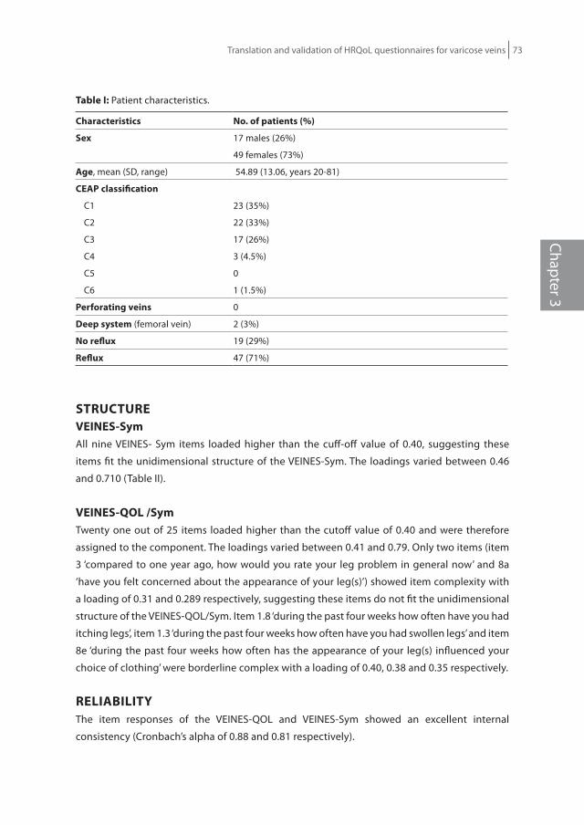
Translation and validation of HRQoL questionnaires for varicose veins 73
Chapter 3
Table i: Patient characteristics.
characteristics no. of patients (%)
sex 17 males (26%)
49 females (73%)
Age, mean (SD, range) 54.89 (13.06, years 20-81)
ceAP classification
C1 23 (35%)
C2 22 (33%)
C3 17 (26%)
C4 3 (4.5%)
C5 0
C6 1 (1.5%)
Perforating veins 0
deep system (femoral vein) 2 (3%)
no reflux 19 (29%)
reflux 47 (71%)
sTrucTureVeines-symAll nine VEINES- Sym items loaded higher than the cuff-off value of 0.40, suggesting these
items fit the unidimensional structure of the VEINES-Sym. The loadings varied between 0.46
and 0.710 (Table II).
Veines-QoL /symTwenty one out of 25 items loaded higher than the cutoff value of 0.40 and were therefore
assigned to the component. The loadings varied between 0.41 and 0.79. Only two items (item
3 ‘compared to one year ago, how would you rate your leg problem in general now’ and 8a
‘have you felt concerned about the appearance of your leg(s)’) showed item complexity with
a loading of 0.31 and 0.289 respectively, suggesting these items do not fit the unidimensional
structure of the VEINES-QOL/Sym. Item 1.8 ‘during the past four weeks how often have you had
itching legs’, item 1.3 ‘during the past four weeks how often have you had swollen legs’ and item
8e ‘during the past four weeks how often has the appearance of your leg(s) influenced your
choice of clothing’ were borderline complex with a loading of 0.40, 0.38 and 0.35 respectively.
reLiABiLiTyThe item responses of the VEINES-QOL and VEINES-Sym showed an excellent internal
consistency (Cronbach’s alpha of 0.88 and 0.81 respectively).

74 Chapter 3
Table ii: The loadings* of a confirmatory primary component analysis, for VEINES-Sym and VEINES-
Sym/QOL separately.
items Veines-sym Veines-sym/QoL
1.1 Heavy legs .61 .54
1.2 Aching legs .73 .59
1.3 Swelling .46 .38
1.4 Night cramps .49 .43
1.5 Burning .64 .52
1.6 Restless legs .65 .50
1.7 Throbbing .63 .58
1.8 Itching .46 .40
1.9 Tingling .68 .55
7 Pain .71 .62
3 Evolution of the leg problem .31
4a Limitation of activities at work .56
4b Limitation of activities at home .79
4c Limitation of standing activities .68
4d Limitation of sitting activities .66
5a Less time spent on work or activities .75
5b Accomplished less .76
5c Limited in the kind of work or activities .56
5d Difficulty performing the work or activities .67
6 Social activities .77
8a Concerned about appearance of the legs .29
8b Felt irritable .66
8c Felt a burden to your family of friends .55
8d Worried about bumping the legs .41
8e Choice of clothing .35
* Loadings >0.40 are included, indicating these items loaded on the expected scale (VEINES-Sym and VEINESSym/QOL separately).
ValidityThe VEINES-Sym is borderline moderately correlated with the physical component and poorly
with the mental component of the SF-36 (rho =0.4 and rho=0.1 respectively). As expected, the
VEINES-QOL is strongly correlated with the physical component and poorly correlated with the
mental component of the SF-36 (rho =0.6 and rho=0.2 respectively) confirming the convergent
validity of the instrument.

Translation and validation of HRQoL questionnaires for varicose veins 75
Chapter 3
In contrast to our hypothesis, the median scores of both the VEINES-Sym and VEINES-QOL
were not affected significantly by severity of varicose veins (C1, C2, C3-C6; p=0.80 and p=0.41
respectively) and varied between 48- 52 and 48-53 respectively. This observation may indicate
a poor construct validity of the instrument.
discussionIn this study, we translated and evaluated the psychometric properties of the Dutch translated
VEINES-QOL/Sym in patients with varicose veins. This venous specific health related quality of
life and symptom instrument showed a good acceptability, validity and high reliability. This
study provided evidence for a high feasibility, since 24 out of 26 items showed no floor or
ceiling effects which indicates a good discriminative effect of the items. The high construct
validity was supported by the strong correlation between the VEINES-QOL and the physical
component of the SF-36.
Interestingly, correlations between the VEINES-QOL and the SF-36 are higher for the
physical rather than for the mental scores suggesting that the psychosocial impact of varicose
veins is more in the functioning domain7. This correlation with functioning is in line with the
study’s of the original English and French VEINES-QOL/Sym versions and validation studies of
the AVVQ and the CIVIQ instruments.6,14-15 As expected, the VEINES- Sym score is not correlated
to the SF-36. This correlates with the original version.6 We therefore suggest that the symptoms
construct differs from the mental and physical components (e.a impairment, disability,
functioning). However, all three belong to the measurement of HRQoL.16-18
We were not able to achieve significant differences between the clinical ‘C’EAP
categories. The poor correlation between the VEINES-Sym and VEINS-QOL scores and the CEAP
classification may be explained by study population (excluding edema patients), small sample
size of patients with severe venous disease, suboptimal measure of disease severity and/or
poor construct validity of the VEINES instrument. In a predominantly C1-3 population, venous
disease may have induced limited HRQoL impairment, but is accompanied by the presence of
symptoms.
In the confirmatory Principle Axis Factoring (PAF) 21 out of 25 items did load sufficiently on
the factor, indicating a good unidimensional underlying construct of these findings. However,
the construct validity of the items would suggest separate scales for symptoms (items 1, 3, 7),
functioning (items 4, 5, 6) and psychological aspects (item 8). A preliminary exploratory PAF
confirmed these scales partly (data not shown). Moreover, it was not the objective of this study
to refine the current instrument.
As we mentioned above, the present study showed a high acceptability. We reported
that two items showed a substantial ceiling effect, including ‘cut down the amount of time
you spent on work or other activities’ and ‘have you felt a burden to your family or friend’.
Not surprisingly, both non-discriminative items are mental aspects of the VEINES-QOL/Sym.
76 Chapter 3
Therefore, it is questionable whether the mental items are specific enough for patients affected
with predominantly mild varicose veins. In this study population, mental health appears to be
less impaired compared to physical health, which is in contrast to the results of other studies
showing that varicose veins may have an impact on the mental health.19
A limitation of the current study is the relatively small group of patients with severe venous
disease. The response rate was adequate, but this study was restricted to patients visiting a
academic center potentially limiting the generalization of our findings. Future psychometric
studies need to confirm the responsiveness (i.e., sensitivity to change) and minimal clinical
important difference of the VEINES-QOL/Sym.
In conclusion, we showed that the psychometric properties of the Dutch VEINESQOL/
Sym were comparable to the original English version. This supports applications of the Dutch
VEINES-QOL/Sym during HRQoL research in the Dutch varicose veins population.

Translation and validation of HRQoL questionnaires for varicose veins 77
Chapter 3
references
1. Eklof B, Perrin M, Delis KT, Rutherford RB, Gloviczki P. Updated terminology of chronic venous disorders: the VEIN-TERM transatlantic interdisciplinary consensus document. Vasc Surg. 2009 Feb;49: 498-501
2. Pannier-Fischer F, Rabe E. Epidemiology of chronic venous diseases. Hautarzt. 2003 Nov;54: 1037 44.3. Wiebe S, Guyatt G, Weaver B, Matijevic S, Sidwell C. Comparative responsiveness of generic and
specific quality-of-life instruments. J Clin Epidemiol. 2003 Jan; 56: 52-604. Launois R, Reboul-marty J, Henry B. Construction and validation of quality of life questionnaire in
Chronic Lower Limb venous Insufficiency (CIVIQ). Qual Life Res. 1996; 5: 539-545. Garratt A, Schmidt L, Mackintosh A, Fitzpatrick R. Quality of life measurement: bibliographic study of
patient assessed health outcome measure. BMJ. 2002 June 324: 1417.6. Lamping DL, Abenheim L, Kurz X, Schroter S, Kahn S.R., and the VEINES group Measuring quality of
life and symptoms in chronic venous disorders of the leg: development and psychometric evaluation of the VEINES-QOL/VEINES-Sym questionnaire. Qual Life Res. 1998; 7: 621-622
7. Muldoon MF, Barger SD, Flory JD, Manuck SB. What are quality of life measurements measuring? BMJ. 1998 Feb; 316: 542-5.
8. De Maeseneer M. The endovenous revolution. Br J Surg. 2011 Aug;98: 1037-89. Bullinger M, Alonso J, Apolone G, et al. Translating health status questionnaires and evaluating their
quality: the IQOLA project approach. J Clin Epidemiol. 1998; 51: 913- 92310. Vasquez MA, Rabe E, McLafferty RB, et al. Revision of the venous clinical severity score: venous
outcomes consensus statement: special communication of the American Venous Forum Ad Hoc Outcomes Working Group. J Vasc Surg. 2010 Nov;52: 1387- 96.
11. De Maeseneer M, Pichot O, Cavezzi A, et al. Duplex ultrasound investigation of the veins of the lower limbs after treatment for varicose veins. Eur J Vasc Endovasc Surg. 2011 Jul;42: 89-102.
12. Ware JE, Gandek B and the IQOLA Project group. The SF-36 Health survey: development and use in mental health research and the IQOLA Projects. Int. J. Ment. Health 1994; 23: 49-73
13. Angoff WH. Perspectives on differential item functioning methodology. In: Holland PW, Wainer H eds. Differential item functioning. New Jersey: Lawrence Erlbaum Associates, Inc., publicers, 1993: 3-24
14. Klem TM, Sybrandy JE, Wittens CH. Measurement of health-related quality of life with the Dutch translated Aberdeen Varicose Vein Questionnaire before and after treatment. Eur J Vasc Endovasc Surg. 2009 Apr;37: 470-6.
15. Biemans AA, van der Velden SK, Bruijninckx CM, Buth J, Nijsten T. Validation of the chronic venous insufficiency quality of life questionnaire in Dutch patients treated for varicose veins. 2011 Aug;42: 246-53.
16. Feinstein AR. Clinimetric perspectives. J Chronic Dis. 1987;40: 635-40 17. McHorney CA, Ware JE Jr, Raczek AE. The MOS 36-Item Short-Form Health Survey (SF-36): II.
Psychometric and clinical tests of validity in measuring physical and mental health constructs. Med Care 1993 Mar;3: 247-63.
18. Bowling A. What things are important in people’s lives? A survey of the public’s judgments to inform scales of health related quality of life. Soc Sci Med. 1995 Nov;41: 1447-62.
19. Sritharan K, Lana TR, Davies AH. The burden of depression in patients with symptomatic varicose veins. Eur J Vasc Endovasc Surg. 2012 Apr;43(4):480


Chapter 4
Comparing Endovenous Laser Ablation, Foam Sclerotherapy and Conventional Surgery for Great Saphenous Varicose Veins
Anke A. BiemansMichael KockaertGeorge P. AkkersdijkRenate R. van den BosMarianne G. de MaeseneerPhilip CuypersMartino H. NeumannTamar Nijsten J Vasc Surg. 2013 Jun 12 (Epub ahead of print)
Chapter 4

80 Chapter 4
ABsTrAcTBackground: Many case-series have been published on treatments of varicose veins, but
comparative randomized controlled trials remain sparse.
objective: to compare the anatomical success rate, frequency of major complications and
quality of life improvement of endovenous laser ablation (EVLA), ultrasound-guided foam
sclerotherapy (UGFS) and conventional surgery (CS), after one year follow-up.
Methods: 240 consecutive patients with primary symptomatic great saphenous vein reflux
were randomized to endovenous laser ablation, ultrasound-guided foam sclerotherapy or
conventional surgery, consisting of high ligation and short stripping. Primary outcome was
anatomical success defined as obliteration or absence of the treated vein on ultrasound
examination after one year. Secondary outcomes were complications, improvement of the ‘C’
class of the CEAP classification and improvement of disease specific (CIVIQ) and general (EQ5D)
quality of life scores.
results: More than 80% of the study population was classified as C2 or C3 venous disease.
After one year, the anatomical success rate was highest after EVLA (88.5%), followed by CS
(88.2%) and UGFS (72.2%) (P<.001). The complication rate was low and comparable between
treatment groups. All groups showed significant (p<0.001) improvement of EQ5D and CIVIQ
scores after therapy. 84.3% of all treated patients showed an improvement of the ‘C’ of the
CEAP classification.
conclusion: After one year follow up EVLA is as effective as CS and superior to UGFS according
to occlusion on ultrasound duplex. Quality of life improves after treatment in all groups
significantly.
Registration number: NCT00529672
Comparing EVLA, UGFS and CS for great saphenous varicose veins 81
Chapter 4
inTroducTionVaricose veins of the legs affect approximately 25% of the population1 and may have a
substantial impact on patient’s health-related quality of life (HRQoL). The treatment of varicose
veins and its complications consume a relatively large proportion of the limited health care
resources.1,2
Until recently, conventional surgery (CS) of the great saphenous vein (GSV), consisting of
high ligation at the saphenofemoral junction (SFJ) and stripping of the above knee GSV, was
the standard of care. In the last decade, minimally invasive techniques such as endovenous
laser ablation (EVLA), radiofrequency ablation (RFA) and ultrasound-guided foam sclerotherapy
(UGFS) have challenged the position of CS for primary varicose veins.3 These techniques are
effective (occlusion rates of EVLA and RFA >90%), and safe.4,5 CS associates stripping of the
refluxing GSV trunk with high ligation at the SFJ and this may induce neovascularization.6,7,8
Endovenous treatment techniques are performed without high ligation, which may be an
advantage. Additional advantages of EVLA, RFA and UGFS over CS are that they can be easily
performed in an ambulatory setting. A comparative meta-analysis of observational studies and
randomized clinical trials (RCTs) demonstrated that both EVLA and RFA were superior to CS
and UGFS.5 In the last few years several important RCTs comparing two different treatment
modalities for varicose veins show that the minimally invasive techniques are at least as
effective as CS, and that they result in faster recovery time, less post-operative pain, and that
they are preferred by patients. 7,9,10,11 In 2011, Rasmussen et al were the first to compare more
than two different treatments in a 4-arm RCT.12 This study showed that thermal ablation and
CS, all performed under tumescent anesthesia, had anatomical results than UGFS however
patient reported outcomes were better after in the UGFS and RFA.
A recent comparative study of UGFS and CS showed significantly higher efficacy rates
two years after CS. However, there were no differences in patient-reported outcomes.13
The objective of this study is to compare the anatomical success rate, frequency of
complications, and HRQoL improvement of EVLA, UGFS, and CS for the treatment of primary
incompetent GSV after one year.
MeThodsOur study was designed as a consecutive single center RCT at the department of dermatology
and vascular surgery of Erasmus Medical Center Rotterdam, The Netherlands, starting in
January 2007. Due to a decreasing inclusion rate, the same departments of Catharina Hospital
Eindhoven were added as second center in May 2009. The last patient was treated in May 2010.
The medical ethical committee of Erasmus MC Rotterdam approved our protocol. (MEC2005-
325).
In Latin, the GSV is called “Vena Saphena Magna” and in the Netherlands we use this
name to indicate the GSV; therefore we have chosen to call our study the MAGNA trial.
82 Chapter 4
Adult patients with a symptomatic primary incompetent GSV at least above the knee with
a diameter of ≥0.5 cm and with an incompetent saphenofemoral junction were eligible to
participate. The incompetence of the GSV was defined as reflux of ≥0.5 seconds at color duplex
ultrasound (Philips, HDI 4500, 10MHz probe). Exclusion criteria were previous treatment of
the ipsilateral GSV, deep venous incompetence or obstruction, agenesis of the deep system,
vascular malformations, use of anticoagulation, pregnancy, heart failure, contraindication
for one of the treatments, (e.g., allergy for aethoxysclerol or lidocaine), immobility, arterial
insufficiency (defined as an Ankle-Brachial Index less than 0.6), age under 18 years, and inability
to provide written informed consent to trial participation.
TreatmentIn this study only the GSV in the thigh (from just below or above knee level in most cases) was
treated. Patients were allocated to one of the three treatments. After written informed consent,
eligible patients were randomized using a computerized list by an independent research
nurse. All treatments were performed by dermatologists or surgeons with more than 5 years of
experience with the treatments (EVLA and UGFS, and CS and EVLA, respectively).
Endovenous laser ablation EVLA was performed under ultrasound guidance with a 940 nm diode laser as previously
described.14 In brief, venous access was obtained by puncturing the vein at knee level, with
a 16 or 18 French needle under ultrasound guidance. After entrance to the varicose vein was
established, a guide wire was passed through the hollow needle into the vein up to the level
of the saphenofemoral junction (SFJ). The needle was removed and a small cutaneous incision
of 3 mm was made, then an introducer sheath was passed over the guide wire and positioned
1-2 centimeters below the SFJ. Subsequently, the laser fiber was introduced after removing
the guide wire. About 250-500 ml (depending on the length of treated vein) of tumescent
anesthesic solution was administered into the saphenous compartment under ultrasound
guidance using a mechanical infusion pump. Withdrawal of the laser fiber was performed in
continuous mode and it was attempted to deliver at least 60 J/cm.15
Ultrasound-guided Foam SclerotherapyUltrasound-guided foam sclerotherapy was performed as reported previously.14
The Tessari-method16 was used to prepare foam (1 cc aethoxysclerol 3%: 3cc air), which
was injected directly under ultrasound guidance in the GSV with the patient in horizontal
position.14,16 The volume of injected foam depended on the length and diameter of the vessel,
with a maximum of 10mL per session (as suggested by the 2nd European Consensus).17 After
injection, the patient remained horizontally for at least 5 minutes. If considered necessary,
UGFS of the included GSV could be repeated after three months.
Comparing EVLA, UGFS and CS for great saphenous varicose veins 83
Chapter 4
Conventional surgeryHigh ligation with short (above knee) stripping was performed under spinal or general
anesthesia. Flush SFJ ligation was followed by ligation of all tributaries back to the second
branch and invaginating stripping of the GSV to knee level. The cribriform fascia, superficial
fascia and skin were closed.
After all treatments ambulatory compressive bandage was applied for 48 hours, followed
by therapeutic compression stockings for 2 weeks post procedure. All patients were observed
for at least half an hour in the clinic after treatment. No specific analgesics were prescribed.
Patients were encouraged to mobilize and to resume their usual activities as soon as possible.
outcomesPrimary outcomePatients were evaluated at 3 and 12 months for clinical examination and duplex ultrasound.
The primary outcome was anatomical success according to duplex ultrasound evaluation. For
EVLA and UGFS this was defined as complete obliteration, without flow or reflux, of the GSV at
the level of the mid-thigh. For CS success was defined as absence of the GSV in the saphenous
compartment at thigh level. We differentiated between obliteration, partial or complete
patency of the treated vein, with or without reflux.
Postoperative neovascularization was assessed at the level of the SFJ using the classification
described by De Maeseneer et al, ‘The degree of neovascularization was determined as ‘grade 1
neovascularization’ (tiny new vein(s) up to 3 mm diameter, not connecting with any superficial
vein) and ‘grade 2 neovascularization’ (tortuous new vein(s) with a diameter ≥4 mm, with
pathological reflux and connecting with thigh varicose veins)’.18
Secondary outcomesAt all visits the ‘C’ of the CEAP classification was recorded. The basic CEAP classification as
described by Eklöf was used, which means patients are classified according to their highest
C score.19 The type and frequency of complications of the different treatments were reported.
The following complications were assessed: superficial vein thrombosis (related to site of
treatment), hyperpigmentation (at treatment site), paresthesia (defined as abnormal skin
sensations such as tingling, tickling, itching, burning or numbness), scotoma, migraine, skin
burns, skin necrosis, anaphylactic shock, wound infection for which antibiotic therapy was
needed, symptomatic deep venous thrombosis (DVT), based on history and confirmed by
duplex examination, and symptomatic pulmonary embolism.
The effect of the treatment on HRQoL was assessed using the disease specific Chronic
Venous Insufficiency Quality of Life Questionnaire (CIVIQ)20 and the generic EuroQol 5D
instrument (EQ-5D)21 questionnaire. Additional phlebectomies were permitted during CS or
EVLA or (in case of residual superficial varicose veins) at three months postoperative. UGFS

84 Chapter 4
could be used as an ‘escape’ therapy in case of failure of the initially allocated treatment of the
GSV.
figure 1: Consort flow.
Randomizedn=240
Enrolment
Allocation
Allocated to EVLA n=80
Underwent intervention
n=76
Procedure failure n=2a
Other intervention n=1b
Allocated to UGFS n=80
Underwent intervention
n=77
Procedure failure n=0
Other intervention n=2b
Allocated to surgery n=80
Underwent intervention
n=65
Procedure failure n=3a
Other intervention n=4b
No intervention n=8b
Follow-up Lost to follow-up n=0
Death n=0
Lost to follow-up n=1b
Death n=0
Lost to follow-up
Death n=0
Analysis Analysed: 68Analysed: 77Analysed: 78
a If there was a procedure failure, patients were treated with UGFS, data analysis was on a intention to treat basis. b Patients who did not receive treatment or received another treatment or were lost of follow up in the first year were excluded from the analysis.
statistical analysisSample size calculation indicated that 240 legs (80 in each group) were needed to detect
a significant difference of 0.05 in anatomical success rate between the three groups with a
beta of 0.20. (i.e. power=80%) Data were analyzed on an intention to treat basis. Continuous
data were first tested for normality using One-Sample Kolmogorov-Smirnov Test. For normal
distribution, data were presented as means with standard deviation (SD) and analysis with one
way ANOVA was done to compare it across three treatment groups.
Categorical data were analyzed by means of χ2 test or, if appropriate, Fisher’s exact test.
Patients with bilateral GSV insufficiency were randomized separately for each leg. For efficacy
analysis, both GSVs were included, but for HRQoL analyses these patients were excluded
because patients are unable to differentiate the impact of varicose veins of each leg on
HRQoL.22
A generalized estimating equation (GEE) model with a logit link was used to model the
odds ratio for total occlusion over time. To take the correlation into account between two

Comparing EVLA, UGFS and CS for great saphenous varicose veins 85
Chapter 4
legs of the same patient and multiple measurements over time, we chose an unstructured
covariance matrix and use the patients as the independent subjects. For EQ5D, Civiq, Health
and CEAP outcomes, we used a linear mixed model (LMM) with empirical standard errors. For
this model we chose a direct product of an unstructured covariance matrix (for the covariance
within a leg) and a compound symmetry correlation matrix (for the correlations within a
patient between the legs). Thereby we allow for an unstructured correlation matrix between
measurements over time within one leg and we assume that the unstructured correlation
matrix is equal for the first and the second leg within one person.
Statistical analysis was performed using SPSS® version 20.0 (SPSS, Chicago, Illinois,
USA) and SAS 9.2 (SAS institute Inc., Cary, NC, USA). All statistical tests were two-sided and
considered significant at the P<.05 level.
resuLTs study populationIn 223 eligible patients, 240 legs were randomized for one of the treatments between January
2007 and December 2009 (Figure 1). Seventeen patients were excluded from the analysis; 16
did not fulfill the inclusion criteria and 1 was lost to follow up.
Five patients were treated with UGFS because initial treatment had failed; two patients
from the EVLA group and three patients from the CS group. Data was analyzed on an ‘intention
to treat’ basis, according to randomization. ‘As treated analysis’ showed no significant difference.
In the study population, 82.3% suffered C2 and/or C3 venous insufficiency. The groups were
well matched for the demographic data, CEAP classification and GSV diameter and HRQoL
impairment (Table I). The patients in the EVLA group were significantly younger than those in
the CS and UGFS group. Patients in the EVLA group received on average 59.16 J/cm (SD 15.20).
Patients in the UGFS group received a mean of 4.7cc (SD 1.19) foam. The majority of patients
were treated in one session of UGFS. Six patients with lasting complaints received a second
treatment within the first year.
Anatomical success EVLA and surgery were comparably effective (88.5%, n 69; 88,2%, n 60 respectively), after one
year follow up (Figure 2, Table II) However, in the CS group 10% (n 7) of patients had grade 1
neovascularization at ultrasound examination of the groin. After one year the occlusion rate
of UGFS was 72,7% (n 56) which was significantly lower than EVLA and CS (P<.02). Twenty-one
patients (27.3%) of the UGFS group had partial obliteration with reflux. In 11 of these patients
initial treatment resulted in complete relief of complaints despite persisting reflux after one
year follow up and therefore they did not undergo any additional treatment.
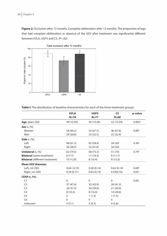
86 Chapter 4
figure 2: Occlusion after 12 months. Complete obliteration after 12 months. The proportion of legs
that had complete obliteration or absence of the GSV after treatment was significantly different
between EVLA, UGFS and CS. (P<.02)
Table i: The distribution of baseline characteristics for each of the three treatment groups.
eVLAn=78
uGfsn=77
csn=68
p-value
Age, years (SD) 49 (15.03) 56 (13.30) 52 (15.59) 0.005a
sex n, (%)Women Men
54 (69.2)24 (30.8)
52 (67.5)25 (32.5)
46 (67.6)22 (32.4)
0.89b
side n, (%)Left Right
48 (61.5)30 (38.5)
45 (58.4)32 (41.6)
34 (50)34 (50)
0.36b
unilateral n, (%) Bilateral (same treatment)Bilateral (different treatment)
62 (79.5)6 (7.7)10 (12.8)
58 (75.3)11 (14.3)8 (10.4)
51 (75)8 (11.7)9 (13.3)
0.74b
Mean GsV diameterLeft, cm (SD)Right, cm (SD)
0.64 ( 0.15)0.59 (0.11)
0.58 (0.14)0.62 (0.14)
0.62 (0.14)0.59(0.16)
0.08a
0.65a
ceAP n, (%)C1C2C3C4C5C6Unknown
037 (47.4)29 (37.2)8 (10.3)004 (5.1)
033 (42.9)30 (39.0)8 (10.4)1 (1.3)05 (6.5)
028 (41.2)21 (30.9)14 (20.6)1 (1.5)04 (5.8)
0.64c
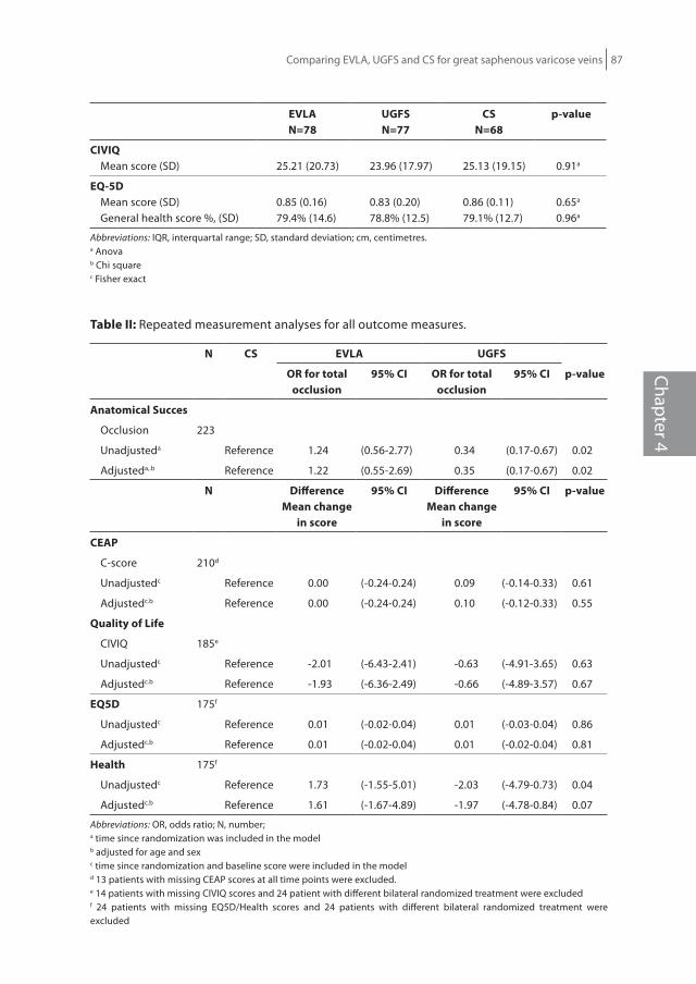
Comparing EVLA, UGFS and CS for great saphenous varicose veins 87
Chapter 4
eVLAn=78
uGfsn=77
csn=68
p-value
ciViQ Mean score (SD) 25.21 (20.73) 23.96 (17.97) 25.13 (19.15) 0.91a
eQ-5d Mean score (SD)General health score %, (SD)
0.85 (0.16)79.4% (14.6)
0.83 (0.20)78.8% (12.5)
0.86 (0.11)79.1% (12.7)
0.65a
0.96a
Abbreviations: IQR, interquartal range; SD, standard deviation; cm, centimetres.a Anovab Chi squarec Fisher exact
Table ii: Repeated measurement analyses for all outcome measures.
n cs eVLA uGfs
or for total occlusion
95% ci or for total occlusion
95% ci p-value
Anatomical succes
Occlusion 223
Unadjusteda Reference 1.24 (0.56-2.77) 0.34 (0.17-0.67) 0.02
Adjusteda,.b Reference 1.22 (0.55-2.69) 0.35 (0.17-0.67) 0.02
n difference Mean change
in score
95% ci difference Mean change
in score
95% ci p-value
ceAP
C-score 210d
Unadjustedc Reference 0.00 (-0.24-0.24) 0.09 (-0.14-0.33) 0.61
Adjustedc.b Reference 0.00 (-0.24-0.24) 0.10 (-0.12-0.33) 0.55
Quality of Life
CIVIQ 185e
Unadjustedc Reference -2.01 (-6.43-2.41) -0.63 (-4.91-3.65) 0.63
Adjustedc.b Reference -1.93 (-6.36-2.49) -0.66 (-4.89-3.57) 0.67
eQ5d 175f
Unadjustedc Reference 0.01 (-0.02-0.04) 0.01 (-0.03-0.04) 0.86
Adjustedc.b Reference 0.01 (-0.02-0.04) 0.01 (-0.02-0.04) 0.81
health 175f
Unadjustedc Reference 1.73 (-1.55-5.01) -2.03 (-4.79-0.73) 0.04
Adjustedc.b Reference 1.61 (-1.67-4.89) -1.97 (-4.78-0.84) 0.07
Abbreviations: OR, odds ratio; N, number;a time since randomization was included in the modelb adjusted for age and sexc time since randomization and baseline score were included in the modeld 13 patients with missing CEAP scores at all time points were excluded.e 14 patients with missing CIVIQ scores and 24 patient with different bilateral randomized treatment were excludedf 24 patients with missing EQ5D/Health scores and 24 patients with different bilateral randomized treatment were excluded
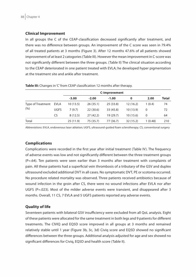
88 Chapter 4
clinical improvementIn all groups the C of the CEAP-classification decreased significantly after treatment, and
there was no difference between groups. An improvement of the C score was seen in 79.4%
of all treated patients at 3 months (Figure 3). After 12 months 47.6% of all patients showed
improvement of at least 2 catogories (Table III). However the mean improvement in C-score was
not significantly different between the three groups. (Table II) The clinical situation according
to the CEAP deteriorated in one patient treated with EVLA; he developed hyper pigmentation
at the treatment site and ankle after treatment.
Table iii: Changes in ‘C’ from CEAP classification 12 months after therapy.
c-improvement
Total-3.00 -2.00 -1.00 0 2.00
Type of Treatment(%)
EVLA 10 (13.5) 26 (35.1) 25 (33.8) 12 (16.2) 1 (0.4) 74
UGFS 7 (9.7) 22 (30.6) 33 (45.8) 10 (13.9) 0 72
CS 8 (12.5) 27 (42.2) 19 (29.7) 10 (15.6) 0 64
Total 25 (11.9) 75 (35.7) 77 (36.7) 32 (15.2) 1 (0.48) 210
Abbreviations: EVLA, endovenous laser ablation; UGFS, ultrasound-guided foam sclerotherapy; CS, conventional surgery
complicationsComplications were recorded in the first year after initial treatment (Table IV). The frequency
of adverse events was low and not significantly different between the three treatment groups
(P=.64). Ten patients were seen earlier than 3 months after treatment with complaints of
pain. All these patients had a superficial vein thrombosis of a tributary of the GSV and duplex
ultrasound excluded additional DVT in all cases. No symptomatic DVT, PE or scotoma occurred.
No procedure related mortality was observed. Three patients received antibiotics because of
wound infection in the groin after CS, there were no wound infections after EVLA nor after
UGFS (P=.023). Most of the milder adverse events were transient, and disappeared after 3
months. Overall, 11 CS, 7 EVLA and 5 UGFS patients reported any adverse events.
Quality of life Seventeen patients with bilateral GSV insufficiency were excluded from all QoL analysis. Eight
of these patients were allocated for the same treatment in both legs and 9 patients for different
treatments. The CIVIQ and EQ5D score improved in all groups at 3 months and remained
relatively stable until 1 year (Figure 3b, 3c, 3d) Civiq score and EQ5D showed no significant
differences between the three groups. Additional analysis adjusted for age and sex showed no
significant differences for Civiq, EQ5D and health score (Table II).

Comparing EVLA, UGFS and CS for great saphenous varicose veins 89
Chapter 4
It is remarkable that the CIVIQ scores improved in 11 patients (14%) of the UGFS group despite
their remaining (segmental) reflux on ultrasound examination and without performing
additional injections.
Table iV: Distribution of the complications for each of the three therapies at 3 and 12 months after
therapy.
eVLAn=78
uGfsn=77
csn=68
p-valuea
3 monthsHyperpigmentation 2 1 0 0.78
Paresthesia 2 1 4 0.30
Superficial vein thrombosis 3 3 4 0.85
Wound infectionb 0 0 3 0.03
Deep vein thrombosis 0 0 0 1.00
Pulmonary emboli 0 0 0 1.00
Death due to therapy 0 0 0 1.00
Total number of patients with complications 7 5 11 0.64
12 monthsHyperpigmentation 1 1 0 1.00
Paresthesia 0 1 1 1.00
a Fisher exactb requiring systemic antibiotics
Additional InterventionsPhlebectomies were permitted during initial treatment with the intention of removing all
varicosities in the same procedure, but because of practical issues this was not possible in all
cases. In 15 patients (19.2%) in the EVLA group and 18 (25.7%) in the CS group phlebectomy
was performed during initial treatment. In 12 (15.3%), 15 (19.5%) and 11 (15.7%) of the EVLA,
UGFS and CS group respectively, phlebectomies were performed after three months. In the
UGFS group 6 patients (4.6%) with lasting reflux and complaints received a second injection
with foam.
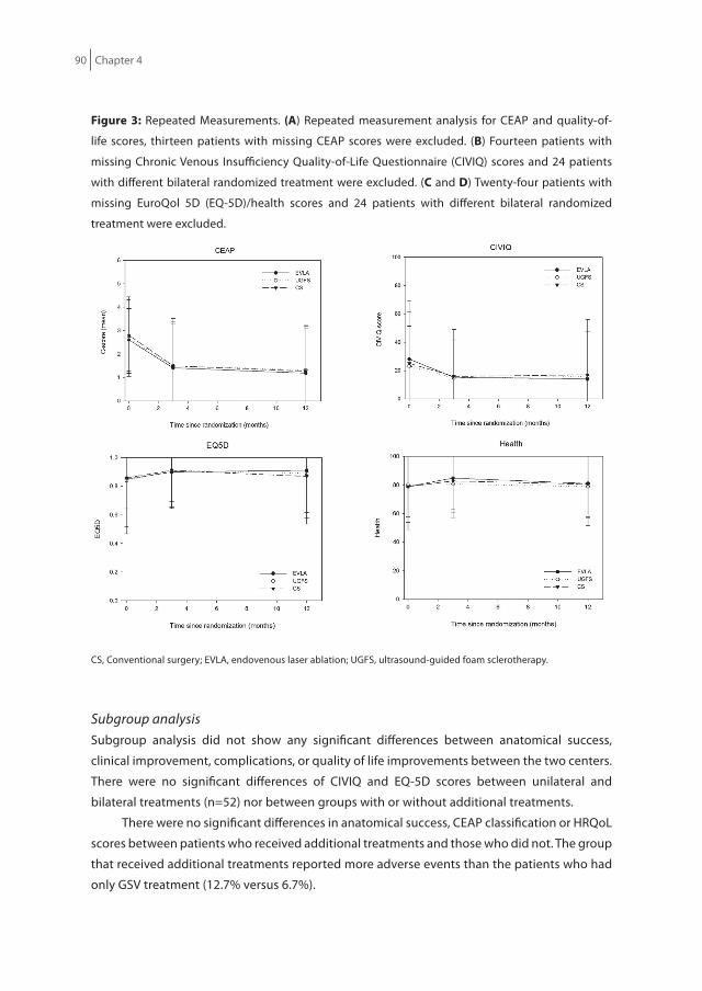
90 Chapter 4
figure 3: Repeated Measurements. (A) Repeated measurement analysis for CEAP and quality-of-
life scores, thirteen patients with missing CEAP scores were excluded. (B) Fourteen patients with
missing Chronic Venous Insufficiency Quality-of-Life Questionnaire (CIVIQ) scores and 24 patients
with different bilateral randomized treatment were excluded. (c and d) Twenty-four patients with
missing EuroQol 5D (EQ-5D)/health scores and 24 patients with different bilateral randomized
treatment were excluded.
CS, Conventional surgery; EVLA, endovenous laser ablation; UGFS, ultrasound-guided foam sclerotherapy.
Subgroup analysisSubgroup analysis did not show any significant differences between anatomical success,
clinical improvement, complications, or quality of life improvements between the two centers.
There were no significant differences of CIVIQ and EQ-5D scores between unilateral and
bilateral treatments (n=52) nor between groups with or without additional treatments.
There were no significant differences in anatomical success, CEAP classification or HRQoL
scores between patients who received additional treatments and those who did not. The group
that received additional treatments reported more adverse events than the patients who had
only GSV treatment (12.7% versus 6.7%).

Comparing EVLA, UGFS and CS for great saphenous varicose veins 91
Chapter 4
discussionIn the last decade, EVLA and RFA were introduced and UGFS has been optimized. These minimal
invasive interventions are increasingly used as alternative to CS for treating saphenous veins.4,5
The MAGNA trial shows that EVLA and CS are comparably effective (almost 90%) and that
both are significantly more effective than UGFS (72.7%) using ultrasound based anatomical
outcomes. Complications are rare and quality of life (CIVIQ and EQ5D) improved significantly
after each treatment. The results of this RCT are strikingly similar to other clinical studies, meta-
analysis and two recent RCTs that included UGFS increasing the validity of the findings .5,12,13,23
One year after treatment, remaining (segmental) flow with reflux was observed in more than
a quarter of the patients treated with UGFS, which is in line with other observational studies
and RCTs.12,13,24,25 Neovascularization occurs in 10% of the patients in the CS group one year
post operatively, which corresponds with results of a previous study focusing on the effect of
closing the cribriform fascia to contain postoperative neovascularization at the SFJ.18 In contrast
to the significant improvement of the CIVIQ, the generic scores of the EQ-5D improved slightly,
suggesting that either saphenous varicose veins have a mild impact on the global HRQoL, this
instrument lacks content validity to assess impact of venous disease in C2-C3 patients and/or
the sensitivity to change the EQ-5D is limited in patients with venous disease. However, other
clinical studies did observe improvement in the SF-36 and the EQ-5D, which may be explained
by study population (82% of patients were C2-3 venous disease), timing of assessment after
therapy (3 months and one year), or limited sample size of this study.12,26 Applying the Norman’s
rule of thumb on the unadjusted data, none of the improvements in the CIVIQ and EQ-5D
appeared to be clinically relevant.27
Despite randomization EVLA treated patients were significantly younger than those in
the other two treatment arms. Adjusting for age and sex in a logistic model, there were no
differences in CIVIQ scores or in CEAP classification between the three groups.
In accordance with other RCTs comparing CS to new minimally invasive treatment
methods, we had difficulty enrolling the required number of patients in the RCT because of the
reluctance among patients to undergo CS. Therefore, an additional center was added during
the study period, but no differences in outcomes were noted between the two centers. Of the
seventeen included patients that withdrew from the trial, ten were assigned to CS suggesting
that informed patients preferred minimally invasive treatments.
The primary outcome of this study was total occlusion and/or absence of the treated GSV
according to ultrasound. This outcome has the advantage to be objective and reproducible
and is possible a proxy of symptom reduction and future clinical relevant recurrence. These
latter assumptions will be tested when the 5-year results become available. Also, defining the
outcome as total occlusion may have been too strict, because ‘remodeling’ of the insufficient
vein, as is often seen after UGFS may be associated with alleviation of symptoms while persisting
flow with or without reflux is present in the treated vein. Eleven patients with residual reflux
92 Chapter 4
after UGFS did decline an additional UGFS session because absence of venous symptoms. This
observation challenges the strict definition of primary outcome criteria such as ‘total occlusion’
and ‘absence of reflux’ used as the gold standard for evaluation after GSV treatment in RCTs.
Moreover, it emphasizes the conviction that we treat patients and not ultrasound findings.28
Recently, the Union Internationale de Phlébologie has proposed a new classification
to describe the fate of the junction and the treated trunk after endovenous ablation.29
This classification allows to describe postoperative findings more detailed, distinguishing
between obliteration, partial or complete patency of the treated vein, and segmental
obliteration or patency of the treated trunk, with or without reflux. Apart from these duplex
ultrasound findings, clinical outcome parameters as well as other outcome measures such
as patient-reported outcomes (e.g., HRQoL, symptoms, satisfaction and preference) should
be considered.13,29,30 In contrast to HRQoL, symptom reduction was not maximally assessed
in this study, which in retrospect is a missed opportunity.13 At time of the study design, we
overlooked the problem of including bilateral GSV. For the HRQoL outcomes, patients with
two different study treatments were excluded from the HRQoL analysis, because patients may
not differentiate HRQoL impairment between both legs. Patients with the same treatment for
both legs were included for HRQoL analysis by taking the correlation between HRQoL scores
of the same patient into account. All patients were included in the efficacy analysis, which was
adjustment for bilateral GSV treatments.
Because of the scarring after CS and in a lesser extent EVLA having a blinded outcome
assessor was not feasible. The ultrasound investigations were done by physicians not
necessarily part of the research team, hopefully limiting the impact of this limitation.
In this study, phlebectomies were allowed during initial treatment of the study GSV in
patients allocated to EVLA or CS or after 3 months for all included patients. Phlebectomies
were proposed to all study patients with residual superficial varicose veins as additional
treatment after three months irrespective of the study treatment. The MAGNA trial includes the
three treatments most frequently used in The Netherlands in 2007 explaining the exclusion of
segmental RFA. In this study, CS was performed using spinal or general anesthesia, which is still
common practice in the Netherlands. Hopefully the long-term results of the minimal invasive
interventions will stimulate surgeons to switch to minimally invasive procedures and/or use
tumescent anesthesia when CS is indicated.28 Using tumescent instead of general anesthesia
will improve patient satisfaction and will lead to shorter down time after intervention.31 The
five year follow-up of the MAGNA trial and other similar ongoing RCT’s will further clarify
whether the observed results of EVLA, UGFS and CS persist over time.
Comparing EVLA, UGFS and CS for great saphenous varicose veins 93
Chapter 4
conclusionThe one-year results of the MAGNA trial show that the short term efficacy, defined as anatomical
success according to duplex ultrasound, is equally high for EVLA and CS and lower for UGFS.
The treatments are equally safe, no severe adverse events were seen. Wound infections and
neovascularization were more common after CS. All therapies resulted in significant clinical
and HRQoL improvement. Long-term efficacy of these three intervention methods needs to be
established and will be available in four years.
AcknowledgementWe like to thank J. Buth, M. de Jong (research nurse), L. Hollestein (statistics), O. van Beuningen
(vascular technologist), all participating vascular surgeons and dermatologists from Catharina
Hospital Eindhoven and Erasmus MC Rotterdam for their assistance.
94 Chapter 4
references
1. Callam MJ. Epidemiology of varicose veins. Br J Surg. 1994; 81: 167-173.2. Nicolaides AN, Allegra C, Bergan J, Bradbury A, Cairols M, Carpentier P, et al. Management of chronic
venous disorders of the lower limbs guidelines according to scientific evidence. Int Angiol. 2008;27:1-59.
3. Pichot O, De Maeseneer M.Treatment of Varicose Veins: Does Each Technique Have a Formal Indication? Perspect Vasc Surg Endovasc Ther. 2012
4. Biemans AA, van den Bos RR, Nijsten T. Endovenous therapies of varicose veins: indications, procedures, efficacy and safety. G Ital Derm Ven. 2010;145:161-74
5. Van den Bos RR, Kockaert MA, Neumann HAM, Nijsten T. Endovenous therapies of lower extremity varicosities: a meta-analysis. J Vasc Surg. 2009;49:230-9.
6. De Maeseneer MG. Neovascularization: an adverse response to proper groin dissection. Bergan JJ (Ed.), The vein book, Elsevier Academic Press, Burlington, San Diego, Londen (2007), pp.239-247.
7. Rasmussen LH, Bjoern L, Lawaetz M, Lawaetz B, Blemings A, Eklof B. Randomised clinical trial comparing endovenous laser ablation with stripping of the great saphenous vein: clinical outcome and recurrence after 2 years. Eur J Vasc Endovasc Surg. 2010; 39: 630–635.
8. Carradice D, Mekako AI, Mazari FAK, Samuel N, Hatfield J and Chetter IC. Clinical and technical outcomes from a randomized clinical trial of endovenous laser ablation compared with conventional surgery for great saphenous varicose veins. British Journal of Surgery 2011; 98: 1117–1123
9. Darwood RJ, Theivacumar N, Dellagrammaticas D, Mavor AI, Gough MJ. Randomized clinical trial comparing endovenous laser ablation of the great saphenous vein with high ligation and stripping in patients with varicose veins: short term results. J Vasc Surg. 2007;46:308-315.
10. Kalteis M, Berger I Messie-Werndl S, Pistrick R, Schimetta EW, Polz W et al. High ligation combined with stripping and endovenous laser ablation of the great saphenous vein: early results of a randomized controlled study. J Vasc Surg. 2008;47:822-829.
11. Carradice D, Mekako AI, Mazari FA, Samuel N, Hatfield J, Chetter IC. Randomized clinical trial of endovenous laser ablation compared with conventional surgery for great saphenous varicose veins. Br J Surg. 2011 Apr;98(4):501-10.
12. Rasmussen LH, Lawaetz M, Bjoern L, Vennits B, Blemings A, Eklof B. Randomized clinical trial comparing endovenous laser ablation, radiofrequency ablation, foam sclerotherapy and surgical stripping for great saphenous varicose veins. Br J Surg. 2011;98:1079-1087.
13. Shadid N, Ceulen R, Nelemans P, Dirksen C, Veraart J, Schurink GW, van Neer P, vd Kley J, de Haan E, Sommer A. Ultrasound-guided foam sclerotherapy versus surgery for the incompetent great saphenous vein: a multicentre randomised controlled non-inferiority trial. Br Journal Surg. Accepted for publication.
14. Nijsten T, van den Bos RR, Goldman MP, Kockaert MA, Proebstle TM, Rabe E, Sadick NS, Weiss RA, Neumann MH. Minimally invasive techniques in the treatment of saphenous varicose veins. J Am Acad Dermatol. 2009 Jan;60(1):110-9.
15. Proebstle TM, Moehler T, Herdemann S. Reduced recanalization rates treatment with increased energy dosing: definition of a tresthold for the endovenous fluence equivalent. J Vasc Surg. 2006;44:834-839.
16. Tessari L, Cavezzi A, Frullini A. Preliminary experience with a new sclerosing foam in the treatment of varicose veins. Dermatol Surg. 2001;27(1):58-60.
17. Breu FX, Guggenbichler, Wollmann JC. 2nd European Consensus Meeting on Foam Sclerotherapy, 28 – 30 April 2006, Tegernsee, Germany. Vasa 2008;37 Supplement 71:1-32.

Comparing EVLA, UGFS and CS for great saphenous varicose veins 95
Chapter 4
18. De Maeseneer MG, Philipsen TE, Vandenbroeck CP, Lauwers PR, Hendriks JM, De Hert SG, Van Schil PE. Closure of the cribriform fascia: an efficient anatomical barrier against postoperative neovascularisation at the saphenofemoral junction? A prospective study. Eur J Vasc Endovasc Surg. 2007 Sep;34(3):361-6.
19. Eklöf B, Rutherford RB, Bergan JJ, Carpentier PH, Gloviczki P, Kistner RL, Meissner MF, Moneta GL, Myers K, Padberg FT, Perrin M, Ruckley CV, Coleridge Smith P, Wakefield TW Revision of the CEAP classification for chronic venous disorders: Consensus statement.,n for the American Venous Forum International Ad Hoc Committee for Revision of the CEAP Classification, Helsingborg, Sweden J Vasc Surg. 2004;40:1248-52.
20. Biemans AA, van der Velden SK, Bruijninckx CA, Buth J, Nijsten T. Validation of the chronic venous insufficiency quality of life questionnaire in Dutch patients treated for varicose veins. Eur J Vasc Endovasc Surg. 2011 Aug;42(2):246-53.
21. EQ5D The EuroQol Group. EuroQol – a new facility for the measurement of health-related quality of life. Health Policy 1990; 16: 199-208.
22. Nesbitt CI, Stansby G. Regarding ‘Prospective randomized trail comparing endovenous laser ablation and surgery for treatent of primary great saphenous varicose veins with a 2-years follow-up.’ J Vasc Surg. 2011
23. Nesbitt C, Eifell RK, Coyne P, Badri H, Bhattacharya V, Stansby G. Endovenous ablation (radiofrequency and laser) and foam sclerotherapy versus conventional surgery for great saphenous vein varices. Cochrane Database Syst Rev. 2011 Oct 5;(10):CD005624.
24. Chapman-Smith P, Browne A. Prospective five-year study of ultrasound-guided foam sclerotherapy in the treatment of great saphenous vein reflux. Phlebology 2009 Aug;24(4):183-8.
25. Thomasset SC, Butt Z, Liptrot S, Fairbrother BJ, Makhdoomi KR. Ultrasound guided foam sclerotherapy: factors associated with outcomes and complications. Eur J Vasc Endovasc Surg. 2010 Sep;40(3):389-92.
26. Michaels JA, Brazier JE, Campbell WB, MacIntyre JB, Palfreyman SJ, Ratcliffe J. Randomized clinical trial comparing surgery with conservative treatment for uncomplicated varicose veins. Br J Surg. 2006 Feb;93(2):175-81.
27. Norman GR., Sloan JA., Wyrwich KW. Interpretation of changes in Health-related Quality of Life. The Remarkable universality of Half a Standard Deviation. Medical Care 2003 ; 41(5), 582-592.
28. De Maeseneer MG. The endovenous revolution. Br J Surg. 2011; 98: 1037-829. De Maeseneer MG, Pichot O, Cavezzi A, Earnshaw J, van Rij A, Lurie F, Smith PC. Duplex ultrasound
investigation of the veins of the lower limbs after treatment for varicose veins – UIP consensus document. Eur J Vasc Endovasc Surg. 2011; 42: 89-102
30. Barrett JM, Allen B, Ockelford A, Goldman MP. Microfoam ultrasound-guided sclerotherapy of varicose veins in 100 legs. Dermatol Surg. 2004 Jan;30(1):6-12.
31. Pronk P, Gauw SA, Mooij MC, Gaastra MT, Lawson JA, van Goethem AR, van Vlijmen-van Keulen CJ. Randomised controlled trial comparing sapheno-femoral ligation and stripping of the great saphenous vein with endovenous laser ablation (980 nm) using local tumescent anaesthesia: one year results. Eur J Vasc Endovasc Surg. 2010 Nov;40(5):649-56.

Chapter 5
The Effect of Single Phlebectomies of Tributary on Great Saphenous Vein Reflux
Anke A. BiemansRenate R. van den BosLoes H. HollesteinBirgit Maessen-VischYvonne VergouwMarianne G. de MaeseneerMartino NeumannTamar Nijsten
Journal of Vascular Surgery 2013 (submitted)
Chapter 5
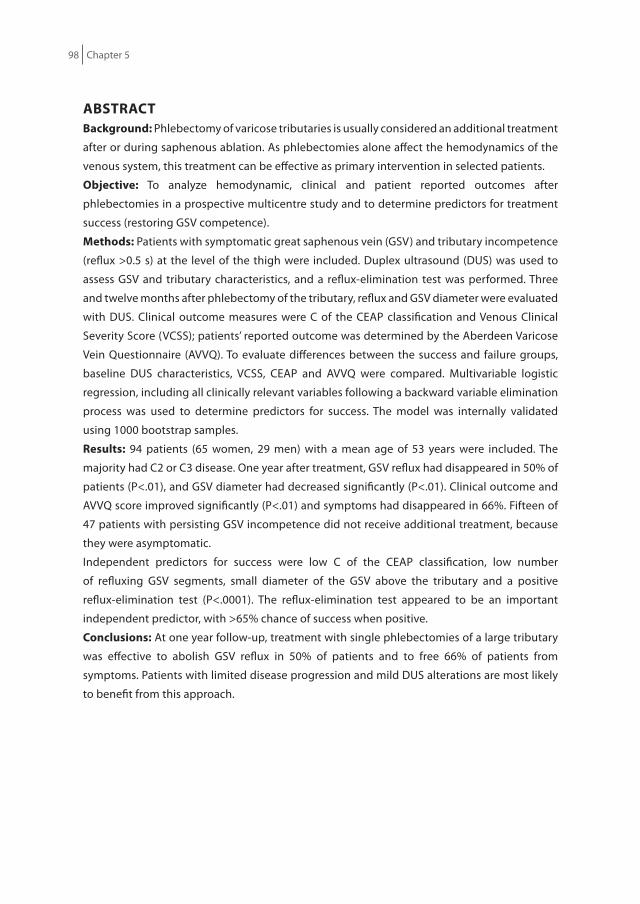
98 Chapter 5
ABsTrAcTBackground: Phlebectomy of varicose tributaries is usually considered an additional treatment
after or during saphenous ablation. As phlebectomies alone affect the hemodynamics of the
venous system, this treatment can be effective as primary intervention in selected patients.
objective: To analyze hemodynamic, clinical and patient reported outcomes after
phlebectomies in a prospective multicentre study and to determine predictors for treatment
success (restoring GSV competence).
Methods: Patients with symptomatic great saphenous vein (GSV) and tributary incompetence
(reflux >0.5 s) at the level of the thigh were included. Duplex ultrasound (DUS) was used to
assess GSV and tributary characteristics, and a reflux-elimination test was performed. Three
and twelve months after phlebectomy of the tributary, reflux and GSV diameter were evaluated
with DUS. Clinical outcome measures were C of the CEAP classification and Venous Clinical
Severity Score (VCSS); patients’ reported outcome was determined by the Aberdeen Varicose
Vein Questionnaire (AVVQ). To evaluate differences between the success and failure groups,
baseline DUS characteristics, VCSS, CEAP and AVVQ were compared. Multivariable logistic
regression, including all clinically relevant variables following a backward variable elimination
process was used to determine predictors for success. The model was internally validated
using 1000 bootstrap samples.
results: 94 patients (65 women, 29 men) with a mean age of 53 years were included. The
majority had C2 or C3 disease. One year after treatment, GSV reflux had disappeared in 50% of
patients (P<.01), and GSV diameter had decreased significantly (P<.01). Clinical outcome and
AVVQ score improved significantly (P<.01) and symptoms had disappeared in 66%. Fifteen of
47 patients with persisting GSV incompetence did not receive additional treatment, because
they were asymptomatic.
Independent predictors for success were low C of the CEAP classification, low number
of refluxing GSV segments, small diameter of the GSV above the tributary and a positive
reflux-elimination test (P<.0001). The reflux-elimination test appeared to be an important
independent predictor, with >65% chance of success when positive.
conclusions: At one year follow-up, treatment with single phlebectomies of a large tributary
was effective to abolish GSV reflux in 50% of patients and to free 66% of patients from
symptoms. Patients with limited disease progression and mild DUS alterations are most likely
to benefit from this approach.

Effect of phlebectomies of tributary on great saphenous vein reflux 99
Chapter 5
inTroducTionTributaries represent the major part of the superficial venous system of the lower limbs.1
From the hemodynamic point of view, they play an important role in patients with varicose
veins. Indeed, in the majority of limbs with varicose veins, blood is refluxing as well along
tributaries as along a segment of the saphenous veins. Tributaries run parallel or beside the
track of an associated saphenous vein but lie outside the saphenous compartment.2,3 A study
on anatomical patterns of the incompetent great saphenous vein (GSV) showed that in 30%
of limbs with GSV incompetence above the knee a large incompetent tributary pierces the
saphenous fascia at thigh level.4 Since tributaries are located in the subcutaneous tissue, they
are suitable for removal by phlebectomies. Currently there are two main strategies for treating
patients having both GSV incompetence and refluxing tributaries. The first strategy consists of
GSV treatment (surgical, thermal or chemical) and phlebectomies in a single procedure,5 while
the second strategy consists of GSV treatment only, awaiting tributaries to diminish.6-12 Both are
based on the theory that varicose veins have a descending origin. A different approach would
be to treat the tributaries first, thereby sparing the GSV in accordance with the ambulatory
selective varices ablation under local anaesthesia (ASVAL) method.13 If GSV incompetence
and symptoms persists, the GSV may be treated in a second stage. It has been shown that
phlebectomies improve the hemodynamics of the venous system in the leg and also have a
beneficial effect on the segments that have been left untreated.13-18 The hemodynamic effects
of removing refluxing tributaries support the theory of an ascending origin of varicose veins.
In a retrospective study with 4 years follow-up, 66% of patients with saphenous and tributary
varicose veins were successfully treated by phlebectomy only.13
The objective of the present prospective multicenter study was to analyze short-term
outcomes after single phlebectomies of a large incompetent tributary joining the GSV at thigh
level in patients who also have GSV incompetence. We aim to identify those patients that could
benefit from this approach by determining predictors with a prediction model and to verify
the value of the reflux-elimination test.
MeThodsParticipantsThe present trial was designed as a prospective multicenter study at the Departments of
Dermatology of the Erasmus MC Rotterdam, Catharina Hospital Eindhoven and Rijnstate
Hospital Arnhem. Recruitment of 100 patients started in April 2010; the last patient was
included in March 2012.
Consecutive adult patients with symptomatic primary GSV incompetence and a clinically
visible incompetent tributary of the GSV at the medial thigh (with or without extension below
the knee) were eligible to participate. Incompetence of the GSV and the tributary was defined

100 Chapter 5
as reflux >0.5 s at color DUS (Philips, HDI 4500, 10MHz transducer, Andover MA, USA). The
tributary had to be clinically visible and/or palpable, making it suitable for phlebectomy.
Exclusion criteria were previous treatment of ipsilateral GSV or tributaries, incompetence
of the accessory anterior saphenous vein and tributaries in the thigh, segmental absence
of the GSV above the knee, deep venous obstruction, agenesis of the deep venous system,
vascular malformations, use of anticoagulant treatment, pregnancy, immobility, significant
arterial disease (ankle : brachial pressure index below 0.6) and inability to provide written
informed consent to trial participation. The ethical medical committee of the Catharina
Hospital Eindhoven approved the study (date 02 March 2010).
diagnostic methodsPatients were assessed with DUS examination in standing position. The saphenofemoral
junction (SFJ), the GSV in the thigh above and below the junction of the tributary, the tributary
itself and the GSV below the knee were studied, as well as the small saphenous vein (SSV),
the common femoral, femoral and popliteal veins. Valvular function was evaluated in the
femoral, popliteal and superficial veins of the thigh by manual calf compression; the Valsalva
manoeuvre was performed to assess reflux at the terminal valve of the SFJ. Reflux was defined
as retrograde flow >0.5 s. Perforating veins were considered incompetent if >3.5 mm with
bidirectional flow.2,3,19
For this study, some additional DUS parameters were recorded (Figure 1). Four anatomic
GSV segments were distinguished: segment 1 from the SFJ down to the site where the tributary
joined the GSV; segment 2 the GSV in the thigh from below the junction of the tributary to the
knee; segment 3 was the GSV from knee to mid-calf; segment 4 from mid-calf to ankle. In
segment 1 en 2 diameter measurement was performed respectively 2 cm above and below the
junction of the refluxing tributary. The total refluxing length of the GSV was recorded. Diameter
of the tributary was measured 2 cm below its junction with the GSV. The reflux-elimination test
was performed by digital compression of the refluxing tributary in the thigh to test whether
this could modify GSV reflux. A slightly modified version of a previously described reflux-
elimination test was used, focusing mainly on the effect of finger compression of the tributary
on the GSV segment above the tributary’s junction.15 The tributary was compressed at least 5
cm from the junction of the tributary with the GSV, to prevent simultaneous compression of
the GSV itself. The reflux-elimination test was considered positive when GSV reflux is eliminated
after compression of the tributary.
TreatmentJust before phlebectomy, patients were examined with DUS, to verify whether they were
validly included. The tributary was marked on the skin with the patient in standing position.
Phlebectomy was performed, using local anesthesia with lidocaine 1%. Small phlebectomy

Effect of phlebectomies of tributary on great saphenous vein reflux 101
Chapter 5
incisions (max. 3 mm) were made and varicose veins were grasped with Oesch phlebectomy
hooks, exteriorized, divided and removed.20
Medical elastic compression stockings were applied over the wound dressings for the
first 48 hours. Patients were instructed to remove the dressings and continue wearing medical
elastic compression stockings during daytime for one week. All patients were observed in
the clinic for at least half an hour after treatment. Patients were encouraged to mobilize and
resume their usual activities as soon as possible.
figure 1: Baseline duplex ultrasound characteristics of GSV and Tributary.
Abbreviations: SFJ, sapheno-femoral junction; TV, terminal valve; PTV, preterminal valve; VF, vena femoralis; GSV, great saphenous vein; S, segment with reflux; T, Tributary.

102 Chapter 5
follow-upPatients were invited for follow-up 3 months after the procedure. C class and VCSS were
recorded, patients were asked to complete the AVVQ questionnaire and underwent DUS as
described above. If patients presented with persisting symptomatic GSV incompetence,
additional treatment was proposed, either by endovenous thermal ablation or ultrasound
guided foam sclerotherapy. Patients without GSV incompetence and/or without symptoms
were scheduled for a follow-up visit 12 months after the initial procedure. The evaluation at 12
months was identical to the one at 3 months. Patients who had undergone additional treatment
were excluded for follow-up, but included in the analysis of total treatment effectiveness; they
were assigned as having reflux.
outcome measurementsHemodynamic outcome The primary outcome was absence of reflux in the entire GSV, measured by DUS evaluation
after one year. In addition, segmental disappearance of reflux and GSV diameter reduction
were studied.
Clinical and patient reported outcomeSecondary outcome measures were C of the CEAP classification,21 VCSS (range 1-10),22 and
AVVQ score (range 1-100).23
independent predictors for disappearance of reflux To test for independent predictors of success (defined as no reflux at 12 months) a logistic
regression model was used including DUS and clinical measurements.
reflux-elimination testA single prediction model analysis was done to evaluate the reflux-elimination test. Because
the reflux-elimination test is quick and easy to perform, it would be of great value in clinical
practice if it proves to be an important predictor.
Phlebectomy reflux-elimination success Test (PresT) Prediction model All variables included in the multivariable logistic regression model were eligible for inclusion
in the prediction model. Final selection was based on statistical significance after backward
deletion. The prediction model is presented as a score chart, from which a total score can be
calculated that correlates with a probability of restoring GSV competence.

Effect of phlebectomies of tributary on great saphenous vein reflux 103
Chapter 5
statistical analysis All continuous variables were normally distributed, using the One-Sample Kolmogorov-
Smirnov Test. Therefore, data were presented as means with standard deviation (SD); analysis
with one sample t-test and paired sample t-test were done to compare groups.
Categorical data were analyzed by means of X2 test or, if appropriate, Fisher’s exact
test. This test was also used to evaluate the relation between reflux at the terminal valve and
diameter of the GSV and their relation with success. Responsiveness of AVVQ after treatment
was tested with paired sample t-test in normally distributed data. The studied patients were
analyzed ‘per protocol’. To test for independent predictors of restoring GSV competence
(defined as no reflux at 12 months) a logistic regression model was used. To avoid overfitting
of the prediction model, we calculated the maximum degrees of freedom that could be spend
during the regression modelling process with our sample size. This was calculated as m/10,
where m is the limited sample size, which is the smallest group among the outcome. Both
success and failure consisted of 47 patients, suggesting that 5 degrees of feedom (df ) could
be spend. Predictors were initially selected based on literature and clinical expertise (reflux
SFJ at terminal valve, number of refluxing segments, C of CEAP, reflux elimination test and
GSV diameter above the tributary). Statistically non-significant variables were subsequently
removed from the model by backward variable selection based on AIC, corresponding to
a P-value of .157 for variables with 1 df. The regression coefficients in the final model were
multiplied with a shrinkage factor, which was obtained by bootstrapping (1000 samples).
The backward variable selection was taken into account in the bootstrapping. Shrinkage is
applied to obtain accurate predictions for new patients; without shrinkage predictions are in
general too extreme, resulting in predictions being too high or too low. The final model with
the shrunken regression coefficients was presented as a score chart to make the prediction
model easy applicable in clinical practice. Calibration (i.e. agreement between predicted and
observed risks) was studied. Discrimination (i.e. distinction between successful treatment and
treatment failure) was studied using a receiver operating curve (ROC) curve.
A single prediction model analysis was done to evaluate the performance of the reflux-
elimination test without any other predictors. We accomplished an internal validation (also
obtained with 1000 bootstrap samples).
Statistical analysis was performed using SPSS® version 20.0 (SPSS, Chicago, Illinois, USA)
and R 2.15.2 (http://www.R-project.org).
resuLTsParticipantsOne hundred eligible patients, with GSV incompetence and an associated incompetent
tributary joining the GSV at thigh level, were included. Six patients were lost to follow-up
(5 patients did not attend the follow-up visit, 1 patient was treated with EVLA of the GSV

104 Chapter 5
before phlebectomy), and were therefore excluded from the analysis. In total 94 patients
were analyzed, 65 women and 29 men. 55.3% patients were classified as C2, 35.1% C3 and
9.6% C4.The mean GSV diameter was 0.55 cm (SD 0.15) above the tributary, and 0.36 cm (SD
0.16) below the tributary. The mean diameter of the tributary was 0.51 cm (SD 0.15) with its
junction located at a mean distance of 19.5 cm (SD 6.64) below the SFJ. Half of the patients had
terminal valve reflux at the SFJ. In approximately half of the patients reflux was present only in
1 segment. The total refluxing GSV length was <10 cm in 14.9% of the patients, and >30 cm in
29.8% of the patients. Mean VCSS was 4 (SD 1.49, range 2-9) and mean AVVQ score was 10.9
(SD 7.75). (Table I) We found a significant relation between reflux in the terminal valve and
diameter of the GSV. In patients without terminal valve reflux, GSV diameter was more often
<5 mm and in patients with terminal valve reflux, the diameter of the GSV was often >5 mm.
(P<.001). Success was related to a smaller GSV diameter (P=.002) and no reflux at the terminal
valve. (P<.001)
hemodynamic outcomesIn 47 patients (50%) reflux disappeared completely after 12 months. In 15 patients (16.0%)
phlebectomy resulted in complete relief of complaints despite persisting GSV reflux after one
year of follow-up. Consequently, these patients did not undergo additional treatment. The
remaining 32 patients had persisting symptoms and underwent additional GSV ablation. In 57
patients (60.6%) the number of refluxing GSV segments had diminished. However, in 4 patients
the number of refluxing segments increased after phlebectomy. The mean diameter of the GSV
above the tributary decreased significantly after 3 and 12 months from 0.55 cm to 0.36 cm and
0.39 cm (P<.001) respectively.
clinical and patient reported outcomeThe C of the CEAP-classification decreased significantly after treatment (P<.001); a lower C class
was found in 73% of all treated patients. No patient deteriorated. The VCSS and AVVQ scores
improved in all patients (independent of hemodynamic effect) after treatment. We found more
improvement of VCSS and AVVQ in the patients with hemodynamic success (i.e., no reflux) at
3 months, which remained relatively stable after 1 year (P<.001). The group with persistent
reflux showed significantly higher baseline scores and less improvement of HRQoL scores after
treatment (Table I). Remarkably, the AVVQ scores improved in 27 patients with persisting GSV
reflux. Because of lack of symptoms 15 patients did not undergo additional treatment.
independent predictors for disappearance of reflux Reflux was significantly more often abolished when the following parameters at baseline
were present: C2 (P<.001); short (<10 cm) refluxing segment (P<.001); reflux in only 1 GSV
segment (P<.001); smaller diameters of GSV and tributary (P<.001 and P<.009); a positive

Effect of phlebectomies of tributary on great saphenous vein reflux 105
Chapter 5
reflux-elimination test (P<.001), and low scores of VCSS (P<.001) and AVVQ (P<.001). Reflux of
the terminal valve was seen more often in patients with a diameter >5 mm (P<.001). Success
was more often the case in patients with a diameter <5 mm (P=.002).
figure 2: Flowchart.
100 patients with GSV insu�ciency and re�uxing tributary at thigh level
Phlebectomy n=94
3 months: Resolved re�uxn=48
3 months: Persistent re�uxn=46
No symptomsn=22
Symptomsn=24
Total: Additional treatment
n=32 EVLA (n=10)UGFS (n=22)
12 months: Resolved re�ux n=47
Total: No re�ux
n=47
12 months: Persistent re�ux
n=23
Total: Persistent re�ux, No symptoms
n=15
Lost to follow-upNo interventionn=6
Abbreviations: n, number; GSV, great saphenous vein; EVLA, endovenous laser ablation; UGFS, ultrasound guided foam sclerotherapy.

106 Chapter 5
Table i: Demographic, clinical and DUS characteristics at baseline and effectiveness after treatment.
Variable Total patients
resolved reflux at month 12
n=47
Persistent reflux at month 12
n=47
P-value
Patient, n=94
Age, years (range) 53 (21-82) 51 (26-79) 55 (21-82) .2#
Women, n (%) 65 (69.1) 34 (72.3) 31 (66.0) .7*
C2, n (%)C3 or C4, n (%)
52 (55.3)42 (44.7)
36 (76.6)11 (23.4)
16 (34.0)31 (66.0) <.001*
AVVQ (SD) 10.95 (7.75) 7.48 (5.44) 14.42 (8.19) <.001#
VCSS (SD) 4.00 (1.50) 3.38 (1.26) 4.62 (1.47) <.001#
duplex characteristics
Reflux SFJ terminal valve, n(%)No reflux terminal valve, n(%)
49 (52.1)45 (47.9)
20 (42.6)27 (60)
29 (61.7)18 (40)
.06*
Refluxing segmentsOne refluxing segment (%)More than one segment (%)
45 (47.9)49 (52.1)
35 (74.5)59 (25.5)
10 (21.3)84 (78.7)
<.001*
Length of refluxing GSV, n (%)<10 cm>10 cm
14 (14.9)80 (85.1)
12 (25.5)35 (74.5)
2 (4.3)45 (97.7)
<.001*
Mean diameter GSV above tributary, mm (SD)
5.5 (1.5) 4.9 (1.2) 6.2 (1.4) <.001#
Mean diameter GSV below tributary, mm (SD)
3.6 (1.6) 3.2 (13) 4.1 (19) .005#
Mean diameter of tributary, Mm (SD) 5.2 (1.5) 4.8 (1.4) 5.6 (1.4) .009#
Location of tributary: distance from SFJ, cm (SD)
19.5 (6.6) 18.7 (7.4) 20.2 (5.8) .3#
Positive reflux-elimination test, n (SD) 61 (64.9) 42 (89.4) 19 (40.4) <.001*
Abbreviations: N, number; SD, standard deviation; %, percentage; SFJ, saphenofemoral junction; cm, centimetre; GSV, great saphenous vein; mm, millimetre; VCSS, Venous Clinical Severity Score; AVVQ, Aberdeen Varicose Veins Questionnaire*analyzed with chi-square# analyzed with independent sample T-test
Univariable logistic regression analyses were performed for all variables. After backward
deletion of the selected variables, reflux SFJ at terminal valve was removed from the model
because of statistical non-significance. In multivariable logistic regression analysis (Table
II), C classification and reflux in 1 segment were significantly associated with a successful
hemodynamic outcome, independent of all other variables included in the model.
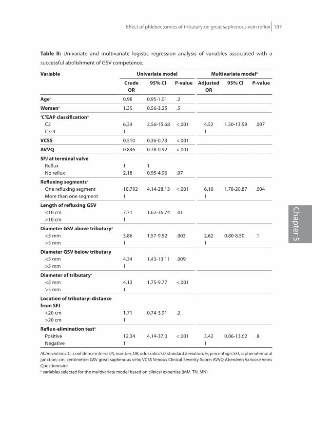
Effect of phlebectomies of tributary on great saphenous vein reflux 107
Chapter 5
Table ii: Univariate and multivariate logistic regression analysis of variables associated with a
successful abolishment of GSV competence.
Variable univariate model Multivariate modela
crudeor
95% ci P-value Adjustedor
95% ci P-value
Agea 0.98 0.95-1.01 .2
Womena 1.35 0.56-3.25 .5
‘c’eAP classificationa
C2C3-4
6.341
2.56-15.68 <.001 4.521
1.50-13.58 .007
Vcss 0.510 0.36-0.73 <.001
AVVQ 0.846 0.78-0.92 <.001
sfJ at terminal valveRefluxNo reflux
12.18
10.95-4.96 .07
refluxing segmentsa
One refluxing segmentMore than one segment
10.7921
4.14-28.13 <.001 6.101
1.78-20.87 .004
Length of refluxing GsV<10 cm>10 cm
7.711
1.62-36.74 .01
diameter GsV above tributarya
<5 mm>5 mm
3.861
1.57-9.52 .003 2.621
0.80-8.50 .1
diameter GsV below tributary<5 mm>5 mm
4.341
1.43-13.11 .009
diameter of tributarya
<5 mm>5 mm
4.131
1.75-9.77 <.001
Location of tributary: distance from sfJ
<20 cm>20 cm
1.711
0.74-3.91 .2
reflux-elimination testa
PositiveNegative
12.341
4.14-37.0 <.001 3.421
0.86-13.62 .8
Abbreviations: CI, confidence interval; N, number; OR, odds ratio; SD, standard deviation; %, percentage; SFJ, saphenofemoral junction; cm, centimetre; GSV great saphenous vein; VCSS Venous Clinical Severity Score; AVVQ Aberdeen Varicose Veins Questionnairea variables selected for the multivariate model based on clinical expertise (MM, TN, MN)

108 Chapter 5
reflux-elimination testThe result of the reflux-elimination test was significantly different between the group without
and the group with persistent reflux. In logistic univariable analysis, a positive test increased
the likelihood of having an effective outcome 12 folds (OR=12; 95% CI 4.1-37.0). Despite it the
test was no longer a significant predictor in the multivariable model, possibly due to too many
variables in a small sample size. A single prediction model analysis for the reflux-elimination
test showed that after internal validation (also obtained with 1000 bootstrap samples), patients
with a positive test have more than 65% chance of success.
development of the PresT prediction model The final prediction model included C of CEAP, number of refluxing segments, GSV diameter
(above the tributary) and the reflux-elimination test (Table III). For internal validation, the
regression coefficients were shrunken by a factor 0.8316, obtained by 1000 bootstrap samples.
The area under the curve of the final model was 0.895. The score chart based on this model
is shown in Table III. For example, for patients with GSV reflux in 1 segment (3 points), C2 (3
points), a positive reflux-elimination test (2 points) and a GSV diameter of 5 mm (6 points), the
model can predict that phlebectomy will be effective in 90% (total of 14 points).
On the other side a patient with 3 refluxing segments (0 points), C3 (0 points), a negative
reflux-elimination test (0) and a GSV diameter of 7 mm (3 points) will not benefit from
phlebectomy only (less than 10%, total score 3) and will probably also need GSV treatment.
Table iii: score chart: predicting the probability of resolving reflux of the GSV after treatment with
phlebectomy only.
Phlebectomy reflux-elimination success Test (PresT) prediction model
Number of refluxing segments 1 segment More than 1 segment
Points 3 0
C of CEAP C2 C3 or C4
Points 3 0
Reflux- elimination test Positive Negative
Points 2 0
Diameter GSV (mm) <3 3 4 5 6 7 8 9 >9
Points 9 8 7 6 5 3 2 1 0
Total Points 2 6 8 10 11 12 14
Probability of restoring GSV Competence
0.01 0.10 0.30 0.50 0.70 0.80 0.90
This scorechart is based on the multivariable model with shrunken beta’s using a shrinkage factor of 0.83, which was obtained by 1000 bootstrap samples. Final model with shrunken beta’s: Logit (Probability of restoring GSV Competence) = 4.19 – 1.61*Number of refluxing segments – 1.37*C of CEAP + 1.07*Reflux- elimination test – 0.64*Diameter of GSV in mm.

Effect of phlebectomies of tributary on great saphenous vein reflux 109
Chapter 5
discussion Phlebectomy of varicose tributaries is mostly used as treatment in addition to saphenous
ablation. However, single phlebectomies affect the hemodynamics of the venous system and
can therefore be effective as primary treatment in selective cases. In the present study, we
demonstrated that treating only the tributary and sparing the GSV led to complete elimination
of GSV reflux in half of the patients. In two thirds of the patients symptoms resolved completely.
Also the length of the refluxing GSV segment was reduced and GSV diameter had decreased
significantly after treatment of the tributary. An advantage of sparing the GSV is that it can
regain its important role as one of the main veins of the superficial venous system. Another
advantage of conserving the GSV is that patients can be treated with fewer and less expensive
operations, which is safer for the patient and costs less. 13 Moreover, the spared GSV can be
used as a natural bypass in case the patient should develop a femoropopliteal deep vein
thrombosis of the ipsilateral limb in later life or it can be used for arterial reconstruction (e.g.,
coronary bypass surgery).
The results of this study support the ascending concept for the origin of varicose veins. The
classic descending pathophysiological concept, stating that superficial venous insufficiency
starts with valvular incompetence at an escape point (SFJ, saphenopopliteal junction or
incompetent perforating vein), and further progresses downwards from the main trunk to
the tributaries, is not the sole concept. Contrarily, there is increasing evidence that superficial
venous disease has a multifocal origin and can be ‘ascending’ from the tributaries towards
the saphenous trunk, and further to the junction.1,24-31 Other studies also found evidence for
the ascending concept with disappearance of GSV reflux following phlebectomy or ablation
of an incompetent tributary,13,15,24 as well as the reduction in GSV diameter after ablation
of a refluxing tributary.13,15,18,25 In a large retrospective study of patients treated by means
of phlebectomies with conservation of the refluxing GSV or SSV, the so-called (ambulatory
selective varices ablation under local anesthesia) ASVAL method, reflux in the saphenous veins
had disappeared in 69.2% of limbs after one year, and in 66.3 % after 4 years. Diameter at the
SFJ had decreased significantly (P<.0001) after 6 months and symptoms were relieved in 84.2%
after one year.13 A prospective study on the effect of isolated phlebectomies on reflux and
diameter of the GSV in 54 patients showed significant reduction of reflux and GSV diameter
after one month follow-up.18
identification of eligible patientsWe found different predictors for hemodynamic success (i.e. abolition of reflux): C2, short
refluxing GSV length, only one refluxing segment, small diameter of the GSV and the tributary,
positive reflux-elimination test, lower VCSS and lower AVVQ score. A correlation between
diameter and reflux of the terminal valve was seen as described before.32 Length of GSV reflux
was also found to be a predictor in a previous study. Several studies showed that the presence

110 Chapter 5
of reflux below the knee and especially down to the malleolus before treatment was much
more frequent in the group of limbs with persisting reflux after phlebectomies.1,13,18,26,27
These findings show that this therapeutic strategy works best in patients with mild
disease and limited DUS changes.
Patients with mild venous disease may benefit from the strategy to remove the tributary
first. If treating the tributary fails to abolish the reflux of the GSV it might be due to progressed
venous disease (i.e. larger GSV diameter, longer refluxing segment or higher C) or to a
descending cause of GSV insufficiency (insufficiency at the SFJ.
reflux-elimination test This test showed a strongly significant positive correlation in patients with hemodynamic
success. When the test is positive, the chance of treatment success is >65%. The outcome
was independent of the C of CEAP, extent of reflux, diameter of GSV or tributary. The reflux-
elimination test is thus an independent and strong predictor for treatment success. Because it
is a quick test and very easy to perform, its introduction in daily practice is feasible. It can be a
valuable addition to the CEAP classification, GSV diameter, extent of reflux and VCSS and AVVQ
scores, which are also good predictors for success.
The prevalence and severity of superficial venous insufficiency increases with age33,34
and, the number of patients with varicose veins will expand. Studies have shown that
treatment of varicose veins is not only cost-effective,35 but also improves patients’ quality of
life.36 Patients’ reported outcome is an important parameter for treatment choice, and it can
be used to prevent overtreatment. In the present study AVVQ score improved in all patients
after treatment. Pittaluga et al.18 also showed that symptoms improved in all patients after
phlebectomy, regardless of abolition of saphenous reflux. Probably, phlebectomies reduce
the total refluxing volume, explaining the clinical and hemodynamic improvements, even if
saphenous reflux persists.18 Obviously, we should not only focus on saphenous reflux, which is
a surrogate outcome parameter, but also take into account patients’ symptoms. The ultimate
goal of any venous treatment should be to improve patients’ quality of life and to prevent
complications, not to improve duplex determined anatomic and hemodynamic findings.37-39
The presence of reflux does not necessarily correlate with functional impairment or clinical
disease.16,36
The present study clearly illustrates that treatment of varicose vein patients should be
individualized. Instead of a ‘one size fits all’ strategy, a well-considered ‘à la carte treatment’
should be preferred. Indeed, every patient is different, not only considering anatomy and
hemodynamics, but also the clinical presentation and the impact of varicose veins on
the patient’s quality of life and symptoms. In modern phlebologic practice, quality of life,
symptoms, clinical signs and DUS findings should be carefully assessed to decide on the
best treatment strategy for each individual patien.38 The predictors for success found in the
Effect of phlebectomies of tributary on great saphenous vein reflux 111
Chapter 5
present study may orient the physician toward a less invasive approach, which may consist of
single phlebectomies in properly selected patients. Even in the presence of saphenous reflux,
phlebectomies may be the first-line treatment avoiding needless saphenous ablation. Also,
multiple treatment sessions can be avoided by selecting the patients who have an advanced
stage of disease; they can be treated with a combination of GSV ablation and phlebectomy in
one session.
LimitationsA possible limitation is the lack of external validation of the prediction model. A new larger
group of patients should be studied to test the validity and reliability of this model. Not all
variables could be included in the prediction-model due to the sample size. A second limitation
is the short follow-up of only one year. This follow-up period was chosen because we wanted
to evaluate short-term effectiveness of single phlebectomies and we wanted to determine
predictors for success. Long-term follow-up is needed to assess whether the favorable
hemodynamic outcomes are durable over time.
A third limitation of this study is that the majority of patients had C2-C3 disease. The
effectiveness of single phlebectomies as a primary treatment may be less in patients with
extended venous disease (C4-6).
conclusionAt one year follow-up, treatment with single phlebectomies of a large tributary was effective to
abolish GSV reflux in 50% of patients and to free 66% of patients from symptoms. Patients with
less extended disease benefit from this approach. Predictors for success are C2, reflux in only
one segment (<10 cm), small diameter of the GSV (<5 mm), and a positive reflux-elimination
test. By using these predictors and the PREST prediction score chart, physicians can evaluate
the probability of success after single phlebectomy for each individual patient.
112 Chapter 5
referenTies
1. Caggiati A, Rosi C, Heyn R, Franceschini M, Acconcia MC. Age-related variations of varicose veins anatomy. J Vasc Surg. 2006;44:1291-5.
2. Coleridge-Smith P, Labropoulos N, Partsch H, Myers K, Nicolaides A, Cavezzi A. Duplex ultrasound investigation of the veins in chronic venous disease of the lower limbs--UIP consensus document. Part I. Basic principles. Eur J Vasc Endovasc Surg. 2006;31:83-92.
3. Cavezzi A, Labropoulos N, Partsch H, et al. Duplex ultrasound investigation of the veins in chronic venous disease of the lower limbs--UIP consensus document. Part II. Anatomy. Vasa 2007;36:62-71.
4. Philipsen TE, De Maeseneer MG, Vandenbroeck CP, Van Schil PE. Anatomical patterns of the above knee great saphenous vein and its tributaries: implications for endovenous treatment strategy. Acta Chir Belg. 2009;109:176-9.
5. Rasmussen LH, Lawaetz M, Bjoern L, Vennits B, Blemings A, Eklof B. Randomized clinical trial comparing endovenous laser ablation, radiofrequency ablation, foam sclerotherapy and surgical stripping for great saphenous varicose veins. Br J Surg. 2011;98:1079-87.
6. Onida S, Lane TR, Davies AH. Phlebectomies: to delay or not to delay? Phlebology 2012;27:103-4.7. Mekako A, Hatfield J, Bryce J, et al. Combined endovenous laser therapy and ambulatory
phlebectomy: refinement of a new technique. Eur J Vasc Endovasc Surg. 2006;32:725-9.8. Monahan DL. Can phlebectomy be deferred in the treatment of varicose veins? J Vasc Surg.
2005;42:1145-9.9. Carradice D, Mekako AI, Hatfield J, Chetter IC. Randomized clinical trial of concomitant or sequential
phlebectomy after endovenous laser therapy for varicose veins. Br J Surg. 2009;96:369-75.10. Doganci S, Demirkilic U. Comparison of 980 nm laser and bare-tip fibre with 1470 nm laser and radial
fibre in the treatment of great saphenous vein varicosities: a prospective randomised clinical trial. Eur J Vasc Endovasc Surg. 2010;40:254-9.
11. Sadick NS, Wasser S. Combined endovascular laser plus ambulatory phlebectomy for the treatment of superficial venous incompetence: a 4-year perspective. J Cosmet Laser Ther. 2007;9:9-13.
12. Welch HJ. Endovenous ablation of the great saphenous vein may avert phlebectomy for branch varicose veins. J Vasc Surg. 2006;44:601-5.
13. Pittaluga P, Chastanet S, Rea B, Barbe R. Midterm results of the surgical treatment of varices by phlebectomy with conservation of a refluxing saphenous vein. J Vasc Surg. 2009;50:107-18.
14. Zamboni P, Marcellino MG, Cappelli M, et al. Saphenous vein sparing surgery: principles, techniques and results. J Cardiovasc Surg. (Torino) 1998;39:151-62.
15. Zamboni P, Cisno C, Marchetti F, Quaglio D, Mazza P, Liboni A. Reflux elimination without any ablation or disconnection of the saphenous vein. A haemodynamic model for venous surgery. Eur J Vasc Endovasc Surg. 2001;21:361-9.
16. Fligelstone LJ, Salaman RA, Oshodi TO, et al. Flush saphenofemoral ligation and multiple stab phlebectomy preserve a useful greater saphenous vein four years after surgery. J Vasc Surg. 1995;22:588-92.
17. Escribano JM, Juan J, Bofill R, Maeso J, Rodriguez-Mori A, Matas M. Durability of reflux-elimination by a minimal invasive CHIVA procedure on patients with varicose veins. A 3-year prospective case study. Eur J Vasc Endovasc Surg. 2003;25:159-63.
18. Pittaluga P, Chastanet S, Locret T, Barbe R. The effect of isolated phlebectomy on reflux and diameter of the great saphenous vein: a prospective study. Eur J Vasc Endovasc Surg. 2010;40:122-8.
19. De Maeseneer M, Pichot O, Cavezzi A, et al. Duplex ultrasound investigation of the veins of the lower limbs after treatment for varicose veins - UIP consensus document. Eur J Vasc Endovasc Surg. 2011;42:89-102.
20. Kabnick LS, Ombrellino M. Ambulatory phlebectomy. Semin Intervent Radiol. 2005;22:218-24.
Effect of phlebectomies of tributary on great saphenous vein reflux 113
Chapter 5
21. Eklof B, Rutherford RB, Bergan JJ, et al. Revision of the CEAP classification for chronic venous disorders: consensus statement. J Vasc Surg. 2004;40:1248-52.
22. Rutherford RB, Padberg FT, Jr., Comerota AJ, Kistner RL, Meissner MH, Moneta GL. Venous severity scoring: An adjunct to venous outcome assessment. J Vasc Surg. 2000;31:1307-12.
23. Klem TM, Sybrandy JE, Wittens CH, Essink Bot ML. Reliability and validity of the Dutch translated Aberdeen Varicose Vein Questionnaire. Eur J Vasc Endovasc Surg. 2009;37:232-8.
24. Theivacumar NS, Darwood RJ, Gough MJ. Endovenous laser ablation (EVLA) of the anterior accessory great saphenous vein (AAGSV): abolition of sapheno-femoral reflux with preservation of the great saphenous vein. Eur J Vasc Endovasc Surg. 2009;37:477-81.
25. Creton D. Diameter reduction of the proximal long saphenous vein after ablation of a distal incompetent tributary. Dermatol Surg. 1999;25:394-7.
26. Sakurai T, Gupta PC, Matsushita M, Nishikimi N, Nimura Y. Correlation of the anatomical distribution of venous reflux with clinical symptoms and venous haemodynamics in primary varicose veins. Br J Surg. 1998;85:213-6.
27. Labropoulos N, Leon M, Nicolaides AN, Giannoukas AD, Volteas N, Chan P. Superficial venous insufficiency: correlation of anatomic extent of reflux with clinical symptoms and signs. J Vasc Surg. 1994;20:953-8.
28. Labropoulos N, Giannoukas AD, Delis K, et al. Where does venous reflux start? J Vasc Surg. 1997;26:736-42.
29. Labropoulos N, Kang SS, Mansour MA, Giannoukas AD, Buckman J, Baker WH. Primary superficial vein reflux with competent saphenous trunk. Eur J Vasc Endovasc Surg. 1999;18:201-6.
30. Engelhorn CA, Engelhorn AL, Cassou MF, Salles-Cunha SX. Patterns of saphenous reflux in women with primary varicose veins. J Vasc Surg. 2005;41:645-51.
31. Pittaluga P, Chastane S, Rea B, Barbe R. Classification of saphenous refluxes: implications for treatment. Phlebology 2008;23:2-9.
32. Cappelli M, Molino Lova R, Ermini S, Zamboni P. Hemodynamics of the sapheno-femoral junction. Patterns of reflux and their clinical implications. Int Angiol. 2004;23:25-8.
33. Kaplan RM, Criqui MH, Denenberg JO, Bergan J, Fronek A. Quality of life in patients with chronic venous disease: San Diego population study. J Vasc Surg. 2003;37:1047-53.
34. Carpentier PH, Maricq HR, Biro C, Poncot-Makinen CO, Franco A. Prevalence, risk factors, and clinical patterns of chronic venous disorders of lower limbs: a population-based study in France. J Vasc Surg. 2004;40:650-9.
35. Michaels JA, Campbell WB, Brazier JE, et al. Randomised clinical trial, observational study and assessment of cost-effectiveness of the treatment of varicose veins (REACTIV trial). Health Technol Assess 2006;10:1-196, iii-iv.
36. Shepherd AC, Gohel MS, Lim CS, Davies AH. A study to compare disease-specific quality of life with clinical anatomical and hemodynamic assessments in patients with varicose veins. J Vasc Surg. 2011;53:374-82.
37. Gibson K, Meissner M, Wright D. Great saphenous vein diameter does not correlate with worsening quality of life scores in patients with great saphenous vein incompetence. J Vasc Surg. 2012;56:1634-41.
38. Pichot O, De Maeseneer M. Treatment of varicose veins: does each technique have a formal indication? Perspect Vasc Surg Endovasc Ther. 2011;23:250-4.
39. De Maeseneer M. The endovenous revolution. Br J Surg. 2011;98:1037-8.


Chapter 6
New Concepts on Recurrence of Varicose Veins According to the Different Treatment Techniques
Marianne G. de MaeseneerAnke A. BiemansOlivier Pichot
Phlebology 2013 (submitted).
Chapter 6

116 Chapter 6
ABsTrAcTRecurrent varicose veins remain a common problem after varicose vein treatment. Several
etiologies have been recognized: tactical and technical failure, neovascularisation, recanal-
isation of a previously obliterated trunk and progression of the disease. With the widespread
use of duplex ultrasound and increasing experience in the field of ultrasound-guided
procedures, the impact of both tactical and technical failure is likely to diminish. However this
issue still needs our attention, as it may induce early recurrence after all types of intervention.
Another etiologic factor is neovascularisation, occurring in particular after surgery at the level
of the saphenofemoral junction (SFJ) or saphenopopliteal junction (SPJ).
To explain recurrence after endovenous ablation (EVA) the focus has rather been on
recanalisation of a previously obliterated trunk. It is well known that such recanalisation occurs
more frequently after chemical ablation with sclerosant foam than after thermal ablation. The
incidence of neovascularisation at the SFJ or SPJ is much lower after EVA than after surgical
procedures. However this does not mean that the junctions are never involved in recurrence
after EVA. It is therefore also important to follow the evolution at the level of the SFJ or SPJ by
means of duplex ultrasound, as new (or persistent) reflux may be detected sonographically.
Progression of the disease cannot be avoided and is an important contributory factor in the
pathophysiology of recurrence at long term. Apart from genetic factors, other patient-related
factors (e.a. BMI ≥30, pregnancy after the intervention) have been claimed to be responsible
for progression of the disease and hence recurrence. Due to disease progression after several
years, tortuous neovascular veins or (newly) refluxing veins at the junction may connect with
superficial varicose veins of the thigh or leg, acting as a ‘joint venture’ and in this way lead to
the clinical situation of a full-blown recurrence of varicose veins.
To increase our understanding of varicose vein recurrence, future studies are needed,
including adequate preoperative duplex ultrasound investigation and long-term follow-up
with serial duplex scans, after different forms of varicose vein treatment.

Recurrence of varicose veins 117
Chapter 6
introduction
Recurrent varicose veins remain a common problem. It has been extensively studied after
surgical treatment of varicose veins, but is now also increasingly encountered in patients
treated with endovenous techniques (Figure 1A and B). After surgery the incidence of clinical
recurrence after 5 years is estimated to be between 25 and 50% according to prospective
studies.1,2 After endovenous thermal ablation (EVTA) the 5 years incidence of recurrence is less
well documented so far, as studies with long term follow-up are still scarce. After treatment of
the great saphenous vein (GSV) with radiofrequency, using the Closure Plus® system, Merchant
et al3 reported an incidence of 27% of varicose vein recurrence after 5 yrs. According to the
3-year results after treatment with radiofrequency powered segmental ablation (Closure
Fast®), the incidence of recurrent varicose veins was 33%.4 Five years after endovenous laser
ablation, recurrent varicose veins were present in 31% of limbs treated without SFJ ligation
and in 49 % of those treated with additional high ligation.5 Finally, although occlusion rates
seem to be inferior after ultrasound guided foam sclerotherapy (UGFS), clinical results are very
comparable to those of surgery and EVTA, at least at short- and mid-term follow-up, according
to recently published randomised controlled trials.6,7 Five year results of these ongoing trials,
including those after UGFS, are still awaited. In summary, the available data illustrate that
clinical reappearance of varicose veins definitely remains a problem after whatever technique
used for primary treatment of patients suffering from varicose veins (C2) with or without
chronic venous insufficiency (C3-C6).
figure 1 A and B: Similar clinical appearance of recurrence 5 years after surgery (A) and 5 years after
endovenous laser ablation (B).

118 Chapter 6
eTioLoGy of VAricose Vein recurrenceAlthough duplex ultrasound has been introduced in phlebological practice all over the
world, there may still be a problem of insufficient understanding of venous anatomy and
haemodynamics, which may indeed be very complex in certain cases. This may lead to tactical
failure. On the other hand, incorrect or insufficient surgical or endovenous intervention may
lead to technical failure. Both tactical and technical failures are obvious causes of recurrence of
varicose veins after treatment.
Neovascularisation has been extensively studied as another cause for developing
recurrence. This term describes new, usually tortuous, venous channels at the site of a previous
(high) ligation e.g. between the saphenous stump on the common femoral vein (CFV) and
a residual GSV, anterior accessory saphenous vein (AASV), posterior accessory saphenous
vein, Giacomini vein or superficial thigh tributaries.8 It is easily detectable by means of
duplex ultrasound after an intervention for varicose veins.9 It has mainly been studied at the
saphenofemoral junction (SFJ) after GSV treatment. However it may equally be seen at the
saphenopopliteal junction (SPJ) after small saphenous vein (SSV) surgery (Figure 2), as well
as after ligation of incompetent perforating veins, or even after phlebectomies. The term
neovascularisation has now been recognized as one of the Vein Terms, and has been defined as:
‘presence of multiple small tortuous veins in anatomic proximity to a previous intervention’.10
A more purely sonographic descriptive term may be used for the typical appearance of these
veins at duplex ultrasound, namely ‘groin varicose network’ at the SFJ or ‘popliteal fossa varicose
network’ at the SPJ.9 Neovascularisation can also be observed after surgery in the strip track.
It appears as a single usually very tortuous refluxing vessel in the saphenous compartment
(Figure 3). In such case, the source of reflux is usually a persisting incompetent junction, an
incompetent perforating vein, or incompetent tributaries draining into the tortuous vein.
Recanalisation of the ablated trunk (after initial obliteration) is a specific concern after
endovenous thermal or chemical ablation. Duplex ultrasound then reveals partial or complete
reopening of the trunk, with or without reflux (Figure 4).9 There may be a connection with a
persisting incompetence of the SFJ terminal valve or of the SPJ, an incompetent perforating
vein, or tributaries with or without reflux.
Finally, the most important cause of recurrence is progression of the disease. One should
never forget that superficial venous disease is a chronic condition, which tends to progress
over time. This means that after an intervention, other previously unaffected superficial veins
or perforating veins may become incompetent and truncal reflux may extend to a previously
competent segment. In some cases abdomino-pelvic venous insufficiency may also play a role
in progression of the disease.11 Other underlying genetic and constitutional risk factors for
disease progression are poorly understood up until now. It is generally accepted that there
is a strong family predisposition, not only for having varicose veins but also for developing
recurrence. The exact nature of the genetic basis for this family predisposition, however, is
far from clear. To shed more light on this issue, it will not be sufficient to study single genes,

Recurrence of varicose veins 119
Chapter 6
potentially implicated in venous disease. Instead, genome wide association studies will be
needed using very large sample sizes, to further unravel the genetic basis of chronic venous
disorders.12 Several constitutional risk factors, which could possibly enhance the tendency for
developing varicose vein recurrence, have been recognized, such as female gender, left sided
disease, obesity, multiple pregnancies and subsequent pregnancies after initial treatment,
severe chronic venous disease (C4-C6 of the CEAP classification), and associated deep vein
incompetence.13
figure 2: Duplex ultrasound of the popliteal fossa. Longitudinal image. Neovascularisation at the
SPJ after small saphenous vein surgery.
figure 3: Duplex ultrasound of the posterior calf (longitudinal image). Neovascularisation in the
strip track of the SSV, 7 years after stripping.

120 Chapter 6
figure 4: Duplex ultrasound of the GSV 10 cm under the SFJ (transverse image). Recanalisation with 2
tortuous channels 3 years after endovenous laser ablation.
PAThoPhysioLoGic MechAnisMs Tactical and technical failureThe pathophysiology of varicose vein recurrence due to tactical and technical failure is rather
obvious. If treatment has been incorrect or incomplete, incompetence may persist which may
lead to early clinical recurrence. If incompetence persists at the SFJ, the ‘pathway’ of reflux
may typically run through the residual AASV, from the SFJ to mid thigh, and then further
down, presenting as recurrent varicosities on the anterior thigh and leg. This case can occur
after surgery but more typically after endovenous ablation. If only the refluxing GSV has
been ablated, and a large refluxing AASV has been left untreated, reflux can persist in the
incompetent SFJ and AASV. This may occur even after correct position of the tip of the thermal
ablation device during the initial procedure, as the AASV usually joins the SFJ only cranially
from the highest point of ablation. Therefore, if both GSV and AASV are large and refluxing, it
is wise to ablate both at once.
neovascularisationAfter a classic surgical intervention for varicose veins, the role of neovascularisation at the
junction has been extensively investigated. It has been reported to account for 8 to 60% of
varicose vein recurrences.2,8,14-17 Contrarily, after EVTA, neovascularisation at the SFJ or SPJ is
a very exceptional finding, with an incidence between 0-1% and this process does not seem
to play an important role in recurrence.5,18 After surgery including high ligation, various
pathophysiologic mechanisms may be involved inducing neovascularisation: angiogenic
stimulation in the stump endothelium, transnodal lymphovenous connections, dilatation of
small adventitial vessels, vasa vasorum of the femoral vein, and disturbed venous drainage of
Recurrence of varicose veins 121
Chapter 6
the ligated tributaries of the SFJ.8,19-22 After EVTA, physiologic drainage of abdominal and pelvic
tributaries is maintained, as ablation only is started distally from the ostium of the superficial
epigastric vein. It is also possible to mimick this situation by performing a surgical intervention,
consisting of ligation of the SFJ distally from the orifice of the superficial epigastric vein,
instead of a ‘flush’ ligation at the very junction. Pittaluga et al.23 reported a very low rate of
neovascularisation (1.8%) two years postoperatively after this kind of procedure. Future
prospective studies will be needed to further elucidate this pathophysiologic issue.
recanalisationAfter endovenous thermal or chemical ablation, recurrence is mainly due to recanalisation of
the ablated trunk. Several factors may influence the rate of recanalization, such as the vein
diameter and the amount of energy delivered to the vein wall. Larger veins, treated with
UGFS, tend to recanalise more easily than smaller veins.24 After thermal ablation recanalisation
may occur more easily if the vein has been treated with insufficient energy.25 After EVTA, it
has also been suggested that vasa vasorum could play a role in the recanalisation process.
Labropoulos et al.26 described tiny arterial vessels entering the vein and postulated these could
be responsible for recanalisation and recurrence. As this issue is far from clear, it would be
worthwhile to investigate this further by means of detailed duplex ultrasound studies after
different types of ablation procedures.
Progression of the diseaseThe above described underlying pathophysiologic mechanisms probably interact with
progression of the disease to cause early or late recurrent varicose veins in an individual
patient. In some patients early recurrence may appear within the first year after a previous
intervention, whereas it may take several years for recurrent varicose veins to develop in
other patients. Probably there is always a ‘joint venture’ between phenomena occurring at
the junction and the presence of superficial refluxing veins in the thigh or leg (Figure 5). It
is not yet clearly understood why these (new) refluxing superficial veins tend to reconnect
over time with those at the junction. Probably there are some chemotactic signs involved in
this reconnection process in addition to other, still to be unraveled, mechanisms. Such a ‘joint
venture’ can be observed after initial surgical treatment as well as after endovenous ablation:
recurrence after surgical treatmentAfter high ligation and stripping of the GSV, recurrence can appear in the early postoperative
stage if residual varicose veins or a refluxing GSV, anterior or posterior accessory saphenous
vein, or Giacomini vein, persist. Prompt reconnection between the latter pathologic veins and
neovascular veins can be quite evident in such situation.16 Recurrence developing late after
surgery is more often primarily due to progression of the disease with neovascularisation
playing only a secondary role in these cases. After a few years new varicose veins develop

122 Chapter 6
in the leg and these can connect with initially tiny neovascular veins in the groin, which at
the long term can become larger and refluxing. This leads to the typical clinical presentation
of thigh or whole leg varicose vein recurrence several years after surgery (Figure 6). In other
cases, one or more perforating veins (e.g. at mid thigh) are acting as a source of reflux, due to
progression of the disease.
figure 5: Joint venture between phenomena at the SFJ or SPJ and truncal or superficial veins in the
periphery may lead to clinically relevant varicose vein recurrence.
figure 6: Patient operated on 12 years earlier (high ligation and stripping of the GSV with
phlebectomies) by one of the authors (MDM). Recurrent varicose veins due to neovascularisation at
the SFJ reconnecting with the AASV and extensive varicose tributaries.

Recurrence of varicose veins 123
Chapter 6
At the level of the SPJ pathophysiology of recurrent reflux has not been studied that extensively.
If surgery has been limited to flush ligation at the SPJ – a common practice in certain countries
like e.g. the Netherlands – large tortuous neovascular vessels can reconnect the SSV stump
with the refluxing SSV trunk. In such case, the refluxing SSV can be easily treated by means of
EVTA or UGFS. After initial stripping of the SSV to mid-calf level, neovascularisation at the SPJ
may result in formation of new tortuous veins running from the popliteal fossa downwards,
either in the saphenous compartment of the SSV (Figure 2) or as superficial veins in the calf.
Another particular feature in the popliteal fossa is the presence of a popliteal fossa perforating
vein, which is easily recognised in front of the lateral condyle of the femur and gives rise to
typical tortuous veins running from the lateral popliteal fossa towards the calf.9 This vein
usually has no connection with the SSV and therefore such varicose veins may rather be related
to progression of the disease after any previous treatment.
recurrence after endovenous treatmentSimilar to what can be observed after surgery, there can be interaction between recurrent or
persisting reflux at the junction and superficial refluxing veins after endovenous ablation as
well. Unfortunately, in the majority of studies looking at outcome after EVTA of the GSV the
fate of the SFJ is not even mentioned. Only a few randomised trials, which compare EVTA with
surgery, have investigated the incidence of new reflux at the SFJ. In the recently published
German RELACS-study,27 duplex-detected reflux at the SFJ appeared to occur significantly
more frequently after endovenous laser ablation (17.8%) than after high ligation and stripping
(1.3%). It should be mentioned that in the surgical group a particular technique had been used
to mitigate the effect of neovascularisation at the SFJ, by invaginating the GSV stump with
a non-absorbable suture. This might explain somehow why the incidence of postoperative
recurrent reflux at the SFJ was so low. Moreover, all procedures were performed under local
tumescent anesthesia, which facilitates dissection at the SFJ and minimizes blood loss. It may
be hypothesized that both these factors reduced surgical trauma and haematoma formation,
and hence the incidence of neovascularisation.19,27
In the recent UIP Consensus Document on duplex evaluation after treatment the
importance of reporting the findings at the SFJ or SPJ after all types of treatment has been
extensively discussed.9 After endovenous thermal or chemical ablation, persistence or re-
appearance of reflux at the SFJ or SPJ and/or at the level of the saphenous stump is always to be
considered pathological. In such case, serial duplex ultrasound examinations can demonstrate
reconnection between the incompetent most cranial part of the GSV in the groin and recurrent
thigh varicosities, even if the main trunk remains obliterated. In case of partial or complete
recanalisation of the GSV after ablation, reflux may of course be transmitted from the SFJ
directly to the recanalised GSV trunk. The same may occur at the level of the SPJ and SSV.
To further clarify this issue, more studies looking at the fate of the SFJ or SPJ after different
treatment forms are to be awaited.

124 Chapter 6
references
1. van Rij AM, Jiang P, Solomon C, Christie RA, Hill GB. Recurrence after varicose vein surgery: A prospective long-term clinical study with duplex ultrasound scanning and air plethysmography. J Vasc Surg. 2003; 38: 935-943
2. Kostas T, Ioannou CV, Touloupakis E, Giannoukos AD, Tsetis D, Katsamouris AN. Recurrent varicose veins after surgery: a new appraisal of a common and complex problem in vascular surgery. Eur J Vasc Endovasc Surg. 2004; 27: 275-82
3. Merchant RF, Pichot O, Closure Study Group. Long-term outcomes of endovenous radiofrequency obliteration of saphenous reflux as a treatment for superficial venous insufficiency. J Vasc Surg. 2005; 42: 502-509.
4. Proebstle TM, Alm J, Göckeritz O, Wenzel C, Noppeney T, Lebard C et al. Three-year European follow-up of endovenous radiofrequency-powered segmental thermal ablation of the great saphenous vein with or without treatment of calf varicosities. J Vasc Surg. 2011; 54: 146-52.
5. Disselhof BC, der Kinderen DJ, Kelder JC, Moll FL. Five-year results of a randomised clinical trial of endovenous laser ablation of the great saphenous vein with and without ligation of the saphenofemoral junction. Eur J Vasc Endovasc Surg. 2011; 41: 685-90.
6. Rasmussen LH, Lawaetz M, Bjoern L, Vennits B, Eklöf B. Randomised trial comparing endovenous laser, radiofrequency, foam sclerotherapy and stripping of the great saphenous vein in patients with varicose veins. One-year results. Br J Surg. 2011; 98: 1079-87.
7. Shadid N, Ceulen R, Nelemans P, Dirksen C, Veraart J, Schurink GW et al. Randomized clinical trial of ultrasound-guided foam sclerotherapy versus surgery for the incompetent great saphenous vein. Br J Surg. 2012, 99: 1062-70.
8. De Maeseneer MGR. The role of postoperative neovascularisation in recurrence of varicose veins: from historical background to today’s evidence. Acta Chir Belg. 2004, 104: 283-289.
9. M. De Maeseneer, Pichot O, Cavezzi A, Earnshaw J, van Rij A, Lurie F, Smith PC. Duplex ultrasound investigation of the veins of the lower limbs after treatment for varicose veins – UIP consensus document. Eur J Vasc Endovasc Surg. 2011; 42: 89-102
10. Eklöf B, Perrin M, Delis KT, Rutherford RB, Gloviczki P. Updated terminology of chronic venous disorders: The VEIN-TERM transatlantic interdisciplinary document. J Vasc Surg. 2009; 49: 498-501
11. Asciutto G, Asciutto KC, Mumme A, Geier B. Pelvic venous incompetence: reflux patterns and treatment results. Eur J Vasc Endovasc Surg. 2009; 38: 381-6.
12. Krysa J, Jones GT, van Rij AM. Evidence for a genetic role in varicose veins and chronic venous insufficiency. Phlebology 2012; 27: 329-35.
13. Fischer R, Chandler JG, Stenger D, Puhan MA, De Maeseneer MG, Schimmelpfennig L. Patient characteristics and physician-determined variables affecting saphenofemoral reflux recurrence after ligation and stripping of the great saphenous vein. J Vasc Surg. 2006; 43: 81-87.
14. van Rij AM, Jones GT, Hill GB, Jiang P. Neovascularization and recurrent varicose veins: more histologic and ultrasound evidence. J Vasc Surg. 2004; 40: 296-302.
15. Perrin MR, Labropoulos N, Leon LR Jr. Presentation of the patient with recurrent varices after surgery (REVAS). J Vasc Surg. 2006; 43: 327-34.
16. Jones L, Braithwaite ED, Selwyn D, Cooke S, Earnshaw JJ. Reprinted article “Neovascularisation is the principal cause of varicose vein recurrence: results of a randomised trial of stripping the long saphenous vein”. Eur J Vasc Endovasc Surg. 2011; 42 Suppl 1:S 57-60
17. Brake M, Lim CS, Shepherd AC, Shalhoub J, Davies AH. Pathogenesis and etiology of recurrent varicose veins. J Vasc Surg. 2013; 57: 860-8.

Recurrence of varicose veins 125
Chapter 6
18. Pichot O, Kabnick LS, Creton D, Merchant RF, Schuller-Petrovic S, Chandler JG. Duplex ultrasound findings two years after great saphenous vein radiofrequency endovenous obliteration. J Vasc Surg. 2004; 39: 189-195.
19. Fischer R, Chandler JG, De Maeseneer MG, Frings N, Lefebvre-Vilardebo M, Earnshaw JJ et al. The unresolved problem of recurrent saphenofemoral reflux. J Am Coll Surg. 2002, 195: 80-94.
20. Rewerk S, Noppeney T, Winkler M, Willeke F, Duczek C, Meyer AJ et al. Pathogenesis of primary and recurrent varicose veins at the sapheno-femoral junction. The role of VEGF and VEGF-receptor. (German) Phlebologie 2007; 36: 137-42.
21. Lemasle P, Lefebvre-Vilardebo M, Uhl JF, Vin F, Baud J. Postoperative recurrence of varices : what if inguinal neovascularisation was nothing more than the development of a pre-existing network ? (French) Phlébologie 2009 ; 62: 42-48.
22. Chandler JG, Pichot O, Sessa C, Schuller-Petrovic S, Osse FJ, Bergan JJ. Defining the role of extended saphenofemoral junction ligation: A prospective comparative study. J Vasc Surg. 2000; 32: 941-953.
23. Pittaluga P, Chastanet S, Guex JJ. Great saphenous vein stripping with preservation of sapheno-femoral confluence: Hemodynamic and clinical results. J Vasc Surg. 2008; 47: 1300-5.
24. Myers KA, Jolley D, Clough A, Kirwan J. Outcome of ultrasound-guided sclerotherapy for varicose veins: medium-term results assessed by ultrasound surveillance. Eur J Vasc Endovasc Surg. 2007 Jan;33(1):116-21.
25. Proebstle TM, Moehler T, Herdemann S. Reduced recanalization rates of the great saphenous vein after endovenous laser treatment with increased energy dosing: definition of a threshold for the endovenous fluence equivalent. J Vasc Surg. 2006 Oct;44(4):834-9.
26. Labropoulos N, Bhatti A, Leon L, Borge M, Rodriguez H, Kalman P. Neovascularization after great saphenous vein ablation. Eur J Vasc Endovasc Surg. 2006; 31: 219-22.
27. Rass K, Frings N, Glowacki P, Hamsch C, Gräber S, Vogt T et al. Comparable effectiveness of endovenous laser ablation and high ligation with stripping of the great saphenous vein. Two-year results of a randomized clinical trial (RELACS study). Arch Dermatol. 2012; 148: 49-58.


Chapter 7
General Discussion
Chapter 7

128 Chapter 7
TreATMenT of ‘choice’The standard of care for treating insufficient saphenous veins has been ligation plus
stripping for more than 100 years. In the last decade, the introduction of minimally invasive
(endovenous) techniques has changed the approach of primary saphenous insufficiency.
Endovenous thermal ablation (EVTA) techniques, always performed under duplex ultrasound
(DUS) guidance, appeared to be very effective treatments with high success rates at short-
term follow-up and an excellent side-effect profile. For these reasons, EVTA treatments were
implemented almost immediately after their introduction in several countries including
the Netherlands. Recently, the Dutch guideline for treatments of venous disease has been
updated; the new guideline recommends EVTA as the first treatment choice for saphenous
insufficiency.1 The Dutch patients association for vascular diseases (‘Hart & Vaatgroep’) has
formulated quality criteria (‘spatader keurmerk’) for performing varicose vein treatments.
One of the requirements for a varicose vein clinic to receive a quality mark is that it performs
more than 95% of the treatments of primary saphenous insufficiency endovenously. These
recent changes indicate that EVTA treatments have expelled classic surgery shortly after their
introduction.
Reimbursement of varicose veins treatments is controversial in many western countries.
The Dutch Health Care Insurance Board (CVZ) has recently restricted reimbursement of varicose
vein treatments. The Board decided to restitute treatment costs only for those patients that are
classified C3 and higher according to the CEAP classification. There are at least three reasons to
object to this decision. First, the CEAP classification is not a classification for varicose veins alone
but for venous disease. The C2 category is the only C that describes varicose veins; all the other
C categories do not comprise the presence of varicose veins. Basing the decision of varicose
vein treatment on the category of CEAP seems therefore very illogical. Secondly, varicose veins
are very likely to progress into advanced stages of venous disease; 30% of patients with C2
progress to C3 and 10% from C3 to C4 (unpublished data from the Bonn Vein Study).2 Therefore,
treatment of varicose veins may prevent progression of venous disease and eventually be cost-
effective. Although there are several indications that venous disease will progress, scientific
evidence of the natural course of venous disease is scarce.3,4 This lack of evidence is the direct
result of the character of venous disease. It is impossible to randomize patients into two groups
(treatment versus no treatment), because patients who have symptoms will have the wish to
get treatment for their varicose veins.5 Longitudinal observational population based cohorts
such as the Bonn Vein Study may help to answer these questions.6,7 The third reason is that the
appearance of varicose veins, the presence of edema or the results of DUS examination are
not a good predictor of patient reported outcomes (PROs).8,9 Therefore, PROs should also be
incorporated in assessing the varicose patient and deciding about treatment.
Another important reimbursement issue in the Netherlands concerns ultrasound guided foam
sclerotherapy (UGFS). Due to the lack of evidence UGFS reimbursement has been debated.
General discussion 129
Chapter 7
Because UGFS has lower occlusion rates in primary saphenous veins than EVTA treatments, it
is reimbursed as such. It can however be reimbursed as standard sclero-compression therapy
which does not include the use of DUS. UGFS is very valuable in specific cases. Sometimes it is
the best and only option, for example in patients with tortuous veins, neovascularization after
stripping and in patients with contraindications for more invasive treatments.10 UGFS is easy
to perform, cheap and well tolerated by patients and it results in the same improvement in
patients reported outcomes as EVLA and conventional stripping.11,12
In addition to reimbursement criteria, there is the issue of treatment indication. Not all
patients are in medical need of treating their varicose veins. In some patients the likelihood of
having symptom reduction after treatment is low. Lower leg symptoms are fairly non-specific
and symptoms such as restless legs may persist even after successful treatment.12 Other patients
primarily seek cosmetic benefit and this should be clearly recognized and acknowledged.
Most importantly, throughout the process of treatment indication, physicians should
take into account the extent of symptoms, venous disease severity and its complications,
patient preferences, impact of the disease on patients’ life (i.e. HRQoL), the likelihood of HRQoL
deterioration due to disease progression, and treatment related adverse events and costs.
Particularly in view of growing expenses for healthcare and limited financial resources proper
patient selection is useful to avoid unnecessary treatments.
AscendinG Theory The principles of venous hemodynamics are much more complex than previously thought.
The classic descending pathophysiological concept is no longer accepted as the only ‘truth’.
The descending theory signifies that superficial venous insufficiency starts with valvular
incompetence at an escape point (i.e., saphenofemoral junction, saphenopopliteal junction,
or perforating vein) and further progresses downwards from the main trunk(s) to the
tributaries. There is now growing evidence that in many cases superficial venous disease is
multifocal and rather ‘ascending’ from the tributaries towards the main trunks and further to
the junctions.3,4,13-16 Both ascending and descending etiology most likely exist. The surgical
approach focusing on the treatment of the ‘varicose reservoir’ (i.e. tributaries) by means of
phlebectomies with conservation of a refluxing saphenous trunk is enjoying renewed attention.
Treating insufficient tributaries of the insufficient saphenous vein may lead to abolition of the
saphenous reflux in the trunk, saving the saphenous vein.17 This re-lived concept challenges
the commonly practiced approach to treatment of venous insufficiency, which usually focuses
on the treatment of the refluxing saphenous trunk. The results of our study imply that by
treating tributaries first, the hemodynamics may improve (Chapter 5), but this should be better
studied in clinical RCT studies. The treatment of tributaries by phlebectomies and UGFS has
been practiced for many years. Our study has contributed to the evidence for the ascending
theory; we found that great saphenous vein (GSV) reflux disappears in 50% of the patients
130 Chapter 7
after phlebectomy of the tributary and in 66% of the patients also symptoms disappear. This
approach seems to be effective in a selective group with less extended venous disease (i.e. C2,
short refluxing segment of the GSV and a diameter less than 5mm). In patients presenting with
extensive varicose veins and a clearly dilated refluxing saphenous trunk from SFJ to the medial
malleolus, it might be wiser to treat the refluxing trunk with concomitant phlebectomies of
large tributaries in direct connection with the saphenous vein in a first stage.18 If treating the
tributaries fails to abolish the reflux of the GSV, it might be due to progressed venous disease
or to a descending origin of the insufficiency (i.e. terminal valve reflux).
ouTcoMe MeAsures of VAricose Vein TreATMenT Complete obliteration and reflux, measured by DUS examination are often the primary and
most important end-points in studies concerning varicose vein treatment. PROs are considered
a secondary outcome. Only a few studies address the importance of HRQoL as effectiveness
measurement for patients with varicose veins.12,18,19 Reflux is generally used as objective
parameter to measure incompetence of veins. When reflux has disappeared after treatment,
the treatment is considered effective. However, there are patients without venous reflux who
have significant venous symptoms. The opposite also exists, some patients with venous reflux
have no symptoms at all (i.e. patients in a compensatory phase). The outcomes of the MAGNA
trial (Chapter 4) illustrate that having reflux does not necessarily result in symptoms.11 We
found that 14% of the patients treated with UGFS did not receive an additional treatment
because their symptoms had disappeared after treatment. The primary outcome measure of
our MAGNA trial was absence of reflux; consequently the UGFS patients with persistent reflux
were considered treatment failures. In fact, these patients could also be considered as treated
successfully, because their symptoms had disappeared and their HRQoL scores had improved
as much as the ones without reflux. The effectiveness of UGFS for primary saphenous veins is
considered to be lower than of that of EVTA treatments and surgery, but this efficacy is based
on DUS based parameters. Based on PROs however, UGFS could be considered as effective as
the other treatment options. Five year results need to confirm the persistence of improved
PROs in time.
Our second clinical trial (Chapter 5) also proved that reflux may not be the only valuable
treatment outcome. In this trial, 16% of the patients did not need an additional treatment since
they lacked symptoms despite their remaining GSV reflux.18 Phlebectomy of tributaries resulted
in reduction of refluxing segments and diameter of the GSV and improvement of PROs. These
results suggest a relation between the extent of the venous reservoir and the symptoms of the
patient. In case the venous reservoir has been diminished after treatment, also symptoms may
disappear independent from persisting reflux.
The results of our studies show that we should not only focus on saphenous reflux as treatment
outcome but take into account patients’ symptoms as well. The ultimate goal of any venous

General discussion 131
Chapter 7
treatment should be to improve patients’ quality of life and to prevent complications, not to
improve findings on anatomic and hemodynamic assessments.8 The presence of reflux does
not necessarily correlate with functional impairment or clinical disease.20,21 These paradoxical
observations confirm that there is a need to clearly identify patients that are most likely to
benefit from venous therapy. Instead of a simple approach (C3 and more) a prediction model
including patient and disease characteristics (symptoms, extension of disease on DUS, disease
severity, and complications) may assist in optimal patient selection.
À LA cArTe TreATMenTTreatment à la carte means individualized treatment, a treatment strategy based on the
specific characteristics of a patient. Every patient is different, not only anatomically and
hemodynamically, but also in his symptoms and in the impact on quality of life that his varicose
veins induce. For deciding about the best treatment strategy for the individual patient, quality
of life, symptoms and DUS investigation should be assessed and evaluated.
We advocate that we should not only treat the varicose reflux, but we have to offer
patients an ‘à la carte’ treatment that is based on assessing the individual patient in multiple
perspectives. For example, an immobile patient with a small refluxing GSV that is associated
with considerable symptoms will have a greater risk to develop venous complications than a
young healthy person with similar GSV reflux. One could decide to treat the GSV of the first
patient and be conservative with the latter patient. To assist in an individualized treatment
plan, the extent of symptoms should be assessed with disease specific PRO instruments.
The superficial venous anatomy is far more complex than what is represented in the
classic anatomical textbooks. Our understanding of the superficial venous anatomy and
hemodynamics has improved considerably during the past 20 years thanks to the introduction
of DUS investigation.22 DUS has led to a refinement in the description of lower limb venous
anatomy, in particular by distinguishing between the main saphenous trunks, within the
fascial compartments, and the tributaries, situated subcutaneously, outside the saphenous
compartment.23 Modern varicose vein treatment is completely based on this ‘duplex anatomy’
and its particular hemodynamics, which may be very different from one patient to the other,
and even may differ between the right and the left leg in an individual patient. This explains
why a ‘one size fits all’ approach is not applicable for patients with varicose veins.
In view of the rapid evolution of available equipment and treatment possibilities for
varicose veins, a more rational approach is mandatory. It is therefore important to identify
objective criteria for choosing an à la carte treatment for each individual patient. Examples of
such criteria could be patient-related, such as a low HRQoL score, and/or based on anatomic
and hemodynamic parameters such as a minimal diameter of the varicose vein.19 Such criteria
will not only allow selecting the most efficient treatment technique(s), but will also help to
exclude inappropriate treatment options.

132 Chapter 7
fuTureWe now have some large well-designed RCTs from different countries showing that the
minimal invasive interventions are preferable to surgical therapy. However these results need
to be confirmed on the long term.
Having several very successful interventions for primary saphenous varicose veins, the
future challenge is to select the patients that are most likely to benefit from these therapies
and to choose the most appropriate therapy for each individual patient. By doing so, we will
avoid unnecessary therapies, reduce costs and provide the best possible care for our patients.
In an era of health technology assessment and cost-effectiveness analysis, treatment related
costs will become increasingly important and this will certainly remain an important issue in
the future. First of all, it is important to identify those patients who are in medical need for
therapy. To be able to select these patients, more evidence of varicose disease progression
is needed. Secondly, consensus criteria should be formulated to assist in management
decisions for individual patients with varicose veins. With the adaptation of minimally invasive
procedures and the choice of techniques on a case-by-case basis and with the abandonment
of needlessly destructive treatments we can improve varicose veins treatment strategies.

General discussion 133
Chapter 7
references
1. Nederlandse Vereniging voor Heelkunde en Nederlandse Vereniging voor Dermatologie en Venereologie. Concept Richtlijn Varices en Diepe veneuze ziekte. Utrecht 2013.
2. Pannier F RE. Progression of chronic venous disorders: results from the Bonn Vein Study II. Paper presented at the 23d Annual Meeting of the American Venous Forum; 23-26 Feb 2011; San Diego, USA J Vasc Surg. 2011;53(1):254-255.
3. Pittaluga P, Chastane S, Rea B, Barbe R. Classification of saphenous refluxes: implications for treatment. Phlebology 2008;23:2-9.
4. Caggiati A, Rosi C, Heyn R, Franceschini M, Acconcia MC. Age-related variations of varicose veins anatomy. J Vasc Surg. 2006;44:1291-5.
5. Michaels JA, Brazier JE, Campbell WB, MacIntyre JB, Palfreyman SJ, Ratcliffe J. Randomized clinical trial comparing surgery with conservative treatment for uncomplicated varicose veins. Br J Surg. 2006;93:175-81.
6. Rabe E, Pannier-Fischer F, Bromen K, et al. Bonner Venenstudie der Deutschen Gesellschaft für Phlebologie – Epidemiologische Untersuchung zur Frage der Häufigkeit und Ausprägung von chronischen Venenkrankheiten in der städtischen und ländlichen Wohnbevölkerung. Phlebologie 2003;32:1-14.
7. Maurins U, Hoffmann BH, Losch C, Jockel KH, Rabe E, Pannier F. Distribution and prevalence of reflux in the superficial and deep venous system in the general population--results from the Bonn Vein Study, Germany. J Vasc Surg. 2008;48:680-7.
8. Gibson K, Meissner M, Wright D. Great saphenous vein diameter does not correlate with worsening quality of life scores in patients with great saphenous vein incompetence. J Vasc Surg. 2012;56:1634-41.
9. Conway AM, Nordon IM, Hinchliffe RJ, Thompson MM, Loftus IM. Patient-reported symptoms are independent of disease severity in patients with primary varicose veins. Vascular 2011;19:262-8.
10. Pichot O, De Maeseneer M. Treatment of varicose veins: does each technique have a formal indication? Perspect Vasc Surg Endovasc Ther. 2011;23:250-4.
11. Biemans AA KM, Akkersdijk GP, van den Bos RR, De Maeseneer MGR, Cuypers P, Stijnen T, Neumann HAM, Nijsten T. Comparing endovenous laser ablation, foam sclerotherapy and conventional surgery for great saphenous varicose veins. Journal of Vascular surgery 2013;Accepted.
12. Shadid N, Ceulen R, Nelemans P, et al. Randomized clinical trial of ultrasound-guided foam sclerotherapy versus surgery for the incompetent great saphenous vein. Br J Surg. 2012;99:1062-70.
13. Pittaluga P, Chastanet S, Rea B, Barbe R. Midterm results of the surgical treatment of varices by phlebectomy with conservation of a refluxing saphenous vein. J Vasc Surg. 2009;50:107-18.
14. Labropoulos N, Giannoukas AD, Delis K, et al. Where does venous reflux start? J Vasc Surg. 1997;26:736-42.
15. Labropoulos N, Kang SS, Mansour MA, Giannoukas AD, Buckman J, Baker WH. Primary superficial vein reflux with competent saphenous trunk. Eur J Vasc Endovasc Surg. 1999;18:201-6.
16. Engelhorn CA, Engelhorn AL, Cassou MF, Salles-Cunha SX. Patterns of saphenous reflux in women with primary varicose veins. J Vasc Surg. 2005;41:645-51.
17. Pittaluga P, Chastanet S, Locret T, Barbe R. The effect of isolated phlebectomy on reflux and diameter of the great saphenous vein: a prospective study. Eur J Vasc Endovasc Surg. 2010;40:122-8.
18. Biemans AA vdBR, Hollestein LH, Maessen-Visch MB, Neumann HAM, de Maeseneer MGR, Nijsten T. The effect of single phlebectomies of a large varicose tributary on great saphenous vein reflux. Journal of Vascular surgery 2013;Submitted.
19. De Maeseneer M. The endovenous revolution. Br J Surg. 2011;98:1037-8.

134 Chapter 7
20. Fligelstone LJ, Salaman RA, Oshodi TO, et al. Flush saphenofemoral ligation and multiple stab phlebectomy preserve a useful greater saphenous vein four years after surgery. J Vasc Surg. 1995;22:588-92.
21. Shepherd AC, Gohel MS, Lim CS, Davies AH. A study to compare disease-specific quality of life with clinical anatomical and hemodynamic assessments in patients with varicose veins. J Vasc Surg. 2011;53:374-82.
22. Coleridge-Smith P, Labropoulos N, Partsch H, Myers K, Nicolaides A, Cavezzi A. Duplex ultrasound investigation of the veins in chronic venous disease of the lower limbs--UIP consensus document. Part I. Basic principles. Eur J Vasc Endovasc Surg. 2006;31:83-92.
23. Cavezzi A, Labropoulos N, Partsch H, et al. Duplex ultrasound investigation of the veins in chronic venous disease of the lower limbs--UIP consensus document. Part II. Anatomy. Vasa 2007;36:62-71.

Chapter 8
Summary / Samenvatting
Chapter 8

136 Chapter 8
suMMAryChapter 1 is a general introduction of this thesis. Venous insufficiency of the lower extremity
is common and the prevalence increases with age. Chronic venous disease (CVD) has a high
impact on patients’ health related quality of life (HRQoL) and is associated with considerable
health care costs. Varicose veins are part of CVD and are very common. Since more than hundred
years, surgical ligation of the junction with or without stripping has been the standard of care
in the treatment of insufficient great and small saphenous veins. In the last decade several
minimally invasive techniques have been introduced to improve efficacy, patients’ HRQoL and
treatment satisfaction, and to reduce serious side effects, costs and postoperative pain. This
chapter ends with the motivation and aims of this thesis.
Chapter 2 gives an overview of the different minimally invasive treatment options. Classic
surgery has a relatively high recurrence rate and not an optimal side-effect profile. Since
more than 10 years minimally invasive treatments are available and they challenge surgery
as the standard of care in patients with saphenous varicose veins. Ultrasound guided foam
sclerotherapy (UGFS), endovenous laser ablation (EVLA) and radiofrequency ablation (RFA) are
the most commonly used endovenous therapies. With a review we inform clinicians about the
therapeutic options for insufficient saphenous varicose veins and describe and compare the
indications, procedures, efficacy and safety profile of each endovenous treatment.
Chapter 3 discusses different instruments that can be used for assessing patient reported
outcomes, both generic and disease-specific instruments. Not surprisingly, patients suffering
from CVD may have substantial HRQoL impairment because of the appearance of varicose
veins, the symptoms and complications of varicose veins and CVD. Several studies confirmed
that treatment of venous disease improved HRQoL. In addition to generic HRQoL instruments,
such as the Short Form (36) Health Survey (SF-36) and the EQ-5D, which have been used in
patients with CVD, disease-specific instruments provide more information about the impact of
CVD and varicose veins on patients’ daily lives. The three most commonly used disease-specific
HRQoL tools for varicose veins and CVD are the Aberdeen Varicose Vein Questionnaire (AVVQ),
the Chronic Venous Insufficiency Quality of Life Questionnaire (CIVIQ) and the VEINES Sym/
Qol. These questionnaires are all designed to provide specific information and can be used
for different study aims. Until now only the AVVQ was translated and validated for the Dutch
language.
In this chapter we describe the translation and validation of the CIVIQ and the VEINES
Sym/Qol in two separate studies for Dutch patients with varicose veins. Several psychometric
properties were tested in heterogeneous groups of Dutch patients treated for varicose veins.
We concluded that both validated questionnaires are feasible and reliable for use and that the
CIVIQ also has a good responsiveness.
Summary / Samenvatting 137
Chapter 8
In chapter 4 we describe the results of the randomized controlled MAGNA-trial. This RCT
compared the anatomical success rate, frequency of complications, and HRQoL improvement
of EVLA, UGFS, and conventional surgery (CS) for the treatment of primary great saphenous
vein incompetence. The study showed that the 1-year efficacy, defined as anatomical success
based on duplex ultrasound examination, was equally high for EVLA and CS and lower for
UGFS. All the treatments were safe, no serious complications were seen. Wound infections and
neovascularisation were more common after CS. All therapies resulted in significant clinical
and HRQoL improvement. Long-term efficacy of these three treatments will be available in
2015.
Chapter 5 describes the results of a prospective study, which evaluates the effect of
phlebectomy on great saphenous vein (GSV) insufficiency. Phlebectomy of varicose tributaries
is usually considered a secondary treatment in addition to saphenous ablation (simultaneous,
or in a second stage). As phlebectomies alone affect the hemodynamics of the venous system,
this treatment can be effective as primary intervention in selected cases.
We found that treatment with single phlebectomies of a large varicose tributary leads to
elimination of GSV reflux in 50% of patients and to relieve of symptoms in 66% of patients. We
concluded that phlebectomies as a single treatment can be effective in patients with limited
varicose vein disease. Different predictors of success were determined and a prediction model
with score chart was made. This model can be used to assist in deciding for which patient
phlebectomies can be used as first step treatment.
In Chapter 6 we review the available literature on varicose vein recurrence after
endovenous treatments. Several etiologies were recognized: tactical and technical failure
of treatment, progression of varicose disease, neovascularisation, and recanalisation of a
previously obliterated vein. The first studies with 5-year follow-up of endovenous laser ablation
are finally published, providing information on long-term efficacy and recurrences. Long term
follow-up of the MAGNA trial will contribute on the knowledge of the recurrence rates and
types of the three most used treatments for varicose veins, being CS, EVLA, and UGFS.
In Chapter 7, we address the difficult discussion on treatment indications for varicose veins
and the reimbursement of varicose vein treatment by health insurance companies. We discuss
the different treatment options and their effectiveness in terms of occlusion and secondary
outcomes. We argue that secondary outcomes such as patient reported outcomes should play
a more important role in deciding about the best treatment option. Furthermore, we discuss
the theory of the ascending etiology of varicose vein in light of the positive hemodynamic
results after phlebectomies. We conclude this chapter by advocating to treat the individual
patient, some patients are better off when the cause of ascending varicose disease is treated
(i.e., tributary treatment) whereas others are better off treated with the opposite approach,
such as a combination of saphenous and tributary treatment.
138 Chapter 8
sAMenVATTinGHoofdstuk 1 is de algemene introductie van dit proefschrift. Veneuze insufficiëntie van
de benen komt veel voor en de incidentie neemt toe met de leeftijd. Chronische veneuze
insufficiëntie (CVI) heeft grote impact op de gezondheid gerelateerde kwaliteit van leven
van patiënten en leidt tot hoge gezondheidskosten. Varices (spataderen) zijn een belangrijk
onderdeel van veneuze insufficiëntie. De standaard behandeling voor insufficiëntie van de
Vena Saphena Magna (VSM) was meer dan honderd jaar chirurgische ligatie van de sapheno-
femorale junctie met of zonder stripping. Rond het jaar 2000 werden meerdere minimaal
invasieve behandelmethoden geïntroduceerd om de effectiviteit, gezondheid gerelateerde
kwaliteit van leven en tevredenheid over de behandeling te verbeteren. Bijkomende voordelen
van deze behandelingen zijn het gunstige bijwerkingenprofiel, lagere kosten en minder
postoperatieve pijn. Het hoofdstuk eindigt met de motivatie en doelen van dit proefschrift.
In Hoofdstuk 2 wordt een overzicht gegeven van de verschillende minimaal invasieve
behandelmogelijkheden. Klassieke chirurgie heeft een relatief hoge recidiefkans, op het
ontstaan van recidief, een iets hogere kans op complicaties en het postoperatieve herstel
duurt langer. Om dit soort nadelen te verminderen werden verscheidene minimaal invasieve
methoden ontwikkeld. Deze nieuwere behandelmethoden dagen de chirurgische methode
uit om als standaard behandelmethode voor patiënten met varices ingezet te worden.
Echogeleide sclerocompressie therapie (ESCT), endoveneuze laser ablatie (EVLA) en
radiofrequente ablatie (RFA) zijn de meest gebruikte endoveneuze methoden. In dit overzicht
informeren we behandelaars over de therapeutische opties voor ablatie van saphene varices
en beschrijven en vergelijken we de indicatie, de procedures, de effectiviteit en de veiligheid
van bovenbeschreven behandelmethoden.
Hoofdstuk 3 beschrijft verschillende instrumenten die gebruikt kunnen worden
om patiënt gerapporteerde resultaten te evalueren. Er zijn generieke en ziektespecifieke
vragenlijsten beschikbaar. Het is niet verwonderlijk dat patiënten met CVI een verlaagde
ziektegerelateerde kwaliteit van leven hebben door de aanwezigheid van varices, en de
symptomen, gevolgen en complicaties van CVI. Verschillende studies bevestigen dat patiënten
na behandeling een verbetering van hun kwaliteit van leven ervaren. In aanvulling op generieke
gezondheidsgerelateerde vragenlijsten, zoals de Short Form (36) Health Survey (SF-36) en de
EuroQual-5D, die gebruikt zijn bij patiënten met CVI, kunnen ziektespecifieke instrumenten
meer informatie geven over de impact van CVI en varices op het dagelijks leven van de
patiënt. De drie meest gebruikte ziektespecifieke instrumenten zijn de Aberdeen Varicose Vein
Questionnaire (AVVQ), de Chronic Venous Insufficiency Quality of Life Questionnaire (CIVIQ) en
de VEINES Sym/Qol. Deze vragenlijsten geven alle op een andere wijze specifieke informatie
en kunnen voor verschillende onderzoeksdoeleinden gebruikt worden. Tot heden was alleen
de AVVQ vertaald in en gevalideerd voor de Nederlandse taal. In dit hoofdstuk vertalen en
valideren we de CIVIQ en de VEINES Sym/Qol in twee separate studies voor Nederlandse

Summary / Samenvatting 139
Chapter 8
patiënten met varices. Verschillende psychometrische parameters werden getest in de
heterogene groep patiënten die werden behandeld voor varices. We concluderen dat beide
gevalideerde vragenlijsten gemakkelijk en betrouwbaar zijn voor gebruik.
Hoofdstuk 4 beschrijft de resultaten van de MAGNA-trial. In deze gerandomiseerde
klinische studie worden anatomisch succes, complicaties en verbetering in gezondheidsge-
relateerde kwaliteit van leven vergeleken tussen EVLA, ESCT en conventionele chirurgie voor
de behandeling van primaire VSM insufficiëntie. Na een jaar bleek het anatomisch succes
gemeten met echo duplex van EVLA en conventionele chirurgie vergelijkbaar en bleken
beide beter dan ESCT. Alle behandelingen bleken veilig, er waren geen ernstige complicaties.
Wondinfecties en neovascularisatie werden alleen gezien in de patiëntengroep die behandeld
was met conventionele chirurgie. Alle behandelingen resulteerden in een significante
verbetering van klinische en patiëntgerapporteerde kenmerken. Lange termijn effect van deze
behandelmethoden zal nog geanalyseerd worden uit de 5-jaars resultaten.
Hoofdstuk 5 beschrijft de resultaten van een prospectieve observationele studie, waarbij
het effect van flebectomie op reflux in de VSM wordt geëvalueerd. Flebectomie wordt meestal
gezien als aanvullende behandeling en wordt meestal tijdens of na behandeling van de stam
verricht. Eerdere studies hebben reeds aangetoond dat flebectomie alléén óók effect kan
hebben op de hemodynamiek van het veneuze systeem. Deze behandeling kan daarom ook
effectief zijn als primaire behandeling in een geselecteerde patiëntengroep.
De studie wees uit dat behandeling met alleen flebectomie van een forse zijtak op het
bovenbeen effectief was om reflux van de VSM te laten verdwijnen in 50% van de patiënten.
Bij 66% van de patiënten verdwenen de klachten volledig. Flebectomie kan een effectieve
behandeling zijn in patiënten met beperkte veneuze afwijkingen. Verschillende predictoren
voor succes werden gevonden en met behulp van deze predictoren werd een predictie model
en score chart gemaakt. De score chart kan gebruikt worden om te beslissen welke patiënt
profijt kan hebben van een behandeling met flebectomie alleen en bij wie het beter is de stam
en de zijtak tegelijk te behandelen.
In Hoofdstuk 6 maken we een samenvatting van de beschikbare literatuur over het
ontstaan van recidieven na endoveneuze behandelingen. Recidieven komen voor na alle
behandelingen van varices. Er wordt onderscheid gemaakt tussen een residu door een
technische fout, neovascularisatie na ligatie en stripping, revascularisatie na EVLA en ESCT
en recidiefvorming door voortgang van ziekte. De eerste studies met 5 jaar follow up na
endoveneuze laser ablatie zijn gepubliceerd. Lange termijn resultaten van de MAGNA-trial
zullen bijdragen aan de kennis over het type en de frequentie van recidieven na EVLA, ESCT en
CS.
Hoofdstuk 7 is de algemene discussie van dit proefschrift. In dit laatste hoofdstuk
worden de indicatiestelling voor de behandeling van varices en de vergoedingsregelingen
van de zorgverzekeraars aangekaart. We bediscussiëren de verschillende behandelopties en

140 Chapter 8
richten ons daarbij op de effectiviteit uitgedrukt in occlusie en secundaire uitkomsten. We
beargumenteren waarom het belangrijk is om patiënt gerapporteerde kwaliteit van leven
scores te implementeren om te beslissen welke behandeling het meest aangewezen is. Dit
hoofdstuk eindigt met de aanbeveling voor iedere patiënt een individueel behandelplan op te
stellen, gebaseerd op meerdere parameters en niet alleen op anatomische of hemodynamische
afwijkingen, zoals reflux.

Chapter 9
DankwoordList of co-authorsList of publicationsCurriculum VitaePhD Portfolio
Chapter 9

142 Chapter 9
dankwoord
Promoveren kun je niet alleen. Men zegt dat je tijdens een zwangerschap 8% minder
hersenfunctie hebt, hopelijk vergeet ik hierdoor geen personen te bedanken. Mocht dit toch
gebeuren, dan bij deze mijn grote dank aan diegene die ik niet persoonlijk benoem! Graag wil
ik een aantal mensen in het bijzonder bedanken, die mij de afgelopen jaren hebben geholpen
deze mijlpaal te behalen.
Mijn promotor, prof.dr. Nijsten, beste Tamar, bedankt voor het vertrouwen dat je in me
hebt gehad om dit project met me aan te gaan. Soms heb ik me als een blok aan je been
gevoeld, maar van dat blok hebben we samen toch een mooi standbeeldje gemaakt. Ik heb
veel geleerd van je bijzondere kijk op statistiek en klinisch relevantie en sta nog dagelijks
versteld van je altijd doorgaande drive, efficiënte en snelle tekstverwerking, multitasking skills
en time-management.
Mijn promotor, prof.dr. Neumann. Het is alweer vijf jaren geleden dat wij elkaar voor
het eerst ontmoeten tijdens het multicenter flebologie overleg (MFO) te Rotterdam. Door
u ben ik destijds aangenomen voor de opleiding tot dermatoloog en door u werd mijn
onderzoeksvoorstel goedgekeurd. Dank voor uw goede vooruitziende blik, de vrijheid die
ik heb gekregen om mijn onderzoek tijdens mijn opleiding te mogen volbrengen, de altijd
goede adviezen en uw hulp bij het afronden van dit proefschrift.
Mijn co-promotor, dr. van den Bos, lieve Renate, mijn reddende engel, letterlijk en
figuurlijk. Zonder jou zou ik hier misschien wel niet meer gestaan hebben en zonder jou
was dit boekje er ook niet geweest. Je was een voorbeeld voor me tijdens de opleiding en je
eigen promotie. Zonder probleem mocht ik in het begin van mijn opleiding je huisgenoot zijn
voor onbepaalde tijd en altijd kon ik bij je terecht voor gezelschap, steun of advies. Bedankt
voor je snelle en vaardige hulp bij alle Engelse tekstverwerkingen en vooral voor je goede
vriendschap. Dikke kus.
Beste prof.dr. de Maeseneer, beste Marianne, bedankt voor al die keren dat we even samen
konden gaan zitten om het een en ander uit te denken en te optimaliseren. Maar natuurlijk
ook voor de drie extra publicaties die we afgelopen jaar nog samen hebben geschreven. Uw
drive is erg motiverend en leerzaam. Ik heb enorme bewondering voor u, voor uw kennis en
nauwkeurigheid. U bent werkelijk een lopende encyclopedie voor varices-literatuur. Bedankt
voor alle uitgebreide correcties, het beoordelen van mijn manuscript en het plaatsnemen in de
kleine commissie.
Prof.dr. Verhagen en prof.dr. Bindels bedankt voor het beoordelen van mijn proefschrift
en het plaatsnemen in de kleine promotiecommissie.

Dankwoord 143
Chapter 9
Dr. Rasmussen, thank you for reading my thesis. It is a great honour to me that you, as an
important researcher in the field of phlebology, are willing to join the doctoral examination
board.
Prof. dr. van Busschbach en prof.dr. Wittens, bedankt voor het plaatsnemen in de grote
commissie.
Dr. Bruijninckx, beste Boy, je bent het prototype vaatchirurg waar ik graag mee samen
werk. Prettig in omgang, nooit om een grapje verlegen en daarnaast zeer kritisch en creatief.
Bedankt dat je plaatsneemt in de grote commissie.
Mijn paranimfen Rick en Simone, bedankt dat jullie mij willen bijstaan op deze bijzondere
dag en bedankt voor alle hulp en steun de afgelopen jaren en maanden. Rick, jij was mijn
maatje al vanaf dag 1 van de opleiding. Altijd stond je klaar voor een luisterend oor, een goede
grap, een heerlijke dolce gusto of een slaapplek. Bedankt voor je feedback en kritisch houding,
ik heb veel van je geleerd en hoop dat we ooit nog eens samen kunnen gaan werken! Veel
succes met jouw promotie.
Simone, aangezien we beiden vanuit Eindhoven naar Rotterdam reisden, samen een
kamer huurden en jij mijn mentor was in het begin van mijn opleiding, zijn we dicht bij elkaar
gekomen. Wat baalde ik toen je klaar was met de opleiding. Bedankt voor de gezellige etentjes
en rustgevende afspraakjes. Dat ik jou paranimf mocht zijn was voor mij een enorme eer.
Bedankt dat je zo’n goed voorbeeld voor me was en voor alle steun en adviezen in de laatste
maanden.
Graag wil ik alle co-auteurs bedanken voor hun inbreng en betrokkenheid. In het bijzonder:
Loes Hollestein, mijn steun en toeverlaat wat betreft statistiek. Bedankt voor je flexibiliteit,
enorme geduld en inzet voor de analyses van de MAGNA-trial en het zijtakproject.
Michael Kockaert, door jou werd de MAGNA-trial opgezet en grotendeels uitgevoerd
en geϊncludeerd. Wat ben ik je dankbaar voor het feit dat ik de data mocht analyseren en
publiceren. Zonder jouw hulp was mijn proefschrift niet volledig geweest. Je bent een
geweldige en kundige arts met een fijn gevoel voor humor.
Dr. Buth, beste Jaap, als ik niet door uw enthousiasme omtrent het vak flebologie was
meegenomen, was ik nooit op die bewuste MFO-avond in Rotterdam geweest. Mijn opleiding
tot dermatoloog en deze promotie zijn daaruit voortgekomen. Bedankt voor de gezellige en
leerzame gesprekken tijdens de terug reizen na het MFO van Rotterdam naar Eindhoven.
Birgit Maessen-Visch bedankt voor de moeite die je hebt gedaan om patiënten te
includeren en te vervolgen voor ons zijtakproject.
Suzan Reeder, ook jij promoveert vandaag. Bedankt voor alle hulp, steun, luisterend oor en
heerlijke kopjes cappuccino die we samen hebben gedronken. Het was fijn om iemand te
hebben om alles door te spreken, niet alleen over de promotie maar ook over onze gezinnetjes.

144 Chapter 9
Ik heb enorm respect voor je, hoe je het afgelopen jaar heb gewerkt om alles op tijd klaar te
krijgen. Het was leuk om deze dag samen met je te organiseren en gaan er straks een goede
‘borrel’ op drinken.
Omdat je onderzoek nooit alleen doet wil graag alle medeauteurs, patiënten, AIOS en
medewerkers van de afdeling dermatologie in het Erasmus MC en afdeling dermatologie en
vaatchirurgie van het Catharina Ziekenhuis bedanken voor alle hulp, medewerking, support
en de tijd die ik op het laatst heb gekregen om alles af te ronden. Marc van Sambeek en
Philip Cuypers bedankt voor de mogelijkheden en de gezelligheid tijdens de follow up van
de MAGNA-trial op jullie vaatpoli. Marjon de Jong, zonder jou was de MAGNA-trial nooit een
succes geworden. Ik vind het verschrikkelijk wat je is overkomen en wens je heel veel sterkte
en kracht toe. Sarah Tellings bedankt voor de vele inclusies tijdens het zijtakproject. Wendy
Malskat en Simone van de Velden bedankt voor de overname van de dataverwerking en follow
up van de MAGNA-trial. Van het 5e jaars artikel gaan we nog een topartikel maken. Daarnaast
wens ik ook jullie veel succes met jullie promoties!
Zonder de onvoorwaardelijke liefde, steun, motivatie en support van mijn opa, oma en
ouders zou ik nooit staan waar ik nu in mijn leven sta. De discipline, het doorzettingsvermogen,
het multitasken en daarbij menselijk en sociaal blijven heb ik van jullie geleerd. Ik kan niet in
woorden uitbrengen hoe dankbaar ik hiervoor ben. Deze levensles ga ik zeker doorgeven aan
mijn kind(eren).
Bart, Josefine, Elise, Diny, Gerry, Rob, Ilse, Stan, Lars, Joost en Suzan, ook jullie mag ik
natuurlijk niet vergeten. Bedankt voor alle interesse, support en ontspanning gedurende de
afgelopen vijf jaar.
Lieve John, bedankt voor alle rust, steun, ruimte en tijd die ik van je heb gekregen om
zoveel tijd in mijn opleiding en promotie te kunnen steken. De tijd die nu vrij komt is voor jou.
In vijf jaar is er veel op ons afgekomen en we zijn er alleen maar sterker van geworden. Ik hou
van je!
Lieve Milan (voor sommige onder jullie beter bekend als ‘kleine pluis’) ook jij hebt mij
hiermee geholpen. Misschien was het door elkaar husselen en verscheuren van mijn concept
noch het laten vallen van de computer niet het handigste, maar je hebt gezorgd voor een
hoop vrolijkheid, knuffels en ontspanning. Jij maakt duidelijk wat er echt toe doet in het leven.
Binnenkort zal mama wat sneller thuis zijn na het werk en kunnen we samen met je nieuwe
kameraadje heerlijk spelen en de wereld gaan verkennen.
List of co-authors 145
Chapter 9
List of co-authors
PresenTed in This ThesisAffiliations at the time at which the research was conducted
Renate R. van den Bos
Department of Dermatology, Erasmus MC Rotterdam, Rotterdam, The Netherlands.
Cornelis M.A. Bruijninckx
Department of Vascular Surgery, Helder Kliniek Rotterdam, The Netherlands.
Jaap Buth
Department of Vascular Surgery, Catharina Hospital Eindhoven, Eindhoven, The Netherlands.
Philip Cuypers
Department of Vascular Surgery, Catharina Hospital Eindhoven, Eindhoven, The Netherlands.
Loes M. Hollestein
Department of Dermatology, Erasmus MC Rotterdam, Rotterdam, The Netherlands.
Michael A. Kockaert
Department of Dermatology, Erasmus MC Rotterdam, Rotterdam, The Netherlands.
Marianne G.R. de Maeseneer
Department of Dermatology, Erasmus MC Rotterdam, Rotterdam, The Netherlands.
M. Birgit Maessen-Visch
Department of Dermatology, Rijnstate Hospital, Arnhem, The Netherlands.
H.A. Martino Neumann
Department of Dermatology, Erasmus MC Rotterdam, Rotterdam, The Netherlands.
Tamar E.C. Nijsten
Department of Dermatology, Erasmus MC Rotterdam, Rotterdam, The Netherlands.
Olivier Pichot
Department of Vascular Medicine, Department of Vascular Surgery, CHU Grenoble, France
146 Chapter 9
Anja Sommer
Department of Dermatology, Academic Medical Center, Maastricht, The Netherlands.
Theo Stijnen
Department of Medical Statistics and Bioinformatics, Leiden University Medical Center, The
Netherlands
Simone K. van der Velden
Department of Dermatology, Academic Medical Center, Maastricht, The Netherlands.
Yvonne Vergouw,
Department of Health Science, Erasmus MC Rotterdam, Rotterdam, The Netherlands.
List of publications 147
Chapter 9
List of publications
Biemans AA, Dikrama PK, Verhaegh M. Acne Fulminans sine fulminans. Ned Tijdschr Derm Ven
2008;8:289-290
Biemans AA, Van Den Bos RR, Nijsten T. Endovenous therapies of varicose veins: indications,
procedures, efficacy and safety. G Ital Dermatol Venereol. 2010 Apr;145(2):161-73. Review.
Biemans AA, Waalboer Spuij R, Koljenovic S, Thio HB. Botryomycosis, goed reagerend op
antibiotica. Ned Tijdschr Derm Ven Nov 2010.
Waalboer Spuij R, de Steenwinkel JEM, Biemans AA, Thio HB. Cutane scedosporiose. Ned
Tijdschr Derm Ven Nov 2010.
Biemans AA, de Hingh IH. A patient presenting with dyspnea and abdominal pain after blunt
trauma. J Emerg Med. 2011 Mar;40(3):59-60
Biemans AA, van der Velden SK, Bruijninckx CM, Buth J, Nijsten T. Validation of the Chronic
Venous Insufficiency Quality of Life Questionnaire in Dutch patients treated for Varicose Veins.
Eur J Vasc Endovasc Surg. 2011 Aug;42(2):246-53.
Biemans AA, Nijsten T. Gerandomiseerd gecontrolleerd onderzoek ter vergelijking van
endovasculaire laser ablatie, echogeleide sclerocompressietherapie en conventionele
chirurgie bij patiënten met varices. Ned Tijdschr Derm Ven Febr 2012.
van der Velden SK, Biemans AA, Nijsten T, Sommer A. Translation and validation of the Dutch
VEINES-QOL/Sym in varicose vein patients. Phlebology. 2013 Apr 4. (Epub ahead of print)
Biemans AA, Kockaert M, Akkersdijk GP, van den Bos RR, De Maeseneer MG, Cuypers P, Stijnen
T, Neumann HA, Nijsten T. Comparing endovenous laser ablation, foam 1 sclerotherapy and
conventional surgery for great saphenous varicose veins. J Vasc Surg 2013 June 12. (Epub
ahead of print)
De Maeseneer MG, Biemans AA. Comment on: ‘Great Saphenous Vein Diameter at the
Saphenofemoral Junction and Proximal Thigh as Parameters of Venous Disease Class’. Eur J
Vasc Endovasc Surg 2013 Apr 26 (Epub ahead of print)
148 Chapter 9
Biemans AA, van den Bos RR, Hollestein LM, Maessen-Visch MB, de Maeseneer MGR, Neumann
HAM, Nijsten T. The effect of single phlebectomies of tributary on great saphenous vein reflux.
J Vasc Surg 2013. Submitted.
De Maeseneer MGR, Biemans AA, Pichot O. New concepts on recurrence of varicose veins
according to the different treatment techniques. Review. Phlebology 2013. Submitted.
Biemans AA, De Maeseneer MGR. Results of the MAGNA trial, a randomized controlled trial
comparing endovenous laser ablation, ultraguided foam sclerotherapy and conventional
surgery in patients with varicose veins. Extended abstract. Vasomed 2013. Submitted.

Curriculum Vitae 149
Chapter 9
curriculum Vitae
In 1980 werd Anke Adriana Maria Biemans geboren op tweede kerstdag (26-12-1980) te Boekel,
een klein dorp in de Peel (Noord-Brabant). Zij behaalde het Atheneum op het Kruisheren
Kollege te Uden in 1999 en startte in datzelfde jaar de studie Geneeskunde aan de Radboud
Universiteit te Nijmegen. In de laatste fase van de studie werkte en reisde zij een half jaar
in de armste gebieden van Midden-Amerika. Tijdens haar studie groeide de interesse voor
praktische vakken, met voorkeur van superficiële chirurgie. Met de gedachte chirurg te worden
startte zij na het artsexamen in 2006 als ANIOS in het Catharina Ziekenhuis te Eindhoven. Zij
werkte achtereenvolgens een jaar op de intensive care en een jaar op de algemene chirurgie.
Tijdens deze werkzaamheden kwam zij in contact met het vak dermatologie en besloot ze om
haar hart te volgen. Zij werkte een jaar als ANIOS dermatologie alvorens zij in 2009 startte met
de opleiding tot dermatoloog in het Erasmus MC te Rotterdam. Haar opleiding combineerde
ze met dit promotie onderzoek. Anke woont sinds 2006 in Eindhoven en is in 2010 getrouwd
met John Mennen. In 2011 werd hun geluk verdubbeld door een zoon, Milan. Op dit moment
is een tweede kindje op komst. Naar verwachting volbrengt zij begin 2014 de opleiding en zal
zij gaan werken in de regio Noord Brabant.


PhD Portfolio 151
Chapter 9
Phd Portfolio
Summary of PhD training and teaching activities
Name PhD student: Anke A.M. Biemans
PhD period: March 2008 – Septembre 2013
Erasmus MC Department: Dermatology
Promotors: Prof.dr. T. Nijsten, Prof.dr. H.A.M. Neumann
Supervisor: Dr. R.R. van den Bos
Year Workload (Hours/ECTS)
1. Phd training
General academic skills − DOO Samenwerken − DOO Ethiek − Dermatosurgery Basic surgical skills − Duplex Cursus, EMC Rotterdam − DOO Communicatie − Superficial Surgery and Anatomy, San Diego − Teach the teacher, EMC − Biomedical English Writing and Communication, EMC − Workshop Endnote (online)
200920102010201020112012201220122012
1 ECTS1 ECTS0,5 ECTS1 ECTS1 ECTS4 ECTS1 ECTS2 ECTS0,5 ECTS
research skills − Masterclass: onderzoeksvaardigheden, Brabant Medical school − Masterclass: Medical Writing, Brabant Medical School − BROK (Basis Registratie Onderzoek Klinische Trails)
200820082011
2 ECTS1 ECTS1 ECTS

152 Chapter 9
Presentations − Congres Cabourg III, Frankrijk. ‘Kwaliteit van leven en validatie van vragenlijsten.’
− MFO Rotterdam. ‘Superficial thrombophlebitis of the venous dorsal arch of the foot and deep venous thrombosis after foam sclerotherapy.’
− MFO Rotterdam. ‘Richtlijn bespreking DVT.’ − MFO Rotterdam. ‘Zin en onzin van antistolling bij EVLA.’ − MFO Rotterdam. ‘Randomized clinical trial of different bandage regimens after foam sclerotherapy for varicose veins.’
− Bijeenkomst Huidfonds, Rotterdam. ‘Recente ontwikkelingen in de spataderwereld’.
− MFO Rotterdam. ‘Validation of the CIVIQ in Dutch patients treated for varicose veins.’
− MFO Rotterdam. ‘Resultaten van de MAGNA trial.’ − Wetenschappelijke vergadering NVDV, Rotterdam. ‘MAGNA trial 1e jaars resultaten’.
− Vascular Rounds cluster Heelkunde, EMC Rotterdam. “Resultaten van de MAGNA trial”.
− Beneluxvergadering, Brugge. ‘Results of the MAGNA trial’ − Cabourg IV, Frankrijk. ‘Effect of phlebectomy on reflux of the GSV.’
− Benelux vergadering, Oisterwijk. ‘Effect of phlebectomy on reflux of the GSV.’ (first price)
2009
2009
200920092009
2010
2010
20122012
2012
20122013
2013
1 ECTS
1 ECTS
1 ECTS1 ECTS1 ECTS
1 ECTS
1 ECTS
1 ECTS1 ECTS
1 ECTS
1 ECTS1 ECTS
1 ECTS
international conferences − European Vascular Course, Amsterdam (15-17 may) − European Vascular Course, Maastricht, the Netherlands (25-27 febr).
− 4th Mediterranean meeting of venous disease, Nice (28-29 may) − Benelux vereniging, Brugge, Belgium (1-2 jun) − Benelux vereniging, Oisterwijk, The Netherlands (31 may- 1 jun)
20082009
201020122013
1 ECTS1 ECTS
1 ECTS1 ECTS1 ECTS
2. Teaching
seminars and workshops − Workshop Ambulante Compressie Therapie − ICK onderwijs Flebologie
20092011
1 ECTS1 ECTS