traumatic intracerebral and subarachnoid hemorrhage due to
TRANSCRIPT

162 Copyright © 2017 Korean Neurotraumatology Society
Introduction
Traumatic pseudoaneurysm of middle meningeal artery (MMA) is uncommon and possible cause for intracerebral hemorrhage (ICH).1,4,6) Medial sphenoid wing dural arte-riovenous fistula (dAVF) is rare disease entity, also.10,11) These lesions usually developed as a result of head trauma, and associated with skull fracture, and could be treated by us-ing endovascular procedures. Symptomatic cerebral vaso-spasm occurs in <20% of patients with traumatic subarach-noid hemorrhage (SAH). However, in the case of traumatic SAH, surveillance for cerebral vasospasm was not routinely
performed and there is no guidance for these. We present a case of a patient with a ruptured traumatic pseudoaneu-rysm of MMA and medial sphenoid wing dAVF presented with an ICH in the left temporal region and SAH. The pa-tient was treated by using endovascular treatment and re-covered without any neurologic deficits. However, delayed neurologic deficits were developed due to cerebral vaso-spasm of branches of middle cerebral artery (MCA).
Case Report
A 69-year-old male patient presented with stuporous men-tality after fall from 2-meter height. On admission, the pa-tient’s Glasgow Coma Scale (GCS) score was 7/15. Neu-rological exam revealed no further abnormalities. Brain computed tomography (CT) scan revealed an ICH in the left temporal lobe with diffuse SAH and subdural hemor-rhage (Figure 1A and B). A brain CT angiography revealed a pseudoaneurysm of MMA (Figure 1C). Sequentially, the patient was submitted to cerebral angiography and the left external carotid angiogram revealed a pseudoaneurysm arisen from the anterior branch of the MMA accompanied
Traumatic Intracerebral and Subarachnoid Hemorrhage Due to a Ruptured Pseudoaneurysm of Middle Meningeal Artery Accompanied by a Medial Sphenoid Wing Dural Arteriovenous Fistula
Jae Won Park and Jong Young LeeDepartment of Neurosurgery, Hallym University Kangdong Sacred Heart Hospital, Hallym University College of Medicine, Seoul, Korea
Traumatic pseudoaneurysms of middle meningeal artery (MMA) and medial sphenoid wing dural arteriovenous fistula (dAVF) are rare. These lesions usually result from traumatic brain injury, and associated with skull fracture. In this paper, the authors report a case of a patient with a ruptured traumatic pseudoaneurysm of MMA and medial sphenoid wing dAVF presented with an intracerebral hemorrhage in the left temporal region and subarachnoid hemorrhage. These le-sions were completely obliterated by endovascular treatment, and the patient was recovered without any neurologic defi-cit. However, 18-day after the procedure, delayed neurologic deficits were developed due to cerebral vasospasm. (Korean J Neurotrauma 2017;13(2):162-166)
KEY WORDS: Aneurysm, false ㆍArteriovenous fistula ㆍSubarachnoid hemorrhage, traumatic.
Received: August 14, 2017 / Revised: October 3, 2017Accepted: October 11, 2017Address for correspondence: Jong Young LeeDepartment of Neurosurgery, Hallym University Kangdong Sacred Heart Hospital, Hallym University College of Medicine, 150 Seon-gan-ro, Gangdong-gu, Seoul 05355, KoreaTel: +82-2-2224-2236, Fax: +82-2-473-7387E-mail: [email protected] cc This is an Open Access article distributed under the terms of Cre-ative Attributions Non-Commercial License (http://creativecommons.org/licenses/by-nc/4.0/) which permits unrestricted noncommercial use, distribution, and reproduction in any medium, provided the original work is properly cited.
CASE REPORTKorean J Neurotrauma 2017;13(2):162-166
pISSN 2234-8999 / eISSN 2288-2243
https://doi.org/10.13004/kjnt.2017.13.2.162

Jae Won Park and Jong Young Lee
http://www.kjnt.org 163
by dAVF at the middle segment of sphenoid ridge (Figure 2). Through the right common femoral artery, a guiding catheter (Envoy 6 Fr; Cordis/Johnson & Johnson, Miami, FL, USA) was positioned in the left external carotid ar-tery. We carefully naviated a microcatheter pre-shaped 45 degrees (Excelsior SL-10; Stryker Neurovascular, Fremont, CA, USA) under the guidance of a 0.014-inch microguide-wire (Synchro 14; Stryker Neurovascular) to reach the jux-taproximal to the pseudoaneurysm via MMA. After the confirmation of the appropriate position of the microcath-eter via repetitive superselective angiography (Figure 3A and B), embolization of a pseudoaneurysm and MMA was performed using 25% n-butyl cyanoacrylate (NBCA) (Hys-
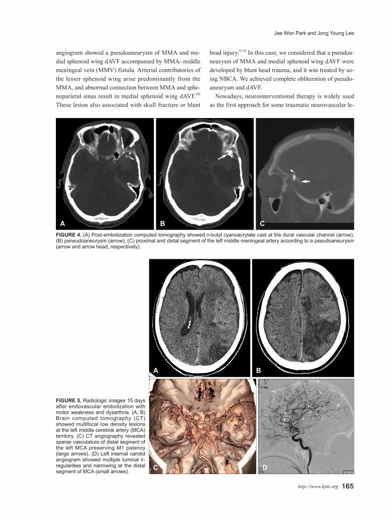
toacryl; B. Braun, Melsnagen, Germany) mixed with io-dized oil (Lipiodol; Guerbet, Aulnay-sous-Boid, France). We achieved angiographically complete occlusion of a pseu-doaneurysm and dural AVF (Figure 3). Follow-up brain CT scan showed no significant hematoma enlargement and well penetrated glue cast at the all the vascular lesions (Figure 4).
The patient presented an improvement in consciousness for GCS score 15 after 12 days. However, at the 18-day af-ter the procedure, the right side weakness and dysarthria were developed. Brain CT angiography and subsequent left internal carotid angiography revealed cerebral vasospasm at the distal segment of MCA with multiple low densitied
A B CFIGURE 1. Initial radiological images after trauma. (A, B) Brain computed tomography (CT) showed intracerebral hemorrhage in the left temporal lobe with diffuse and large amount of subarachnoid hemorrhage and subdural hemorrhage at the left hemisphere. (C) Brain CT angiography revealed that abnormal vascular structure, considered as pseudoaneurysms, at the distal segment of the left lesser sphenoid ridge (arrow).
A B CFIGURE 2. Left external carotid angiogram. (A) anteroposterior view, (B) lateral view showed a multilobulated pseudoaneurysm at the distal segment of the lesser sphenoid ridge (large arrow). Retrograde contrast filling through the left superior ophthalmic vein (double arrow) and middle meningeal artery-middle meningeal vein fistula (double arrow head) was also shown. (C) Cone-beam computed tomography image showed a multilobulated pseudoaneurysm at the distal segment of the lesser sphenoid ridge (large arrow) and the left superior ophthalmic vein (double arrow). Between these, complex vascular connections at the lesser sphenoid ridge resulting in medial sphenoid ridge dural arteriovenous fistula was shown.

164 Korean J Neurotrauma 2017;13(2):162-166
Ruptured Pseudoaneurysm of MMA and Medial Sphenoid Wing DAVF
at the left MCA territories (Figure 5). Cerebral vasospasm was treated using intra-arterial (IA) nimodipin infusion, and it was improved. At discharge, dysarthria was im-proved, but right side weakness was remained (Grade 3).
Discussion
Usually, traumatic pseudoaneurysms of MMA cause
extradural hematoma.7) However, pseudoaneurysm of the MMA, even though rare, has been considered as a possi-ble etiology of ICH.1,4,9) Pseudoaneurysms of MMA tend to gradually enlarge, resulting in a delayed rupture and clinical deterioration, which is associated with a mortality up to 50%.1,6) These lesions usually associated with skull fracture. In the present case, however, skull fracture was not detected by brain CT and skull X-ray. On the other hand,
A B C
D E FFIGURE 3. Superselective angiogram of the left middle meningeal artery (MMA). (A) anteroposterior view, (B) lateral view revealed an appropriate position of distal microcatheter tip (large arrow) showing a pseudoaneurysm (arrow head) and retrograde contrast filling through the left superior ophthalmic vein (double arrow). (C, D) N-butyl cyanoacrylate cast after embolization (large arrow). (E, F) Post-embolization external carotid angiogram showed complete occlusion of pseudoaneurysm, medial sphenoid ridge dural ar-teriovenous fistula and MMA-middle meningeal vein fistula.
Jae Won Park and Jong Young Lee
http://www.kjnt.org 165
angiogram showed a pseudoaneurysm of MMA and me-dial sphenoid wing dAVF accompanied by MMA- middle meningeal vein (MMV) fistula. Arterial contributories of the lesser sphenoid wing arise predominantly from the MMA, and abnormal connection between MMA and sphe-noparietal sinus result in medial sphenoid wing dAVF.10) These lesion also associated with skull fracture or blunt
head injury.8,11) In this case, we considered that a pseudoa-neurysm of MMA and medial sphenoid wing dAVF were developed by blunt head trauma, and it was treated by us-ing NBCA. We achieved complete obliteration of pseudo-aneurysm and dAVF.
Nowadays, neurointerventional therapy is widely used as the first approach for some traumatic neurovascular le-
A B CFIGURE 4. (A) Post-embolization computed tomography showed n-butyl cyanoacrylate cast at the dural vascular channel (arrow), (B) psneudoaneurysm (arrow), (C) proximal and distal segment of the left middle meningeal artery according to a pseudoaneurysm (arrow and arrow head, respectively).
FIGURE 5. Radiologic images 15 days after endovascular embolization with motor weakness and dysarthria. (A, B) Brain computed tomography (CT) showed multifocal low density lesions at the left middle cerebral artery (MCA) territory. (C) CT angiography revealed sparse vasculature of distal segment of the left MCA preserving M1 patency (large arrows). (D) Left internal carotid angiogram showed multiple luminal ir-regularities and narrowing at the distal segment of MCA (small arrows).
A
C
B
D

166 Korean J Neurotrauma 2017;13(2):162-166
Ruptured Pseudoaneurysm of MMA and Medial Sphenoid Wing DAVF
sions, being minimally invasive, feasible, and effective.1) In term of endovascular techniques treating a pseudoaneu-rysm, a definitive treatment can be obtained with occlu-sion of the proximal parent vessel segment, the pseudoan-eurysm lumen, and the distal segment, avoiding the risk of distal retrograde filling. Definitive embolic agents are ob-viously preferable, with glue being the most used. Because the adhesive characteristic of NBCA, only one session of procedure is permitted per one selected feeding artery. Es-pecially, medial sphenoid wing dAVF was accompanied in the present case, it is important to identify the exact vas-cular structure around the lesions for the position of the microcatheter. To achieve complete occlusion of a pseudo-aneurysm and medial sphenoid wing dAVF via one session of glue injection, we performed repetitive superselective angiography to identify the exact vascular structure of pseu-doaneurysm and dAVF, and confirm the most appropriate position of the microcatheter. Finally, we could achieve a complete occlusion of all the vascular lesions using one ses-sion of procedure. Postoperative CT revealed glue cast at the pseudoaneurysm, dural vascular channel, proximal and distal segment of MMA according to a pseudoaneurysm.
In this case, delayed neurologic deterioration was devel-oped after full recovery. Angiogram revealed vasospasm of the left MCA, especially distal segment of MCA was mainly involved. There are important differences and sim-ilarities between cerebral vasospasm that occurs as a re-sult of traumatic brain injury compared with aneurismal SAH, e.g. 20% to 30% of aneurismal SAH, and less than 20% with traumatic brain injury.2,5) For these reasons, even though SAH is accompanied, the importance of vasospasm usually underestimated in case of traumatic brain injury, and any practice guidelines do not recommend routine sur-veillance for cerebral vasospasm in patient with traumatic brain injury of any severity.2) Moreover, there is no suggest-ed monitoring period for symptoms of delayed cerebral ischemia as there is for patient with aneurismal SAH.3) In the same manner as aneurysmal SAH, high suspicion level for cerebral vasospasm should be maintained in case of trau-matic SAH.
Conclusion
Ruptured pseudoaneurysm of MMA and medial sphe-noidal ridge dAVF is uncommon, but a ruptured pseudoa-
neurysm has a potential aggressive natural history. There-fore, once diagnosed, it must be treated. These lesions usually treated by using NBCA, and identification of an exact vas-cular structures and appropriate position of microcatheter is crucial to achieve complete occlusion of the pseudoan-eurysm and dAVF using only one session of glue injection. In addition, cerebral vasospasm should not be neglected in case of traumatic SAH after complete obliteration of pri-mary focus.
■ The authors have no financial conflicts of interest.
REFERENCES1) Bruneau M, Gustin T, Zekhnini K, Gilliard C. Traumatic false an-
eurysm of the middle meningeal artery causing an intracerebral hemorrhage: case report and literature review. Surg Neurol 57:174-178, 2002
2) Carney N, Totten AM, O'Reilly C, Ullman JS, Hawryluk GW, Bell MJ, et al. Guidelines for the management of severe traumatic brain injury, fourth edition. Neurosurgery 80:6-15, 2017
3) Connolly ES Jr, Rabinstein AA, Carhuapoma JR, Derdeyn CP, Dion J, Higashida RT, et al. Guidelines for the management of an-eurysmal subarachnoid hemorrhage: a guideline for healthcare pro-fessionals from the American Heart Association/american Stroke Association. Stroke 43:1711-1737, 2012
4) Lim DH, Kim TS, Joo SP, Kim SH. Intracerebral hematoma caused by ruptured traumatic pseudoaneurysm of the middle meningeal artery : a case report. J Korean Neurosurg Soc 42:416-418, 2007
5) Oertel M, Boscardin WJ, Obrist WD, Glenn TC, McArthur DL, Gravori T, et al. Posttraumatic vasospasm: the epidemiology, se-verity, and time course of an underestimated phenomenon: a pro-spective study performed in 299 patients. J Neurosurg 103:812-824, 2005
6) Paiva WS, de Andrade AF, Amorim RL, Figueiredo EG, Teixeira MJ. Traumatic pseudoaneurysm of the middle meningeal artery causing an intracerebral hemorrhage. Case Rep Med 2010:219572, 2010
7) Salazar Flores J, Vaquero J, Garcia Sola R, Rossi E, Martinez R, Martinez P, et al. Traumatic false aneurysms of the middle menin-geal artery. Neurosurgery 18:200-203, 1986
8) San Millán Ruíz D, Fasel JH, Rüfenacht DA, Gailloud P. The sphe-noparietal sinus of breschet: does it exist? An anatomic study. AJNR Am J Neuroradiol 25:112-120, 2004
9) Sandin JA, 3rd, Salamat MS, Baskaya M, Dempsey RJ. Intracere-bral hemorrhage caused by the rupture of a nontraumatic middle meningeal artery aneurysm. Case report and review of the litera-ture. J Neurosurg 90:951-954, 1999
10) Shi ZS, Ziegler J, Feng L, Gonzalez NR, Tateshima S, Jahan R, et al. Middle cranial fossa sphenoidal region dural arteriovenous fis-tulas: anatomic and treatment considerations. AJNR Am J Neu-roradiol 34:373-380, 2013
11) Unterhofer C, Chemelli A, Waldenberger P, Bauer R, Ortler M. Traumatic fistula between the middle meningeal artery and the sphenoparietal sinus. Acta Neurochir (Wien) 151:1301-1304, 2009