trauma learning objectives - paramedic advantage · 2 every day thousands of people become the...
TRANSCRIPT

1
MODULE V
TRAUMA
1. Provide care to a patient in shock (hypoperfusion).
o State methods of emergency medical care of external bleeding.
o List signs and symptoms of shock (hypoperfusion).
o State the steps in the emergency medical care of the patient with signs and
symptoms of shock (hypoperfusion).
2. Provide care to a patient with a suspected spinal injury.
o State the signs and symptoms of a potential spine injury.
o Describe how to stabilize the spine.
3. Provide care to a patient with a suspected head injury.
o Relate mechanism of injury to potential injuries of the head and spine.
4. Provide care to a patient with a soft-tissue injury.
o Describe the emergency medical care of the patient with a closed soft
tissue injury.
o Describe the emergency medical care of the patient with an open soft
tissue injury.
5. Perform a rapid extrication of a trauma patient.
o Describe the indications for the use of rapid extrication.
o List steps in performing rapid extrication.
LEARNING OBJECTIVES
Upon completion of this course, the student will be able to:

2
Every day thousands of people become the accidental victims of trauma. Approximately
43,000 people die in motor vehicle crashes each year in the United States. With an
estimated 2.9 million people injured in these accidents, the impact on the EMS system is
significant. Head-on collisions account for approximately 11% of all fatal accidents
(NHTSA, 2004). Such collisions commonly produce lower-extremity injuries to the
knees, femurs, and hips, as well as chest and abdominal injuries from impact with the
steering wheel or airbag.
Trauma is the leading cause of death in the United States for persons between the ages of
1 and 44. Understanding the mechanism of injury, relevant signs and symptoms, and
appropriate intervention techniques is essential when dealing with the traumatized
patient. Airbags have reduced the incidence of significant head injuries for restrained
drivers; however, drivers are still at risk for cervical spine and head injuries in this type
of collision.
Rear-end collisions generally do not cause fatal injuries; however, they commonly result
in cervical spine injuries caused by the neck's hyperextending—when the occupant is
propelled forward by the force of the accident—and then snapping back (whiplash).
Properly adjusted head rests are designed to reduce the incidence and severity of neck
injuries.
Driver experiencing whiplash.
Lateral and side-impact collisions occur less frequently but they are the most serious,
resulting in approximately 24% of all traffic-related fatalities (NHTSA, 2004). In a lateral
or side-impact collision, the amount of structural steel between the occupant and the
impacting vehicle is markedly less than for frontal or rear-end collisions, and this
frequently results in significant internal injuries. Both upper- and lower-extremity injuries
may occur on the impacted side, as well as head and cervical spinal injuries.
When a vehicle is struck from an oblique angle, the vehicle rotates as a result of the
collision. This is referred to as a rotational impact. Though the damage to the vehicle may
be significant in these types of collisions, they generally do not result in significant

3
injuries to the occupant because they do not cause the vehicle to stop suddenly; instead,
the vehicle comes to rest more slowly, resulting in fewer deceleration injuries.
Rollover collisions result in approximately 22% of annual traffic fatalities. Significant
injuries may result based on the number of impacts associated with the rollover and
whether the occupant was restrained. The greatest risk associated with rollover collisions
is that of being partially or fully ejected from the vehicle if occupants are improperly
restrained.
The use of seatbelts has steadily increased over the past twenty years, with many states
enacting strict laws governing their use. With the exception of public transportation, most
states require seatbelt use for all front-seat occupants. In some states however, seatbelt
violations are considered secondary violations, meaning that an officer can only enforce
seatbelt laws when the driver has been pulled over for a violation of another law. The
NHTSA estimates that the use of manual three-way seatbelts reduces the risk of death in
motor vehicle collisions by 45% for front-seat occupants.
In 1995 approximately 84% of all new cars sold were equipped with dual air bags for the
driver and front-seat passenger. In frontal collisions, which represent the primary
collision type for which airbags were designed to reduce injury, drivers with airbags had
a reduced fatality rate of 21% (NHTSA, 1996).
Since the widespread use of airbags began in the mid-1990s, just under 300 adults and
children have died as a result of their use. In that same period it is estimated that 20,000
lives have been saved by using airbags. New technology for airbags, coupled with public
awareness campaigns, has resulted in more infants and children being secured in the rear
seat, away from airbags, further reducing fatalities.
Injuries associated with airbags are related to the speed with which the devices inflate,
and are usually limited to superficial injuries to the upper extremities. This does not apply
to infants or children in the front seat; significant airbag injuries can result from placing
infants in rear-facing child seats or children under age 12 or less than 100 pounds in front
passenger seats.
In 2004 an estimated 70,000 pedestrians were injured in motor vehicle collisions and
approximately 4,600 pedestrians were killed by motor vehicles (IIHS, 2005). Most
pedestrians are struck by the front of the vehicle, and they are generally at fault in these
types of accidents. Nevertheless, many accidents, most of which involve children, are
events in which pedestrians are backed into or over by a vehicle. The speed of the vehicle
at impact, the size of the pedestrian, and the height of the vehicle all factor significantly
in the injury patterns noted on these patients.

4
SCENARIO
At 2200 you are called to respond to a motor vehicle accident (MVA) on a
rural two-lane highway. A helicopter is also being dispatched to transport
any critical patient to the nearest trauma center, which is 45 miles away.
Witnesses report that traffic had stopped for a tractor crossing the road. A
truck traveling at a high rate of speed failed to stop, lost control, and skidded
off the roadway at 70 mph. The truck rolled and struck a tree, ejecting the
occupant. You find a 22-year-old male unresponsive, lying 25 feet from the
vehicle.
The patient is prone and motionless. While maintaining manual c-spine
control, you and your partner log roll him onto his back. You maintain c-
spine control while your partner palpates a carotid pulse of 100 bpm but is
unable to palpate a radial pulse. You place a cervical collar on the patient's
neck and then the two of you log roll the patient onto a long spine board and
check for breathing.
The patient has a respiratory rate of 10, so your partner places an
oropharyngeal airway (OPA) and begins ventilating the patient with a bag
valve mask (BVM) using high-flow oxygen, while you do a quick
assessment for uncontrolled bleeding. [Note: In your district you are
authorized to intubate and start IVs.]
Your partner reports that it is becoming difficult to ventilate the patient due
to blood and secretions in the airway. You suction and then intubate the
patient to secure his airway. Your partner resumes ventilation with a BVM
using high-flow oxygen.
After making sure the head and neck are stabilized and rest of the body is
secured to the board, you load the patient into the back of the ambulance.
The heater is turned up to keep him warm while you expose the body to
assess for other injuries. While your partner ventilates the patient with high-
flow oxygen, you complete a focused assessment.
You find that the patient has deformity to his upper right leg and a 3-inch
laceration to the lower right leg with moderate bleeding. You learn that the
helicopter will arrive in 10 minutes. You bandage the laceration with dry
sterile dressings, place a traction splint on the deformed upper leg, initiate
two large-bore IVs, and cover the patient to keep him warm. When the
helicopter arrives, you hand off the patient to the helicopter crew.

5
SHOCK (HYPOPERFUSION
SYNDROME)
Severity
To ensure normal perfusion of tissues, the human body must have:
Adequate air exchange in the lungs to allow oxygen into the bloodstream
An intact vascular system to delivery the oxygenated blood to the body's tissues
An adequate volume of fluid, including red blood cells and plasma
A functioning pump to circulate the volume
The lower airway, showing gas exchange.

6
In shock states, one or more of these components has been disrupted, causing inadequate
tissue perfusion. Cell and organ malfunction and death can result from shock; therefore,
prompt recognition and treatment is vital to patient survival.
Though the most common shock observed in trauma is related to hemorrhage, shock can
be classified into three general categories (BTLS, 2004):
Low-volume shock (absolute hypovolemia), caused by hemorrhage or other
major body fluid loss
High-space shock (relative hypovolemia), caused by spinal injury, vasovagal
syncope, sepsis, and certain drug overdoses
Mechanical (obstructive) shock, caused by pericardial tamponade, tension
pneumothorax, or myocardial contusion
In the early stages of low-volume or hypovolemic shock, the body's sympathetic nervous
system responds to a decrease in circulating volume by releasing catecholamines that
cause an increase in heart rate and vasoconstriction. The body mounts this response in an
attempt to maintain adequate blood flow to the brain and heart.
Peripheral vasoconstriction at this stage can cause the patient's skin to be cool and pale
and capillary refill to slow. As low-volume shock progresses, the patient may show
mental status changes indicating that the body's compensatory mechanisms are failing to
perfuse the vital organs adequately. A late sign that the body is moving from a
compensated shock state to a decompensated state is a drop in the patient's blood
pressure.
High-space shock, commonly referred to as neurogenic shock, can be observed in
patient's who have spinal cord injuries. This type of shock causes a disruption in the
sympathetic nervous system, which prevents a catecholamine release and
vasoconstriction in response to accompanying hemorrhage.
Patients with high-space shock present with different symptoms from those of a low-
volume shock patients in that they will not exhibit an increased heart rate, cool and pale
skin, or changes in capillary refill time. These patients generally exhibit pink, warm and
dry skin, mild hypotension, relative bradycardia, and frequently have no changes in
mental status, but they do have flaccid paralysis.
High-space shock states can be missed in the prehospital setting because these patients
often do not appear to be critically injured. You must rely on evaluation of the
mechanism of injury and a comprehensive neurologic assessment to determine the
potential for underlying hemorrhage. If present, it may necessitate appropriate treatment
such as fluid replacement, oxygen administration, and destination determination.
Normally the body circulates approximately five liters of blood per minute. In order to
maintain this volume of distribution, the left side of the heart must be able to relax and
contract sufficiently to pump the volume through the arterial system. Likewise, the right

7
side of the heart must be able to relax and contract sufficiently to accept the return of de-
oxygenated blood from the venous system and push it out to the lungs to be re-
oxygenated.
Mechanical shock can result from trauma to the chest, causing conditions such as tension
pneumothorax and pericardial tamponade. A tension pneumothorax causes a shift in the
structures of the mediastinum, which reduces venous return of blood. In pericardial
tamponade, the space surrounding the heart fills with blood, which prevents the heart
from filling and pumping adequately.
Pneumothorax resulting from trauma, showing the collapse of the lung due to air entering
the pleural space. Note deviated trachea.
In order to choose the appropriate treatment modality it is important to be aware of all
three types of shock when assessing a trauma patient. The foundation of good trauma
care is developing a working diagnosis based on a thorough assessment of the mechanism
of injury, the patient's age and medical history, presenting signs and symptoms, and the
patient's response to treatment. Close attention paid to the patient's response may indicate
a need to re-assess in order to respond appropriately to the patient's condition.
Signs and Symptoms of Shock
The signs and symptoms of shock can be assessed in three ways: mental status, perfusion
status and vital signs. As shock progresses, the signs exhibited by the patient will become
more pronounced.

8
Class I hypovolemic shock is characterized by a fluid loss of up to 15%, or 750 ml of
circulating volume. At this stage minimal signs of shock are present. Mental status is
generally unchanged, skin signs are warm and dry, heart rate remains less than 100 bpm,
and blood pressure and respiratory rate are within normal limits.
Class II shock represents a more significant volume loss of 15% to 30%, or up to 1500 ml
of circulating volume. At this stage, patients may exhibit mental status changes such as
restlessness or anxiety. The skin may become pale and cool with a delay in capillary
refill, and both heart rate and respiratory rate increase.
A loss of 30% to 40% of the circulating volume, or 1500 to 2000 ml, represents profound
shock. As the patient transitions into class III shock there will be marked anxiety and
confusion. A further delay in capillary refill time, with increasing pallor and cool skin,
are signs of advancing peripheral shut down. Blood pressure begins to drop, and heart
and respiratory rates continue to increase as the body fights to survive. These are signs
that the body's compensatory mechanisms are beginning to fail. Patients in class III shock
who survive the initial insult often succumb to organ failure before discharge.
Class IV shock is the final stage of decompensated shock. The body has suffered a fluid
loss of more than 2000 ml, or 40% of the total circulating volume. The patient becomes
lethargic or unresponsive with cool, sometimes waxy skin. As the patient moves into
class IV shock, the heart rate may be in excess of 140 bpm and is accompanied by
profound hypotension. As the compensatory mechanisms continue to shut down, both the
heart rate and respiratory rate begin to decrease (WNMEDS, 2004).
In addition to the signs and symptoms discussed earlier in this section, patients in shock
may also exhibit dilated pupils as the result of stimulation of the sympathetic nervous
system, and cyanosis to the lips, fingers, and toes as a result of peripheral
vasoconstriction. Patients may also exhibit nausea and vomiting related to the
catecholamine release.
You should have a very high index of suspicion for pediatric patients who have been
involved in a traumatic event. Infants and children can maintain their blood pressure until
their blood volume is more than half gone. However when pediatric patients start to show
signs of decompensation, they deteriorate very rapidly.
Pediatric patients have less reserve than their adult counterparts, which will result in rapid
exsanguination if there is major uncontrolled bleeding, and a shorter duration of time
during which the body's fight-or-flight mechanism will maintain vital functions in the
presence of significant traumatic injuries. By the time hypotension ensues, the pediatric
patient may be near death.
Emergency Medical Care
As with all other patient encounters, body substance isolation is of utmost importance
when you are assessing and treating trauma patients. In accident settings, it is a good idea

9
to have an extra pair of gloves available in the event your gloves are compromised by
glass, sharp metal, or other debris at an accident scene. In addition, accident events often
involve multiple victims, which may require you to rapidly assess and triage more than
one patient. To prevent exposing a patient to the bodily fluids of another, gloves should
be changed before each encounter with a new patient.
Airway management of the patient in shock may range from providing supplemental
oxygen via a nonrebreather mask for a patient who is not exhibiting signs of respiratory
distress to endotracheal intubation for a patient who cannot effectively maintain a patent
airway. All trauma patients who are exhibiting signs of shock should at a minimum
receive supplemental oxygen by mask. Only competent patients who refuse oxygen by
mask should be placed on a nasal cannula (BTLS, 2004).
Suctioning may be required for patients who are unable to clear their own secretions, or
to remove blood, emesis, or foreign objects from the airway. Suctioning the airway prior
to attempting intubation will improve intubation success if fluids or foreign objects are
present. Even patients with minor to moderate oral or facial injuries who are alert and
oriented may not be able to clear their airways effectively when placed supine with spinal
immobilization. Continual re-assessment is required to ensure that the patient maintains
an open airway throughout transport.
Some patients require mechanical assistance to maintain adequate ventilation and
oxygenation. The oropharyngeal airway (OPA) and nasopharyngeal airway (NPA) are
designed for use in patients with an altered level of consciousness to prevent the patient's
tongue from falling to the back of the oropharynx and interfering with ventilation.
If a patent airway can not be maintained utilizing an OPA/NPA and a bag valve mask
with supplemental oxygen, endotracheal intubation or use of a dual-lumen airway device
may be required; however, if oxygenation and ventilation can be maintained with a
OPA/NPA airway, rapid transport to an appropriate trauma facility should take
precedence over extending scene time to secure an advanced airway in the field.
Control of external bleeding requires a variety of techniques, depending on the location
and severity of the injury. The initial step to control bleeding involves direct pressure,
performed by placing fingertip pressure directly on the site of the bleeding. Elevation of a
bleeding extremity may be used secondary to and in conjunction with direct pressure if
needed.

10
To intervene with a bleeding wound, apply sterile gauze and direct pressure and elevate
the extremity if possible. (Illustration by Jason M. McAlexander, MFA. Copyright ©
2007 Wild Iris Medical Education.)
Large gaping wounds may require packing with sterile gauze and direct hand pressure if
direct fingertip pressure fails to control bleeding. If bleeding is still uncontrolled, or soaks
through the dressings, additional pressure should be applied to the site. In the past, it was
taught that if bleeding soaks through a dressing, additional dressings should be placed on
top of the original dressing. However, ITLS now instructs caregivers to remove the
soaked dressing and redress the wound once to ensure that pressure is being applied
directly to the location of the bleeding (BTLS, 2004). In the upper and lower extremities,
pressure points may also be used to control significant bleeding.
Elevation of the lower extremities by 8 to 12 inches may be effective in improving
circulation to vital organs for a patient in shock; however, it should not be attempted if
the patient has suspected injuries to the pelvis, lower extremities, head, chest, abdomen,
neck, or spine—which accounts for the majority of traumatic injuries.
If signs of shock are present and the lower abdomen is tender, or pelvic injury is
suspected, application of the pneumatic anti-shock garment (PASG) may be indicated if
allowed by local protocol or medical direction. The PASG device cannot be used in
patients with suspected chest injury because the device increases pressure in the chest,
potentially worsening bleeding from chest injuries.
For patients exhibiting signs of shock, the goal is rapid transport with spinal precautions
in place. Splinting of suspected fractures and dislocations may decrease the incidence of
pain for the patient; however, if rapid transport is indicated, splinting of long-bone
11
fractures may be all that time allows. If the patient has a suspected or obvious femur
fracture, use of a traction splint will decrease the incidence of pain and prevent free
motion of the bone ends, which could lacerate the femoral artery or vein, thus increasing
blood loss and worsening the progressing shock.
In order to complete a thorough primary assessment, exposure of all or most of the
patient is critical to finding all injuries; however, patients should be covered with a
blanket as quickly as possible to preserve the patient's privacy and to conserve body heat.
In some cases, complete exposure can be delayed until the patient is in the back of a
warm ambulance, prior to transport.
Keeping in mind that a patient can lose up to 2000 ml of fluid from a femur fracture, it is
important to identify potentially significant trauma early in your assessment. The goal is
to transport the patient expeditiously to an appropriate facility capable of surgical
intervention. Direct pressure, splinting, and fluid replacement are only temporizing
measures; trauma is a surgical disease.
EMERGENCY MEDICAL CARE
Chest Injury
Chest injuries fall into two general categories; open and closed. Appropriate management
of closed chest injuries relies almost exclusively on a comprehensive evaluation of the
mechanism of injury and on patient assessment. Closed chest injuries may be present
with minimal or no outward signs immediately following the event. Patients may
complain of chest pain that worsens on palpation, or of difficulty breathing.
Often chest trauma is associated with multi-system trauma, which may distract the patient
from identifying symptoms related to an underlying chest injury. Closed chest injuries
may include pulmonary or cardiac contusion, fractures of the sternum, scapulae, or ribs,
pneumothorax, or hemothorax.
Following trauma, patients who present with difficulty breathing, unilateral diminished
lung sounds, and hypotension may have an underlying tension pneumothorax, which
requires immediate intervention. In some cases, tracheal deviation may be apparent:
however this is a late sign and it is difficult to visualize on some patients. A simple
pneumothorax that does not result in respiratory or vascular compromise does not
necessitate prehospital treatment. Advanced life support treatment for tension
pneumothorax is needle decompression of the affected side.
Up to 15% of all deaths following motor vehicle collisions are due to injury to the
thoracic aorta. Many of these patients are dead at the scene from complete aortic
transection. Patients who survive to the emergency department usually have small tears
or partial-thickness tears of the aortic wall (PHTLS, 2003).
12
Most blunt aortic injuries occur in the proximal thoracic aorta, although any portion of
the aorta is at risk. The proximal descending aorta, where the relatively mobile aortic arch
can move against the fixed descending aorta, is at greatest risk from the shearing forces
of sudden deceleration. Thus the aorta is at greatest risk in frontal or side impacts and
falls from heights (Trauma.org, 2004).
Patients with blunt aortic injuries surviving to arrival at the hospital usually have partial
transections, and should be managed with blood pressure control until surgical repair.
The priority in the management of hemodynamically unstable patients with potential
aortic injuries is to rapidly identify and control ongoing hemorrhage from other sites, and
to avoid over-resuscitation. Overly aggressive fluid resuscitation can lead to complete
aortic rupture for these patients by increasing intra-aortic pressure. Clinical signs of
traumatic aortic injury are rarely present, and diagnosis is based on a high index of
suspicion related to the mechanism of injury (Trauma.org, 2004).
Management of open chest wounds is dependent on the location and severity of the
injury. Penetrating trauma to the chest should be managed with an occlusive dressing,
such as a petroleum jelly (Vaseline) or bi-occlusive dressing. Upon initial identification, a
gloved hand should be used to occlude the injury site while further assessment takes
place to identify other life-threatening injuries. Once an occlusive dressing has been
placed, the patient should be frequently re-evaluated for signs of a developing tension
pneumothorax. If signs or symptoms of a tension pneumothorax develop, lift an edge of
the occlusive dressing to allow trapped air to escape.
All patients with obvious or suspected chest injuries should receive supplemental oxygen
at an appropriate flow rate, and be placed in a position of comfort if spinal injuries are not
suspected.
Abdominal Injuries
Depending upon the underlying structures affected, abdominal injuries can result in
significant hemorrhage. Most of the abdominal organs—including the liver, spleen,
stomach, pancreas and the reproductive organs—are located in the peritoneal cavity. The
peritoneum is a smooth transparent membrane that lines the abdomen and doubles back
over the surfaces of the internal organs to form a continuous sac. Some organs and major
vessels such as the kidneys, inferior vena cava, and aorta, are located posterior to the
peritoneum in what is referred to as the retroperitoneal space. The primary function of the
peritoneum is to protect the internal organs from injury.

13
The abdominal organs. Think about what organs might be injured from seatbelt
placement.
Improperly positioned seatbelts can cause liver or spleen ruptures that may result in
significant internal blood loss. In blunt abdominal trauma, the most commonly injured
organs are the spleen and the liver. The liver is frequently injured in penetrating trauma
due to its relatively large size. The liver is a very vascular organ and bleeds profusely
when it is injured (Health Care Information, 2006). A patient who has experienced a
traumatic injury and who presents with signs of shock having no apparent cause should
be considered to have abdominal bleeding until proven otherwise.
Appropriate treatment for a suspected closed abdominal injury is dictated by the patient's
presentation. Patients exhibiting clinical signs of hypoperfusion such as an altered mental
status and cool, pale skin should be transported expeditiously to an appropriate trauma
receiving facility. Supplemental oxygen is administered to all patients exhibiting signs or
symptoms related to a traumatic event. Advanced life support treatment includes
initiating IV access and titrating fluids to maintain adequate perfusion as evidenced by a
systolic blood pressure of 90 to 100 mm Hg (BTLS, 2004).
Treatment for an open abdominal injury focuses on reducing the risk of infection by
protecting the exposed tissue and organs. Exposed organs should not be touched, and you
should make no effort to replace organs that are protruding out of the abdominal cavity.
Replacing organs that have been exposed to environmental contaminants may result is
severe infection and complications in the patient's long-term recovery.
Open wounds and organs that are exposed should be covered with a sterile dressing,
moistened with saline or sterile water, and secured in place. If you do not suspect lower-
extremity injuries, placing the patient in a position that allows flexion of the hips and
14
knees will accommodate relaxation of the abdominal muscles and possibly decrease
patient discomfort.
Amputations
Traumatic amputations are a dramatic finding at the scene. The mechanism of injury that
results in an amputation can frequently produce other significant injuries as well;
however, it is easy to become distracted by the amputation. Initial management of
traumatic amputations includes a complete primary assessment of the patient to rule out
other life-threatening injuries including potential hemorrhage from the amputation site.
If a patient has sustained a partial amputation, the area should be immobilized and
dressed to prevent further injury. In rare circumstances, partial amputations have required
that the amputation be completed in order to free an entrapped patient; however,
generally no attempt is made to complete a partial amputation.
Once initial stabilization has been addressed, efforts are made to preserve the integrity of
the amputated part. The part should be wrapped in a sterile dressing moistened slightly
with saline or sterile water, and then placed in a plastic bag and kept cool. To avoid tissue
damage, an amputated part should never be placed directly on ice or in contact with a
chemical cold pack.
In the chaos of a scene that may include multiple patients and multiple ambulances, or the
utilization of helicopters to facilitate transport to trauma centers, be careful to ensure that
the amputated part is transported with the patient.
Burns
While the leading cause of fatal fires is related to smoking materials such as cigarettes,
the majority of residential fires that result in burn injuries are caused by cooking
equipment (Stanford, 2006). Though the majority of deaths caused by fire are related to
smoke inhalation, many severely burned patients survive the fire but later succumb to
infection.
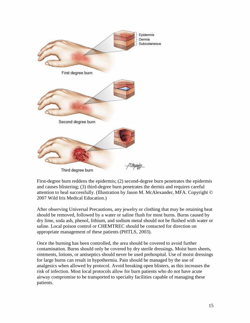
Burns are commonly categorized as first-, second-, or third-degree, which are sometimes
known as superficial, partial-thickness, or full-thickness, respectively. Superficial burns
are painful burns that are characterized by inflamed red skin. Though field treatment of
superficial and partial-thickness burns is directed at controlling pain, the first priority for
all burns is ensuring that the burning process has stopped.
15
First-degree burn reddens the epidermis; (2) second-degree burn penetrates the epidermis
and causes blistering; (3) third-degree burn penetrates the dermis and requires careful
attention to heal successfully. (Illustration by Jason M. McAlexander, MFA. Copyright ©
2007 Wild Iris Medical Education.)
After observing Universal Precautions, any jewelry or clothing that may be retaining heat
should be removed, followed by a water or saline flush for most burns. Burns caused by
dry lime, soda ash, phenol, lithium, and sodium metal should not be flushed with water or
saline. Local poison control or CHEMTREC should be contacted for direction on
appropriate management of these patients (PHTLS, 2003).
Once the burning has been controlled, the area should be covered to avoid further
contamination. Burns should only be covered by dry sterile dressings. Moist burn sheets,
ointments, lotions, or antiseptics should never be used prehospital. Use of moist dressings
for large burns can result in hypothermia. Pain should be managed by the use of
analgesics when allowed by protocol. Avoid breaking open blisters, as this increases the
risk of infection. Most local protocols allow for burn patients who do not have acute
airway compromise to be transported to specialty facilities capable of managing these
patients.

16
Burn patients should be continually reevaluated for airway compromise. Painful burns
may distract a patient from recognizing symptoms of advancing airway edema. Stridor,
coughing, singed facial hair, and soot around the mouth or nares may indicate potential
airway involvement. All patients who have experienced thermal burns should receive
supplemental oxygen to treat possible hypoxia related to inhalation and compromised
circulation.
INJURIES TO BONES AND JOINTS
Signs and Symptoms
Bone and joint injuries are rarely fatal; however, they are usually quite painful. Signs and
symptoms associated with bone and joint injuries may include:
Deformity or angulation
Pain and tenderness
Grating
Swelling
Bruising
Exposed bone ends
Joint locked into position
Fractures are categorized as open (compound) or closed (simple). A compound fracture
is the result of an underlying fractured bone or bones breaking through the surface of the
skin. The bones may protrude through the skin and remain out, or they may break the
skin surface and then retract beneath the skin. Any open injury associated with a possible
fracture should be treated as a compound fracture, regardless of whether bone is visible.
When in doubt about the existence of a fracture, a painful extremity injury should be
splinted even if none of the above signs and symptoms is present. The presence of pain is
indicative of injury, and splinting may serve to decrease the patient's discomfort.
Emergency Care of Bone or Joint Injuries
When assessing a patient with a suspected fracture, it is important to obtain a good
history in order to determine the mechanism of injury, and the potential for other injuries
in addition to the painful fracture.
As an EMS professional, one of the most important things to keep in mind when you are
assessing a trauma patient who has an extremity fracture is that the pain associated with
the fracture may distract the patient from recognizing injuries in other areas. In the
presence of a painful extremity fracture, the patient may not clearly identify cervical
spine pain on palpation. For this reason, you need to consider spinal immobilization for

17
patients who have experienced a high-risk mechanism of injury resulting in isolated
extremity trauma even if the patient does not complain of neck or back pain.
After following Universal Precautions, assess the patient for life-threatening injuries. In
the absence of life-threatening injuries, splint fractures prior to transport to control pain
and prevent further injury. For the critical patient, splinting should be accomplished as
time allows once transport has been initiated.
Supplemental oxygen is considered and applied as indicated by the patient's presentation.
Elevation and the application of a cold pack to a painful, swollen, or deformed extremity
will reduce both pain and swelling to the area. Keep in mind that cold packs should be
applied only following a dressing so that the cold pack does not come in direct contact
with the injury site.
General Rules of Splinting
Before splinting, remove clothing or cut it away and assess the extremity for pulse,
movement, and sensation distal to the injury site. The proximal and distal joint should be
immobilized for a suspected bone injury, and the proximal and distal bone should be
immobilized for a suspected joint injury. Following splinting, re-assess pulse, movement,
and sensation and record both assessments on the patient care report.
Open fractures are managed by applying a sterile dressing and splinting the extremity in a
position of comfort. Dressings moistened with sterile water may improve healing if
transport time is prolonged. To reduce the risk of infection and further tissue damage,
protruding bones should not be pushed back under the skin. If during the application of a
traction splint the bone ends retract beneath the surface, do not increase the amount of
traction. Take care when placing the splint to ensure that it is well padded and does not
put pressure on the protruding bone (BTLS, 2004).
Closed fractures may be less dramatic in their presentation but associated hemorrhage can
be difficult to determine. Bilateral closed femur fractures can result in a blood loss of up
to 2 liters, and pelvic fractures can result in life-threatening hemorrhage with significant
bleeding into the abdominal cavity before symptoms become apparent. As with open
fractures, closed fractures are splinted in a position of comfort.
Ensuring that the splint is well padded will reduce discomfort to the patient. Pain control
with analgesics should be considered for patients with isolated extremity fractures but
may be contraindicated in the presence of multi-system trauma. Analgesics can
complicate the physician's assessment in the emergency room, making it difficult to
ascertain the location of the patient's injuries.
If a fractured extremity is angulated and pulses are absent, you may apply gentle traction
in an attempt to restore circulation; however, excessive manipulation can result in further
damage to underlying structures. If a receiving facility is close, expeditious transport may
be the most appropriate treatment.

18
SPINAL INJURIES
Mechanism of Injury with High Index of Suspicion
In 2006 the Spinal Cord Injury Information Network estimated that annual incidence of
spinal cord injury, not including those who die at the scene of the accident, is
approximately 40 cases per million population in the United States, or approximately
11,000 new cases each year (SPIIN, 2006).
Spinal cord injury primarily affects young adults, with most injuries occurring between
the ages of 16 and 30. However, the number of injured adults over 60 years of age has
steadily increased from 4.7% prior to 1980 to 11.5% since 2000. About 78% of spinal
cord injuries occur in males (SPIIN, 2006).
Motor vehicle crashes account for 46.9% of reported spinal cord injuries cases. The next
most common cause of these injuries is falls, followed by acts of violence (primarily
gunshot wounds). Recreational sporting activities also account for a number of spinal
cord injury cases annually (SPIIN, 2006).
Signs and Symptoms of Spine Injuries
Patients not exhibiting an altered level of consciousness and those without a distracting
injury or neurologic deficit may exhibit tenderness or pain on palpation or with
movement. Never move patients or ask them to move or perform range of motion (ROM)
exercises to elicit a pain response. Pain that is independent of movement or palpation
may manifest not only in the spinal column but also in the legs if there is swelling or
injury to nerves along the spinal column. Pain may also be caused by muscle contractions
that can be intermittent in nature.
Obvious deformity to the spine may be noted on palpation. Depending on the degree of
deformity, this may present an issue when placing the patient supine on a long spine
board. This issue can usually be overcome by padding the board to accommodate the
deformity.
Signs of neurologic injury may include:
Numbness, weakness, or tingling in the extremities
Paralysis in one or more extremities, or below the level of injury
Loss of sensation to one or more extremities, or below the level of injury
Incontinence
Priapism

19
Injuries to the spinal cord resulting in paralysis or loss of sensation may be related to a
number of specific injury types. Cord contusion involves bruising or bleeding into the
tissues or the spinal cord, and may result in a temporary loss of function below the level
of the injury. Cord compression results from pressure on the spinal cord that is causing
swelling, which can result in tissue ischemia. A cord laceration describes cord tissue
that is torn or cut. Both compression and laceration can result in either permanent or
transient disability. Complete cord transection interrupts all cord function distal to the
injury site, resulting in permanent disability (PHTLS, 2003).
Though injury to nerves that control the bladder may result in urinary incontinence,
incontinence in the presence of trauma may also be caused by a traumatic brain injury.
In male patients, spinal injuries may result in priapism. Priapism is a prolonged erection
of the penis resulting from unopposed parasympathetic stimulation caused by an insult to
the spinal cord (Mistovich et al., 2003).

20
Assessing the Potentially Spine-Injured Patient
For responsive patients, you attain a history of the event from both the patient and any
bystanders. When asking "What happened?" you may find the bystander's account of the
incident differs from the patient's recollection of the event. This suggests that the patient
had an associated loss of consciousness that may be denied upon questioning.
The patient should be asked if there is any pain in the neck or back. If the patient
indicates that there is tenderness or pain, palpate the area to localize the site and to
determine if there is any associated deformity. To avoid unnecessary movement of the
spine, do not ask the patient to point to the area of injury.
To check for appropriate neurologic response, ask patients to move their hands and feet,
and palpate their fingers and toes while asking if they can feel your touch. Assess the
extremities for equality of strength by asking the patient to grip your hands, and gently
push their patient's feet against your hands. The neurologic assessment should be
performed both before and after placing the patient in spinal immobilization, and
intermittently throughout transport, with all findings chronologically documented on the
patient care report.
In the trauma setting, all unresponsive patients and all patients who demonstrate an
altered level of consciousness should be considered to have a spinal injury. In attempting
to transport an unresponsive trauma patient quickly to a trauma center, spinal precautions
are often overlooked. Though expeditious transport is very important to these patients,
preventing further injury to the spine is an important consideration in the patient's long-
term quality of life following the accident.
When assessing an unresponsive patient, your determining the mechanism of injury is
important to the receiving physician. When a patient is unable to provide an account of
the event, the only other clues as to what occurred are at the scene They include:
Bystander accounts of the incident
The patient's mental status preceding the event
Damage to vehicles
Height of potential falls
After managing any life-threatening injuries, make a quick survey to reveal any
contusions, deformities, lacerations, punctures, penetrations, or swelling.
Patients are transported after being secured to a padded long spine board, with
appropriate cervical immobilization. As with any other trauma patient, supplemental
oxygen is administered, titrated appropriately.

21
Emergency Medical Care
As with all other trauma patients, patients with suspected spinal injuries should be treated
for life-threatening injuries before managing the spinal injury; however, every effort
should be made to limit movement to the spine while performing these treatments.
Manual in-line immobilization is maintained while advanced airway procedures are
performed and bleeding is controlled.
Patients with a spinal injury are immobilized in a supine position on a rigid spine board in
a neutral in-line position. The head, neck, torso, and pelvis must each be immobilized to
prevent further movement (PHTLS, 2003).
Place an appropriately sized cervical collar to immobilize the cervical spine. Cervical
collars do not adequately immobilize the cervical spine unless the patient is motion-
restricted on a spine board with head blocks in place.
Spine boards should be padded to provide comfort to the patient, and to minimize the risk
of soft-tissue injuries to the patient's skin. Additional padding may be needed under the
patient's shoulders to accommodate patients immobilized with a helmet, or for pediatric
patients whose heads are anatomically disproportionate to their body size.
Supplemental oxygen is provided to the patient, and the patient is transported to an
appropriate receiving facility.
HEAD INJURIES
Mechanism of Injury with a High Index of Suspicion
Approximately 1.4 million people suffer traumatic brain injuries (TBI) annually in the
United States, which results in 50,000 deaths and 235,000 hospitalizations. Of these
injuries, 28% are caused by falls, 20% are the result of motor vehicle accidents, and 11%
are the result of an assault (CDC, 2006).
The CDC estimates that at least 5.3 million Americans, approximately 2% of the U.S.
population, currently have a long-term or lifelong need for help to perform activities of
daily living (ADLs) as a result of a TBI.
Traumatic brain injuries can cause a wide range of functional changes that affect
thinking, sensation, language, and emotions. They can also cause epilepsy and increase
the risk for conditions such as Alzheimer's disease, Parkinson's disease, and other brain
disorders that become more prevalent with age (CDC, 2006).
When evaluating the scene of a motor vehicle accident, inspect the windshield for blood,
hair, starring, or deformity. If your patient was involved in an accident while wearing a
22
helmet, inspect the helmet for scratches, dents or breaks, as well as for the presence of
blood or brain matter. It is important to note and report to the receiving facility the extent
of damage as well as the type of helmet worn.
There are essentially three types of helmets—full-face, three-quarter shell, and half-
shell—that offer varying degrees of protection. Evaluating the type of helmet worn may
help in determining the extent of possible injuries to both the face and head.
Signs and Symptoms of Head Injury
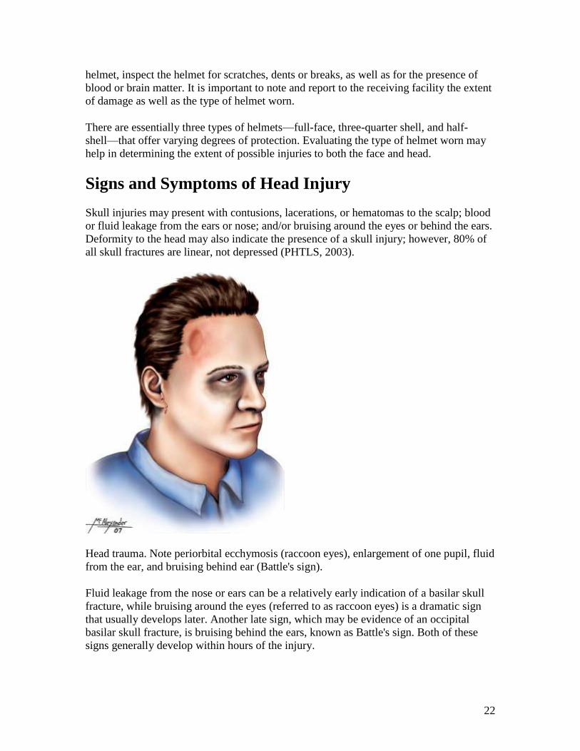
Skull injuries may present with contusions, lacerations, or hematomas to the scalp; blood
or fluid leakage from the ears or nose; and/or bruising around the eyes or behind the ears.
Deformity to the head may also indicate the presence of a skull injury; however, 80% of
all skull fractures are linear, not depressed (PHTLS, 2003).
Head trauma. Note periorbital ecchymosis (raccoon eyes), enlargement of one pupil, fluid
from the ear, and bruising behind ear (Battle's sign).
Fluid leakage from the nose or ears can be a relatively early indication of a basilar skull
fracture, while bruising around the eyes (referred to as raccoon eyes) is a dramatic sign
that usually develops later. Another late sign, which may be evidence of an occipital
basilar skull fracture, is bruising behind the ears, known as Battle's sign. Both of these
signs generally develop within hours of the injury.

23
Though skull fractures may make for a dramatic presentation, the underlying injury to the
brain, and the symptoms associated with that injury, dictate the appropriate management
of the patient.
Head injuries are categorized as open or closed. Both may produce an altered or
decreasing mental status demonstrated as confusion, disorientation, repetitive
questioning, or unresponsiveness. Other signs may include neurologic disability, nausea
and/or vomiting, seizures, or unequal pupils. Changes to the patient's breathing may also
be present depending on the location of the injury and the degree of intracranial pressure.
Though drugs, alcohol, and other altered physiology may result in unusual presentation of
the pupils, these findings, when associated with an altered mental status, may also
indicate the severity and location of a traumatic brain injury. In this setting, dilated
nonreactive pupils indicate a brain stem injury with a grim prognosis; however, if the
pupils are dilated but reactive, the injury may be reversible. Unequal pupils (anisocoria)
with one dilated but reactive pupil are a sign of increasing cerebral pressure, and
anisocoria with a dilated, nonreactive pupil is an extreme emergency mandating rapid
transport and hyperventilation (BTLS, 2004).
Emergency Medical Care
After taking Universal Precautions, the first step in management of the head-injured
patient is addressing any life-threatening conditions that may be present. After those
conditions have been addressed, the focus moves to managing cerebral perfusion by
maintaining adequate oxygenation of cerebral tissues and maintaining the patient's blood
pressure (PHTLS, 2003).
An initial assessment with spinal immobilization is done on scene, with a complete
detailed physical exam performed en route. Prehospital care for head-injured patients is
directed at preventing further injury or insult and delivering the patient to a higher level
of care for definitive treatment. Quick transport to an appropriate receiving facility is a
high priority.
Because altered patients may not be able to effectively maintain an open airway, airway
management is a priority for these patients. Supplemental oxygen is indicated for the
patient's hypoperfused cells. Some patients will require endotracheal intubation; however,
keep in mind that multiple attempts at intubation have been shown to increase intracranial
pressure (ICP).
Advanced airway management for head-injured patients should be performed by the most
experienced provider on scene. If a head-injured patient requires intubation, cervical
spinal precautions must be maintained throughout the procedure. Use of alternative
airway devices, such as the Combitube, King Laryngeal Tube, or EOA may be
considered, and may minimize manipulation of the cervical spine.
24
Open head injuries often bleed profusely. Usually the bleeding is from scalp injuries
rather than within the cranium. Bleeding is controlled with direct pressure and pressure
dressings; however, pressure should not be applied to an open or depressed skull injury.
Any impaled object is bandaged in place to minimize manipulation.
An open head injury may present with the presence of brain matter. The presence of brain
matter outside of the cranial vault is not in and of itself an indication of death. A patient
who is still breathing, and has a pulse, should be treated and transported in the same
manner as any other head-injured patient and treated in accordance with the assessment
findings.
Head-injured patients need to be frequently reassessed. As ICP increases, changes to the
patient's vital signs may indicate a need to adjust your treatment plan. If a patient who
was initially confused becomes unresponsive, you may consider intubation. If changes to
the pupils are noted, as discussed in the previous section, it may be appropriate to adjust
the ventilation rate you are delivering.
Assume that any patient with a head injury has an associated spinal injury. Spinal
precautions should be taken to reduce the risk of further exacerbating unidentified spinal
injuries.
RAPID EXTRICATION
Indications
Patients who are in immediate danger of death may require emergency rescue from a
vehicle or structure. Dangerous scenes such as those that involve active fire, rising
waters, or imminent risk of explosion or structure collapse may require the rescuer to
move a patient to safety emergently.
Patients who are unstable on initial assessment may also require rapid extrication. For
example, a patient involved in a motor vehicle collision who is hemorrhaging from a
traumatic injury may need to be rapidly extricated in order to manage the wound
effectively and prevent exsanguination (bleeding out).
Another situation in which rapid extrication may be warranted is when one patient is
blocking access to another seriously injured patient, requiring that the first patient be
moved to provide care to the second patient.
Rapid extrication should only be performed if there is an immediate risk of death or
serious injury to the patient or rescuer, and the reason for rapid extrication should be well
documented on the patient care report. Since the focus of rapid extrication is on moving
quickly, usually delaying full immobilization of the spine until the patient is removed
from the scene, it should only be performed in truly urgent cases.

25
Procedure
Multiple rescuers should be used to perform rapid extrication so that the risk of injury to
the patient is reduced as much as possible. Keep in mind that even though this is a rapid
extrication every effort must be made to limit movement of the spine.
To perform the extrication, one rescuer gets behind the patient and brings the cervical
spine into neutral in-line position while providing manual stabilization. A second rescuer
applies a cervical immobilization device as a third rescuer places a rigid spine board as
near the patient as possible. Working along the long axis of the patient, one rescuer
continues to hold and stabilize the head while a second rescuer supports the thorax and a
third rescuer frees the lower extremities from any obstacles. With the first and second
rescuers coordinating their movements to limit rotation of the spine, the patient is
maneuvered onto the spine board in short controlled movements.
As the patient is removed from the vehicle or structure, an external rescuer or bystander
should assume control of the patient's head and cervical spine as the patient is extricated.
If possible, a gurney should be as close as possible so that the board can be placed on the
gurney immediately when it is freed from the wreckage.
It may be necessary to move the patient to a safer location before fully securing the
patient to the board. This is accomplished more safely with the patient on the gurney
rather than being carried on the board. Once the rescuers and patient are no longer in
danger, the patient should be appropriately secured to the spine board and cervical
immobilization with a collar and head blocks completed as usual.