transposons - link.springer.com · transposons adam r. parksa and joseph e. petersb* amolecular...
TRANSCRIPT

Transposons
Adam R. Parksa and Joseph E. Petersb*aMolecular Control and Genetics Section, Gene Regulation and Chromosome Biology Laboratory, National CancerInstitute, National Institutes of Health, Frederick, MD, USAbDepartment of Microbiology, Cornell University, Ithaca, NY, USA
Synopsis
Transposons are mobile genetic elements that can move between different DNAmolecules or withinan individual DNA molecule. The donor and target DNA molecules do not require any sequencehomology for mobilization to occur. Transposons are often characterized by the biochemicalstrategy used to carry out DNA breaking and joining reactions. Recombinase families include theDDE-, HUH-, DEDD-type and serine transposases. Transposons may be removed from a donorDNA entirely by being “cut out,” or they may be left in place while a copy of the element is “copiedout.” During insertion, the element may be either “pasted in,” moved entirely into the recipientmolecule, or they may be “copied in.” Transposons that are copied out as RNA are referred to asretrotransposons, whereas transposons that move exclusively as a DNAmolecule and do not requirean RNA intermediate are simply referred to as DNA transposons. The assembly and coordination ofall of the components involved in transposition is a complex and highly regulated process.
Introduction
Transposons are mobile genetic elements that can move between different DNAmolecules or withinan individual DNA molecule. They are distinct from other mobile elements in that they are notrequired to move between sites that share sequence homology; they may move to sites with nosequence similarity at all. The most intensively studied type of recombinase that carries out thisfunction is the DDE-type transposase (also called an integrase in eukaryotes); however, there aremany other biochemical strategies for recombination. Transposons are often characterized by thebiochemical strategy used to carry out DNA breaking and joining reactions. These recombinasefamilies include the DDE-, HUH-, DEDD-type and serine transposases (Curcio and Derbyshire2003; Siguier et al. 2014). In addition to differences in biochemical strategies used for DNAcleavage and strand transfer, the overall mechanisms of element movement can differ significantly.Other details about the transposition process are also useful for comparing closely related elements.These details can include the sequence and arrangement of terminal inverted-repeat sequences,target-site duplications, arrangement of transposase and accessory genes, and target-site specificity.
A convenient way to think about different strategies that mobile elements use for mobility is toconsider how they are removed from their original, or donor, DNA molecule and how they areinserted into a new, target or recipient, DNA molecule (Curcio and Derbyshire 2003). Transposonsmay be removed from a donor DNA entirely by being “cut out,” or they may be left in place whilea copy of the element is “copied out” (Fig. 1). Similarly, elements may be either “pasted in,”movedentirely into the recipient molecule, or they may be “copied in.” The terms describing the element’s
*Email: [email protected]
Molecular Life SciencesDOI 10.1007/978-1-4614-6436-5_154-1# Springer Science+Business Media New York 2014
Page 1 of 14

removal and insertion are combined to describe the entire mobilization process, for example,a cut-and-paste transposon is removed entirely from the donor DNA molecule and pasted directlyinto a recipient DNA molecule. Transposons that are copied out as RNA are referred to asretrotransposons, whereas transposons that move exclusively as a DNAmolecule and do not requirean RNA intermediate are simply referred to as DNA transposons.
Anatomy of a Transposon
In their most basic form, the genetic structures of transposons are composed of terminal inverted-repeat sequences flanking a single open reading frame encoding a transposase. These simpleelements are called insertion sequences (IS) (Siguier et al. 2014). The inverted-repeat(IR) sequences at the ends of the element have the binding site or sites for the transposase protein(Fig. 2). If multiple binding sites are in the element, the most terminal site will indicate where thetransposase carries out strand breakage and joining. In simple IS elements, an identical IR that flanksthe element with a single transposase binding site may be all that is required; however, morecomplex elements often have multiple transposase binding sites at their ends. Some elements,such as Tn7, have different numbers of transposase binding sites in the right and left ends, allowingdistinction between left and right ends (Craig 2002). For Tn7, this characteristic has implications inregulation of transposition. IRs often contain additional information, such as a promoter for thetransposase gene.
a
b
c
d
Cut-Out
Copy-Out Copy-In
Paste-In
Mobile ElementMobile Element
Mobile Element
Mobile Element
Mobile Element
RNAP
3’
3’
5’
5’
Mob
ile E
lem
ent
Fig. 1 Examples of mobile elements that illustrate the cutout, copy-out, paste-in, and copy-in terms are shown. (a)Elements such as Tn10 and Tn5 use a cutout strategy to remove the element from donor DNA. The transposase (greencircles) “cuts out” the element from the donor DNA by joining the top and bottom strands at either end of the element,leaving behind a double-strand DNA break. (b) Retroelements such as Ty1 and L1 are “copied out” of donor DNA asRNA transcripts. (c) Mobile elements that are “pasted in,” such as Tn10 and Tn5, undergo a strand exchange reaction inwhich the DNA that encodes the element is joined with the target DNA, as opposed to a polymerase enzyme makinga copy of the element at the new site (see d copy-in). (d) Elements that are “copied in” require the activity of a DNApolymerase to make a new copy of the element at the target site, using the target-site DNA as a primer for synthesis andusing DNA or RNA element as a template. Tn3 is an example of an element that uses this strategy
Molecular Life SciencesDOI 10.1007/978-1-4614-6436-5_154-1# Springer Science+Business Media New York 2014
Page 2 of 14
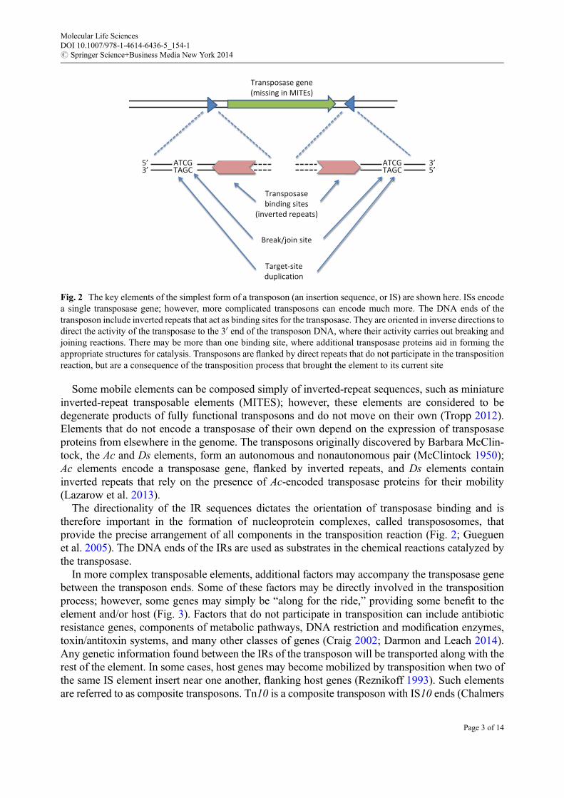
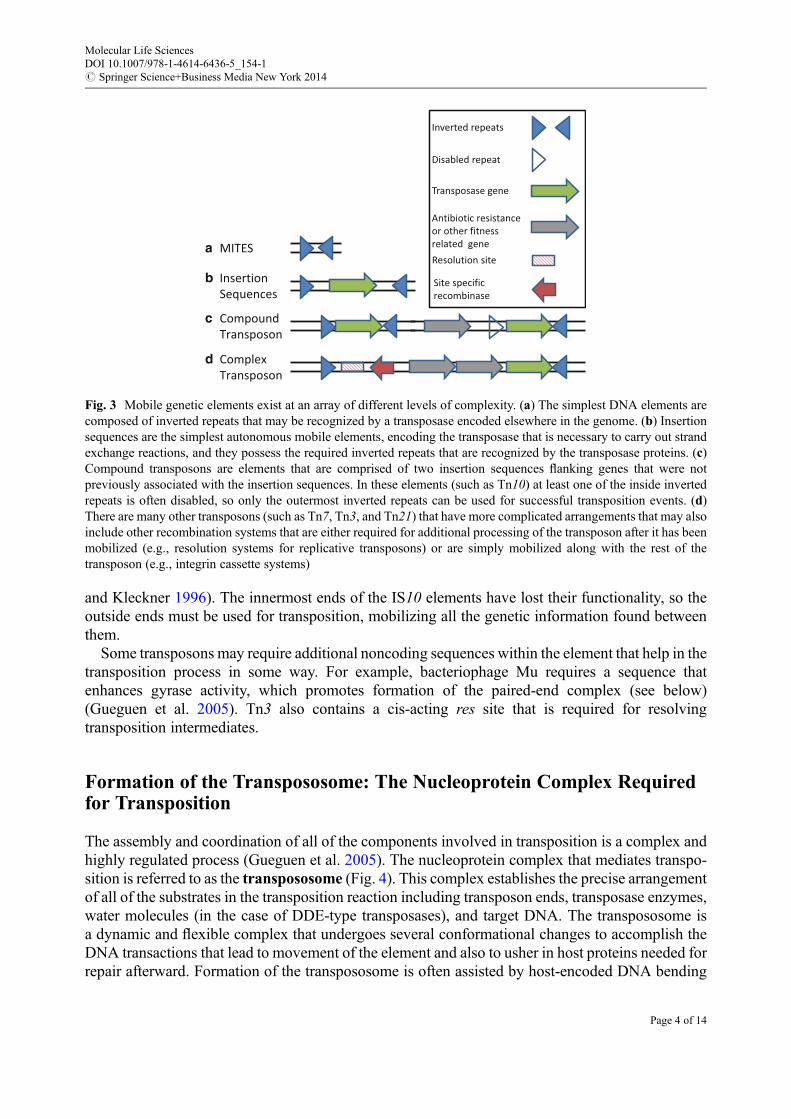
Some mobile elements can be composed simply of inverted-repeat sequences, such as miniatureinverted-repeat transposable elements (MITES); however, these elements are considered to bedegenerate products of fully functional transposons and do not move on their own (Tropp 2012).Elements that do not encode a transposase of their own depend on the expression of transposaseproteins from elsewhere in the genome. The transposons originally discovered by Barbara McClin-tock, the Ac and Ds elements, form an autonomous and nonautonomous pair (McClintock 1950);Ac elements encode a transposase gene, flanked by inverted repeats, and Ds elements containinverted repeats that rely on the presence of Ac-encoded transposase proteins for their mobility(Lazarow et al. 2013).
The directionality of the IR sequences dictates the orientation of transposase binding and istherefore important in the formation of nucleoprotein complexes, called transpososomes, thatprovide the precise arrangement of all components in the transposition reaction (Fig. 2; Gueguenet al. 2005). The DNA ends of the IRs are used as substrates in the chemical reactions catalyzed bythe transposase.
In more complex transposable elements, additional factors may accompany the transposase genebetween the transposon ends. Some of these factors may be directly involved in the transpositionprocess; however, some genes may simply be “along for the ride,” providing some benefit to theelement and/or host (Fig. 3). Factors that do not participate in transposition can include antibioticresistance genes, components of metabolic pathways, DNA restriction and modification enzymes,toxin/antitoxin systems, and many other classes of genes (Craig 2002; Darmon and Leach 2014).Any genetic information found between the IRs of the transposon will be transported along with therest of the element. In some cases, host genes may become mobilized by transposition when two ofthe same IS element insert near one another, flanking host genes (Reznikoff 1993). Such elementsare referred to as composite transposons. Tn10 is a composite transposon with IS10 ends (Chalmers
ATCGTAGC
ATCGTAGC
5’3’ 5’
3’
Target-siteduplication
Transposasebinding sites
(inverted repeats)
Break/join site
Transposase gene(missing in MITEs)
Fig. 2 The key elements of the simplest form of a transposon (an insertion sequence, or IS) are shown here. ISs encodea single transposase gene; however, more complicated transposons can encode much more. The DNA ends of thetransposon include inverted repeats that act as binding sites for the transposase. They are oriented in inverse directions todirect the activity of the transposase to the 30 end of the transposon DNA, where their activity carries out breaking andjoining reactions. There may be more than one binding site, where additional transposase proteins aid in forming theappropriate structures for catalysis. Transposons are flanked by direct repeats that do not participate in the transpositionreaction, but are a consequence of the transposition process that brought the element to its current site
Molecular Life SciencesDOI 10.1007/978-1-4614-6436-5_154-1# Springer Science+Business Media New York 2014
Page 3 of 14
and Kleckner 1996). The innermost ends of the IS10 elements have lost their functionality, so theoutside ends must be used for transposition, mobilizing all the genetic information found betweenthem.
Some transposons may require additional noncoding sequences within the element that help in thetransposition process in some way. For example, bacteriophage Mu requires a sequence thatenhances gyrase activity, which promotes formation of the paired-end complex (see below)(Gueguen et al. 2005). Tn3 also contains a cis-acting res site that is required for resolvingtransposition intermediates.
Formation of the Transpososome: The Nucleoprotein Complex Requiredfor Transposition
The assembly and coordination of all of the components involved in transposition is a complex andhighly regulated process (Gueguen et al. 2005). The nucleoprotein complex that mediates transpo-sition is referred to as the transpososome (Fig. 4). This complex establishes the precise arrangementof all of the substrates in the transposition reaction including transposon ends, transposase enzymes,water molecules (in the case of DDE-type transposases), and target DNA. The transpososome isa dynamic and flexible complex that undergoes several conformational changes to accomplish theDNA transactions that lead to movement of the element and also to usher in host proteins needed forrepair afterward. Formation of the transpososome is often assisted by host-encoded DNA bending
MITES
Insertion Sequences
Compound Transposon
Inverted repeats
Transposase gene
Disabled repeat
Antibiotic resistanceor other fitnessrelated geneResolution site
Site specific recombinase
Complex Transposon
a
b
c
d
Fig. 3 Mobile genetic elements exist at an array of different levels of complexity. (a) The simplest DNA elements arecomposed of inverted repeats that may be recognized by a transposase encoded elsewhere in the genome. (b) Insertionsequences are the simplest autonomous mobile elements, encoding the transposase that is necessary to carry out strandexchange reactions, and they possess the required inverted repeats that are recognized by the transposase proteins. (c)Compound transposons are elements that are comprised of two insertion sequences flanking genes that were notpreviously associated with the insertion sequences. In these elements (such as Tn10) at least one of the inside invertedrepeats is often disabled, so only the outermost inverted repeats can be used for successful transposition events. (d)There are many other transposons (such as Tn7, Tn3, and Tn21) that have more complicated arrangements that may alsoinclude other recombination systems that are either required for additional processing of the transposon after it has beenmobilized (e.g., resolution systems for replicative transposons) or are simply mobilized along with the rest of thetransposon (e.g., integrin cassette systems)
Molecular Life SciencesDOI 10.1007/978-1-4614-6436-5_154-1# Springer Science+Business Media New York 2014
Page 4 of 14
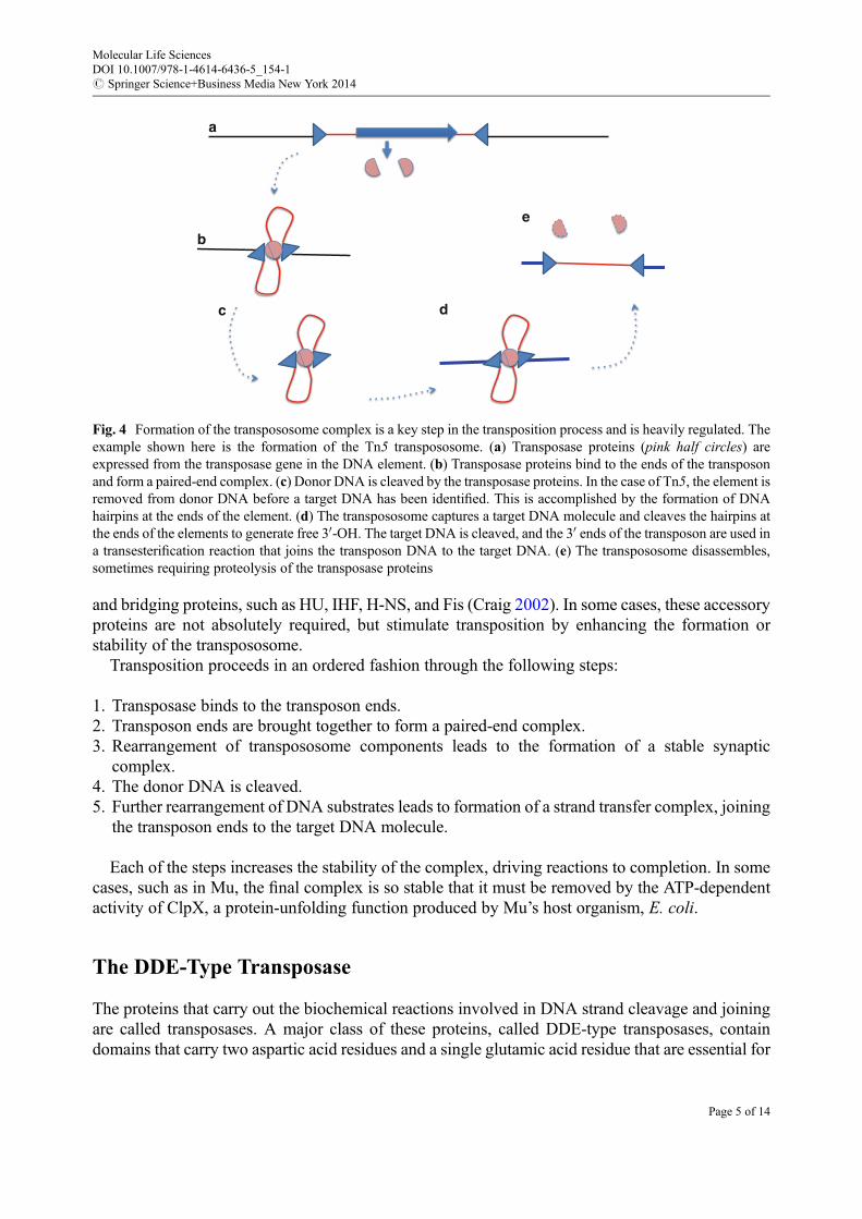
and bridging proteins, such as HU, IHF, H-NS, and Fis (Craig 2002). In some cases, these accessoryproteins are not absolutely required, but stimulate transposition by enhancing the formation orstability of the transpososome.
Transposition proceeds in an ordered fashion through the following steps:
1. Transposase binds to the transposon ends.2. Transposon ends are brought together to form a paired-end complex.3. Rearrangement of transpososome components leads to the formation of a stable synaptic
complex.4. The donor DNA is cleaved.5. Further rearrangement of DNA substrates leads to formation of a strand transfer complex, joining
the transposon ends to the target DNA molecule.
Each of the steps increases the stability of the complex, driving reactions to completion. In somecases, such as in Mu, the final complex is so stable that it must be removed by the ATP-dependentactivity of ClpX, a protein-unfolding function produced by Mu’s host organism, E. coli.
The DDE-Type Transposase
The proteins that carry out the biochemical reactions involved in DNA strand cleavage and joiningare called transposases. A major class of these proteins, called DDE-type transposases, containdomains that carry two aspartic acid residues and a single glutamic acid residue that are essential for
a
b
c d
e
Fig. 4 Formation of the transpososome complex is a key step in the transposition process and is heavily regulated. Theexample shown here is the formation of the Tn5 transpososome. (a) Transposase proteins (pink half circles) areexpressed from the transposase gene in the DNA element. (b) Transposase proteins bind to the ends of the transposonand form a paired-end complex. (c) Donor DNA is cleaved by the transposase proteins. In the case of Tn5, the element isremoved from donor DNA before a target DNA has been identified. This is accomplished by the formation of DNAhairpins at the ends of the element. (d) The transpososome captures a target DNA molecule and cleaves the hairpins atthe ends of the elements to generate free 30-OH. The target DNA is cleaved, and the 30 ends of the transposon are used ina transesterification reaction that joins the transposon DNA to the target DNA. (e) The transpososome disassembles,sometimes requiring proteolysis of the transposase proteins
Molecular Life SciencesDOI 10.1007/978-1-4614-6436-5_154-1# Springer Science+Business Media New York 2014
Page 5 of 14

the coordination of two divalent magnesium ions, similar to the arrangement used by RNAseH enzymes (Fig. 5). Some elements actually use slight variations on this theme, such as DDD,DDN, or DDH, but function in the same way (Dyda et al. 2012; Siguier et al. 2014). The magnesiumions function in stabilizing the transition state that is required both for cleavage of the DNAbackbone in the donor DNA molecule and for mediating the strand exchange reaction that joinsthis newly cleaved DNAwith the target DNA. In strand cleavage, a single water molecule is used asa nucleophile in a reaction that generates a free 30-OH group at the end of the transposon. This 30-OHis, in turn, used as the nucleophile in joining the transposon end to the target DNA molecule. Thisreaction is referred to as a transesterification reaction because it results in the exchange ofphosphodiester bonds between the sugar–phosphate backbone of donor and target DNA molecules.The result is one strand of DNA, from the end of the transposon, joined within a new targetDNA. The target DNA now has a free 30-OH group where target DNA has been exchanged withthe transposon end. The remaining 30-OH group is then used by the host DNA polymerase and ligaseenzymes to restore the target DNA molecule to a fully double-stranded DNA form.
The activities of the transposases must be coordinated at both ends of the element to ensure thatthe entire transposition reaction goes to completion (Nagy and Chandler 2004). For this reason, bothends of the transposon, bound by the transposase proteins, are drawn together in a nucleoproteincomplex that sometimes includes the target DNA molecule as well. The assembly of this nucleo-protein complex ensures that the breaking and joining reactions can be coordinated at both ends ofthe element, prevents insertion of one end of the element to a site that is internal to the transposon,and enables both ends of the transposon to insert at the same site in the target DNA, as opposed tosites that are distant from one another, which would potentially result in large deletions (Darmon andLeach 2014). Since both transposon ends, along with the transposase proteins, cannot occupy thesame physical space on the DNA, the sites of strand exchange are staggered, typically by 2–9 nt.
OO
OO
O
O
HO P
O
H
Mg2+
Mg2+
3’
5’3’
5’
BASE
BASE
E
DDTransposase
P
O
OH
OO
P
O
OH
HOO
P
O
OH
OO
HO
OHH
+
P
O
OH
HOO
HO
OHH
+
Fig. 5 DDE-type transposase proteins contain a highly conserved aspartate (D), aspartate (D), and glutamate (E) motifthat allows them to coordinate two divalent magnesium cations. This arrangement is characteristic of RNase H-likedomains. The magnesium ions stabilize the transition state of the DNA hydrolysis and transesterification reactionsinvolved in DNA strand exchange. The sequence of reactions is shown in the inset, where black lines represent donorDNA, and thick green lines represent target DNA
Molecular Life SciencesDOI 10.1007/978-1-4614-6436-5_154-1# Springer Science+Business Media New York 2014
Page 6 of 14

This principle of transposons leads to one of the hallmarks of the transposition process, target-siteduplications. The staggering of the join sites leads to duplications because the top strand of the targetis joined to one end of the transposon and the bottom strand is joined to the opposite end. Followingrepair of the gap, DNA sequence derived from the top strand of the target will be present at one endof the element, and identical DNA sequence information derived from the bottom strand of the targetsite will be present at the other end. The target-site duplications are generally 2–9 nt in length, butcan sometimes be as long as �250 nt, depending on the distance between top- and bottom-strandjoining.
At its simplest form, joining both ends of the transposon to the target DNA is enough to mobilizethe element (Fig. 6). If strand transfer at the ends of the element is not accompanied by cleavage ofthe strand that has not been transferred, the so-called second strand, then the target DNA and thedonor DNA will remain joined in a cointegrated DNA molecule (Tropp 2012). As previouslymentioned, the 30-OH remaining at both ends flanking the element after cleavage of the targetDNA molecule are used to prime DNA synthesis from both ends, proceeding through the DNAelement and ending where the opposite end of the element was joined. Since DNA replication isinvolved in inserting the DNA element at the new site, this mechanism is called replicativetransposition. The element is both copied out of the donor DNA molecule and copied into the targetDNA molecule, so it is considered a copy-out and copy-in mechanism. In some cases, such as Tn3and Tn917, the cointegration of donor DNA and target DNA is resolved by a separate site-specificrecombination system. In other elements, such as bacteriophage Mu, the cointegrate is leftunresolved.
Mobile Element
Mobile Element Mobile Element
Mobile Element
Mobile Element
Mobile Element
Mobile Element
Mobile Element
Mobile Element
Mobile Element
a b
c d
e
Fig. 6 Second strand cleavage (at the 50 end of the element) can be carried out by (a) formation of DNA hairpinstructures at the ends of the transposon DNA (e.g., Tn5 and Tn10). (b) Cleavage by a second protein that is dedicatedonly to nicking the DNA at the 50 end (e.g., TnsA of Tn7). (c) Cleavage in an event separate from the transesterificationreaction, mediated by the transposase protein (e.g., Mos1). (d) Asymmetrical cleavage of one DNA strand, generatinga circular intermediate that must be resolved by DNA replication. (e) Transcription and subsequent reverse transcriptionof the element (e.g., Ty1, HIV Int). This mechanism still requires 30 processing by the integrase to generate a new 30-OH
Molecular Life SciencesDOI 10.1007/978-1-4614-6436-5_154-1# Springer Science+Business Media New York 2014
Page 7 of 14
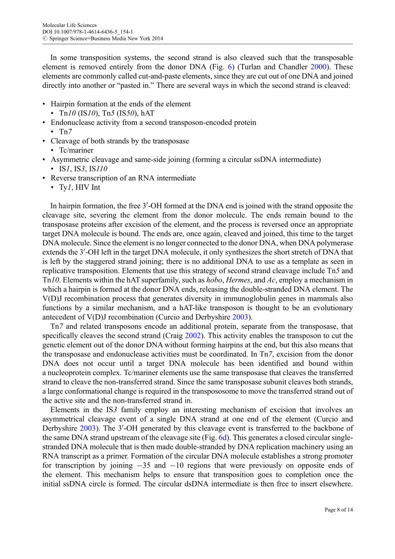
In some transposition systems, the second strand is also cleaved such that the transposableelement is removed entirely from the donor DNA (Fig. 6) (Turlan and Chandler 2000). Theseelements are commonly called cut-and-paste elements, since they are cut out of one DNA and joineddirectly into another or “pasted in.” There are several ways in which the second strand is cleaved:
• Hairpin formation at the ends of the element• Tn10 (IS10), Tn5 (IS50), hAT
• Endonuclease activity from a second transposon-encoded protein• Tn7
• Cleavage of both strands by the transposase• Tc/mariner
• Asymmetric cleavage and same-side joining (forming a circular ssDNA intermediate)• IS1, IS3, IS110
• Reverse transcription of an RNA intermediate• Ty1, HIV Int
In hairpin formation, the free 30-OH formed at the DNA end is joined with the strand opposite thecleavage site, severing the element from the donor molecule. The ends remain bound to thetransposase proteins after excision of the element, and the process is reversed once an appropriatetarget DNA molecule is bound. The ends are, once again, cleaved and joined, this time to the targetDNAmolecule. Since the element is no longer connected to the donor DNA, when DNA polymeraseextends the 30-OH left in the target DNA molecule, it only synthesizes the short stretch of DNA thatis left by the staggered strand joining; there is no additional DNA to use as a template as seen inreplicative transposition. Elements that use this strategy of second strand cleavage include Tn5 andTn10. Elements within the hATsuperfamily, such as hobo,Hermes, and Ac, employ a mechanism inwhich a hairpin is formed at the donor DNA ends, releasing the double-stranded DNA element. TheV(D)J recombination process that generates diversity in immunoglobulin genes in mammals alsofunctions by a similar mechanism, and a hAT-like transposon is thought to be an evolutionaryantecedent of V(D)J recombination (Curcio and Derbyshire 2003).
Tn7 and related transposons encode an additional protein, separate from the transposase, thatspecifically cleaves the second strand (Craig 2002). This activity enables the transposon to cut thegenetic element out of the donor DNAwithout forming hairpins at the end, but this also means thatthe transposase and endonuclease activities must be coordinated. In Tn7, excision from the donorDNA does not occur until a target DNA molecule has been identified and bound withina nucleoprotein complex. Tc/mariner elements use the same transposase that cleaves the transferredstrand to cleave the non-transferred strand. Since the same transposase subunit cleaves both strands,a large conformational change is required in the transpososome to move the transferred strand out ofthe active site and the non-transferred strand in.
Elements in the IS3 family employ an interesting mechanism of excision that involves anasymmetrical cleavage event of a single DNA strand at one end of the element (Curcio andDerbyshire 2003). The 30-OH generated by this cleavage event is transferred to the backbone ofthe same DNA strand upstream of the cleavage site (Fig. 6d). This generates a closed circular single-stranded DNA molecule that is then made double-stranded by DNA replication machinery using anRNA transcript as a primer. Formation of the circular DNA molecule establishes a strong promoterfor transcription by joining �35 and �10 regions that were previously on opposite ends ofthe element. This mechanism helps to ensure that transposition goes to completion once theinitial ssDNA circle is formed. The circular dsDNA intermediate is then free to insert elsewhere.
Molecular Life SciencesDOI 10.1007/978-1-4614-6436-5_154-1# Springer Science+Business Media New York 2014
Page 8 of 14

This seemingly complicated mechanism is among the most common and is often referred to asa copy-out and paste-in strategy.
When RNA Is an Intermediate: Retrotransposons
Retroelements are copied out of the donor DNA molecule as an RNA by a host-encoded RNApolymerase (Fig. 6e). In order to be inserted back into a new DNA molecule, the RNA moleculemust be converted to DNA; host genetic systems do not tolerate a mix of RNA and DNAwithin theirgenomes. This activity is accomplished by a reverse transcriptase that is typically encoded by theelement itself. In some cases elements may use reverse-transcriptase proteins that are encoded byother mobile elements, or in the case of plants, these reverse transcriptases may actually be encodedwithin the host genome. The retrotransposons are comprised of two major groups, the LTR (longterminal repeat) transposons and the non-LTR transposons (Craig 2002). Their names refer todifferences in the sequence composition of the elements at their ends; however, the mechanismsby which these elements are mobilized are much more profound. LTR elements move by a copy-outand paste-in mechanism, while non-LTR elements move in a copy-out and copy-in manner.
LTR transposons contain long repeated sequences that delineate the ends of the element and willeventually serve as a binding site for proteins that are involved in the elements’ insertion into a newDNA molecule. These elements are evolutionarily related to retroviruses, and many (e.g., the yeastTy1, Ty3, and Tf1) also contain functional proteins that enable them to make “viruslike particles”(VLPs) outside the nucleus, where they are processed by the reverse-transcriptase protein (oftenreferred to as “Pol”) (Curcio and Derbyshire 2003; Tropp 2012). The similarity betweenretrotransposons and retrovirus makes them useful systems for the study of key functions that affectretroviral replication. These elements are typically transcribed by the host’s RNA polymerase IIenzyme, which is responsible for transcription of most cellular mRNAs. After transcription theelements are converted from RNA to DNA by the activity of the reverse-transcriptase protein.A DDE-type recombinase protein (often called “integrase” with these elements) binds to the LTRregions at the ends of the element, activating them for transposition by exposing free 30-OH groups atthe 30 end of both strands. The protein–DNA complex then identifies an appropriate target site, andthe integrase mediates a strand exchange reaction in which the activated 30-OH ends are used asa nucleophile to attack the 50 PO4 within the backbone of the recipient DNA molecule. This processhappens simultaneously at both ends, but at staggered sites on the recipient DNA molecule. Sincethe integration of the ends is staggered, a duplication of the target-site sequence results, one copy justoutside each end of the element.
DEDD Transposases
Some elements use transposases that resemble RuvC-like enzymes that are involved in resolution ofHolliday junctions that are homologous recombination intermediates (Siguier et al. 2014). Very littleis known about how DEDD-type transposases function in transposition, but given their similarity toRuvC, it is likely that this transposition mechanism also involves Holliday junction formation.RuvC, as well as DEDD-type transposases, contains a conserved RNase H fold, just as DDE-typetransposases do (Buchner et al. 2005). These recombinases do not generate target-site duplications(Prosseda et al. 2006). IS110 is an example of a transposon that encodes DEDD-type transposase.
Molecular Life SciencesDOI 10.1007/978-1-4614-6436-5_154-1# Springer Science+Business Media New York 2014
Page 9 of 14

HUH Transposases
Transposons that employ HUH-type transposases function by a mechanism that is very differentfrom DDE- and DEDD-type transposase. HUH transposases form covalent phosphotyrosine inter-mediates during the process of transposition (Fig. 7; Chandler et al. 2013; Dyda et al. 2012).Superficially, this mechanism is reminiscent of tyrosine recombinase reactions used in some site-specific recombination systems; however, HUH transposases and tyrosine recombinases areunrelated. This class of transposition does not generate target-site duplications. HUH transposasesleave behind a free 30-OH in the donor DNA, following strand cleavage, which can be extended byDNA replication machinery, whereas tyrosine site-specific recombinases leave behind a 50-OH,which cannot be extended. This distinction is significant in that DNA replication plays an importantrole in Y2 transposons that are part of the HUH family. As in other transposition systems, HUHtransposons do not require specific binding sites for mobilization. This class of element is mecha-nistically related to relaxases or Rep proteins that are involved in the transfer of conjugal plasmidsand replication of rolling-circle plasmids, respectively. HUH transposases receive their name basedon the conserved histidine (H)–bulky hydrophobic residue (U)–histidine motif that is important fortheir function. These residues form a motif that helps coordinate a divalent metal ion, usually Mg2+
or Mn2+, which is used in cleavage of the donor DNA backbone, an arrangement similar to that seenin DDE-type transposases. An additional key feature of these transposases includes at least oneconserved tyrosine, or serine, that is used to form a covalent protein–DNA bond. Transposons thatattach to DNA by a single tyrosine are called Y1 transposons. Those that involve phosphoserinelinkage are called S1 transposons. Yet another class of transposase uses covalent linkage with twoseparate tyrosine residues and is referred to as Y2 transposons.
Fig. 7 HUH transposases generate a covalent 50 phosphotyrosine intermediate (Y) to cleave DNA. The conservedhistidine residues (H) in these transposases are used to help coordinate a metal ion that is used in the activation andcoordination of the DNA nicking reaction. In this example, glutamine (Q) is also involved in coordinating the metal ion.Red arrows indicate the transfer of electrons in the process. The process is reversed to rejoin DNA ends
Molecular Life SciencesDOI 10.1007/978-1-4614-6436-5_154-1# Springer Science+Business Media New York 2014
Page 10 of 14
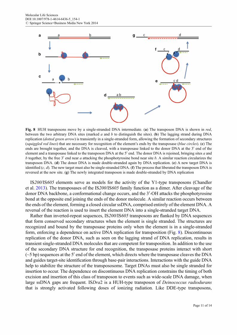
IS200/IS605 elements serve as models for the activity of the Y1-type transposons (Chandleret al. 2013). The transposases of the IS200/IS605 family function as a dimer. After cleavage of thedonor DNA backbone, a conformational change occurs, and the 30-OH attacks the phosphotyrosinebond at the opposite end joining the ends of the donor molecule. A similar reaction occurs betweenthe ends of the element, forming a closed circular ssDNA, comprised entirely of the element DNA. Areversal of the reaction is used to insert the element DNA into a single-stranded target DNA.
Rather than inverted-repeat sequences, IS200/IS605 transposons are flanked by DNA sequencesthat form conserved secondary structures when the element is single stranded. The structures arerecognized and bound by the transposase proteins only when the element is in a single-strandedform, enforcing a dependence on active DNA replication for transposition (Fig. 8). Discontinuousreplication of the donor DNA, such as seen on the lagging strand of DNA replication, results intransient single-stranded DNA molecules that are competent for transposition. In addition to the useof the secondary DNA structure for end recognition, the transposase proteins interact with short(~5 bp) sequences at the 50 end of the element, which directs where the transposase cleaves the DNAand guides target-site identification through base-pair interactions. Interactions with the guide DNAhelp to stabilize the structure of the transpososome. Target DNAs must also be single stranded forinsertion to occur. The dependence on discontinuous DNA replication constrains the timing of bothexcision and insertion of this class of transposon to events such as wide-scale DNA damage, whenlarge ssDNA gaps are frequent. ISDra2 is a HUH-type transposon of Deinococcus radioduransthat is strongly activated following doses of ionizing radiation. Like DDE-type transposons,
a b
Y
Y
a b
a b
c d
Y
Y
c d
c da
b
c
d
e
f
g
Fig. 8 HUH transposons move by a single-stranded DNA intermediate. (a) The transposon DNA is shown in red,between the two arbitrary DNA sites (marked a and b to distinguish the sites). (b) The lagging strand during DNAreplication (dotted green arrow) is transiently in a single-stranded form, allowing the formation of secondary structures(squiggled red lines) that are necessary for recognition of the element’s ends by the transposase (blue circles). (c) Theends are brought together, and the DNA is cleaved, with a transposase linked to the donor DNA at the 30 end of theelement and a transposase linked to the transposon DNA at the 50 end. The donor DNA is rejoined, bringing sites a andb together, by the free 30 end near a attacking the phosphotyrosine bond near site b. A similar reaction circularizes thetransposon DNA. (d) The donor DNA is made double-stranded again by DNA replication. (e) A new target DNA isidentified (c, d). The new target must also be single-stranded DNA. (f) The process that liberated the transposon DNA isreversed at the new site. (g) The newly integrated transposon is made double-stranded by DNA replication
Molecular Life SciencesDOI 10.1007/978-1-4614-6436-5_154-1# Springer Science+Business Media New York 2014
Page 11 of 14
IS200/IS605 have nonautonomous derivatives. Small IS elements that lack transposase genes havebeen identified and referred to as bacterial interspersed mosaic elements (BIMEs). These elementscan alter gene regulation and higher-order DNA structure and can serve as specific target sites forcertain IS elements.
Y2 transposons include the IS91 family and the helitrons of eukaryotes (Curcio and Derbyshire2003). Not many detailed descriptions of these elements are available; however, they are presumedto mobilize by a mechanism that resembles rolling-circle replication of some plasmids. Two tyrosineresidues within the recombinase protein are essential for transposition, hence the name Y2. BothssDNA and dsDNA closed circle intermediates have been detected. As previously mentioned, donorstrand cleavage generates a free 30-OH that is used to prime DNA replication. Leading-strand DNAreplication is thought to displace the element. As a consequence of the inefficient termination ofreplication in these elements, ~1 % of the mobilized elements also transfer genetic information fromthe donor molecule that lies adjacent to the typical end of the Y2 element. IS91 elements do containinverted repeats, which are involved in end recognition.
Serine Transposases
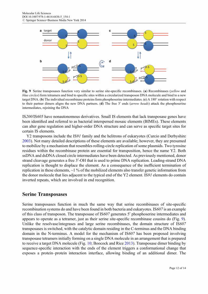
Serine transposases function in much the same way that serine recombinases of site-specificrecombination systems do and have been found in both bacteria and eukaryotes. IS607 is an exampleof this class of transposon. The transposase of IS607 generates 50 phosphoserine intermediates andappears to operate as a tetramer, just as their serine site-specific recombinase cousins do (Fig. 9).Unlike the resolvase/integrases and large serine recombinases, the domain structure of IS607transposases is switched, with the catalytic domain residing in the C-terminus and the DNA bindingdomain in the N-terminus. A model for the mechanism of IS607 has been proposed involvingtransposase tetramers initially forming on a single DNAmolecule in an arrangement that is preparedto receive a target DNA molecule (Fig. 10; Boocock and Rice 2013). Transposase dimer binding bysequence-specific interaction with the ends of the element triggers a conformational change thatexposes a protein–protein interaction interface, allowing binding of an additional dimer. The
S
S
S
O-S
S
S
S
SS
S-O PP
O-SPS-O P
O-S
S-O P
P
O-SP
S-O P
targeta
b
c
d
donor
Fig. 9 Serine transposases function very similar to serine site-specific recombinases. (a) Recombinases (yellow andblue circles) form tetramers and bind to specific sites within a circularized transposon DNA molecule and bind to a newtarget DNA. (b) The individual recombinase proteins form phosphoserine intermediates. (c) A 180� rotation with respectto their partner dimers aligns the new DNA partners. (d) The free 30 ends (arrow heads) attack the phosphoserineintermediates, rejoining the DNA
Molecular Life SciencesDOI 10.1007/978-1-4614-6436-5_154-1# Springer Science+Business Media New York 2014
Page 12 of 14

assembly of the tetramer positions the DNA binding domains of newly arrived dimer, making themready to bind target DNA. This preassembly step may increase the affinity of the complex forrandom DNA sequence, as opposed to the stepwise assembly of inactive dimers seen in site-specificrecombination. This model assumes that the randomDNA binding ability of each monomer is weak,but the overall complex is of sufficient stability to coordinate the DNA interaction needed in thereaction. The assembled IS607 transposase tetramers then bridge donor and target DNAs and cleaveDNA in trans – the DNA binding domains of monomers that bind to the target DNA – whereas thecatalytic domain of the same monomer cleaves the donor DNA, and vice versa, much like howmanyDDE-type transposases operate. Just as seen in other serine recombinases, the transposase formsa phosphoserine covalent bond. After cleavage, a 180� rotation of the two transposase dimersrepositions target and donor DNA ends, and the reverse reaction is carried out. The ends of thetransposon are then joined with the target DNA. Further study of IS607-like elements is required toconfirm this model.
Cross-References
▶DNA Repair Polymerases▶DNA Replication▶Double-strand Break Repair▶Homologous Recombination in Lesion Bypass▶Mechanisms of DNA Recombination▶V(D)J Recombination
Fig. 10 IS607 is an example of a serine transposon. (a) Transposition occurs through a circular double-stranded DNAintermediate. The transposase proteins have an N-terminal DNA binding domain (inset, blue rectangle) and a C-terminalcatalytic domain (inset, blue circle). (b) A dimer of transposase proteins (blue) binds a specific site on the circularizedtransposon DNA molecule. This dimer recruits an additional dimer to make a tetramer. (c) Since the DNA bindingdomains are preassembled, the nonspecific DNA binding ability of the tetramer is much greater than that of the dimer, soit can now bind to target DNA nonspecifically. (d) Strand exchange reactions join transposon and target DNAmolecules
Molecular Life SciencesDOI 10.1007/978-1-4614-6436-5_154-1# Springer Science+Business Media New York 2014
Page 13 of 14
References
Boocock MR, Rice PA (2013) A proposed mechanism for IS607-family serine transposases. MobDNA 4:24
Buchner JM, Robertson AE, Poynter DJ, Denniston SS, Karls AC (2005) Piv site-specific invertaserequires a DEDD motif analogous to the catalytic center of the RuvC Holliday junctionresolvases. J Bacteriol 187:3431–3437
Chalmers RM, Kleckner N (1996) IS10/Tn10 transposition efficiently accommodates diversetransposon end configurations. EMBO J 15:5112–5122
Chandler M, de la Cruz F, Dyda F, Hickman AB, Moncalian G, Ton-Hoang B (2013) Breaking andjoining single-stranded DNA: the HUH endonuclease superfamily. Nat Rev Microbiol11:525–538
Craig NL (2002) Mobile DNA II. ASM Press, Washington, DCCurcio MJ, Derbyshire KM (2003) The outs and ins of transposition: from mu to kangaroo. Nat Rev
Mol Cell Biol 4:865–877Darmon E, Leach DR (2014) Bacterial genome instability. Microbiol Mol Biol Rev 78:1–39Dyda F, Chandler M, Hickman AB (2012) The emerging diversity of transpososome architectures.
Q Rev Biophys 45:493–521Gueguen E, Rousseau P, Duval-Valentin G, Chandler M (2005) The transpososome: control of
transposition at the level of catalysis. Trends Microbiol 13:543–549Lazarow K, Doll ML, Kunze R (2013) Molecular biology of maize Ac/Ds elements: an overview.
Methods Mol Biol 1057:59–82McClintock B (1950) The origin and behavior of mutable loci in maize. Proc Natl Acad Sci U S
A 36:344–355Nagy Z, Chandler M (2004) Regulation of transposition in bacteria. Res Microbiol 155:387–398Prosseda G, Latella MC, Casalino M, Nicoletti M, Michienzi S, Colonna B (2006) Plasticity of the
P junc promoter of ISEc11, a new insertion sequence of the IS1111 family. J Bacteriol188:4681–4689
Reznikoff WS (1993) The Tn5 transposon. Annu Rev Microbiol 47:945–963Siguier P, Gourbeyre E, Chandler M (2014) Bacterial insertion sequences: their genomic impact and
diversity. FEMS Microbiol Rev doi: 10.1111/1574-6976.12067. [Epub ahead of print] 1–28Tropp BE (2012) Molecular biology: genes to proteins, 4th edn. Jones & Bartlett Learning, SudburyTurlan C, Chandler M (2000) Playing second fiddle: second-strand processing and liberation of
transposable elements from donor DNA. Trends Microbiol 8:268–274
Molecular Life SciencesDOI 10.1007/978-1-4614-6436-5_154-1# Springer Science+Business Media New York 2014
Page 14 of 14