transporters in the blood-brain barrier
TRANSCRIPT

5
Transporters in the Blood-Brain Barrier
Masaki Ueno, Toshitaka Nakagawa and Haruhiko Sakamoto Faculty of Medicine, Kagawa University
Japan
1. Introduction
The mammalian brain restricts the entrance of ions and solutes circulating in the
bloodstream by two cellular barriers, namely, the blood-brain barrier (BBB) and blood-
cerebrospinal fluid (CSF) barrier (Brightman et al., 1970; Ballabh, 2004). The BBB is built up
by a monolayer of endothelial cells (ECs) lining the brain capillaries that restricts the
movement of small polar molecules and macromolecules between the blood and the brain
interstitial fluid (Reese & Karnovsky, 1967; Brightman & Reese, 1969; Vorbrodt, 1988). The
endothelial barrier is supplemented with capillary pericytes that share the basement
membrane with the ECs. Moreover, perivascular end-feet of the astrocyte almost totally
cover the abluminal surface of the microvascular basement membrane. The blood-CSF
barrier is built up by a monolayer of epithelial cells of the choroid plexus separating the
blood from the CSF. This blood-CSF epithelial barrier is of great functional importance
because the fenestrated endothelium of the choroid plexus capillaries is leaky and
permeable to blood-borne solutes. Although the choroid plexus is traditionally considered
the major component of the blood-CSF barrier, a similar barrier is formed by the functional
complexes between the arachnoid cells. This barrier is also important because substances
passing into the stroma of the choroid plexus after intravenous presentation may find their
way into the CSF by crossing the ependyma adjacent to the root of the choroid plexus; this is
as described as a “functional leak” by Brightman et al. (1970), while van Deurs (1978) denies
the existence of this functional leak. These barriers maintain a constant chemical
environment within the central nervous system (CNS), which is optimal for the function of
neurons.
The brain capillaries were characterized morphologically as the site of the BBB by Reese and Karnovsky (1967) after introduction of electron microscopy and the use of horseradish peroxidase as a macromolecular tracer. Further ultrastructural studies (van Deurs, 1980; Brightman, 1989) revealed that the continuous endothelium of brain capillaries possesses several unique structural and functional features (Vorbrodt & Dobrogowska, 2003). First, the paracellular cleft between adjacent ECs is sealed by continuous strands of tight junctions (TJs). Second, the endocytic (pinocytic) and transcytotic activities are very low, and therefore, the transendothelial traffic of solutes (via plasmalemmal vesicles) is low. Third, the uptake of essential nutrients from the bloodstream into the brain interstitial fluid is selectively mediated through specific transport-related molecules such as receptors and carriers. Fourth, the presence of numerous mitochondria in the EC cytoplasm suggests a high metabolic activity and an energy-requiring function of these cells (Oldendorf et al.,
www.intechopen.com
Alzheimer’s Disease Pathogenesis-Core Concepts, Shifting Paradigms and Therapeutic Targets
88
1977). In contrast, classical studies on the passage of tracers from the blood have demonstrated that the barrier function is defective or absent in certain regions of the brain, since these regions become stained by intravenously administered dyes (Brightman, 1989; Brightman & Tao-Cheng, 1993; Broadwell, 1992; Broadwell & Sofroniew, 1993). These regions have been described collectively as the circumventricular organs (CVOs), which comprise the median eminence, the neurohypophysis, the pineal gland, the organum vasculosum of the lamina terminalis, the subfornical organ, the subcommissural organ, and the area postrema. These are specialized tissues that are not typical of the CNS. The median eminence, one of the CVOs, is the site of the portal system of capillaries that receives the releasing hormones and transports them to the anterior pituitary. In addition, the CVOs contribute to transport from the bloodstream to the brain by bypassing the BBB (Broadwell, 1992a; Broadwell & sofroniew, 1993), while the BBB protects against the passive entrance of solutes circulating in the bloodstream. On the other hand, in order to selectively receive nutrients and essential molecules and discharge undesirable substances from the brain, there are several kinds of active transporters such as carrier-mediated, active efflux, ion, and receptor-mediated transporters in the BBB.
2. The transcytotic pathway in the BBB
2.1 The transendothelial pathways Non-lipid-soluble micromolecules and macromolecules are capable of circumventing the “fluid-brain barrier” by intracellular routes related to three separate and distinct endocytic processes (Broadwell & Balin, 1988; Broadwell 1992b), namely, fluid-phase endocytosis, adsorptive endocytosis, and receptor-mediated endocytosis. First, fluid-phase endocytosis is a constitutive process for acquiring extracellular macromolecules and recycling of the plasma membrane. This internalization process occurs indiscriminantly and without binding to the cell surface (Broadwell & Balin, 1988). Second, adsorptive endocytosis concerns molecules such as lectins that bind to carbohydrate moieties on the cell surface (e.g., wheat germ agglutinin), and positively charged (cationized) molecules that bind to negatively charged cell surface components. Third, receptor-mediated endocytosis has been identified in clathrin-coated vesicles with the binding of a ligand (e.g., insulin, transferrin (Tf)) to a cell surface receptor specific for that ligand; the binding then triggers the internalization of the receptor-ligand complex. Clathrin-mediated endocytosis from the plasma membrane allows cells to internalize proteins and other biomolecules from their environment via specific receptors. Receptors are endocytosed by their capture in clathrin-coated vesicles budding from the plasma membrane. In addition, vesiculo-vacuolar organelle (VVO) (Kohn et al., 1992) and vesiculo-tubular structures (VTS) (Tagami et al., 1983; Lossinsky et al., 1983) have been suggested as transendothelial pathways for macromolecular extravasation. Clathrin is the main scaffold protein of the coat formed by trimers, the so-called triskelions, that oligomerize both in vivo and in vitro to form polygonal clathrin cages (Keen et al., 1979; Liu et al., 2001). In receptor-mediated endocytosis, clathrin coats assemble on the cytoplasmic face of the plasma membrane forming pits that invaginate and pinch off the receptor-containing portion of the membrane to form clathrin-coated vesicles (Kirchhausen, 1999). One clathrin-independent route for endocytosis involves caveolae (small caves), which are specialized micro domains of the plasma membranes (Dautry-Varsat, 2001; Stan, 2002; Parton et al., 2006; Mehta & Malik, 2006). Caveolae are small flasked-shaped
www.intechopen.com

Transporters in the Blood-Brain Barrier
89
membrane invaginations that can be distinguished from coated pits by their size (50-80 nm diameter, compared to 100-110nm for coated pits). Caveolae are involved in many cellular functions, such as not only endocytosis but also signal transduction, mechano-transduction, potocytosis, and cholesterol trafficking. In addition, it is thought that endothelial caveolae are involved in capillary permeability via their participation in the transcytosis process. Supporting this role in endocytosis are several reports on the uptake of cholera toxin and also studies on SV40 virus internalization (Pelkmans et al., 2001; Pelkmans & Helenius, 2002). On the other hand, the involvement of caveolae in the transcytosis of macromolecules was recently questioned by the caveolin knockout mouse model (Drab et al., 2001). Hommelgaard et al. (2005) describe that most caveolae are stable microdomains at the cell surface and that only a small fraction of caveolae are constitutively internalized, leading to a quantitatively minor uptake of ligands and receptors. On the other hand, Pelkman and Zerial (2005) have shown the dynamic nature of caveolae trafficking. Using highly advanced techniques, it will be possible to determine whether or how the trafficking of caveolae from the apical to the basal side of the endothelium regulates endothelial permeability. In contrast to normal microvessels, vessels that supply tumors are strikingly
hyperpermeable to circulating macromolecules such as plasma proteins. Tracer studies have
shown that macromolecules cross the tumor vascular endothelium by way of a cytoplasmic
organelle, VVO. VVO is made up of grape-like clusters of interconnecting uncoated vesicles
and vacuoles, bound by trilaminar unit membranes, that span the entire thickness of the
vascular endothelium, thereby providing a potential trans-endothelial connection between
the vascular lumen and the extravascular space (Kohn et al., 1992; Feng et al., 1996; Dvorak
et al., 1996). Macromolecular tracers preferentially cross hyperpermeable tumor
microvessels through VVOs. Study results indicate that VVOs provide a major pathway for
the extravasation of circulating macromolecules across the endothelia of venules in response
to several mediators and suggest that upregulated VVO function accounts for the well-
known hyperpermeability of tumor blood vessels.
Based on morphologic evidence from studies of BBB injuries, some authors have discovered
a unique EC system that fuses together forming transendothelial cell channels. Later, a
similar EC was profiled as vesiculo-canalicular or VTS. The VTS was described originally in
brain injury (Tagami et al., 1983), and subsequently considered by others to represent a
possible structural mechanism for inflammatory or tumor cell transport across the BBB
(Lossinsky et al., 1983; Azzarelli et al., 1984; Lossinsky et al., 1989; Nag, 1990; Lossinsky &
Shivers, 2004).
2.2 Transporters in brain microvasculature (Figs. 1, 2, & 3) The blood-to-brain influx transporters supply hydrophilic nutrients and other essential molecules such as glucose (Pardridge & Oldendorf, 1975), lactate/monocarboxylates (Cremer et al., 1979), and creatine (Ohtsuki et al., 2002). In addition, L-tyrosine, L-tryptophan, and L-histidine are precursors of neurotransmitters, and are transported from the blood to the brain via a Na+-independent neutral amino acid transporter (system L) at the BBB (Ohtsuki & Terasaki, 2007). The system L is potentially important for drug delivery to the brain. L-Dopa is transported across the BBB by system L, and is ready biotransformed in the brain to dopamine (Gomes & Soares-da-Silva, 1999). On the other hand, there are several kinds of efflux transporters at the BBB such as ATP-binding cassette (ABC) transporters, organic anion transport (OAT) systems, aminoacid transport systems, and so
www.intechopen.com

Alzheimer’s Disease Pathogenesis-Core Concepts, Shifting Paradigms and Therapeutic Targets
90
Fig. 1. Representative immunoelectron micrographic images of RAGE (a,b), LRP1 (c,d), and LDLR (e,f) are shown in the ECs of hippocampal vessels in Wistar-Kyoto (a,c,e) and SHRSP (b,d,f) rats. The rat brains were removed after perfusion with physiological saline and perfusion-fixed with 4% paraformaldehyde in 0.1M phosphate buffer (PB). The brain tissue was embedded in LR White resin after additional fixation in 1% glutaraldehyde in 0.1M PB for 1 hour. (a,b) Ultrathin sections were stained with goat anti-RAGE antibody (Santa Cruz Biotechnol), followed by incubation in a solution of anti-goat IgG antibody conjugated with colloidal gold particles of 25 nm diameter (Aurion), diluted with phosphate buffered saline (1:20), for 1 h at RT. (c,d) Ultrathin sections were stained with rabbit anti-LRP1 antibody (Santa Cruz), followed by incubation in a solution of anti-rabbit IgG antibody conjugated with colloidal gold particles of 10 nm diameter (Aurion), diluted with phosphate buffered saline (1:20), for 1 h at RT. (e,f) Ultrathin sections were stained with goat anti-LDLR antibody (Santa
www.intechopen.com
Transporters in the Blood-Brain Barrier
91
Cruz), followed by incubation in a solution of anti-goat IgG antibody conjugated with colloidal gold particles of 25 nm diameter (Aurion), diluted with phosphate buffered saline (1:20), for 1 h at RT. The ultrathin sections were stained with uranyl acetate and Reynold’s lead citrate, and were examined in a JEM-1200EX electron microcope (JEM, Tokyo, Japan). Labeling by 25, 10, or 25-nm gold particles conjugated with the antibody against RAGE, LRP1, or LDLR is found in the cytoplasm of the ECs including the luminal (arrowheads) and abluminal (arrows)
membranes, and the basal lamina. Scale bars indicate 0.5 m.
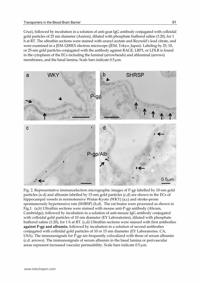
Fig. 2. Representative immunoelectron micrographic images of P-gp labelled by 10-nm gold particles (a-d) and albumin labelled by 15-nm gold particles (c,d) are shown in the ECs of hippocampal vessels in normotensive Wistar-Kyoto (WKY) (a,c) and stroke-prone spontaneously hypertensive rats (SHRSP) (b,d). The rat brains were processed as shown in Fig.1. (a,b) Ultrathin sections were stained with mouse anti-P-gp antibody (Abcam, Cambridge), followed by incubation in a solution of anti-mouse IgG antibody conjugated with colloidal gold particles of 10 nm diameter (EY Laboratories), diluted with phosphate buffered saline (1:20), for 1 h at RT. (c,d) Ultrathin sections were stained with first antibodies against P-gp and albumin, followed by incubation in a solution of second antibodies conjugated with colloidal gold particles of 10 or 15 nm diameter (EY Laboratories, CA, USA). The immunosignals for P-gp are frequently colocalized with those of serum albumin (c,d: arrows). The immunosignals of serum albumin in the basal lamina or perivascular areas represent increased vascular permeability. Scale bars indicate 0.5 m.
www.intechopen.com

Alzheimer’s Disease Pathogenesis-Core Concepts, Shifting Paradigms and Therapeutic Targets
92
Fig. 3. Representative active efflux and influx transporters of A proteins are shown according to data from Figs. 1 & 2. Pgp, LRP1, and LDLR of efflux transporters are expressed on each membrane of the endothelial cytoplasm, while RAGE, the influx transporter, is also expressed on each membrane of the endothelial cytoplasm.
www.intechopen.com

Transporters in the Blood-Brain Barrier
93
on (Ohtsuki & Terasaki 2007). Major representatives of the ABC efflux transporters are the multidrug resistance protein (MDR), multidrug resistance associated protein (MRP), and breast cancer resistance protein (BCRP) (Tamai & Tsuji, 2000). P-glycoprotein (P-gp), which belongs to the MDR family, preferentially transports cationic and/or zwitterionic compounds as substrates, whereas the MRP family preferentially transports anionic compounds, although there is some overlap between them (Hollo et al., 1996). Newly discovered categories of transporters are the OAT family, the organic cation transporter (OCT) family, the organic cation transporter novel type (OCTN)/carnitine transporter family (Bart et al., 2000; sekine et al., 2000), and the monocarboxylic acid transporter (MCT) family (Price et al., 1998), which is expected to be responsible for the transport of some organic anions from the brain to the EC and/or from the EC to the blood (Tamai & Tsuji, 2000). In addition, the concentrative nucleoside transporter, equilibrative nucleoside transporter subfamilies, and receptor-mediated transport systems such as the transferrin receptors and the scavenger receptors have also been detected in brain capillaries or brain capillary EC lines (Brett et al., 1993; de Boer et al., 2003).
(a) P-gp (Figs. 2 & 3)
The multidrug resistance efflux transporter P-gp was the plasma membrane protein first demonstrated in cancer cells by reducing intracellular levels of chemotherapeutic drugs (Ling, 1995). However, P-gp is also expressed in various normal tissues such as the liver, kidney, intestine, and brain, where it functions to protect the tissue against potentially toxic exogenous compounds (Bodo et al., 2003; Fromm, 2003; Schinkel & Jonker, 2003). In addition, it is known that P-gp is identified not only in normal epithelial cells with secretory/excretory functions but also in the ECs of capillary blood vessels in the brain (Schinkel et al., 1994) and the testis (Melaine et al., 2002). Until quite recently, P-gp in the brain had been thought to be primarily located in the apical (luminal) membrane of capillary ECs that form the BBB and to become part of the mechanisms involved in protecting the brain from xenobiotics (Schinkel, 1999; Bendayan et al., 2002; de Boer, 2003). A study using a new polyclonal antibody against P-gp (Schlachetzki & Pardridge, 2003) demonstrated dual expression of P-gp at astrocytes and the endothelium in normal primate brains. In addition, Bendayan et al. (2006) recently reported that P-gp localized to both the luminal and abluminal membranes of capillary ECs as well as in adjacent pericytes and astrocytes. We also confirmed the localization of P-gp to the luminal and abluminal membranes of cerebral ECs (Fig. 2). These authors reported that P-gp was distributed along the nuclear envelope, in the caveolae, cytoplasmic vesicles, Golgi complex, and rough endoplasmic reticulum. They stated that this glycoprotein might regulate drug transport processes in the CNS at both the cellular and subcellular levels. P-gp substrates include not only a wide variety of antineoplastic agents but also many other hydrophobic compounds such as immunosuppressive agents, cardiac glycosides, opioid analgesics, antibiotics, pesticides, antiepileptics, antidepressants, and human immunodeficiency virus protease inhibitors (Schinkel & Jonker, 2003). Inhibition of P-gp can be achieved by antidepressants (Weiss et al., 2003), suggesting the possibility that the usage of a medicine together with an antidepressant may lead to an increase in the brain concentration of the medicine. It has also been shown that at clinically relevant doses given orally, oxytetracycline is able to saturate P-gp and, subsequently, the net absorption of other drugs increases (Schrickx & Fink-Gremmels, 2007). In addition, the large number of psychoactive drugs that are substrates of P-gp could be potentially involved in a significant
www.intechopen.com

Alzheimer’s Disease Pathogenesis-Core Concepts, Shifting Paradigms and Therapeutic Targets
94
number of drug-drug interactions related to P-gp. Because of overlapping substrates specificities between CYP3A4 and P-gp, many drug interactions may involve both CYP2A4 and P-gp (Linnet & Ejing, 2008). Therefore, it is important to distinguish CYP3A4-mediated inhibition from P-gp-mediated one in order to make appropriate interpretation of drug interaction data. P-gp deficiency induces an undesirable effect on the brain. It has been hypothesized that
A proteins are deposited in periarterial interstitial fluid drainage pathways of the brain,
contributing significantly to cerebral amyloid angiopathy in Alzheimer’s disease (Weller
et al., 1998). Vogelgesang et al. (2004) reported that A deposition occurred first in
arterioles, where P-gp expression was primarily low, and accordingly, the P-gp
expression disappeared completely with the accumulation of A proteins. In addition,
Cirrito et al. (2005) reported that P-gp deficiency at the BBB increased amyloid-
deposition in a murine model of Alzheimer’s disease, suggesting that P-gp normally
discharges A out of the brain or periarterial interstitial fluid, and that perturbation of A
efflux directly affects A accumulation within the brain or perivascular areas. P-gp
expression was increased in the BBB-damaged vessels of a stroke-prone hypertensive rat
(Fig. 2). It is likely that the expression of P-glycoprotein increases as a temporary
physiological compensatory response in BBB-damaged vessels to discharge intracerebral
or periarterial undesirable substances from the brain. These findings suggest that
endothelial P-gp contributes to efflux of undesirable substances from the brain or
periarterial interstitial fluid. Concerning transendothelial transport of -amyloid protein,
the receptor for advanced glycation end products (RAGE) is thought to be a primary
transporter of -amyloid across the BBB into the brain from systemic circulation, while the
low-density lipoprotein receptor-related protein (LRP)-1 mediates transport of -amyloid
out of the brain (Donahue et al., 2006; Zlokovic, 2005). RAGE versus LRP balance
regulates Alzheimer amyloid -peptide clearance through transport across the BBB
(Deane et al., 2004). In addition, BBB efflux function of the P-gp transport system was
decreased at later disease stages of Parkinson’s disease, suggesting that the P-gp
dysfunction contributes to neuronal damage due to increased accumulation of toxins such
as insoluble -synuclein (Bartels et al., 2008). According to a paper reported by Widder et
al. (2007), the P-gp is a major exporter of oxidized glutathione and plays a crucial role in
the genesis of multiple vascular abnormalities that accompany hypertension. Moreover,
its presence is essential for the hypertensive response to angiotension II. These findings
suggest that the increased expression of P-gp in vessels may directly induce the BBB
damage. We showed the colocalization of P-gp with serum albumin (Figs. 2c, 2d),
suggesting that the expression of P-gp is upregulated in the vessels with mild BBB
damage.
(b) MRP
MRP1 is a member of the ATP-binding cassette superfamily and is expressed in non-P-gp expressing MDR cell lines (Cole et al., 1992). Of the MRP family, MRP1, MRP3, and MRP5 are expressed in the BBB (Kool et al., 1997; Huai-Yun et al., 1998; Regina et al., 1998). Since MRP is involved in extrusion of conjugated xenobiotics that may be harmful to the brain, some authors suggest that MRP1 and/or its closely related proteins are expressed at the luminal side of the brain capillaries (Kusuhara et al., 1998; Seethataman et al., 1998). However, this has not been proven experimentally.
www.intechopen.com

Transporters in the Blood-Brain Barrier
95
(c) Scavenger receptors (Figs. 1a,b & 3)
Scavenger receptors are multifunctional receptors with a wide substrate specificity [98]. Particularly, the class A, type I scavenger receptor (SR-AI) and the class B, type I scavenger receptor (SB-BI) are expressed at endothelial cells of cerebral capillaries (de Vries et al., 1993; Silver & Tall, 2001; Goti et al., 2001). In addition, these receptors are widely expressed in mammalian tissues, particularly the liver, macrophages, endothelial cells, etc. This makes these receptors less suitable for targeting drugs to the brain. Their role at the BBB seems to be a very important one because the SR-AI receptor seems to be involved with neurodegenerative diseases. In addition, the SR-BI receptor has been shown to play a role in the transport of cholesteryl esters at the BBB. Malfunction of this receptor can also result in atherosclerotic events leading to neurodegenerative processes in the brain. Mackic et al. (1998) found binding of the soluble monomeric 1-40 amino acid peptide Alzheimer amyloid-
beta (A) at the SR-AI and the receptor for advanced glycation end products (RAGE) at endothelial cells of brain capillaries.
(d) Transporters of A protein (Figs. 1, 2, & 3)
Concerning A clearance in the brain, continuous removal of toxic substances such as A-peptide species from the central nervous system is important for preventing their potentially neurotoxic accumulations in brain interstitial fluid (Deane et al., 2004). It has been suggested
that vascular A receptors are expressed in endothelial cells, transfer A across the BBB into
circulation, and thus mediate clearance of A from the brain (Zlokovic, 2004, 2008a, 2008b).
Alternatively, A receptors may also mediate A clearance via phagocytosis of A by microglia and astrocytes. Both the low-density lipoprotein receptor (LDLR) and the LDLR-
related protein 1 (LRP1) act as receptors for A efflux (Fryer et al., 2005; Abdulkarim & Hameed, 2006; Sagare et al., 2007) (Figs. 1c, 1d, 1e, 1f & Fig. 3). LDLR also regulates apolipoprotein E (apoE) levels in the CNS and LDLR-deficient Alzheimer transgenic mice
show increased cerebral A deposition (Cao et al., 2006). The LDLR is an important apoE receptor that regulates human and murine apoE endocytosis and levels in the brain (Mahley, 1988). In addition, it has been clarified that the LDLR itself regulates the level of apoE in the CNS and LDLR deficiency causes an increase in murine apoE level (Sagare et al., 2007). Accordingly, it is likely that LDLR expression is inversely related with the level of apoE. Interestingly, the apoE displays antioxidant activity (Hayek et al., 1994; Miyata & Smith, 1996). LRP1 is a member of the LDLR family and functions both as a multi-functional scavenger and signaling receptor and as a transporter and metabolizer of cholesterol and
apoE-containing lipoproteins (Herz & Marschang, 2003). LRP1 binds both ApoE/A
complexes and A and regulates their clearance from brain to blood (Zlokovic, 2004; Shibata
et al., 2000). Besides the LDLR family, some other potential A-binding receptors have been identified. P-glycoprotein (multidrug resisitance 1, MDR1) (Lam et al., 2001), scavenger receptor CD36 (Coraci et al., 2002), the formylpeptide receptor-like-1 (FPRL1) (Le et al., 2001), and the transmembrane amyloid precursor protein (APP) itself (Lorenzo et al., 2000)
also function as A receptors.
In contrast, the RAGE binds A proteins and transports them from blood to brain (Deane et
al., 2003). It is thought that the RAGE versus LRP balance regulates Alzheimer A-peptide clearance through transport across the BBB (Zlokovic, 2004) (Figs. 1a, 1b, 1c, 1d & Fig. 3).
The net flux of A into or out of the brain is the algebric sum of the inward flux and outward flux and presumably depends upon the density and activity of these receptors.
www.intechopen.com

Alzheimer’s Disease Pathogenesis-Core Concepts, Shifting Paradigms and Therapeutic Targets
96
RAGE is a member of the immunoglobulin superfamily of cell surface molecules and engages diverse ligands relevant to distinct pathological processes (Schmidt et al., 1999). The
RAGE ligands include not only A proteins but also glycation products, termed advanced glycation end products (AGEs), which occur at sites of oxidant stress in diabetes and atherosclerosis. The AGEs diminish vascular barrier function in the ECs of diabetic vasculopathy (Wautier et al., 1996). The engagement of RAGE with AGEs is shown to elicit oxidative stress generation and subsequently evoke inflammatory responses in ECs, thus being involved in atherosclerosis (Schmidt & Stern, 2000). In addition, the exogenously administered soluble form of RAGE may capture and eliminate circulating AGEs, thus protecting against the AGE-induced vascular cell damage by acting as a decoy receptor for AGEs (Park et al., 1998). Accordingly, oxidative damage may be induced in conditions with
excess AGEs or few RAGEs. Actually, concerning the localization of A transporters in the rat ECs, the immunoreaction of LRP1, LDLR, P-gp, and RAGE is seen in the cytoplasm of the ECs including the luminal and abluminal membranes (Figs. 1, 2 & 3). It is likely that the localization may move to the other areas of the ECs or appear in another cell in pathological conditions. In vessels of normotensive rats without BBB damage, the immunosignals of P-gp are located in luminal and abluminal membranes of the ECs (Figs. 2a, 2c). In contrast, in vessels of hypertensive rats, which were reported to show mild BBB damage, more immunosignals of P-gp are located to abluminal membranes of the ECs and the basal lamina than in vessels of normotensive rats (Figs. 2b, 2d). In addition, the immunosignals of P-gp are frequently colocalized with those of albumin (Fig. 2c, 2d). These may be a response to discharge intracerebral or periarterial undesirable substances from the brain.
3. Glycocalyx in endothelial surface
The glycocalyx is a negatively charged, surface coat of proteoglycans, glycosaminoglycans, and adsorbed plasma proteins lining the luminal surface of the ECs (Luft, 1966). Some researchers have put forward the concept that the endothelial glycocalyx contributes to the vasculoprotective effects of the vessel wall (Nieuwdorp et al., 2005). This layer has also been shown to be involved in maintaining vascular permeability (Henry & Duling, 1999). The endothelial glycocalyx can be evaluated by measuring the binding capacity of cationized ferritin on the luminal endothelial surface. In addition, the glycocalyx harbours a wide array of enzymes that might contribute to its vasculoprotective effect. Extracellular superoxide dismutase, an enzyme that converts oxygen radicals to hydrogen peroxide, is bound to heparan sulphate proteoglycans within glycocalyx (Li et al., 1998). Damage to the glycocalyx is accompanied by increased shedding of extracellular superoxide dismutase, which is probably related to the decreased availability of heparan sulphate binding sites. The glycocalyx damage shifts the balance towards a pro-oxidant state. These observations are of particular interest because altered vascular permeability, attenuated nitric oxide bioavailability, and redox dysregulation are the earliest characteristics of atherogenesis (Libby, 2002). In addition, disappearance of the glycocalyx is expected to be followed by exposure of adhesion molecules on ECs and subsequent leukocyte rolling, tethering, and transmigration, which are critical in the course of atherogenesis (Mulivor & Lipowsky, 2002). This evidence suggests that intact glycocalyx is necessary for the maintenance of normal vascular function, and that disruption of glycocalyx by atherogenic stimuli increases the vascular vulnerability to atherogenesis. Moreover, it is known that endothelial glycocalyx is disturbed in various types of vascular diseases (Luft, 1966). It is also known
www.intechopen.com

Transporters in the Blood-Brain Barrier
97
that inflammation induces glycocalyx shedding (Mulivor & Lipowsky, 2002). One of the most common chemokines expressed in the CNS during inflammation is monocyte chemoattractant protein-1 (Mulivor & Lipowsky, 2002). High chemokine expression is found in many pathological settings accompanied by inflammation, providing a chemoattractant gradient for leukocyte influx to the brain (Murphy, 1994; Rollins, 1997).
4. Extracellular pathways bypassing the BBB
It is known that a blood-borne protein gaining extracellular access to non-BBB sites can move not only within the CSF of the subarachnoid space but also into the brain parenchyma adjacent to each of the leaky sites (Broadwell & Sofroniew, 1993). Once in the Virchow-Robin and superficial perivascular clefts, blood-borne protein is free to circulate in the perivascular tree throughout the CNS, conveyed by the pulsatile activity of beating arterioles, and for endocytosis by perivascular phagocytes (Roher et al., 2003). It is possible in experimental animals that blood-borne macromolecules escaping the subfornical organ, a BBB-free area, have ready access not only to the white matter of the corpus callosum (Broadwell & Sofroniew, 1993), but also to the hippocampus. In addition, a drainage pathway through the subarachnoid spaces of olfactory nerves from the brain to deep cervical lymph nodes has been proposed by Bradbury et al. (1981).
5. Potential pathway of blood-borne compounds into the brain
As mentioned above, the endothelial glycocalyx with extracellular enzymes covers the luminal surface of the ECs and accordingly works at the first line of the BBB. The ECs of brain capillaries are morphologically characterized by limited vesicular transcytosis and tight junctions. Enzymatic constituents in the endothelial cytoplasm of brain capillaries inactivate some substrates. The endothelial transcytosis in brain capillaries is limited to specific substrates because several kinds of influx and efflux transporters are located at the BBB. In these ways, the BBB impedes the influx of intravascular compounds from the blood to the brain. In order to work medicines on brain function, medicines should be transferred into the brain through the BBB, and the medicines entering the brain should be prevented from discharging into the blood by the transporters or modification by the enzymes. Various trials for medicines to pass the BBB into the brain parenchyma have been performed. Osmotic opening of TJs has been reported in several types of animal models (Neuwelt & Dahlborg, 1989), since the original reports by Broman and Olsson in the 1940s (Brosman & Olsson, 1948). It is likely that reversible opening of TJs would be useful for delivery of medicines into the brain. It is also known that the intra-arterial administration of alkylglycerols transiently increases the penetration of drugs and macromolecules across the BBB, suggesting that the administration of alkylglycerols could be a unique method for enhanced drug delivery to the brain and to brain tumors (Erdlenbruch et al., 2000; Lee et al., 2002). It is most likely that increased lipophilicity of drugs makes transportation into the brain easy. Compounds bound to lectins are thought to be easily transported by adsorptive vesicular transport. Enhanced vesicular transport can be used to deliver compounds into the brain. Transient inhibition of P-gp by medicines such as antidepressants may be useful for delivery of anticancer drugs into the brain. It is likely that the manipulation of P-gp will be useful for delivery of medicines in the brain with cerebrovascular diseases. The RAGE
versus LRP balance regulates Alzheimer A-peptide clearance through transport across the
www.intechopen.com

Alzheimer’s Disease Pathogenesis-Core Concepts, Shifting Paradigms and Therapeutic Targets
98
BBB (Zlokovic, 2004). Accordingly, the influx/efflux of some substances is regulated under the expressions of these receptors. If a targeted region is situated near one of the circumventricular organs (CVOs), the delivery of medicines to that region could be achieved via CVO capillaries. There are extracellular pathways bypassing the BBB. Blood-borne proteins gaining extracellular access to non-BBB sites can move not only within the cerebrospinal fluid of the subarachnoid space, but also into the brain parenchyma adjacent to each of the leaky sites (Broadwell & Sofroniew, 1993). It is possible in experimental animals that blood-borne macromolecules escaping the subfornical organ, a BBB-free area, have ready access not only to the white matter of the corpus callosum (Broadwell & Sofroniew, 1993), but also to the hippocampus. A drainage pathway through the subarachnoid spaces of olfactory nerves from the brain to deep cervical lymph nodes has been also proposed by Bradbury et al. (1981). Accordingly, nasally inhaled medicines can affect parts of the brain through the subarachnoid spaces of olfactory nerves. In addition, it has been investigated in experimental animals whether the treatment of brain diseases is possible by using gene targeting technology that delivers the gene across the BBB after i.v. administration of nonviral formulation of the gene (Shi et al., 2001). In the experiment, the plasmid DNA was targeted to the brain with pegylated immunoliposomes using a targeting ligand such as an antibody to transferrin receptor or insulin receptor. Thus, detailed information on the BBB is necessary and useful to plan a strategy and develop therapies against various brain and vascular diseases.
6. Conclusion
The blood-brain barrier (BBB) not only impedes the influx of intravascular substances from
blood to brain, but also promotes transport of substances from blood to brain or from brain
to blood through several transport systems such as carrier-mediated transport, active efflux
transport, ion transport, or receptor-mediated transport systems. The multidrug resistance
transporter P-glycoprotein is an ATP-dependent efflux pump and contributes to efflux of
many drugs such as anti-cancer drugs and undesirable substances such as amyloid- (A)
proteins from the brain or periarterial interstitial fluid into the blood. The deficiency of P-
glycoprotein, a representative efflux transporter of A, at the BBB increases A deposition in
an Alzheimer disease mouse model. Continuous removal of toxic substances such A-
peptide species from the central nervous system is important for preventing their potentially
neurotoxic accumulation in brain interstitial fluid. It has been suggested that vascular A
transporters in endothelial cells transfer A across the BBB into circulation and thus mediate
clearance of A from the brain. The low-density lipoprotein receptor-related protein 1
(LRP1) is a major efflux transporter for A. In addition, the low-density lipoprotein receptor
(LDLR) may also act as A receptors. In the central nervous system, LDLR also regulates the
level of apolipoprotein E (apoE), which displays antioxidant activity. LDLR-deficient
Alzheimer transgenic mice show increased cerebral A deposition. Besides the LDLR
family, some other potential A-binding receptors have been identified. Scavenger receptor
CD36, the formylpeptide receptor-like-1 (FPRL1), and the transmembrane amyloid
precursor protein (APP) itself also function as A receptors. In contrast, the receptor for
advanced glycation end products (RAGE) binds A proteins and transports them from
blood to brain. It is thought that the influx versus efflux transporters balance regulates
Alzheimer A-peptide clearance through transport across the BBB. The RAGE ligands
www.intechopen.com

Transporters in the Blood-Brain Barrier
99
include not only A proteins but also advanced glycation end products (AGEs), which occur
at sites of oxidant stress in diabetes and atherosclerosis. The AGEs diminish vascular barrier
function in the endothelium of diabetic vasculopathy. The net flux of A into or out of the
brain is the algebric sum of the inward flux and outward flux and presumably depends
upon the density and activity of these receptors. Thus, A clearance in the brain endothelial
cells is an important function of the BBB. Dysfunction of the BBB with efflux and influx
transporters of A proteins may contribute to the pathogenesis of several kinds of
degenerative neuronal dysfunction or disorders including Alzheimer’s disease.
7. Acknowledgement
This research was supported by grant 22590958 from the Ministry of Education, Culture, Sports, Science and Technology of Japan (Masaki Ueno). We would like to thank Ms M. Kawauchi for technical assistance and Ms A. Kimura for editorial assistance.
Abbreviations: A = amyloid-, ABC = ATP-binding cassette, AGEs = advanced glycation
end products, apoE = apolipoprotein E, AVP = arginine-vasopressin, BBB = blood-brain barrier, BCRP = breast cancer resistance protein, CNS = central nervous system, CSF = cerebrospinal fluid, CVO = circumventricular organ, DSIP = delta-sleep inducing peptide, EC = endothelial cell, LDLR = low-density lipoprotein receptor, LHRH = luteinizing-hormone releasing hormone, LRP1 = low-density lipoprotein receptor-related protein 1, MCT = monocarboxylic acid transporter, MDR = multidrug resistance protein, MRP = multidrug resistance associated protein, OAT = organic anion transporter, OCT = organic cation transporter, OCTN = organic cation transporter novel type, P-gp = P-glycoprotein, PTS = peptide transport system, RAGE = receptor for advanced glycation end product, Tf = transferrin, Tf-R = transferrin-receptor, TJ= tight junction, Tyr-MIF-1 = tyrosine melanocyte-stimulating inhibitory factor 1, VTS = vesiculo-vacuolar organelle, VVO = vesiculo-tubular structure
8. References
Abdulkarim, Y.; Hameed, Z. (2006). Is the LDL receptor involved in cortical amyloid protein clearance? Neurochem. Res., 31, 839-847.
Azzarelli, B.; Mirkin, L.D.; Goheen, M.; Muller, J.; Crockett, C. (1984). The leptomeningeal vein. A site of re-entry of leukemic cells into the systemic circulation. Cancer, 54, 1333-1343.
Ballabh, P.; Braun, A.; Nedergaard, M. (2004). The blood-brain barrier: an overview. Structure, regulation, and clinical implications. Neurobiol. Disease, 16, 1-13.
Bart, J.; Groen, H.J.M.; Hendrikse, N.H.; van der Graaf, W.T.A.; Vaalburg, W.; de Vries, E.G.E. (2000). The blood-brain barrier and oncology: new insights into function and modulation. Cancer Treat. Rev., 26, 449-462.
Bartels, A.L.; Willemsen, A.T.M.; Kortekaas, R.; de Jong, B.M.; de Vries, R.; de Klerk, O.; van Oostrom, J.C.H.; Portman, A.; Leenders, K.L. (2008). Decreased blood-barin barrier P-glycoprotein function in the progression of Parkinson’s disease, PSp and MSA. J. Neural. Transm., 115, 1001-1009.
Bendayan, R.; Lee, G.; Bendayan, M. (2002). Functional expression and localization of P-glycoprotein at the blood brain barrier. Microsc. Res. Tech., 57, 365-380.
www.intechopen.com
Alzheimer’s Disease Pathogenesis-Core Concepts, Shifting Paradigms and Therapeutic Targets
100
Bendayan, R.; Ronaldson, P.T.; Gingras, D.; Bandayan, M. (2006). In situ localization of P-glycoprotein (ABCB1) in human and rat brain. J. Histochem. Cytochem., 54, 1159-1167.
Bodo, A.; Bakos, E.; Szeri, F.; Varadi, A.; Sarkadi, B. (2003). The role of multidrug transporters in drug availability, metabolism and toxicity. Toxicol. Lett., 140-141, 133-143.
Brett, J.; Schmidt, A.M.; Zou, Y.S.; Yan, S.D.; Weidman, E.; Pinsky, D.J.; Neeper, M.; Przysiecki, M.; Shaw, A.; Migheli, A.; Stern, D.M. (1993). Survey of the distribution of a newly characterized receptor for advanced glycation end products in tissues. Am. J. Pathol., 143, 1699-1712.
Bradbury, M.W.B.; Cserr, H.F.; Westrop, R.J. (1981). Drainage of cerebral interstitial fluid into deep cervical lymph of the rabbit. Am. J. Physiol., 240, F329-F336.
Broman, T.; Olsson, O. (1948). The tolerance of cerebral blood vessels to a constrast medium of the diodrast group. Acta Radiol. (Stockh), 30, 326-636.
Brightman, M.W.; Reese, T.S.; Feder, N. (1970). Assessment with the electron microscope of the permeability to peroxidase of cerebral endothelium and epithelium in mice and shrks. In: Capillary permeability, Crone C, Lassen NA, Eds.; Munksgaard: Copenhagen, pp 468-476.
Brightman, M.W.; Reese, T.S. (1969). Junctions between intimately apposed cell membranes in the vertebrate brain. J. Cell Biol., 40, 648-677.
Brightman, M.W. (1989). The anatomic basis of the blood-brain barrier. In: Implications of the Blood-Brain Barrier and Its Manipulation, Neuwelt, E.A.; Ed; Vol. 1, Plenum Medical: New York, Vol. 1, pp. 53-83.
Brightman, M.W.; Tao-Cheng, J.H. (1993). Tight junctions of brain endothelium and epithelium. In: The Blood-Brain Barrier, Pardridge, W.M. Ed.; Raven Press: New York, pp. 107-125.
Broadwell, R.D.; Balin, B.J.; Salcman, M. (1988). Transcytotic pathway for blood-borne protein through the blood-brain barrier. Proc. Natl. Acad. Sci. U.S.A., 85, 632636.
Broadwell, R.D. (1992a) Pathways into, through, and around the fluid-brain barrier. NIDA Res. Monogr., 120, 230-258.
Broadwell, R.D. (1992b). Pathways into, through, and around the fluid-brain barrier. NIDA Res. Monogr., 120, 230-258.
Broadwell, R.D.; Sofroniew, M.V. (1993) Serum proteins bypass the blood-brain fluid barriers for extracellular entry to the central nervous system. Exp. Neurol., 120, 245-263.
Cao, D.; Fukuchi, K.; Wan, H.; Kim, H.; Li, L. (2006). Lack of LDL receptor aggravates learning and amyloid deposits in Alzheimer transgenic mice. Neurobiol. Aging, 27, 1632-1643.
Cirrito, J.R.; Deane, R.; Fagan, A.M.; Sprinner, M.L.; Parsadanian, M.; Finn, M.B.; Jiang, H.; Prior, J.L.; Sagare, A.; Bales, K.R.; Paul, S.M.; Zlokovic, B.V.; Piwnica-Worms, D.; Hotzman, D.M. (2005). P-glycoprotein deficiency at the blood-brain barrier
increases amyloid- deposition in an Alzheimer disease mouse model. J. Clin. Invest., 115, 3285-3290.
Cole, S.P.; Bhardwaj, G.; Gerlach, J.H.; Mackie, J.E.; Grant, C.E.; Almquist, K.C.; Stewart, A.J.; Kurz, E.U.; Duncan, A.M.; Deeley, R.G. (1992). Overexpression of a transporter gene in a multidrug-resistant human lung cancer cell line. Science, 258, 1650-1654.
www.intechopen.com

Transporters in the Blood-Brain Barrier
101
Coraci, I.S.; Husemann, J.; Berman, J.W.; Hulette, C.; Dufour, J.H.; Campanella, G.K.; Luster, A.D.; Silverstein, S.C.; El-Khoury, J.B. (2002). CD36, a class B scavenger receptor, is expressed on microglia in Alzheimer’s disease brains and can mediate production of reactive oxygen species in response to beta-amyloid fibrils. Am. J. Pathol., 160, 101-112.
Cremer, J.E.; Cunningham, V.J.; Pardridge, W.M.; Braun, L.D.; Oldendorf, W.H. (1979). Kinetics of blood-brain barrier transport of pyruvate, lactate and glucose in sucking, weanling and adult rats. J. Neurochem., 33, 439-445.
Dautry-Varsat, A. Clathrin-independent endocytosis. (2001). In: Endocytosis, Marsh, M. Ed.: Oxford University Press. Oxford, pp26-57.
de Boer, A.G.; van der Sandt, I.C.; Gaillard, P.J. (2003). The role of drug transporters at the blood-brain barrier. Annu. Rev. Pharmacol. Toxicol., 43, 629-656.
de Vries, H.E.; Kuiper, J.; de Boer, A.G.; van Berkel, T.J.C.; Breimer, D.D. (1993). Characterization of the scavenger receptor on bovine cerebral endothelial cells in vitro. J. Neurochem., 61, 1813-1821.
Deane, R.; Du, Y.S.; Submamaryan, R.K.; LaRue, B.; Jovanovic, S.; Hogg, E.; Welch, D.; Manness, L.; Lin, C.; Yu, J.; Zhu, H.; Ghiso, J.; Frangione, B.; Stern, A.; Schmidt, A.M.; Armstrong, D.L.; Arnold, B.; Liliensiek, B.; Nawroth, P.; Hofman, F.; Kindy,
M.; Stern, D.; Zlokovic, B. (2003). RAGE mediates amyloid- peptide transport across the blood-brain barrier and accumulation in brain. Nat. Med., 9, 907 913.
Deane, R.; Wu, Z.; Zlokovic, B.V. (2004). RAGE (Yin) versus LRP (Yang) balance regulates
Alzheimer amyloid -peptide clearance through transport across the blood-brain barrier. Stroke, 35, 2628-2631.
Donahue, J.E.; Flaherty, S.L.; Johanson, C.E.; Duncan, III J.A.; Silverberg, G.D.; Miller, M.C.; Tavares, R.; Yang, W.; Wu, Q.; Sabo, E.; Hovanesian, V.; Stopa, E.G. (2006). RAGE, LRP-1, and amyloid-beta protein in Alzheimer’s disease. Acta Neuropathol., 112, 405-415.
Drab, M.; Verkade, P.; Elger, M.; Kasper, M.; Lohn, M.; Lauterbach, B.; Menne, J.; Lindschau, C.; Mende, F.; Luft, F.C.; Schedl, A.; Haller, H.; Kurzchalia, T.V. (2001). Loss of caveolae, vascular dysfunction, and pulmonary effects in caveolin-1 gene-disrupted mice. Science, 293, 2449-2452.
Dvorak, A.M.; Kohn, S.; Morgan, E.S.; Fox, P.; Nagy, J.A.; Dvorak, H.F. (1996). The vesiculo-vacuolar organelle (VVO): a distinct endothelial cell structure that provides a transcellular pathway for macromolecular extravasation. J. Leukoc. Biol., 59, 100-115.
Erdlenbruch, B.; Jendrossek, V.; Eibl, H.; Lakomek, M. (2000). Transient and controllable opening of the blood-brain barrier to cytostatic and antibiotic agents by alkylglycerols in rats. Exp. Brain Res., 135, 417-422.
Feng, D.; Nagy, J.A.; Hipp, J.; Dvorak, H.F.; Dvorak, A.M. (1996). Vesiculo-vacuolar organelles and the regulation of venule permeability to macromolecules by vascular permeability factor, histamine, and serotonin. J. Exp. Med., 183, 1981-1986.
Fromm, M.F. (2003). Importance of P-glycoprotein for drug disposition in humans. Eur. J. Clin. Invest., 33, 6-9.
Fryer, J.D.; DeMattos, R.B.; McCormick, L.M.; O’Dell, M.A.; Spinner, M.L.; Bales, K.R.; Paul, S.M.; Sullivan, P.M.; Parsadanian, M.; Bu, G.; Holtman, D.M. (2005). The low density lipoprotein receptor regulates the level of central nervous system and
www.intechopen.com
Alzheimer’s Disease Pathogenesis-Core Concepts, Shifting Paradigms and Therapeutic Targets
102
murine apolipoprotein E but does not modify amyloid plaque pathology in PDAPP mice. J. Biol. Chem., 280, 25754-25759.
Gomes, P.; Soares-da-Silva, P. (1999). L-Dopa transport properties in an immortalized cell line of rat capillary cerebral endothelial cells. RBE4. Brain Res., 829, 143-150.
Goti, D.; Hrzenjak, A.; Levak-Frank, S.; Frank, S.; van der Westhyzen, D.R.; Malle, E.; Sattler, W. (2001). Scavenger receptor class B type 1 is expressed in porcine brain capillary endothelial cells and contributes to selective uptake of HDL-associated vitamine E. J. Neurochem., 76, 498-508.
Hayek, T.; Oiknine, J.; Brook, J.G.; Aviram, M. (1994). Increased plasma and lipoprotein and lipid peroxidation in APOE-deficient mice. Biochem. Biophys. Res. Commun., 201, 1567-1574.
Henry, C.B.; Duling, B.R. (1999). Permeation of the luminal capillary glycocalyx is determined by hyaluronan. Am. J. Physiol., 277, H508-H514.
Herz, J.; Marschang, P. (2003). Coaxing the LDL receptor family into the fold. Cell, 112, 289-292.
Hollo, Z.; Homolya, L.; Hegedus, T.; Sarkadi, B. (1996). Transport properties of the multidrug resistance-associated protein (MRP) in human tumor cells. FEBS Lett., 383, 99-104.
Hommelgaard, A.M.; Roepstoff, K.; Vilhardt, F.; Torgersen, M.L.; Sandvig, K.; van Deurs, B. (2005). Caveolae: stable membrane domains with a potential for internalization. Traffic, 6, 720-724.
Huai-Yun, H.; Secrest, D.T.; Mark, K.S.; Carnev, D.; Braudquist, C.; Elmquist, W.F.; Miller, D.W. (1998). Expression of multidrug resistance-associated protein (MRP) in brain microvessel endothelial cells. Biochem. Biophys. Res. Commun., 243, 816-820.
Keen, J.H.; Willingham, M.C.; Pastan, I.H. (1979). Clathrin-coated vesicles: isolation, dissociation and factor-dependent reassociation of clathrin baskets. Cell, 16, 303-312.
Kirchhausen T. (1999). Adaptors for clathrin-mediated traffic. Annu. Rev. Cell Dev. Biol., 15, 705-732.
Kohn, S.; Nagy, J.A.; Dvorak, H.F.; Dvorak, A.M. (1992). Pathways of macromolecular tracer transport across venules and small veins. Structural basis for the hyperpermeability of tumor blood vessels. Lab. Invest., 67, 596-607.
Kool, M.; de Haas, M.; Scheffer, G.L.; Scheper, R.J.; van Ejik, M.J.; Juijn, J.A.; Baas, F.; Borst, P. (1997). Analysis of expression of cMOAT (MRP2), MRP3, MRP4, and MRP5, homologues of the multidrug resistance-associated protein gene (MRP1), in human cancer cell lines. Cancer Res., 57, 3537-3547.
Kusuhara, H.; Suzuki, H.; Naito, M.; Tsuruo, T.; Sugiyama, Y. (1998). Characterization of efflux transport of organic anions in mouse brain capillary endothelial cells. J. Pharmacol. Exp. Ther., 285, 1260-1265.
Lam, F.C.; Liu, R.; Lu, P.; Shapiro, A.B.; Renoir, J.M.; Sharom, F.J.; Reiner, P.B. (2001). Beta-amyloid efflux mediated by P-glycoprotein. J. Neurochem., 76, 1121-1128.
Le, Y.; Gong, W.; Tiffany, H.L.; Tumanov, A.; Nedospasov, S.; Shen, W.; Dunlop, N.M.; Gao, J.L.; Murphy, O.M.; Oppenheim, J.J.; Wang, J.M. (2001). Amyloid (beta) 42 activates a G-protein-couples chemoattractant receptor, FRP-like-1. J. Neurosci., 21, RC123-RC127.
www.intechopen.com

Transporters in the Blood-Brain Barrier
103
Lee, H.J.; Zhang, Y.; Pardridge, W.M. (2002). Blood-brain barrier disruption following the internal carotid arterial perfusion of alkyl glycerols. J. Drug Target, 463-467.
Li, Q.; Bolli, R.; Qiu, Y.; Tang, X.L.; Murphree, S.S.; French, B.A. (1998). Gene therapy with extracellular superoxide dismutase attenuates myocardial stunning in conscious rabbits. Circulation, 98, 1438-1448.
Libby, P. (2002). Inflammation in atherosclerosis. Nature, 420, 868-874. Ling, V. (1995). P-glycoprotein: its role in drug resistance. Am. J. Med., 99, 315-345. Linnet, K.; Ejsing T.B. (2008). A review on the impact of P-glycoprotein on the penetration of
drugs into the brain. Focus on psychotropic drugs. Eur. Neuropsychopharmacol., 18, 157-169.
Liu, S.-H.; Mallet, W.G.; Brodsky, F.M. (2001). Clathrin-mediated endocytosis. In: Endocytosis, Marsh, M. Ed.; Oxford University Press: Oxford, pp.1-25.
Lorenzo, A.; Yuan, M.; Zhang, Z.; Paganetti, P.A.; Sturchler-Pierrat, C.; Staufenbiel, M.; Mautino, J.; Vigo, F.S.; Sommer, B.; Yankner, B.A. (2000). Amyloid beta interacts with the amyloid precursor protein: a potential toxic mechanism in Alzheimer’s disease. Nat. Neurosci., 3, 460-464.
Lossinsky, A.S.; Vorbrodt, A.W.; Wisniewski, H.M. (1983). Ultrastructural studies of vesicular and canalicular transport structures in the injured mammalian blood-brain barrier. Acta Neuropathol., 61, 239-245.
Lossinsky, A.S.; Badmajew, V.; Robson, J.; Moretz, R.C.; Wisniewski, H.M. (1989). Sites of egress of inflammatory cells and horseradish peroxidase transport across the blood-brain barrier in a murine model of chronic relapsing experimental allergic encephalomyelitis. Acta Neuropathol., 78, 359-371.
Lossinsky, A.S.; Shivers, R.R. (2004). Structural pathways for macromolecular and cellular transport across the blood-brain barrier during inflammatory conditions. Review. Histol. Histopathol., 19, 535-564.
Luft, J.H. (1966). Fine structures of capillary and endocapillary layer as revealed by ruthenium red. Fed. Proc., 25, 1773-1783.
Mackic, J.; Stins, M.; McComb, J.; Calero, M.; Ghiso, J.; Kim, K.; Yan, S.; Stern, D.; Schmidt, A.; Frangione, B.; Zlokovic, B. (1998). Human blood-brain barrier receptors for Alzheimer’s amyloid-beta 1-40. Asymmetrical binding, endocytosis, and transcytosis at the apical side of brain microvascular endothelial monolayer. J. Clin. Invest., 102, 734-743.
Mahley, R.W. (1988). Apolipoprotein E: cholesterol transport protein with expanding role in cell biology. Science, 240, 622-630.
Mehta, D.; Malik, A.B. (2006). Signaling mechanisms regulating endothelial permeability. Physiol. Rev., 86, 279-367.
Melaine, N.; Lienard, M.-O.; Dorval, I.; Gaoscogne, C.L.; Lejeune, H.; Jegou, B. (2002). Multidrug resistance genes and P-glycoprotein in the testis of the rat, mouse, guinea pig, and human. Biol. Reprod., 67, 1699-1707.
Miyata, M.; Smith, J. (1996). Apolippprotein E allele-specific antioxidant activity and effects
on cytotoxicity insults and -amyloid peptides. Nature Genet., 14, 55-61. Mulivor, A.W.; Lipowsky, H.H. (2002). Role of glycocalyx in leukocyte-endothelial cell
adhesion. Am. J. Physiol. Heart Circ. Physiol., 282, H1282-H1291. Murphy, P.M. (1994). The molecular biology of leukocyte chemoattractant receptors. Annu.
Rev. Immunol., 12, 593-633.
www.intechopen.com

Alzheimer’s Disease Pathogenesis-Core Concepts, Shifting Paradigms and Therapeutic Targets
104
Nag, S. (1990). Presence of transendothelial channels in cerebral endothelium in chronic hypertension. Acta Neurochir. Suppl. (Wien), 51, 335-337.
Neuwelt, E.A.; Dahlborg, S.A. (1989). Blood-brain barrier disruption in the treatment of brain tumors: Clinical implications. In: Implications of the blood-brain barrier and its manipulation. Neuwelt, E.A. Ed.; New York, Plenum Publishing Corp. Vol. II , pp. 195-261.
Nieuwdorp, M.; Meuwese, M.C.; Vink, H.; Hoekstra, J.B.L.; Kastelen, J.J.P.; Stroes, E.S.G. (2005). The endothelial glycocalyx: a potential barrier between health and vascular disease. Curr. Opin. Lipidol., 16, 507-511.
Ohtsuki, S.; Tachikawa, M.; Takanaga, H.; Shimizu, H.; Watanabe, M.; Hosoya, K.; Terasaki, T. (2002). The blood-brain barrier creatine transporter is a major pathway for supplying creatine to the brain. J. Cereb. Blood Flow Metab., 22, 1327-1335.
Ohtsuki, S.; Terasaki, T. (2007). Contribution of carrier-mediated transport systems to the blood-brain barrier as a supporting and protecting interface for the brain; importance for CNS drug discovery and development. Pharm. Res., 24, 1745-1758.
Oldendorf, W.H.; Cornford, M.E.; Brown, W.J. (1977). The large apparent work capacity of the blood-brain barrier: a study of mitochondrial content of capillary endothelial cells in brain and other tissues of rat. Ann. Neurol., 1, 409-417.
Pardridge, W.M.; Oldendorf, W.H. (1975). Kinetics of blood-brain transport of hexoses. Biochim. Biophys. Acta., 382, 377-392.
Park, L.; Raman, K.G.; Lee, K.J.; Lu, Y.; Jr. Ferran, L.J.; Chow, W.S.; Stern, D.; Schmidt, A.M. (1998). Suppression of accelerated diabetic atherosclerosis by the soluble receptor for advanced glycation end products. Nat. Med., 4, 1025-1031.
Parton, R.G.; Hanzal-Bayer, M.; Hancock, J.F. (2006). Biogenesis of caveolae: a structural model for caveolin-induced domain formation. J. Cell Sci., 119, 787-796.
Pelkmans, L.; Kartenbeck, J.; Helenius, A. (2001). Caveolar endocytosis of simian virus 40 reveals a new two-step vesicular-transport pathway to the ER. Nat. Cell Biol., 3, 473-483.
Pelkmans, L.; Helenius, A. (2002). Endocytosis via caveolae. Traffic, 3, 311-320. Pelkmans, L.; Zerial, M. (2005). Kinase-regulated quantal assemblies and kiss-and-run
recycling of caveolae. Nature, 436, 128-133. Price, N.T.; Jackson, V.N.; Halestrap, A.P. (1998). Cloning and sequencing of four new
mammalian monocarboxylate transporter (MCT) homologues confirms the existence of a transporter family with an ancient past. Biochem. J., 329, 321-328.
Reese, T.S.; Karnovsky, M.J. (1967). Fine structural localization of a blood-brain barrier to exogenous peroxidase. J. Cell Biol., 34, 207-217.
Regina, A.; Koman, A.; Piciotti, M.; El Hafny, B.; Center, M.S.; Bergmann, R.; Couranud, P.O.; Roux, F. (1998). MRP1 multidrug resistance-associated protein and P-glycoprotein expression in rat brain microvessel endothelial cells J. Neurochem., 71, 705-715.
Roher, A.E.; Kuo, Y.-M.; Esh, C.; Knebel, C.; Weiss, N.; Kalback, W.; Luehrs, D.C.; Childress, J.L.; Beach, T.G.; Weller, R.O.; Kokjohn, T.A. (2003). Cortical and leptomeningeal cerebrovascular amyloid and white matter pathology in Alzheimer’s disease. Mol. Med., 9, 112-122.
Rollins, B.J. (1997). Chemokines. Blood, 90, 909-928.
www.intechopen.com

Transporters in the Blood-Brain Barrier
105
Sagare, A.; Deane, R.; Bell, R.D.; Johnson, B.; Hamm, K.; Pendu, R.; Marky, A.; Lenting, P.J.; Wu, Z.; Zarcone, T.; Goate, A.; Mayo, K.; Perlmutter, D.; Coma, M.; Zhong, Z.;
Zlokovic, B.V. (2007). Clearance of amyloid- by circulating lipoprotein receptors. Nat. Med., 13, 1029-1031.
Schinkel, A.H.; Smit, J.J.; van Tellingen, O.; Beijnen, J.H.; Wagennaar, E.W.; van Deemter, L.; Mol, C.A.; van der Valk, M.A.; Robanus-Maandag, E.C.; Riele, H.P.; Berns, A.J.M.; Borst, P. (1994). Distribution of the mouse mdr1a P-glycoprotein gene leads to a deficiency in the blood-brain barrier and to increased sensitivity to drugs. Cell, 77, 491-502.
Schinkel, A.H. (1999). P-glycoprotein, a gatekeeper in the blood-brain barrier. Adv. Drug Deliv. Rev., 36, 179-194.
Schinkel, A.H.; Jonker, J.W. (2003). Mammalian drug efflux transporters of the ATP binding cassette (ABC) family: an overview. Adv. Drug Deliv. Rev., 55, 3-29.
Schlachetzki, F.; Pardridge, W.M. (2003). P-glycoprotein and caveolin-1 in endothelium and astrocytes of primate brain. Neuroreport, 14, 2041-2046.
Schmidt, A.M.; Yan, S.D.; Wautier, J.-L.; Stern, D. (1999). Activation of receptor for advanced glycation end products. A mechanism for chronic vascular dysfunction in diabetic vasculopathy and atherosclerosis. Circ. Res., 84, 489-497.
Schmidt, A.M.; Stern, D. (2000). Atherosclerosis and diabetes: the RAGE connection. Curr. Atheroscler. Rep., 5, 430-436.
Schrickx, J.; Fink-Gremmels, J. (2007). P-glycoprotein-mediated transport of oxytetracycline in the Caco-2 cell model. J. Vet. Pharmacol. Therap., 30, 25-31.
Seetharaman, S.; Barrand, M.A.; Maskell, L.; Scheper, R.J. (1998). Multidrug resistance-related transport proteins in isolated human brain microvessels and in cells cultures from these isolates. J. Neurochem., 70, 1151-1159.
Sekine, T.; Cha, S.H.; Endou, H. (2000). The multispecific organic anion transporter (OAT) family. Pflügers Arch. – Eur. J. Physiol., 440, 337-350.
Shi, N.; Zhang, Y.; Boado, R.J.; Pardridge, W.M. (2001). Brain-specific expression of an exogenous gene after i.v. administration. Proc. Natl. Acad. U.S.A., 98, 12754-12759.
Shibata, M.; Yamada, S.; Kumar, M.; Calero, J.; Bading, B.; Frangione, D.; Holtzman, C.; Miller, C.; Strickland, D.; Ghiso, J.; Zlokovic, B. (2000). Clearance of Alzheimer’s amyloid-ss(1-40) peptide from brain by LDL receptor-related protein-1 at the blood-brain barrier. J. Clin. Invest., 106, 1489-1499.
Silver, D.L.; Tall, A.R. (2001). The cellular biology of scavenger receptor class B type 1. Curr. Opin. Lipidol., 12, 497-504.
Stan, R.-V. (2002). Structure and function of endothelial caveolae. Microsc. Res. Tech., 57, 350-364.
Tagami, M.; Kubota, A.; Sunaga, T.; Fujino, H.; Maezawa, H.; Kihara, M.; Nara, Y.; Yamori, Y. (1983). Increased transendothelial channel transport of cerebral capillary endothelium in stroke-prone SHR. Stroke, 14, 591-596.
Tamai, I.; Tsuji, A. (2000). Transporter-mediated permeation of drugs across the blood-brain barrier. J. Pharm. Sci., 89, 1371-1388.
van Deurs, B. (1978). Microperoxidase uptake into the rat choroids plexus epithelium. J. Ultrastruct. Res., 62, 168-180.
www.intechopen.com

Alzheimer’s Disease Pathogenesis-Core Concepts, Shifting Paradigms and Therapeutic Targets
106
van Deurs, B. (1980). Structural aspects of brain barriers, with special reference to the permeability of the cerebral endothelium and choroidal epithelium. Int. Rev. Cytol., 65, 117-191.
Vogelgesang, S.; Warzok, R.W.; Cascorbi, I.; Kunert-Keil, C.; Schroeder, E.; Kroemer, H.K.; Siegmund, W.; Walker, L.C.; Pahnke, J. (2004). The role of P-glycoprotein in cerebral amyloid angiopathy; implications for the early pathogenesis of Alzheimer’s disease. Curr. Alzheimer Res., 1, 121-125.
Vorbrodt, A.W. (1988). Ultrastructural cytochemistry of blood-brain barrier endothelia in Prog. Histochem. Cytochem., Gustav Fisher Verlag: Stuttgart, New York, pp. 1-99.
Vorbrodt, A.W.; Dobrogowska, D.H. (2003). Molecular anatomy of intracellular junctions in brain endothelial and epithelial barriers: electron microscopist’s view. Brain Res. Rev., 42, 221-242.
Wautier, J.-L.; Zoukourian, C.; Chappey, O.; Wautier, M.-P.; Guillausseau, P.-J.; Cao, R.; Hori, O.; Stern, D.; Schmidt, A.M. (1996). Receptor-mediated endothelial cell dysfunction in diabetic vasculopathy. J. Clin. Invest., 97, 238-243.
Weiss, J.; Gregor, S.-M.; Dormann, G.; Martin-Facklam, M.; Kerpen, C.J.; Ketabi-Kiyanvash, N.; Haefeli, W.E. (2003). Inhibition of P-glycoprotein by newer antidepressants. J. Pharmacol. Exp. Ther., 305, 197-204.
Weller, R.O.; Massey, A.; Newman, T.A.; Hutchings, M.; Kuo, Y.-M.; Roher, A.E. (1998). Cerebral amyloid angiopathy: amyloid beta accumulates in putative interstitial fluid drainage pathways in Alzheimer’s disease. Am. J. Pathol., 153, 725-733.
Widder, J.D.; Guzik, T.J.; Mueller, C.F.H.; Clempus, R.E.; Schmidt, H.H.H.W.; Dikalov, S.I.; Griendling, K.K.; Jones, D.P.; Harrison, D.G. (2007). Role of the multidrug resistance protein-1 in hypertension and vascular dysfunction caused by angiotensin II. Arterioscler. Thromb. Vasc. Biol., 27, 762-768.
Zlokovic, B.V. (2004). Clearning amyloid through the blood-brain barrier. J. Neurochem., 89, 807-811.
Zlokovic, B.V. (2005). Neurovascular mechanisms of Alzheimer’s neurodegeneration. Trends Neurosci., 28, 202-208.
Zlokovic, B.V. (2008a). New therapeutic targets in the neurovascular pathway in Alzheimer’s disease. Neurotherapeutics, 5, 409-414.
Zlokovic, B.V. (2008b). The blood-brain barrier in health and chronic neurodegenerative disorders. Neuron, 57, 178-201.
www.intechopen.com

Alzheimer's Disease Pathogenesis-Core Concepts, ShiftingParadigms and Therapeutic TargetsEdited by Dr. Suzanne De La Monte
ISBN 978-953-307-690-4Hard cover, 686 pagesPublisher InTechPublished online 12, September, 2011Published in print edition September, 2011
InTech EuropeUniversity Campus STeP Ri Slavka Krautzeka 83/A 51000 Rijeka, Croatia Phone: +385 (51) 770 447 Fax: +385 (51) 686 166www.intechopen.com
InTech ChinaUnit 405, Office Block, Hotel Equatorial Shanghai No.65, Yan An Road (West), Shanghai, 200040, China
Phone: +86-21-62489820 Fax: +86-21-62489821
Alzheimer's Disease Pathogenesis: Core Concepts, Shifting Paradigms, and Therapeutic Targets, delivers theconcepts embodied within its title. This exciting book presents the full array of theories about the causes ofAlzheimer's, including fresh concepts that have gained ground among both professionals and the lay public.Acknowledged experts provide highly informative yet critical reviews of the factors that most likely contribute toAlzheimer's, including genetics, metabolic deficiencies, oxidative stress, and possibly environmentalexposures. Evidence that Alzheimer's resembles a brain form of diabetes is discussed from differentperspectives, ranging from disease mechanisms to therapeutics. This book is further energized by discussionsof how neurotransmitter deficits, neuro-inflammation, and oxidative stress impair neuronal plasticity andcontribute to Alzheimer's neurodegeneration. The diversity of topics presented in just the right depth willinterest clinicians and researchers alike. This book inspires confidence that effective treatments could bedeveloped based upon the expanding list of potential therapeutic targets.
How to referenceIn order to correctly reference this scholarly work, feel free to copy and paste the following:
Masaki Ueno, Toshitaka Nakagawa and Haruhiko Sakamoto (2011). Transporters in the Blood-Brain Barrier,Alzheimer's Disease Pathogenesis-Core Concepts, Shifting Paradigms and Therapeutic Targets, Dr. SuzanneDe La Monte (Ed.), ISBN: 978-953-307-690-4, InTech, Available from:http://www.intechopen.com/books/alzheimer-s-disease-pathogenesis-core-concepts-shifting-paradigms-and-therapeutic-targets/transporters-in-the-blood-brain-barrier

© 2011 The Author(s). Licensee IntechOpen. This chapter is distributedunder the terms of the Creative Commons Attribution-NonCommercial-ShareAlike-3.0 License, which permits use, distribution and reproduction fornon-commercial purposes, provided the original is properly cited andderivative works building on this content are distributed under the samelicense.