transplantation of adult mouse ips cell-derived photoreceptor
TRANSCRIPT

Transplantation of Adult Mouse iPS Cell-DerivedPhotoreceptor Precursors Restores Retinal Structure andFunction in Degenerative MiceBudd A. Tucker1*, In-Hyun Park3, Sara D. Qi2, Henry J. Klassen6, Caihui Jiang1, Jing Yao2,7, Stephen
Redenti1, George Q. Daley4,5, Michael J. Young2
1 Department of Ophthalmology, Institute for Vision Research, Carver College of Medicine, University of Iowa, Iowa City, Iowa, United States of America, 2 Department of
Ophthalmology, Schepens Eye Research Institute, Harvard Medical School, Boston, Massachusetts, United States of America, 3 Division of Pediatric Hematology/Oncology,
Children’s Hospital Boston and Dana Farber Cancer Institute, Boston, Massachusetts, United States of America, 4 Department of Biological Chemistry and Molecular
Pharmacology, Harvard Medical School, Boston, Massachusetts, United States of America, 5 Manton Center for Orphan Disease Research, Harvard Stem Cell Institute,
Howard Hughes Medical Institute, Children’s Hospital Boston, Boston, Massachusetts, United States of America, 6 Department of Ophthalmology, School of Medicine,
Gavin Herbert Eye Institute, University of California Irvine, Orange, California, United States of America, 7 Department of Ophthalmology, Eye&ENT Hospital, Shanghai
Medical School, Fudan University, Shanghai, China
Abstract
This study was designed to determine whether adult mouse induced pluripotent stem cells (iPSCs), could be used toproduce retinal precursors and subsequently photoreceptor cells for retinal transplantation to restore retinal function indegenerative hosts. iPSCs were generated using adult dsRed mouse dermal fibroblasts via retroviral induction of thetranscription factors Oct4, Sox2, KLF4 and c-Myc. As with normal mouse ES cells, adult dsRed iPSCs expressed thepluripotency genes SSEA1, Oct4, Sox2, KLF4, c-Myc and Nanog. Following transplantation into the eye of immune-compromised retinal degenerative mice these cells proceeded to form teratomas containing tissue comprising all threegerm layers. At 33 days post-differentiation a large proportion of the cells expressed the retinal progenitor cell marker Pax6and went on to express the photoreceptor markers, CRX, recoverin, and rhodopsin. When tested using calcium imagingthese cells were shown to exhibit characteristics of normal retinal physiology, responding to delivery of neurotransmitters.Following subretinal transplantation into degenerative hosts differentiated iPSCs took up residence in the retinal outernuclear layer and gave rise to increased electro retinal function as determined by ERG and functional anatomy. As such,adult fibroblast-derived iPSCs provide a viable source for the production of retinal precursors to be used for transplantationand treatment of retinal degenerative disease.
Citation: Tucker BA, Park I-H, Qi SD, Klassen HJ, Jiang C, et al. (2011) Transplantation of Adult Mouse iPS Cell-Derived Photoreceptor Precursors Restores RetinalStructure and Function in Degenerative Mice. PLoS ONE 6(4): e18992. doi:10.1371/journal.pone.0018992
Editor: Branden Nelson, Seattle Children’s Research Institute, United States of America
Received October 22, 2010; Accepted March 23, 2011; Published April 29, 2011
Copyright: � 2011 Tucker et al. This is an open-access article distributed under the terms of the Creative Commons Attribution License, which permitsunrestricted use, distribution, and reproduction in any medium, provided the original author and source are credited.
Funding: BAT is supported by grants from the National Institutes of Health (NIH) (DP2 new innovators award) and the Foundation Fighting Blindness; MJY issupported by grants from the Lincy and Discovery Eye Foundations and Research to Prevent Blindness; GQD is supported by grants from the NIH, and is aninvestigator of the Manton Center for Orphan Disease Research and the Howard Hughes Medical Institute. The funders had no role in study design, data collectionand analysis, decision to publish, or preparation of the manuscript.
Competing Interests: The authors have declared that no competing interests exist.
* E-mail: [email protected]
Introduction
Retinal degenerative diseases such as retinitis pigmentosa (RP)
and age-related macular degeneration (AMD) are currently the
leading cause of incurable blindness in the western world [1,2,3].
These diseases are characterized by death of the light sensing
photoreceptor cells of the outer neural retina. As the intrinsic
regenerative capacity of the mammalian retina is extremely
limited, the only viable treatment option for people suffering from
photoreceptor cell loss is cellular replacement.
Over the past decade, stem/progenitor cell transplantation as a
means of inducing tissue reconstruction and functional regener-
ation has garnered extensive interest in the field of regenerative
medicine. Within the retina in particular, many exciting advances
have been made. One significant achievement came in 2004
when a subset of transplanted retinal progenitor cells was shown
to develop into a variety of mature retinal neurons, including
retinal ganglion and photoreceptor cells [4]. Since then,
numerous studies reporting varying degrees of success have
utilized an assortment of different cell types ranging from the
fate-restricted photoreceptor precursor [5] to the pluripotent
embryonic stem (ES) cell [6,7,8,9]. ES cells in particular are of
interest due to their ability to undergo unlimited expansion and
subsequent tissue specific differentiation. These inherent proper-
ties may allow one to generate a sufficiently large number of cells
in order to perform clinical transplantation from single isolations
rather than requiring multiple new donations, as is potentially the
problem when using more terminally differentiated cell types.
However, like photoreceptor precursor and retinal progenitor
cells, issues pertaining to host donor compatibility and ethics of
cellular isolation exist (i.e. human versions of the above
mentioned cells are isolated during embryonic development).
Thus, generation/selection of a cell type more suited for clinical
application would be desirable.
PLoS ONE | www.plosone.org 1 April 2011 | Volume 6 | Issue 4 | e18992

A cell type that avoids such problems is the recently generated,
induced pluripotent stem cell (iPSC). Initially produced by
Takahashi and Yamanaka (2006), iPSCs were generated via
genetic reprogramming of dermal fibroblasts to pluripotency using
retroviral transduction with the four transcription factors Oct4,
Sox2, KLF4 and c-Myc [10]. Although the originally published
protocols remain the most effective and efficient means of inducing
pluripotency in adult dermal fibroblasts, a variety of new protocols
with variations in cell type, delivery methods and reprogramming
factors have been developed [11,12,13,14,15,16,17,18,19]. Such
variations are aimed at reducing the need for virally induced
genetic insertion of the potentially tumorogenic factors c-Myc and
KLF4. In doing so, it has become evident that reprogramming is
exponentially more efficient, requiring less genetic manipulation,
when cells isolated from developmentally immature tissues are
used. For instance, unlike fibroblasts isolated from adult skin,
embryonic fibroblasts have been successfully reprogrammed using
the transcription factors OCT4 and Sox2 alone [20]. However, as
retinal degenerative disorders are in large part diseases of
adulthood, to be clinically relevant derivation of patient specific
iPSCs from accessible adult tissue will be required.
As demonstrated using the above mentioned cell types,
production of iPSC derived retinal cells expressing a variety of
retinal proteins, including those specific to photoreceptors, have
been achieved [21,22]. Furthermore it has recently shown that
retinal neurons produced from human iPSCs can engraft the
mouse retina following delivery to the subretinal space [23].
However, to date the safety and capacity for functional integration
following ocular transplantation have yet to be investigated. In this
study, iPSCs were generated from adult dsRed-mouse dermal
fibroblasts, differentiated toward retinal photoreceptor precursors,
and transplanted into retinal degenerative hosts. Rod and cone
photoreceptor cell repopulation, synapse formation and cellular
integration associated with restoration of electrophysiological and
anatomical correlates of retinal function were observed. These
findings establish a proof-of-principle for applications of autolo-
gous iPSCs for the safe and effective treatment of retinal
degenerative diseases.
Results and Discussion
Forced expression of the transcription factors Oct4, Sox2, Klf4,
and c-MYC induced formation of cell colonies that were
indistinguishable from those of control mouse ES cells (Fig. 1 A,
G ). Immunocytochemical analysis revealed that dsRed-iPS cell
colonies expressed levels of the markers Oct4 (B), c-MYC (C),
Sox2 (D), Nanog (E) and Klf4 (F) that were comparable to mouse
ES cells (G, H, I, J&K). Similarly, a focused microarray analysis
showed that 23 genes indicative of pluripotency were similarly
expressed between mouse ES and dsRed-iPS cells (Fig. 1M). These
results demonstrate that the iPS cells generated in this study are
similar to established ES cell colonies.
To test for pluripotency, 2.56106 undifferentiated dsRed-iPS
cells were injected into the eye of SCID mice. At 21 days post-
transplantation, histological analysis revealed teratomas (Fig. 1N)
containing tissue specific to each of the three embryonic germ
layers (Fig 1O, P neural rosettes, neuroepithelia—ectoderm; Q, R
adipocytes, chondrocytes—mesoderm; S glandular epithelium—
endoderm). Similarly, immunocytochemical staining of the
teratomas revealed GFAP-positive neural rosettes (Fig. S1A,
ectoderm), bIII tubulin-positive neural tissue (Fig. S1B, ectoderm)
and a-smooth muscle actin-positive arterial structures (Fig. S1C,
mesoderm). Collectively, these findings demonstrate that we have
successfully produced a reprogrammed adult mouse dsRed-iPS
cell line that possesses the ability to produce cell types of all three
germ layers, similar to embryonic stem cells.
To produce retinal neurons for subretinal transplantation, a
stepwise differentiation protocol was developed. This protocol
combines different aspects of previously published ES and iPS cell
differentiation paradigms [7,8,9,24,25] so as to maximize the
percentage of photoreceptor cells produced for transplantation
(Fig. 2A). This protocol takes into account the role of bone
morphogenic protein (BMP) and Wnt signaling pathway inhibition
in neuroectodermal development [26,27,28], as well as the role of
IGF-1 in anterior neural/eye field development [29] and Notch
pathway inhibition in photoreceptor development [30]. This
paradigm takes into account basic knowledge established for
eyefield and retinal development, using extrinsic chemical
signaling pathways to manipulate cell fate.
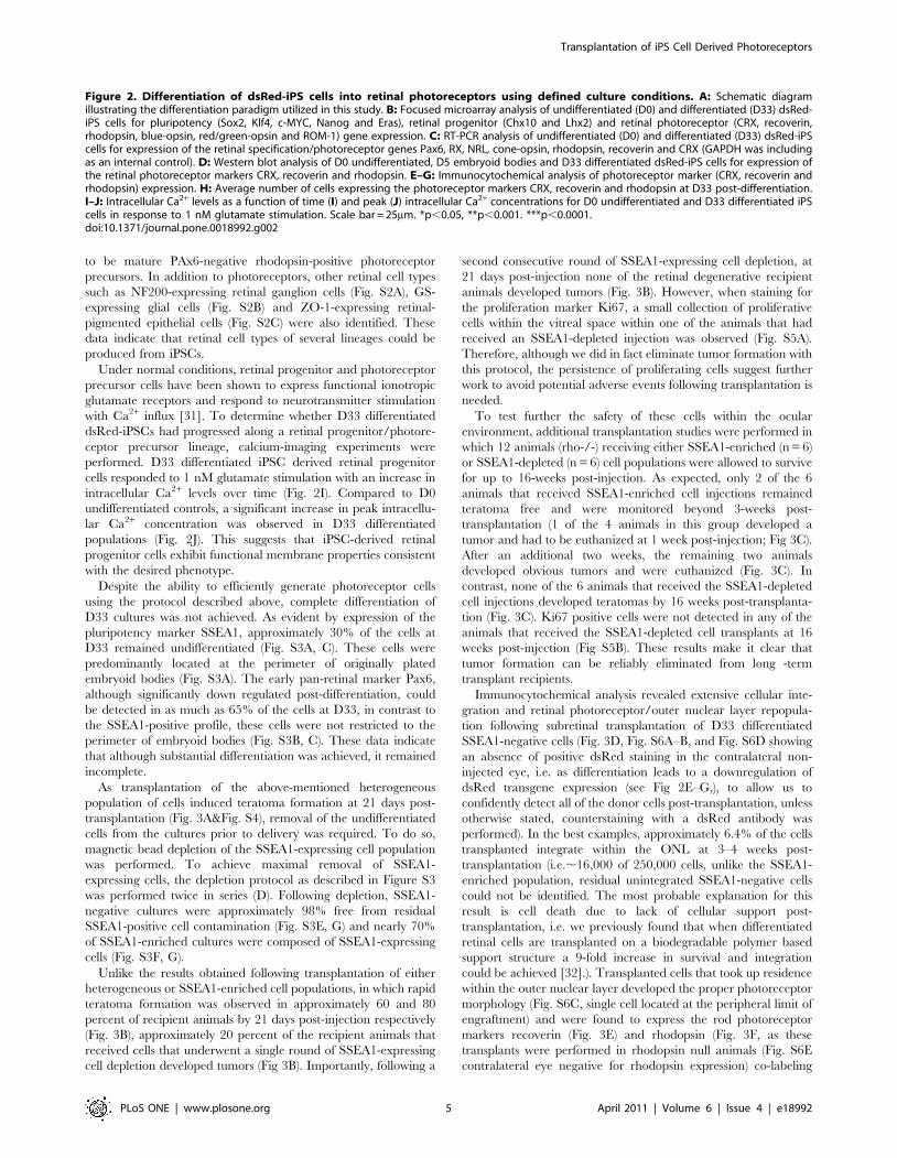
To determine whether the above protocol was effective at
inducing retinal cell differentiation, experiments using total RNA
isolated from D0 undifferentiated and D33 differentiated dsRed-
iPS cells were performed. As shown in Figure 2B, the pluripotency
genes Sox2, c-MYC, Nanog, and Eras decreased (red bars), while
the retinal progenitor cell genes Chx10 and Lhx2 (green bars) and
the photoreceptor cell genes CRX, recoverin, rhodopsin, blue-
opsin, red/green-opsin and ROM-1 (blue bars) increased in D33
differentiated cells in comparison to D0 undifferentiated controls.
Similarly, increased expression of the retinal cell markers Pax6 and
RX and the retinal photoreceptor markers NRL, cone-opsin,
recoverin, rhodopsin and CRX was detected by RT-PCR (Fig. 2C,
gene specific primer sequences can be found in Table S1). These
data indicate that following a 33 day differentiation protocol leads
to production retinal neurons from iPS cells.
To confirm the presence of retinal photoreceptor marker
expression, lysates from D0 undifferentiated cells, D5 embryoid
bodies and D33 differentiated cells were analyzed via western
blotting. Blots were probed for expression of the pan photorecep-
tor marker CRX and the rod photoreceptor markers recoverin
and rhodopsin (Fig. 2D). As expected, neither CRX, recoverin nor
rhodopsin could be detected in D0 undifferentiated cultures
(Fig. 2D). Although a slight increase in CRX expression was
detected in D5 embryoid bodies, neither recoverin nor rhodopsin
expression could be detected at this time point (Fig. 2D). In
comparison to cells at either D0 or D5, D33 differentiated cells
showed significantly elevated expression of CRX, recoverin and
rhodopsin (Fig. 2D), further supporting the fact that retinal
photoreceptors were being formed following a 33 day differenti-
ation paradigm.
Microscopically, at D33 clonal areas of differentiation were
evident, i.e. clusters of differentiated and undifferentiated cells
could be identified. Within the differentiated cell clusters,
immunocytochemical staining revealed that approximately 55%
of the cells expressed CRX (Fig. 2E, H), 40% of the cells expressed
recoverin (Fig. 2F, H), and 28% of the cells expressed rhodopsin
(Fig. 2G, H). To provide a representative depiction of the entire
heterogenous cell population at the end of the differentiation
protocol, all cells within the culture system, differentiated and non-
differentiated alike, were counted. As shown in figure 2, CRX,
recoverin, and rhodopsin were expressed by approximately 33%,
22%, and 12% of the cells respectively (H, total bars). To
determine the percentage of mature photoreceptor precursor cells
present at day 33 post-differentiation, immunocytochemical
analysis targeted against Pax6 and rhodopsin was performed (i.e.
Pax6 is expressed in developing retinal progenitor cells and turned
off in mature photoreceptors). As indicated by the PAx6-/Rho+bar in figure 2H, approximately 1–3% of the cells within the
heterogeneous cultures at day 33 post-differentiation were found
Transplantation of iPS Cell Derived Photoreceptors
PLoS ONE | www.plosone.org 2 April 2011 | Volume 6 | Issue 4 | e18992

Figure 1. Characterization of adult mouse dsRed-iPS cells. A–L: Microscopic/Immunocytochemical analysis comparing ES and dsRed-iPS cellmorphology (A, G) and expression of the pluripotency markers Oct4 (B, H), c-MYC (C, I), Sox2 (D, J), Nanog (E, K) and Klf4 (F, L). M: Focusedmicroarray analysis performed on RNA isolated from both ES and dsRed-iPS cells for the expression of genes known to be associated withpluripotency. N–S: Histological analysis of dsRed-iPS cell generated teratomas (N) for production of cells/tissues specific to ectodermal (O&P)mesodermal (Q&R) and endodermal (S) germ layers. Scale Bar = 100 mm.doi:10.1371/journal.pone.0018992.g001
Transplantation of iPS Cell Derived Photoreceptors
PLoS ONE | www.plosone.org 3 April 2011 | Volume 6 | Issue 4 | e18992

Transplantation of iPS Cell Derived Photoreceptors
PLoS ONE | www.plosone.org 4 April 2011 | Volume 6 | Issue 4 | e18992
to be mature PAx6-negative rhodopsin-positive photoreceptor
precursors. In addition to photoreceptors, other retinal cell types
such as NF200-expressing retinal ganglion cells (Fig. S2A), GS-
expressing glial cells (Fig. S2B) and ZO-1-expressing retinal-
pigmented epithelial cells (Fig. S2C) were also identified. These
data indicate that retinal cell types of several lineages could be
produced from iPSCs.
Under normal conditions, retinal progenitor and photoreceptor
precursor cells have been shown to express functional ionotropic
glutamate receptors and respond to neurotransmitter stimulation
with Ca2+ influx [31]. To determine whether D33 differentiated
dsRed-iPSCs had progressed along a retinal progenitor/photore-
ceptor precursor lineage, calcium-imaging experiments were
performed. D33 differentiated iPSC derived retinal progenitor
cells responded to 1 nM glutamate stimulation with an increase in
intracellular Ca2+ levels over time (Fig. 2I). Compared to D0
undifferentiated controls, a significant increase in peak intracellu-
lar Ca2+ concentration was observed in D33 differentiated
populations (Fig. 2J). This suggests that iPSC-derived retinal
progenitor cells exhibit functional membrane properties consistent
with the desired phenotype.
Despite the ability to efficiently generate photoreceptor cells
using the protocol described above, complete differentiation of
D33 cultures was not achieved. As evident by expression of the
pluripotency marker SSEA1, approximately 30% of the cells at
D33 remained undifferentiated (Fig. S3A, C). These cells were
predominantly located at the perimeter of originally plated
embryoid bodies (Fig. S3A). The early pan-retinal marker Pax6,
although significantly down regulated post-differentiation, could
be detected in as much as 65% of the cells at D33, in contrast to
the SSEA1-positive profile, these cells were not restricted to the
perimeter of embryoid bodies (Fig. S3B, C). These data indicate
that although substantial differentiation was achieved, it remained
incomplete.
As transplantation of the above-mentioned heterogeneous
population of cells induced teratoma formation at 21 days post-
transplantation (Fig. 3A&Fig. S4), removal of the undifferentiated
cells from the cultures prior to delivery was required. To do so,
magnetic bead depletion of the SSEA1-expressing cell population
was performed. To achieve maximal removal of SSEA1-
expressing cells, the depletion protocol as described in Figure S3
was performed twice in series (D). Following depletion, SSEA1-
negative cultures were approximately 98% free from residual
SSEA1-positive cell contamination (Fig. S3E, G) and nearly 70%
of SSEA1-enriched cultures were composed of SSEA1-expressing
cells (Fig. S3F, G).
Unlike the results obtained following transplantation of either
heterogeneous or SSEA1-enriched cell populations, in which rapid
teratoma formation was observed in approximately 60 and 80
percent of recipient animals by 21 days post-injection respectively
(Fig. 3B), approximately 20 percent of the recipient animals that
received cells that underwent a single round of SSEA1-expressing
cell depletion developed tumors (Fig 3B). Importantly, following a
second consecutive round of SSEA1-expressing cell depletion, at
21 days post-injection none of the retinal degenerative recipient
animals developed tumors (Fig. 3B). However, when staining for
the proliferation marker Ki67, a small collection of proliferative
cells within the vitreal space within one of the animals that had
received an SSEA1-depleted injection was observed (Fig. S5A).
Therefore, although we did in fact eliminate tumor formation with
this protocol, the persistence of proliferating cells suggest further
work to avoid potential adverse events following transplantation is
needed.
To test further the safety of these cells within the ocular
environment, additional transplantation studies were performed in
which 12 animals (rho-/-) receiving either SSEA1-enriched (n = 6)
or SSEA1-depleted (n = 6) cell populations were allowed to survive
for up to 16-weeks post-injection. As expected, only 2 of the 6
animals that received SSEA1-enriched cell injections remained
teratoma free and were monitored beyond 3-weeks post-
transplantation (1 of the 4 animals in this group developed a
tumor and had to be euthanized at 1 week post-injection; Fig 3C).
After an additional two weeks, the remaining two animals
developed obvious tumors and were euthanized (Fig. 3C). In
contrast, none of the 6 animals that received the SSEA1-depleted
cell injections developed teratomas by 16 weeks post-transplanta-
tion (Fig. 3C). Ki67 positive cells were not detected in any of the
animals that received the SSEA1-depleted cell transplants at 16
weeks post-injection (Fig S5B). These results make it clear that
tumor formation can be reliably eliminated from long -term
transplant recipients.
Immunocytochemical analysis revealed extensive cellular inte-
gration and retinal photoreceptor/outer nuclear layer repopula-
tion following subretinal transplantation of D33 differentiated
SSEA1-negative cells (Fig. 3D, Fig. S6A–B, and Fig. S6D showing
an absence of positive dsRed staining in the contralateral non-
injected eye, i.e. as differentiation leads to a downregulation of
dsRed transgene expression (see Fig 2E–G,), to allow us to
confidently detect all of the donor cells post-transplantation, unless
otherwise stated, counterstaining with a dsRed antibody was
performed). In the best examples, approximately 6.4% of the cells
transplanted integrate within the ONL at 3–4 weeks post-
transplantation (i.e.,16,000 of 250,000 cells, unlike the SSEA1-
enriched population, residual unintegrated SSEA1-negative cells
could not be identified. The most probable explanation for this
result is cell death due to lack of cellular support post-
transplantation, i.e. we previously found that when differentiated
retinal cells are transplanted on a biodegradable polymer based
support structure a 9-fold increase in survival and integration
could be achieved [32].). Transplanted cells that took up residence
within the outer nuclear layer developed the proper photoreceptor
morphology (Fig. S6C, single cell located at the peripheral limit of
engraftment) and were found to express the rod photoreceptor
markers recoverin (Fig. 3E) and rhodopsin (Fig. 3F, as these
transplants were performed in rhodopsin null animals (Fig. S6E
contralateral eye negative for rhodopsin expression) co-labeling
Figure 2. Differentiation of dsRed-iPS cells into retinal photoreceptors using defined culture conditions. A: Schematic diagramillustrating the differentiation paradigm utilized in this study. B: Focused microarray analysis of undifferentiated (D0) and differentiated (D33) dsRed-iPS cells for pluripotency (Sox2, Klf4, c-MYC, Nanog and Eras), retinal progenitor (Chx10 and Lhx2) and retinal photoreceptor (CRX, recoverin,rhodopsin, blue-opsin, red/green-opsin and ROM-1) gene expression. C: RT-PCR analysis of undifferentiated (D0) and differentiated (D33) dsRed-iPScells for expression of the retinal specification/photoreceptor genes Pax6, RX, NRL, cone-opsin, rhodopsin, recoverin and CRX (GAPDH was includingas an internal control). D: Western blot analysis of D0 undifferentiated, D5 embryoid bodies and D33 differentiated dsRed-iPS cells for expression ofthe retinal photoreceptor markers CRX, recoverin and rhodopsin. E–G: Immunocytochemical analysis of photoreceptor marker (CRX, recoverin andrhodopsin) expression. H: Average number of cells expressing the photoreceptor markers CRX, recoverin and rhodopsin at D33 post-differentiation.I–J: Intracellular Ca2+ levels as a function of time (I) and peak (J) intracellular Ca2+ concentrations for D0 undifferentiated and D33 differentiated iPScells in response to 1 nM glutamate stimulation. Scale bar = 25mm. *p,0.05, **p,0.001. ***p,0.0001.doi:10.1371/journal.pone.0018992.g002
Transplantation of iPS Cell Derived Photoreceptors
PLoS ONE | www.plosone.org 5 April 2011 | Volume 6 | Issue 4 | e18992

Figure 3. Transplantation of SSEA1- dsRed-iPS derived photoreceptor precursor cells induces retinal outer nuclear layerrepopulation. A: Histological staining of a teratoma containing Rho-/- eye at 21 days post-injection of a heterogeneous population of SSEA1-containing D33 differentiated cells. B: Percentage of animals developing teratomas after receiving either heterogeneous undepleted SSEA1-containing (n = 5), SSEA1-enriched (n = 5), one round of SSEA1-depleted (n = 10) or two round of SSEA1-depleted cell transplants at 21 days postinjection. C: Number of animals to and time taken for the development of teratomas in animals receiving either SSEA1-enriched or SSEA1-depletedcell transplants over a 16 week post-op period. D–L: Immunocytochemical analysis performed on rho-/- retinal degenerative eyes 21 days after
Transplantation of iPS Cell Derived Photoreceptors
PLoS ONE | www.plosone.org 6 April 2011 | Volume 6 | Issue 4 | e18992

with dsRed was not required) and the rod outer segment marker
ROM-1 (Fig. 3G). Under higher magnification it was evident that
ROM-1 tightly co-localized with the donor cell reporter gene
dsRed (Fig. 3H, arrows). In contrast, host photoreceptors, which
lack rod outer segments in the rho-/- model [33,34], do not
express this proposed pattern of ROM-1 staining, especially
following degeneration-induced rod photoreceptor denudation
[34] (Fig. 3G, arrowheads, Fig. S6D control contralateral eye). As
with rod photoreceptor markers, cone-opsin (i.e. co-labeled with
antibodies directed against both red/green and blue cone-opsin)
was also expressed by a significant population of donor-derived
photoreceptor cells (indicated by co-localization of dsRed and
cone-opsins (red/green and blue cone-opsin) within both the
newly formed cell bodies and cone outer segments; Fig. 3I, arrows
and Fig. S7C). Very few of the cone photoreceptor cells identified
in these studies were of host rather than donor origin (Fig. 3I and
Fig. S7A-C arrowheads), indicating non-dsRed positive cell and
outer segments expressing pan cone-opsin (Fig. 3I, Fig. S7C, co-
labeled with red/green and blue cone-opsin), blue cone-opsin
(Fig. S7A) and red/green cone-opsin (Fig. S7B) respectively.
These data demonstrate that engrafted progenitor cells can
differentiate into mature cone photoreceptors following trans-
plantation.
To determine whether transplanted iPS cell-derived photore-
ceptors integrate within the host retinal circuitry, immunocyto-
chemical analysis of synaptic marker expression was performed.
Pronounced co-localization of the synaptic marker synaptophysin
and the donor cell marker dsRed was observed (Fig. 3J,
arrowheads), particularly at the level of the outer plexiform layer.
Similarly, co-localization of synapsin and dsRed (Fig. S8A,
arrowheads) and VAMP2 and dsRed (Fig. S8B, arrowheads) were
identified. To further demonstrate coaptation of dsRed positive
iPS cell-derived photoreceptors processes with host bipolar cells
post-transplantation, immunocytochemical staining targeted
against the synaptic marker bassoon, the bipolar cell marker
PKCa and the donor cell marker dsRed were performed. As
shown in figure 3, extensive co-localization among all 3 markers,
with clear punctate bassoon staining within the OPL, was
identified (Fig. 3K&L arrowheads, low and high magnification
images respectively). We also demonstrate the differentiation/
integration of a modest number of cells into other retinal cell types,
i.e. staining for NF200 and GFAP indicative of retinal ganglion
cells (Figure S9A) and Muller glia (Figure S9B). These results make
it clear that grafted cells form new synapses with the host retina,
leading us to investigate the possibility of functional recovery in
recipient animals.
To determine whether integrated iPS-derived photoreceptor
cells enable recovery of retinal functional, electroretinographic
(ERG) analysis of rho-/- animal eyes, with or without transplants
at 28 days post-op, was performed (at the time of recording
animals were 8–10 weeks of age). To ensure that changes in visual
function post-transplantation were due to the presence of new
graft-derived photoreceptor cells and not related to residual host
cones, retinal degenerative recipient animals were analyzed pre-
injection and chosen based on absence of detectable b-wave
amplitude. As shown in Figure 4, a statistically significant
increase of approximately 95 mv in b-wave amplitude was
observed at 21 days post-subretinal transplant as compared to
the contralateral uninjected eye (A, B). Similar findings have been
reported for ES cell-derived photoreceptor precursors in CRX-/-
mice, whereby an approximate 40–50 mV increase in b-wave
amplitude was observed post-transplantation [6]. The difference
in recovered b-wave amplitude can potentially be explained by a
variety of factors including increased photoreceptor production
via the modified differentiation protocol used here, the use of
allogeneic as opposed to xenogeneic donor cells, the fact that
remaining undifferentiated cells were removed from the D33
differentiated population prior to transplantation and finally the
use of a different host/retinal degeneration model systems (i.e.
Rho-/- vs. CRX-/-).
To further demonstrate the relationship between cellular
integration and recovery of retinal function, a linear regression
analysis was performed. As illustrated in Figure 4, a significant
positive correlation between peak b-wave amplitude and donor
dsRed-positive photoreceptor cell integration was identified
(Fig. 3C, r2 = 0.7316, n = 10, p#0.005). These findings suggest
that as more cells integrated within the dystrophic retinal
architecture, a larger increase in recovery of electro-retinal
function was seen. Although these data suggests that transplanted
iPSC-RPCs are integrating functionally within the host retinal
architecture (i.e. a recordable ERG could only be detected in 8–10
week old mice post-transplantation), we cannot exclude the
possibility that transplant induced restoration of residual host
cone viability is partially contributing to the increased ERG
response.
To further assess retinal function in grafted versus ungrafted
eyes, functional anatomy focused on activation of neurons within
the retinal inner nuclear layer (INL) was performed. As light
exposure, and subsequent photoreceptor activation/phototrans-
duction, is known to induce nuclear c-Fos expression in the inter-
neurons of the INL [35], immunocytochemical analysis targeted
against the expression of this marker was employed. As compared
to control contralateral ungrafted eyes, which do not express
functional rhodopsin and in turn lack significant light induced
phototransduction (Fig 4D&E), eyes that had received iPSC-
derived photoreceptor precursors were found to have significantly
increased light induced INL c-Fos expression at 21 days post-
transplantation (Fig. 4E arrowheads&F). For instance, an approx-
imate 7-fold increase in the number of INL cells expressing c-Fos
was detected (Fig. 4F). In large part, c-Fos expressing cells were
located in close proximity to newly engrafted dsRed-iPSC derived
photoreceptor processes (Fig. 4E arrowheads).
Collectively, these data suggest that adult dermal fibroblast-
derived iPS cells represent a useful source of replacement retinal
neurons, particularly photoreceptors, and that this method of
treatment is capable of providing at least partial restoration of
retinal function. It is worth noting that such an approach in
genetic dystrophies such as retinitis pigmentosa will require either
gene correction, as employed in an animal model of sickle cell
anemia [36], or engraftment with normal allogeneic tissue, as
patient specific cells will express the same genetic defect found in
host photoreceptors. Moreover, further investigations into large
animal models of diseases resembling age-related macular
degeneration will be needed if any restorative transplantation
strategies are to be employed in this disorder.
These data also demonstrate the potential utility of active
removal of residual pluripotent cells prior to transplantation. As
embryonic stem cells that have not been subject to manipulation
receiving subretinal injections of SSEA1-depleted cells targeted against expression of the photoreceptor markers recoverin (E), rhodopsin (F), ROM-1(G&H) and opsin (I), the synapse markers synaptophysin (J) and bassoon (K&L), the bipolar cell marker PKCa (K&L) and the iPS cell marker dsRed (D–J).TP = transplant site. Scale bar = 50 mm.doi:10.1371/journal.pone.0018992.g003
Transplantation of iPS Cell Derived Photoreceptors
PLoS ONE | www.plosone.org 7 April 2011 | Volume 6 | Issue 4 | e18992

with viral vectors do not appear to possess the same propensity
toward tumor formation post-transplantation [6,37,38], alternate
means of adult dermal cell reprogramming, which do not utilize
genome-incorporating viruses, would be advantageous. It is
reasonable to expect that sustained over-expression of the
transgenes used to induce pluripotency might interfere with
terminal differentiation and the data presented in Figure 2B,
showing maintenance of Klf4 gene expression in heterogenous
unselected cultures following differentiation, would support this.
Efficient removal of undifferentiated SSEA1-expressing cells
mitigates the risk of teratoma formation, thereby allowing for
improved experimental outcome including orthotopic replacement
of photoreceptors and partial restoration of electroretinal function.
Materials and Methods
Ethics statementAll experiments were conducted with the approval of the
Schepens Eye Research Institute Animal Care and Use Commit-
tee (Animal welfare assurance # A3177-01, ACUC approval # S-
170-0710) and the ARVO Statement for the Use of Animals in
Ophthalmic and Vision Research.
AnimalsAdult 4–6 week old dsRed-positive C57Bl6 mice (Jackson
Laboratory, Bar Harbor, ME) were used as fibroblast donors;
adult 4–6 week old rhodopsin-null mice that lack rod outer
segments and do not form functional rod photoreceptors [33]
(rho-/-, Peter Humphries, Trinity College, Dublin) were used as
retinal degenerative transplant recipients. Severe combined
immunodeficient mice (SCID, Jackson Laboratory, Bar Harbor,
ME) were used for assessment of teratoma formation.
iPS cell differentiationTo maintain pluripotency, adult dsRed-iPS cells were cultured
on inactive mouse embryonic fibroblasts in LIF containing
pluripotency media. To begin differentiation, iPS cells are
removed from the culture substrate via incubation in a 1 mg/ml
type I collagenase (Sigma-Aldrich) solution, resuspended in
embryoid body media (DMEM F-12 media (Gibco) containing
10% knockout serum replacement (Gibco) 2% B27 supplement
(Gibco) 1% N2 supplement (Gibco), 1% L-glutamine (Gibco), 1%
100x NEAA (Gibco), 1% penicillin/streptomycin (Gibco), 0.2%
Fungizone (Gibco), 1 ng/ml noggin (R&D Systems, Minneapolis,
MN), 1 ng/ml Dkk-1 (R&D Systems), 1 ng/ml IGF-1 (R&D
Systems) and 0.5 ng/ml bFGF (R&D Systems)), and plated at a
density of ,50 cell clumps/cm2 in ultra low cluster plates
(Corning, Lowell, MA). Cell clumps are cultured for 5 days as
indicated above, after which the embryoid bodies are removed,
washed and plated at a density of 25–30/cm2 in fresh
differentiation media 1 (DMEM F-12 media (Gibco), 2% B27
supplement (Gibco) 1% N2 supplement (Gibco), 1% L-glutamine
(Gibco), 1% 100x NEAA (Gibco) 10 ng/ml noggin (R&D
Systems), 10 ng/ml Dkk-1 (R&D Systems), 10 ng/ml IGF-1
(R&D Systems) and 1 ng/ml bFGF (R&D Systems)) in 6-well
culture plates coated with poly-D-lysine (BD Bioscience, San Jose,
CA, 10 mg/ml), collagen (BD Bioscience, 25 mg/ml), laminin
(Gibco, 50 mg/ml) and fibronectin (Sigma-Aldrich, 100 mg/ml).
Cultures are fed every other day for 10 days with differentiation
media 1, then every other day for an additional 6 days with
Figure 4. Transplantation of SSEA1-dsRed-iPS derived photoreceptor precursor cells induces increased electroretinal function asdetermined by ERG and light induced c-Fos expression. A–C: Representative ERG (A, 5 db flash under scotopic conditions), average peak b-wave amplitudes for rho-/- mice 21 days after receiving subretinal SSEA1-depleted cell injections (B, n = 6 of 10 animals that received transplants, onlyanimals with recovery in ERG response above baseline were chosen for this analysis, the 4 animals that were not chosen were found to have poorcellular integration due to extensive cell death post- transplantation), and correlation between peak b-wave amplitude and iPS cell derivedphotoreceptor layer repopulation (C, n = 10). D–E: Immunocytochemical analysis targeted against the immediate early gene c-Fos and the iPSCmarker dsRed performed on rho-/- transplant and contralateral control mouse eyes at 21 days post-subretinal injection of SSEA1-negative dsRed-iPSderived photoreceptor precursor cells. F: Number of cells per microscopic section expressing c-Fos. A significant increase in the number of cellswithin the retinal inner nuclear layer expressing c-Fos was detected in Rho-/- eyes that had received subretinal injections of SSEA1-negative dsRed-iPSC derived photoreceptor precursor cells as compared to contralateral control eyes following light exposure. Scale bar = 50 mm.doi:10.1371/journal.pone.0018992.g004
Transplantation of iPS Cell Derived Photoreceptors
PLoS ONE | www.plosone.org 8 April 2011 | Volume 6 | Issue 4 | e18992

differentiation media 2 (differentiation media 1+10 uM of the
Notch signaling inhibitor, DAPT (Calbiochem, Gibbstown, NJ),
followed every other day for an additional 12 days with
differentiation media 3 (differentiation media 2+2 ng/ml of aFGF
(R&D Systems)).
ERG8–10 week old rhodopsin null transplant recipient mice were
dark-adapted for 12 hrs prior to testing, anesthetized, had their
pupils dilated by topical application of tropicamide (Akorn, Lake
Forest, IL) and placed on a heated recording stage maintained at
37uC. Contact lens electrodes were placed directly onto the
corneal surface of eyes precoated with a 2.5% hydroxypropyl-
methylcellulose solution (Gonak, Akorn), while a copper
reference electrode was placed beneath the scalp and another
ground was inserted beneath the tail skin. Responses to 5 test
flashes were recorded for each mouse and all ERGs were carried
out under scotopic conditions using a standard 5 db flash. ERG
signals were amplified 10,000x, filtered between 1 Hz to 3 kHz,
and sampled at 5 kHz. All recordings were performed under
infrared light and data was analyzed using EMwin software
(LKC Technologies, Inc., Gaithersburg, MD). Data present in
Figure 3 were taken from responsive animals at 21 days post-
transplantation (M).
Functional anatomyTo further assess retinal function post-transplantation, analysis
of c-Fos expression was performed. Rho-/- null transplant
animals housed in a 12 hr dark/light cycle were sacrificed at
approximately 1.5 hr after the lights had been turned on in their
housing environment (,280 mW/cm2). Animals were subse-
quently sacrificed and enucleated. The eyes were fixed in
4%PFA, cryosectioned and immunostained with a primary
antibody targeted against c-Fos (Calbiochem, Gibbstown, NJ)
and detected using a cy2 conjugated secondary antibody
(Jackson, West Grove, PA).
For further details pertaining to Retroviral production and iPS
cell generation, Focused Microarray Analysis, Subretinal Trans-
plantation, SSEA1 Cell depletion, Calcium imaging, Immuno-
staining, Immunoblotting, RNA isolation and RT-PCR, Cell
Counting, and Statistical Analysis see Methods S1.
Supporting Information
Figure S1 Analysis of dsRed-iPS cell pluripotency. A–C:Immunocytochemical analysis performed on dsRed-iPS cell
derived teratomas targeted against the ectodermal markers GFAP
(A: glia) and bIII tubulin (B, neural), and the mesodermal marker
a-SMA (C: vascular). Expression of ectodermal (GFAP and
NF200) and mesodermal (a-SMA) markers within iPS cell derived
teratomas indicate that the parent iPS cells are pluripotent.
(TIF)
Figure S2 Identification of dsRed-iPS cell derivedretinal cells. A–C: Immunocytochemical analysis performed
on dsRed-iPS cell cultures at D33 post-differentiation directed
against the ganglion cell marker NF200 (A), the glial cell marker
GS (B), and the RPE cell/tight junction marker ZO-1 (D). Scale
bar = 25 mm.
(TIF)
Figure S3 Depletion of D33 undifferentiated SSEA1-positive dsRed-iPS cells prior to transplantation pre-vents teratoma formation. A–B: Immunocytochemical anal-
ysis of SSEA1 and Pax6 expression in D33 cultures post-
differentiation. C: Percentage of cells expressing SSEA1 and
Pax6 in D33 cultures post-differentiated. D: Schematic diagram
illustrating the procedures used for depletion of remaining SSEA1-
positive undifferentiated cells from D33 cultures post-differentia-
tion. E–F: Immunocytochemical analysis of SSEA1 expression in
SSEA1 cell-depleted and -enriched D33 post-differentiation
cultures. G: percent of SSEA1-negative cells in SSEA1-depleted
cultures and SSEA1-positive cells in SSEA1-enriched cultures
following successive rounds of depletion/isolation. Scale
bar = 50 mm.
(TIF)
Figure S4 Transplantation of a heterogeneous popula-tion of D33 differentiated dsRed-iPS cells inducesteratoma formation. A–B: Immunocytochemical analysis of
recoverin (A) and rhodopsin (B) expression post-subretinal
transplantation of heterogeneous D33 differentiated cells. Trans-
plantation of a heterogeneous population of undepleted cells (i.e.
SSEA1 positive population included) isolated at D33 post-
differentiation induced either teratomas or at the very least
collections of cells suggestive of incipient tumors at 21 days post-
transplantation. Cells contained within these masses were found to
express both recoverin (A) and rhodopsin (B).
(TIF)
Figure S5 Proliferative cells identified at 21-days post-transplantation are absent at 16-weeks post-transplan-tation. A–B: Immunocytochemical analysis performed on rho-/-
recipient mouse eyes at 21-days and 16-weeks post-intravitreal
injection of SSEA1-negative dsRed-iPS cells targeted against the
cell cycle marker Ki67. Scale bar = 50 mm.
(TIF)
Figure S6 Transplantation of SSEA1- dsRed-iPS derivedphotoreceptor precursor cells induces extensive cellularintegration and outernuclear layer repopulation. A–C:Immunocytochemical analysis performed on rho-/- recipient
mouse eyes at 21 days post-subretinal injection of SSEA1-negative
dsRed-iPS cells targeted against the host donor cell marker dsRed.
A–B: Low magnification images used to show the extent of cellular
integration post-transplantation. C: High magnification image
taken at the outer limit of cellular migration in figure B where
sparse cellular integration was observed. This image was taken in
an attempt to show detailed donor cell morphology. As shown in
these images extensive cellular integration and retinal ONL
repopulation was identified across a wide area of the host retina at
3 weeks post-transplantation (A–B). Cells that integrate within the
retinal degenerative environment adopt a photoreceptor morphol-
ogy represented by a single cell body with and outer segment
extended toward the RPE and an inner process ending with a
synaptic pedicle that extends into the host plexiform layer (C). D–E: Immunocytochemical analysis performed on control rho-/- un-
injected contralateral mouse eyes at 21 days post-op against
dsRed, ROM1 and recoverin (D) or rhodopsin (E). Contralateral
6–8 week old rhodopsin null mouse eyes do not express ROM1,
dsRed or rhodopsin, indicating that rod photoreceptors detected
in Rho-/- eye at 21-days post-subretinal injection are iPSC
transplant derived.
(TIF)
Figure S7 Generation of cone photoreceptors followingtransplantation of SSEA1- dsRed-iPS derived photore-ceptor precursor cells. A–C: Immunocytochemical analysis
performed on rho-/- recipient mouse eyes at 21 days post-
subretinal injection of SSEA1-negative dsRed-iPS cells targeted
against the blue cone photoreceptor marker blue-opsin (A), the
Transplantation of iPS Cell Derived Photoreceptors
PLoS ONE | www.plosone.org 9 April 2011 | Volume 6 | Issue 4 | e18992

red/green cone photoreceptor marker red/green-opsin (B) and the
pan cone photoreceptor marker pan-cone-opsin (C).
(TIF)
Figure S8 Synaptic integration of dsRed-iPS cell derivedphotoreceptor precursor cells following subretinaltransplantation. A–C: Immunocytochemical analysis per-
formed on rho-/- recipient mouse eyes at 21 days post-subretinal
injection of SSEA1-negative dsRed-iPS cells targeted against the
synaptic markers synapsin (A) and Vamp-2 (B). Scale bar = 10 mm.
Transplanted dsRed-expressing iPS cell derived photoreceptor
precursors that integrate into the outer nuclear layer of retinal
degenerative mice following subretinal injection form synaptic
connections at the level of the outer plexiform layer within the host
retina.
(TIF)
Figure S9 Integration of cell types other then retinalphotoreceptors at 21-days post-injection. A-C: Immuno-
cytochemical analysis performed on rho-/- recipient mouse eyes at
21 days post-injection of SSEA1-negative dsRed-iPS cells targeted
against the retinal ganglion cell marker NF200 (A) and the glial cell
marker GFAP (B). Scale bar = 10 mm. In addition to photorecep-
tors, transplanted dsRed-expressing iPS cells also gave rise to
NF200 expressing retinal ganglion and GFAP-expressing glial cells
following ocular injection. Importantly, both cell types took up
residence within the appropriate retinal layer and appeared to
develop morphologically into the correct cell types.
(TIF)
Table S1 Gene specific primer sequences used for RT-PCR.
(DOC)
Methods S1 Retroviral production and iPS cell genera-tion.
(DOC)
Author Contributions
Conceived and designed the experiments: BAT MJY. Performed the
experiments: BAT I-HP SDQ. Analyzed the data: BAT I-HP SDQ HJK
GQD. Wrote the paper: BAT MJY SDQ HJK GQD. Performed calcium
imaging analysis: SR. Provided transplantation and clinical support: CJ JY.
References
1. Mitchell J, Bradley C (2006) Quality of life in age-related macular degeneration:
a review of the literature. Health Qual Life Outcomes 4: 97.
2. Chopdar A, Chakravarthy U, Verma D (2003) Age related macular
degeneration. Bmj 326: 485–488.
3. Earnshaw SR, Moride Y, Rochon S (2007) Cost-effectiveness of pegaptanibcompared to photodynamic therapy with verteporfin and to standard care in the
treatment of subfoveal wet age-related macular degeneration in Canada. ClinTher 29: 2096–2106.
4. Klassen HJ, Ng TF, Kurimoto Y, Kirov I, Shatos M, et al. (2004) Multipotent
retinal progenitors express developmental markers, differentiate into retinalneurons, and preserve light-mediated behavior. Invest Ophthalmol Vis Sci 45:
4167–4173.
5. MacLaren RE, Pearson RA, MacNeil A, Douglas RH, Salt TE, et al. (2006)Retinal repair by transplantation of photoreceptor precursors. Nature 444:
203–207.
6. Lamba DA, Gust J, Reh TA (2009) Transplantation of human embryonic stem
cell-derived photoreceptors restores some visual function in Crx-deficient mice.
Cell Stem Cell 4: 73–79.
7. Lamba DA, Karl MO, Ware CB, Reh TA (2006) Efficient generation of retinal
progenitor cells from human embryonic stem cells. Proc Natl Acad Sci U S A103: 12769–12774.
8. Ikeda H, Osakada F, Watanabe K, Mizuseki K, Haraguchi T, et al. (2005)
Generation of Rx+/Pax6+ neural retinal precursors from embryonic stem cells.Proc Natl Acad Sci U S A 102: 11331–11336.
9. Osakada F, Ikeda H, Mandai M, Wataya T, Watanabe K, et al. (2008) Toward
the generation of rod and cone photoreceptors from mouse, monkey and humanembryonic stem cells. Nat Biotechnol 26: 215–224.
10. Takahashi K, Yamanaka S (2006) Induction of pluripotent stem cells frommouse embryonic and adult fibroblast cultures by defined factors. Cell 126:
663–676.
11. Carey BW, Markoulaki S, Hanna J, Saha K, Gao Q, et al. (2009)Reprogramming of murine and human somatic cells using a single polycistronic
vector. Proc Natl Acad Sci U S A 106: 157–162.
12. Gonzalez F, Barragan Monasterio M, Tiscornia G, Montserrat Pulido N,Vassena R, et al. (2009) Generation of mouse-induced pluripotent stem cells by
transient expression of a single nonviral polycistronic vector. Proc Natl AcadSci U S A 106: 8918–8922.
13. Kim JB, Greber B, Arauzo-Bravo MJ, Meyer J, Park KI, et al. (2009) Direct
reprogramming of human neural stem cells by OCT4. Nature.
14. Nakagawa M, Koyanagi M, Tanabe K, Takahashi K, Ichisaka T, et al. (2008)
Generation of induced pluripotent stem cells without Myc from mouse and
human fibroblasts. Nat Biotechnol 26: 101–106.
15. Okita K, Nakagawa M, Hyenjong H, Ichisaka T, Yamanaka S (2008)
Generation of mouse induced pluripotent stem cells without viral vectors.Science 322: 949–953.
16. Shao L, Feng W, Sun Y, Bai H, Liu J, et al. (2009) Generation of iPS cells using
defined factors linked via the self-cleaving 2A sequences in a single open readingframe. Cell Res 19: 296–306.
17. Sommer CA, Stadtfeld M, Murphy GJ, Hochedlinger K, Kotton DN, et al.
(2009) Induced pluripotent stem cell generation using a single lentiviral stem cellcassette. Stem Cells 27: 543–549.
18. Welstead GG, Brambrink T, Jaenisch R (2008) Generating iPS cells from MEFSthrough forced expression of Sox-2, Oct-4, c-Myc, and Klf4. J Vis Exp.
19. Park IH, Lerou PH, Zhao R, Huo H, Daley GQ (2008) Generation of human-
induced pluripotent stem cells. Nat Protoc 3: 1180–1186.
20. Huangfu D, Osafune K, Maehr R, Guo W, Eijkelenboom A, et al. (2008)
Induction of pluripotent stem cells from primary human fibroblasts with only
Oct4 and Sox2. Nat Biotechnol 26: 1269–1275.
21. Meyer JS, Shearer RL, Capowski EE, Wright LS, Wallace KA, et al. (2009)
Modeling early retinal development with human embryonic and induced
pluripotent stem cells. Proc Natl Acad Sci U S A.
22. Osakada F, Jin ZB, Hirami Y, Ikeda H, Danjyo T, et al. (2009) In vitro
differentiation of retinal cells from human pluripotent stem cells by small-
molecule induction. J Cell Sci 122: 3169–3179.
23. Lamba DA, McUsic A, Hirata RK, Wang PR, Russell D, et al. (2010)
Generation, purification and transplantation of photoreceptors derived from
human induced pluripotent stem cells. PLoS One 5: e8763.
24. Reh TA, Lamba D, Gust J (2010) Directing human embryonic stem cells to a
retinal fate. Methods in molecular biology 636: 139–153.
25. Tucker BA, RS, Park I-H, Daley GQ, Young MJ (2009) Generation of retinal
precursors from murine iPS cells. Association for Research in Vision and
Ophthalmology.
26. Mukhopadhyay M, Shtrom S, Rodriguez-Esteban C, Chen L, Tsukui T, et al.
(2001) Dickkopf1 is required for embryonic head induction and limb
morphogenesis in the mouse. Dev Cell 1: 423–434.
27. Anderson RM, Lawrence AR, Stottmann RW, Bachiller D, Klingensmith J
(2002) Chordin and noggin promote organizing centers of forebrain develop-
ment in the mouse. Development 129: 4975–4987.
28. Lamb TM, Knecht AK, Smith WC, Stachel SE, Economides AN, et al. (1993)
Neural induction by the secreted polypeptide noggin. Science 262: 713–718.
29. Pera EM, Wessely O, Li SY, De Robertis EM (2001) Neural and head induction
by insulin-like growth factor signals. Dev Cell 1: 655–665.
30. Jadhav AP, Mason HA, Cepko CL (2006) Notch 1 inhibits photoreceptor
production in the developing mammalian retina. Development 133: 913–
923.
31. Sun W, Seigel GM, Salvi RJ (2002) Retinal precursor cells express functional
ionotropic glutamate and GABA receptors. Neuroreport 13: 2421–2424.
32. Tomita M, Lavik E, Klassen H, Zahir T, Langer R, et al. (2005) Biodegradable
polymer composite grafts promote the survival and differentiation of retinal
progenitor cells. Stem Cells 23: 1579–1588.
33. Humphries MM, Rancourt D, Farrar GJ, Kenna P, Hazel M, et al. (1997)
Retinopathy induced in mice by targeted disruption of the rhodopsin gene. Nat
Genet 15: 216–219.
34. Lee ES, Burnside B, Flannery JG (2006) Characterization of peripherin/rds and
rom-1 transport in rod photoreceptors of transgenic and knockout animals.
Invest Ophthalmol Vis Sci 47: 2150–2160.
35. Huerta JJ, Llamosas MM, Cernuda-Cernuda R, Garcia-Fernandez JM (1997)
Fos expression in the retina of rd/rd mice during the light/dark cycle. Neurosci
Lett 232: 143–146.
36. Hanna J, Wernig M, Markoulaki S, Sun CW, Meissner A, et al. (2007)
Treatment of sickle cell anemia mouse model with iPS cells generated from
autologous skin. Science 318: 1920–1923.
37. Schraermeyer U, Thumann G, Luther T, Kociok N, Armhold S, et al. (2001)
Subretinally transplanted embryonic stem cells rescue photoreceptor cells from
degeneration in the RCS rats. Cell Transplant 10: 673–680.
Transplantation of iPS Cell Derived Photoreceptors
PLoS ONE | www.plosone.org 10 April 2011 | Volume 6 | Issue 4 | e18992

38. Vugler A, Carr AJ, Lawrence J, Chen LL, Burrell K, et al. (2008) Elucidating the
phenomenon of HESC-derived RPE: anatomy of cell genesis, expansion andretinal transplantation. Exp Neurol 214: 347–361.
39. Jiang C, Klassen H, Zhang X, Young M (2010) Laser injury promotes migration
and integration of retinal progenitor cells into host retina. Molecular vision 16:983–990.
Transplantation of iPS Cell Derived Photoreceptors
PLoS ONE | www.plosone.org 11 April 2011 | Volume 6 | Issue 4 | e18992