transient peaked t waves during exercise stress testing...
TRANSCRIPT

Transient peaked T waves during exercise stress testing: an unusual manifestation of reversible cardiac ischemia
Ondas T pontiagudas transitórias durante o teste ergométrico: uma manifestação incomum de isquemia cardíaca reversível
From Raimundo Barbosa Barros M.D. Coronary Center Hospital de Messejana Dr. Carlos Alberto Studart Gomes Fortaleza-Ceará-Brazil
Finals comments Andrés Ricardo Pérez-Riera M.D. Ph.D.In charge of Electrovectorcardiogram sector- Cardiology Discipline – ABC Faculty – ABC Foundation
– Santo André- São Paulo – [email protected]
Phone Cell (011) 84693388 ; Residence(011) 5044-6243; Office (011) 5621-2390
Prezado Andrés gostaria de compartilhar este interessante caso com os colegas do foro.Colega(médico)assintomático,46 anos, hipertenso, sobrepeso e história familiar positiva para doençaarterial coronariana.Foi submetido à avaliação cardiológica com teste ergométrico.Durante o esforço não apresentou sintomas.Com 1 minuto da fase de recuperação começou a apresentar dor no peito tipo opressiva que cedeu após8 minutos.Observe onda T apiculada tipo isquémica no segundo traçado que normalizou no terceiro ECG.Indicado uma coronariografia que revelou suboclusão proximal da arteria descedente anterior.Foi submetido com sucesso à implante de stent farmacológico.Seria interessante um debate sobre esta rara manifestação.Comentários?
Dear Andrés: I would like to share this interesting case with the forum´s colleagues.Physician, asymptomatic, 46 years old, systemic hypertension, overweight and positive family history for coronary artery disease.He underwent cardiac evaluation with exercise stress testing.During the effort had no symptoms.After one minute of the recovery phase started to get chest pain oppressive type that yielded after 8 minutes.Note the peaked T wave in the second ECG normalized in the third one.Coronariography showed a proximal subocclusion on left anterior descending coronary artery (LAD).He underwent successful implantation of drug-eluting stent.It would be an interesting debate on this rare manifestation of coronary insuficence.Any comments?
Raimundo Barbosa Barros M.D.
------------------------------------------------------------------------------------------------------------------------

Teste ErgométricoExercise stress testing
ECG de repousoECG at rest
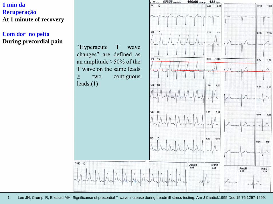
1 min da RecuperaçãoAt 1 minute of recovery
Com dor no peitoDuring precordial pain
“Hyperacute T wave changes” are defined as an amplitude >50% of the T wave on the same leads ≥ two contiguous leads.(1)
1. Lee JH, Crump R, Ellestad MH. Significance of precordial T-wave increase during treadmill stress testing. Am J Cardiol.1995 Dec 15;76:1297-1299.
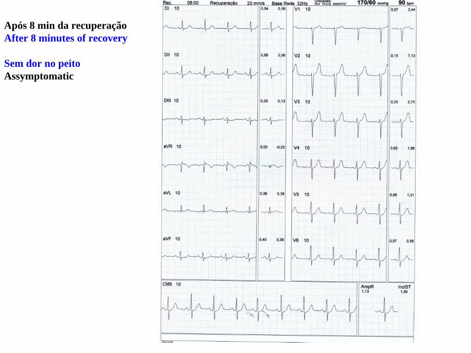
Após 8 min da recuperaçãoAfter 8 minutes of recovery
Sem dor no peitoAssymptomatic

Colleagues comments

An interesting ECG, Tall hyperacute T wave (in this case with ST elevation) during acute ischemia were discussed a.o by Goldberger (1), Also called hyperacute T-wave pattern and may appear during acute myocarditis (Electrocardiography in Clinical practice,2008 by Surawicz),but usually proximal LAD occlusion( Fig 10.4 in my textbook(2),The incidence of ST elevation and no previous MI during stress test ranged from 0.2 to 1.7% in 2 studies (3;4)
Regards Borys Surawicz M.D. M.A.C.C. Professor EmeritusIndiana University School of MedicineSenor Research AssociatedKrannet Institute of CardiologyMember of The Care Group Indianopolis, Indiana----------------------------------------------------------------------------------------------------------------------------------
Un interesante ECG, onda T alta hiperaguda (en este caso con elevación del segmento ST) durante la isquemia aguda como o discutiera Goldberger (1), también llamado patrón de onda T hiperagudo. Este patrón pueden aparecer durante la miocarditis aguda aguda (Electrocardiografía en la práctica clínica, 2008 por Surawicz ), pero por lo general indica lesión oclusiva proximal LAD (Fig. 10.4 en mi libro de texto (2),La incidencia de la elevación del segmento ST y sin antecedentes de infarto previo durante prueba de esfuerzo varió de 0,2 a 1,7% en 2 estudios (3, 4)
1. Goldberger AL. Hyperacute T waves revisited. Am Heart J. 1982 Oct;104(4 Pt 1):888-90.2. Morton Tavel Chapter 10 Stress Test pp218-219 In Surawicz/Knilans. Chou´s Electrocardiography in Clinical Practice Adult &
pediatric 5th ed. Borys Surawicz, Tymithy Knilans3. Sriwattanakomen S, Ticzon AR, Zubritzky SA, et al. S-T segment elevation during exercise: electrocardiographic and
arteriographic correlation in 38 patients. Am J Cardiol. 1980 Apr;45:762-768.4. Bruce RA, Fisher LD, Pettinger M, Weiner DA, Chaitman BR. ST segment elevation with exercise: a marker for poor ventricular
function and poor prognosis. Coronary Artery Surgery Study (CASS) confirmation of Seattle Heart Watch resultsCirculation. 1988 Apr;77:897-905.

Dear Andres, I’m not so concerned about the peaked T waves as I am about the significant ST segment elevation in V1-3 during exercise, along with some ST segment elevation in lead aVR. This is usually indicative of high-grade proximal LAD occlusion resulting in transmural ischemia. Exercise induced ST segment depression usually best seen in the lateral precordial leads is indicative of subendocardial ischemia and is generally non-localizing with regard to specific coronary artery lesion location. This is the usual ischemic finding during exercise ECG testing. On the other hand, exercise-induced ST segment elevation, a manifestation of transmural ischemia, is localizing in a similar way that STEMI ECG findings localize the particular lesions in acute myocardial infarction. Findings such as this during exercise usually result in a trip to the cath lab – as was done in this patient.Regards,Frank Yanowitz, MD Professor of Medicine University of Utah School of MedicineMedical Director, ECG Department LDS Hospital Salt Lake City, Utah 8th Ave. and C Street Salt Lake City, Utah 84143 USAfyanow@mac.com--------------------------------------------------------------------------------------------------------------------Estimado Andrés, No estoy tan preocupado por las ondas T picudas como lo estoy en relación a la significativa elevación del segmento ST en V1-3 durante el ejercicio, junto con alguna elevación del segmento ST en aVR. Esto suele ser indicativo de alto grado de oclusión proximal de la DA queresulta en isquemia transmural. Depresión del segmento ST inducida por el ejercicio por lo general se ve mejor en las derivacionesprecordiales laterales y es indicativo de isquemia subendocárdica y por lo general no localiza el lugarde la lesión de la arteria coronaria. Contrariamente la elevación del ST, inducida por el ejercicio es manifestación de isquemia transmural, semejante a la que se observa en la fase hiper-aguda del infarto de miocardio. Estos hayazgos durante el ejercicio deben hacernos mandar de inmediato al paciente al laboratorio de hemodinamia - como se hizo en este caso.Un cordial saludo

Dear Raimundo & AndresThanks again for all the excellent materialThe second ECG is consistent with CAD ---The T waves are symmetric. Peaking can be a catecholamine effect.Other observations: Absent septal q on all ECGs =CADHe has interatrial block and probable LA enlargement.His P axis is about 80º = emphysema and/ or smoker.Awaiting your discussionFraternallyDavid H. Spodick, MD, FACC, MACP, FCCP, FAHADirector Emeritus of the Cardiovascular Medicine Fellowship Program and at the University of Massachusetts Medical School where he is Professor of Medicine Emeritus.
Estimado Andrés y RaimundoGracias nuevamente por todo el excelente materialEl segundo ECG es compatible con el enfremdad coronaria --- Las ondas T son simétricas. El ser picudas puede ser un efecto de catecolaminas.Otras observaciones: q ausencia del tabique en todos los ECG = CADTiene bloqueo interauricular y probable sobrecarga auricular izquierda.El eje de P está en 80 º = enfisema y / o fumador.En espera de su discusiónfraternalmente

Again an interesting case! Myocardial ischemia during an exercise test presenting with ST depressions in the ECG does not localize the ischemia – the same leads show ST depressions independently of coronary arterydisease severity. In a few cases, ST elevations are provoked during the stress test as a sign of critical coronaryartery obstruction (or in some cases Prinzmetal angina). In the present case, the ECG shows signs of transmural ischemia and the ECG pattern indicates a proximal LAD lesion, because there is ST elevation alsoin leads aVR and V1. In our own material, 1/3 of patients with anterior STEMI, who had a proximal LAD lesion presented with ST elevation in aVR and V1, while V6 showed reciprocal ST depression (="aVR pattern). 2/3 of the patients with proximal LAD occlusion presented with ST elevations in I and aVL (="aVLpattern"). The T waves are very prominent especially in the precordial leads, while the J-point elevations are moderate. Maybe one should classify the ischemia severity as Sclarovsky-Birnbaum Grade 1.
Una vez más un caso interesante! La isquemia miocárdica durante una prueba de esfuerzo se presenta con depresiones del ST y en el ECG no localiza la isquemia - las mismas derivaciones muestran depresiones del ST de forma independiente de la severidad de la enfermedad arterial coronaria. En algunos casos, elevación del ST es provocada durante la prueba de esfuerzo como un signo de obstrucción crítica de la arteria coronaria (o en algunos casos angina de Prinzmetal). En el presente caso, el ECG muestra signos de isquemia transmural y el patrón del ECG indica una lesión proximal de LAD, porque hay elevación también del segmento ST en aVR y V1. En nuestro propio material, 1/3 de los pacientes con STEMI anterior, que tenía una lesión proximal de LAD se presentaron con elevación del ST en aVR y V1, mientras V6 mostró depresión recíproca del segmento ST (= "patrón de aVR). 2/3 de los pacientes con oclusión proximal LAD presentaban elevaciones del ST en I y aVL (= "patrón aVL"). Las ondas T son muy prominentes, especialmente en las derivaciones precordiales, mientras que las elevaciones de los puntos J-son moderadas. Tal vez se deba clasificar la gravedad de la isquemia como Sclarovsky Birnbaum-Grado 1.
Kjell NikusTampere, Finland

Los ECG N° 1 y 3 no parecen registrar alguna alteración. ECG N° 2: Ritmo Sinusal, frecuencia cardíaca de 110 lpm, eje eléctrico en los +50° aproximadamente. Buena progresión de la onda R en precordiales.Alteraciones registradasIsquemia subendocárdica expresada por las ondas T altas, acuminadas y de base estrecha desde V2 a V6 y en cara inferior, DII-DIII-aVF. Supradesnivel de V2 a V4, cóncavo hacia arriba. En V1 se observa supradesnivel recto de 1 mm, que luego desaparece en el último electrocardiograma. Existepatente de lesión subendocárdica en la cara inferior, DII-DIII-aVF, que también podría ser la imagenen espejo de lo que sucede en la cara anterior. La causa de estas patentes es la insuficiencia coronaria, pero también en teoría tendríamos que pensar en una hiperpotasemia. Conceptualmente la isquemia subendocárdica es fugaz, difícil de registrar.Saudação. Muito obrigado.Lucas Barbieri Argentina--------------------------------------------------------------------------------------------------------------------------------The ECG No. 1 and 3 do not seem to register any alteration.ECG # 2: Sinus rhythm, heart rate 110 bpm, QRS axis at +50 °. Good R wave progression in precordial leads.Alterations registeredSubendocardial ischemia expressed by tall T waves, with acuminate and narrow base from V2 to V6 and in inferior leads, II-III-aVF. ST segment elevation from V2 to V4, concave upward. In V1 is seen straight elevation 1 mm, which then disappears in the last electrocardiogram. Subendocardial injury pattern is observed in inferior leads II-III-aVF, which could also be the mirror image of what happens in the anterior wall. The cause of these patents is coronary heart disease, but also in theory would have to think of hyperkalemia.Conceptually subendocardial ischemia is fleeting, difficult to record.Greeting and thank you very much.Lucas Barbieri Argentine
En esta ergometría no sólo se veo aumento de la onda T sino que leve en V1 pero más claramente en V2 y V3 se observa un supradesnivel de ST de casi 3 mm, lo cual no es una presentación habitual perono es atípica, está hablando claramente que la isquemia es severa al punto que con el aumento de consumo de O2 comienza con onda de lesión subepicárdica. El supradesnivel del ST en ergometría es una urgencia y se debe pedir la cinecoronariografía como se hizo en este caso.Ignácio Retamar----------------------------------------------------------------------------------------------------------------------------------In this exercise stress test not only I see an increase voltage of the T wave in V1 but a mild but more clear ST elevation of about 3 mm in V2 and V3, which is not a usual presentation but not atypical. This phenomena, clearly speaking in severe ischemia with subepicardial injury. The ST segment elevation in exercise testing is an emergency and must be ordered coronary angiography as in this case.Ignacio RetamarReplica Andrés
Querido Ignacio excelente observación. Apenas debo corregirte un punto: tu dices que es una presentación no habitual pero que no es atípica. El primer adjetivo(no habitual) concuerdo contigo pero eso de no ser atipica se te ocurrió a ti. Está escrito que es poco usual, incomun o poco comun no hemos escrito atípica.Reply from Andrés to RetamarDear Ignacio excellent observation. I just correct you one thing: you say it is an unusual presentation but not atypical. The first adjective (not usual) I agree with you but that is not to be atypical happened to you. It is written that is unusual, uncommon we have not written the adjetive atypical.“Transient peaked T waves during exercise stress testing: an unusual manifestation of reversible cardiac ischemia”Andrés.

Prezados Andrés e RaimundoNão temos o ECG da freqüência máxima na ergometria mas durante a dor, há nítido supradesnível do
segmento ST em V1-V2 com a onda T alta e apiculada. Isto indicativo de isquemia aguda como se estivéssemos na presença de um infarto em fase inicial. O supradesnivel é raro na ergometria, mas geralmente indica lesão grave de descendente anterior. Ficaria muito interessante se houvesse onda T negativa após o desaparecimento da dor.
Um grande abraço
Jose Claudio Kruse Porto Alegre Brasil-------------------------------------------------------------------------------------------------------------------------------Dear Andrés and Raimundo
We have not the highest frequency in the exercise stress testing but during the chest pain, there is clear ST-segment elevation in V1-V2 with tall and spiked T wave. This occurrence is indicative of acute ischemia such as if we were in the presence of a heart attack at an early stage. The ST segment elevation is rare during the Exercise stress testing, but generally indicates a severe lesion of the left anterior descending artery. It would be very interesting if there were negative T wave after the pain disappear.
A big hug
Jose Claudio Kruse M.D. Porto Alegre, Brazil

In patients with severe LAD disease, when neither ST elevation nor depression occurs, an increase in T amplitude in V2 and V3 may occur indicating severe myocardial ischemia. Changes in V2 are the most reliable and when a cut-off of at least 2.5 mm is used as the increase from rest to peak exercise, the sensitivity is 20% but the specificity is 89%.It appears that a T-wave amplitude increase of ≥ 2.5 mm in lead V2 during a treadmill stress test may be specific (95%), even though this finding only occurs occasionally. Therefore, a T-wave amplitude increase during an exercise test may aid in the diagnosis of the few patients who develop this abnormality, especially if there is no ST depression, as has occurred during several recent exercise tests.(1)
Augusto Uchida M.D. Ph.D. treadmill stress test sector Instututo do Coração São Paulo Brazil
En pacientes con severa obstrucción da LAD, cuando ni elevación del segmento ST ni la depresión ocurre, un aumento de la amplitud da onda T en V2, y V3 puede producirse indicando isquemia miocárdica grave. Los cambios en V2 son los más confiables y cuando un corte de al menos 2,5 mm se utiliza como el valor del aumento desde el reposo al pico del esfuerzo, la sensibilidad es del 20% pero la especificidad es del 89%.
1. Lee JH, Crump R, Ellestad MH. Significance of precordial T-wave increase during treadmill stress testing. Am J Cardiol.1995 Dec 15;76:1297-1299.

Queridos amigos del forum. Es universalmente aceptado que una obstrucción coronaria puede inducir a depresión del ST-T en mayor medida en V4-V5 como consequencia de taquicardia sinusal en presencia de una arteria significativamente obstruida ,( >75 %) por ocasionar aumento de la presión diastólica final del VI, induciendo una isquemia circumferential, independientemente de la arteria epicárdica obstruida. Cuando se induce depression del ST y T negativa en V4 -V5 con frecuencias cardiacas menores (entre 80 -100lpm) debemos sospechar enfermedad coronaria triarterial critica o de obstrucción significativa del tronco de la coronaria izquierda (Left Main Coronary Artery obstruction). Existen excepciones a este comportamiento electrocardiográfico cuando una arteria secundaria aislada esta criticamente cerrada ocasionará elevación del ST y T positiva, porque al irrigar un território menor no puede inducir a una isquemia circunferencial por no producir aumento de la presion diastólica final del VI, y consecuentemente no ocasiona depression del ST e inversión de la T en V4-V5Cuando en una prueba de esfuerzo aparece una depression del segmento ST-con onda T negativa de V1aV3 pensamos en obstrucción de algunas de las ramas marginales de la circunfleja. Cuando se observe depressión del ST y onda T negativa en DII ,DIII, aVF y ST elevado en aVL con T positiva estaremos casi seguros de que la obstrucción está en la marginal superior. Si se observa solo depression del segmento ST en V1,V2 ,V3 se puede diagnosticar obstrucción de la primara rama diagonal.Cuando una artéria descendente anterior distal esta cerrada o criticamente obstruida el esfuerzo con baja
frecuencia cardíaca induce elevación del segmento ST y onda T positiva en V2-V3 Finalmente, en el escenario correspondiente al caso presentado por nuestros amigos hay una arteria descendente anterior criticamente o totalmente obstruida asociada a profusa y suficiente circulación colateral lo que impide el descenso del ST. En fin queridos amigos ,yo me inclino por esta última posibilidadUn fraternal abrazoSamuel Sclarovsky
1. Sagie A ,Sclarovsky S et al ; Acute anterior wall infarction presenting with positive T waves and without segment ST shift Chest 1989; 95; 1211-15

Dear friends of the forum,It is universally accepted that a coronary obstruction may lead to ST-T depression in a greater extent in V4-V5, due to sinus tachycardia in the presence of critically obstructedartery (>75%) causing increase of end diastolic pressure of the LV, inducing circumferential ischemia, regardless of the obstructed epicardial artery. When with a lower heart rate (between 80-100 bpm) ST depression and negative T is induced in V4-V5, we should suspect more diffuse ischemia, suggesting the existence of critical three-artery disease or critical LMC artery obstruction.There are exceptions to this electrocardiographic behavior when an isolated secondary artery is criticallyclosed, causing ST elevation and positive T, because it cannot induce per se, an extended circumferentialischemia since it does not produce increase in LV end diastolic pressure and consequently, it does notproduce ST depression and T inversion in V4-V5. When in a stress test ST segment depression appears with negative T wave in V1, V2, V3, we considerobstruction in some of the marginal branches of the LCX. When ST depression and negative T wave is observed in II, III, aVF and elevated ST in aVL with positive T wave, we will almost be certain that the obstruction is in the superior marginal branch. If only ST segment depression is observed in V1, V2, V3, obstruction of the first diagonal branch can bediagnosed. When a distal LAD artery is closed or critically obstructed, strain with low heart rate induces ST segmentelevation and positive T wave in V2, V3. Finally, in a scenario corresponding to the case presented by our friends, there is an LAD critically ortotally obstructed with abundant and enough collateral circulation. Anyway, dear friends, I lean toward the last possibility. Warm regards,
Samuel Sclarovsky Israel
1. Sagie A ,Sclarovsky S et al ; Acute anterior wall infarction presenting with positive T waves and without segment ST shift Chest 1989; 95; 1211-1215
FINAL COMMENTS
By Andrés Ricardo Pérez-Riera M.D.Ph.D.

A reading of possible myocardial ischemia in a person who isn't actively having symptoms of a heart attack is meaningless. Thats why machines have not replaced doctors. A machine has no common sense. What I can tell you. T wave changes are common. On a screening ECG they sometimes relate to mild enlargement of the heart, they sometimes relate to electrolytes, they sometimes relate to nothing.A screening ECG is used to tell us if you already had a heart attack. In that case we would see Q waves, a totally different thing than the T wave. Some times people had a silent heart attack and a screening ECG helps us pick this up. A screening ECG has NO PREDICTIVE VALUE.The question which help predict if you are at risk for heart disease are: Are you as smoker? Are you a diabetic?Are you an hypertension subject?Do you have a family history of heart disease?Did someone in your family have a heart attack before age 45?Do you exercise?Do you have high cholesterol?Have you overweight?Do you get chest pain or pressure with physical exertion?How many flights of stairs can you walk up before getting out of breath?You answers to these questions relate to your risk. With increasing amounts of risk we may do tests for heartThe T wave represents the repolarization (or recovery) of the ventricles. It represent the uncanceled potential differences of ventricular repolarization. Ischemia occurs when blood flow is decreased through one or more of the arteries in the heart muscle; in this case, to the anterior portion of the heart. The main risk factors that can lead to ischemia are elevated cholesterol levels, elevated blood pressure, and diabetes. Ischemia can be the result of coronary artery disease (atherosclerosis), in which plaques formed of cholesterol and possibly other cellular waste products build up on an artery wall and restrict the blood flow. This is the most common cause of myocardial (heart muscle) ischemia. Some other conditions include blood clots, causing a sudden ischemic event which may lead to heart attack; coronary artery spasm, which is brief and temporary; and severe illnesses, such as those leading to blood loss.
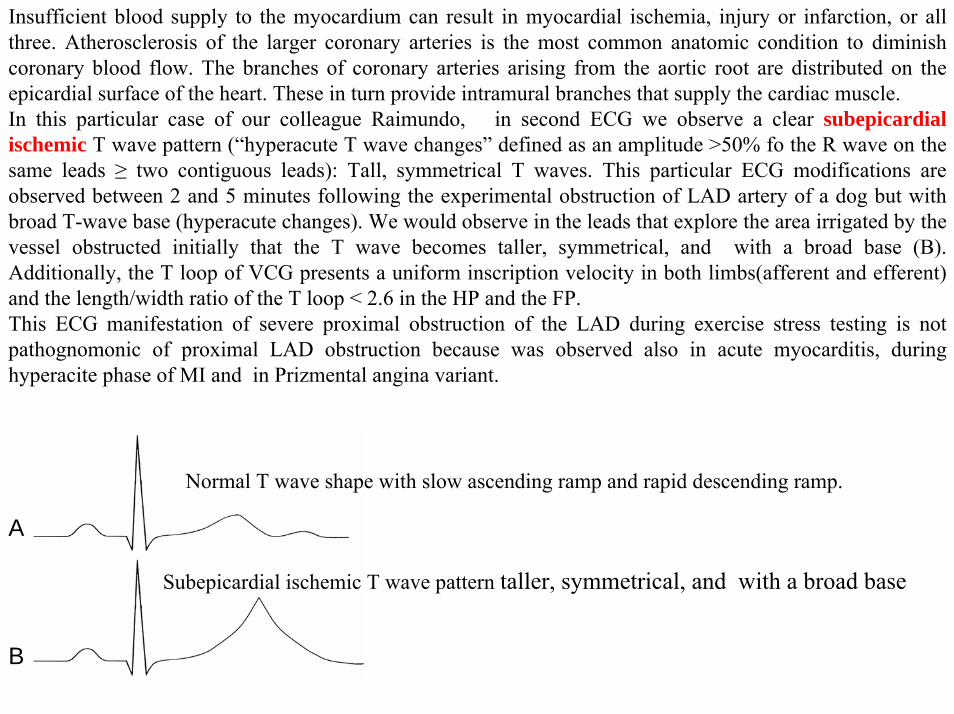
Insufficient blood supply to the myocardium can result in myocardial ischemia, injury or infarction, or all three. Atherosclerosis of the larger coronary arteries is the most common anatomic condition to diminish coronary blood flow. The branches of coronary arteries arising from the aortic root are distributed on the epicardial surface of the heart. These in turn provide intramural branches that supply the cardiac muscle.In this particular case of our colleague Raimundo, in second ECG we observe a clear subepicardial ischemic T wave pattern (“hyperacute T wave changes” defined as an amplitude >50% fo the R wave on the same leads ≥ two contiguous leads): Tall, symmetrical T waves. This particular ECG modifications are observed between 2 and 5 minutes following the experimental obstruction of LAD artery of a dog but with broad T-wave base (hyperacute changes). We would observe in the leads that explore the area irrigated by the vessel obstructed initially that the T wave becomes taller, symmetrical, and with a broad base (B). Additionally, the T loop of VCG presents a uniform inscription velocity in both limbs(afferent and efferent) and the length/width ratio of the T loop < 2.6 in the HP and the FP.This ECG manifestation of severe proximal obstruction of the LAD during exercise stress testing is not pathognomonic of proximal LAD obstruction because was observed also in acute myocarditis, during hyperacite phase of MI and in Prizmental angina variant.
Subepicardial ischemic T wave pattern taller, symmetrical, and with a broad base
Normal T wave shape with slow ascending ramp and rapid descending ramp.
A
B

V1
V2
V3aVR
Concomitantly with tall T waves a subepicardial injury is observed from V1 to V3. (Clear ST segment elevation from V2 -V3 and minimally in V1 ) I thinck that ST segment elevation is absent in aVR. The ST segment elevation is unusul feature during the exercise stress testing, but when present generally is indicative of severe proximal obstruction of the LAD artery. (transmural ischemia.) associated with sufficient collateral circulation.( absence of augmentation of end LV diastolic pressure.) WhenST segment elevation develops during exercise in a non-Q wave leads in a patient without a previous IM , the findings should be considered as likely evidence of transmural myocardial ischemia caused by coronary vasospasm or a high-grade coronary narrowing. This finding is uncommon occurring ≈1% of patients with obstructive CAD. Scintigraphy usually reveals a defect in the territory involved.(1)
1.Mobilia G, Zanco P, Desideri A, et al, T wave normalization in infarct-related electrocardiographic leads during exercise testing for detection of residual viability: comparison with positron emission tomography. J Am Coll Cardiol. 1998 Jul;32:75-82.

Rarely. patients with acute proximal thrombotic occlusion of the LAD, tall subepicardial ischemic T waves never evolve into ST-segment elevation. This was recently inaccurately reported as a "novel sign" of proximal LAD occlusion. It has been speculated that the absence of ST-segment elevation could be attributed to the large area of transmural ischemia, the anatomic variant of Purkinje fibers, or to lack of activation of sarcolemal adenosine triphosphate-potassium channels. This ECG picture was recently explained by changes in the subendocardial but not in the epicardial action potential, suggesting subendocardial ischemia as the underlying mechanism. Stankovic et al (1) present a patient with thromboticlesion of proximal LAD, static precordial ST-segment depression, and tall T waves who underwent primary PCI and stent placement. Surprisingly, total thrombotic stent occlusion on the following day was associated with ST-segment elevation in precordial leads, indeed supporting the concept of the regional subendocardialischemia that was first described more than a decade ago.Verouden et al (2) described patients with a distinct ECG pattern without ST-segment elevation in the presence of an acute MI consequence of occlusion of the proximal LAD artery who were referred for PCI. Of 1890 patients who underwent primary PCI of the LAD artery, they could identify 35 patients (2%) with a static, distinct ECG pattern ST-segment depression at the J-point of at least 1 mm in precordial leads with upsloping ST-segments continuing into tall, symmetrical T-waves. Patients with this distinct ECG pattern were younger, more often male and more often had hypercholesterolaemia compared to patients with anterior myocardial infarction and ST-segment elevation. This ECG pattern signifies proximal LAD artery occlusion. It is important for cardiologists and emergency care physicians to recognise this distinct ECG pattern, so they can triage such patients for immediate reperfusion therapy.
1. Stankovic I, Ilic I, Panic M, Vlahovic-Stipac A, Putnikovic B, Neskovic AN.The absence of the ST-segment elevation in acute coronary artery thrombosis: what does not fit, the patient or the explanation?JElectrocardiol. 2011 Jan-Feb;44:7-10.
2. Verouden NJ, Koch KT, Peters RJ, et al. Persistent precordial "hyperacute" T-waves signify proximal left anterior descending artery occlusion.Heart.2009 Oct;95:1701-1706.

THEORETICAL CONSIDERATIONS ABOUT NORMAL AND PATHOLOGICAL T WAVES

In electrocardiography, the T wave represents the repolarization (or recovery) of the ventricles and the uncanceled potentials differences of ventricular repolarization. Repolarization is an electrical phenomenon opposite to depolarization. In the ventricles, repolarization starts in the epicardium towards the endocardium and from the base to the point. It occurs during mechanical systole, a fact that explains the inversion of the sequence regarding depolarization. Repolarization (vector T), is electrically inverse to depolarization. The vector that is represented begins in the epicardium and it moves backwards, pointing its positive end (+) towards this region and thus, it gains positive charges towards the endocardium, where the origin (-) of the vector is located. The interval from the beginning of the QRS complex to the apex of the T wave is correspond to as the absolute refractory period. The last half of the T wave is referred to as the relative refractory period (or vulnerable period). Smirk and Palmer(1) highlight the risk of SCD from VF particularly when PVCs occur at the same time as the T wave on relative refractory period or vulnerable period. The 'R on T' phenomenon. The T wave contains more information than the QT interval The T wave can be described by its symmetry, skewness, slope of ascending and descending limbs, amplitude and subintervals like the Tpeak–Tend interval. In most leads, the T wave is positive. However, a negative T wave is normal in lead aVR. Lead V1 may have a positive, negative, or biphasic (positive followed by negative) T wave. In addition, it is not uncommon to have an isolated negative T wave in lead III, aVL, or aVF. In 1856 Rudolph von Koelliker and Heinrich Muller confirm that an electrical current accompanies each heart beat by applying a galvanometer to the base and apex of an exposed ventricle. They also applied a nerve-muscle preparation, similar to Matteucci's, to the ventricle and observed that a twitch of the muscle occured just prior to ventricular systole and also a much smaller twitch after systole. These twitches would later be recognised as caused by the electrical currents of the QRS and T waves.(2)
1. Smirk FH, Palmer DG. A myocardial syndrome, with particular reference to the occurrence of sudden death and of premature systoles interrupting antecedent T waves. Am J Cardiol 1960;6:620.
2. von Koelliker A, Muller H. Nachweis der negativen Schwankung des Muskelstroms am naturlich sichkontrahierenden Herzen. Verhandlungen der Physikalisch-Medizinischen Gesellschaft in Wurzberg. 1856;6:528-33.

Clinical significanceT-wave inversion (negative T waves) can be a sign of coronary ischemia, Wellens' syndrome, left ventricular hypertrophy CNS disorder., or intraventricular conduction disturbance When left bundle branch block(LBBB) or right bundle branch block (RBBB) is present, the T wave should be deflected opposite the terminal deflection of the QRS complex. This is known as appropriate T wave discordance. A periodic beat-to-beat variation in the amplitude or shape of the T wave may be termed T wave alternans. Tall and narrow ("peaked" or "tented") symmetrical T waves may indicate hyperkalemia or congenital short QT syndrome. Flat T waves (less than 1 mV in the limb leads and less than 2 mV in the precordial leads)may indicate coronary ischemia or hypokalemia.The earliest electrocardiographic finding of ST-elevation MI (STEMI) acute myocardial infarction is sometimes the hyperacute T wave, which can be distinguished from hyperkalemia by the broad base and slight asymmetry. This may also be seen in Prinzmetal angina and acute myocarditis.
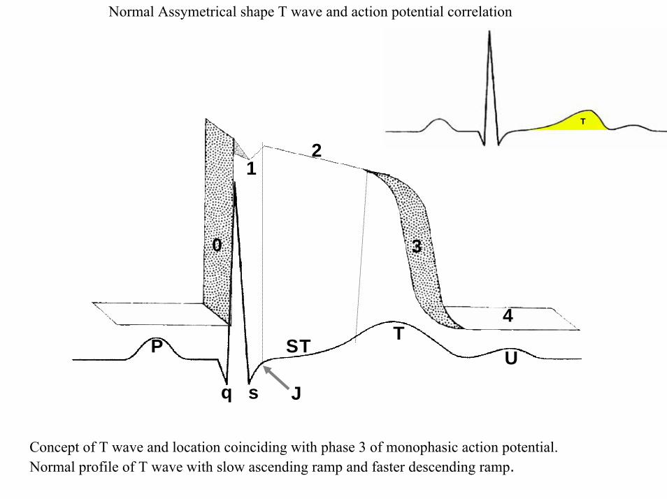
Normal Assymetrical shape T wave and action potential correlation
P
q s J
ST TU
2
4
30
1
TT
Concept of T wave and location coinciding with phase 3 of monophasic action potential. Normal profile of T wave with slow ascending ramp and faster descending ramp.
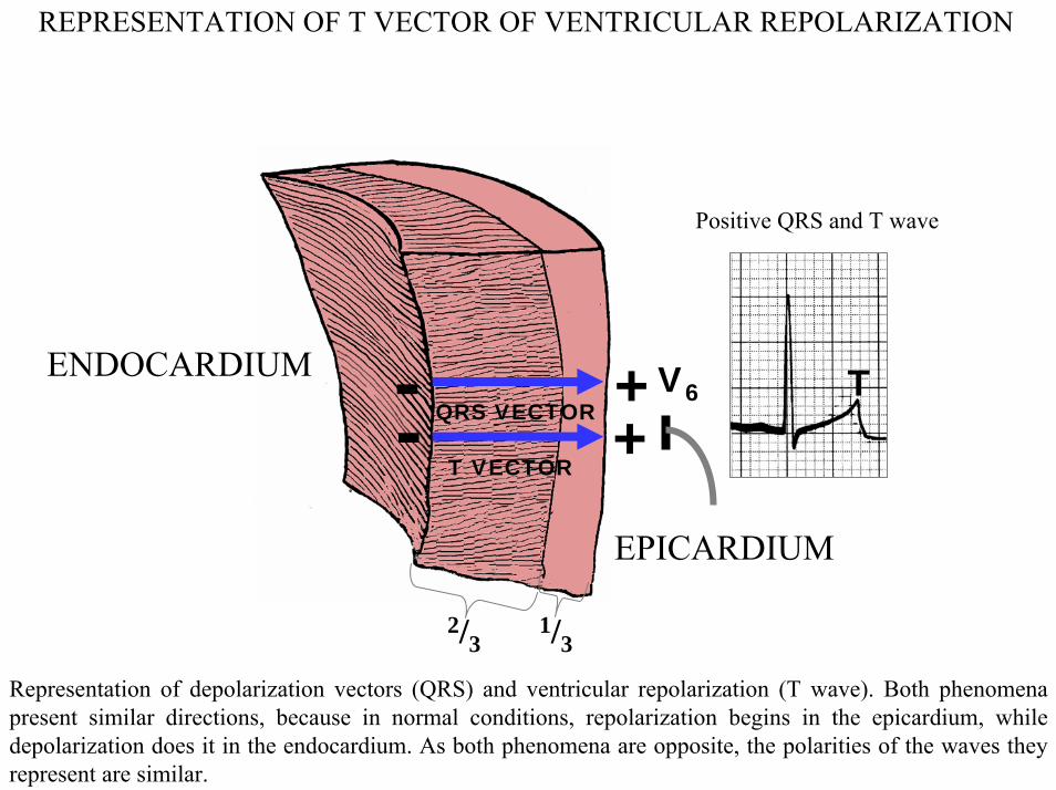
REPRESENTATION OF T VECTOR OF VENTRICULAR REPOLARIZATION
ENDOCARDIUM
EPICARDIUM
+-T VECTOR
QRS VECTOR- +V6 T
1/3
Positive QRS and T wave
2/3
Representation of depolarization vectors (QRS) and ventricular repolarization (T wave). Both phenomena present similar directions, because in normal conditions, repolarization begins in the epicardium, while depolarization does it in the endocardium. As both phenomena are opposite, the polarities of the waves they represent are similar.
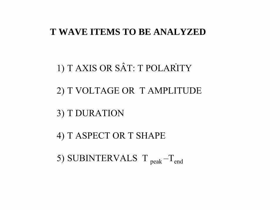
T WAVE ITEMS TO BE ANALYZED
1) T AXIS OR SÂT: T POLARITY
2) T VOLTAGE OR T AMPLITUDE
3) T DURATION
4) T ASPECT OR T SHAPE
5) SUBINTERVALS T peak –Tend
-
I
DIIDIII
aVF
aVR aVL
-35 0
+15
+80 0
SÂT
I
IIIII
aVF
aVR aVL-35º
+15º
+80 0
SÂT
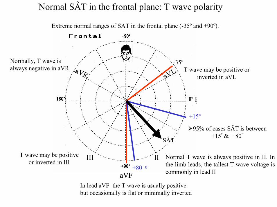
95% of cases SÂT is between +15º & + 80°
Extreme normal ranges of SAT in the frontal plane (-35º and +90º).
Normally, T wave is always negative in aVR
Normal T wave is always positive in II. In the limb leads, the tallest T wave voltage is commonly in lead II
In lead aVF the T wave is usually positive but occasionally is flat or minimally inverted
T wave may be positive or inverted in aVL
T wave may be positive or inverted in III
Normal SÂT in the frontal plane: T wave polarity

NORMAL T AXIS WAVE OR SÂT IN THE FRONTAL PLANE: T POLARITY IN NORMAL ADULTS
SÂT in the FP is between +15º & +80º In adults normal T vector is oriented leftward, inferiorly
FP
00 I
aVR
aVF+ 900
DII
Y
III
aVL
+600
+1800
-1500 - 300
+1200
SÂT
T wave polarity is always positive in II , nearly always positive in aVF and I; variable (biphasic or inverted) in aVL and III; and always negative in aVR.
I
DIIDIII
aVF
aVR aVL
-350
+15º
+800
SÂT
Y
X I
DIIIII
aVF
aVR aVL
-35º
+
+800 SÂTY
XNormal T Vector
Location in adults of normal T wave axis (SÂT) in the frontal plane (near the +60º).

SAT & SAQRS IN THE FRONTAL PLANEIN NORMAL ADULTS
SÂQRS
SÃT
X 0ºI
Y+90ºaVF
The normal angle between the mean SAT & the mean SAQRS is always < 60º. Both are oriented leftward and inferiorly
The sketch shows the angle between QRS and the T wave is narrow in the frontal plane in adults: < 60º. SAT: it means axis of T wave for the ECG or T loop for the VCG. The acronym comes from English, and means S = spatial and A = angle. SAQRS: it means axis of the QRS complex for the ECG or the QRS loop for VCG. The acronym comes from English, and means S = spatial and A = angle.
SÂQRS: mean QRS vectorSÃT: mean T-vector

SAT & SAQRS IN THE FRONTAL PLANE IN NORMAL ELDERLY PEOPLE
SÂQRSSÂT
X 0º I
Y+90ºaVF
The angle between the SÂT and the SÂQRS is always wider: close to 90º.
The outline shows that the angle between QRS and the T wave is wider in the frontal plane in the elderly: near the 90º.
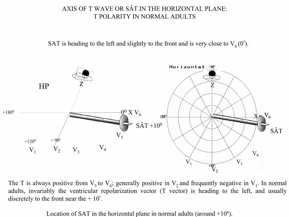
AXIS OF T WAVE OR SÂT IN THE HORIZONTAL PLANE: T POLARITY IN NORMAL ADULTS
SAT is heading to the left and slightly to the front and is very close to V6 (0º).
V1V2
V3
V4
V6X
Z
SÂT
V1V2
V3
V4
V6X
Z
00 X V6
V2
V5+ 900
SÂT +100
ZZHP
+1800
+1200
V4V1 V3
The T is always positive from V3 to V6; generally positive in V2 and frequently negative in V1. In normal adults, invariably the ventricular repolarization vector (T vector) is heading to the left, and usually discretely to the front near the + 10º.
Location of SAT in the horizontal plane in normal adults (around +10º).

HORIZONTAL PLANE: NORMAL SÂT IN ADULTS
V1V2
V3
V4
V6X
Z
+ 10 0
V5
SÂT
In adults over 30 years of age the T wave always positive from V3 to V6
V1V2
V3
V4
V6X
Z
V5
SÂT+10°
In adults over 30 years of age, the T wave is always positive from V3 to V6; generally positive in V2 and frequently negative in V1 .

POLARITY OF T WAVE FROM V1 TO V3 IN ARVC/D
V1
V2
V3
In absence of CRBBB in patients >12 years old, negative T wave from V1 to V3 is a sign with great value for diagnosis for ARVC/D.
In normal, young patients, there is usually positive T polarity in V1; however, it may flatten and nearly always has a positive polarity in V2.
In symptomatic patients carriers of ARVC/D, the ECG generally shows T wave inversion in V1 and V2, which may reach up to V6
1.
T wave from V1 to V3 in ARVC/D.
1. Fontaine G, Tsezana R, Lazarus A, Lascault G, Tonet J, Frank R. Repolarization and intraventricular conduction disorders in arrhythmogenic right ventricular dysplasiaAnn CardiolAngeiol (Paris).1994 Jan;43:5-10.

T LOOP BEHAVIOR IN ARVC/D AND ITS RELATIONSHIP WITH RV END DIASTOLIC VOLUME
V6
V2
HP
V3V4
V5
V1
Z
+ 900+1200
+1800
SAT +150
SAT -100
COUNTERCLOCK WISE ROTATION
When the RV end diastolic volume is not very increased (in average 100 ml/m2).The T loop presents counterclockwise rotation in the HP and axis between +15º y –10º (average +5º).
Value of VCG in ARVD.
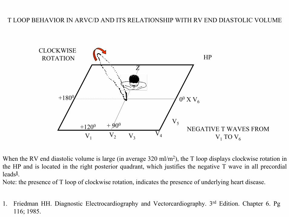
T LOOP BEHAVIOR IN ARVC/D AND ITS RELATIONSHIP WITH RV END DIASTOLIC VOLUME
V2 V3V4
V5
V1
Z
+ 900+1200
+1800
CLOCKWISE ROTATION HP
00 X V6
NEGATIVE T WAVES FROM V1 TO V6
When the RV end diastolic volume is large (in average 320 ml/m2), the T loop displays clockwise rotation in the HP and is located in the right posterior quadrant, which justifies the negative T wave in all precordial leads1.Note: the presence of T loop of clockwise rotation, indicates the presence of underlying heart disease.
1. Friedman HH. Diagnostic Electrocardiography and Vectorcardiography. 3rd Edition. Chapter 6. Pg 116; 1985.

T loop in 9 patients in the HP, carriers of ARVD. T loops are arranged on the basis of progressive RVE.
T loop (nº 1) has a RV end diastolic volume of 100 ml/m2 and the last loop (nº 9) has 320 ml/m2.
Note the progressive alteration of the T loop from 1 to 9.
T LOOP BEHAVIOR IN ARVC/D AND ITS RELATIONSHIP WITH RV END DIASTOLIC VOLUME
1 2 3
4 5 6
7 8 9
V1V2
V3
V6
NEGATIVE T WAVES FROM V1 TO V6
Value of VCG in ARVC/D.
1. Nava A, Canciani B, Buja G, etal.Electrovectorcardiographic study of negative T waves on precordial leads in arrhythmogenic right ventricular dysplasia: relationship with right ventricular volumes. J Electrocardiol. 1988 Aug; 21: 239-245.

NORMAL SÂT IN FULL-TERM NEWBORN BABIES IN THE HORIZONTAL PLANE
SÂTV1
V3
V4
V6X
Z
+750
SATV1 V3
V4
VX
Z
+750
In newborn babies, T wave: it may be of positive polarity in V1 in the first day of life, (SÂT is heading towards the V3 lead, i.e. around + 75º.) being negative since the third day. Positive T wave beyond this time suggests RVH/E. SAT in the HP at the moment of birth points to the front and to the left, near the V3 lead: +75º. Between 1h and 6h of life, SAT dislocates even more to the right, close to +100º. Therefore, occasionally the T wave may be negative in V6. (SAT in +100º ).
V2
V5
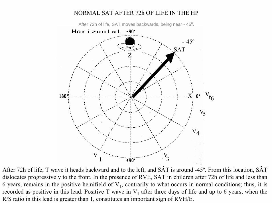
NORMAL SAT AFTER 72h OF LIFE IN THE HP
SÂT
V1
V3
V4
V6X
Z
- 45 0
V
SAT
V1
V3
V4
V6X
Z
- 45º
V5
After 72h of life, SAT moves backwards, being near - 450.
After 72h of life, T wave it heads backward and to the left, and SÂT is around -45º. From this location, SÂT dislocates progressively to the front. In the presence of RVE, SAT in children after 72h of life and less than 6 years, remains in the positive hemifield of V1, contrarily to what occurs in normal conditions; thus, it is recorded as positive in this lead. Positive T wave in V1 after three days of life and up to 6 years, when the R/S ratio in this lead is greater than 1, constitutes an important sign of RVH/E.

SAT & SAQRS IN THE FRONTAL PLANE IN NORMAL CHILDREN
SÂQRS
SÂTX 0º I
Y+90ºaVF
The SAQRS is more anterior and the SAT mare posterior: the anglecould be wide.
The outline shows that the T wave is more posterior in relation to the QRS in the frontal plane in children.
2) T VOLTAGE OR T AMPLITUDE
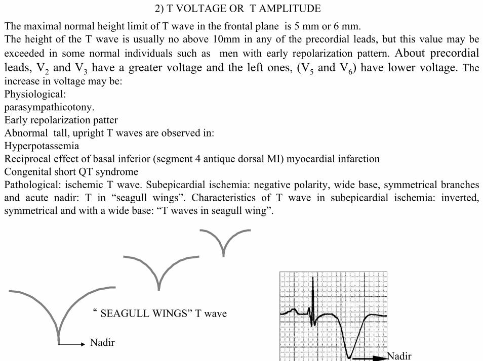
SEAGULL WINGS” T wave“
Nadir
The maximal normal height limit of T wave in the frontal plane is 5 mm or 6 mm. The height of the T wave is usually no above 10mm in any of the precordial leads, but this value may be exceeded in some normal individuals such as men with early repolarization pattern. About precordial leads, V2 and V3 have a greater voltage and the left ones, (V5 and V6) have lower voltage. The increase in voltage may be: Physiological: parasympathicotony.Early repolarization patter Abnormal tall, upright T waves are observed in:HyperpotassemiaReciprocal effect of basal inferior (segment 4 antique dorsal MI) myocardial infarctionCongenital short QT syndromePathological: ischemic T wave. Subepicardial ischemia: negative polarity, wide base, symmetrical branches and acute nadir: T in “seagull wings”. Characteristics of T wave in subepicardial ischemia: inverted, symmetrical and with a wide base: “T waves in seagull wing”.
Nadir
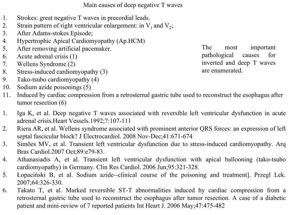
Main causes of deep negative T waves
1. Strokes: great negative T waves in precordial leads. 2. Strain pattern of right ventricular enlargement: in V1 and V2;3. After Adams-stokes Episode;4. Hypertrophic Apical Cardiomyopathy (Ap.HCM)5. After removing artificial pacemaker.6. Acute adrenal crisis (1)7. Wellens Syndrome (2)8. Stress-induced cardiomyopathy (3)9. Tako-tsubo cardiomyopathy (4)10. Sodium azide poisonings (5)11. Induced by cardiac compression from a retrosternal gastric tube used to reconstruct the esophagus after
tumor resection (6)
The most important pathological causes for inverted and deep T waves are enumerated.
1. Iga K, et al. Deep negative T waves associated with reversible left ventricular dysfunction in acute adrenal crisis.Heart Vessels.1992;7:107-111
2. Riera AR, et al. Wellens syndrome associated with prominent anterior QRS forces: an expression of left septal fascicular block? J Electrocardiol. 2008 Nov-Dec;41:671-674
3. Simões MV, et al. Transient left ventricular dysfunction due to stress-induced cardiomyopathy. ArqBras Cardiol.2007 Oct;89:e79-83.
4. Athanasiadis A, et al. Transient left ventricular dysfunction with apical ballooning (tako-tsubocardiomyopathy) in Germany. Clin Res Cardiol. 2006 Jun;95:321-328.
5. Łopaciński B, et al. Sodium azide--clinical course of the poisoning and treatment]. Przegl Lek. 2007;64:326-330.
6. Takato T, et al. Marked reversible ST-T abnormalities induced by cardiac compression from a retrosternal gastric tube used to reconstruct the esophagus after tumor resection. A case of a diabetic patient and mini-review of 7 reported patients Int Heart J. 2006 May;47:475-482

Name: E.A.D.; Age: 68 y.; Sex: Fem. Race: White Date: 01/21/1999.; Weight: 65 Kg Height: 1.65 mMedication in use: Enalapril Hydrochlorothiazide
Clinical diagnosis: Subarachnoid bleeding.ECG diagnosis: long QT interval, largely wide and inverted T waves: “giant T waves”.
ECG that shows inverted T waves, with great width and wide base with prolonged QT interval in a patient with subarachnoid bleeding.
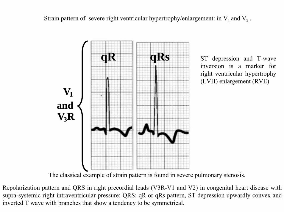
Strain pattern of severe right ventricular hypertrophy/enlargement: in V1 and V2 .
VV11
eVV33RR
qR qRs
VV11
andVV33RR
qR qRs
The classical example of strain pattern is found in severe pulmonary stenosis.
ST depression and T-wave inversion is a marker for right ventricular hypertrophy (LVH) enlargement (RVE)
Repolarization pattern and QRS in right precordial leads (V3R-V1 and V2) in congenital heart disease with supra-systemic right intraventricular pressure: QRS: qR or qRs pattern, ST depression upwardly convex and inverted T wave with branches that show a tendency to be symmetrical.

STRAIN PATTERN OF RVH/E SEVERE PULMONARY STENOSIS
V1
Suprasystemic right intraventricular pressure. V2 and V3 continue showing QRS predominantly positive. Inverted T wave and with a tendency to be symmetrical (primary).
qR
V1
Repolarization pattern and QRS in right precordial leads (V3R-V1 and V2) in congenital heart disease with suprasystemic right intraventricular pressure (severe pulmonary stenosis): QRS: qR pattern, ST and inverted T wave with branches that show a tendency to be symmetrical.

T WAVES AFTER ADAMS-STOKES ATTACKS ASSOCIATED WITH COMPLETE HEART BLOCK
P P P
P
PP P P
P
P
Negative T wave after Adams-Stokes episode in complete AV block.
ECG strip that shows total AV block in a patient that suffered a recent episode of Stokes-Adams: giant T waves, deeply inverted and with prolonged QT interval. This situation causes a tendency to appearance of polymorphic ventricular tachycardia of the torsade des pointes (TdP) type.

Name: SFS. Age: 15y. Sex: M. Race: W.Weight: 70Kg. Height: 1.72m. Number: 718. Date: 03/31/98 Medication in use: beta-blocker.
HCM obstructive form. Apical area of the septum with 32 mm of diastolic thickness. LAE. Systolic pattern of LVE by important alteration secondary to ventricular repolarization in anterolateral and inferior wall.
Typical ECG of obstructive form of hypertrophic cardiomyopathy in a 15-year-old teenager. Left chamber enlargement, important depression of ST segment upwardly convex and followed by wide-based and deeply inverted T waves: alteration secondary to ventricular repolarization in anterolateral and inferior wall.
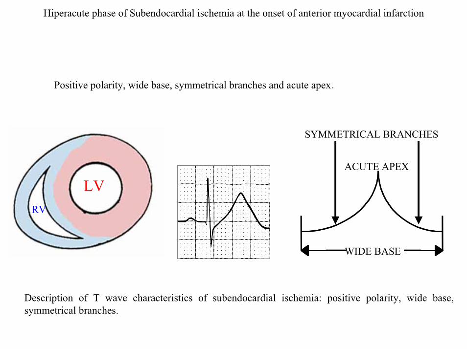
Hiperacute phase of Subendocardial ischemia at the onset of anterior myocardial infarction
Positive polarity, wide base, symmetrical branches and acute apex.
LVRV
ACUTE APEX
SYMMETRICAL BRANCHES
WIDE BASE
Description of T wave characteristics of subendocardial ischemia: positive polarity, wide base, symmetrical branches.

CAUSES OF ISCHEMIC & PSEUDO-ISCHEMIC SUBEPICARDIAL T WAVE
- Coronary insufficiency: a) Hyperacute phase of anterior AMIb) Reciprocal alterations in the inferior wall by posterior ischemia;
- Pericarditis:- Ventricular enlargement of volumetric or diastolic type;- Alcoholism;- Normal variant: in athletic men.
Enumeration of the main causes of T waves of subendocardial ischemia and pseudo subepicardial ischemia.
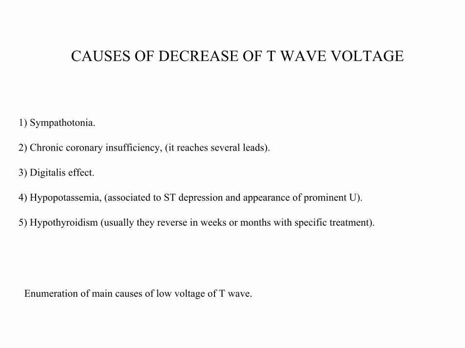
CAUSES OF DECREASE OF T WAVE VOLTAGE
1) Sympathotonia.
2) Chronic coronary insufficiency, (it reaches several leads).
3) Digitalis effect.
4) Hypopotassemia, (associated to ST depression and appearance of prominent U).
5) Hypothyroidism (usually they reverse in weeks or months with specific treatment).
Enumeration of main causes of low voltage of T wave.

90 ms 100ms to 250ms
3) DURATION OF T WAVE
100ms to 250ms (up to five times more than ventricular depolarization).
Comparison of ECGs with normal duration of QRS (90ms) and T wave (100ms to 250ms).

4) NORMAL ASPECT OR SHAPE OF THE T WAVE
RAMPA ASCENDENTE
LENTA
RAMPA DESCENDENTE
RÁPIDAAsymmetrical T-wave: with an ascending slow slopewith superior concavity and faster descending slope.
When positive, T wave is characterized by being asymmetrical with its ascending slope being slow and of superior concavity, and fast descending slope.
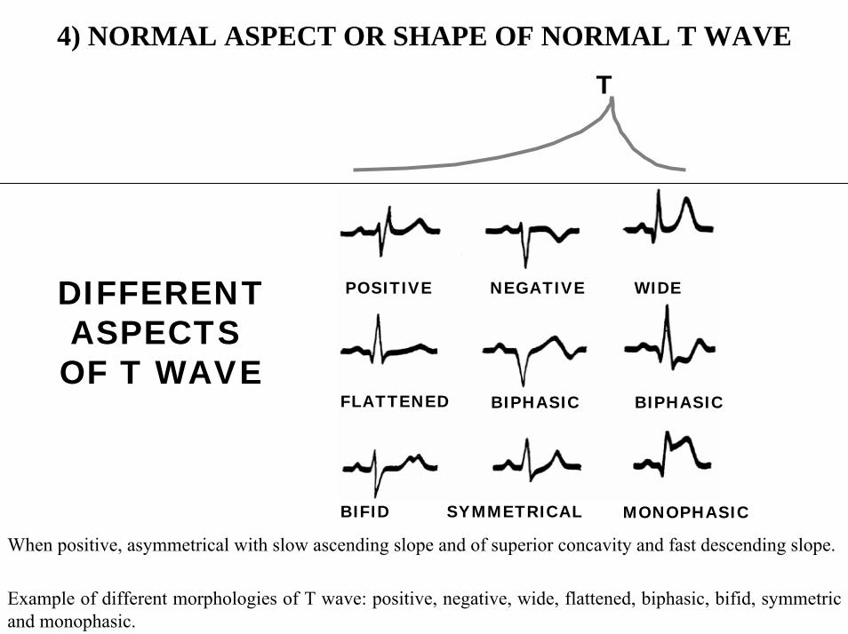
4) NORMAL ASPECT OR SHAPE OF NORMAL T WAVE
T
POSITIVE NEGATIVE WIDE
FLATTENED BIPHASIC BIPHASIC
BIFID SYMMETRICAL MONOPHASIC
DIFFERENTASPECTS OF T WAVE
When positive, asymmetrical with slow ascending slope and of superior concavity and fast descending slope.
Example of different morphologies of T wave: positive, negative, wide, flattened, biphasic, bifid, symmetric and monophasic.

5) Tpeak-Tend interval or Tpe.
In electrocardiography, T wave represents repolarization or recovery of the ventricles. The interval from the QRS complex onset up to the T wave apex or peak, is known as absolute refractory period. The last portion of the T wave, is the faster descending part. It is known as relative or vulnerable refractory period. The normal value of Tpeak/Tend interval (Tpe) is ≤94 ms in men and ≤92 in women when measured in the V5 lead. Tpe prolongation to values ≥120 ms is associated to a greater number of events in patients carriers of BrS.(1-5.)
Representation of Tpeak-Tend interval or Tpe
Interval elapsed from the apex to the end of T wave
1. Wang JF, Shan QJ, Yang B, et al. Tpeak-Tend interval and risk of cardiac events in patients with Brugada syndrome Zhonghua Xin Xue Guan Bing Za Zhi. 2007; 35: 629-632.
2. Haarmark C, Graff C, Andersen MP, et al. Reference values of electrocardiogram repolarization variables in a healthy population. J Electrocardiol. 2010 Jan-Feb;43: 31-39.
3. Gupta P, Patel C, Patel H, et al. T(p-e)/QT ratio as an index of arrhythmogenesis. J Electrocardiol. 2008 Nov-Dec;41:567-74.
4. Lambiase PD.Tpeak-Tend interval and Tpeak-Tend/QT ratio as markers of ventricular tachycardia inducibility in subjects with Brugada ECG phenotype. Europace. 2010 Feb;12:158-159.
5. Letsas KP, Weber R, Astheimer et al. Tpeak-Tend interval and Tpeak-Tend/QT ratio as markers of ventricular tachycardia inducibility in subjects with Brugada ECG phenotype. Europace. 2010 Feb; 12: 271-274.
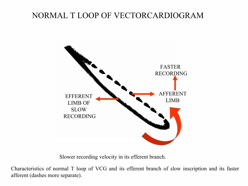
NORMAL T LOOP OF VECTORCARDIOGRAM
AFFERENTLIMB
EFFERENT LIMB OF
SLOW RECORDING
FASTER RECORDING
Slower recording velocity in its efferent branch.
Characteristics of normal T loop of VCG and its efferent branch of slow inscription and its faster afferent (dashes more separate).

HYPERPOTASSEMIC T WAVE
T
K+ 7.8 mEq/L
POINTY T WAVE IN “DESERT TENT”
Narrow-based, symmetrical, pointy T wave, of increased voltage, in “desert tent”. QRScomplex widens and p wave is flattened or disappears.
Tent-shaped hyperpotassemic T wave: tall and narrow-based. This wave is observed with slightly increased serum potassium levels. It is present only in 22% of the cases of hyperpotassemia. The signal is not too sensitive but quite specific. Similar T wave type may be observed in congenital familial short QT syndrome.

Typical hyperkalemic/ hyperpotassemic T wave
SYMMETRICAL, NARROW-BASED, POINTY T WAVE,
IN “DESERT TENT”
It is observed when the rate of potassium reaches 5.5 mEq/l. The sensitivity is just 22% of cases. It is visible and may be confused with the T wave observed in bradycardia, diastolic LVE, subendocardial ischemia, schizophrenia, short QT syndrome and stroke. Hyperkalemia affects up to 8% of hospitalized patients mainly in the setting of compromised renal function. The ECG manifestation of hyperkalemia depends on serum K+ level. At 5.5-7.0 mmol/L K+, tall peaked, narrow-based T waves are seen.(1)1. El-Sherif N, Turitto G. Electrolyte disorders and arrhythmogenesis. Cardiol J. 2011;18:233-245.

HYPERPOTASSEMIA ASSOCIATED TO HYPOCALCEMIAQT INTERVAL PROLONGATION AT THE EXPENSE OF THE ST SEGMENT
+
HYPERPOTASSEMIA UREMIA
+
HYPOCALCEMIA
HYPOCALCEMIA
Characteristics of repolarization in uremia: prolonged ST segment (hypocalcemia) followed by T wave of great voltage with narrow base (hyperpotassemia).

CONGENITAL SHORT QT SYNDROME AND TALL T WAVES
Hereditary, congenital, or familial short QT syndrome is a clinico-electrocardiographic entity, and part of the so-called channelopathies; dominant autosomal or sporadic, genetically heterogeneous, which affects the electric system of the heart, clinically characterized by a large set of signs and symptoms, such as: syncope, sudden death, dizziness and high tendency to appearance of episodes of paroxysmal runs of atrial fibrillation.
Electrocardiogram characterized by: extremely short QT interval (QTc interval ≤ 300 ms) that is not significantly modified with heart rate changes, T waves of great voltage and narrow base, which resemble T wave in “desert tent” or tour Eiffel pattern of hyperkalemia/ hyperpotasemia.From the structural point of view, the heart is normal and electrophysiologically, there is significant shortening of refractory periods of atria and ventricles, being inducible (sustained VF) by programmed stimulation.
Several families have been identified, Thus far, mutations in six different genes encoding potassium and calcium channel subunits have been reported. Three affecting K channels with a gain of function: SQT1 (Iks), SQT2 (Ikr) and SQT3 (Ik1) and three variant related with calcium channels.
The variants SQT1 (Iks), SQT2 (Ikr) and SQT3 (Ik1) are the opposite of long QT syndrome, since they exert opposite effects regarding potassium rectifier channels function: SQTS causes increase in the function of such channels; on the other hands, long QT syndrome causes decrease of function.
Congenital short QT syndrome: concept and features.
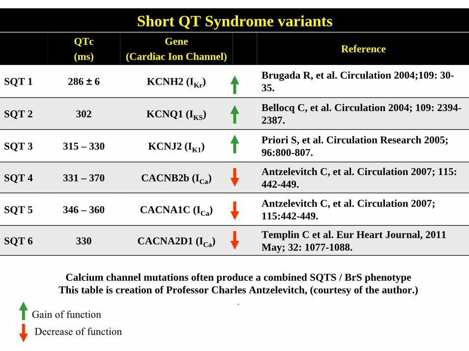
Short QT Syndrome variantsQTc (ms)
Gene (Cardiac Ion Channel)
Reference
SQT 1 286 ± 6 KCNH2 (IKr)Brugada R, et al. Circulation 2004;109: 30-35.
SQT 2 302 KCNQ1 (IKS) Bellocq C, et al. Circulation 2004; 109: 2394-2387.
SQT 3 315 – 330 KCNJ2 (IK1)Priori S, et al. Circulation Research 2005; 96:800-807.
SQT 4 331 – 370 CACNB2b (ICa)Antzelevitch C, et al. Circulation 2007; 115: 442-449.
SQT 5 346 – 360 CACNA1C (ICa)Antzelevitch C, et al. Circulation 2007; 115:442-449.
SQT 6 330 CACNA2D1 (ICa)Templin C et al. Eur Heart Journal, 2011 May; 32: 1077-1088.
Calcium channel mutations often produce a combined SQTS / BrS phenotypeThis table is creation of Professor Charles Antzelevitch, (courtesy of the author.)
.Gain of functionDecrease of function

Name: JSVB; Age: 27; Sex: Male; Race: White; Weight: 67 Kg.Height: 1.72 m. Date: 06/24/2004; Medication in use: none.
Rhythm: sinus; HR: 65 bpm; P wave: SAP axis: +54º in the FP and to the front in the HP; Duration: 80 ms; Voltage: 1 mm; PR interval: 134 ms; QRS: SAQRS: +106º; in the FP and to the front in the HP; QRS duration (QRSD): 120 ms; QRS morphology: triphasic rSR' pattern in V1 and broad S wave in left leads DI, aVL V5 and V6 (right terminal forces); intrinsic deflection in V1 > 50 ms. T wave: morphology: tall T wave from V3 through V5 with narrow base and a tendency to be symmetrical (the patient does not have serum potassium increase); SAT: +42º in the FP and discretely heading to the front and below in the HP; QT/QTc interval: 302/315: short for this rate (the inferior limit for a 67 bpm heart rate in men is 324ms1); JT/JTc interval: 182/199 ms: extremely short (QT-QRSD = JT. 302-120 = 182 ms). (The inferior limit for a 67 bpm heart rate in men is 224 ms). Conclusion: 1) CRBBB; 2) Increase of QRS duration; 3) Short QT interval with no use of drugs, electrolytic disorders or any associated pathophysiological state; 4) Very short JT interval; 5) Probable early repolarization pattern.
Typical ECG of congenital short QT syndrome. 1) Sagie A, et al. Am J Cardiol 1992; 70:797-801.

V1
JT170 ms
QT
281 ms
V6
170 ms
281 msQT
JT
V1
JT170 ms
QT
281 ms
V6
170 ms
281 msQT
JT
V6
170 ms
281 msQT
JT
Characteristics of JT and QT intervals in congenital short QT syndrome.

VECTOCARDIOGRAMName: JSVB; Age: 27; Sex: Male; Race: White; Weight: 67 Kg.
Height: 1.72 m. Date: 06/24/2004; Medication in use: None.
Vectocardiogram in congenital short QT syndrome and correlation with electrocardiogram.

ECG/VCG CORRELATION FRONTAL PLANE
aVR aVL
DI
DIIDIII
aVF
T
X
Y
P
ECD
CRBBB
ASYMMETRICALT LOOP
QRS LOOP DURATION120 ms
aVR aVL
DI
T
X
Y
P
END CONDUCTION DELAY (ECD)
Vectocardiogram in congenital short QT syndrome and correlation with electrocardiogram.
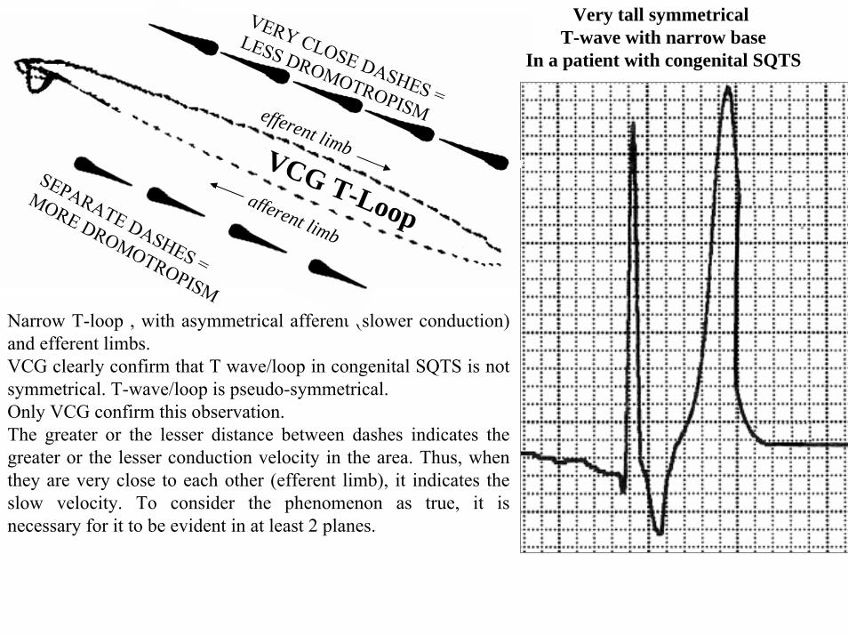
VCG T-Loop
Very tall symmetrical T-wave with narrow base
In a patient with congenital SQTS
efferent limb
afferent limb
Narrow T-loop , with asymmetrical afferent (slower conduction) and efferent limbs.VCG clearly confirm that T wave/loop in congenital SQTS is not symmetrical. T-wave/loop is pseudo-symmetrical.Only VCG confirm this observation.The greater or the lesser distance between dashes indicates the greater or the lesser conduction velocity in the area. Thus, when they are very close to each other (efferent limb), it indicates the slow velocity. To consider the phenomenon as true, it is necessary for it to be evident in at least 2 planes.
SEPARATE DASHES =
MORE DROMOTROPISM
VERY CLOSE DASHES =
LESS DROMOTROPISM

ECG/VCG CORRELATION HORIZONTAL PLANE
V6
V1
V2
V3
V4
V5
T
P
X
Z
ECD
SHORT QT INTERVAL
V6
V1
V2
V3
V4
V5
T
P
X
Z
TALL T WAVES WITH NARROW BASE
FROM V3 THROUGH V5
TRIPHASIC rSR’ PATTERN
GRISHMAN-TYPE CRBBB:AFFERENT LOOP BEHIND THE X LINE
Vectocardiogram in congenital short QT syndrome and correlation with electrocardiogram.
ECG/VCG CORRELATION RIGHT SAGITTAL PLANE
aVF
Y
Z
T
P
aVF
Y
Z
T
P
T LOOP HEADING DOWN AND TO THE FRONT
ASYMMETRICAL T LOOP
Vectocardiogram in congenital short QT syndrome and correlation with electrocardiogram.

LONG DURATION ELECTROCARDIOGRAM RECORDING (HOLTER)
In this tracing we can see a short period of gross atrial fibrillation. The patient described palpitations. Congenital short QT syndrome is associated to high incidence of paroxysmal atrial fibrillation, the electrophysiological mechanism of which would be caused by heterogeneous shortening of the cardiac potential and refractory period of atrial cardiomyocytes.
Long duration electrocardiogram recording (Holter) in congenital short QT syndrome, which displays paroxysmal atrial fibrillation run.

LONG-DURATION OF ELECTROCARDIOGRAM RECORDING (HOLTER)
Approximately 8 hours later during the same test, the patient spontaneously reversed into sinus rhythm.
Long duration electrocardiogram recording (Holter) in congenital short QT syndrome, which shows spontaneous sinus rhythm during the same recording (8 hours later).

CAUSES OF SHORT QT SYNDROME
Classification of short QT syndrome.
A) Acquired formsAcidosis.Autonomic tone alterations.Rufinamide it is a new antiepileptic drug for the add-on treatment of Lennox-Gastaut syndrome (1)Toxicity and digitalis effect.Hypercalcemia.Hyperthermia.Hyperkalemia/Hyperpotasemia.
B) Congenital, inherited or familial variants1. SQT1: By mutation in the rapid potassium rectifier channel Ikr / HERG (KCNH2): the mutation causes
gain in Ikr channel function, causing heterogeneous shortening in action potential and refractoriness, reducing channel activity by blockers2.(2)
2. SQT2: By mutation in the slow potassium rectifier channel Iks in the KCNQ1 gene.(3)3. SQT3 variant by mutation in the inward rectifier K+ current or Ik1 channel.(4) 4. SQT4 mutation in CACNB2b decrease of function of calcium channel(5)5. SQT5: mutation inCACNA1C decrease of function of calcium channel(6)6. SQTS6: By mutatation in the CACNA2D1 gene in humans, causing the SQTS6 variant.(7)
1. Schimpf R, Veltmann C, Papavassiliu T, et al, Drug-induced QT-interval shortening following antiepileptic treatment with oral rufinamide.Heart Rhythm. 2012 Jan 11. [Epub ahead of print]
2. Brugada R, et al. Circulation. 2004; 109: 30-35.3. Bellocq C, et al. Circulation. 2004; 109:2394-2397.4. 3Priori SG, et al. Circ Res. 2005; 96:800-807. 5. Antzelevitch, C. Circulation 2007; 115:442. 6. Antzelevitch, C Circulation 2007; 115:442,7. Templin C Eur Heart J. 2011 May;32:1077-1088.

Tall, narrow, and symmetrical T-waves (or pseudosymmetrical) in congenital Short QT syndrome.
Short QT syndrome (SQTS) is a genetically determined ion-channel disorder, which may cause malignant tachyarrhythmias and SCD. Templin et al (1) presented, a novel loss-of-function mutation coding for an L-type calcium channel subunit.The ECG of the affected member of a single family revealed a QT interval of 317 ms (QTc 329 ms) with tall, narrow, and symmetrical T-waves. Invasive EPS testing showed short ventricular refractory periods and increased vulnerability to induce VF. DNA screening of the patient identified a new variant at a heterozygous state in the CACNA2D1 gene (nucleotide c.2264G > C; amino acid p.Ser755Thr), coding for the Ca(v)α(2)δ-1 subunit of the L-type calcium channel. The pathogenic role of the p.Ser755Thr variant of the CACNA2D1 gene was analysed by using co-expression of the two other L-type calcium channel subunits, Ca(v)1.2α1 and Ca(v)β(2b), in HEK-293 cells. Barium currents (I(Ba)) were recorded in these cells under voltage-clamp conditions using the whole-cell configuration. Co-expression of the p.Ser755Thr Ca(v)α(2)δ-1 subunit strongly reduced the I(Ba) by more than 70% when compared with the co-expression of the wild-type (WT) variant. Protein expression of the three subunits was verified by performing western blots of total lysates and cell membrane fractions of HEK-293 cells. The p.Ser755Thr variant of the Ca(v)α(2)δ-1 subunit was expressed at a similar level compared with the WT subunit in both fractions. Since the mutant Ca(v)α(2)δ-1 subunit did not modify the expression of the pore-forming subunit of the L-type calcium channel, Ca(v)1.2α1, it suggests that single channel biophysical properties of the L-type Ca2+ channel are altered by this variant.The authors (1) reported the first pathogenic mutation in the CACNA2D1 gene in humans, which causes the SQT6 variant. It remains to be determined whether mutations in this gene lead to other manifestations of the J-wave syndrome.
1. Templin C, Ghadri JR, Rougier JS, et al. Identification of a novel loss-of-function calcium channel gene mutation in short QT syndrome (SQTS6).Eur Heart J. 2011 May;32:1077-1088.

T-WAVE ALTERNANS ("macroscopic" TWA)
The alternans of T wave polarity is a characteristic of patients carriers of long QT syndrome (LQTS). Isolated T wave alternans is not related to tachycardia or extra-systole, and it usually indicates advanced heart disease or severe electrolytic disorder. T-wave alternans has long been recognized as a marker of electrical instability in acute ischemia, where it may precede ventricular tachyarrhythmia. Studies have shown that T wave (or ST-T) alternans can also precede non-ischemic ventricular tachyarrhythmias. Considerable interest has recently been shown in the detection of microvolt T wave alternans as a noninvasive marker of the risk of ventricular tachyarrhythmia in patients with chronic heart disease. Assessment of left ventricular ejection fraction, Holter monitoring, and signal-averaged late potentials are the principal noninvasive means of determining the risk of ventricular arrhythmias after myocardial infarction. However, these measures of vulnerability to arrhythmias have been found to be less predictive of arrhythmic events than invasive electrophysiologic testing.Microvolt T-wave alternans testing is performed by placing high-resolution electrodes, designed to reduce electrical interference, on a patient’s chest prior to a period of controlled exercise (CMS, 2005). These electrodes detect tiny beat-to-beat changes, on the order of one-millionth of volt, in the EKG T-wave. Spectral analysis is used to calculate these minute voltage changes. Spectral analysis is a sensitive mathematical method of measuring and comparing time and the electrocardiogram signals. Software then analyzes these microvolt changes and produces a report to be interpreted by a physician.
Alternans of T wave polarity, characteristic of patients carriers of long QT syndrome (LQTS).

CAUSES OF ISOLATED T-WAVE ALTERNANS
- Tachycardia.- Sudden changes in cycle length or HR cycle.- Severe hyperpotassemia of uremia.- Experimentally, in hypocalcemia in dogs.- Severe myocardial impairment: cardiomyopathy.- Acute myocardial ischemia, particularly in variant angina.- Post-resuscitation.- Acute pulmonary embolism.- After administration of amiodarone or quinidine (rare).- Congenital long QT syndromes of the Romano-Ward or Jervell-Lange Nielsen types.- Brugada syndrome.
The main causes of isolated T wave alternans.