transcriptional analysis of pluripotency reveals the hippo pathway
TRANSCRIPT

Transcriptional analysis of pluripotency reveals theHippo pathway as a barrier to reprogramming
Han Qin1,2, Kathryn Blaschke1,2, Grace Wei1, Yuki Ohi1,2, Laure Blouin1,2, Zhongxia Qi3,
Jingwei Yu3, Ru-Fang Yeh4,{, Matthias Hebrok1 and Miguel Ramalho-Santos1,2,∗
1Eli and Edythe Broad Center of Regeneration Medicine and Stem Cell Research, and Diabetes Center, 2Departments
of Ob/Gyn and Pathology, and Center for Reproductive Sciences, 3Department of Laboratory Medicine and 4Division
of Biostatistics, University of California, San Francisco, CA, USA
Received November 17, 2011; Revised December 23, 2011; Accepted January 23, 2012
Pluripotent stem cells are derived from culture of early embryos or the germline and can be induced by re-programming of somatic cells. Barriers to reprogramming that stabilize the differentiated state and havetumor suppression functions are expected to exist. However, we have a limited understanding of whatsuch barriers might be. To find novel barriers to reprogramming to pluripotency, we compared the transcrip-tional profiles of the mouse germline with pluripotent and somatic cells, in vivo and in vitro. There is a re-markable global expression of the transcriptional program for pluripotency in primordial germ cells(PGCs). We identify parallels between PGC reprogramming to pluripotency and human germ cell tumorigen-esis, including the loss of LATS2, a tumor suppressor kinase of the Hippo pathway. We show that knockdownof LATS2 increases the efficiency of induction of pluripotency in human cells. LATS2 RNAi, unlike p53 RNAi,specifically enhances the generation of fully reprogrammed iPS cells without accelerating cell proliferation.We further show that LATS2 represses reprogramming in human cells by post-transcriptionally antagonizingTAZ but not YAP, two downstream effectors of the Hippo pathway. These results reveal transcriptional par-allels between germ cell transformation and the generation of iPS cells and indicate that the Hippo pathwayconstitutes a barrier to cellular reprogramming.
INTRODUCTION
Pluripotent stem cells can be propagated almost indefinitelywithout undergoing senescence and can give rise to all celltypes of the body, both in vitro and in vivo. Because ofthese properties, pluripotent stem cells are an excellentsystem to study cellular differentiation in normal and dis-eased states and may contribute to the development of cell-replacement therapies (1–4). Embryonic stem (ES) cells arethe prototypical pluripotent stem cells and are derived fromin vitro culture of the inner cell mass (ICM) of the blastocyst(5–7). Remarkably, pluripotent stem cells can be generatedby over-expressing particular key transcription factors ormicroRNAs in somatic cells (8–18). This approach allowsthe generation of disease-specific induced pluripotent stem(iPS) cells (19–21) and holds enormous promise in Regen-erative Medicine. However, the efficiency of iPS cell
generation is very low, and this is likely due to genes or path-ways that act as barriers to reprogramming to pluripotency.Senescence has been reported as a barrier to reprogramming.Preventing senescence by over-expressing SV40T antigen orhTERT (15), or down-regulating p53 or p21 (22–29), cansignificantly increase the efficiency of iPS cell generation.However, these manipulations appear to facilitate reprogram-ming largely by inducing a higher rate of cell proliferation,and thereby increasing the probability of stochastic eventsthat may underlie reprogramming (23). Targets of the EScell-specific cell cycle-regulating (ESCC) family of miRNAhave also been shown to antagonize reprogramming (17). Inaddition, lineage-specific transcription factors may also actas barriers to reprogramming (30,31). Therefore, the assayof iPS cell generation provides an opportunity to dissect themechanisms that act as barriers to reprogramming and antag-onize cellular transformation (32).
†Present address: Genentech Inc., South San Francisco, CA, USA.
∗To whom correspondence should be addressed. Tel: +1 4155029584; Fax: +1 4155142346; Email: [email protected]
# The Author 2012. Published by Oxford University Press. All rights reserved.For Permissions, please email: [email protected]
Human Molecular Genetics, 2012, Vol. 21, No. 9 2054–2067doi:10.1093/hmg/dds023Advance Access published on January 27, 2012
Dow
nloaded from https://academ
ic.oup.com/hm
g/article/21/9/2054/580086 by guest on 12 Decem
ber 2021

A cell lineage where barriers to reprogramming may be ofparticular importance is the germline. Primordial germ cells(PGCs) are the embryonic precursors to the gametes, which re-establish the totipotent zygote upon fertilization. When PGCsare cultured in vitro they give rise to pluripotent stem cellsvery similar to ES cells, called embryonic germ (EG) cells(33–35). Unlike the reprogramming of somatic cells to iPScells, reprogramming of PGCs to EG cells does not requireintroduction of exogenous genes. This is largely due to thefact that critical regulators of ES cell pluripotency and repro-gramming, such as the transcription factors Oct4 and Nanog,are highly expressed in PGCs and indeed are essential fortheir development (36–38). However, important differencesbetween PGCs and pluripotent stem cells must exist. PGCs,unlike ES cells or EG cells, proliferate for only a shortperiod of time and do not contribute to chimeras when injectedinto blastocysts (39). Germ cell tumors are thought to arisefrom loss of tumor suppressor mechanisms that are active inPGCs but not in pluripotent stem cells (40). A direct compari-son of transcriptional profiles between PGCs and other pluri-potent cell types would therefore be expected to shed lighton the mechanisms that protect PGCs against cellular trans-formation, and potentially also reveal novel barriers to repro-gramming of somatic cells to pluripotency. While severalrecent studies have described transcriptional analyses ofPGCs (41–47), no study to date has directly compared thetranscriptome of the ICM, ES cells, PGCs and EG cells, andno insights into potential barriers to reprogramming havebeen reported.
We report a comparative study of the gene-expression pro-files of mouse pluripotent stem cells and the cells in theembryo from which they are derived, including PGCs. Ourresults reveal a core transcriptional program present in allpluripotent cells analyzed, including a remarkable global ex-pression of the transcriptional program for pluripotency inPGCs. We find that reprogramming of PGCs to the pluripotentstem cell state involves transcriptional changes that parallelboth human germ cell tumorigenesis and the generation ofiPS cells. The tumor suppressor Lats2 is highly expressed inPGCs but not in pluripotent stem cells or human germ celltumors. Lats2 is a kinase of the Hippo pathway, a signalingcascade that regulates cell growth and tumorigenesis in bothDrosophila and mammals (48–50). We show that LATS2acts as a barrier to induction of pluripotency in human cells,and that this effect is mediated by suppression of TAZ, adownstream target of the Hippo pathway. We discuss the po-tential implications of our results for the parallels betweengerm cell transformation and the generation of iPS cells.
RESULTS
Identification of the transcriptional profiles ofpluripotent cells
We first used microarrays to identify and compare the tran-scriptional profiles of mouse pluripotency associated cells invivo, the ICM and PGCs, as well as the pluripotent stem cellsthey give rise to when cultured: ES cells and EG cells, respect-ively (Fig. 1A). ICMs were isolated by immunosurgery fromembryonic day E3.5 blastocysts (51). PGCs were purified
from mouse embryos by fluorescence-activated cell sorting(FACS) using the Oct4/GFP transgenic mouse line GOF18/delta PE/GFP (52–54) (Fig. 1A). These mice express GFPunder the control of the Oct4 promoter specifically in PGCs(52,53). We initially focused our analysis on PGCs isolatedfrom E11.5 mouse embryos. PGCs at this stage are still sexual-ly indifferent and are capable of giving rise to EG cells (55). EScells and EG cells were cultured in vitro and removed fromfeeder cells before analysis. In order to identify the
Figure 1. Pluripotent cells in vivo and in vitro express a shared transcriptionalprogram. (A) The transcriptional profiles of mouse inner cell mass (ICM) ofthe blastocyst, embryonic stem (ES) cells, primordial germ cells (PGCs) andembryonic germ (EG) cells were determined. ICMs were isolated by immuno-surgery and PGCs by FACS using Oct4/GFP transgenic embryos. GFP fluor-escence images in transgenic E11.5 and E13.5 embryos are shown. Note thatE11.5 PGCs, but not E13.5 PGCs, give rise to EG cells when cultured in vitro.Controls used were: Somatic cells of the genital ridge/mesonephros (SGM)and mouse embryonic fibroblasts (MEFs). All cell types were analyzed withthree to six replicas per cell type using Affymetrix microarrays. Also analyzedwere previously collected data on gene-expression profiles of adult hematopoi-etic and NSCs (56). (B) Principal component analysis (PCA) of the transcrip-tional profiles of pluripotent and somatic cells, freshly isolated or in vitrocultured.
Human Molecular Genetics, 2012, Vol. 21, No. 9 2055
Dow
nloaded from https://academ
ic.oup.com/hm
g/article/21/9/2054/580086 by guest on 12 Decem
ber 2021
transcriptional program of the various pluripotency associatedcells analyzed, we used the following non-pluripotent cells ascontrols: freshly isolated somatic cells of the genital ridge/mesonephros area at E11.5 (SGM, in vivo control) and E13.5cultured mouse embryonic fibroblasts (MEFs, in vitrocontrol; Fig. 1A). All cell types were analyzed with aminimum of three and a maximum of six biological replicates.In addition, our analysis included our previous data on the tran-scriptional profiles of two adult stem cells: hematopoietic stemcells (HSCs) and neural stem/precursor cells (NSCs) (56). Weanalyzed all samples in C57Bl/6 background, with the only ex-ception of ICM samples (C57Bl/6xC3H F1), which were con-trolled for as described in the Materials and Methods section.Detailed information on all samples used for microarraystudies is provided in Supplementary Material, File S1. Theraw data can be obtained from GEO (http://www.ncbi.nlm.nih.gov/geo/, GSE35416). The full normalized log 2-transformedexpression data can be found in Supplementary Material, File S2.
We analyzed the broad similarities and differences betweenthe various cell types using principal component analysis(PCA) of the entire expression data (Fig. 1B). Figure 1Bshows that pluripotent cells, regardless of whether they arefreshly isolated from embryos (ICM and PGCs) or culturedin vitro (ES and EG cells), cluster closely together. Adultstem cells (HSCs and NSCs) or other somatic cells (SGM,MEFs) are clearly distant from pluripotent cells. Theseresults indicate that pluripotent cells share similarities intheir transcriptional programs that distinguish them fromsomatic cells.
We addressed in more detail the relative transcriptionalsimilarities between all pluripotent cells. Hierarchical cluster-ing indicates that the similarities between the transcriptionalprofiles of the various pluripotent cells are very high, to thepoint that sample clustering changes depending on the statis-tical method used (Supplementary Material, Fig. S1). Wealso determined the number of genes differentially expressedbetween ES cells and various other cell types, quantifiedalong a continuum of fold-change cutoff, as a measure ofthe relative similarities between these samples. The reasoningbehind this method is that if the transcriptional profiles of twocell types are similar, there will be few genes that are differ-entially expressed between them. As expected, there arelarge numbers of genes whose expression changes betweenES cells and the somatic cell controls, SGM and MEFs (Sup-plementary Material, Fig. S2). ES cells would be predicted tohave high transcriptional similarities to the ICM, the cellsfrom which they are derived, or to EG cells, which are alsocultured pluripotent stem cells. In agreement with these pre-dictions, there are few differentially expressed genes whenES cells are compared with EG cells or the ICM (Supplemen-tary Material, Fig. S2). Interestingly, there are also few differ-entially expressed genes when ES cells are compared withPGCs (Supplementary Material, Fig. S2). The similaritiesbetween ES cells and PGCs are validated by quantitativereverse transcription-polymerase chain reaction (qRT-PCR).Out of 34 comparisons by qRT-PCR, 33 (97%) confirmedthe microarray data, and one was ambiguous (SupplementaryMaterial, Fig. S3). Taken together the PCA (Fig. 1B), hier-archical clustering results (Supplementary Material, Fig. S1),differential gene-expression data (Supplementary Material,
Fig. S2) and qRT-PCR analysis (Supplementary Material,Fig. S3) indicate that there are high transcriptional similaritiesbetween ES cells and E11.5 PGCs, comparable with the simi-larities between ES cells and the ICM or EG cells. We find thatPGCs downregulate the pluripotency program as they progressto sexual differentiation from E11.5 and E13.5 (Supplemen-tary Material, Fig. S4), in agreement with a previous report(42). Importantly, E11.5 PGCs are functionally distinct fromES cells and do not contribute to chimeras (39). Nevertheless,these results suggest that the transcriptional program of pluri-potency is globally maintained in E11.5 PGCs.
Transcriptional differences between E11.5 PGCs andpluripotent stem cells
The global similarities between the transcriptional profiles ofE11.5 PGCs and pluripotent stem cells raise the question ofwhat are the specific differences that may underlie the distinctbiology and tumorigenic potential of these two cell types. Inparticular, the expression profile of PGCs is expected todiffer from pluripotent stem cells in ways that protectagainst cellular transformation (39,40). One possibilitywould be that, while there are overall transcriptional similar-ities between E11.5 PGCs and ES cells, E11.5 PGCs do notexpress the critical core regulators of ES cell pluripotency,or express them at inappropriate levels. We observed thatthis is not the case: both E11.5 PGCs and ES cells expressOct4, Sox2, Nanog, Sall4, Utf1, Rex1, Fbx15 and Dppa4,among other pluripotency regulators or markers, at similarlyhigh levels (Fig. 2A). The similarity in expression levelsbetween E11.5 PGCs and ES cells for these genes is striking:there is no significant difference in gene levels between thetwo cell types (Fig. 2A). These observations are validated byqRT-PCR (Supplementary Material, Table S1). This is thecase even for Oct4, the levels of which have to be verytightly regulated in ES cells to avoid differentiation (57).Therefore, E11.5 PGCs express the core transcriptional regula-tors of pluripotency at levels very similar to those in ES cells.
We next sought to identify genes expressed at similar levelsin the ICM, ES cells and EG cells, but highly differentiallyexpressed (by greater than 4-fold) between each of these celltypes and E11.5 PGCs. Only 17 genes fulfilled these criteria(Fig. 2B and Supplementary Material, File S3), further high-lighting the global transcriptional similarities between E11.5PGCs and pluripotent stem cells. These results suggest that rela-tively few transcriptional changes may underlie transformationof germ cells to the tumorigenic pluripotent stem cell state.
Transcriptional differences between PGCs and pluripotentstem cells are recapitulated in human germ cell tumors
Given that germ cell tumors are thought to arise from trans-formation of PGCs (40), we then tested whether geneshighly differentially expressed between PGCs and pluripotentstem cells (Fig. 2B and Supplementary Material, File S3) alsoshow differential expression in germ cell tumors. We analyzedthe expression of human orthologs of the genes in Figure 2B,making use of microarray data on the transcriptional profilesof human germ cell tumors (58). Interestingly, the average ex-pression of the genes not expressed in PGCs (top part of
2056 Human Molecular Genetics, 2012, Vol. 21, No. 9
Dow
nloaded from https://academ
ic.oup.com/hm
g/article/21/9/2054/580086 by guest on 12 Decem
ber 2021

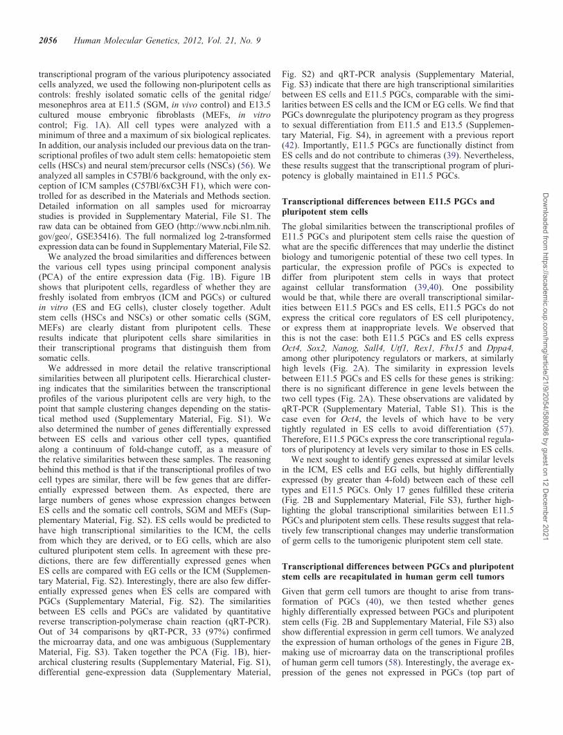
Fig. 2B), without any bias in gene selection other than the ex-istence of a human ortholog and its detection by the humanmicroarray used, shows a significant up-regulation in semino-mas (t-test P-value ¼ 0.009) and a slight up-regulation in em-bryonic carcinomas (P ¼ 0.054), but not in yolk sac tumorsand teratomas (Fig. 3). Seminomas and embryonic carcinomasare two types of germ cell tumors with transcriptional similar-ities to pluripotent stem cells (58). Yolk sac tumors and tera-tomas, on the other hand, have transcriptional similarities todifferentiated extra-embryonic and somatic tissues, respectively(58). This trend is more pronounced in the expression of spe-cific genes such as UPP1, DNMT3A and KLF4 (Fig. 3). UPP1is a uridine phosphorylase that has been proposed to be a prog-nostic factor in breast cancer (59), pancreatic cancer (60) andoral squamous cell carcinoma (61). DNMT3A is a de novoDNA methyl-transferase that regulates imprinting and gene
silencing, which when dysregulated may lead to cancer (62).Of note, the pluripotency-inducing factor Klf4 (9–16) ishighly upregulated in pluripotent stem cells relative to PGCs(Fig. 2B) and is induced in human germ cell tumors(Fig. 3). These data suggest that there may be molecular simi-larities between transformation of PGCs to the tumorigenicpluripotent stem cell state and induction of pluripotency insomatic cells.
Knockdown of LATS2 facilitates the generation ofhuman iPS cells
We found that Rhox6/Psx1, Trap, Xlr and Lats2 have the ex-pression pattern opposite to Klf4, i.e. they are highly expressedin PGCs but not in pluripotent stem cells (Fig. 2B). In particu-lar, the tumor suppressor Lats2 is the most differentially
Figure 2. Few genes are highly differentially expressed between E11.5 PGCs and pluripotent stem cells. (A) Expression of core transcriptional regulators andmarkers of pluripotency of ES cells and E11.5 PGCs. ES cells and E11.5 PGCs express similarly high levels of Oct4, Sox2, Nanog, Sall4, Utf1, Rex1, Fbx15 andDppa4. The table shows fold changes in the expression of these genes in ES cells versus E11.5 PGCs, ES cells versus MEFs and E11.5 PGCs versus MEFs. Thedata are validated by qRT-PCR (Supplementary Material, Table S1). (B) Genes showing high differential expression between E11.5 PGCs and ICM, ESC cellsand EG cells. Genes shown were identified as follows: they do not change between ES cells and ICM by more than 4-fold but are differentially expressedbetween ES cells and E11.5 PGCs and between EG cells and E11.5 PGCs by greater than 4-fold. Differential gene expression in the samples indicated is color-coded: red represents up-regulation, green represents down-regulation. Note the high differential expression of Klf4 and Lats2. The absent/present calls in themicroarray data (not shown) and qRT-PCR (Supplementary Material, Table S1) confirm that Klf4 is either expressed at very low levels or not at all in E11.5PGCs.
Human Molecular Genetics, 2012, Vol. 21, No. 9 2057
Dow
nloaded from https://academ
ic.oup.com/hm
g/article/21/9/2054/580086 by guest on 12 Decem
ber 2021
expressed gene in this comparison (Fig. 2B and Supplemen-tary Material, File S3) and shows a striking down-regulationin all human germ cell tumor types (Fig. 3). We thereforefocused on its role in reprogramming for our next experiments.Loss of Lats2 in Drosophila leads to tumorigenesis (63) andsilencing of LATS2 has been associated with a variety ofhuman cancers (64). Interestingly, Lats2 is a target ofmiRNAs that are highly expressed in germ cell tumors (65)and that can induce pluripotency in somatic cells (8). Lats2is also strongly downregulated when MEFs are reprogrammedto iPS cells (66). We therefore hypothesized that Lats2 mayrepresent a barrier to reprogramming to pluripotency. We
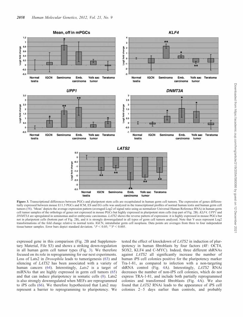
tested the effect of knockdown of LATS2 in induction of plur-ipotency in human fibroblasts by four factors (4F: OCT4,SOX2, KLF4 and C-MYC). Indeed, three different shRNAsagainst LATS2 all significantly increase the number ofhuman iPS cell colonies positive for the pluripotency markerTra-1-81, as compared to infection with a non-targetingshRNA control (Fig. 4A). Interestingly, LATS2 RNAidecreases the number of non-iPS cell colonies, which do notexpress TRA-1-81, and include both partially reprogrammedcolonies and transformed fibroblasts (Fig. 4A). We alsofound that LATS2 RNAi leads to the appearance of iPS cellcolonies 2–5 days earlier than controls, and probably
Figure 3. Transcriptional differences between PGCs and pluripotent stem cells are recapitulated in human germ cell tumors. The expression of genes differen-tially expressed between mouse E11.5 PGCs and ICM, ES and EG cells was analyzed in the transcriptional profiles of normal human testis and human germ celltumors (58). ‘Mean’ depicts the average expression pattern (averaged Log2 of signal ratio using as normalizer Universal Human Reference RNA) in human germcell tumor samples of the orthologs of genes not expressed in mouse PGCs but highly expressed in pluripotent stem cells (top part of Fig. 2B). KLF4, UPP1 andDNMT3A are upregulated in seminomas and/or embryonic carcinomas. LATS2 shows the reverse pattern of expression: it is highly expressed in mouse PGCs butnot in pluripotent cells (bottom part of Fig. 2B), and it is strongly downregulated in all types of germ cell tumors analyzed. Note that Y-axes represent Log2transformations of the fold change relative to normal testis. IGCN, intratubular germ cell neoplasia. Data points are averages from three to four independenttissue/tumor samples. Error bars depict standard deviation. ∗P , 0.05; ∗∗P , 0.005.
2058 Human Molecular Genetics, 2012, Vol. 21, No. 9
Dow
nloaded from https://academ
ic.oup.com/hm
g/article/21/9/2054/580086 by guest on 12 Decem
ber 2021

because of this it increases the size of iPS cell colony(Fig. 4B). Efficient knockdown of LATS2 mRNA was con-firmed by qRT-PCR (Supplementary Material, Fig. S5A).While LATS2 RNAi enhances reprogramming efficiency, itdoes not replace any of the reprogramming factors (data notshown).
Once human iPS cell lines generated with LATS2 RNAi areestablished, they have normal growth rates (data not shown)and express human ES/iPS cell-specific surface markers in-cluding SSEA3, SSEA4, TRA-1-60 and TRA-1-81 (Fig. 4C).RT-PCR showed that these cells activate the endogenous ex-pression of pluripotency markers OCT4, SOX2 and NANOG,and silence the viral transgenes (Supplementary Material,Fig. S5B), all of which are indicative of faithful
reprogramming. These results indicate that, as suggested byour expression-profiling studies (Figs 2B and 3), LATS2 con-stitutes a novel barrier to reprogramming to the pluripotentstem cell state.
LATS2 represses human iPS cell generation byantagonizing TAZ
We sought to understand the mechanism by which LATS2represses reprogramming. The reported role for Lats2 ingenomic stability of mouse cells (67) prompted us toanalyze the karyotypes of human iPS cells generated withLATS2 RNAi. All of these lines were found to be karyotypi-cally normal (Fig. 4D and Supplementary Material,
Figure 4. Knockdown of LATS2 increases the efficiency of human iPS cell generation. (A) The number of Tra1-81-positive iPS cell and Tra-1-81-negativenon-iPS cell colonies was counted on d20 after infection of human BJ foreskin fibroblasts with 4F alone (4), 4F + non-targeting shRNA (4 + NT) and 4F +LATS2 shRNA (three different short hairpins targeting LATS2 were independently tested, 4 + LATS2 i1, 4 + LATS2 i2 and 4 + LATS2 i3). Infectionswere performed in triplicate. Knockdown of LATS2 resulted in a significant increase in the number of Tra1-81-positive iPS cell colonies, and in a significantreduction in the number of Tra1-81-negative iPS cell colonies when compared with 4F + NT. (B) The diameter of iPS cell colonies was measured on d24after infection of BJ foreskin fibroblasts with 4F alone (4), 4 + NT and 4F + LATS2 i1/2/3. For each condition 10 iPS cell colonies were randomly picked.Knockdown of LATS2 resulted in a significant increase in the diameter of iPS cell colonies. Phase-contrast representative images of a colony for each conditionare also shown. (C) The iPS cell clones (P5) generated by 4F alone and 4F + LATS2 i1/2 showed strong, positive staining for all human ES cell-specific markersanalyzed by immunostaining. (D) iPS cells (P10) generated by 4F + LATS2 i2 showed a normal male karyotype (46, XY). In all relevant panels, error barsrepresent standard deviation, and scale bars represent 300 mm. ∗P , 0.05; ∗∗P , 0.01; ∗∗∗P , 0.001.
Human Molecular Genetics, 2012, Vol. 21, No. 9 2059
Dow
nloaded from https://academ
ic.oup.com/hm
g/article/21/9/2054/580086 by guest on 12 Decem
ber 2021

Figure 5. LATS2 antagonizes human cell reprogramming by repressing TAZ. (A) Growth curves of human fibroblasts infected with 4 factor, 4F + non-targetingshRNA (4 + NT), 4F + LATS2 shRNA (4 + Li1/2/3), counted on d0, d1, d4, d7, d10 and d13 post-infection. Infections were performed in triplicates. LATS2RNAi did not increase total cell numbers during the first 13 days of reprogramming. Data shown are representative of two independent experiments, and errorbars represent standard deviations. (B) TAZ knockdown suppresses the LATS2 RNAi-mediated increase in efficiency of iPS cell generation. The number of iPScell colonies was counted on d21 after infection of BJ foreskin fibroblasts with 4F alone (control), 4F + non-targeting shRNA (pLKO-NT) and 4F + LATS2
2060 Human Molecular Genetics, 2012, Vol. 21, No. 9
Dow
nloaded from https://academ
ic.oup.com/hm
g/article/21/9/2054/580086 by guest on 12 Decem
ber 2021
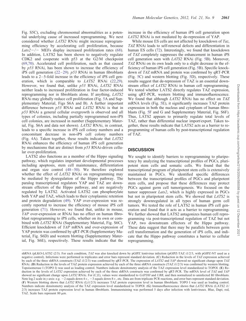
Fig. S5C), excluding chromosomal abnormalities as a poten-tial underlying cause of increased reprogramming. We nextconsidered whether LATS2 RNAi might increase reprogram-ming efficiency by accelerating cell proliferation, becauseLats22/2 MEFs display increased proliferation rates (68).In addition, LATS2 has been shown to negatively regulateCDK2 and cooperate with p53 at the G2/M checkpoint(69,70). Accelerated cell proliferation, such as that causedby p53 RNAi, has been shown to increase the efficiency ofiPS cell generation (22–29). p53 RNAi in human fibroblastsleads to a 2–5-fold increase in the efficiency of iPS cell gen-eration, which is comparable to LATS2 RNAi (22,29).However, we found that, unlike p53 RNAi, LATS2 RNAineither leads to increased proliferation in four factor-inducedreprogramming nor in fibroblasts alone. If anything, LATS2RNAi may globally reduce cell proliferation (Fig. 5A and Sup-plementary Material, Figs S6A and B). A further importantdifference between p53 RNAi and LATS2 RNAi is that inp53 RNAi a general cellular overgrowth is observed and alltypes of colonies, including partially reprogrammed non-iPScell colonies, are increased in number (Supplementary Mater-ial, Fig. S6A and data not shown). LATS2 RNAi, in contrast,leads to a specific increase in iPS cell colony numbers and aconcomitant decrease in non-iPS cell colony numbers(Fig. 4A). Taken together, these results indicate that LATS2RNAi enhances the efficiency of human iPS cell generationby mechanisms that are distinct from p53 RNAi-driven cellu-lar over-proliferation.
LATS2 also functions as a member of the Hippo signalingpathway, which regulates important developmental processesincluding apoptosis, stem cell maintenance, differentiationand organ size control (48–50). We therefore exploredwhether the effect of LATS2 RNAi on reprogramming maybe mediated by dysregulation of the Hippo pathway. Theparalog transcriptional regulators YAP and TAZ are down-stream effectors of the Hippo pathway, and are negativelyregulated by LATS2. Activated LATS2 can phosphorylateboth YAP and TAZ, which leads to their cytoplasmic retentionand protein degradation (49). YAP over-expression was re-cently reported to increase the efficiency of mouse iPS cellgeneration (71). However, we found that, unlike in mouse,YAP over-expression or RNAi has no effect on human fibro-blast reprogramming to iPS cells, whether on its own or com-bined with LATS2 RNAi (Supplementary Material, Fig. S6C).Efficient knockdown of YAP mRNA and over-expression ofYAP protein was confirmed by qRT-PCR (Supplementary Ma-terial, Fig. S6D) and western blotting (Supplementary Mater-ial, Fig. S6E), respectively. These results indicate that the
increase in the efficiency of human iPS cell generation uponLATS2 RNAi is not mediated by de-repression of YAP.
While mouse ES cells are not affected by knockdown of Taz,TAZ RNAi leads to self-renewal defects and differentiation inhuman ES cells (72). Interestingly, we found that knockdownof TAZ completely suppresses the enhancement in human iPScell generation seen with LATS2 RNAi (Fig. 5B). Moreover,TAZ RNAi on its own leads only to a slight decrease in the ef-ficiency of human iPS cell generation (Fig. 5B). Specific knock-down of TAZ mRNA and protein was confirmed by qRT-PCR(Fig. 5C) and western blotting (Fig. 5D), respectively. Theseresults suggest that de-repression of TAZ is an essential down-stream effect of LATS2 RNAi in human cell reprogramming.We tested whether LATS2 directly regulates TAZ expression,using qRT-PCR, western blotting and immunofluorescence.We found that although LATS2 RNAi has no effect on TAZmRNA levels (Fig. 5E), it significantly increases TAZ proteinexpression in both the nucleus and cytoplasm of human fibro-blasts (Fig. 5F and G and Supplementary Material, Fig. S6F).Thus, LATS2 appears to primarily regulate total levels ofTAZ, rather than differential nuclear import/export. Taken to-gether, these results indicate that LATS2 acts as a barrier to re-programming of human cells by post-transcriptional regulationof TAZ.
DISCUSSION
We sought to identify barriers to reprogramming to pluripo-tency by analyzing the transcriptional profiles of PGCs, pluri-potent stem cells and somatic cells. We found that thetranscriptional program of pluripotent stem cells is extensivelymaintained in PGCs. We identified specific differencesbetween the transcriptional profiles of PGCs and pluripotentstem cells, and propose that these differences may protectPGCs against germ cell tumorigenesis. We focused on thetumor suppressor Lats2, which is highly expressed in PGCsbut not in pluripotent stem cells. We showed that LATS2 isstrongly downregulated in all types of human germ celltumors. We tested the role of LATS2 in human iPS cell gen-eration and found that it acts as a barrier to reprogramming.We further showed that LATS2 antagonizes human cell repro-gramming via post-transcriptional regulation of TAZ but notYAP, two downstream effectors of the Hippo pathway.These data suggest that there may be parallels between germcell transformation and the generation of iPS cells, and indi-cate that the Hippo pathway constitutes a barrier to cellularreprogramming.
shRNA (pLKO-LATS2 i2/3). For each condition, TAZ was also knocked down by pGIPZ lentivirus infection (pGIPZ-TAZ i1/2/3, with pGIPZ-NT used as anegative control). Infections were performed in triplicates and error bars represent standard deviation. (C) Reduction in the levels of TAZ expression achievedby each of the three shRNA constructs (TAZ i1/2/3) was confirmed by qRT-PCR. The expression of LATS2 and YAP showed no significant change upon TAZRNAi. (D) Reduction in the levels of TAZ protein expression achieved by each of the three shRNA constructs (TAZ i1/2/3) was confirmed by western blotting.Topoisomerase I (TOPO I) was used as loading control. Numbers indicate densitometry analysis of the TAZ expression level standardized to TOPOI. (E) Re-duction in the levels of LATS2 expression achieved by each of the three shRNA constructs was confirmed by qRT-PCR. The mRNA level of TAZ and YAPshowed no significant change upon LATS2 RNAi. For (C,E), values were standardized to GAPDH and UBB, and then normalized to uninfected BJ fibroblasts.Note log 2 scale in y-axis: e.g. 22 equals down 4×, 23 equals down 8×, etc. Data are from triplicate PCR reactions, and error bars represent standard deviation.(F) Western blotting shows that LATS2 RNAi (Li1/2/3) increases TAZ protein expression level in human fibroblasts. TOPO I was used as loading control.Numbers indicate densitometry analysis of the TAZ expression level standardized to TOPOI. (G) Immunofluorescence shows that LATS2 RNAi (LATS2 i1/2/3) increases TAZ protein expression level in human fibroblasts. Immunostaining was performed 5 days after infection with lentiviruses. Blue, Dapi; red,TAZ. Scale bars represent 80 mm.
Human Molecular Genetics, 2012, Vol. 21, No. 9 2061
Dow
nloaded from https://academ
ic.oup.com/hm
g/article/21/9/2054/580086 by guest on 12 Decem
ber 2021
Relationship between PGCs, germ cell tumorigenesis andinduction of pluripotency
Our data reveal transcriptional parallels between reprogram-ming of PGCs to the pluripotent stem cell state, germ celltumorigenesis and the generation of iPS cells. It is generallyassumed that iPS cells represent an artificial manipulation ofsomatic cells in vitro that leads to the re-acquisition of theES cell state, which in turn represents a ‘freezing in time’ ofthe late ICM of the blastocyst (73). An alternative and not mu-tually exclusive possibility suggested by our data is that iPScells can represent the implementation in somatic cells of arecipe for germ cell transformation to a self-renewing tumori-genic state, which occurs naturally at low frequencies in bothmice and humans. Similar alternative routes to achieving theES cell state upon blastocyst culture, one involving a directICM–ES cell transition and another involving an intermediatePGC-like state, have recently been hypothesized and proposedto be dependent on culture conditions (74,75).
Possible role for Lats2 in the suppression of germ celltumors
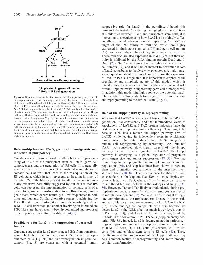
Our data suggest that Lats2 may protect PGCs from transform-ation. The high expression of Lats2 in PGCs relative to pluripo-tent stem cells (Fig. 2B) and its downregulation in germ celltumors (Fig. 3) are consistent with a potential tumor-
suppressive role for Lats2 in the germline, although thisremains to be tested. Considering the high global transcription-al similarities between PGCs and pluripotent stem cells, it isinteresting to speculate as to how Lats2 is so strikingly differ-entially expressed between these cell types (Fig. 6). Lats2 is atarget of the 290 family of miRNAs, which are highlyexpressed in pluripotent stem cells (76) and germ cell tumors(65), and can induce pluripotency in somatic cells (8,18).These miRNAs are also expressed in PGCs (77), but their ac-tivity is inhibited by the RNA-binding protein Dead end 1,Dnd1 (78). Dnd1 mutant mice have a high incidence of germcell tumors (79), and it will be of interest to determine if lossof Lats2 contributes to the Dnd2/2 phenotype. A major unre-solved question about this model concerns how the expressionof Dnd1 in PGCs is regulated. It is important to emphasize thespeculative and simplistic nature of this model, which isintended as a framework for future studies of a potential rolefor the Hippo pathway in suppressing germ cell tumorigenesis.In addition, this model highlights some of the potential paral-lels identified in this study between germ cell tumorigenesisand reprogramming to the iPS cell state (Fig. 6).
Role of the Hippo pathway in reprogramming
We show that LATS2 acts as a novel barrier to human iPS cellgeneration. We consistently find that intermediate levels ofknockdown of LATS2 and TAZ protein induction have thebest effects on reprogramming efficiency. This might bebecause such levels reduce the Hippo pathway arm ofLATS2 while leaving its independent roles in cytokinesis/ploidy intact. Our data indicate that LATS2 antagonizeshuman cell reprogramming by repressing TAZ, but notYAP, two conserved downstream targets of the Hippopathway that are directly regulated by LATS2. The Hippopathway is emerging as a global regulator of progenitorcells, organ size and tumor suppression (48–50). We hadfound Yap to be upregulated in multiple mouse stem cellpopulations (56), and Yap has since been shown to regulatestem and progenitor compartments in the intestine, liver,skin and brain (80–82). There is evidence for shared as wellas specific roles for Yap and Taz. Yap2/2 mice display em-bryonic lethality at E8.5, whereas Taz2/2 mice can surviveto adulthood but with defects in the kidneys and lungs (83–86). However, Yap and Taz likely act redundantly during pre-implantation because Yap2/2;Taz2/2 embryos arrest priorto morula development (87). Yap and Taz are thought to regu-late commitment to the trophectoderm lineage in the morulaand early blastocyst and are repressed by Lats1/2 in the ICM(87). These findings are compatible with ours because wedetect Lats2 in the ICM, albeit at much lower levels than inPGCs (Fig. 2B), and Lats2 is further downregulated by�3-fold in the conversion ICM–ES cells (Supplementary Ma-terial, File S3). Indeed, Lats2 is downregulated in various set-tings of reprogramming to the pluripotent stem cell state, suchas ICM–ES cells, PGC–EG cells (this work), MEF to iPScells (66) and epiblast stem cells to ES cells (88). Theseresults suggest that suppression of the Hippo pathway maybe a common feature of reprogramming and, more broadly,cellular transformation.
Figure 6. Speculative model for the role of the Hippo pathway in germ celltumorigenesis and reprogramming. Lats2 may be under tight control inPGCs via Dnd1-mediated inhibition of miRNAs of the 290 family. Loss ofDnd1 in PGCs may allow these miRNAs to inhibit their targets, includingLats2. ‘Other’ represents targets of the miRNA 290 family other than Lats2.Question mark (‘?’) represents functions of Lats2 independent of the Hippopathway effectors Yap and Taz, such as in cell cycle and mitotic stability.Loss of Lats2 de-represses Yap or Taz, which promote reprogramming tothe tumorigenic pluripotent stem cell state. Checkmarks indicate caseswhere a gene has been implicated in germ cell tumorigenesis and/or iPScell generation in the literature (Dnd1, mir290, Yap) or in this study (Lats2,Taz). The different role for Yap and Taz in mouse versus human cell repro-gramming may be due to species- or stage-specific differences. See Discussionsection for details.
2062 Human Molecular Genetics, 2012, Vol. 21, No. 9
Dow
nloaded from https://academ
ic.oup.com/hm
g/article/21/9/2054/580086 by guest on 12 Decem
ber 2021

Interestingly, TAZ and YAP have divergent roles in mouseand human cells. YAP regulates mouse ES cell self-renewal(89) and can increase the efficiency of mouse iPS cell gener-ation (71), but has no role in human ES cells (90) or in iPScell generation (this work). The reverse is the case for TAZ:it is important for human ES cell self-renewal (72) and iPScell generation (this work), but has no role in mouse EScells (72). We speculate that this difference may be due tothe distinct signaling requirements of mouse versus humanpluripotent stem cells. YAP can act as a co-activator of theBMP signaling pathway, while TAZ is important for TGFb/Activin signaling (72,91). Both YAP and TAZ have, in add-ition, been shown to regulate the Wnt signaling pathway(92,93). The BMP pathway, with which Yap interacts, is crit-ical for mouse ES/iPS cell self-renewal, but not for human. Onthe other hand, the TGFb/Activin pathway, with which TAZinteracts, is essential for human ES/iPS cell self-renewal, butnot for mouse (94,95). In addition, human ES cells correspondto a developmental stage that is more similar to mouse Epi-blast stem cells than to ES cells (88,96), and therefore the dis-tinct roles for TAZ and YAP in mouse versus human may berelated to stage-specific differences in Hippo pathway signal-ing. Further work will be needed to dissect the distinct signal-ing interactions mediated by TAZ and YAP in different typesof pluripotent stem cells.
MATERIALS AND METHODS
Culture of mouse ES cells and EG cells
C57Bl/6 (B6) ES cells were cultured in standard conditions inthe presence of fetal bovine serum (FBS, Hyclone), MEFs andleukemia inhibitory factor (LIF) as previously described (97).ES cells were removed from MEFs by serial re-plating (56) orserial re-plating followed by culture for one passage in theabsence of MEFs in gelatin-coated dishes. B6 EG cells (Patri-cia Labosky, Vanderbilt U.), derived from E12.5 male PGCs(55), were cultured in the presence of FBS, STO feeder fibro-blasts and LIF, as described (98). Feeders were removed byserial re-plating (56) followed by culture for one passage ingelatin-coated dishes.
Isolation of ICMs
Throughout this study, we collected mouse samples on aninbred B6 background. The only exception was that B6inbred mice (100% B6), upon super-ovulation, yielded fewproperly staged blastocysts for ICM isolation. We thereforecollected 50% B6–50% C3H (B6C3H) ICMs. We tested theeffect of reducing the B6 background on the transcriptionalprofiles of ICMs. We collected one ICM sample that is only25% B6 (ICM-1) and compared its transcriptional profilewith the other two ICM replicates (ICM-2 and ICM-3),which are 50% B6. If the genetic background was a major
factor in our analysis, we would have expected the two repli-cates with 50% B6 (ICM-2 and ICM-3) to be the most closelycorrelated of the three replicates, with ICM-1 being an outlier.We observed that not to be the case. Analysis of the correl-ation coefficients (CC) between the ICM datasets indicatesthat reducing the B6 contribution by half did not significantlybias the data: CC ICM-1/2 ¼ 0.9840; CC ICM-1/3 ¼ 0.9775;CC ICM-2/3 ¼ 0.9799. B6C3H (B6 × C3H F1) femaleswere super-ovulated between 5–8 weeks of age with 10 IUPMS (Calbiochem) followed by 10 IU HCG (Calbiochem)46 h later and mated to B6C3H males overnight. Embryoswere flushed at E3.5 with M2 (Sigma), washed with Dulbec-co’s modified Eagle’s medium (DMEM)/10% FBS beforebeing transferred to a droplet of rabbit anti-mouse serum(Sigma) under oil. Embryos were incubated in serum for30 min at 378C before being washed three times in M2.They were then transferred to a droplet of guinea pig serum(Sigma) under oil and incubated at 378C for 30–60 min.Embryos were examined for trophectoderm lysis and washedthree times in DMEM/FBS before being transferred into adroplet of 0.5% pronase (Sigma) at 378C. When the zona pel-lucida started to disintegrate, embryos were pipetted vigorous-ly to strip them of the zonas as well as lysed trophectodermcells. ICMs were then washed in M2 before being transferredinto RNA lysis buffer (RLT, Qiagen).
Isolation of primordial germ cells
Male mice of the Oct4/EGFP transgenic line (53), kept on aB6 background, were crossed to B6 females. The morningof the day of vaginal plug was considered E0.5. PGCs wereisolated at E11.5 and E13.5. E11.5 PGCs are still sexually in-different, so a mixture of male and female embryos was used.E13.5 PGCs have initiated sexual differentiation, and at thisstage male and female embryos were processed separately.Embryo fragments were dissected and dissociated in the pres-ence of 0.25% trypsin (Invitrogen) and 1 mg/ml DNAse(Worthington) in phosphate-buffered saline (PBS, Invitrogen)at 378C for 10 min, with occasional vortexing and pipetting.Trypsinization was stopped with the addition of FBS to 5%.Cells were pelleted at 2000 rpm for 4 min, re-suspended inPBS with 1% fetal calf serum and 1 mg/ml propidum iodide(PI, Invitrogen) and passed through a 40 mm cell strainer.PGCs (PI2; EGFP+) and E11.5 SGM (PI2;EGFP2) frac-tions were purified in the UCSF Diabetes Center CellSorting Facility using a MoFlo cell sorter (Cytomation). Cellfractions were collected directly into RLT.
RNA amplification and microarray hybridization
We collected three to six replicates per cell type. The cellnumbers used for the following cell types were:
ICM ESC E11.5 PGC EGC E13.5F PGC E13.5M PGC E11.5 SGMCell number 50–75 #(no. of ICMs) 500–15 000 1500–25 400 1000 2100–11 900 5400–7000 20 000–41 000
Human Molecular Genetics, 2012, Vol. 21, No. 9 2063
Dow
nloaded from https://academ
ic.oup.com/hm
g/article/21/9/2054/580086 by guest on 12 Decem
ber 2021
RNA was isolated using the RNeasy kit (Qiagen) within-column DNAse digestion. mRNA was amplified using atwo-round in vitro transcription protocol as described (56). Al-ternatively, mRNA was amplified using the RiboAmp HS kit(Arcturus), which is a modified two-round in vitro transcrip-tion protocol. Samples amplified using the RiboAmp HS kitwere treated as a separate batch and, after batch effect calcu-lation (see below), clustered correctly with replicates of thesame tissues amplified using our protocol (56). For example,the mRNA for ES-1 and ES-2 were amplified using our proto-col and had previously been reported (56), whereas ES-3 wasamplified using the RiboAmp HS kit (Figs 1B and 2). Twelvemicrograms of biotin-labeled amplified RNA was hybridizedto Affymetrix U74Av2 arrays at the UCSF Gladstone Genom-ics Core Facility, according to the manufacturer’s instructions.The raw data can be downloaded from GEO (GSE35416).Data on the transcriptional profiles of mouse adult stem cellshave been previously described (56).
Statistical analyses
All the statistical analyses were performed using customscripts and packages in the free statistical computing environ-ment R/Bioconductor (www.r-project.org) (99). The probe in-tensities of the Affymetrix CEL files were backgroundsubtracted, quantile normalized and summarized for eachprobe set into a logarithm base 2 intensity value using therobust multi-array average method implemented in the Affy-metrix package (100). Subsequently, we applied the meancenter global adjustment to remove apparent batch effectsacross the experiments to facilitate between-experiment com-parisons (101). Differential expression analysis between con-ditions were performed using the moderated t-statistics ascomputed by the limma package (102). P-values were adjustedto control for false discovery rate (FDR) using the Benjamini–Hochberg method (103) to account for multiple testing. PCAwas done as described (104,105). The expression of geneshighly differentially expressed between E11.5 PGCs andICM, ES cells and EG cells (Supplementary Material, FileS3 and Fig. 2B) was analyzed in human germ cell tumordata obtained from GEO dataset GDS1742 (58). Genes withhuman orthologs whose expression was assayed for in themicroarrays used (58) were the following: PPRS1, KLF4,UPP1, ACSL4, TDGF1, FABP3, SGK, PGRMC1, DNMT3A,GSTA4, TTRAP and LATS2. P-values were calculated usinga two-tailed t-test.
Quantitative real-time RT-PCR
Independent samples containing approximately the samenumber of ES cells, PGCs and ICM cells were obtained asdescribed above. RNA was isolated using the RNeasy MiniRNA Isolation kit (Qiagen) and reverse-transcribed using theiScript first strand cDNA synthesis kit (BioRad) or the High-Capacity cDNA Reverse Transcription kit (Applied BioSys-tems). The cDNA reaction was diluted 1:5 in TE (10 mM
Tris–Cl/1 mM EDTA, pH 7.6) and used in Sybr Green real-time PCR reactions (BioRad or Applied BioSystems). PCRprimers were designed to amplify 100–200 bp fragmentsspanning exons. Housekeeping genes used were Ubiquitin-b
and Ribosomal protein L7, which were determined from themicroarray data to not be differentially expressed in thesamples analyzed, or as indicated. Reactions were run in dupli-cates or triplicates on a MyiQ qPCR machine (BioRad) or a7900HT machine (Applied BioSystems) according to the man-ufacturer’s instructions. Only samples with single and match-ing end-point melting curve peaks were used for subsequentanalysis. Cycle threshold values were imported into theREST software (106) for fold-change calculations of ES orPGC relative to ICM, using the housekeeping genes as con-trols, or as indicated. When a gene was detected in onetissue but not another, no fold change was calculated andinstead the Present/Absent (P/A) notation was used. Primersequences are listed in Supplementary Material, Table S2.
Immunostaining and western blotting
For immunofluorescence, cells were fixed directly in culturingplates with 4% paraformaldehyde or cold methanol, and per-meabilized with 0.1% Triton X-100. Cells were then stainedwith primary antibodies against SSEA-3 (MAB4303, Milli-pore, 1:100), SSEA-4 (MAB4304, Millipore, 1:100), Tra1–60 (ab16288, Abcam, 1:100), Tra1-81 (MAB4381, Millipore,1:100), V5 (46-0705, Invitrogen, 1:5000 for western blotting),TAZ (no.4883, Cell Signaling, 1:200; no.560235, BD, 1:1000for western blotting), Topoisomerase I (ab85038, Abcam,1:800 for western blotting), Bex1/Rex3 (Frank Margolis,U. Maryland, 1:20 000) and DMRT1 (Silvana Guioli, NIMR,London, 1:1000). Respective secondary antibodies wereconjugated to either Alexa Fluor 594 or Alexa Fluor 488 (Invi-trogen) and used at 1:500. Sodium dodecyl sulfate–polyacryl-amide gel electrophoresis and western blotting using the sameprimary antibodies and respective secondary antibodies conju-gated with horse radish peroxidase were performed accordingto standard protocols.
Lentivirus production
Lentiviral vectors that lead to the expression of shRNAwere obtained from OpenBiosystems. shRNA for LATS2(RHS3979–9569292, RHS3979–9569293, RHS3979–9569294) is in pLKO.1 backbone, and shRNA for YAP(RHS4430–98525388, RHS4430–98818907, RHS4430–98893379) and TAZ (RHS4430–98514207, RHS4430–99293240, RHS4430–101097950) is in pGIPZ. For virusproduction, 293T cells at 60–70% confluency were transfectedin 10 cm plates with 4 mg of the lentiviral vectors together with1 mg each of the packaging plasmids VSV-G, MDL-RRE andRSVr using Fugene 6 (Roche). After 72 h, viral supernatantswere harvested, filtered and stored at 2808C.
Generation of human iPS cells
Human primary newborn foreskin (BJ) fibroblasts wereobtained from ATCC (reference no.: CRL-2522) and culturedin DMEM with 10% FBS, 1× glutamine, 1× non-essentialamino acids, 1× sodium pyruvate, 2× penicillin/streptomycinand 0.06 mM b-mercaptoethanol (fibroblast medium). Fibro-blasts were seeded at 60 000 cells per well of a six-well platethe day before infection. Cells were infected with 0.5 ml each
2064 Human Molecular Genetics, 2012, Vol. 21, No. 9
Dow
nloaded from https://academ
ic.oup.com/hm
g/article/21/9/2054/580086 by guest on 12 Decem
ber 2021

of concentrated retroviruses (obtained from the Harvard GeneTherapy Initiative) leading to the over-expression of OCT4,SOX2 and KLF4 and 0.05 ml in the case of c-MYC, alone orin combination with 20 ml (for LATS2) or 100 ml (for YAPand TAZ) of non-concentrated lentivirus for shRNA. Cellswere infected in 1 ml human ES cell medium (DMEM/F12with 20% KSR, 0.5× glutamine, 1× non-essential aminoacids, 2× penicillin/streptomycin, 0.1 mM b-mercaptoethanol,10 ng/ml bFGF) and 8 mg/ml polybrene. Cells remained in thepresence of virus for 48 h and on the day after virus addition,1 ml of fibroblast medium was added. Forty-eight hours afterinfection, virus was removed and cells were cultured inhuman ES cell medium. On d20–d28 after infection, liveTra1-81 (MAB4381, Millipore) staining was performed inorder to identify fully reprogrammed iPS cell colonies.
Cytogenetic analysis
Human iPS cells were treated with 10 ng/ml of Colcemid(Invitrogen) overnight at 378C. Cells were harvested andG-banded according to standard cytogenetic protocols (107).Metaphase cells were analyzed under the microscope and kar-yotyped according to an International System for HumanCytogenetic Nomenclature (108) using CytoVision system(Applied Imaging).
SUPPLEMENTARY MATERIAL
Supplementary Material is available at HMG Online.
ACKNOWLEDGEMENTS
We are grateful to Hans Scholer for Oct4/GFP mice, TrishLabosky for EG cells, Frank Margolis and Silvana Guioli forantibodies, Kevin Eggan for pMXs-4F plasmids and DeepaSubramanyam for advice on human iPS cell generation. Wethank the Santos laboratory and Matt Cook for discussionsand Robert Blelloch, Marco Conti, Susan Fisher, ReneeReijo Pera, Diana Laird, Marica Grskovic, Connie Wong andChristina Chaivorapol for critical reading of the manuscript.
Conflict of Interest statement. The authors declare no compet-ing financial interests.
FUNDING
Work in M.H.’s laboratory is supported by grants from theJDRF and the Leona M. and Harry B. Helmsley CharitableTrust. Work in the Santos laboratory is supported by an NIHDirector’s New Innovator Award, the Leona M. and HarryB. Helmsley Charitable Trust and CIRM.
REFERENCES
1. Keller, G. (2005) Embryonic stem cell differentiation: emergence of anew era in biology and medicine. Genes Dev., 19, 1129–1155.
2. Smith, A.G. (2001) Embryo-derived stem cells: of mice and men. Annu.Rev. Cell Dev. Biol., 17, 435–462.
3. Stadtfeld, M. and Hochedlinger, K. (2011) Induced pluripotency: history,mechanisms, and applications. Genes Dev., 24, 2239–2263.
4. Kiskinis, E. and Eggan, K. (2010) Progress toward the clinicalapplication of patient-specific pluripotent stem cells. J. Clin. Invest., 120,51–59.
5. Evans, M.J. and Kaufman, M.H. (1981) Establishment in culture ofpluripotential cells from mouse embryos. Nature, 292, 154–156.
6. Martin, G.R. (1981) Isolation of a pluripotent cell line from early mouseembryos cultured in medium conditioned by teratocarcinoma stem cells.Proc. Natl Acad. Sci. USA, 78, 7634–7638.
7. Thomson, J.A., Itskovitz-Eldor, J., Shapiro, S.S., Waknitz, M.A.,Swiergiel, J.J., Marshall, V.S. and Jones, J.M. (1998) Embryonic stemcell lines derived from human blastocysts. Science, 282, 1145–1147.
8. Anokye-Danso, F., Trivedi, C.M., Juhr, D., Gupta, M., Cui, Z., Tian, Y.,Zhang, Y., Yang, W., Gruber, P.J., Epstein, J.A. et al. (2011) Highlyefficient miRNA-mediated reprogramming of mouse and human somaticcells to pluripotency. Cell Stem Cell, 8, 376–388.
9. Takahashi, K. and Yamanaka, S. (2006) Induction of pluripotent stemcells from mouse embryonic and adult fibroblast cultures by definedfactors. Cell, 126, 663–676.
10. Okita, K., Ichisaka, T. and Yamanaka, S. (2007) Generation ofgermline-competent induced pluripotent stem cells. Nature, 448,313–317.
11. Wernig, M., Meissner, A., Foreman, R., Brambrink, T., Ku, M.,Hochedlinger, K., Bernstein, B.E. and Jaenisch, R. (2007) In vitroreprogramming of fibroblasts into a pluripotent ES-cell-like state.Nature, 448, 318–324.
12. Maherali, M., Sridharan, R., Xie, W., Utikal, J., Eminli, S., Arnold, K.,Stadtfeld, M., Yachechko, R., Tchieu, J., Jaenisch, R. et al. (2007)Directly reprogrammed fibroblasts show global epigenetic remodelingand widespread tissue contribution. Cell Stem Cell, 1, 55–70.
13. Takahashi, K., Tanabe, K., Ohnuki, M., Narita, M., Ichisaka, T.,Tomoda, K. and Yamanaka, S. (2007) Induction of pluripotent stem cellsfrom adult human fibroblasts by defined factors. Cell, 131, 861–872.
14. Yu, J., Vodyanik, M.A., Smuga-Otto, K., Antosiewicz-Bourget, J.,Frane, J.L., Tian, S., Nie, J., Jonsdottir, G.A., Ruotti, V., Stewart, R.et al. (2007) Induced pluripotent stem cell lines derived from humansomatic cells. Science, 318, 1917–1920.
15. Park, I.H., Zhao, R., West, J.A., Yabuuchi, A., Huo, H., Ince, T.A.,Lerou, P.H., Lensch, M.W. and Daley, G.Q. (2008) Reprogramming ofhuman somatic cells to pluripotency with defined factors. Nature, 451,141–146.
16. Lowry, W.E., Richter, L., Yachechko, R., Pyle, A.D., Tchieu, J.,Sridharan, R., Clark, A.T. and Plath, K. (2008) Generation of humaninduced pluripotent stem cells from dermal fibroblasts. Proc. Natl Acad.
Sci. USA, 105, 2883–2888.17. Subramanyam, D., Lamouille, S., Judson, R.L., Liu, J.Y., Bucay, N.,
Derynck, R. and Blelloch, R. (2011) Multiple targets of miR-302 andmiR-372 promote reprogramming of human fibroblasts to inducedpluripotent stem cells. Nat. Biotechnol., 29, 443–448.
18. Miyoshi, N., Ishii, H., Nagano, H., Haraguchi, N., Dewi, D.L., Kano, Y.,Nishikawa, S., Tanemura, M., Mimori, K., Tanaka, F. et al. (2011)Reprogramming of mouse and human cells to pluripotency using maturemicroRNAs. Cell Stem Cell, 8, 633–638.
19. Brennand, K.J., Simone, A., Jou, J., Gelboin-Burkhart, C., Tran, N.,Sangar, S., Li, Y., Mu, Y., Chen, G., Yu, D. et al. (2011) Modellingschizophrenia using human induced pluripotent stem cells. Nature, 473,221–225.
20. Howden, S.E., Gore, A., Li, Z., Fung, H.L., Nisler, B.S., Nie, J., Chen,G., McIntosh, B.E., Gulbranson, D.R., Diol, N.R. et al. (2011) Geneticcorrection and analysis of induced pluripotent stem cells from a patientwith gyrate atrophy. Proc. Natl Acad. Sci. USA, 108, 6537–6542.
21. Liu, G.H., Barkho, B.Z., Ruiz, S., Diep, D., Qu, J., Yang, S.L.,Panopoulos, A.D., Suzuki, K., Kurian, L., Walsh, C. et al. (2011)Recapitulation of premature ageing with iPSCs from Hutchinson-Gilfordprogeria syndrome. Nature, 472, 221–225.
22. Zhao, Y., Yin, X., Qin, H., Zhu, F., Liu, H., Yang, W., Zhang, Q., Xiang,C., Hou, P., Song, Z. et al. (2008) Two supporting factors greatlyimprove the efficiency of human iPSC generation. Cell Stem Cell, 3,475–479.
23. Hanna, J., Saha, K., Pando, B., van Zon, J., Lengner, C.J., Creyghton,M.P., van Oudenaarden, A. and Jaenisch, R. (2009) Direct cellreprogramming is a stochastic process amenable to acceleration. Nature,462, 595–601.
Human Molecular Genetics, 2012, Vol. 21, No. 9 2065
Dow
nloaded from https://academ
ic.oup.com/hm
g/article/21/9/2054/580086 by guest on 12 Decem
ber 2021
24. Hong, H., Takahashi, K., Ichisaka, T., Aoi, T., Kanagawa, O.,Nakagawa, M., Okita, K. and Yamanaka, S. (2009) Suppression ofinduced pluripotent stem cell generation by the p53-p21 pathway.Nature, 460, 1132–1135.
25. Banito, A., Rashid, S.T., Acosta, J.C., Li, S., Pereira, C.F., Geti, I.,Pinho, S., Silva, J.C., Azuara, V., Walsh, M. et al. (2009) Senescenceimpairs successful reprogramming to pluripotent stem cells. Genes Dev.,23, 2134–2139.
26. Utikal, J., Polo, J.M., Stadtfeld, M., Maherali, N., Kulalert, W., Walsh,R.M., Khalil, A., Rheinwald, J.G. and Hochedlinger, K. (2009)Immortalization eliminates a roadblock during cellular reprogramminginto iPS cells. Nature, 460, 1145–1148.
27. Marion, R.M., Strati, K., Li, H., Murga, M., Blanco, R., Ortega, S.,Fernandez-Capetillo, O., Serrano, M. and Blasco, M.A. (2009) Ap53-mediated DNA damage response limits reprogramming to ensureiPS cell genomic integrity. Nature, 460, 1149–1153.
28. Li, H., Collado, M., Villasante, A., Strati, K., Ortega, S., Canamero, M.,Blasco, M.A. and Serrano, M. (2009) The Ink4/Arf locus is a barrier foriPS cell reprogramming. Nature, 460, 1136–1139.
29. Kawamura, T., Suzuki, J., Wang, Y.V., Menendez, S., Morera, L.B.,Raya, A., Wahl, G.M. and Belmonte, J.C. (2009) Linking the p53 tumoursuppressor pathway to somatic cell reprogramming. Nature, 460,1140–1144.
30. Hanna, J., Markoulaki, S., Schorderet, P., Carey, B.W., Beard, C.,Wernig, M., Creyghton, M.P., Steine, E.J., Cassady, J.P., Foreman, R.et al. (2008) Direct reprogramming of terminally differentiated mature Blymphocytes to pluripotency. Cell, 133, 250–264.
31. Mikkelsen, T.S., Hanna, J., Zhang, X., Ku, M., Wernig, M., Schorderet,P., Bernstein, B.E., Jaenisch, R., Lander, E.S. and Meissner, A. (2008)Dissecting direct reprogramming through integrative genomic analysis.Nature, 454, 49–55.
32. Ramalho-Santos, M. (2009) iPS cells: insights into basic biology. Cell,138, 616–618.
33. Matsui, Y., Zsebo, K. and Hogan, B.L. (1992) Derivation ofpluripotential embryonic stem cells from murine primordial germ cells inculture. Cell, 70, 841–847.
34. Resnick, J.L., Bixler, L.S., Cheng, L. and Donovan, P.J. (1992)Long-term proliferation of mouse primordial germ cells in culture.Nature, 359, 550–551.
35. Shamblott, M.J., Axelman, J., Wang, S., Bugg, E.M., Littlefield, J.W.,Donovan, P.J., Blumenthal, P.D., Huggins, G.R. and Gearhart, J.D.(1998) Derivation of pluripotent stem cells from cultured humanprimordial germ cells. Proc. Natl Acad. Sci. USA, 95, 13726–13731.
36. Okamura, D., Tokitake, Y., Niwa, H. and Matsui, Y. (2008) Requirementof Oct3/4 function for germ cell specification. Dev Biol., 317, 576–584.
37. Kehler, J., Tolkunova, E., Koschorz, B., Pesce, M., Gentile, L., Boiani, M.,Lomeli, H., Nagy, A., McLaughlin, K.J., Scholer, H.R. et al. (2004) Oct4 isrequired for primordial germ cell survival. EMBO Rep., 5, 1078–1083.
38. Chambers, I., Silva, J., Colby, D., Nichols, J., Nijmeijer, B., Robertson,M., Vrana, J., Jones, K., Grotewold, L. and Smith, A. (2007) Nanogsafeguards pluripotency and mediates germline development. Nature,450, 1230–1234.
39. Donovan, P.J. (1994) Growth factor regulation of mouse primordial germcell development. Curr. Top Dev. Biol., 29, 189–225.
40. Stevens, L.C. (1967) Origin of testicular teratomas from primordial germcells in mice. J. Natl Cancer Inst., 38, 549–552.
41. Kurimoto, K., Yabuta, Y., Ohinata, Y., Shigeta, M., Yamanaka, K. andSaitou, M. (2008) Complex genome-wide transcription dynamicsorchestrated by Blimp1 for the specification of the germ cell lineage inmice. Genes Dev., 22, 1617–1635.
42. Sabour, D., Arauzo-Bravo, M.J., Hubner, K., Ko, K., Greber, B., Gentile,L., Stehling, M. and Scholer, H.R. (2011) Identification of genes specificto mouse primordial germ cells through dynamic global gene expression.Hum. Mol. Genet., 20, 115–125.
43. Mise, N., Fuchikami, T., Sugimoto, M., Kobayakawa, S., Ike, F., Ogawa,T., Tada, T., Kanaya, S., Noce, T. and Abe, K. (2008) Differences andsimilarities in the developmental status of embryo-derived stem cells andprimordial germ cells revealed by global expression profiling. GenesCells, 13, 863–877.
44. Grskovic, M., Chaivorapol, C., Gaspar-Maia, A., Li, H. andRamalho-Santos, M. (2007) Systematic identification of cis-regulatorysequences active in mouse and human embryonic stem cells. PLoSGenet., 3, e145.
45. Sharov, A.A., Piao, Y., Matoba, R., Dudekula, D.B., Qian, Y.,VanBuren, V., Falco, G., Martin, P.R., Stagg, C.A., Bassey, U.C. et al.(2003) Transcriptome analysis of mouse stem cells and early embryos.PLoS Biol., 1, E74.
46. Sharova, L.V., Sharov, A.A., Piao, Y., Shaik, N., Sullivan, T., Stewart,C.L., Hogan, B.L. and Ko, M.S. (2007) Global gene expression profilingreveals similarities and differences among mouse pluripotent stem cellsof different origins and strains. Dev. Biol., 307, 446–459.
47. Guo, G., Huss, M., Tong, G.Q., Wang, C., Li Sun, L., Clarke, N.D. andRobson, P. (2010) Resolution of cell fate decisions revealed bysingle-cell gene expression analysis from zygote to blastocyst. Dev. Cell,18, 675–685.
48. Pan, D. (2007) Hippo signaling in organ size control. Genes Dev., 21,886–897.
49. Zhao, B., Li, L., Lei, Q. and Guan, K.L. (2010) The Hippo-YAP pathwayin organ size control and tumorigenesis: an updated version. Genes Dev.,24, 862–874.
50. Chan, S.W., Lim, C.J., Chen, L., Chong, Y.F., Huang, C., Song, H. andHong, W. (2011) The Hippo pathway in biological control and cancerdevelopment. J. Cell Physiol., 226, 928–939.
51. Solter, D. and Knowles, B.B. (1975) Immunosurgery of mouseblastocyst. Proc. Natl Acad. Sci. USA, 72, 5099–5102.
52. Yeom, Y.I., Fuhrmann, G., Ovitt, C.E., Brehm, A., Ohbo, K., Gross, M.,Hubner, K. and Scholer, H.R. (1996) Germline regulatory element ofOct-4 specific for the totipotent cycle of embryonal cells. Development,122, 881–894.
53. Yoshimizu, T., Sugiyama, N., De Felice, M., Yeom, Y.I., Ohbo, K.,Masuko, K., Obinata, M., Abe, K., Scholer, H.R. and Matsui, Y. (1999)Germline-specific expression of the Oct-4/green fluorescent protein(GFP) transgene in mice. Dev. Growth Differ., 41, 675–684.
54. Szabo, P.E., Hubner, K., Scholer, H. and Mann, J.R. (2002)Allele-specific expression of imprinted genes in mouse migratoryprimordial germ cells. Mech. Dev., 115, 157–160.
55. Labosky, P.A., Barlow, D.P. and Hogan, B.L. (1994) Mouse embryonicgerm (EG) cell lines: transmission through the germline and differencesin the methylation imprint of insulin-like growth factor 2 receptor (Igf2r)gene compared with embryonic stem (ES) cell lines. Development, 120,3197–3204.
56. Ramalho-Santos, M., Yoon, S., Matsuzaki, Y., Mulligan, R.C. andMelton, D.A. (2002) ‘Stemness’: transcriptional profiling of embryonicand adult stem cells. Science, 298, 597–600.
57. Niwa, H., Miyazaki, J. and Smith, A.G. (2000) Quantitative expressionof Oct-3/4 defines differentiation, dedifferentiation or self-renewal of EScells. Nat. Genet., 24, 372–376.
58. Skotheim, R.I., Lind, G.E., Monni, O., Nesland, J.M., Abeler, V.M.,Fossa, S.D., Duale, N., Brunborg, G., Kallioniemi, O., Andrews, P.W.et al. (2005) Differentiation of human embryonal carcinomas in vitro andin vivo reveals expression profiles relevant to normal development.Cancer Res., 65, 5588–5598.
59. Kanzaki, A., Takebayashi, Y., Bando, H., Eliason, J.F., Watanabe Si, S.,Miyashita, H., Fukumoto, M., Toi, M. and Uchida, T. (2002) Expressionof uridine and thymidine phosphorylase genes in human breastcarcinoma. Int. J. Cancer, 97, 631–635.
60. Sahin, F., Qiu, W., Wilentz, R.E., Iacobuzio-Donahue, C.A., Grosmark,A. and Su, G.H. (2005) RPL38, FOSL1, and UPP1 are predominantlyexpressed in the pancreatic ductal epithelium. Pancreas, 30, 158–167.
61. Miyashita, H., Takebayashi, Y., Eliason, J.F., Fujimori, F., Nitta, Y.,Sato, A., Morikawa, H., Ohashi, A., Motegi, K., Fukumoto, M. et al.(2002) Uridine phosphorylase is a potential prognostic factor in patientswith oral squamous cell carcinoma. Cancer, 94, 2959–2966.
62. Baylin, S.B. (2005) DNA methylation and gene silencing in cancer. Nat.Clin. Pract. Oncol., 2(Suppl. 1), S4–S11.
63. Xu, T., Wang, W., Zhang, S., Stewart, R.A. and Yu, W. (1995)Identifying tumor suppressors in genetic mosaics: the Drosophila latsgene encodes a putative protein kinase. Development, 121, 1053–1063.
64. Visser, S. and Yang, X. (2010) LATS tumor suppressor: a new governorof cellular homeostasis. Cell Cycle, 9, 3892–3903.
65. Voorhoeve, P.M., le Sage, C., Schrier, M., Gillis, A.J., Stoop, H., Nagel,R., Liu, Y.P., van Duijse, J., Drost, J., Griekspoor, A. et al. (2006) Agenetic screen implicates miRNA-372 and miRNA-373 as oncogenes intesticular germ cell tumors. Cell, 124, 1169–1181.
66. Samavarchi-Tehrani, P., Golipour, A., David, L., Sung, H.K., Beyer,T.A., Datti, A., Woltjen, K., Nagy, A. and Wrana, J.L. (2010) Functional
2066 Human Molecular Genetics, 2012, Vol. 21, No. 9
Dow
nloaded from https://academ
ic.oup.com/hm
g/article/21/9/2054/580086 by guest on 12 Decem
ber 2021

genomics reveals a BMP-driven mesenchymal-to-epithelial transition inthe initiation of somatic cell reprogramming. Cell Stem Cell, 7, 64–77.
67. McPherson, J.P., Tamblyn, L., Elia, A., Migon, E., Shehabeldin, A.,Matysiak-Zablocki, E., Lemmers, B., Salmena, L., Hakem, A., Fish, J.et al. (2004) Lats2/Kpm is required for embryonic development,proliferation control and genomic integrity. EMBO J., 23, 3677–3688.
68. Yabuta, N., Okada, N., Ito, A., Hosomi, T., Nishihara, S., Sasayama, Y.,Fujimori, A., Okuzaki, D., Zhao, H., Ikawa, M. et al. (2007) Lats2 is anessential mitotic regulator required for the coordination of cell division.J. Biol. Chem., 282, 19259–19271.
69. Li, Y., Pei, J., Xia, H., Ke, H., Wang, H. and Tao, W. (2003) Lats2, aputative tumor suppressor, inhibits G1/S transition. Oncogene, 22,4398–4405.
70. Aylon, Y., Michael, D., Shmueli, A., Yabuta, N., Nojima, H. and Oren,M. (2006) A positive feedback loop between the p53 and Lats2 tumorsuppressors prevents tetraploidization. Genes Dev., 20, 2687–2700.
71. Lian, I., Kim, J., Okazawa, H., Zhao, J., Zhao, B., Yu, J., Chinnaiyan, A.,Israel, M.A., Goldstein, L.S., Abujarour, R. et al. (2010) The role ofYAP transcription coactivator in regulating stem cell self-renewal anddifferentiation. Genes Dev., 24, 1106–1118.
72. Varelas, X., Sakuma, R., Samavarchi-Tehrani, P., Peerani, R., Rao,B.M., Dembowy, J., Yaffe, M.B., Zandstra, P.W. and Wrana, J.L. (2008)TAZ controls Smad nucleocytoplasmic shuttling and regulates humanembryonic stem-cell self-renewal. Nat. Cell Biol., 10, 837–848.
73. Nichols, J., Silva, J., Roode, M. and Smith, A. (2009) Suppression of Erksignalling promotes ground state pluripotency in the mouse embryo.Development, 136, 3215–3222.
74. Nichols, J. and Smith, A. (2011) The origin and identity of embryonicstem cells. Development, 138, 3–8.
75. Zwaka, T.P. and Thomson, J.A. (2005) A germ cell origin of embryonicstem cells? Development, 132, 227–233.
76. Marson, A., Levine, S.S., Cole, M.F., Frampton, G.M., Brambrink, T.,Johnstone, S., Guenther, M.G., Johnston, W.K., Wernig, M., Newman, J.et al. (2008) Connecting microRNA genes to the core transcriptionalregulatory circuitry of embryonic stem cells. Cell, 134, 521–533.
77. Hayashi, K., Chuva de Sousa Lopes, S.M., Kaneda, M., Tang, F., Hajkova,P., Lao, K., O’Carroll, D., Das, P.P., Tarakhovsky, A., Miska, E.A. et al.
(2008) MicroRNA biogenesis is required for mouse primordial germ celldevelopment and spermatogenesis. PLoS One, 3, e1738.
78. Kedde, M., Strasser, M.J., Boldajipour, B., Oude Vrielink, J.A.,Slanchev, K., le Sage, C., Nagel, R., Voorhoeve, P.M., van Duijse, J.,Orom, U.A. et al. (2007) RNA-binding protein Dnd1 inhibits microRNAaccess to target mRNA. Cell, 131, 1273–1286.
79. Youngren, K.K., Coveney, D., Peng, X., Bhattacharya, C., Schmidt, L.S.,Nickerson, M.L., Lamb, B.T., Deng, J.M., Behringer, R.R., Capel, B.et al. (2005) The Ter mutation in the dead end gene causes germ cell lossand testicular germ cell tumours. Nature, 435, 360–364.
80. Cao, X., Pfaff, S.L. and Gage, F.H. (2008) YAP regulates neuralprogenitor cell number via the TEA domain transcription factor. Genes
Dev., 22, 3320–3334.81. Avruch, J., Zhou, D., Fitamant, J. and Bardeesy, N. (2011) Mst1/2
signalling to Yap: gatekeeper for liver size and tumour development.Br. J. Cancer, 104, 24–32.
82. Camargo, F.D., Gokhale, S., Johnnidis, J.B., Fu, D., Bell, G.W., Jaenisch,R. and Brummelkamp, T.R. (2007) YAP1 increases organ size and expandsundifferentiated progenitor cells. Curr. Biol., 17, 2054–2060.
83. Hossain, Z., Ali, S.M., Ko, H.L., Xu, J., Ng, C.P., Guo, K., Qi, Z.,Ponniah, S., Hong, W. and Hunziker, W. (2007) Glomerulocystic kidneydisease in mice with a targeted inactivation of Wwtr1. Proc. Natl Acad.
Sci. USA, 104, 1631–1636.84. Makita, R., Uchijima, Y., Nishiyama, K., Amano, T., Chen, Q.,
Takeuchi, T., Mitani, A., Nagase, T., Yatomi, Y., Aburatani, H. et al.
(2008) Multiple renal cysts, urinary concentration defects, andpulmonary emphysematous changes in mice lacking TAZ.Am. J. Physiol. Renal Physiol., 294, F542–F553.
85. Tian, Y., Kolb, R., Hong, J.H., Carroll, J., Li, D., You, J., Bronson, R.,Yaffe, M.B., Zhou, J. and Benjamin, T. (2007) TAZ promotes PC2degradation through a SCFbeta-Trcp E3 ligase complex. Mol. Cell Biol.,27, 6383–6395.
86. Morin-Kensicki, E.M., Boone, B.N., Howell, M., Stonebraker, J.R.,Teed, J., Alb, J.G., Magnuson, T.R., O’Neal, W. and Milgram, S.L.(2006) Defects in yolk sac vasculogenesis, chorioallantoic fusion, and
embryonic axis elongation in mice with targeted disruption of Yap65.Mol. Cell Biol., 26, 77–87.
87. Nishioka, N., Inoue, K., Adachi, K., Kiyonari, H., Ota, M., Ralston, A.,Yabuta, N., Hirahara, S., Stephenson, R.O., Ogonuki, N. et al. (2009)The Hippo signaling pathway components Lats and Yap pattern Tead4activity to distinguish mouse trophectoderm from inner cell mass. Dev.Cell, 16, 398–410.
88. Tesar, P.J., Chenoweth, J.G., Brook, F.A., Davies, T.J., Evans, E.P.,Mack, D.L., Gardner, R.L. and McKay, R.D. (2007) New cell lines frommouse epiblast share defining features with human embryonic stem cells.Nature, 448, 196–199.
89. Tamm, C., Bower, N. and Anneren, C. (2011) Regulation of mouseembryonic stem cell self-renewal by a Yes-YAP-TEAD2 signalingpathway downstream of LIF. J. Cell Sci., 124, 1136–1144.
90. Chia, N.Y., Chan, Y.S., Feng, B., Lu, X., Orlov, Y.L., Moreau, D.,Kumar, P., Yang, L., Jiang, J., Lau, M.S. et al. (2010) A genome-wideRNAi screen reveals determinants of human embryonic stem cellidentity. Nature, 468, 316–320.
91. Alarcon, C., Zaromytidou, A.I., Xi, Q., Gao, S., Yu, J., Fujisawa, S.,Barlas, A., Miller, A.N., Manova-Todorova, K., Macias, M.J. et al.(2009) Nuclear CDKs drive Smad transcriptional activation and turnoverin BMP and TGF-beta pathways. Cell, 139, 757–769.
92. Varelas, X., Miller, B.W., Sopko, R., Song, S., Gregorieff, A., Fellouse, F.A.,Sakuma, R., Pawson, T., Hunziker, W., McNeill, H. et al. (2010) The Hippopathway regulates Wnt/beta-catenin signaling. Dev. Cell, 18, 579–591.
93. Heallen, T., Zhang, M., Wang, J., Bonilla-Claudio, M., Klysik, E.,Johnson, R.L. and Martin, J.F. (2011) Hippo pathway inhibits Wntsignaling to restrain cardiomyocyte proliferation and heart size. Science,332, 458–461.
94. Hyslop, L.A., Armstrong, L., Stojkovic, M. and Lako, M. (2005) Humanembryonic stem cells: biology and clinical implications. Expert Rev.Mol. Med., 7, 1–21.
95. Zhang, H. and Wang, Z.Z. (2008) Mechanisms that mediate stem cellself-renewal and differentiation. J. Cell Biochem., 103, 709–718.
96. Brons, I.G., Smithers, L.E., Trotter, M.W., Rugg-Gunn, P., Sun, B.,Chuva de Sousa Lopes, S.M., Howlett, S.K., Clarkson, A.,Ahrlund-Richter, L., Pedersen, R.A. et al. (2007) Derivation ofpluripotent epiblast stem cells from mammalian embryos. Nature, 448,191–195.
97. Joyner, A. (2000) The practical approach series. In Alexandra, L.J. (ed.),Gene Targeting, a practical approach. Oxford University Press, Oxford,Vol. 212, pp. 293.
98. Labosky, P.A. and Hogan, B.L. (1999) Mouse primordial germ cells.Isolation and in vitro culture. Methods Mol. Biol., 97, 201–212.
99. Gentleman, R.C., Carey, V.J., Bates, D.M., Bolstad, B., Dettling, M.,Dudoit, S., Ellis, B., Gautier, L., Ge, Y., Gentry, J. et al. (2004)Bioconductor: open software development for computational biologyand bioinformatics. Genome Biol., 5, R80.
100. Irizarry, R.A., Hobbs, B., Collin, F., Beazer-Barclay, Y.D., Antonellis,K.J., Scherf, U. and Speed, T.P. (2003) Exploration, normalization, andsummaries of high density oligonucleotide array probe level data.Biostatistics, 4, 249–264.
101. Johnson, W.E., Rabinovic, A. and Li, C. (2006) Adjusting batch effectsin microarray expression data using Empirical Bayes methods.Biostatistics, 8, 118–127.
102. Smyth, G.K. (2004) Linear models and empirical bayes methods forassessing differential expression in microarray experiments. Stat. Appl.Genet. Mol. Biol., 3, Article3.
103. Benjamini, Y. and Hochberg, Y. (1995) Controlling the false discoveryrate: a practical and powerful approach to multiple testing. J. R. Stat.Soc. Ser. B, 57, 289–300.
104. Mardia, K.V., Kent, J.T. and Bibby, J.M. (1979) Multivariate analysis.Academic Press, London/New York.
105. Venables, W.N., Ripley, B.D. and Venables, W.N. (2002) Modernapplied statistics with S. Springer, New York.
106. Pfaffl, M.W., Horgan, G.W. and Dempfle, L. (2002) Relative expressionsoftware tool (REST) for group-wise comparison and statistical analysis ofrelative expression results in real-time PCR. Nucleic Acids Res., 30, e36.
107. Rooney, D.E. (2001) Human cytogenetics: constitutional analysis, apractical approach. Oxford University Press, New York.
108. Shaffer, L.G., Slovak, M.L. and Campbell, L.J. (2009) An internationalsystem for human cytogenetic nomenclature. Karger Publishers, Inc.,Switzerland.
Human Molecular Genetics, 2012, Vol. 21, No. 9 2067
Dow
nloaded from https://academ
ic.oup.com/hm
g/article/21/9/2054/580086 by guest on 12 Decem
ber 2021