tracheoesophageal fistula resulting from invasive ... · tracheoesophageal fistula (tef) in adult...
TRANSCRIPT
Tracheoesophageal fistula (TEF) in adult patients hasbeen reported to mainly occur in patients with carcino-ma of the lung and esophagus. TEF is an uncommoncomplication of leukemia and lymphoma. Aspergillushas newly emerged at a leading cause of death due to in-fectious fungal organisms in the immunocompromisedhost (1). Aspergillus bronchitis is a relatively indolentprocess that is typically diagnosed only on autopsy (2).
We report here on a case of TEF that evolved in a pa-tient during chemotherapy for acute lymphoblasticleukemia (ALL).
Case Report
A 46-year-old woman presented with a 2-month histo-ry of a palpable, non-tender mass in her right lowerneck. On admission, the laboratory studies were re-markable for the blast form cells; her white blood cell
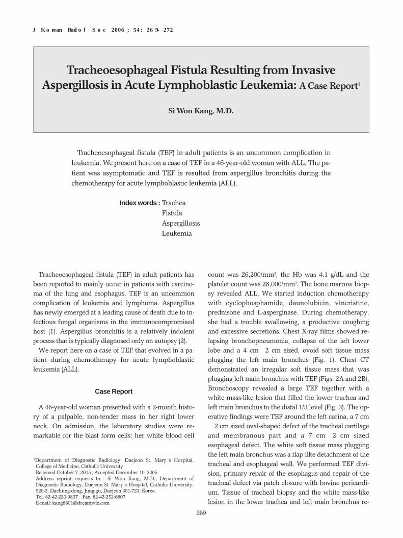
count was 26,200/mm3, the Hb was 4.1 g/dL and theplatelet count was 28,000/mm3. The bone marrow biop-sy revealed ALL. We started induction chemotherapywith cyclophosphamide, daunolubicin, vincristine,prednisone and L-asperginase. During chemotherapy,she had a trouble swallowing, a productive coughingand excessive secretions. Chest X-ray films showed re-lapsing bronchopneumonia, collapse of the left lowerlobe and a 4 cm×2 cm sized, ovoid soft tissue massplugging the left main bronchus (Fig. 1). Chest CTdemonstrated an irregular soft tissue mass that wasplugging left main bronchus with TEF (Figs. 2A and 2B).Bronchoscopy revealed a large TEF together with awhite mass-like lesion that filled the lower trachea andleft main bronchus to the distal 1/3 level (Fig. 3). The op-erative findings were TEF around the left carina, a 7 cm×2 cm sized oval-shaped defect of the tracheal cartilageand membranous part and a 7 cm×2 cm sizedesophageal defect. The white soft tissue mass pluggingthe left main bronchus was a flap-like detachment of thetracheal and esophageal wall. We performed TEF divi-sion, primary repair of the esophagus and repair of thetracheal defect via patch closure with bovine pericardi-um. Tissue of tracheal biopsy and the white mass-likelesion in the lower trachea and left main bronchus re-
J Korean Radiol Soc 2006;54:269-272
─ 269 ─
Tracheoesophageal Fistula Resulting from InvasiveAspergillosis in Acute Lymphoblastic Leukemia: A Case Report1
Si Won Kang, M.D.
1Department of Diagnostic Radiology, Daejeon St. Mary’s Hospital,College of Medicine, Catholic University Received October 7, 2005 ; Accepted December 10, 2005Address reprint requests to : Si Won Kang, M.D., Department ofDiagnostic Radiology, Daejeon St. Mary’s Hospital, Catholic University,520-2, Daehung-dong, Jung-gu, Daejeon 301-723, Korea.Tel. 82-42-220-9837 Fax. 82-42-252-6807E-mail: [email protected]
Tracheoesophageal fistula (TEF) in adult patients is an uncommon complication inleukemia. We present here on a case of TEF in a 46-year-old woman with ALL. The pa-tient was asymptomatic and TEF is resulted from aspergillus bronchitis during thechemotherapy for acute lymphoblastic leukemia (ALL).
Index words : Trachea Fistula Aspergillosis Leukemia

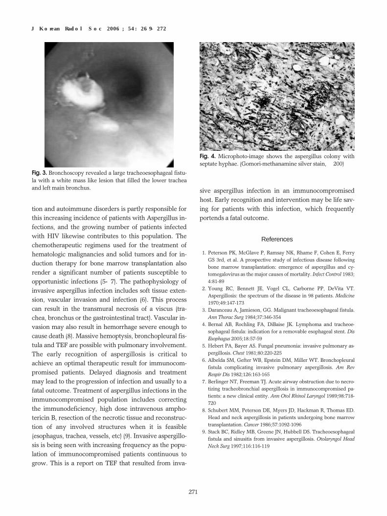
vealed aspergillosis with infarction (Fig. 4). The patientdisplayed persistent leakage of secretions at the opera-tion site and an increasing right pleural effusion.Esophagogram showed leakage of water soluble con-trast media at the primary repaired site of theesophageal defect. So, we performed a second operationof cervical esophagogastrostomy and feeding jejunosto-my. Thereafter, her general condition was improvedwithout leakage of secretions at the operation site, andthe right pleural effusion was gradually decreased. Shewas gradually administered a regular diet.
Discussion
The term TEF is used to refer any fistula formation be-tween the esophagus and the trachea or lung, regardlessof the origin of the lesion, including bronchoesophagealfistula. The development of TEF in the setting of malig-nancy is generally a poor prognostic event (3), and aspi-ration pneumonia is the immediate cause of death in thegreater proportion of patients. Medical, surgical and en-doscopic methods have been described to treat TEF.The only effective treatment is to exclude the fistulafrom the alimentary tract. Self-expanding metal stentsare used for palliative reasons to seal the TEF and allowthe patient to resume an oral food intake (4). The pa-tients with a large fistula who are in a poor medical con-dition or who suffer from repeated bouts of aspirationpneumonia require more urgent treatment. Our case al-so had a large fistula and relapsing aspiration pneumo-
nia. Since long survival following treatment forleukemia is common, the early recognition and repair ofTEF can be lifesaving.
Aspergillus is a ubiquitous air-borne fungal agent thatis frequently found to colonize the paranasal sinuses ofpatients with chronic sinusitis, and it may form as-pergillomata (5). Aspergillus infections are on the in-crease as the number of immunocompromised patientscontinuous to grow. The most frequent underlying con-ditions in the patients with invasive pulmonary as-pergillosis are leukemia and lymphoma, and prolongedgranulocytopenia is a major risk factor. The increaseduse of immunosuppressive agents for organ transplanta-
Si Won Kang: Tracheoesophageal Fistula Resulting from Invasive Aspergillosis in Acute Lymphoblastic Leukemia
─ 270 ─
A BFig. 2. A. Axial contrast enhanced CT (5-mm collimination) at the mediastinal window setting shows the tracheoesophageal fistula. B. CT scan at the lung window setting shows an irregular, soft tissue mass (aspergilloma, white arrow) in the left main bronchus,tracheoesophageal fistula (black arrow) and focal consolidations in the right upper lobe.
Fig. 1. Chest PA shows about a 4 cm×2 cm sized, ovoid softtissue mass plugging the left main bronchus (black arrows)and an air-filled esophagus (white arrows).
tion and autoimmune disorders is partly responsible forthis increasing incidence of patients with Aspergillus in-fections, and the growing number of patients infectedwith HIV likewise contributes to this population. Thechemotherapeutic regimens used for the treatment ofhematologic malignancies and solid tumors and for in-duction therapy for bone marrow transplantation alsorender a significant number of patients susceptible toopportunistic infections (5-7). The pathophysiology ofinvasive aspergillus infection includes soft tissue exten-sion, vascular invasion and infection (6). This processcan result in the transmural necrosis of a viscus (tra-chea, bronchus or the gastrointestinal tract). Vascular in-vasion may also result in hemorrhage severe enough tocause death (8). Massive hemoptysis, bronchopleural fis-tula and TEF are possible with pulmonary involvement.The early recognition of aspergillosis is critical toachieve an optimal therapeutic result for immunocom-promised patients. Delayed diagnosis and treatmentmay lead to the progression of infection and usually to afatal outcome. Treatment of aspergillus infections in theimmunocompromised population includes correctingthe immunodeficiency, high dose intravenous ampho-tericin B, resection of the necrotic tissue and reconstruc-tion of any involved structures when it is feasible(esophagus, trachea, vessels, etc) (9). Invasive aspergillo-sis is being seen with increasing frequency as the popu-lation of immunocompromised patients continuous togrow. This is a report on TEF that resulted from inva-
sive aspergillus infection in an immunocompromisedhost. Early recognition and intervention may be life sav-ing for patients with this infection, which frequentlyportends a fatal outcome.
References
1. Peterson PK, McGlave P, Ramsay NK, Rhame F, Cohen E, FerryGS 3rd, et al. A prospective study of infectious disease followingbone marrow transplantation: emergence of aspergillus and cy-tomegalovirus as the major causes of mortality. Infect Control 1983;4:81-89
2. Young RC, Bennett JE, Vogel CL, Carborne PP, DeVita VT.Aspergillosis: the spectrum of the disease in 98 patients. Medicine1970;49:147-173
3. Daranceau A, Jamieson, GG. Malignant tracheoesophageal fistula.Ann Thorac Surg 1984;37:346-354
4. Bernal AB, Rochling FA, DiBaise JK. Lymphoma and tracheoe-sophageal fistula: indication for a removable esophageal stent. DisEsophagus 2005;18:57-59
5. Hebert PA, Bayer AS. Fungal pneumonia: invasive pulmonary as-pergillosis. Chest 1981;80:220-225
6. Albelda SM, Gefter WB, Epstein DM, Miller WT. Bronchopleuralfistula complicating invasive pulmonary aspergillosis. Am RevRespir Dis 1982;126:163-165
7. Berlinger NT, Freeman TJ. Acute airway obstruction due to necro-tizing tracheobronchial aspergillosis in immunocompromised pa-tients: a new clinical entity. Ann Otol Rhinol Laryngol 1989;98:718-720
8. Schubert MM, Peterson DE, Myers JD, Hackman R, Thomas ED.Head and neck aspergillosis in patients undergoing bone marrowtransplantation. Cancer 1986;57:1092-1096
9. Stack BC, Ridley MB, Greene JN, Hubbell DS. Tracheoesophagealfistula and sinusitis from invasive aspergillosis. Otolaryngol HeadNeck Surg 1997;116:116-119
J Korean Radiol Soc 2006;54:269-272
─ 271 ─
Fig. 4. Microphoto-image shows the aspergillus colony withseptate hyphae. (Gomori-methanamine silver stain, ×200)
Fig. 3. Bronchoscopy revealed a large tracheoesophageal fistu-la with a white mass like lesion that filled the lower tracheaand left main bronchus.

Si Won Kang: Tracheoesophageal Fistula Resulting from Invasive Aspergillosis in Acute Lymphoblastic Leukemia
─ 272 ─
대한영상의학회지 2006;54:269-272
림프모구성 백혈병 환자에서 침습성 아스페르길루스증 합병으로 발생된기관지 식도 누출공: 증례 보고1
1대전성모병원영상의학과
강 시 원
백혈병 환자에서 기관지 식도 누출공은 매우 드믄 합병증이다. 저자들은 최근 46세 여자, 림프모구성 백혈병환자에
서 화학요법 중에, 침습성 아스페르길루스증 합병으로 발생된 기관지 식도 누출공 1예를 경험하였기에 문헌고찰과 함
께 보고한다.