trace elements in corallium spp. as indicators for origin and … · each of the skeletons was...
TRANSCRIPT

1
Trace Elements in Corallium spp. as Indicators for Origin and Habitat 1
2
3
Hiroshi Hasegawa*, 1; M. Azizur Rahman2, 3; Nguyen Trong Luan3; Teruya Maki1; Nozumu 4
Iwasaki4 5
6
7
8
1Institute of Science and Engineering, Kanazawa University, Kakuma, Kanazawa 920-1192, 9
Japan 10
2Centre for Environmental Sustainability, School of the Environment, Faculty of Science, 11
University of Technology Sydney, P.O. Box 123, Broadway, NSW 2007, Australia 12
3Graduate School of Natural Science and Technology, Kanazawa University, Kakuma, 13
Kanazawa 920-1192, Japan 14
4Faculty of Geo-environmental Science, Rissho University, Magechi, Kumagaya, Saitama 15
360-0194, Japan 16
17
18
*Corresponding author 19
E-mail: [email protected] (H. Hasegawa) 20
Tel/Fax: 81-76-234-4792 21
2
Abstract 22
Precious corals have been commercially exploited for many centuries around the world. 23
The skeletons of these corals consist of calcium carbonate, and have been used as amulets or 24
gemstones since ancient times. Different Corallium species of Coralidae family (e.g., 25
Corallium rubrum, Corallium elatus, Corallium konojoi, and Paracorellium japonicum) were 26
collected from different locations of the Mediterranean Sea (off Italy) and Pacific Ocean (off 27
Japan and off Midway Island), and trace elements in their skeletons were analyzed. Results 28
show that trace element concentrations in the skeletons of Corallium spp. were attributable to 29
their habitat and origin. In particular, Mg/Ca and Ba/Ca ratios in the skeletons of Corallium 30
spp. from the Mediterranean Sea and Japanese and the Midway Islands’ waters were found to 31
be habitat-specific. This study also reveals that trace elements in the skeletons can be used as 32
ecological indicator of the coral’s origin, and are expected to play an important part in the 33
cultural study and sustainable management of precious corals. Findings of this study will also 34
be of great relevance to the coral industry to authenticate and identify the habitat and origin of 35
the corals. 36
37
38
Keywords: Precious coral, Coralidae, Corallium spp., Trace element, Habitat 39
40
3
1. Introduction 41
Precious corals are some of the most valuable living marine resources, and are 42
harvested only in limited areas in the world. They belong to the functional group of deep 43
corals and are important structure-forming organisms that provide shelter for other organisms 44
and increase marine biodiversity (Tsounis et al., 2010). Precious corals are different from 45
reef-forming corals in that their skeletons are closely-packed with high magnesium calcite, 46
while the reef-building corals consist mostly of aragonite, and are porous because of its 47
loosely-packed crystals. 48
Taxonomically, precious corals belong primarily to three orders of the class Anthozoa, 49
and the most valuable species are red and pink corals of the genus Corallium and 50
Paracorallium of the family Coralidae, which consist of 19 and 7 species, respectively 51
(Tsounis et al., 2010). They are found mainly in the Mediterranean Sea and Pacific Ocean 52
(Japanese waters and off Taiwan, off the Midway Islands and off the Hawaiian Islands) 53
(Iwasaki and Suzuki, 2010). Some of the important precious corals include Corallium rubrum, 54
Paracorallium japonicum, Corallium elatius, Corallium konojoi and Corallium secundum 55
(Tsounis et al., 2010). The Corallium spp. are commonly known as deep-sea coral, and the red 56
coral (C. rubrum) is produced in the Mediterranean Sea. The pink coral (C. secundum) is 57
distributed in the seas around Hawaii and the Midway Islands, and also found in the waters 58
close to the Midway Island (Grigg, 1993). 59
4
Japanese red coral (P. japonicum), pink coral (C. elatius) and white coral (C. konojoi) 60
are distributed and harvested in waters near Japan (Iwasaki et al., 2009). Paracorallium 61
japonicum is found at depths of 76-280 m on the rocky bottom in Sagami Bay (Pacific coast 62
of Japan), in the waters from the Ogasawara Islands (Japan), and off the coast near the Goto 63
Islands, Nagasaki (Japan) (Seki, 1991). Corallium elatius is distributed on the rocky bottom at 64
a depth of 100-276 m in the waters near Wakayama (Pacific coast of Japan), from the 65
Ogasawara Islands (Japan) to the northern South China Sea, and off the Goto Islands, 66
Nagasaki, Japan (Iwasaki and Suzuki, 2010). Corallium konojoi is distributed on the rocky 67
bottom at a depth of 76-276 m in the waters of Wakayama (Pacific coast of Japan), in the 68
waters from the Ogasawara Islands (Japan) to the northern South China Sea, and off the Goto 69
Islands, Nagasaki, Japan (Seki, 1991; Nonaka et al., 2004). Corallium secundum has been 70
found to grow on flat exposed substrata whereas C. regale prefer encrusted uneven rocky 71
bottom habitat in the Hawaiian Islands, and both species are absent from the shelf areas (<400 72
m depth) (Grigg, 1974). 73
Some Corallium species have a hard calcium skeleton of intense red and the others 74
are pink and of pink (Iwasaki and Suzuki, 2010). Both spicules and skeletons of red coral (C. 75
rubrum) are mainly made of calcium carbonate (CaCO3) crystallized in the form of calcite, 76
though small amounts of other trace elements such as magnesium (Mg), strontium (Sr), iron 77
(Fe), aluminum (Al) and sulphur (S) are also found (Maté et al., 1986). Previously, Velimirov 78
5
and Bohm (1976) analyzed calcium (Ca) and Mg by atomic absorption spectroscopy and 79
ethylenediaminetetraacetate (EDTA) titration with the aim of providing information on the 80
mineral composition of gorgonians and the possible variations in different growth regions. 81
They showed that CaCO3, MgCO3 and total mineral content increase markedly from branch to 82
stem. Weinbauer and Velimirov (1995) determined Mg, Ca, and Sr in sclerites of four 83
Mediterranean gorgonians and suggested that Mg/Ca and Sr/Ca ratios were very low 84
(0.064-0.098 and 0.004-0.0025, respectively). They also revealed that calcium concentrations 85
did not vary with geographical origin, while the variations of Mg/Ca and Sr/Ca ratios were 86
related to water depth. Besides, there was a direct relationship between Mg concentration and 87
temperature, and the Mg/Ca ratios increased significantly with the ambient water temperature 88
(Weinbauer and Velimirov, 1995). 89
Reef-building coral is better understood, and concentrations of trace elements in its 90
carbonate skeletons have been determined. The validity of their use as indicators of past 91
environmental conditions, such as water temperatures, nutrients and pollution levels has been 92
confirmed in earth and environmental science studies (Weber and Woodhead, 1970; Weber, 93
1973; Mitsuguchi et al., 1996; Mitsuguchi et al., 2001; Mitsuguchi et al., 2003). In contrast, 94
studies on trace elements in precious coral have been focused mostly on Mediterranean red 95
coral C. rubrum (Weinbauer and Velimirov, 1995; Weinbauer et al., 2000). Studies on trace 96
elements in other precious corals, especially the Japanese white coral (C. konojoi), from 97
6
various locations is limited. Research on precious corals in Japanese waters has recently been 98
started. 99
Precious corals have attracted worldwide attention as sparse biological resources, and 100
Corallidae have been recently proposed for inclusion in Appendix II of the Convention on 101
International Trade in Endangered Species of Wild Fauna and Flora (CITES) that regulates 102
the international trade in endangered species by listing them in its appendices. The problem is 103
that appendix II permits the export and/or import of corals from well managed stocks, while it 104
prohibits that corals from unmanaged areas can pass the costumes borders. Therefore, 105
appropriate scientific methods for the authentication of the uniqueness and origin of 106
Corallidae are necessary to protect the coral resources and for international trade. This refers 107
to both geographical and bathymetric origin, because shallow coral may be protected in one 108
area, while deeper stocks may be harvested for commercial uses. In the present study, trace 109
element concentrations in the skeletons of Corallium spp. of Corallidae family from different 110
geographical locations (origins) were determined to investigate if the concentrations and 111
distribution of trace elements were related to their origin and habitat. The identification of the 112
origin of the corals via trace metal analysis will provide the opportunity to reveal smuggling 113
of illegal corals with fake papers. 114
115
2. Materials and Methods 116

7
2.1. Sampling site 117
Samples were collected from Japanese waters, the Midway Islands’ waters and the 118
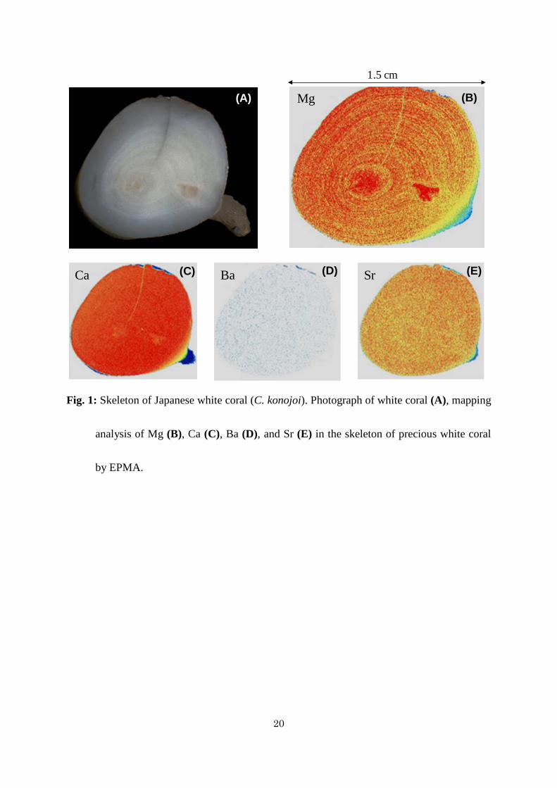
Mediterranean Sea (off Italy) from fishermen, coral traders, and research institutes. A 119
deep-sea coral (Corallium sp.) was collected by Marine Geological Research Vessel “Hakurei 120
Maru” cruise GH85-1 conducted by the National Institute of Advanced Industrial Science and 121
Technology. Two specimens of Japanese red coral (P. japonicum) were collected by a manned 122
submersible “Hakuyo” from the Kochi Prefectural Deep Seawater Laboratory. The species 123
and the locations from where the samples were collected are shown in Table 1. 124
Corals of the Mediterranean Sea and Japanese waters were sampled from a depth of < 150 125
m, while some samples of the Midway Islands were collected from a depth of 400-500 m and 126
the other were from a depth of 900-1200 m. White coral (C. konojoi; Fig. 1) was collected 127
from a depth of 100 m, off Cape Muroto, Kochi, Japan, in July 2004. 128
129
2.2. Chemical analysis of skeleton composition 130
Barium (Ba), Ca, Mg, and Sr concentrations were analyzed in skeleton of the corals. 131
Each of the skeletons was ground in an agate mortar into a particle size of 5 mm in diameter 132
and 0.1 g of it was taken into 10-mL polypropylene test tubes with three replications. The 133
skeletons of precious corals were then cleaned following sequential methods of ultrasonic, 134
oxidation, and reduction treatments (Shen and Boyle, 1988). At first the samples were treated 135

8
with ultrasonic waves in 1 mL of purified water and then in 1 mL of 0.2 M nitric acid for 10 136
min each. The samples were rinsed with purified water between the treatments. This 137
procedure was repeated three times. After drying at room temperature, they were ground 138
further in the agate mortar and were sieved through a 25-50 Teflon mesh screen of a 139
polypropylene sieve. The samples were then collected from three different sieved samples in 140
the same colony (n = 3). 141
After ultrasonic cleaning, the samples in 10-mL polypropylene test tubes were oxidized 142
by addition of 1 mL solution prepared from 1:1 (v/v) 30% hydrogen peroxide (H2O2) and 0.2 143
M sodium hydroxide (NaOH). They were then placed in a steam and an ultrasonic bath for 2 144
min each for a total of 10 repeats. They were then sequentially treated with ultrasonic waves 145
in 1 mL of 0.2 M nitric acid for 3 min, in 1 mL of purified water for 10 min and in 1 mL of 146
0.2 M nitric acid for 3 min, and then the oxidation treatment was applied once again. A 147
reduction treatment was followed by oxidation treatment in which the samples were treated 148
with 1 mL solution containing 97% hydrogen, concentrated ammonia, and 0.3 M citric acid in 149
the ratio of 1:6:3. They were then placed in a hot (70 °C) and an ultrasonic bath for 2 min 150
each for a total of 16 repeats. Finally the samples were cleaned by repeating oxidation 151
treatments, treating ultrasonic waves (2-min) in 1 mL of 0.2 M nitric acid three times, and 152
rinsing twice in 1 mL of 0.2 M nitric acid. Then the samples were dissolved in 1 mL of 2 M 153
nitric acid. 154

9
The sample solutions were diluted to 1000 times with 0.5 M nitric acid, and the 155
determination of trace element concentrations in the samples were carried out by inductively 156
coupled plasma atomic emission spectroscopy (ICP-AES, Perkin Elmer, Optima 3300XL) in 157
triplicates using a calibration curve method. 158
159
2.3. Analysis of inorganic elements with electron probe micro-analyzer (EPMA) 160
Each skeleton sample with dried organic tissues attached on the surface of white coral 161
(C. konojoi) was embedded in polyester resin for EPMA. The skeletons were cut at 5 mm 162
intervals perpendicular to the growth direction using a diamond saw, and one of the thin slices 163
was ground to 100-200 μm with 600, 1000, and 2000 grits silicon carbide abrasive papers. 164
The surface of the samples was then polished to a mirror finish using an alumina wrapping 165
sheet (Marumoto Kogyo, Japan) with a particle diameter of 0.1 μm, and was coated with a 166
10-μm carbon film by evaporative deposition of carbon. EPMA was performed using an 167
EPMA-8705 (Shimadzu Corporation, Japan). Two-dimensional images of elemental 168
distribution were obtained by stage mapping the sample along the x and y axes with 29 μm 169
raster spacing. Measurement parameters were set as follows: accelerating voltage at 15 keV, 170
beam current at 0.3 µA, and a measurement time 0.04 sec. for Ca and Sr, and 0.22 sec. for Mg 171
and Ba. 172
173

10
3. Results and Discussion 174
3.1. Trace element distribution in the skeletons of precious coral 175
Ca and Mg measurements in the cross-section of white coral (C. konojoi) skeleton 176
reveal that Ca is distributed homogeneously (Fig. 1C), while Mg concentration is distributed 177
concentrically forming growth rings (Fig. 1B). About 38 growth rings of Mg were observed in 178
tiers on a 6 mm radius. If these growth rings are formed annually, the radial growth rate of 179
this white coral will be 0.32 mm yr-1. This rate agrees with the radial growth rate of the same 180
coral obtained through infrared spectroscopy using synchrotron radiation as well as through 181
210Pb dating (Hasegawa et al., 2010). As in C. rubrum, annual rings can also be observed by 182
staining the organic matrix with toluidine blue. Vielzeuf et al. (2008) performed EPMA 183
mapping on the skeleton of C. rubrum and found a negative correlation between Mg 184
concentrations and the organic matter, except in the center part of the skeleton, indicating that 185
Mg concentrations corresponded to annual rings. Thus, Mg growth rings can be considered as 186
annual rings of precious coral. 187
Mg-rich layers grow in warm seasons, and the variations in Mg concentrations in the 188
skeleton are very likely to be related to water temperatures. Weinbauer and Velimirov (1995) 189
compared the Mg/Ca ratios in the skeletons of several C. rubrum species from different 190
depths and found a positive correlation between the Mg/Ca ratios and water temperatures. 191
Variations in Mg concentrations within an individual coral skeleton can be assessed as the 192
11
Mg/Ca molar ratio. The Mg/Ca ratio in white coral varies from 10 to 15% of the mean values, 193
while that in C. rubrum obtained from EPMA study of Weinbauer et al. (2000) varied from 13 194
to 30%. 195
Other than these elements, Sr, Ba, I, and Mo concentrations have also been confirmed in 196
Japanese red coral by two-dimensional images produced in a synchrotron-radiation X-ray 197
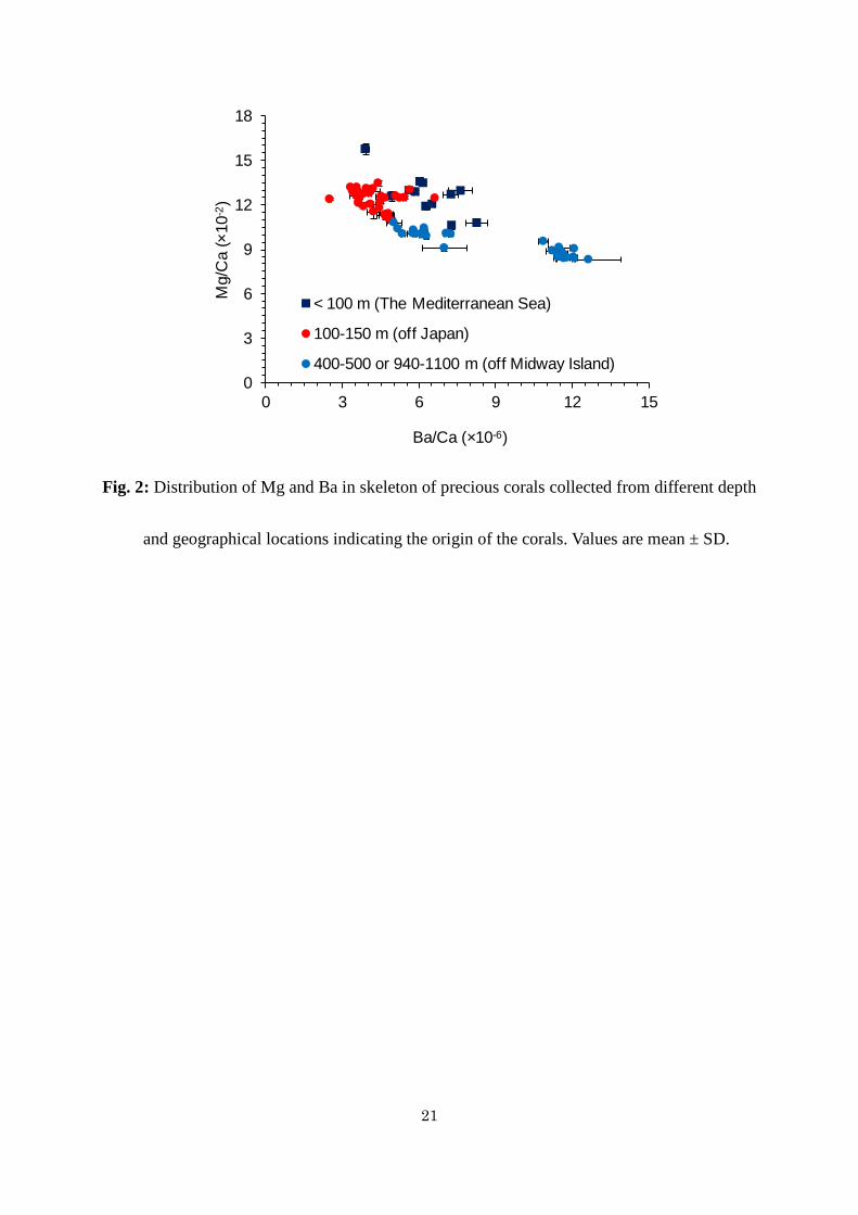
Fluorescence (XRF) study (Hasegawa et al., 2010). These elements are distributed 198
homogeneously across the cross-sections of the skeleton, and the variations of their 199
concentrations are lower compared to Mg concentration. Weak correlation between Sr 200
concentration and the number of growth rings on the skeleton of Japanese red coral obtained 201
by XRF mapping analysis has been reported by Hasegawa et al. (2010). The present study 202
also reveals a weak correlation between Sr concentration and growth rings on the skeleton of 203
white coral obtained by EPMA analysis (Fig. 1E). The millimeter-scale variations of the Sr/Ca 204
ratio do not correspond to those of growth rings, which is between 100 and 200 μm. On the 205
other hand, the Sr/Ca ratio in the skeletons of C. rubrum has been reported to vary 206
significantly in proportion to skeleton density (Weinbauer et al., 2000). 207
208
3.2. Trace element compositions reflecting the characteristics of coral habitats 209
The Mg/Ca and Ba/Ca ratios in the skeletons of C. rubrum, Japanese red, pink and white 210
corals, and Midway corals are shown in Figure 2. Japanese red, pink and white corals co-habit 211
12
in sea-floors around Japan. The Mg/Ca and Ba/Ca ratios in these three corals collected from 212
the same area were within similar ranges without species-specific differences. On the other 213
hand, Mg/Ca and Ba/Ca ratios in corals from the Mediterranean Sea, Japanese waters and the 214
sea around the Midway Islands differed depending on their habitats (Fig. 2) except Sr/Ca ratio 215
(0.31-0.33×10⁻² mol mol-1). Mg/Ca and Sr/Ca ratios in the skeletons of precious corals (C. 216
rubrum and Japanese red, pink and white corals) of present study determined by EPMA 217
analysis are well agreed with those in the skeletons of C. rubrum measured by XRF 218
(Weinbauer and Velimirov, 1995). This study also showed that Ba/Ca ratio in the Midway 219
deep-sea corals is higher compared to that in other samples. 220
The Mg/Ca ratios in precious corals of the Mediterranean Sea and Japanese waters 221
ranged from 10-15x10-2 mol mol-1, while those in pink and white corals of the Midway 222
Islands were in the ranges of 9-11x10-2 mol mol-1 and 8-9x10⁻² mol mol-1 for 400-500 m and 223
900-1200 m (deep-sea corals), respectively. Because Mg and Ca are the major salts in 224
seawater, and Mg/Ca ratio is almost constant in all parts of the ocean, it is extremely unlikely 225
that the observed variations in the Mg/Ca ratio were influenced by the Mg/Ca ratio in 226
seawater. In a previous study, Weinbauer and Velimirov (1995) observed that the Mg/Ca ratio 227
in the skeletons of C. rubrum was usually related to ocean’s depth. They incorporated other 228
reports and estimated that the Mg/Ca ratio in C. rubrum was directly proportional to water 229
temperature, increasing by 0.004-0.006 mol mol-1 per 1 ˚C. Water temperatures near the 230
13
sediments, where the coral samples for the present study were collected (Table 1), were in the 231
range of 13-24 °C in the Mediterranean Sea and Japanese waters, and 8-11 and 2-3 °C at a 232
depth of 400-500 m and 900-1200 m in the Midway Islands, respectively. The difference in 233
the water temperature could be predicted by the Mg/Ca ratios assuming that an increase of 234
0.004-0.006 mol mol-1 Mg/Ca ratio per 1 °C was applicable to all precious corals in the 235
subclass Octocorallia. The difference in temperature between the Mediterranean Sea/Japanese 236
waters and the Midway shallow area is 2-16 ˚C, and between the Mediterranean Sea/Japanese 237
waters and the Midway deep area is 10-22 ˚C, which can be calculated as 0.008-0.096 and 238
0.040-0.132 mol mol-1 of Mg/Ca ratios, respectively. Because these figures are close to the 239
difference in the Mg/Ca ratios in this study (Fig. 2), variations in the Mg/Ca ratio of precious 240
corals are likely to be explained by difference in water temperature during coral formation. 241
Ba/Ca ratio in the Midway coral from a depth of 900-1200 m was significantly higher 242
(10-15×10-6 mol mol-1) than that of Midway coral from a depth of 400-500 m (5-8×10⁻⁶ mol 243
mol-1) and Japanese corals from a depth of 100-150 m (2-6×10⁻⁶ mol mol-1). In contrast, the 244
Ba/Ca ratios in C. rubrum showed a wider range (4-14×10-6 mol mol-1). The proportions of 245
Mg, Ca, and Sr concentrations, the major constituents of seawater, are almost identical to 246
those in seawater, while Ba concentration increases with the depth of the ocean. For example, 247
dissolved Ba concentrations of 30-40 mol kg-1 at 50-200 m, 40-55 mol kg-1 at 400-500 m, and 248
90-110 mol kg-1 at 900-1200 m were reported from Corallium (what species) in the North 249
14
Pacific (Chow and Goldberg, 1960; Boyle et al., 1976). These figures agree well with the 250
Ba/Ca ratios in the skeletons of analyzed precious corals collected from the waters around 251
Japan and around the Midway Islands. 252
Some overlaps of Mg/Ca and Ba/Ca ratios in corals from the Mediterranean and 253
Japanese waters were observed in the present study. The data presented in Fig. 2 showed that 254
the Mg/Ca and Ba/Ca rations in only 5 samples of Japanese corals and 6 samples of the 255
Mediterranean corals are in the range of 12-13x10-2 and 4.6-6.0x10⁻⁵ mol mol-1, respectively, 256
while 33 samples of the Japanese corals and 13 samples of the Mediterranean corals are 257
distributed separately. One sample of the Japanese coral with 12.5x10⁻² mol mol-1 of Mg/Ca 258
and 6.6x10⁻⁵ mol mol-1 of Ba/Ca, and one sample of Mediterranean corals with 15.8×10⁻² mol 259
mol-1 of Mg/Ca and 3.9×10⁻² mol mol-1 Ba/Ca might be exceptions. Thus, some degree of 260
error was unavoidable in the same manner as other scientific methods for identification of 261
origin and habitat of corals. Combined use of the information on color and appearance of 262
coral samples would be helpful for reducing the error. In another study, we have detected 263
other trace elements such as I, Mo, Sn, Mn, Zn, Cd and Br in skeletons of precious corals 264
(Hasegawa et al., 2010), which would have the potential for additional indicators of 265
geographical origin. 266
267
Conclusion 268
15
Apart from Mg concentrations that vary slightly with annual rings, trace elements in 269
precious coral skeletons are distributed homogeneously. Our study reveals that the trace 270
elements in skeletons of precious corals are habitat-specific rather than species-specific. The 271
Mg/Ca and Ba/Ca ratios in skeletons of precious corals, particularly, are the indicators of their 272
habitats and environments and, therefore, can be used to identify the harvested areas of coral 273
products. X-ray fluorescent analysis is another useful method that can serve to identify the 274
bathymetrical and geographic origin of coral (and other) products. 275
The proposal to list all species in the family Corallidae in Appendix II of the 276
Convention on International Trade in Endangered Species of Wild Fauna and Flora was 277
rejected in the Conferences of the Parties (CoP) 14 (FAO, 2007) and CoP 15 (FAO, 2010). 278
The difficulties in the identification of coral products in trade have been highlighted in the 279
debate about the feasibility to enforce such a listing effectively. Identification of raw coral to 280
species level is easy to the coral specialists, and a taxonomic reference guide has been 281
recently published by World Wildlife Fund Canada (WWF Canada) to help customs officials 282
to identify raw coral to species level, however, it is likely that identification might not be 283
possible when coral products such as jewelries of Corallium sp. and of other species that are 284
resembled to Corallium sp. after dying (FAO, 2007; 2010). The findings of the present study 285
would guide to a possible solution to this problem, and would contribute in developing a 286
nondestructive analytical method, such as XRF, for the identification of raw corals as well as 287

16
coral products. Further research is necessary to develop a fully functional, cost-effective and 288
readily applicable method. 289
290
Acknowledgements 291
We wish to thank Noriyoshi Yoshimoto of Pacific Coral Co. Ltd., Masaki Kikuchi of 292
Kikuchi Coral, Hisaichi Kawaguchi, Yuichi Furukawa, Shuichiro Deguchi of Deguchi Coral, 293
SNK Ocean Co., Ltd., the National Institute of Advanced Industrial Science and Technology, 294
and the Kochi Prefectual Deep Seawater Laboratory for giving the specimens. We would like 295
to express our gratitude to Osamu Nagata and Hideto Kameya, Kanazawa University, and 296
Masayoshi Kobayashi, Niigata University, for measurements of trace elements in the 297
skeletons. This research was support partly by Japan Society for the Promotion of Science as 298
Grant-in-Aid for Scientific Research 21651101, which is gratefully acknowledged. We also 299
would like to thank Dr. Anne Colville, School of Environmental Sciences, University of 300
Technology Sydney (UTS), Australia, for her valuable time in reviewing the manuscript. 301
302
References 303
Boyle, E.A., Sclater, F., Edmond, J.M., 1976. On the marine geochemistry of cadmium. 304 Nature 263(5572), 42-44. 305
Chow, T.J., Goldberg, E.D., 1960. On the marine geochemistry of barium. Geochim. 306 Cosmochim. Acta 20(3-4), 192-198. 307
FAO, 2007. Report of the second FAO Ad hoc expert advisory panel for the assessment of 308
17
proposals to amend appendices I and II of CITES concerning commercially-expoited 309 aquatic species. Food and Agriculture Organization of the United Nations, Rome, pp. 310 133. 311
FAO, 2010. Report of the third FAO expert advisory panel for the assessment of proposals to 312 amend appendices I and II of CITES concerning commercially- exploited aquatic species. 313 Food and Agriculture Organization of the United Nations, Rome, pp. 150. 314
Grigg, R.W., 1974. Growth rings: Annual periodicity in two gorgonian corals. Ecology 55(4), 315 876-881. 316
Grigg, R.W., 1993. Precious coral fisheries of Hawaii and the US Pacific Islands. Mar. Fish. 317 Rev. 55(2), 50-60. 318
Hasegawa, H., Iwasaki, N., Suzuki, A., Maki, T., Hayakawa, S., 2010. Distribution of trace 319 elements in biogenic carbonate minerals of precious corals by X-ray fluorescence 320 analysis. Bunseki Kagaku 59(6), 521-530. 321
Iwasaki, N., Hasegawa, H., Suzuki, T., Yamasa, M., 2009. Biology of Japanese Corallium and 322 Paracorallium. In: Bruckner, A.W., Roberts, G.G. (Eds.), Proceedings of the First 323 International Workshop on Corallium Science, Management, and Trade. NOAA Technical 324 Memorandum NMFS-OPR-43 and CRCP-8, Silver Spring, MD, Hong Kong, China, pp. 325 153. 326
Iwasaki, N., Suzuki, T., 2010. Biology of precious corals. In: Iwasaki, N. (Ed.), Biohistory of 327 Precious corals- Scientific,Cultural and Historical Perspectives. Tokai Univeristy Press, 328 Tokai, Japan. 329
Maté, P., Revenge, S., Masso, C., 1986. Estudio preliminar de la composicion quimica del 330 coral rojo (Corallium rubrum L.) de distintas zonas del Mediterraneo Espanol. Bol. Inst. 331 Esp. Oceanogr. 3, 53–60. 332
Mitsuguchi, T., Matsumoto, E., Abe, O., Uchida, T., Isdale, P.J., 1996. Mg/Ca thermometry in 333 coral skeletons. Science 274(5289), 961. 334
Mitsuguchi, T., Matsumoto, E., Uchida, T., 2003. Mg/Ca and Sr/Ca ratios of porites coral 335 skeleton: Evaluation of the effect of skeletal growth rate. Coral Reefs 22(4), 381-388. 336
Mitsuguchi, T., Uchida, T., Matsumoto, E., Isdale, P.J., Kawana, T., 2001. Variations in Mg/Ca, 337 Na/Ca, and Sr/Ca ratios of coral skeletons with chemical treatments: Implications for 338 carbonate geochemistry. Geochim. Cosmochim. Acta 65(17), 2865-2874. 339
18
Nonaka, M., Muzik, K., Uchida, S., 2004. Capture, study and display of precious corals, 10th 340 International Coral Reef Symposium, Okinawa, Japan, pp. 38-42. 341
Seki, K., 1991. Study of precious coral diver survey with open circuit air SCUBA diving at 342 108 m. Ann. Physiol. Anthropol. 10(3), 189-192. 343
Shen, G.T., Boyle, E.A., 1988. Determination of lead, cadmium and other trace metals in 344 annually-banded corals. Chem. Geol. 67(1-2), 47-62. 345
Tsounis, G., Rossi, S., Grigg, R., Santangelo, G., Bramanti, L., Gili, J.M., 2010. The 346 exploitation and conservation of precious corals. Ocean. Mar. Biol. 48, 161-212. 347
Velimirov, B., Böhm, E., 1976. Calcium and magnesium carbonate concentrations in different 348 growth regions of gorgonians. Mar. Biol. 35(3), 269-275. 349
Vielzeuf, D., Garrabou, J., Baronnet, A., Grauby, O., Marschal, C., 2008. Nano to macroscale 350 biomineral architecture of red coral (Corallium rubrum). Am. Mineral. 93(11-12), 351 1799-1815. 352
Weber, J.N., 1973. Incorporation of strontium into reef coral skeletal carbonate. Geochim. 353 Cosmochim. Acta 37(9), 2173-2190. 354
Weber, J.N., Woodhead, P.M.J., 1970. Carbon and oxygen isotope fractionation in the skeletal 355 carbonate of reef-building corals. Chem. Geol. 6, 93-117. 356
Weinbauer, M.G., Brandstatter, F., Velimirov, B., 2000. On the potential use of magnesium 357 and strontium concentrations as ecological indicators in the calcite skeleton of the red 358 coral (Corallium rubrum). Mar. Biol. 137(5), 801-809. 359
Weinbauer, M.G., Velimirov, B., 1995. Calcium, magnesium and strontium concentrations in 360 the calcite sclerites of Mediterranean gorgonians (Coelenterata: Octocorallia). Estuar. 361 Coast. Shelf Sci. 40(1), 87-104. 362
363
364
365
366
367
19
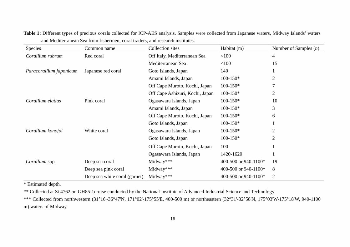
Table 1: Different types of precious corals collected for ICP-AES analysis. Samples were collected from Japanese waters, Midway Islands’ waters and Mediterranean Sea from fishermen, coral traders, and research institutes.
Species Common name Collection sites Habitat (m) Number of Samples (n) Corallium rubrum Red coral Off Italy, Mediterranean Sea <100 4
Mediterranean Sea <100 15 Paracorallium japonicum Japanese red coral Goto Islands, Japan 140 1
Amami Islands, Japan 100-150* 2 Off Cape Muroto, Kochi, Japan 100-150* 7 Off Cape Ashizuri, Kochi, Japan 100-150* 2
Corallium elatius Pink coral Ogasawara Islands, Japan 100-150* 10 Amami Islands, Japan 100-150* 3 Off Cape Muroto, Kochi, Japan 100-150* 6 Goto Islands, Japan 100-150* 1
Corallium konojoi White coral Ogasawara Islands, Japan 100-150* 2 Goto Islands, Japan 100-150* 2
Off Cape Muroto, Kochi, Japan 100 1 Ogasawara Islands, Japan 1420-1620 1
Corallium spp.
Deep sea coral Midway*** 400-500 or 940-1100* 19 Deep sea pink coral Midway*** 400-500 or 940-1100* 8 Deep sea white coral (garnet) Midway*** 400-500 or 940-1100* 2
* Estimated depth. ** Collected at St.4762 on GH85-1cruise conducted by the National Institute of Advanced Industrial Science and Technology. *** Collected from northwestern (31°16'-36°47'N, 171°02'-175°55'E, 400-500 m) or northeastern (32°31'-32°58'N, 175°03'W-175°18'W, 940-1100 m) waters of Midway.
20
1.5 cm
Ca Ba Sr
Mg (B)(A)
(C) (D) (E)
Fig. 1: Skeleton of Japanese white coral (C. konojoi). Photograph of white coral (A), mapping
analysis of Mg (B), Ca (C), Ba (D), and Sr (E) in the skeleton of precious white coral
by EPMA.
21
0
3
6
9
12
15
18
0 3 6 9 12 15
Mg/
Ca
(×10
-2)
Ba/Ca (×10-6)
< 100 m (The Mediterranean Sea)
100-150 m (off Japan)
400-500 or 940-1100 m (off Midway Island)
Fig. 2: Distribution of Mg and Ba in skeleton of precious corals collected from different depth
and geographical locations indicating the origin of the corals. Values are mean ± SD.