towards a structural understanding of the fibrillization pathway in
TRANSCRIPT
doi:10.1016/j.jmb.2005.08.061 J. Mol. Biol. (2005) 353, 642–654
Towards a Structural Understanding of the FibrillizationPathway in Machado-Joseph’s Disease: Trapping EarlyOligomers of Non-expanded Ataxin-3
Luıs Gales1, Luısa Cortes2, Carla Almeida2, Carlos V. Melo2
Maria do Carmo Costa3, Patrıcia Maciel3, David T. Clarke4
Ana Margarida Damas1 and Sandra Macedo-Ribeiro2*
1ICBAS-Instituto de CienciasBiomedicas de Abel Salazar andIBMC-Instituto de BiologiaMolecular e CelularUniversidade do Porto, Portugal
2MacromolecularCrystallography Group, Centerfor Neuroscience and CellBiology (CNC), 3004-517Coimbra, Portugal
3Health and Life SciencesResearch Institute/School ofHealth Sciences, University ofMinho, Campus de Gualtar4710-057 Braga, Portugal
4CCLRC Daresbury LaboratoryDaresbury, Warrington WA44AD, UK
0022-2836/$ - see front matter q 2005 E
Abbreviations used: EM, electronoligomers, high molecular mass oligMachado-Joseph’s disease; MJDL-Celegans ataxin-3 like protein containiresidue (1Q) in the region corresponglutamine domain; MJD1-1, humancontaining 14 glutamine residues (1region; poly(Q), polyglutamine; UIMing motif; ThT, thioflavin T; NIs, ne
E-mail address of the [email protected]
Machado-Joseph’s disease is caused by a CAG trinucleotide repeatexpansion that is translated into an abnormally long polyglutamine tractin the protein ataxin-3. Except for the polyglutamine region, proteinsassociated with polyglutamine diseases are unrelated, and for all of thesediseases aggregates containing these proteins are the major components ofthe nuclear proteinaceous deposits found in the brain. Aggregates of theexpanded proteins display amyloid-like morphological and biophysicalproperties.
Human ataxin-3 containing a non-pathological number of glutamineresidues (14Q), as well as its Caenorhabditis elegans (1Q) orthologue, showeda high tendency towards self-interaction and aggregation, under near-physiological conditions. In order to understand the discrete steps in theassembly process leading to ataxin-3 oligomerization, we have separatedchromatographically high molecular mass oligomers as well as mediummass multimers of non-expanded ataxin-3. We show that: (a) oligomeriza-tion occurs independently of the poly(Q)-repeat and it is accompanied byan increase in b-structure; and (b) the first intermediate in theoligomerization pathway is a Josephin domain-mediated dimer of ataxin-3. Furthermore, non-expanded ataxin-3 oligomers are recognized by aspecific antibody that targets a conformational epitope present in solublecytotoxic species found in the fibrillization pathway of expandedpolyglutamine proteins and other amyloid-forming proteins. Imaging ofthe oligomeric forms of the non-pathological protein using electronmicroscopy reveals globular particles, as well as short chains of suchparticles that likely mimic the initial stages in the fibrillogenesis pathwayoccurring in the polyglutamine-expanded protein. Thus, they constitutepotential targets for therapeutic approaches in Machado-Joseph’s disease,as well as valuable diagnostic markers in disease settings.
q 2005 Elsevier Ltd. All rights reserved.
Keywords: SCA3; self-assembly domain; neurodegenerative; polygluta-mine; amyloid
*Corresponding authorlsevier Ltd. All rights reserve
microscopy; HMMomers; MJD,
E, Caenorhabditisng a single glutamineding to the poly-ataxin-3 isoform 24Q) in the repeat
, ubiquitin interact-uronal inclusions.ing author:
Introduction
Several human disorders are caused by trinucleo-tide expansions in a heterogeneous group of genes.1
CAG expansions leading to increased polygluta-mine repeats within the affected proteins areresponsible for nine slowly progressive disorderscollectively known as polyglutamine (poly(Q))diseases. Despite ubiquitous expression of boththe normal and disease proteins throughout the
d.
Trapping Early Oligomers of Non-expanded Ataxin-3 643
brain and other tissues, only a particular subset ofneurons is selectively affected in each disease.Nuclear and sometimes cytoplasmatic neuronalinclusions (NIs) containing the mutant proteins,are now clearly established as the identifyingfingerprint of poly(Q) diseases.2,3 Longer poly(Q)repeats cause more severe disease symptoms,implying that the degree of toxicity is directlyproportional to the size of the repeat.
The structural and functional effects of poly(Q)expansions in the affected proteins as well as themechanisms of polyglutamine toxicity are still notclear. An enormous research effort has been put intounderstanding the structure of polyglutamine andthe structural changes induced by polyglutamineexpansion.4,5 Current experimental data show that,independently of the repeat length, monomericpolyglutamine sequences exist predominantly in arandom coil conformation. A conformational tran-sition occurs upon aggregation: in close connectionwith the pathological threshold in polyglutamino-pathies, the tendency to aggregate into b-richfibrillar structures increases with poly(Q) length.6
The amyloid nature of these polyglutamineinsoluble structures has been demonstrated bynumerous experiments in vitro7–9 and in vivo.7,10
Furthermore, poly(Q) aggregates are recognized by“pan-amyloid” antibodies able to associate withgeneric amyloid epitopes in fibrils formed from adiverse number of proteins.11 Whether the cellular
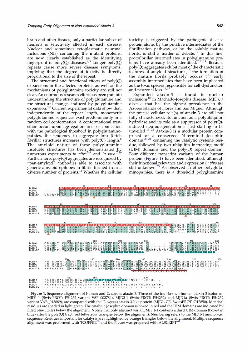
Figure 1. Sequence alignment of human and C. elegans ataMJD1-1 (SwissPROT: P54252 variant VSP_002784), MJD2-1variant VAR_013689), are compared with the C. elegans ataxinresidues are shaded in light green. The catalytic Josephin domfilled blue circles below the alignment. Notice that only ataxinblue) after the poly(Q) tract (red left-arrow triangles below thsequence. Residues important for catalysis are highlighted byalignment was preformed with TCOFFEE60 and the Figure w
toxicity is triggered by the pathogenic diseaseprotein alone, by the putative intermediates of thefibrillization pathway, or by the soluble maturefibrils, is still a matter of debate.12 In this line,protofibrillar intermediates in polyglutamine pro-teins have already been identified.9,13,14 Becausepoly(Q) aggregates exhibit most of the characteristicfeatures of amyloid structures,15 the formation ofthe mature fibrils probably occurs via earlyassembly intermediates that have been implicatedas the toxic species responsible for cell dysfunctionand neuronal loss.16,17
Expanded ataxin-3 is found in nuclearinclusions18 in Machado-Joseph’s disease (MJD), adisease that has the highest prevalence in theAzores islands of Flores and Sao Miguel. Althoughthe precise cellular role(s) of ataxin-3 are still notfully characterized, its function as a polyubiquitinhydrolase and its role as a suppressor of poly(Q)-induced neurodegeneration is just starting to beunveiled.19–22 Ataxin-3 is a modular protein com-prised of a conserved N-terminal Josephindomain,23,24 containing the catalytic cysteine resi-due, followed by two ubiquitin interacting motif(UIM) domains and the poly(Q) repeat domain.Four different transcript variants of the humanprotein (Figure 1) have been identified, althoughtheir functional relevance and expression in vivo arestill unknown.25 As observed in other polygluta-minopathies, there is a threshold polyglutamine
xin-3. Three of the four known human ataxin-3 isoforms:(SwissPROT: P54252) and MJD1a (SwissPROT: P54252
-3-like protein (MJDL-CE, SwissPROT: O17850). Identicalain is boxed in red and the UIM domains are indicated by-3 variant MJD1-1 contains a third UIM domain (boxed ine alignment). Numbering refers to the MJD1-1 amino acidorange triangles below the alignment. Multiple sequenceas prepared with ALSCRIPT.61

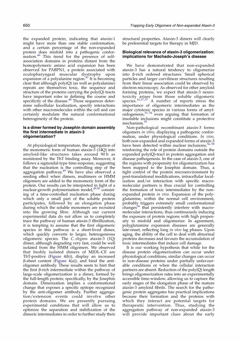
Figure 2. Non-expanded ataxin-3 oligomerizes undernear-physiological conditions. (a) Monomeric humanataxin-3 can be separated from high molecular massoligomers by size-exclusion chromatography (Hiprep 26/60 Sephacryl S-200 HR). Elution profiles of native humanataxin-3 eluted from the HisTrap column with 100 mM(black line) imidazole, monitored at 280 nm. The elutionprofile, in combination with the SDS-PAGE analysis of theisolated fractions (inset A), indicate that the purifiedprotein exists in different oligomeric forms. The proteinelutes with a predominant monomeric peak (peak 3),although partially contaminated with the other oligo-meric forms (peaks 1 and 2). The oligomeric speciespartition into two overlapping peaks, one eluting around106 ml (peak 2), possibly representing a mixture of homo-multimers with different subunit compositions, and thevoid volume eluate (peak 1), representing higher-ordersoluble ataxin-3 oligomers. Monomeric ataxin-3 elutesfaster than expected for a globular 44 kDa protein, but thisbehaviour might reflect the non-spherical shape of thisprotein.8 For comparison purposes, the elution profile ofthe catalytic Josephin domain (peak 4, predicted molecu-lar mass 23 kDa) is also shown (broken black line); theblack arrow highlights the shoulder preceding the majorpeak of the Josephin domain, representing Josephindimers (see the text and Figure 3). The purifiedmonomeric form of full-length ataxin-3 and the Josephindomain alone were concentrated to w10 mg/ml andanalysed by SDS-PAGE under reducing conditions (insetB). (b) Size-exclusion chromatography (Hiprep 26/60Sephacryl S-200 HR) elution profile of MJDL-CE, mon-itored at 280 nm. The protein applied to the column waseluted from the HisTrap column with 500 mM imidazole,and it is separated into three well-resolved peaks, of highmolecular mass oligomers (peak 1), multimers (peak 2),and monomers (peak 3). Fractions of 5 ml were collectedand analysed by SDS-PAGE (12% polyacrylamide gels)(inset A). All gels are stained with Coomassie brilliantblue. Asterisks (*) denote the positions of the molecular
644 Trapping Early Oligomers of Non-expanded Ataxin-3
length (14–40 residues) for ataxin-3, below whichthe pathways leading to selective neural loss are nottriggered within the human lifespan.26 Remarkably,it has been shown that non-expanded protein canalso form amyloid fibrils with increased b-sheetstructure upon partial unfolding induced byincreased temperature and pressure,27,28 as well asby chemical denaturation.29
In order to understand the structural mechanismsthat link polyglutamine expansion with disease,and to clarify the role of the poly(Q) repeats inataxin-3 aggregation, we expressed and purified thehomologous ataxin-3 protein from Homo sapiens(14Q), and the orthologue protein fromCaenorhabditiselegans (26.9% identity, 1Q). Human ataxin-3, aswell as heterologous C. elegans protein form, undernear-native conditions, high molecular mass(HMM) oligomers and smaller multimers that weisolated chromatographically from the monomericforms for further characterization.
Results
Non-expanded ataxin-3 self-associates to formsoluble high molecular mass oligomers
We have cloned and over-produced in Eschrerichiacoli the isoform MJD1-1 (Figure 1) of non-expandedhuman ataxin-3, with a poly(Q) tract of 14 residues,and the C. elegans ataxin-3-like protein (MJDL-CE,Figure 1), containing a single glutamine residue inthe region corresponding to the poly(Q). TheC. elegans ataxin-3-like protein and the humanprotein display around 27% identity, being mostlysimilar within the catalytic Josephin domain (43%identity).
Gel-filtration chromatography analysis of theataxin-3 containing fractions, eluted from themetaloaffinity column, showed that for bothhuman (14Q) and C. elegans (1Q) proteins solubleoligomeric and monomeric forms coexist. Theseresults indicate that ataxin-3 is able to self-assembleinto homo-multimeric species of different sizes,independently of the length of the poly(Q)-tract(Figure 2). The gel-filtration profile of proteinfractions purified from the HisTrap column showedthat human ataxin-3 separates into a well-resolvedpredominant peak containing monomeric protein(Figure 2(a), peak 3), and two overlapping oligo-meric fractions (Figure 2(a), peaks 1 and 2). Incontrast, MJDL-CE separates into three major well-resolved peaks (see Figure 2(b)) that we calledHMM oligomers (peak 1), multimers (peak 2), andmonomers (peak 3). Despite the fact that MJDL-CEcontains a single glutamine rersidue, this protein
mass standards, from left to right: blue dextran(2000 kDa), thyroglobulin (669 kDa), catalase (232 kDa),aldolase (158 kDa), bovine serum albumin (67 kDa),ovalbumin (43 kDa), and chymotrypsinogen (25 kDa).

Trapping Early Oligomers of Non-expanded Ataxin-3 645
forms a higher percentage of oligomeric species,and it is less stable than the human protein,showing a much greater tendency towards precipi-tation and degradation.
Although the predicted molecular mass ofhuman ataxin-3 is w44 kDa (Figure 2(a), inset A),the lower molecular mass peak (peak 3) elutes fromthe gel-filtration column before the 67 kDa molecu-lar mass marker. This behaviour is observed also formonomeric MJDL-CE (Figure 2(b), peak 3; expectedmolecular mass w39 kDa). The anomalousmigration observed for the two proteins likelyindicates that both MJD1-1 and MJDL-CE displayan overall elongated shape, similar to what has beenobserved for isoform MJD1a (Figure 1).8
To further evaluate the oligomerization state ofthe soluble ataxin-3 aggregates, aliquots from thepeaks separated by gel-filtration chromatographywere crosslinked with glutaraldehyde to fix theaggregation state of the proteins, allowing theiranalysis under denaturing conditions by SDS-PAGE(Figure 3). This confirmed that the lower molecularmass peaks (Figure 2(a) and (b), peak 3) docorrespond to monomeric species. The faint bandof dimers observed upon longer incubations ofmonomeric ataxin-3 with glutaraldehyde(Figure 3(a)), likely represents non-specific cross-linking effects resulting from random collisions ofmonomers. Similar results are observed uponcrosslinking of lysozyme under identical exper-imental conditions (data not shown). The MJD1-1protein eluting just before the monomer, with acalculated molecular mass of 140 kDa (Figure 2(a),peak 2), is likely enriched in ataxin-3 dimers as
Figure 3. Ataxin-3 self-assembles into multimericspecies composed of dimers, pentamers and hexamers.Crosslinking was performed with 0.02% glutaraldehydefor human ataxin-3 fractions collected from (a) peak 3 and(b) peak 2 and for the C. elegans ataxin-3 fractionscollected from (c) peaks 2 and 3. The protein wasincubated at room temperature and 10 ml aliquots werecollected at the indicated time-points, the reaction wasstopped by adding sample buffer and heating to 100 8Cfor 5 min. (d) Crosslinking of protein isolated from thesmall shoulder preceding the major peak of the Josephindomain, indicating that this domain alone is able tooligomerize. Samples were analyzed by SDS-PAGE andthe gels in (a), (b) and (d) were stained with Coomassiebrilliant blue. The gel in (c) was stained with silver.
observed by the appearance of a predominantw100 kDa species on SDS-PAGE, accompanied bythe simultaneous disappearance of the 44 kDaband, after crosslinking with glutaraldehyde(Figure 3(a)). Those dimers are not homogeneousand consistently appear contaminated with twodiscrete bands with molecular mass greater than250 kDa, possibly representing MJD1-1 pentamersand hexamers, and for this reason we refer to thesespecies as multimers. Those multimers of thehuman protein could not be fully separated fromthe other oligomeric species and are very unstable,quickly converting into higher molecular massstructures (see Supplementary Data, Figure 1).Crosslinking analysis of the MJDL-CE intermediatepeak (Figure 2(b), peak 2) indicates that it is also adimer separating very well from higher molecularmass oligomers (Figure 3(c)). In contrast, cross-linking of the HMM oligomers (Figure 2(a) and (b);peak 1) yielded species that did not enter the gel,even when lower concentrations of glutaraldehydewere used (data not shown). Those species haveapparent molecular mass of 300–600 kDa, as calcu-lated by comparison with known molecular massstandards in gel-filtration chromatography.
Oligomer formation seems to be an intrinsicproperty of ataxin-3 proteins, independent of thelength of the polyglutamine domain. In agreement,in vitro cell studies with cellular models expressingexpanded and non-expanded ataxin-3 have shownthat both proteins form large macromolecularcomplexes.30 Indeed, under the same experimentalconditions we can observe the formation of HMMoligomers of mouse (6Q) and chicken (7Q) ataxin-3homologues (S. M.-R., unpublished results). Con-sidering that the domain with the highest degree ofconservation across species is the Josephin domain,we have decided to further investigate the proper-ties of this catalytic domain of human ataxin-3. TheJosephin domain migrates with the predictedmolecular mass on the gel-filtration column(Figure 2(a), inset B), as expected from a globularprotein, and in agreement with previously reportedexperimental data.23,31 However, no HMM oligo-meric species are observed during the purificationof the Josephin domain alone, although thepresence of a small shoulder preceding the majorpeak (Figure 2(a), peak 4), indicates the presence ofa small percentage of dimers that cannot beseparated completely from the monomeric forms.The partial dimerization of the Josephin domainwas confirmed by crosslinking with glutaraldehyde(Figure 3(d)). Under partially denaturing con-ditions, we did observe precipitation of the proteininto amyloid-like structures capable of bindingCongo red (S. M.-R., unpublished results) asreported recently.31
Altogether, our results provide evidence thatnon-expanded ataxin-3 has an intrinsic propensityto oligomerize, and that oligomerization is notmediated by direct poly(Q) interactions. Further-more, the fact that we could observe the formationof dimers of both full-length human and C. elegans

646 Trapping Early Oligomers of Non-expanded Ataxin-3
ataxin-3, and that the truncated human Josephindomain alone shows propensity for dimerization,clearly implicates this highly conserved domain inataxin-3 dimerization. Further oligomerizationevents seem to be mediated and/or induced, atleast under our experimental conditions, by theother protein domains.
Circular dichroism analysis suggests that aconformational change occurs uponoligomerization
To further characterize the nature and propertiesof the oligomeric forms of ataxin-3, a combination ofmethods has been used. They include binding tohistological dyes, such as thioflavin T (ThT), andcircular dichroism (CD) to elucidate the constituentsecondary structure.
Figure 4. Ataxin-3 early oligomers are b-rich amyloid-like structures. (a) The CD spectra of (a1) human and (a2)C. elegans ataxin-3 in the monomeric and oligomericforms. The secondary structure content was calculatedusing the Contin method and is displayed (in %). Theb-sheet conversion accompanies protein aggregation andis more marked for human ataxin-3. (b) Thioflavin-Tbinding assays of (b1) human and (b2) C. elegans ataxin-3.The oligomeric species are all thioflavin-T positive,reflecting the amyloid-like properties of those earlyataxin-3 aggregates. Multimers (*) of human ataxin-3 area heterogeneous mixture of different oligomerizationstates (see Supplementary Data, Figure 1).
The secondary structure of MJD1-1 in the mono-meric and in the oligomeric forms was determined byCD spectroscopy. The spectra of the monomeric formof the human protein display a high content ofa-helix, 36%, in agreement with previous results,8,23
and a relatively lower percentage of b-sheet (16%),and b-turn (17%, Figure 4(a)). There is also asignificant fraction of random coil conformation(31%), suggesting the presence of a flexible portionof the protein, which is in agreement with previousstudies.23 The relative proportion of a-helix, b-sheetand b-turn is relatively well conserved for MJDL-CE,indicating a conservation of the overall secondarystructure for this ataxin-3 ortholog (Figure 4(a)).
Oligomerization induces important alterations inthe secondary structure of the protein. CD analysisof the HMM MJD1-1 oligomers, that display greaterhomogeneity than the multimeric species after afreeze–thaw cycle (see Supplementary Data,Figure 1), show that there is a drastic reduction inthe a-helix content accompanied by the proportionalincrease in b-sheet structure. The b-turn and randomcoil contents show minor alterations upon oligo-merization (Figure 4(a)). The C. elegans ataxin-3-likeprotein (1Q) displays the same behaviour as thehuman ataxin-3 (14Q) (increase in b-sheet anddecrease in a-helix content), although to a signifi-cantly lesser extent. The HMM oligomers, and eventhe earlier multimeric species of both the human andC. elegans proteins, are ThT-positive (Figure 4(b)).This fluorescent dye is known to bind b-rich fibrillarstructures.
From our observations, freezing (at K80 8C) andthawing the protein between purification steps canincrease the proportion of HMM oligomers up to90% of the total soluble MJD1-1 ataxin-3 purified.Furthermore, it consistently forms fibrillar precipi-tates upon thawing. This susceptibility towardsaggregation induced by freezing has been reportedfor poly(Q)-containing peptides.6 Those ataxin-3precipitates bind Congo red, a known reporter ofrepetitive b-structures, and display red-greenbirefringence characteristic of amyloid-like fibrils(Figure 5(a)). Electron microscopy analysis revealedthe presence of elongated fibrils and a smallnumber of granular aggregates (Figure 5(b)).These structures are reminiscent of the precursoraggregates (protofibrils) that precede formation ofamyloid fibrils.16,32,33
Seeding effects in ataxin-3 aggregation
The aggregation progress of poly(Q) proteins canbe followed consistently using techniques such asCD spectroscopy, HPLC, light-scattering and ThTfluorescence.34 In this work, we monitored thepolymerisation kinetics of MJD1-1 by ThT fluores-cence. Furthermore, we evaluated the seeding effectin the aggregation progress by adding, at timezero, a small amount (4%, w/w) of dimers and ofHMM oligomers (10%, w/w). We observed thatthere is an initial lag phase, which is reducedsignificantly by seeding with pre-formed dimers or

Figure 5. Amyloid-like properties of ataxin-3 insolubleaggregates. Non-expanded human ataxin-3 precipitatesinto amyloid-like structures (a) as detected by red-greenbirefringence upon binding Congo red and (b) electronmicrographs of the negatively stained insoluble material.
Figure 6. Dimers and HMM oligomers seed humanataxin-3 fibril formation. Aggregation kinetics at 37 8C ofhuman ataxin-3, as monitored by thioflavin-T fluor-escence. Polymerisation progress at pH 7.5 without seed(†), with addition at time 0 of 4% (w/w) of preformeddimers (-), and with addition at time 0 of 10% (w/w) ofhigh molecular mass soluble oligomers (%). Aggregationfollows a nuclear-growth model with a marked seedingeffect.
Trapping Early Oligomers of Non-expanded Ataxin-3 647
HMM oligomers, followed by a rapid growth phase(Figure 6). This behaviour is typical of poly(Q)aggregation in vitro,35 and it suggests a nuclear-growth polymerisation model.
Without the addition of preformed oligomers, weobserved a complete time-course of aggregation ofaround 90 h at 37 8C and an initial lag phase of lessthan 10 h. A previous work on the MJD1a variantsof ataxin-3 (Figure 1) containing 15, 28 and 50residues in their glutamine tract, reported noaggregation, as monitored by ThT fluorescence,within 24 h for ataxin-3(15Q) and ataxin-3(28Q),and aggregation to near-completion of the patho-logical ataxin-3(50Q) in 24 h.36 Indeed, we haveobserved for the MJD1-1 variant that during the first24 h of incubation at 37 8C the increase of ThTfluorescence is relatively small. This initial stage isfollowed by a rapid growth phase in the case of theMJD1-1 variant, which can be clearly monitored byThT fluorescence.
The complete time-course of polymerisation of
The fibril diameter is 8–12 nm. The short and curly fibrilsare characteristic of protofibrils or “immature” amyloidfibrils. Arrows show granular oligomeric structures. Thescale bar represents 100 nm.

648 Trapping Early Oligomers of Non-expanded Ataxin-3
MJD1-1, and the length of the initial lag phase inparticular, are very sensitive to seeding withdimers, multimers (data not shown) and HMMoligomers. The strong effect of adding a smallamount of dimers at time zero suggests thatnucleation involves either the refolding of themonomer, as was proposed by Chen and co-workers based on the aggregation kinetics ofpoly(Q) peptides,34 or the formation of a dimer.
Transmission electron microscopy revealsthe morphological characteristics ofnon-pathological ataxin-3 along the aggregationpathway
We are currently able to sample three distinctsoluble oligomeric states of ataxin-3: non-globularmonomers, multimers, and HMM oligomers. Thoseprofiles are obtained consistently by gel-filtration
chromatography, independently of the total con-centration of ataxin-3. The HMM oligomers arerather homogeneous and remain stable uponstorage at K80 8C. The multimers of humanataxin-3 are not particularly stable, convertingquickly to higher molecular mass forms. Theheterogeneity of the multimeric species is higherfor the human than for the C. elegans protein.
The multimers of MJD1-1 were separated freshlyby gel-filtration chromatography (SupplementaryData, Figure 1) and electron microscopy was used toexamine the structure of isolated species in theindividual peaks. Examination of the dimeric and ofthe oligomeric samples by transmission electronmicroscopy reveals the presence of sphericalstructures, although the corresponding diameterfor the oligomers (w7 nm) (Figure 7) seems slightlylarger than the diameter of the dimers (w6 nm)(Supplementary Data, Figure 1). Moreover, the
Figure 7. Spherical ataxin-3oligomers are recognized by theanti-oligomer antibody. Character-isation of the different species ofhuman recombinant ataxin-3 bydot blot analysis and electronmicroscopy. Soluble HMM oligo-mers, multimers and monomers of(a) human and (c) C. elegansrecombinant ataxin-3 wereapplied to a nitrocellulose mem-brane and either probed witholigomer-specific antibody oranti-His antibody. Oligomer-specific antibody recognized alloligomeric species, and did notrecognize monomers of recombi-nant ataxin-3, whereas anti-Hisantibody recognized all oligomer-ization forms of the recombinantprotein (data not shown). EMimages show the spherical mor-phology of (b) human and (d)C. elegans ataxin-3 oligomers (thescale bars represent 100 nm).(e) The anti-oligomer antibodyrecognizes an epitope present inthe dimeric Josephin domain thatis absent from the monomericform of this protein.

Trapping Early Oligomers of Non-expanded Ataxin-3 649
oligomeric sample looks more heterogeneous and aresidual population consisting of two or three alignedrod-like structures is present, which should corre-spond to the early stage of the elongation phase. Thehigher-order soluble aggregates have an elongatedcurvilinear structure. They are unbranched, withdiameters of 7–9 nm and length up to 200 nm(Figure 7). These structures precede the formation ofthe protofilaments, which are mostly unbranchedwith a variable length up to 2 mm and a diameter of8–12 nm (Figure 5). Also present along with theseprecursors of the mature fibrils are rod-like aggre-gates with diameters of 20–30 nm. The beadedmorphology and width dimensions of the fibrilsresemble those formed by expanded (78Q) ataxin-3.8
The MJDL-CE protein is separated by size-exclusion chromatography into three major well-resolved peaks (see Figure 2(b)), HMM oligomers,multimers and monomers. These three distinctsamples were examined by electron microscopy.The monomeric sample was constituted by sphericalstructures and was, as expected, homogeneous(data not shown). The sample containing multimerswas more heterogeneous and there were alreadyaggregates, forming a few spherical structures(Figure 7). The HMM oligomers are formed byslightly elongated structures, with dimensions inthe shorter direction of w7 nm and in the longerdirection ranging between 7 nm and 25 nm.
Characterisation of the oligomer-specificimmunoreactivity
It has been shown recently that amyloid-b peptide,a-synuclein and poly(Q) peptides adopt a structuralepitope found only in soluble oligomeric structures,which correlates with their cellular toxicity.16,17,37
Ataxin-3 multimers and HMM oligomers, bindthe anti-oligomer antibody (Figure 7) independentlyof the length of the poly(Q) tract, joining thegrowing family of amyloidogenic intermediatesthat share a toxic conformation. This suggests thatthey might represent toxic intermediates withrelevance for the pathogenesis of Machado-Joseph’sdisease, as shown for other amyloid-related degen-erative diseases. Interestingly, the protein fractionenriched in a dimeric form of the Josephin domain(Figure 3(d)), also exposes an epitope clearlyrecognized by the anti-oligomer antibody that isabsent from the monomeric form of the protein(Figure 7(e)). This indicates that dimerization of theJosephin domain is accompanied by a structuralrearrangement that might constitute the firstmisfolding event of the ataxin-3 aggregation pathway.
Discussion
Poly(Q)-independent oligomerization
Amyloid is generally considered to be a non-native quaternary structure generated by defects inprotein folding or clearance pathways. A diverse
number of proteins with an abnormally longpolyglutamine expansion display a strong tendencyto form amyloid-like aggregates and are implicatedin several progressive disorders. Ataxin-3, thesmallest of these poly(Q) proteins, is associatedwith Machado-Joseph’s disease.
Using human ataxin-3 containing a non-patho-logical number of glutamine residues (14Q), theC. elegans (1Q) orthologue we were able to (a) provethat the glutamine expansion is not essential forataxin-3 amyloid fibril formation in vitro, and (b)trap for the first time early oligomers that arerecognized by an oligomer-specific antibody,known to target soluble cytotoxic intermediates ofmany amyloidogenic proteins, including poly(Q)proteins.16 Previous studies showed the formationof insoluble amyloid fibrils by non-expandedataxin-3,27–29 and specifically by the isolated Jose-phin domain.31 Those studies, focused on the end-stage of ataxin-3 aggregation reactions, showednon-expanded protein aggregation following par-tial unfolding induced by pressure, temperature orchemical denaturation. The formation of amyloidfibrils upon partial protein denaturation has beendemonstrated for a number of proteins, even thoseunrelated to amyloid pathologies.38 In contrast, wehave established near-physiological experimentalconditions whereby non-pathological ataxin-3forms soluble oligomeric structures. In agreementwith previous results showing that the Josephindomain alone is able to form insoluble amyloidfibrils,31 our results demonstrate that at least one ofthe protein oligomerization sites also maps to thiscatalytic domain. Even though this domain seemsto be necessary for ataxin-3 oligomerization, ourcurrent CD data show also that the extent ofthe a-helix to b-sheet structure conversion uponoligomerization correlates with the length of thepoly(Q) tract.
The heterogeneity of ataxin-3 oligomerizationresembles very closely the data obtained foroligomerization of the chymotrypsin inhibitor 2upon introduction of a four to ten residue poly(Q)stretch in the inhibitory loop.39 As shown in thecrystal structure of the isolated dimer, oligomeriza-tion occurred by a domain swapping mechanism,40
and, although induced by the introduction of apolyglutamine repeat, was not mediated by thedirect poly(Q) association.41 All our present dataindicate that a similar mechanism could induce thefirst steps in ataxin-3 oligomerization and thatthis association, most probably mediated by theJosephin domain, does not involve direct inter-glutamine interactions. The 3D domain swapping isa mechanism for proteins to form oligomers byexchanging identical domains,42 and has beenproposed as a possible mechanism leading toamyloid formation.43 The presence of self-associ-ation regions with relevance for protein aggregationoutside the poly(Q) tract has been described forataxin-1.44,45 Furthermore, it was demonstratedthat high levels of wild-type ataxin-1 can causedegenerative phenotypes similar to those caused by
650 Trapping Early Oligomers of Non-expanded Ataxin-3
the expanded protein, indicating that ataxin-1might have more than one stable conformation,and a certain percentage of the non-expandedprotein does misfold into a pathogenic confor-mation.46 This trend for the presence of self-association domains in proteins distant from thehomopolymeric amino acid expansion has beenobserved for PABPN1, a protein associated withoculopharyngeal muscular dystrophy uponexpansion of a polyalanine region.47 It is becomingclear that although poly(Q) (as well as polyalanine)repeats are themselves toxic, the sequence andstructure of the proteins carrying the poly(Q) tractshave important roles in defining the course andspecificity of the disease.48 Those sequences deter-mine subcellular localization, specify interactionswith other macromolecules within the cell and cancertainly modulate the natural conformationalheterogeneity of the protein.
Is a dimer formed by Josephin domain assemblythe first intermediate in ataxin-3oligomerization?
At physiological temperature, the aggregation ofthe monomeric form of human ataxin-3 (14Q) intoamyloid-like structures takes around 90 h asmonitored by the ThT binding assay. Moreover, itfollows a sigmoidal-type time-response, suggestingthat the nucleation is the controlling step of theaggregation pathway.49 We have also observed aseeding effect when dimers, multimers or HMMoligomers are added to the monomeric form of theprotein. Our results can be interpreted in light of anuclear-growth polymerisation model,49,50 consist-ing of a time-controlled nucleation phase duringwhich only a small part of the soluble proteinparticipates, followed by an elongation phaseduring which the soluble monomers are recruitedinto the growing fibre. Although our currentexperimental data do not allow us to completelytrace the pathway of human ataxin-3 fibrillization,it is tempting to assume that the first oligomericspecies in this pathway is a short-lived dimer,which quickly converts to larger, heterogeneousoligomeric species. The C. elegans ataxin-3 (1Q)dimer, although degrading very fast, could be wellisolated from the HMM oligomers. We observedthat freshly isolated dimers of MJDL-CE areThT-positive (Figure 4(b)), display an increasedb-sheet content (Figure 4(a)), and bind the anti-oligomer antibody. These results seem to hint thatthe first b-rich intermediate within the pathway oflarge-scale oligomerization is a dimer, formed bythe full-length protein; specifically, by the Josephindomain. Dimerization implies a conformationalchange that exposes a specific epitope recognizedby the anti-oligomer antibody. Further aggrega-tion/extension events could involve otherprotein domains. We are presently pursuingexperimental conditions that will allow us tooptimize the separation and stabilization of thedimeric intermediates in order to further study their
structural properties. Ataxin-3 dimers will clearlybe preferential targets for therapy in MJD.
Biological relevance of ataxin-3 oligomerization:implications for Machado-Joseph’s disease
We have demonstrated that non-expandedataxin-3 has a natural tendency to oligomerizeinto b-rich ordered structures. Small sphericalparticles and larger curvilinear structures resultingfrom their linear association could be observed byelectron microscopy. As observed for other amyloidforming proteins, we expect that ataxin-3 neuro-toxicity arises from those soluble oligomericspecies.16,17,37 A number of reports stress theimportance of oligomeric intermediates as themajor cytotoxic species in various forms of amyl-oidogenesis,51–54 even arguing that formation ofinsoluble inclusions might constitute a protectivemechanism.55
Non-pathological recombinant ataxin-3 formsoligomers in vitro, displaying a pathogenic confor-mation, under physiological conditions. In vivo,both non-expanded and expanded forms of ataxin-3have been detected within nuclear inclusions,56–58
reinforcing the role of protein domains outside theexpanded poly(Q)-tract in protein aggregation anddisease pathogenesis. In the case of ataxin-3, one ofthe regions with propensity for oligomerization hasbeen mapped to the Josephin domain. Thus, atight control of the protein microenvironment bypost-translational modifications, intracellular local-ization and/or interaction with specific macro-molecular partners is thus crucial for controllingthe formation of toxic intermediates by the non-expanded protein in vivo. Expansion of the poly-glutamine, within the normal cell environment,probably triggers extremely small conformationalchanges36 that persistently interfere with macro-molecular interactions, thus continuously inducingthe exposure of protein regions with high propen-sity to misfold and oligomerize. In agreement,polyglutamine expansion diseases are generallylate-onset, reflecting long in vivo lag phases. Uponaging, the ability of the cell to deal with abnormalproteins decreases and favours the accumulation oftoxic intermediates that induce cell damage.
It is our working hypothesis that while for thedisease protein oligomerization can occur underphysiological conditions, similar changes can occurin non-disease proteins under partially unfavour-able conditions or when the cellular interactionpartners are absent. Reduction of the poly(Q) lengthbrings oligomerization rates into an experimentallyaccessible time-window, allowing us to capture theearly stages of the elongation phase of the matureataxin-3 amyloid fibrils. The search for the patho-genic protein aggregates has practical implicationsbecause their formation and the proteins withwhich they interact are potential targets fortherapeutic intervention. Thus, studying theaggregation pathway of non-expanded ataxin-3will provide important clues about the early

Trapping Early Oligomers of Non-expanded Ataxin-3 651
aggregation mechanisms in MJD. Moreover, as wecan monitor the formation of the amyloid pre-cursors, which includes the isolation of earlyoligomers, we are now able to screen drugs thatmay interfere with the aggregation process andeven identify at which stage of the polymerizationthey act.
Materials and Methods
Plasmid construction
A construct encoding the human ataxin-3 cDNAcorresponding to the splice variant MJD1-1 (Swiss-ProtP54252_2, variant VSP_002784) has been described.25 TheEST clone yk77c4 (kindly provided by Yuji Kowara)corresponding to C. elegans mjd-1 cDNA was sequencedand its missing 5 0 end completed using the RACE system(Gibco) following the manufacturer’s instructions. ThecDNA was obtained from total RNA using randomprimers. The PCR reaction was performed using thereverse primer ceNde 5 0-GCA CAA ACC AGT GCT CTCTGA G-3 0 and SL1 primer (spliced leader) with anadditional PstI restriction site 5 0-GAG GCT GCA GGTTTA ATT ACC CAA GTT TGA G-3 0. The fragment wassubcloned into the NdeI and PstI restriction sites of theinitial yk77c4 construct and the full-length cDNA plasmidobtained (ceMJD) was confirmed by sequencing.
The full-length cDNAs encoding MJD gene splicevariant MJD1-1, and the C. elegans orthologue proteinwere first amplified by PCR using primers including the5 0 and 3 0 attB site-specific recombination sequences(Gateway Cloning System, Invitrogen Life Technologies)attB1-MJD (5 0-GGG GAC AAG TTT GTA CAA AAA AGCAGG CTG GAT GGA GTC CAT CTT CCA CGA G-3 0)/attB2-MJD1-1 (5 0-GGG GAC CAC TTT GTA CAA GAAAGC TGG GTC TTA TTT TTT TCC TTC TGT TTT 3 0),attB1-ceMJD (5 0-GGG GAC AAG TTT GTA CAA AAAAGC AGG CTG GAT GTC AAA AGA CGA TCC GATC-3 0)/attB2-ceMJD (5 0-GGG GAC CAC TTT GTA CAAGAA AGC TGG GTC TTA TTT TTC ATC GTT CCT CTC-3 0). These attB-PCR products were cloned into pDONR201vectors (Invitrogen Life Technologies), and the lattersubcloned into pDEST17 expression vectors through site-specific recombination, using the Gateway CloningSystem, according to the manufacturer’s protocol.
Plasmid pDEST17-J1, encoding the catalytic Josephindomain of human ataxin-3, was obtained by introductionof a stop codon mutation at position 548–550 in the openreading frame, through QuickChange Site-DirectedMutagenesis (Stratagene). The mutagenesis primerswere: Jos-1_For: 5 0CGA AGC TGA CCA ACT CCT ACAGAT GAT TAG GTG ACA ACA GAT GCC TCG ACC3 0
and Jos-1_Rev: 5 0GGT CGA GGC ATC TGT TGT CACCTA ATC ATC TGT AGG AGT TGG TCA GCT TCG3 0. Allbacterial expression constructs were verified by restric-tion enzyme digestion and automatic sequencing.
Protein expression and purification
The hexahistidine-tagged proteins were expressed inE. coli BL21(DE3)-SI cells (Invitrogen Life Technologies).Cells were grown at 37 8C in Luria broth medium withoutNaCl containing 100 mg/l of ampicillin and 0.2% (w/v)glucose. When cultures reached an absorbance at 600 nmof 0.5–0.7, expression of the fusion protein was induced
by adding NaCl to a final concentration of 300 mM. After3–4 h induction at 30 8C, the cells were collected bycentrifugation, resuspended in cell lysis buffer L (25 mMsodium phosphate (pH 7.5), 500 mM NaCl, 10 mMimidazole) containing 50 mg/l of lysozyme and frozenat K20 8C. Before purification, a protease inhibitorcocktail (Complete EDTA-free, Roche) was added to theprotein extract, and the supernatant obtained aftercentrifugation was applied to a HisTrap chelating column(Amersham Biosciences) and eluted in three steps byaddition of buffer L containing 50 mM, 100 mM and500 mM imidazole. Fractions suitable for further purifi-cation were selected by SDS-PAGE analysis. The fractionseluting after metalloaffinity chromatography (w10 ml)were applied to a Hiprep 26/60 column equilibrated inbuffer A (20 mM Hepes (pH 7.5), 200 mM NaCl, 5% (v/v)glycerol) and eluted with a flow rate of 0.5 ml/min at6 8C. The eluted proteins were monitored at 280 nm andcompared to the elution profile of the following molecularmass standards: blue dextran (2000 kDa), 87.4 ml; thyr-oglobulin (669 kDa), 87.81 ml; ferritin (440 kDa), 90.02 ml;catalase (232 kDa), 104.3 ml; aldolase (158 kDa), 106.5 ml;bovine serum albumin (67 kDa), 125.3 ml; ovalbumin(43 kDa), 138.9 ml; and chymotrypsinogen A (25 kDa),173.8 ml. After analysis by SDS-PAGE, and analytical gel-filtration chromatography (Superose 12 10/300 GL) thepurest fractions were pooled and concentrated onVivaspin15 concentrators (cutoff 10 kDa, Vivascience,Sartorius) to 2–10 mg/ml, immediately frozen in liquidnitrogen and stored at K80 8C. Protein concentrationswere determined by measuring the absorbance at 280 nmusing extinction coefficients of 36,160 MK1 cmK1, 34,640 MK1 cmK1 and 34,760 MK1 cmK1 for human ataxin-3,C. elegans ataxin-3 and the Josephin domain, respectively.
In order to confirm purity after concentration, 50 ml ofeach concentrated ataxin-3 sample was applied on aSuperose 12 10/300 GL column (Amersham Biosciences)equilibrated in buffer A, at room temperature. The gel-filtration column was calibrated with gel-filtrationstandard proteins (Amersham Biosciences), with thefollowing elution volumes: thyroglobulin (669 kDa),8.65 ml; aldolase (158 kDa), 11.72 ml; bovine serumalbumin (67 kDa), 12.67 ml; ovalbumin (43 kDa),13.54 ml; chymotrypsinogen A (25 kDa), 15.20 ml; ribo-nuclease A (13.7 kDa), 15.69 ml. The void volume of thecolumn was determined by applying blue dextran, whicheluted at 8.21 ml. The apparent molecular mass values(Mapp) were calculated from an interpolation of a semi-logplot of partition coefficient (Kav) of the protein standardsversus molecular mass.
Crosslinking with glutaraldehyde
A sample (10 ml) of 0.1 mg/ml of purified protein inbuffer A was mixed with a freshly prepared 0.02% (v/v)glutaraldehyde solution, and incubated at room tempera-ture. The reactions were allowed to proceed for 1 min,5 min, 10 min and 20 min, and stopped by addition of10 ml of 2! SDS sample buffer, followed by heating to100 8C for 5 min. Samples were analyzed by SDS-PAGE.
Thioflavin-T staining
This assay is based on a previously describedprotocol.59 Briefly, ThT (final concentration 30 mM) wasadded to 100 mg of protein in 50 mM glycine/NaOHbuffer (pH 9.0), in an assay volume of 1 ml. Excitation andemission slits were set at 5 nm and 10 nm, respectively.

652 Trapping Early Oligomers of Non-expanded Ataxin-3
The excitation spectra (400–500 nm) were recorded byspectrofluorimetry (FP-770; JASCO) at 25 8C with emis-sion recorded at 482.0 nm.
Fibril formation and seeding effects monitored by theThT fluorescence assay
Series of solutions (1 ml) of human ataxin-3 (14Q) wereprepared (0.3 mg/ml) in buffer B (20 mM Hepes (pH 7.5),200 mM NaCl, 5 mM EDTA, 1 mM DTT, 1 mM PMSF).Solutions of monomers, monomers plus preformeddimeric species (4%, w/w) and monomers plus HMMsoluble oligomers (10%, w/w), were incubated at 37 8Calong with a blank buffer solution. ThT fluorescence wasmonitored over a period of 90 h in a SPECTRAmaxmicroplate spectrofluorimeter at 37 8C. The fluorescencemeasurements were performed as follows. At increasingincubation times, 100 ml of the protein solutions and of theblank buffer solution were added to 150 ml of 16.7 mM ThTin 50 mM glycine/NaOH buffer (pH 9.0) (ThT finalconcentration 10 mM). The fluorescence was measuredimmediately setting the excitation wavelength to 440 nmand collecting the emission at 482 nm. The valuesobtained for the protein solutions were corrected withthe blank buffer reading.
Circular dichroism
Synchrotron Radiation CD spectra were collected onbeam line CD12 of the CLRC Daresbury Laboratory’sSynchrotron Radiation Source (SRS). Spectrosil cuvettesof 0.05 mm, 0.02 mm and 0.01 mm pathlengths were usedwith protein samples at concentrations of 2.1–12.6 mg/mlin buffer A. The spectra were measured at 4 8C between170 nm and 260 nm, corrected with a blank buffer readingand scaled to molar ellipticity using the CD spectrum of(C)-10-camphorsulfonic acid at 1 mg/ml. The analysis ofthe secondary structure content was performed withContin, Selcon and CDsstr methods. Spectra deconvolu-tion results were very consistent for the three methodsalthough, for most spectra, the lowest experimental/calculated RMSD was achieved with the Contin method.
Congo red staining
Ataxin-3 soluble oligomers and aggregates wereincubated for 20 min with 80% (v/v) ethanol saturatedwith NaCl followed by 1% (w/w) Congo red in 80%alkaline ethanol saturated with NaCl and left to dry ontomicroscopy slides. The samples were examined forbirefringence under polarized light in an Olympuspolarization light microscope.
Electron microscopy
EM images were acquired using a Zeiss (60 kV) electronmicroscope. For each experiment, 10 ml of protein solutionwas placed on a formvar and carbon-coated grid andblotted off after 5 min. The sample was then stained with3 ml of (2%, w/v) uranyl acetate, dried and observed at amagnification of 10,000–25,000!.
Dot blot assay
Samples (1 mg) of HMM oligomers, multimers andmonomers of the recombinant proteins were applied to anitrocellulose membrane, blocked with non-fat milk inTris-buffered saline (TBS) containing 0.01% (v/v) Tween
20 (TBS-T), at room temperature for 1 h, washed threetimes for 5 min each with TBS-T and incubated for 1 h atroom temperature with the oligomer-specific antibodykindly provided by C.G. Glabe (0.1 mg/ml in 3% (w/v)BSA in TBS-T)16 or with the anti-His antibody(Amersham Biosciences) (1:10,000 (v/v) in 3% BSA inTBS-T). The membranes were washed three times for5 min each with TBS-T, incubated with alkaline-phospha-tase conjugated anti-rabbit IgG or anti-mouse IgG(Amersham-Pharmacia) diluted 1:10,000 (v/v) in 3%BSA in TBS-T and incubated for 1 h at room temperature.The blots were washed three times for 5 min each withTBS-T and developed with the ECF kit (AmershamBiosciences).
Acknowledgements
This work was supported by Fundacao para aCiencia e a Tecnologia, Portugal (Project POCTI/MGI/47550/2002, co-financed by FEDER), byFundacao Luso-Americana para o Desenvolvi-mento and by the National Ataxia Foundation.The authors acknowledge R. Fernandes for excel-lent technical assistance in TEM, P. J. B. Pereira forcritical reading of the manuscript, and E. Pires forcontinuous support. We thank R. Kayed and C. G.Glabe for the generous gift of the anti-oligomerantibody.
Supplementary Data
Supplementary data associated with this articlecan be found, in the online version, at doi:10.1016/j.jmb.2005.08.061
References
1. Cummings, C. J. & Zoghbi, H. Y. (2000). Fourteen andcounting: unraveling trinucleotide repeat diseases.Hum. Mol. Genet. 9, 909–916.
2. Perutz, M. (1994). Polar zippers: their role in humandisease. Protein Sci. 3, 1629–1637.
3. Rubinsztein, D. C., Wyttenbach, A. & Rankin, J. (1999).Intracellular inclusions, pathological markers indiseases caused by expanded polyglutamine tracts?J. Med. Genet. 36, 265–270.
4. Perutz, M. F., Johnson, T., Suzuki, M. & Finch, J. T.(1994). Glutamine repeats as polar zippers: theirpossible role in inherited neurodegenerative diseases.Proc. Natl Acad. Sci. USA, 91, 5355–5358.
5. Masino, L. & Pastore, A. (2002). Glutamine repeats:structural hypotheses and neurodegeneration. Bio-chem. Soc. Trans. 30, 548–551.
6. Chen, S., Berthelier, V., Hamilton, J. B., O’Nuallain, B.& Wetzel, R. (2002). Amyloid-like features of poly-glutamine aggregates and their assembly kinetics.Biochemistry, 41, 7391–7399.
7. Scherzinger, E., Lurz, R., Turmaine, M., Mangiarini,L., Hollenbach, B., Hasenbank, R. et al. (1997).Huntingtin-encoded polyglutamine expansions formamyloid-like protein aggregates in vitro and in vivo.Cell, 90, 549–558.
Trapping Early Oligomers of Non-expanded Ataxin-3 653
8. Bevivino, A. E. & Loll, P. J. (2001). An expandedglutamine repeat destabilizes native ataxin-3 struc-ture and mediates parallel beta-fibrils. Proc. Natl Acad.Sci. USA, 98, 11955–11960.
9. Poirier, M. A., Li, H., Macosko, J., Cai, S., Amzel, M. &Ross, C. A. (2002). Huntingtin spheroids and proto-fibrils as precursors in polyglutamine fibrilization.J. Biol. Chem. 277, 41032–41037.
10. McGowan, D. P., van Roon-Mom, W., Holloway, H.,Bates, G. P., Mangiarini, L., Cooper, G. J. S. et al. (2000).Amyloid-like inclusions in Huntington’s disease.Neuroscience, 100, 677–680.
11. O’Nuallain, B. & Wetzel, R. (2002). ConformationalAbs recognizing a generic amyloid fibril epitope. Proc.Natl Acad. Sci. USA, 99, 1485–1490.
12. Michalik, A. & Van Broeckhoven, C. (2003). Patho-genesis of polyglutamine disorders: aggregationrevisited. Hum. Mol. Genet. 12, R173–R186.
13. Iuchi, S., Hoffner, G., Verbeke, P., Djian, P. & Green, H.(2003). Oligomeric and polymeric aggregates formedby proteins containing expanded polyglutamine.Proc. Natl Acad. Sci. USA, 100, 2409–2414.
14. Tanaka, M., Machida, Y., Nishikawa, Y., Akagi, T.,Hashikawa, T., Fujisawa, T. & Nukina, N. (2003).Expansion of polyglutamine induces the formation ofquasi-aggregate in the early stage of protein fibrilliza-tion. J. Biol. Chem. 278, 34717–34724.
15. Tycko, R. (2004). Progress towards a molecular-levelstructural understanding of amyloid fibrils. Curr.Opin. Struct. Biol. 14, 96–103.
16. Kayed, R., Head, E., Thompson, J. L., McIntire, T. M.,Milton, S. C., Cotman, C. W. & Glabe, C. G. (2003).Common structure of soluble amyloid oligomersimplies common mechanism of pathogenesis. Science,300, 486–489.
17. Kayed, R., Sokolov, Y., Edmonds, B., McIntire, T. M.,Milton, S. C., Hall, J. E. & Glabe, C. G. (2004).Permeabilization of lipid bilayers is a commonconformation-dependent activity of soluble amyloidoligomers in protein misfolding diseases. J. Biol. Chem.279, 46363–46366.
18. Paulson, H. L., Das, S. S., Crino, P. B., Perez, M. K.,Patel, S. C., Gotsdiner, D. et al. (1997). Machado-Joseph disease gene product is a cytoplasmic proteinwidely expressed in brain. Ann. Neurol. 41, 453–462.
19. Burnett, B., Li, F. & Pittman, R. N. (2003). Thepolyglutamine neurodegenerative protein ataxin-3binds polyubiquitylated proteins and has ubiquitinprotease activity. Hum. Mol. Genet. 12, 3195–3205.
20. Burnett, B. G. & Pittman, R. N. (2005). The poly-glutamine neurodegenerative protein ataxin 3 regu-lates aggresome formation. Proc. Natl Acad. Sci. USA,102, 4330–4335.
21. Chai, Y., Berke, S. S., Cohen, R. E. & Paulson, H. L.(2004). Poly-ubiquitin binding by the polyglutaminedisease protein ataxin-3 links its normal function toprotein surveillance pathways. J. Biol. Chem. 279,3605–3611.
22. Warrick, J. M., Morabito, L. M., Bilen, J., Gordesky-Gold, B., Faust, L. Z., Paulson, H. L. & Bonini, N. M.(2005). Ataxin-3 suppresses polyglutamine neuro-degeneration in Drosophila by a ubiquitin-associatedmechanism. Mol. Cell. 18, 37–48.
23. Masino, L., Musi, V., Menon, R. P., Fusi, P., Kelly, G.,Frenkiel, T. A. et al. (2003). Domain architecture of thepolyglutamine protein ataxin-3: a globular domainfollowed by a flexible tail. FEBS Letters, 549, 21–25.
24. Scheel, H., Tomiuk, S. & Hofmann, K. (2003). Elucida-tion of ataxin-3 and ataxin-7 function by integrativebioinformatics. Hum. Mol. Genet. 12, 2845–2852.
25. Goto, J., Watanabe, M., Ichikawa, Y., Yee, S. B., Ihara,N., Endo, K. et al. (1997). Machado-Joseph diseasegene products carrying different carboxyl termini.Neurosci. Res. 28, 373–377.
26. Maciel, P., Gaspar, C., DeStefano, A. L., Silveira, I.,Coutinho, P., Radvany, J. et al. (1995). Correlationbetween CAG repeat length and clinical features inMachado-Joseph disease. Am. J. Hum. Genet. 57, 54–61.
27. Shehi, E., Fusi, P., Secundo, F., Pozzuolo, S., Bairati, A.& Tortora, P. (2003). Temperature-dependent, irrevers-ible formation of amyloid fibrils by a soluble humanataxin-3 carrying a moderately expanded polygluta-mine stretch (Q36). Biochemistry, 42, 14626–14632.
28. Marchal, S., Shehi, E., Harricane, M. C., Fusi, P., Heitz,F., Tortora, P. & Lange, R. (2003). Structural instabilityand fibrillar aggregation of non-expanded humanataxin-3 revealed under high pressure and tempera-ture. J. Biol. Chem. 278, 31554–31563.
29. Chow, M. K., Paulson, H. L. & Bottomley, S. P. (2004).Destabilization of a non-pathological variant ofataxin-3 results in fibrillogenesis via a partially foldedintermediate: a model for misfolding in polygluta-mine disease. J. Mol. Biol. 335, 333–341.
30. Chai, Y., Wu, L., Griffin James, D. & Paulson Henry, L.(2001). The role of protein composition in specifyingnuclear inclusion formation in polyglutamine disease.J. Biol. Chem. 276, 44889–44897.
31. Masino, L., Nicastro, G., Menon, R. P., Piaz, F. D.,Calder, L. & Pastore, A. (2004). Characterization ofthe structure and the amyloidogenic properties of thejosephin domain of the polyglutamine-containingprotein ataxin-3. J. Mol. Biol. 344, 1021–1035.
32. Cardoso, I., Goldsbury, C. S., Muller, S. A., Olivieri, V.,Wirtz, S., Damas, A. M. et al. (2002). Transthyretinfibrillogenesis entails the assembly of monomers: amolecular model for in vitro assembled transthyretinamyloid-like fibrils. J. Mol. Biol. 317, 683–695.
33. Rochet, J. C., Conway, K. A. & Lansbury, P. T., Jr (2000).Inhibition of fibrillization and accumulation ofprefibrillar oligomers in mixtures of human andmouse alpha-synuclein. Biochemistry, 39, 10619–10626.
34. Chen, S., Ferrone, F. A. & Wetzel, R. (2002).Huntington’s disease age-of-onset linked to poly-glutamine aggregation nucleation. Proc. Natl Acad. Sci.USA, 99, 11884–11889.
35. Chen, S., Berthelier, V., Yang, W. & Wetzel, R. (2001).Polyglutamine aggregation behavior in vitro supportsa recruitment mechanism of cytotoxicity. J. Mol. Biol.311, 173–182.
36. Chow, M. K., Ellisdon, A. M., Cabrita, L. D. &Bottomley, S. P. (2004). Polyglutamine expansion inataxin-3 does not affect protein stability: Implicationsfor misfolding and disease. J. Biol. Chem. 279,47643–47651.
37. Demuro, A., Mina, E., Kayed, R., Milton, S. C., Parker,I. & Glabe, C. G. (2005). Calcium dysregulation andmembrane disruption as a ubiquitous neurotoxicmechanism of soluble amyloid oligomers. J. Biol.Chem. 280, 17294–17300.
38. Stefani, M. & Dobson, C. M. (2003). Protein aggrega-tion and aggregate toxicity: new insights into proteinfolding, misfolding diseases and biological evolution.J. Mol. Med. 81, 678–699.
39. Stott, K., Blackburn, J. M., Butler, P. J. G. & Perutz, M.(1995). Incorporation of glutamine repeats makes
654 Trapping Early Oligomers of Non-expanded Ataxin-3
protein oligomerize - implications for neuro-degenerative diseases. Proc. Natl Acad. Sci. USA, 92,6509–6513.
40. Chen, Y. W., Stott, K. & Perutz, M. F. (1999). Crystalstructure of a dimeric chymotrypsin inhibitor 2mutant containing an inserted glutamine repeat.Proc. Natl Acad. Sci. USA, 96, 1257–1261.
41. Duncan, J. G., Carbajo, R. J., Stott, K. & Neuhaus, D.(2001). Solution studies of chymotrypsin inhibitor-2glutamine insertion mutants show no interglutamineinteractions. Biochem. Biophys. Res. Commun. 280,855–860.
42. Bennett, M. J. & Eisenberg, D. (2004). The evolvingrole of 3D domain swapping in proteins. Structure, 12,1339–1341.
43. Jaskolski, M. (2001). 3D domain swapping, proteinoligomerization, and amyloid formation. Acta Bio-chim. Pol. 48, 807–827.
44. Burright, E. N., Davidson, J. D., Duvick, L. A., Koshy,B., Zoghbi, H. Y. & Orr, H. T. (1997). Identification of aself-association region within the SCA1 gene product,ataxin-1. Hum. Mol. Genet. 6, 513–518.
45. de Chiara, C., Menon, R. P., Adinolfi, S., de Boer, J.,Ktistaki, E., Kelly, G. et al. (2005). The AXH domainadopts alternative folds the solution structure ofHBP1 AXH. Structure, 13, 743–753.
46. Fernandez-Funez, P., Nino-Rosales, M. L., de Gouyon,B., She, W. C., Luchak, J. M., Martinez, P. et al. (2000).Identification of genes that modify ataxin-1-inducedneurodegeneration. Nature, 408, 101–106.
47. Fan, X., Dion, P., Laganiere, J., Brais, B. & Rouleau,G. A. (2001). Oligomerization of polyalanineexpanded PABPN1 facilitates nuclear protein aggre-gation that is associated with cell death. Hum. Mol.Genet, 10, 2341–2351.
48. Paulson, H. (2003). Polyglutamine neurodegenera-tion: minding your Ps and Qs. Nature Med. 9, 825–826.
49. Chen, S., Ferrone Frank, A. & Wetzel, R. (2002).Huntington’s disease age-of-onset linked to poly-glutamine aggregation nucleation. Proc. Natl Acad. Sci.USA, 99, 11884–11889.
50. Harper, J. D. & Lansbury, P. T., Jr (1997). Models ofamyloid seeding in Alzheimer’s disease and scrapie:mechanistic truths and physiological consequences ofthe time-dependent solubility of amyloid proteins.Annu. Rev. Biochem. 66, 385–407.
51. Bucciantini, M., Giannoni, E., Chiti, F., Baroni, F.,
Formigli, L., Zurdo, J. et al. (2002). Inherent toxicity ofaggregates implies a common mechanism for proteinmisfolding diseases. Nature, 416, 507–511.
52. Walsh, D. M. & Selkoe, D. J. (2004). Oligomers onthe brain: the emerging role of soluble proteinaggregates in neurodegeneration. Protein Pept. Letters,11, 213–228.
53. Schaffar, G., Breuer, P., Boteva, R., Behrends, C.,Tzvetkov, N., Strippel, N. et al. (2004). Cellular toxicityof polyglutamine expansion proteins: mechanism oftranscription factor deactivation. Mol. Cell. 15, 95–105.
54. Kazlauskaite, J., Young, A., Gardner, C. E.,Macpherson, J. V., Venien-Bryan, C. & Pinheiro, T. J.(2005). An unusual soluble beta-turn-rich confor-mation of prion is involved in fibril formation andtoxic to neuronal cells. Biochem. Biophys. Res. Commun.328, 292–305.
55. Arrasate, M., Mitra, S., Schweitzer, E. S., Segal, M. R. &Finkbeiner, S. (2004). Inclusion body formationreduces levels of mutant huntingtin and the risk ofneuronal death. Nature, 431, 805–810.
56. Fujigasaki, H., Uchihara, T., Koyano, S., Iwabuchi, K.,Yagishita, S., Makifuchi, T. et al. (2000). Ataxin-3 istranslocated into the nucleus for the formation ofintranuclear inclusions in normal and Machado-Joseph disease brains. Expt. Neurol. 165, 248–256.
57. Fujigasaki, H., Uchihara, T., Takahashi, J., Matsushita,H., Nakamura, A., Koyano, S. et al. (2001). Preferentialrecruitment of ataxin-3 independent of expandedpolyglutamine: an immunohistochemical study onMarinesco bodies. J. Neurol. Neurosurg. Psychiatry, 71,518–520.
58. Lieberman, A. P., Trojanowski, J. Q., Leonard, D. G.,Chen, K. L., Barnett, J. L., Leverenz, J. B. et al. (1999).Ataxin 1 and ataxin 3 in neuronal intranuclearinclusion disease. Ann. Neurol. 46, 271–273.
59. Gales, L., Cardoso, I., Fayard, B., Quintanilha, A.,Saraiva, M. J. & Damas, A. M. (2003). X-ray absorptionspectroscopy reveals a substantial increase of sulfuroxidation in transthyretin (TTR) upon fibrillization.J. Biol. Chem. 278, 11654–11660.
60. Poirot, O., O’Toole, E. & Notredame, C. (2003).Tcoffee@igs: a web server for computing, evaluatingand combining multiple sequence alignments. Nucl.Acids Res. 31, 3503–3506.
61. Barton, G. J. (1993). ALSCRIPT: a tool to formatmultiple sequence alignments. Protein Eng. 6, 37–40.
Edited by F. E. Cohen
(Received 4 June 2005; received in revised form 16 August 2005; accepted 25 August 2005)Available online 12 September 2005