total mandibular joint replacement surgical guidelines · temporomandibular joint surgery (middle...
TRANSCRIPT

Total Mandibular Joint Replacement Surgical Guidelines
Peter D. Quinn, DMD, MD
Peter D. Quinn, DMD, MD
• Vice Dean for Professional Services, School of Medicine
• Schoenleber Professor of Oral and Maxillofacial Surgery School of Dental Medicine
Dr. Quinn is the Schoenleber Professor of Oral and Maxillofacial Surgery and Pharmacology. His primary research interests are in the alloplastic reconstruction of the temporomandibular joint and vascularmalformations of the maxillofacial skeleton. He also serves as the Vice Dean for Professional Services at the University of Pennsylvania School of Medicine and as Senior Vice-President in the University of Pennsylvania Health System. Dr. Quinn served as the principal and lead developer ofthe Biomet Microfixation Total Mandibular Joint Replacement System.
Contributors to this Surgical Guideline
M. Franklin DolwickDMD, PhD
University of FloridaProfessor and ChairmanDepartment of Oral &Maxillofacial Surgery and Diagnostic SciencesCollege of DentistryGainesville, FL
Douglas P. SinnDDS
Clinical ProfessorUniversity of TexasSouthwestern Medical CenterOral & Maxillofacial SurgeryPrivate Practice, Advanced Facial and Oral SurgeryMansfield, Texas
Total Mandibular Joint Replacement Surgical Guidelines
I

Peter D. Quinn, DMD, MD
Introduction
The anatomy, function and pathology of the temporomandibular joint is clearly themost complex of all the articulations in the human body. The history of alloplastic temporomandibular joint reconstruction has been characterized by multiple failuresbased on inappropriate design, lack of attention to biomechanical principles, andinattention to the exceptional wisdom of the orthopaedic experience.
Between 1992 and 1995, the design and testing phase of the Biomet® Microfixationtotal mandibular joint replacement system was completed. In the ten year clinicaltrial (from 1995 to 2005), over 400 prosthesis have been implanted with a patient success rate of over 96%.* The current indications for alloplastic jointreconstruction include:
• Arthritic conditions: osteoarthritis, traumatic arthritis, rheumatoid arthritis
• Ankylosis, including but not limited to, recurrent ankylosis with excessive heterotopic bone formation
• Revision procedures where other treatments have failed (e.g. alloplastic reconstruction, autogenous grafts)
• Avascular necrosis
• Multiply operated joints
• Fracture
• Functional deformity
• Benign neoplasms
• Malignancy (e.g. post-tumor excision)
• Degenerated or resorbed joints with severe anatomic discrepancies
• Developmental abnormality
The Biomet Microfixation total mandibular joint replacement system received FDAapproval in September of 2005.
Peter D. Quinn, DMD, MDProfessor and Chair, Oral and Maxillofacial Surgery, University of Pennsylvania
*Success rate derived from clinical study conducted for system approval
II

Total Mandibular Joint Replacement Surgical Guidelines
Table of contents
Step Description Page
III
1
2
3
4
5
6
7
8
9
10
11
12
13
14
15
16
17
Exposure of the Ramus
Exposure of the Zygomatic Arch
Exposure of the entire joint
PDQ Retractors to prepare for osteotomy
Dissection medial to condylar neck
Ramal exposure
Dissecting around the mandibular branch of the facial nerve
Masseter reflection
Ramal exposure
Begin osteotomy with 1mm fissure burr
T-bar osteotome to finish osteotomy
Two-step osteotomy - Phase II
Reciprocating rasp flattens fossa eminence
Sizing and Implantation of Fossa Component
Inter-maxillary fixation
Fitting and Placement of the Mandibular Component
Final Screw Placement
1
2
3
4
5
5
6
6
7
7
8
9
10
11
12
12/13
14

Step 1: Exposure of the Ramus
The Biomet Microfixation total mandibular joint replacement prosthesis is placed through a combination of an endaural incision and a posterior mandibular incision. It is important to completethe dissections for both the superior and inferior incisions before the osteotomies are performed to allow for optimal visualizationand control of hemorrhage from branches of the external carotid should it occur during the procedure.
1
Peter D. Quinn, DMD, MD
1c

Step 2: Exposure of the Zygomatic Arch
Dissection is carried down to the posterior root of the zygomatic arch to keep the dissection as far posteriorly as possible to avoid any damage to the branches of the facial nerve.
2
Total Mandibular Joint Replacement Surgical Guidelines

Step 3: Exposure of the entire joint
Condylar retractors are used to isolate the neck of the condyleto avoid damage to the internal maxillary artery and its branches.
3
Peter D. Quinn, DMD, MD

Step 4: PDQ Retractors to prepare for osteotomy
With a combination of condylar retractors and a specifically designed condylar neck retractor (PDQ Retractor), the condyle is isolated in preparation for the two-step osteotomy.
Total Mandibular Joint Replacement Surgical Guidelines
4

Step 5: Dissection medial to condylar neck
The superficial temporal artery is ligated as part of the initial dissection down to the root of the zygoma. Adequate dissection of the soft tissue medial to the neck of the condyle is important to avoid hemorrhage from the internal maxillary artery and the branches most commonly involved with hemorrhage during temporomandibular joint surgery (middle meningeal artery and deep temporal artery). This is especially important in the multiple operated patient where scarring and fibrosis may bring these vessels in closer proximity to the osteotomy cuts.
Step 6: Ramal exposure
The posterior mandibular incision is largely a vertical plane of dissection anterior to the sternocleidomastoid muscle. This gives greater visualization of the entire ramus for placement of the condylar prosthesis and allows rapid access to the upper portion of the external carotid in case it is necessary to ligate that vessel to control hemorrhage on the medial surface of the condylar neck and superior ramus. Ligation of the external carotid, above the posterior auricular branch and below the transverse facial, has been shown to be more efficacious in decreasing flow to the internal maxillary artery and its branches compared to ligation at the bifurcation of the common carotid.
5
Peter D. Quinn, DMD, MD
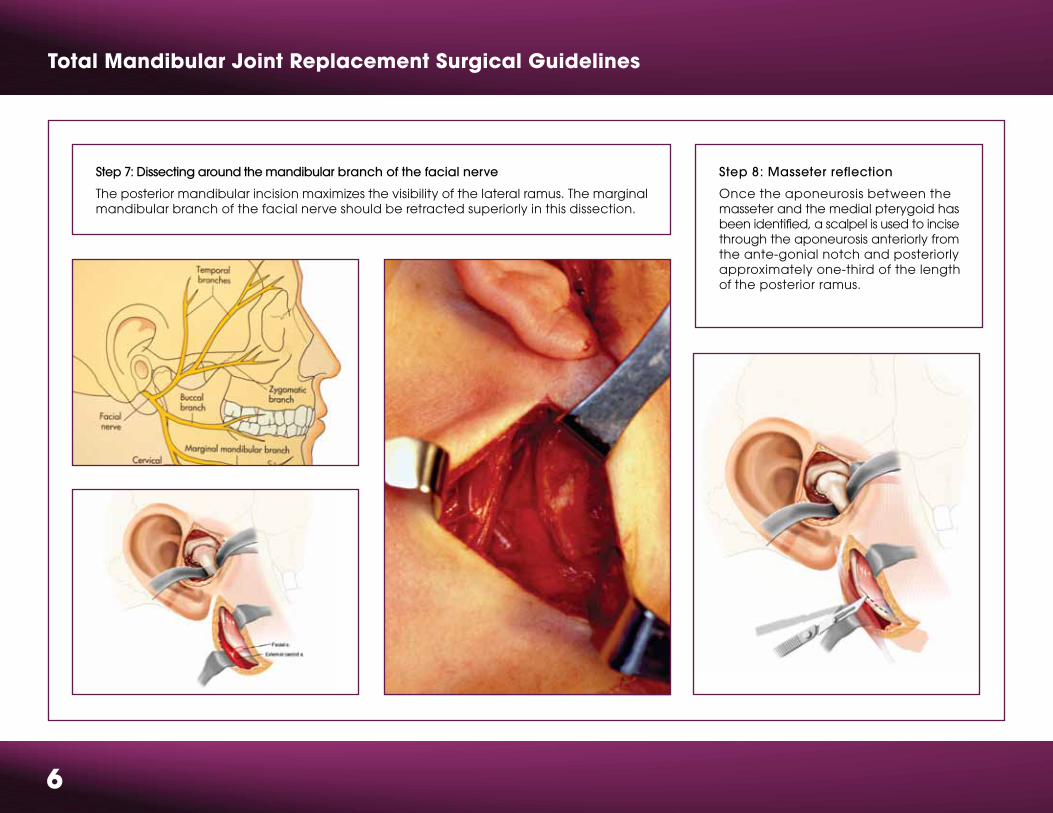
Step 8: Masseter reflection
Once the aponeurosis between the masseter and the medial pterygoid has been identified, a scalpel is used to incise through the aponeurosis anteriorly from the ante-gonial notch and posteriorly approximately one-third of the lengthof the posterior ramus.
Total Mandibular Joint Replacement Surgical Guidelines
6
Step 7: Dissecting around the mandibular branch of the facial nerve
The posterior mandibular incision maximizes the visibility of the lateral ramus. The marginal mandibular branch of the facial nerve should be retracted superiorly in this dissection.

Step 9: Ramal exposure
A periosteal elevator is used to cleanly strip the masseter muscleat its aponeurotic insertion to avoid incising muscle fibers and causing undue hemorrhage.
Step 10: Begin osteotomy with 1mm fissure burr
Once the lateral ramus is stripped from its masseteric insertion and temporalis insertion, if necessary, attention is directed back to the endaural incision for the first phase of the two-step osteotomy.A 1mm fissure bur is used to perform a standard condylectomywith appropriate protection from the condylar retractor
7
Peter D. Quinn, DMD, MD
Total Mandibular Joint Replacement Surgical Guidelines
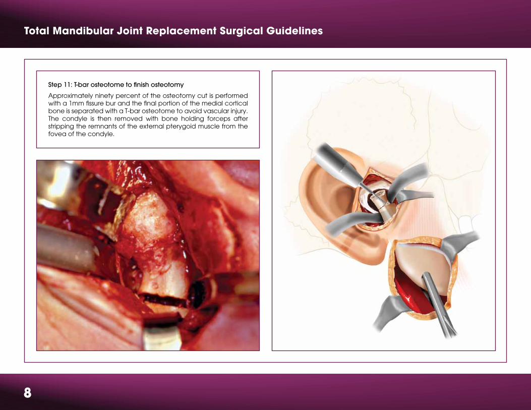
Step 11: T-bar osteotome to finish osteotomy
Approximately ninety percent of the osteotomy cut is performed with a 1mm fissure bur and the final portion of the medial cortical bone is separated with a T-bar osteotome to avoid vascular injury. The condyle is then removed with bone holding forceps after stripping the remnants of the external pterygoid muscle from the fovea of the condyle.
8

9
Peter D. Quinn, DMD, MD
Step 12: Two-step osteotomy - Phase II
Specially designed bone holding forceps are used to maintain asecure purchase point on the inferior border of the mandible. Theramus is then pushed superiorly up into the space created by stepone of the two-step osteotomy. This now allows better visualizationof the lower portion of the condylar neck and superior ramus for performance of this second step osteotomy. Approximately5 to 7mm of additional bone is now removed so the osteotomycut is actually below the lowest point of the sigmoid notch. It is important to remove adequate bone to allow for the thickness of the polyethylene fossa implant. If adequate bone is not removed,then a superior portion of the condyle-ramus may impinge on the fossa prosthesis when the patient is placed in intermaxillary fixation. Again, the retractors are used to protect the medial tissues duringthe osteotomy.

Total Mandibular Joint Replacement Surgical Guidelines
Step 13: Reciprocating rasp flattens fossa eminence
A coarse diamond reciprocating rasp has been designed to flatten the articular eminence to allow “tri-pod” stability of the fossa implant againstthe base of the skull. The same rasp can also be used to uniformly level the lateral ramus, especially in patients who have had a history of bruxism, where there may be a sharply defined ridge of cortical bone along the inferior border where the hypertrophic masseter muscle had attached.
10

11
Peter D. Quinn, DMD, MD
Step 14: Sizing and Implantation of Fossa Component
With the use of the fossa templates, the appropriate size of thefossa implant is selected (small, medium, large). It is important to make sure that the fossa is approximately parallel to the Frankfurt horizontal line. “Tipping” the fossa anteriorly at an excessive angle could potentially lead to displacement of the condylar prosthesis during function. It is recommended to place the fossa prosthesis with two screws initially to make sure the mating between thefossa prosthesis and the condylar prosthesis is optimal before placing the recommended four screws.

Total Mandibular Joint Replacement Surgical Guidelines
Step 15: Inter-maxillary fixation
Prior to the extra-oral incisions, Erich arch bars or Ivy loops wereplaced to allow for intermaxillary fixation in the desired occlusionat this stage of the operation. Once the fossa prosthesis is placed,the wound should be covered with antibiotic-soaked sterilesponges, and the surgeon and assistant now enter the oral cavityto place the patient in rigid maxillary fixation. This is done with aseparate set of instruments specifically for the intra-oral procedure.After the occlusion is secured, the surgical team should changetheir gowns and gloves to return to the extra-oral surgicaldissections.
12
Step 16: Fitting and Placement of the Mandibular Component
With the patient in intermaxillary fixation, a condylar prosthesisis now selected using the 45, 50 and 55mm templates. Eitherthe narrow or standard design can be used depending onthe adequacy of bone and defects from previous surgeries.As illustrated on the following page (Figure 16d), it is extremely important to position the head of the condyle as far posteriorly as possible so that there can be some degree of“pseudo-translation” of the condylar head in the fossa asthe patient opens to the expected range of 32 to 35mm. Positioning the condyle too far anteriorly in the closed position, as shown in the right portion of the illustration, could lead to dislocation of the condyle anterior of the fossa. Also note that the fossa is parallel to the Frankfurt horizontal plane and is not tipped anteriorly or superiorly.
Fig. 16a

13
Peter D. Quinn, DMD, MD
Fig. 16b
Fig. 16c Fig. 16d
Total Mandibular Joint Replacement Surgical Guidelines
14
Warnings and Precautions for use of the Total Mandibular Joint Replacement
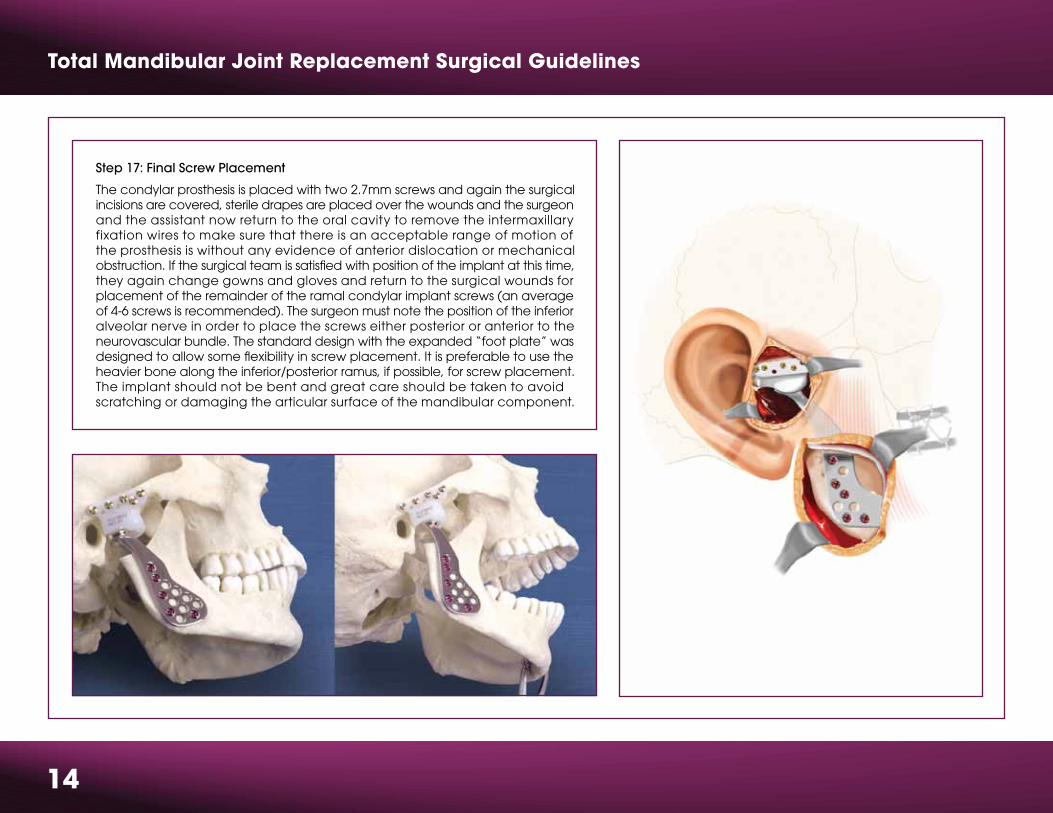
Step 17: Final Screw Placement
The condylar prosthesis is placed with two 2.7mm screws and again the surgical incisions are covered, sterile drapes are placed over the wounds and the surgeonand the assistant now return to the oral cavity to remove the intermaxillaryfixation wires to make sure that there is an acceptable range of motion ofthe prosthesis is without any evidence of anterior dislocation or mechanical obstruction. If the surgical team is satisfied with position of the implant at this time,they again change gowns and gloves and return to the surgical wounds for placement of the remainder of the ramal condylar implant screws (an averageof 4-6 screws is recommended). The surgeon must note the position of the inferior alveolar nerve in order to place the screws either posterior or anterior to the neurovascular bundle. The standard design with the expanded “foot plate” was designed to allow some flexibility in screw placement. It is preferable to use the heavier bone along the inferior/posterior ramus, if possible, for screw placement.The implant should not be bent and great care should be taken to avoidscratching or damaging the articular surface of the mandibular component.

15
Warnings and Precautions for use of the Total Mandibular Joint Replacement
Caution
Federal Law (USA) restricts this device to sale, distribution, or use, by or on the order of a physician.
Description
The Total Temporomandibular Joint (TMJ) Replacement System is implanted in the jaw to functionally reconstruct a diseased and/or damaged temporomandibular joint.
The Total TMJ Replacement System is a two-component system comprised of mandibular condyle and glenoid fossa components.
Both components are available in multiple sizes as right and left side specific designs and are attached to the bone by screws. Included in the system are trials, instruments and instrument cases.
Materials
• Mandibular Component—Cobalt-Chromium-Molybdenum (Co-Cr-Mo) alloy with titanium alloy coating or Titanium (Ti-6Al-4V) alloy with titanium alloy coating
• Fossa Component—ultra high molecular weight polyethylene (UHMWPE)
• Screws—titanium alloy
• Trials: Mandibular—aluminum, Fossa—Radel® plastic• Instruments: TMJ flat diamond rasp, TMJ diamond burrs, TMJ double-ended drill guide, retractors—stainless steel
• Instrument Case—stainless steel, silicone, Radel® plastic
Indications
The Total Temporomandibular Joint Replacement System is indicated for reconstruction of the temporomandibular joint. The reconstruction is necessary due to one of the following diagnoses:
• Arthritic conditions: osteoarthritis, traumatic arthritis, rheumatoid arthritis
• Ankylosis including but not limited to recurrent ankylosis with excessive heterotopic
bone formation
• Revision procedures where other treatments have failed (e.g. alloplastic
reconstruction, autogenous grafts)
• Avascular necrosis
• Multiply operated joints
• Fracture
• Functional deformity
• Benign neoplasms
• Malignancy (e.g. post-tumor excision)
• Degenerated or resorbed joints with severe anatomic discrepancies
• Developmental abnormality
Contraindications
• Active or chronic infection.
• Patient conditions where there is insufficient quantity or quality of bone to support the components.
Adverse Events
Adverse events that may occur following placement of the Total TMJ Replacement System are listed below. See Tables 7 and 8 located in the insert # 01-50-1000for more detailed information on adverse events from the clinical trial.
• Removal of components(s) including, but not limited to the following: - implant changes caused by loading and/or wear - degenerative changes within the joint surfaces from disease or previous implants - implant materials producing particles or corroding
• Loosening or displacement with or without removal of the implant
• Infection (systemic or superficial)
• Foreign body or allergic reaction to implant components
• Fossa wear through
• Facial swelling and/or pain
• Facial nerve dysfunction
• Excision of tissue
• Heterotopic bone formation
• Neuroma formation
• Ear problems
• Dislocation
Patient Counseling Information
• Discussion of the following points is recommended prior to surgery.
• The importance of prompt medical attention if they experience unusual swelling in the area of the implant.
• The risks associated with a Total TMJ System (see Warnings and Adverse Events).
• Post-operative pain relief and return of function varies from patient to patient.
• Additional treatment may be required including but not limited to extended physical therapy, bite splint, dental braces, and/or orthognathic and reconstructive surgery.
Sterility
The Total Temporomandibular Joint Replacement System mandibular and fossa components are sterilized by exposure to a minimum of 25 kGy of gammairradiation. DO NOT RESTERILIZE. Screws, trials, and the TMJ Instrument Case containing instruments are supplied non-sterile and should be wrapped with an FDA cleared sterilization wrap prior to steam sterilization in order to maintain sterility.
• Systemic disease with increased susceptibility to infection.
• Patients with extensive perforations in the mandibular fossa and/or bony deficiencies in the articular eminence or zygomatic arch that would severely compromise support for the artificial fossa component.
• Partial TMJ joint reconstruction.
• Known allergic reaction to any materials used in the components. NOTE: Patients with known or suspected nickel sensitivity should not have Co-Cr-Mo devices implanted since this material contains nickel.
• Patients with mental or neurological conditions who are unwilling or unable to follow postoperative care instructions.
• Skeletally immature patients.
• Patients with severe hyper-functional habits (e.g. clenching, grinding etc.)
• Patients with a foreign body reaction due to previous implants.
Warnings
• Mandibular and fossa components are provided STERILE. DO NOT RESTERILIZE.
• Screws, trials, instruments and instrument cases are provided NON-STERILE. CLEAN AND STERILIZE BEFORE USE.
• DO NOT USE if there is a loss of sterility of the devices.
• DO NOT USE damaged implants and only use implants that are packaged in unopened or undamaged containers.
• DO NOT USE the individual components of this total system (e.g. mandibular components, fossa components, or screws) for partial joint reconstruction. Bone cement or other grouting agents should not be used when implanting these devices. Safety and efficacy have not been established for the use of bone cement or other grouting agents with these implants.
• DO NOT USE IN CHILDREN. The Total TMJ Replacement was designed for skeletally mature patients.
Precautions
The device is limited to surgeons who are adequately trained in the use of the device through hands-on and educational course work. In all cases sound medical practiceis to be followed and the surgeon must select the type of device appropriate for treatment. The patient is to be warned that the system does not replace normal healthy bone in their TMJ and they may continue to have chronic pain and limited range of motion. The system can break or loosen as a result of stress, activity, or trauma. Patients with severe hyper-functional habits may have an undesirable outcome. The presence of existing mandibular and/or zygomatic arch screws or screw holes may compromise fixation. Note that placement of the implant in one joint onlymay result in harmful effects to the joint on the opposite side. Placement of the implantmay produce an improper relationship between teeth surfaces that should contact during biting. The patient is to be made aware of surgical risks and possible adverse effects prior to surgery and warned that failure to follow postoperative care instructionscan cause failure of the implant and the treatment. Specialized instruments/trials are designed for use with the Total TMJ Replacement System to aid in the accurate implantation of the components. DO NOT USE trials/instruments or cases that are disfigured, cracked, corroded, or otherwise damaged. Instruments/trials are subject towear with normal usage and are susceptible to fracture when exposed to extensiveuse or excessive force. All trials/instruments and cases should be regularlyinspected for wear or disfigurement. These should be disposed of appropriately.

r01k0911 BMF00-2185
This brochure is presented to demonstrate the technique and post-operative care regimen of Peter Quinn, MD. As the manufacturer of this device, Biomet Microfixation does not practice medicine and does not recommend this product or surgical technique for use on a specific patient. The surgeon who performs any implant procedure must determine the appropriate device and surgical procedure for each individual patient. Devices shown in this brochure may not be cleared or licensed for use or sale in your individual country.
Please contact your local distributor for information regarding availability of this product. Information contained in this brochure is intended for surgeon or distributor information only and is not intended for patient distribution. All surgeries carry risks.For additional information, please see package insert, or visit our web site at www.biometmicrofixation.com or call 1-800-874-7711.