total curve™ herbal breast enhancement pills · 2020-05-11 · natural breast curve. over the...
TRANSCRIPT

We imagine for you...
Volufi line™

VOLUFILINETM
Patent filed
For a progressively shapelier bust
All use of this file for commercial or advertising purposes requires the prior written authorization of the SEDERMA Company
Non- Warranty: The information in this publication is given in good faith by Sederma by way of general guidance and for reference purposes only. The information should not be construed as granting a license to practice any methods or compositions of matter covered by patents. Sederma assumes no liability in relation to the information and gives no warranty regarding the suitability of the product and/or ingredients described for a particular use. The use made of the product and/or ingredients described by the recipient and any claims or representations made by the recipient in respect of such product and/or ingredients are the responsibility of the recipient. The recipient is solely responsible for ensuring that products marketed to consumers comply with all relevant laws and regulations.

VOLUFILINETM
VOLUFILINETM
SYNOPSIS
Description Sarsasapogenin as the active substance promoting the shapeliness of the décolleté
INCI name Hydrogenated Polyisobutene (and) Anemarrhena Asphodeloides Root Extract Demonstrated cosmetic activity
In vitro: Effects of sarsasapogenin on:
The proliferation of adipocytes in the differentiation phase: o 20.5% increase with a concentration equivalent to 0.5% VOLUFILINE™ o 35.2% increase with a concentration equivalent to 2% VOLUFILINE™
The differentiation of pre-adipocytes as determined by G3PDH activity:
o 103% increase with a concentration equivalent to 0.5% VOLUFILINE™ o 535% increase with a concentration equivalent to 1.75% VOLUFILINE™
Lipid uptake in cultured adipocytes 3T3-L1 and human adipocytes:
o For cultured adipocytes 3T3-L1: 492% increase with a concentration equivalent to 1.75% VOLUFILINE™
o For human adipocytes: 641% increase with a concentration equivalent to 1% VOLUFILINE™
Adipocyte volume: o 26-fold increase in cell volume with 1.5% VOLUFILINE™.
In vivo:
56-day clinical trial on 28 volunteers who applied a cream gel containing 5% VOLUFILINE™ to one breast twice daily. Each subject acted as her own control (one untreated breast). Application was randomized. Measurement of the remodeling effect using the FOITS method:
1.4% increase in the volume of the 5% VOLUFILINE™-treated breast zone over 28 days; up to a 2.2% increase at T56 days For the best responders: 6.4% increase at 28 days and 8.4% increase at 56 days
Recommendations for use: - soluble in an oil phase - withstands temperatures of up to 80°C for 4h - no particular recommendation with regard to pH Recommended concentration for use: 5%
Toxicology (as per the UNITIS Charter): Patch-test on humans (10 volunteers)
HET CAM RIPT on 100 volunteers Ames' test
Total Curve

VOLUFILINETM
Restriction on use of the product Sederma has filed a patent covering the use of sarsasapogenin (VOLUFILINE™) in the stimulation of lipogenesis. This application has never been described before and is thus patentable. To Sederma's knowledge, L’Oréal holds several patents on sapogenins, including a set of patents (European patent No. 1 092 422, US patent No. 6,294, 157, US patent No. 6,528,043, JP patent No. 3646148) concerning a sapogenin solubilization system. In consequence, in order to prevent any risk of infringement of industrial property rights in Europe, Japan and the United States, Sederma advises its customers not to include VOLUFILINE™ in formulations containing: The following triple combinations:
- ester(s)* + branched fatty alcohol(s) ** + VOLUFILINE™ and
- ester(s)* + lipophilic UV filter(s) + VOLUFILINE™ are also to be avoided since the triple combinations have been patented by L’Oréal. All esters including plant oils can be used in combination with VOLUFILINE™ providing that they are not formulated with BRANCHED FATTY ALCOHOL(S) (e.g. Guerbet alcohol). The same applies to lipophilic sun filters. (*) The term “ester(s)” refers to non-emulsifying esters including plant oils, and
fatty alcohols and acids whose hydrocarbon chain contains at least 8 carbon atoms, such as Octyl Palmitate, C10/C12 Caprylyl/Capric Triglycerides, etc.
(**) The term “branched fatty alcohol(s)” refers to, for example, Guerbet alcohol
such as Eutanol™ G (Cognis) and similar substances.

VOLUFILINETM
CONTENTS 1. INTRODUCTION p. 1 to 6/31 2. EFFICACY TESTS
2.1. In vitro studies p. 7 to 21/31 2.2. In vivo studies p. 22 to 27/31
3. CONCLUSION p. 28 to 29/31 REFERENCES p. 30/31 APPENDIX p. 31/31
04/2007/V3

VOLUFILINETM
1/31 1. INTRODUCTION Sung by the poets or delicately disclosed by painters, the subject of dreams or temptation, the shapeliness of the breasts is the alpha and omega of femininity. Natural Breast curve.Over the ages, the criteria of feminine seductiveness have markedly changed: the nurturing function of the breasts was essential in the Middle Ages and, at that time, the ideal woman had a small, high bosom. Women with a larger bosom strapped up their breasts. It was only after the 13th century that the bust was revealed and the seductiveness of the female body was more strongly expressed. Today, sensuality predominates and the image of female beauty includes a generous bosom partially revealed and deploying its erotic potential. Women nonetheless remain very sensitive to the ambivalence - nurturing breasts/seductive breasts - and for many women it is important to care of their bosom with an appropriate lifestyle. It has also become essential to take care of the appearance of the bust in order to feel confident in oneself and one's wellbeing.
VOLUFILINETM
2/31 ANATOMY AND PHYSIOLOGY OF THE BREASTS From birth to puberty, there is no difference between the male and female mammary gland. Subsequently, the combination of the hormones estrogen and progesterone induce profound changes in the breasts of pubertal girls and the breasts undergo a complete transformation in 3 to 4 years. At the end of that stage, the breast consists in lactiferous ducts associated with lobes that are separated by connective and adipose tissue. The hormone-sensitive connective and glandular lobes develop in the subcutaneous fat which they invade and raise: the mammary gland thus consists of lobes separated by fatty tissue and maintained by the cutaneous overlay. The volume and shape of the breasts are thus determined by the quantities of glandular and adipose tissue that they contain. Thus, generally between the age of 30 and 50 years, the breast consists in adipose tissue, which constitutes one half of its volume, and mammary gland, which constitutes the other half The space between the skin and the mammary structure is very homogeneous and fat-filled, divided by the “milk line” or mammary ridge which maintains the skin and gland together .http://www.med.univ-rennes1.fr The subcutaneous fat is outside of the glandular lobes (and the interlobular fat) and its thickness varies.
VOLUFILINETM
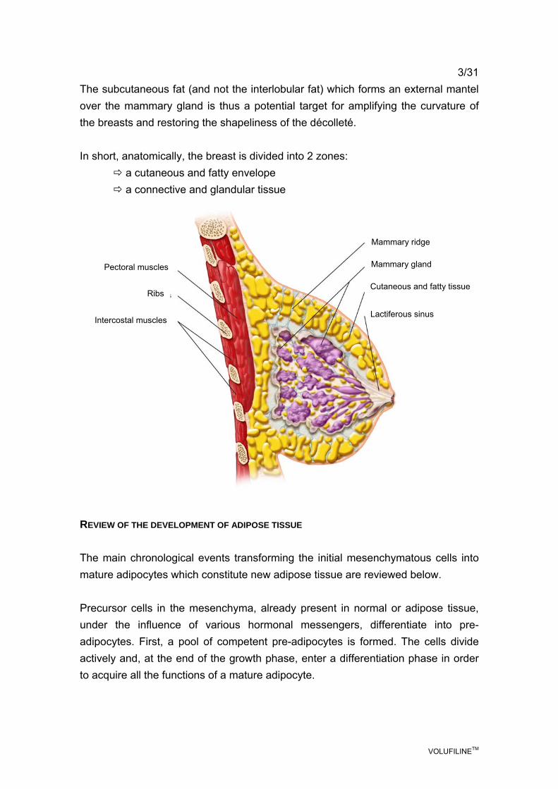
3/31 The subcutaneous fat (and not the interlobular fat) which forms an external mantel over the mammary gland is thus a potential target for amplifying the curvature of the breasts and restoring the shapeliness of the décolleté. In short, anatomically, the breast is divided into 2 zones:
a cutaneous and fatty envelope a connective and glandular tissue
REVIEW OF THE DEVELOPMENT OF ADIPOSE TISSUE The main chronological events transforming the initial mesenchymatous cells into mature adipocytes which constitute new adipose tissue are reviewed below. Precursor cells in the mesenchyma, already present in normal or adipose tissue, under the influence of various hormonal messengers, differentiate into pre-adipocytes. First, a pool of competent pre-adipocytes is formed. The cells divide actively and, at the end of the growth phase, enter a differentiation phase in order to acquire all the functions of a mature adipocyte.
Mammary ridge
Mammary gland
Cutaneous and fatty tissue
Lactiferous sinus
Pectoral muscles
Ribs
Intercostal muscles

VOLUFILINETM
4/31 The arrest of cell division and the triggering of differentiation is accompanied by profound changes in the activity of several genes, in particular: activation of the transcription genes promoting lipogenesis (of which: pref-1, PPARγ, C/EBPα, lipogenesis enzymes) (GREGOIRE, 1998; ROSEN, 2000), activation of the genes involved in lipid metabolism (acetyl-CoA, GDPH, GAPDH, FAS). The change enables the adipocytes to assume their mature phenotype. At that late stage of differentiation, the adipocytes express various receptors, including the insulin-sensitivity receptor and adrenergic receptors. At that stage, proteins specialized in the formation and transport of lipid droplets (adipophilin, perilipin, vimentin) are activated (SHI, 2000) as is secretion of agents promoting the installation of fat mass (monobutyrin, angiogenic factors, extracellular matrix proteins). The various stages are summarized in the following diagram (GREGOIRE, 1998):
molecular event
stem cell
mesenchymatous precursor
pre-adipocyte
mature adipocyte
Pref-1
C/EBP β and δ
PPARγ
adipocyte genes
lipogenesis enzyme
secretion factor
others
modification of the extracellular matrix
and
remodeling of the cytoskeleton

VOLUFILINETM
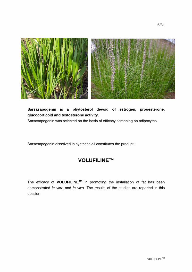
5/31 SUBCUTANEOUS FAT AND ESTHETIC FAT While interference in any way with the connective and glandular tissue is outside of the scope of cosmetology, it is possible to attempt to promote the installation of subcutaneous fat to enhance the appearance of the breasts. This was the strategy adopted by Sederma for its new active substance that promotes the shapeliness of the décolleté. SARSASAPOGENIN Sarsasapogenin is of plant origin and is abundantly present in certain plants of the Liliaceae family such as sarsaparilla (Smilax officinalis) or Asparagus officinalis. Decoctions of the rhizomes of those plants, which contain a mixture of sapogenins, are used for their anti-inflammatory and antioxidant virtues and to treat skin disorders such as leprosy, psoriasis and eczema. Sarsasapogenin: C27H44O3 Mr = 416.64
O
O
CH3
CH3 H
HHO
CH3
CH3
Sederma decided to extract and purify sarsasapogenin from the plant Anemarrhena asphodeloides (family, initially Liliaceae, but phylogenetically reclassified as Agavaceae in 2003). The Asian plant, known as “Zhi Mu”' in China, where it is in widespread use, is a perennial herbaceous plant reaching a height of 1 meter and flowering in the summer. The roots contain 6% of saponins. Rhizome decoctions are traditionally used as febrifuges, laxatives and antibacterials. Decoctions are also used as mouthwashes and alleviate gingival ulceration.
VOLUFILINETM
6/31 Sarsasapogenin is a phytosterol devoid of estrogen, progesterone, glucocorticoid and testosterone activity. Sarsasapogenin was selected on the basis of efficacy screening on adipocytes. Sarsasapogenin dissolved in synthetic oil constitutes the product:
VOLUFILINE™
The efficacy of VOLUFILINETM in promoting the installation of fat has been demonstrated in vitro and in vivo. The results of the studies are reported in this dossier.
VOLUFILINETM
7/31 2. EFFICACY TESTS 2.1. In vitro studies The studies of the ability of VOLUFILINE™ to promote fat installation were implemented in two phases: DNA array study of the activated genes in order to elucidate the action mechanism and studies on adipose cells to quantify product efficacy. 2.1.1. Effect of sarsasapogenin on gene expression profile
Gensodi study (November 2005 - February 2006) - SEDERMA exclusive Principle Two studies were conducted: one addressed the effect of sarsasapogenin on differentiation using a pre-adipocyte culture; the other addressed the effect of sarsasapogenin on mature adipocytes.
Protocol Human pre-adipocyte cells were inoculated and cultured (multiplication) for 5 days. Then:
- either the cells were exposed to the induction medium in the presence or absence of sarsasapogenin for 8 or 24h before culture arrest (study of the effect of sarsasapogenin on differentiation)
- or the cells were exposed to the differentiation medium for 10 days, yielding mature adipocytes, then exposed or not exposed to sarsasapogenin for 24h (study of the effect of sarsasapogenin on maturation).
At each time point, the mRNA of the cells was extracted using a commercially available kit (Roche Kit V6 - Extraction of total RNA). In parallel, solvent controls were run and subjected to extraction in the same manner. source: http://www.total-curve.net/

VOLUFILINETM
8/31 The expression profile of the gene sequences was determined using CodeLink Whole Genome chips for each sample of RNA purified. The values obtained were normalized on the controls.
Results
On the basis of the results obtained, sarsasapogenin is reported to stimulate adipocytic differentiation by various mechanisms:
Sarsasapogenin induces activation of the gene coding for protein MNT after 8 and 24h of differentiation (MAX binding protein). However, protein MNT in association with Max represses the activation induced by Myc-Max. The latter is known to inhibit adipocytic differentiation. The inhibitory effect on the inhibitor is reported to activate differentiation.
In addition, sarsasapogenin acts upstream of peroxisome proliferator-activated receptor gamma (PPARγ) activation and potentiates the function of that receptor.
Various genes associated with activation of PPARγ were observed to be markedly up-regulated: for example, genes COPS3 and COPS5 (>5) coding for the complex known as “COP9 signalosome” and cyclin D3, which, in association with CDK4, orients differentiation at the expense of proliferation and affords conditions propitious to phosphorylation of PPARγ.
Phosphorylated PPARγ forms a heterodimer with retinoid X receptor (RXR) which binds to the PPRE transcription response sequence and induces differentiation into adipocytes.
Induction at 8 and 24h
On mature adipocytes
Induction at 24h
Induction at 24h
Activation
Transcription activation
Binding
Interaction
Inhibition
Biological activity
Legend of the figures
On differentiating adipocytes Induction at 8h Induction at 24h
VOLUFILINETM
9/31
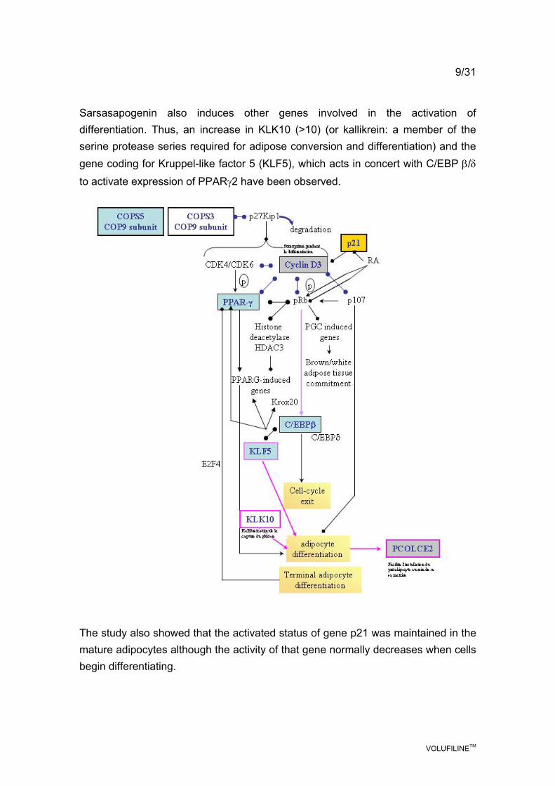
Sarsasapogenin also induces other genes involved in the activation of differentiation. Thus, an increase in KLK10 (>10) (or kallikrein: a member of the serine protease series required for adipose conversion and differentiation) and the gene coding for Kruppel-like factor 5 (KLF5), which acts in concert with C/EBP β/δ to activate expression of PPARγ2 have been observed.
The study also showed that the activated status of gene p21 was maintained in the mature adipocytes although the activity of that gene normally decreases when cells begin differentiating.
VOLUFILINETM
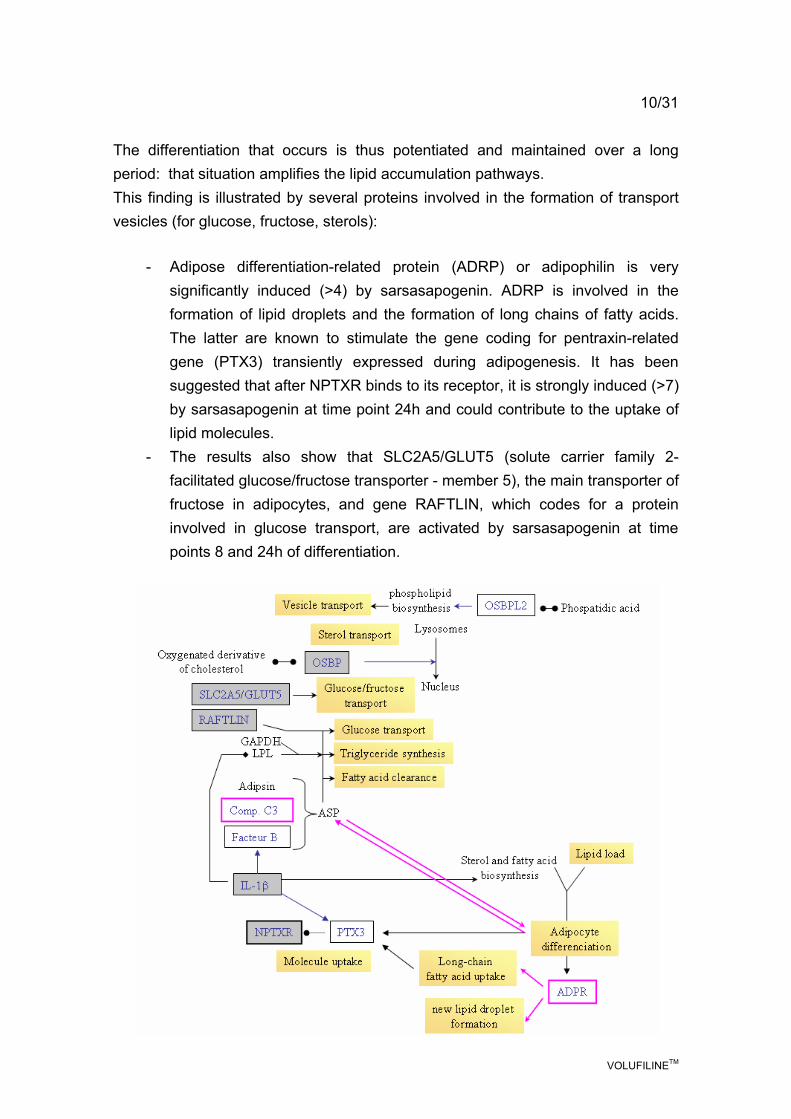
10/31 The differentiation that occurs is thus potentiated and maintained over a long period: that situation amplifies the lipid accumulation pathways. This finding is illustrated by several proteins involved in the formation of transport vesicles (for glucose, fructose, sterols):
- Adipose differentiation-related protein (ADRP) or adipophilin is very significantly induced (>4) by sarsasapogenin. ADRP is involved in the formation of lipid droplets and the formation of long chains of fatty acids. The latter are known to stimulate the gene coding for pentraxin-related gene (PTX3) transiently expressed during adipogenesis. It has been suggested that after NPTXR binds to its receptor, it is strongly induced (>7) by sarsasapogenin at time point 24h and could contribute to the uptake of lipid molecules.
- The results also show that SLC2A5/GLUT5 (solute carrier family 2-facilitated glucose/fructose transporter - member 5), the main transporter of fructose in adipocytes, and gene RAFTLIN, which codes for a protein involved in glucose transport, are activated by sarsasapogenin at time points 8 and 24h of differentiation.
VOLUFILINETM
11/31
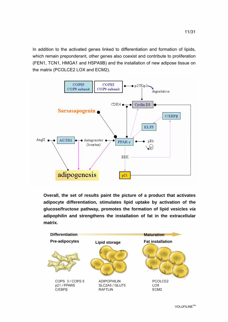
In addition to the activated genes linked to differentiation and formation of lipids, which remain preponderant, other genes also coexist and contribute to proliferation (FEN1, TCN1, HMGA1 and HSPA9B) and the installation of new adipose tissue on the matrix (PCOLCE2 LOX and ECM2).
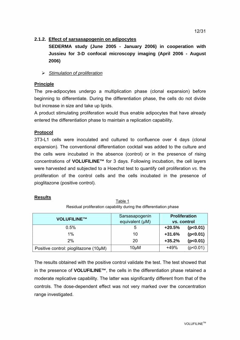
Overall, the set of results paint the picture of a product that activates adipocyte differentiation, stimulates lipid uptake by activation of the glucose/fructose pathway, promotes the formation of lipid vesicles via adipophilin and strengthens the installation of fat in the extracellular matrix.
Differentiation Pre-adipocytes Lipid storage
Maturation Fat installation
VOLUFILINETM
12/31
2.1.2. Effect of sarsasapogenin on adipocytes SEDERMA study (June 2005 - January 2006) in cooperation with Jussieu for 3-D confocal microscopy imaging (April 2006 - August 2006)
Stimulation of proliferation
Principle The pre-adipocytes undergo a multiplication phase (clonal expansion) before beginning to differentiate. During the differentiation phase, the cells do not divide but increase in size and take up lipids. A product stimulating proliferation would thus enable adipocytes that have already entered the differentiation phase to maintain a replication capability.
Protocol 3T3-L1 cells were inoculated and cultured to confluence over 4 days (clonal expansion). The conventional differentiation cocktail was added to the culture and the cells were incubated in the absence (control) or in the presence of rising concentrations of VOLUFILINE™ for 3 days. Following incubation, the cell layers were harvested and subjected to a Hoechst test to quantify cell proliferation vs. the proliferation of the control cells and the cells incubated in the presence of pioglitazone (positive control). Results
VOLUFILINE™ Sarsasapogenin equivalent (µM)
Proliferation vs. control
0.5% 5 +20.5% (p<0.01) 1% 10 +31.6% (p<0.01) 2% 20 +35.2% (p<0.01)
Positive control: pioglitazone (10µM) 10µM +49% (p<0.01)
The results obtained with the positive control validate the test. The test showed that
in the presence of VOLUFILINE™, the cells in the differentiation phase retained a
moderate replicative capability. The latter was significantly different from that of the
controls. The dose-dependent effect was not very marked over the concentration
range investigated.
Table 1 Residual proliferation capability during the differentiation phase
VOLUFILINETM
13/31
Conclusion
VOLUFILINE™ maintains a replicative capability (20 to 35% greater than that of the controls) in cells in the differentiation phase. The results corroborate the DNA array data: activation of genes contributing to proliferation (FEN1, TCN1, HMGA1 and HSPA9B).
Stimulation of differentiation
Principle
After an in vivo proliferation phase, the pre-adipocytes begin differentiating under the influence of specific endogenous factors (in particular, glucocorticoids and insulin). In vitro, those factors are included in the conventional cocktail used to trigger differentiation.
The maintenance or amplification of differentiation when the cells are subsequently cultured in a simple maintenance medium is determined by assay of glycerol-3-phosphate dehydrogenase, G3PDH (or GPDH). The enzyme is involved in energy metabolism. Its activity regularly increases through the differentiation process.
Protocol
As previously, 3T3-L1 cells were cultured to confluence, then underwent differentiation induction with the conventional cocktail and were incubated in the absence or presence of VOLUFILINE™ at various concentrations. After 3 days of incubation, the differentiation cocktail was replaced by culture maintenance medium in the presence or absence of VOLUFILINE™. After 3 days of incubation, the cell layers were harvested and G3PDH activity determined.

VOLUFILINETM
14/31
Results
VOLUFILINE™ Sarsasapogenin equivalent (µM)
Change in G3PDH activity vs. control (%)
Significance vs. control
0.5% 5 +103% (p<0.01) 1% 10 +201% (p<0.01)
1.5% 15 +429% (p<0.01) 1.75% 17.5 +535% (p<0.01)
Positive control: pioglitazone (10µM)
10µM +521% (p<0.01)
The assay was validated by comparison with the positive control, pioglitazone, with which a 521% increase in differentiation stimulation was observed.
Dose-dependent stimulation was obtained in the presence of sarsasapogenin: differentiation was increased 103% at a concentration equivalent to 0.5% VOLUFILINE™ and 535% for 1.75% VOLUFILINE™.
The cell data correlate with the DNA array results (maintenance of differentiation via gene p21 and cyclin-CDK4).
Table 2 Percentage change in G3PDH activity expressed relative to the control cells
not treated with sarsasapogenin. Mean for n=3 tests
Plot 1: Potentiation of differentiation in the presence of VOLUFILINE™
VOLUFILINE™
Cha
nge
in G
3PD
H a
ctiv
ity
vs. c
ontro
l
Positive control
0.5% 1% 1.5% 1.75%

VOLUFILINETM
15/31
Stimulation of lipid uptake by 3T3-L1 cells and human adipocytes
Principle
Adipocyte differentiation is a decisive stage in the transformation of pre-adipocytes into lipid-laden mature adipocytes. The intracellular accumulation of lipids is determined by conventional red oil staining (neutral lipid marker). The stained lipids can then be quantified under the microscope.
Protocol
3T3-L1 cells were inoculated and cultured for 4 days (multiplication). This was followed by a differentiation phase (incubation with the conventional differentiation cocktail) and a maturation phase (maturation cocktail) in the absence or presence of VOLUFILINE™ at various concentrations.
At the end of that period, the cells were washed, fixed and stained with red oil. The cell layers were digitally photographed and the red staining quantified by image analysis.
The percentage red oil areas, shown in table 1, were expressed relative to the untreated control cells and the test was validated by comparison with pioglitazone (10µM), the positive control for differentiation stimulation.
Results
VOLUFILINE™ Sarsasapogenin equivalent (µM)
% change in red oil content vs. control
Significance vs. control
0.5% 5 +135% (p<0.01) 1% 10 +274% (p<0.01)
1.75% 17.5 +492% (p<0.01) Positive control: pioglitazone (10µM)
10µM +359% (p<0.01)
As expected, an increase in triglycerides was observed in the presence of
pioglitazone.
Table 3 Change in the quantity of lipids accumulated in 3T3-L1 cells
in the presence of VOLUFILINE™. Mean values for n=3 tests

VOLUFILINETM
16/31
The cells incubated in the presence of VOLUFILINE™ showed an increase in dose-dependent lipid incorporation that was greater than 492% vs. the control cells, for a VOLUFILINE™ concentration of 1.75%.
The results were reproduced using human adipocytes:
Following culture and confluence, the human adipocytes were incubated in the differentiation maintenance medium in the absence (control) or presence of VOLUFILINE™ at various concentrations for 6 days.
At the end of that period, the cells were washed, fixed and stained with red oil. The cell layers were digitally photographed and the red staining quantified by image analysis.
VOLUFILINE™ Sarsasapogenin equivalent (µM)
% change in red oil content vs. control
Significance vs. control
0.5% 5 +229% (p<0.01) 1% 10 +641% (p<0.01)
Table 4 Change in the quantity of lipids accumulated in human adipocytes
in the presence of VOLUFILINE™
VOLUFILINE™
Plot 2
Cha
nge
in re
d oi
l con
tent
vs.
con
trol
(%)
VOLUFILINE™
0.5% 1%

VOLUFILINETM
17/31
Photographs of human adipocytes:
Figure a: control
Figure b: 0.5%VOLUFILINE™
Figure c: 1% VOLUFILINE™
VOLUFILINETM
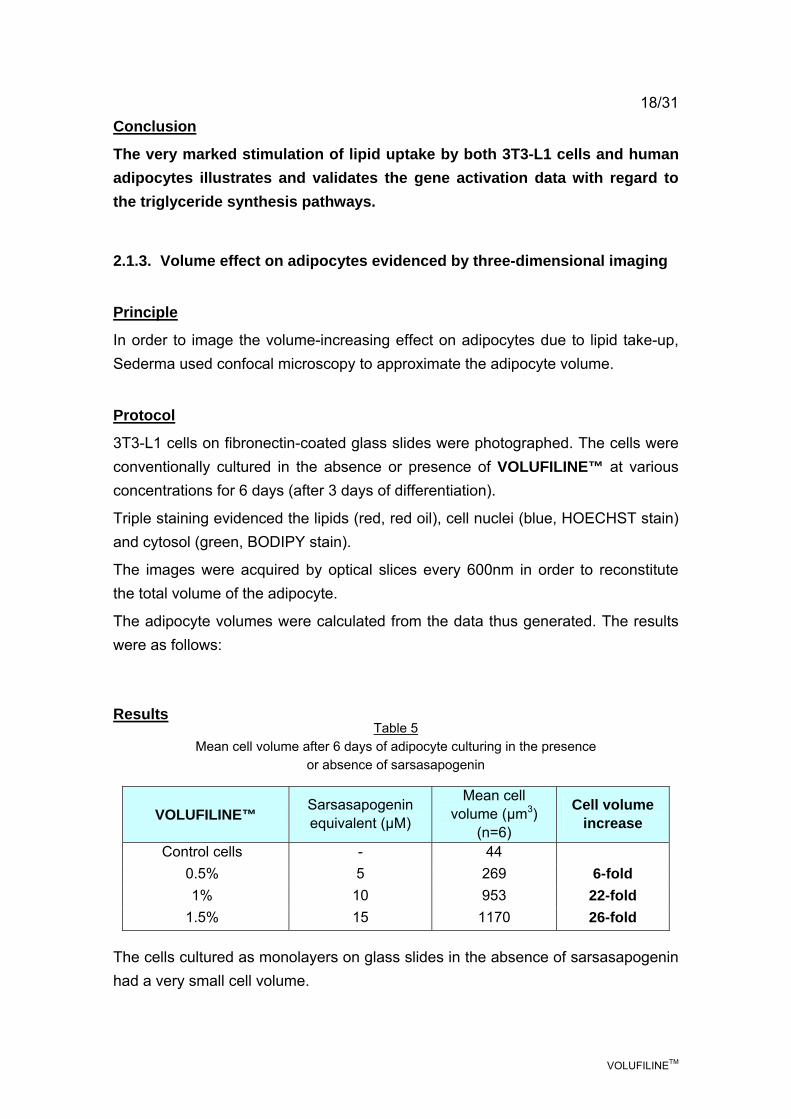
18/31 Conclusion
The very marked stimulation of lipid uptake by both 3T3-L1 cells and human adipocytes illustrates and validates the gene activation data with regard to the triglyceride synthesis pathways.
2.1.3. Volume effect on adipocytes evidenced by three-dimensional imaging
Principle
In order to image the volume-increasing effect on adipocytes due to lipid take-up, Sederma used confocal microscopy to approximate the adipocyte volume.
Protocol
3T3-L1 cells on fibronectin-coated glass slides were photographed. The cells were conventionally cultured in the absence or presence of VOLUFILINE™ at various concentrations for 6 days (after 3 days of differentiation).
Triple staining evidenced the lipids (red, red oil), cell nuclei (blue, HOECHST stain) and cytosol (green, BODIPY stain).
The images were acquired by optical slices every 600nm in order to reconstitute the total volume of the adipocyte.
The adipocyte volumes were calculated from the data thus generated. The results were as follows: Results
VOLUFILINE™ Sarsasapogenin equivalent (µM)
Mean cell volume (µm3)
(n=6)
Cell volume increase
Control cells - 44 0.5% 5 269 6-fold 1% 10 953 22-fold
1.5% 15 1170 26-fold The cells cultured as monolayers on glass slides in the absence of sarsasapogenin had a very small cell volume.
Table 5 Mean cell volume after 6 days of adipocyte culturing in the presence
or absence of sarsasapogenin
VOLUFILINETM
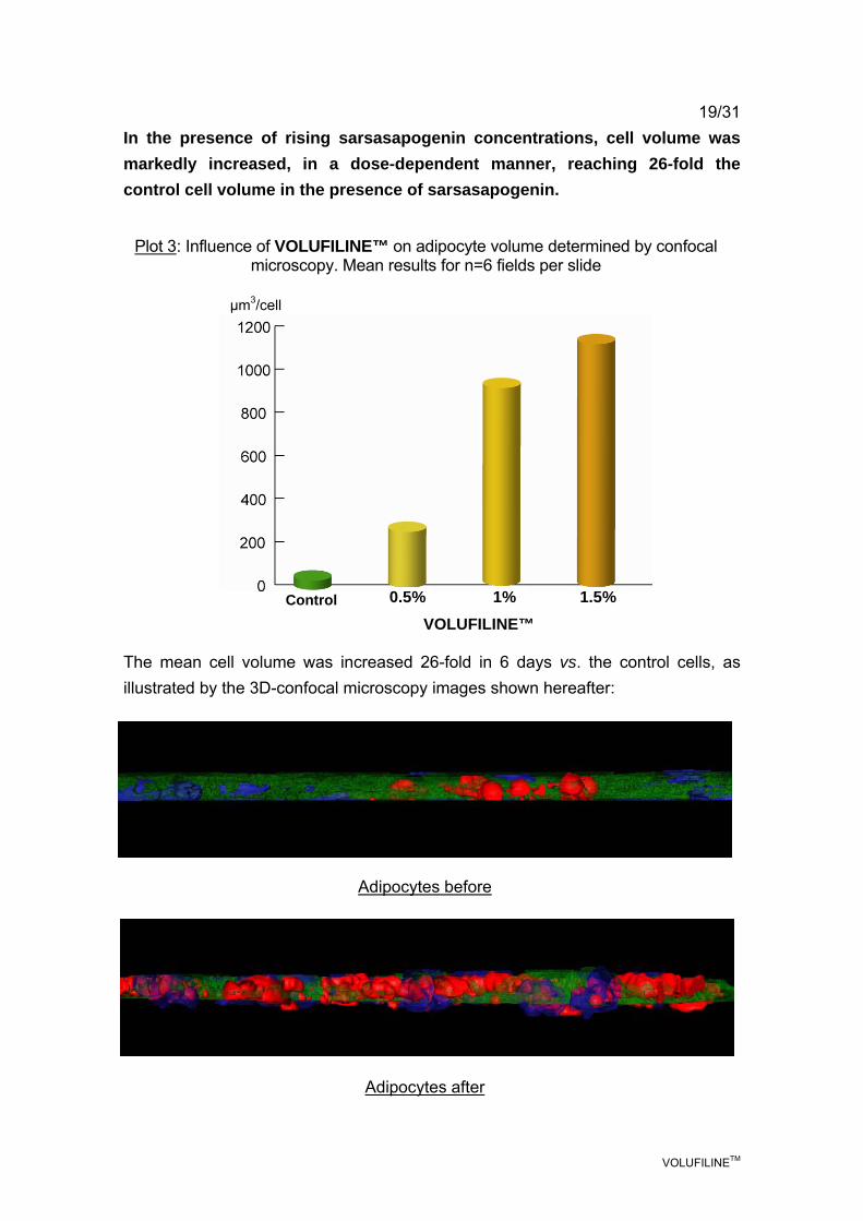
19/31 In the presence of rising sarsasapogenin concentrations, cell volume was markedly increased, in a dose-dependent manner, reaching 26-fold the control cell volume in the presence of sarsasapogenin.
The mean cell volume was increased 26-fold in 6 days vs. the control cells, as illustrated by the 3D-confocal microscopy images shown hereafter:
Plot 3: Influence of VOLUFILINE™ on adipocyte volume determined by confocal microscopy. Mean results for n=6 fields per slide
Adipocytes before
Adipocytes after
VOLUFILINE™
µm3/cell
Control 0.5% 1% 1.5%

VOLUFILINETM
20/31 Conclusion of the in vitro studies
The set of results obtained show complete consistency between the DNA array activated-gene profile and the cell effects observed.
VOLUFILINE™ maintained a moderate proliferative activity throughout the differentiation phase: 20 to 35%.
VOLUFILINE™ amplified differentiation (430% increase) and maintained it, activating lipid accumulation (640% increase).
Those effects result in a volume-enhancing effect on adipose tissue, which was demonstrated by confocal microscopy.
Before After

VOLUFILINETM
21/31 2.2. In vivo studies SPINCONTROL study (March 2006 - May 2006) Principle The volume study was conducted using interference-fringe topometry consisting in analysis of the optical deformation of the fringes projected on the skin surface under laser illumination (FOITS method).
Protocol For this study in order to ensure the most precise repositioning possible, it was necessary to develop special equipment consisting in:
- a horizontal breast support system (identical to that used for breast radiography) the height of which was precisely determined relative to the seat
- a back-rest system situated at a constant distance from the breast-rest. The system is shown below:
VOLUFILINETM
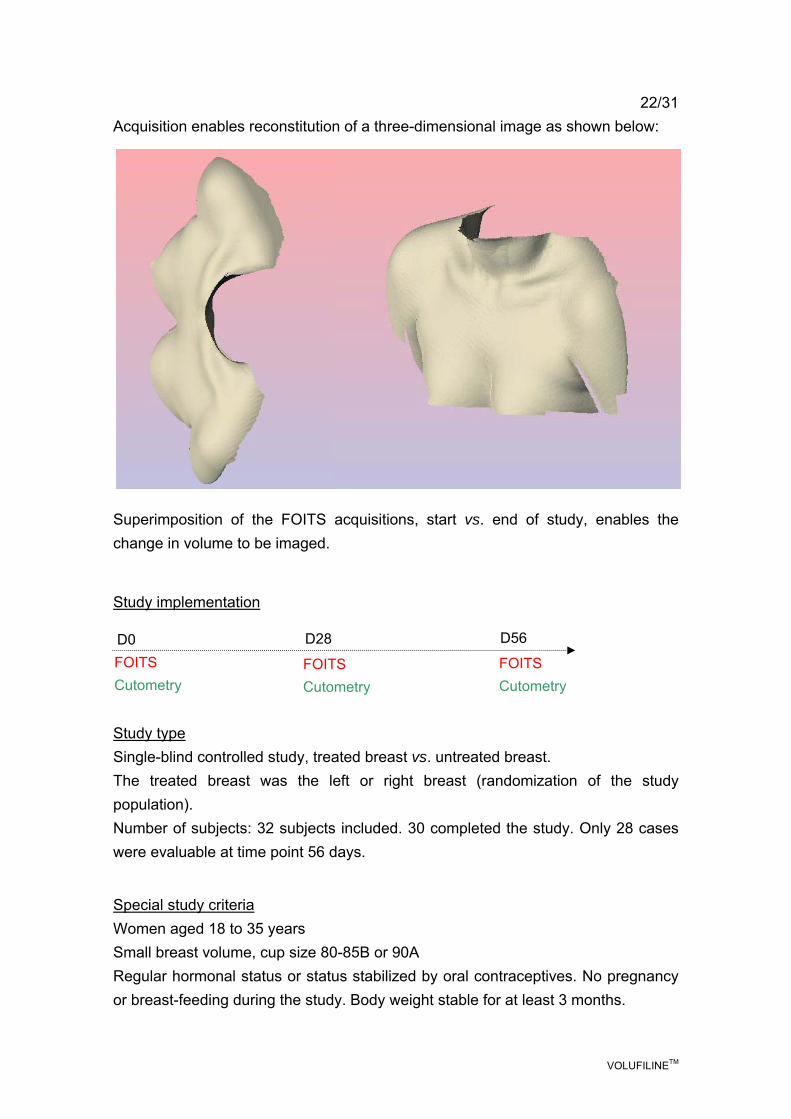
22/31 Acquisition enables reconstitution of a three-dimensional image as shown below: Superimposition of the FOITS acquisitions, start vs. end of study, enables the change in volume to be imaged. Study implementation
Study type Single-blind controlled study, treated breast vs. untreated breast. The treated breast was the left or right breast (randomization of the study population). Number of subjects: 32 subjects included. 30 completed the study. Only 28 cases were evaluable at time point 56 days.
Special study criteria Women aged 18 to 35 years Small breast volume, cup size 80-85B or 90A Regular hormonal status or status stabilized by oral contraceptives. No pregnancy or breast-feeding during the study. Body weight stable for at least 3 months.
FOITS Cutometry
D0 D28
FOITS Cutometry
FOITS Cutometry
D56

VOLUFILINETM
23/31 Product application The product was a cream gel containing 5% VOLUFILINE™ (Appendix 1), which was applied twice daily for 56 days.
Results Safety The product was well tolerated by all the volunteers.
Volume by FOITS
Out of the 32 volunteers included in the study, only the data for n=30 subjects at time point 28 days and n=28 subjects at time point 56 days were evaluable.
The breast volume determined by FOITS was that of the superior part of the breast (décolleté zone), the lower part being excluded by the positioning system.
In order to facilitate result reading, the volumes at T0 were normalized on a reference value of 5,000 mm3. Depending on breast size, the raw data ranged from 4,000 to 10,000 mm3.
The raw datum determined on T28 or 56 thus became 5,000 + the absolute value
of the difference vs. T0.
Treated side
Volume (mm3) Untreated side Volume (mm3)
T0 5000 5000 T28 days 5070 4997 Significance p=0.3 p=0.95 T56 days 5108 5046 Significance p=0.10 p=0.52 Best responders at T56 (8 subjects=1st quartile)
5330 4900
For the control breast, the change in volume was practically null after 28 days and the very slight variation observed at 56 days was of very little significance (p=0.52). For the treated breast, an increase, although not very significant, was observed at T28 (p=0.3). The increase was confirmed at T56 days with 90% certainty (p=0.1).
Table 6 Change in volume of the superior part of the breast following application
of 5% VOLUFILINE™ for 56 days
VOLUFILINETM
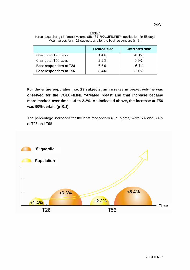
24/31
Treated side Untreated side
Change at T28 days 1.4% -0.1% Change at T56 days 2.2% 0.9% Best responders at T28 6.6% -6.4% Best responders at T56 8.4% -2.0%
For the entire population, i.e. 28 subjects, an increase in breast volume was observed for the VOLUFILINE™-treated breast and that increase became more marked over time: 1.4 to 2.2%. As indicated above, the increase at T56 was 90% certain (p=0.1). The percentage increases for the best responders (8 subjects) were 5.6 and 8.4% at T28 and T56.
Table 7 Percentage change in breast volume after 5% VOLUFILINE™ application for 56 days
Mean values for n=28 subjects and for the best responders (n=8).
1st quartile
Population
Time +1.4%
+6.6%
+2.2%
+8.4%

VOLUFILINETM
25/31 Conclusion While the increase in breast volume was moderate: on average 2.2% at time point 56 days for the 28 subjects, they were nonetheless significant at the p=0.1 level. This trend toward an increase, which became more marked over time, is a second indication that concords with the results obtained and suggests that the long-term results may be even better. An example of the FOITS analyses is given below:
T0 T56
Before After: treated left breast

VOLUFILINETM
26/31
An example of the superimposed images acquired at T0 and T56 days is given below. The subject's left breast was treated while the right breast acted as the control.
Cutometry
Principle
Foreword: cutometry is not usually used to characterize lipogenesis since the method was developed to investigate the contribution of supporting fibers (collagen, elastin) to the viscoelastic characteristics of the skin. However, cutometric data were used to verify that the increase in adiposity did not affect the intrinsic mechanical parameters.
The parameter, Ue (immediate extension), does not take into account deep viscoelasticity but the latter is reflected in parameter Uv.
Parameter Ur reflects elastic recoil after deformation.
TREATED CONTROL
T0
T+56 days

VOLUFILINETM
27/31
Change vs. T0 Uv Ur
T28 days -0.15% +2.1%
T56 days -2.3% +2.4%
Significance at T56 NS NS
The stability of parameters Uv and Ur over time shows the absence of an effect on the elasticity parameters. Uv, elasticity, related to deep viscoelasticity, did not change. Ur, the intrinsic elastic recoil of the skin after stretching, did not change either. Conclusion The cutometric parameters selected to characterize the effect of VOLUFILINE™ show no change in the elasticity or viscoelasticity of the deeper planes.
Table 8 Change in the cutometric parameters relating to deep viscoelasticity
Results after 28 and 56 days of 5% VOLUFILINE™ application. Mean values for n=28 subjects

VOLUFILINETM
28/31
3. CONCLUSION
In order to meet strong esthetic demand relating to the sightlines of the décolleté, it was imperative to spare the physiology of the mammary gland while offering remodeling of the contours of the décolleté.
Sederma thus decided to develop an original and mild active substance: VOLUFILINE™. The properties of VOLUFILINE™ derive from sarsasapogenin, extracted from the plant Anemarrhena asphodeloides. VOLUFILINE™ gradually stimulates the installation and development of a subcutaneous fat layer which locally rounds the cutaneous mantel.
The results of DNA array studies on adipocytes correlated with the results of the in vitro study and showed: a marked pro-differentiation action with a
535% increase (related to activation of genes COPS3/COPS 5, PPARγ,
Kallikrein 10, p21); stimulation of lipid synthesis and storage with a 492% increase via enhanced glucose and fructose transport (SLC2A5/GLUT5/RAFTLIN); triglyceride uptake to form lipid vesicles, which was increased by 641%, thanks to adipophilin. In addition, installation of fatty tissue was promoted (by activation of PCOLCE2, LOX and ECM2).
The effects were illustrated at cell level using 3D-confocal microscopy. The mean cell volume at time point 6 days was 26-fold greater than the control cell volume.
A clinical trial conducted on 28 female volunteers with a small bust (FOITS method) demonstrated a gradual volume-enhancing effect after 28 and 56 days of 5% VOLUFILINE™ application.

VOLUFILINETM
29/31
The mean increase was 2.2% (significant at the p=0.1 level) at time point 56 days, while the control breast showed no significant remodeling.
The 8 best responders (1st quartile) showed increases of 6.6% at 28 days and 8.4% at 56 days vs. a negative result for the control breast.
The continued increase in volume between days 28 and 56 suggests even more pronounced results over a longer application period.
VOLUFILINE™ is a new cosmetic product intended to remodel the contours of the décolleté through a progressive and targeted action that spares the mammary gland.
SEDERMA recommends using VOLUFILINE™ at a concentration of 5%.
VOLUFILINETM
30/31
REFERENCES GREGOIRE FM, SMAS CM and SUL HS, 1998 Understanding Adipocyte differentiation Physiological Reviews, 78 (3), p 783-809 ROSEN ED, WALKEY CJ, PUIGSERVER P, and SPIEGELMAN BM, 2000 Transcriptional regulation of adipogenesis Genes & Development, 14 p1293-1307 Eur J Pharmacol. 2000 May 26; 397(1):187-95. SHI H, HALVORSEN YD, ELLIS PN, WILKISON WO, and ZEMEL MB, 2000 Role of intracellular calcium in human adipocyte differentiation Physiological genomics, 3, p75-82.

VOLUFILINETM
31/31 APPENDIX 1
Formula of the products used in the in vivo study.
Starting materials INCI name Supplier Concentration (%)
Phase 1
Demineralized water Water (Aqua) q.s. 100
Natrosol 330+ Cetyl hydroxyethylcellulose 0.30
Phase 2
Ultrez 10 Carbomer 0.20
Demineralized water Water (Aqua) 20.00
Phase 3
Glycerol Glycerin 1.00
Preservatives q.s.
Phase 4
VOLUFILINE™ (cf. synopsis) SEDERMA 5.00
Marcol 82 Mineral oil 1.00
Crillet 1 Polysorbate 20 CRODA 1.00
Pemulen TR2 Acrylates/C10-30 alkyl acrylate crosspolymer
0.30
Phase 5
Potassium sorbate Potassium sorbate 0.10
Phase 6
Demineralized water Water (Aqua) 5.00
30% NaOH Sodium hydroxide 0.50
Procedure: Stage 1: Disperse phase 1 under impeller stirring at a rate of 300 rpm. Stage 2: Phase 2: sprinkle Ultrez 10 on the water and allow to swell for 30 min. Stage 3: Heat phase 3 until dissolution is complete. Stage 4: Mix phase 1 with phase 2 and blend. Stage 5: Run phase 2+1 into phase 3 under impeller stirring at a rate of
300 rpm. Stage 6: Weigh and mix phase 4. Stage 7: Run phase 4 into phase 1+2+3 and blend. Stage 8: Run in phase 5 and mix thoroughly. Stage 9: Neutralize with phase 6 and mix thoroughly.
Sederma SAS29, rue du Chemin VertF-78612 Le Perray en YvelinesTel ++ 33 1 34 84 10 10Fax ++ 33 1 34 84 11 [email protected]
Sederma, Inc.300-A Columbus CircleEdison, NJ 08837 USATel ++ (732) 692 1652Fax ++ (732) 417 [email protected]
Sederma GmbHHerrenpfad-Süd 3341334 Nettetal GermanyTel ++ 49 21 75 817318Fax ++ 49 21 57 [email protected]
Sederma © 2006
member of Croda International Plc
ACTIVE INGREDIENTSCosmetic