topological analysis and sub-network mining of … analysis and sub-network mining of protein ......
TRANSCRIPT

Topological Analysis and Sub-Network Mining of Protein–Protein Interactions
Daniel Wu, Xiaohua Hu College of Information Science and Technology, Drexel University,
Philadelphia, PA 19104 [email protected], [email protected]
1. Introduction Proteins are important players in executing the genetic program. When carrying out a particular biological function or serving as molecular building blocks for a particular cellular structure, proteins rarely act individually. Rather, biological complexity is encapsulated in the structure and dynamics of the combinatorial interactions among proteins as well as other biological molecules (such as DNA and RNA) at different levels, ranging from the simplest biochemical reactions to the complex ecological phenomena [1]. Therefore, one of the key challenges in the post genomic era is to understand these complex molecular interactions that confer the structure and dynamics of a living cell. Traditionally, knowledge about protein-protein interactions (PPI) has been accumulated from the so-called small scale biochemical and biophysical studies. The results obtained through these small scale experiments are considered to be reliable and become the foundation of our understanding of the complex bio-molecular interaction networks. Recent years, however, have seen a tremendous increase in the amount of data about protein-protein interactions attributed to the development of high-throughput data collection techniques. On one hand, the collection of this high volume of data provides a great opportunity for further investigations including those employing computational approaches for modeling and thus understanding the structure and dynamics of the complex biological systems. On the other hand, the data available are still incomplete and appear to be noisy, posting a great challenge for further analysis. Nonetheless, analyzing these PPI data is widely believed to be important and may provide valuable insights into proteins, protein complexes, signaling pathways, cellular processes, and even complex diseases [2]. Modeling protein-protein interactions often takes the form of graphs or networks, where vertices represent proteins and edges represent the interactions between pairs of proteins. Research on such PPI networks has revealed a number of distinctive topological properties, including the “small world effect”, the power-law degree distribution, clustering (or network transitivity), and the community structure [3]. These topological properties, shared by many biological networks, appear to be of biological significance. One example of such biological relevance is the correlation reported between gene knock-out lethality and the connectivity of the encoded protein [4]. Correlation is also found between the evolutionary conservation of proteins and their connectivity [5-7]. Not surprisingly, topological information has been exploited in the predictive functional assignment of uncharacterized proteins and the theoretical modeling for the evolution of PPI networks [8-12]. In this chapter, we present a comprehensive evaluation of the topological structure of PPI networks across different species. We also introduce a novel and efficient approach, which exploits the network topology, for mining the PPI networks to detect a protein community from a given seed. We begin with a review of related work, followed by a description of the data sets and metrics we use to analyze the topological structure of PPI networks. We then present the algorithm for detecting a protein community from a seed. Finally, we report our findings and conclude the chapter with a discussion. 2. Background

We can study the topological properties of networks either globally or locally. Global properties describe the entire network to provide a bird-eye view of a given network. While useful, global properties in general are not capable of describing the intricate differences among different networks. Especially when data about networks are incomplete and noisy, such as PPI networks, the ability of global properties to accurately describe a given network suffers. On the contrary, local properties study only parts of the entire networks. They measure local sub-graphs or patterns. In regarding to studying incomplete and noisy networks, local properties have one obvious advantage in that they may describe these networks more accurately because sub-graphs in these networks are believed more likely to be complete than the whole graph. Most research in the area of network topological analysis thus far has been focus on such properties as network diameters, degree distribution, clustering co-efficient, and the community structure. The diameter of a network is the average distance between any two vertices in the network. The distance between two vertices is measured by the shortest path lengths between these two vertices. Despite their large sizes, many real-world networks, such as biological and social networks, have small diameters. The so-called “small world” property refers to such small diameters in the network. The “small world” model was first proposed by Watts and Strogatz [13] who started a large area of research related to the small world topology. The degree (or connectivity) of a vertex v is the number of edges connecting v to other vertices in the network. The degree distribution, denoted as P(k), is defined as the probability that a given vertex v in an undirected graph has exact degree of k. P(k) has been used to characterize the distribution of degrees in a network. In their pioneering work, Barabasi and Albert [14] discovered a highly heterogeneous PPI network with non-Poisson, scale-free degree distribution in the yeast. The signature of scale-free networks, as opposing to random networks, is that the degrees of vertices are distributed following a power-law, ( )P k k γ− , where P(k) is the probability of a vertex having a degree of k and 0γ > . The power law degree distribution has been observed in many real-world networks such as World Wide Web, social, and biological networks including PPI networks of S. cerevisiae, H. pylori, E. coli, C.elegans, and D. melanogaster [15-20]. Therefore, since its emergence, the scale-free network model has been widely adopted. In network analysis, the term “clustering” is used exchangeable with “network transitivity” to describe the phenomenon of an increased probability of two vertices being adjacent if both share a common neighbor, i.e. if a vertex A is connected to vertex B, and vertex C is also connected to vertex B, then there is a heightened probability that A has a direct connection to C. Clustering property is normally measured by the clustering coefficient, which is the average probability that two neighbors of a given vertex are adjacent. Formally, the clustering coefficient of vertex v, denoted as vC [14], is defined by:
( )1 / 2
vv
En n
C =−
,
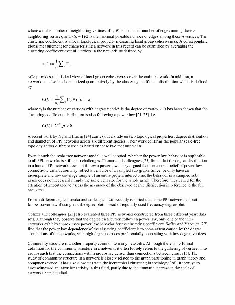
where n is the number of neighboring vertices of v, vE is the actual number of edges among these n neighboring vertices, and n(n – 1)/2 is the maximal possible number of edges among these n vertices. The clustering coefficient is a local topological property measuring local group cohesiveness. A corresponding global measurement for characterizing a network in this regard can be quantified by averaging the clustering coefficient over all vertices in the network, as defined by
1vv
C Cn
< >= ∑ ,
<C> provides a statistical view of local group cohesiveness over the entire network. In addition, a network can also be characterized quantitatively by the clustering coefficient distribution which is defined by
1( ) , |v vvk
C k C v d kn
= ∀ =∑ ,
where kn is the number of vertices with degree k and vd is the degree of vertex v. It has been shown that the clustering coefficient distribution is also following a power law [21-23], i.e. ( ) , 0C k k β β− > , A recent work by Ng and Huang [24] carries out a study on two topological properties, degree distribution and diameter, of PPI networks across six different species. Their work confirms the popular scale-free topology across different species based on these two measurements. Even though the scale-free network model is well adopted, whether the power-law behavior is applicable to all PPI networks is still up to challenges. Thomas and colleagues [25] found that the degree distribution in a human PPI network does not follow a power law. They argued that the current belief of power-law connectivity distribution may reflect a behavior of a sampled sub-graph. Since we only have an incomplete and low coverage sample of an entire protein interactome, the behavior in a sampled sub-graph does not necessarily imply the same behavior for the whole graph. Therefore, they called for the attention of importance to assess the accuracy of the observed degree distribution in reference to the full proteome. From a different angle, Tanaka and colleagues [26] recently reported that some PPI networks do not follow power law if using a rank-degree plot instead of regularly used frequency-degree plot. Colizza and colleagues [23] also evaluated three PPI networks constructed from three different yeast data sets. Although they observe that the degree distribution follows a power law, only one of the three networks exhibits approximate power law behavior for the clustering coefficient. Soffer and Vazquez [27] find that the power law dependence of the clustering coefficient is to some extent caused by the degree correlations of the networks, with high degree vertices preferentially connecting with low degree vertices. Community structure is another property common to many networks. Although there is no formal definition for the community structure in a network, it often loosely refers to the gathering of vertices into groups such that the connections within groups are denser than connections between groups [3]. The study of community structure in a network is closely related to the graph partitioning in graph theory and computer science. It has also close ties with the hierarchical clustering in sociology [28]. Recent years have witnessed an intensive activity in this field, partly due to the dramatic increase in the scale of networks being studied.

Because communities are believed to play a central role in the functional properties of complex networks [28], the ability to detect communities in networks could have practical applications. Studying the community structure of biological networks is of particular interest and challenging, given the high data volume and the complex nature of interactions. In the context of biological networks, communities might represent structural or functional groupings. They can be synonymous with molecular modules, biochemical pathways, gene clusters, or protein complexes. Being able to identify the community structure in a biological network may help us to understand better the structure and dynamics of biological systems. Many algorithms for detecting community structure in networks have been proposed. They can be roughly classified into two categories, divisive and agglomerative. The divisive approach takes the route of recursive removal of vertices (or edges) until the network is separated into its components or communities, whereas the agglomerative approach starts with isolated individual vertices and joins together small communities. One important algorithm is proposed by Girvan and Newman (the GN algorithm) [3]. The GN algorithm is based on the concept of betweenness, a quantitative measure of the number of shortest paths passing through a given vertex (or edge). The vertices (or edges) with the highest betweenness are believed to play the most prominent role in connecting different parts of a network. The GN algorithm detects communities in a network by recursively removing these high betweenness vertices (or edges). It has produced good results and is well adopted by different authors in studying various networks [28]. However, it has a major disadvantage which is its computational cost. For sparse networks with n vertices, the GN algorithm is of 3n time. Various alternative algorithms have been proposed [29-33], attempting to improve either the quality of the community structure or the computational efficiency. The GN algorithm has been applied to a number of metabolic networks from different organisms to detect communities that relate to functional units in the networks [34]. It has also been adapted to analyze a network of gene relationships as established by co-occurrence of gene names in published literature and to detect communities of related genes [35]. One goal of our work here is to address a slightly different question about the community structure in a PPI network, i.e. what is the community a given protein (or proteins) belongs to. We are motivated by two main factors. Firstly, due to the complexity and modularity of biological networks, it is more feasible computationally to study a community containing one or a few proteins of interest. Secondly, sometimes the whole community structure of the network may not be our primary concern. Rather, we may be more interested in finding the community which contains a protein (or proteins) of interest. Hashimoto and colleagues [36] have used a similar approach to growing genetic regulatory networks from seed genes. Their work is based on probabilistic Boolean networks and sub-networks are constructed in the context of a directed graph using both the coefficient of determination and the Boolean function influence among genes. The similar approach is also taken by Flake and colleagues [37] to find highly topically related communities in the Web based on the self-organization of the network structure and on a maximum flow method. Related works also include those that predict co-complex proteins. Jansen and colleagues [38] use a procedure integrating different data sources to predict the membership of protein complexes for individual genes based on two assumptions: first, the function of any protein complex depends on the functions of its subunits; and second, all subunits of a protein complex share certain common properties. Bader and Hogue [39] report a Molecular Complex Detection (MCODE) clustering algorithm to identify molecular complexes in a large protein interaction network. MCODE is based on local network density -

a modified measure of the clustering coefficient. Bu and colleagues [10] use a spectral analysis method to identify the topological structures such as quasi-cliques and quasi-bipartites in a protein-protein interaction network. These topological structures are found to be biologically relevant functional groups. In our previous work, we developed a spectral-based clustering method using local density and vertex neighborhood to analyze the chromatin network [11-12]. Two recent works along this line of research are based on the concept of network modularity introduced by Hartwell and colleagues [40]. The works by [21] and [41] both used computational analyses to cluster the yeast PPI network and discovered that molecular modules are densely connected with each other but sparsely connected with the rest of the network. 3. Method We intuitively model a protein-protein interaction network as a simple graph, meaning that it is undirected, unweighted, and without self-loops. Each vertex of the graph represents a protein and each edge represents an interaction between the two proteins connected by it. 3.1 Data Sets
We analyze the topology of PPI networks using the species-specific data set which includes E. coli, H. pylori, S. cerevisiae, D. melanogaster, C. elegans, M. musculus, and H. sapiens PPI networks. The data sets were downloaded from the Database for Interacting Proteins (DIP) [42]. To test our algorithm, we use a data set of interactions for Saccharomyces cerevisae downloaded from the General Repository for Interaction Datasets (GRID) [43]. The GRID database contains all published large-scale interaction datasets as well as available curated interactions. The GRID yeast data set we downloaded has 4,907 proteins and 17,598 interactions. 3.2 Measurements of Network Topology The basic properties are measured for each PPI network, including: 1) the number of proteins, measured by the number of vertices; 2) the number of interactions, measured by the number of edges; 3) the number of connected components within the network; 4) the size of the largest (or giant) component, measured by the size of the largest connected sub-graph; We also measure three degree related metrics: the maximum degree (kmax), the average degree (<k>), defined as
2 | |Ekn
< >= ,
where |E| is the total number of edges and n is the total number of vertices, and the degree distribution (P(k)) which is the frequency of a vertex having degree k in the network. The diameter of a network, <l>, is defined as the average distance between any two vertices. The distance between two vertices is defined as the number of edges along their shortest path. For a vertex i, we adopt the definition of the clustering coefficient vC from [15] as defined in Section 2. A global measurement related to this is the average clustering coefficient <C> also defined in Section 2. Assuming the same degree distribution, we adopt the following definition to obtain an average clustering coefficient of a random network [44]:

2 2
3( )
randk kC
n k< > − < >
< >=< >
.
We also calculate a local property called vertex density <D>. The definition of vertex density is inspired by Bader and Hogue [39] who define a local density by expanding the definition of the clustering coefficient for vertex v to include v itself in the formula when calculating vC . All statistical analyses are performed using SPSS software package. 3.3 The Algorithm 3.3.1 Notation An undirected graph, G = (V, E), is comprised of two sets, vertices V and edges E. An edge e is defined as a pair of vertices (u, v) denoting the direct connection between vertices u and v. The graphs we use in this paper are undirected, unweighted, and simple – meaning no self-loops or parallel edges.
For a subgraph 'G G⊂ and a vertex i belonging to G’, we define the in-community degree for vertex i, k in
i (G’), to be the number of edges connecting vertex i to other vertices belonging to G’ and the out-
community degree, k outi (G’), to be the number of edges connecting vertex i to other vertices that are in G
but do not belong to G’.
In our algorithm, we adopt the quantitative definitions of community defined by Radicchi and colleagues [45]. In this definition, a subgraph G’ is a community in a strong sense if for each vertex i in G’, its in-community degree is greater than out-community degree. More formally, G’ is a community in a strong sense if ( ') ( '), ', 'in out
i iK G K G i G G G> ∀ ∈ ⊂ . In a weak sense if the sum of all degrees within G’ is greater than the sum of all degrees from G’ to the rest of the graph, i.e., G’ is a community in a weak sense if ( ') ( '), ', 'in out
i ii iK G K G i G G G> ∈ ⊂∑ ∑ .
3.3.2 The algorithm The algorithm, called CommBuilder, accepts the seed protein s, gets the neighbors of s, finds the core of the community to build, and expands the core to find the eventual community.
The two major components of CommBuilder are FindCore and ExpandCore. In fact, FindCore performs a naïve search for maximum clique from the neighborhood of the seed protein by recursively removing vertices with the lowest in-community degree until all vertices in the core set have the same in-community degree.
The algorithm performs a breadth first expansion in the core expanding step. It first builds a candidate set containing the core and all vertices adjacent to each vertex in the core. It then adds to the core a vertex that either meets the quantitative definition of community in a strong sense or the fraction of in-community degree over a relaxed affinity threshold f of the size of the core. The affinity threshold is 1

when the candidate vertex connects to each of vertices in the core set. This threshold provides flexibility when expanding the core, because it is too strict requiring every expanding vertex to be a strong sense community member.
The FindCore is a heuristic search for a maximum complete subgraph in the neighborhood N of seed s. Let K be the size of N, then the worst-case running time of FindCore is 2( )O k . The ExpandCore part costs in worst-case approximately |V| + |E| + overhead. |V| accounts for the expanding of the core, at most all vertices in V, minus what are already in the core, would be included. |E| accounts for calculating the in- and out-degrees for the candidate vertices that are not in the core but in the neighborhood of the core. The overhead is caused by recalculating the in- and out-degrees of neighboring vertices every time the FindCore is recursively called. The number of these vertices is dependent on the size of the community we are building and the connectivity of the community to the rest of the network, but not the overall size of the network.
Algorithm 1 CommBuilder(G, s, f) 1: G(V, E) is the input graph with vertex set V and edge set E. 2: s is the seed vertex, f is the affinity threshold. 3: N ← {Adjacency list of s } ∪{s} 4: C ← FindCore(N) 5: C’ ← ExpandCore(C, f) 6: return C’ 7: FindCore(N) 8: for each v ∈N 9:
calculate kinv (N)
10: end for 11:
Kmin ← min { kinv (N), v ∈N}
12: Kmax ← max { k
inv (N), v ∈N}
13: if Kmin = Kmax then return N 14:
else return FindCore(N – {v}, kinv (N) = Kmin)
15: ExpandCore(C, f) 16: D ←
CwCvEwv ∉∈∈∪
,,),({v, w}
17: C’ ← C 18: for each t ∈D and t ∉C 19:
calculate kint (D)
20: calculate k
outt (D)
21: if k
int (D) > k
outt (D) or k
int (D)/|D| > f then
C’ ← C’ ∪ {t} 22: end for 23: if C’ = C then return C 24: else return ExpandCore(C’, f)
4. Results 4.1 Basic properties of the PPI networks Table 1 lists the basic properties of all PPI networks used for our analysis. The sizes of networks vary significantly across species, indicating the varied status in data collecting and documenting for the

specific data source and virtually our understanding of PPI for these organisms. Table 1 shows the small sizes of so called giant components for H. sapiens and M. musculus, meaning that we have a fairly large number of unconnected small sub-graphs in these two networks. Table 1 PPI networks of different species.
SPECIES PROTEINS INTERACTIONS #COMPONENTS GIANT COMPONENT(*)
E. coli 1640 6658 200 1396 (85.1%) H. pylori 702 1359 9 686 (97.7%) S. cerevisiae (Core) 2614 6379 66 2445 (93.5%) D. melanogaster 7441 22636 52 7330 (98.5%) C. elegans 2629 3970 99 2386 (90.8%) M. musculus 327 274 79 49 (15.0%) H. sapiens 1059 1318 119 563 (53.2%)
*Number inside the parenthesis: percentage of the size of the giant component in the entire network. 4.2 Average global topological properties of PPI networks In Table 2, we report the average global topological properties of PPI networks. Across species, PPI networks all exhibit small values of average degree and diameters, even though the absolute values differ significantly. Also, except for C. elegans, PPI networks for all other species have larger average clustering coefficient comparing to the corresponding random clustering coefficient, indicating a non-random and hierarchical structure within these networks. Contrary to the average clustering coefficient, the average vertex density shows much lesser variability across species. Table 2 Average global topological properties of PPI networks.
NETWORK Kmax <k> <l> <D> <C> <Crand> E. coli 152 8.12 3.73 0.7053 0.5889 0.1168 H. pylori 54 3.87 4.14 0.4514 0.0255 0.0403 S. cerevisiae (Core) 111 4.88 5.00 0.5609 0.2990 0.0103 D. melanogaster 178 6.08 4.39 0.3920 0.0159 0.0097 C. elegans 187 3.02 4.81 0.4885 0.0490 0.0462 M. musculus 12 1.68 3.57 0.6082 0.1011 0.0062 H. sapiens 33 2.49 6.80 0.5703 0.1658 0.0098
4.3 Degree distribution Degree distribution P(k) is the probability that a selected protein has exactly degree k. We evaluate the distribution of degrees P(k) as a function of k. Figure 1 shows the degree distribution for the networks across different species. The log-log plot clearly demonstrates the power law dependence of P(k) on degree k. For our analysis, we select to use directly the raw data, instead of following [15] with exponential cutoff. The results of statistical analysis are listed in Table 3. Without exponential cutoff, our regression analysis yields power law exponents γ between 1.36 and 2.36, in fairly good agreement with previously reported results. Even though the regression analysis and figures clearly show strong power-law degree distribution, we want to conduct further statistical analysis to test if the power law model adequately captures all the

features in the testing data. Using SPSS software package, we create a scatter plot of residues by fit values for the power law model. The result is shown in Figure 2, which clearly indicate a pattern in the data that is not captured by the power law model. This means that the power law is a model that has excellent fit statistics, but has poor residuals, indicating the inadequacy of the model.
0.00001
0.0001
0.001
0.01
0.1
1
10
1 10 100 1000k
P(k)
E. coliH. pyloriS. cerevisiaeD. melanogasterC. elegansM. musculusH. sapiens
Figure 1 Degree distribution P(k) of PPI networks across species.
Figure 2 Residuals vs fit values

Table 3 Statistical analysis of PPI networks.
NETWORKS γ† (R2) α† (R2) β† (R2) E. coli 1.355 (0.882) 0.562 (0.656) 0.536 (0.756) H. pylori 1.651 (0.899) 0.495 (0.373) 0.826 (0.985) D. melanogaster 1.945 (0.923) 3.050 (0.311) 0.836 (0.989) S. cerevisiae (Core) 1.977 (0.911) 0.893 (0.721) 0.759 (0.867) C. elegans 1.599 (0.839) 0.625 (0.362) 0.833 (0.976) M. musculus 2.360 (0.931) 0.598 (0.431)* 0.689 (0.965) H. sapiens 2.025 (0.931) 0.657 (0.190)* 0.626 (0.699)
† P(k) ~ k-γ, C(k) ~ k-α, D(k) ~ k-β * p > 0.05 4.4 The average clustering coefficient distribution We have shown results of average clustering coefficient for PPI networks in a previous section. We now take a closer look at the distribution of the clustering coefficient by averaging the clustering coefficient over vertices with degree k, C(k), as defined in Section 2. The results, as shown in Figure 3, indicate that while E. coli and S. cerevisiae (also shown in Table 3) PPI networks show somewhat weak power law distribution, networks of other species do not follow a power law. Regression analysis shows that there is no statistical significance for fitting a power law for networks from M. musculus and H. sapiens. Even though the remaining networks from H. pylori, D. melanogaster, and C. elegans have p values less than 0.05, the values of 2R are fairly low.
0.0001
0.001
0.01
0.1
1
10
1 10 100 1000k
C(k
)
E. coliH. pyloriS. cerevisiaeD. melanogasterC. elegansM. musculusH. sapiens
Figure 3 Average clustering coefficient C(k) as a function of degree k in PPI networks across different species.

4.5 The average vertex density distribution We evaluate the distribution of the average vertex density over the vertices with degree k. The results for the vertex density spectrum (D(k) over degree k) display consistent power law behavior for all the networks (Figure 4).
0.001
0.01
0.1
1
10
1 10 100 1000k
D(k
)
E.coliH.pyloriS.cerevisiaeD.melanogasterC.elegansM.musculusH.sapiens
Figure 4 Average vertex density D(k) as a function of degree k in PPI networks across different species.
4.6 Protein Communities We applied our algorithm against the network built from the downloaded data set as described in Section 3. The average running time for finding a community of around 50 members is about 20 ms.
Because there is no alternative approach to our method, we decide to compare the performance of our algorithm to the work on predicting protein complex membership by Asthana and colleagues [46]. Asthana and colleagues reported results of queries with four complexes using probabilistic network reliability (we will refer their work as PNR method in the following discussion). Four communities are identified by CommBuilder using one protein as seed from each of the query complexes used by the PNR method. The seed protein is selected randomly from the “core” protein set. The figures for visualizing the identified communities are created using Pajek [47]. The community figures are extracted from the network we build using the above mentioned data set omitting connections and proteins outside the detected community. The proteins in each community are annotated with a brief description obtained from the MIPS complex catalogue database [48] as shown in Table 4-7. As a comparison, we use Complexpander, an implementation of the PNR method [46] and available at http://llama.med.harvard.edu/Software.html, to predict co-complex using the core protein set that contains the same seed protein used by CommBuilder. For all our queries when using Complexpander, we select the option to use the MIPS complex catalogue database. We record in Table 4-7 the ranking of the members in our identified communities that also appear in the co-complex candidate list predicted by Complexpander.

The first community, shown in Figure 5, is identified using TAF6 (in red) as seed. TAF6 is a component of the SAGA complex which is a multifunctional co-activator that regulates transcription by RNA polymerase II [49]. The SAGA complex is listed in MIPS complex catalogue as a known cellular complex consisting of 16 proteins. As shown in Table 4, the community identified by our algorithm contains 39 members, including 14 of the 16 SAGA complex proteins listed in MIPS (indicated by an asterisk in the Alias column). The community also contains 14 of 21 proteins listed in MIPS as Kornberg’s mediator (SRB) complex. The rest of the proteins in the community are either TATA-binding proteins or transcription factor IID (TFIID) subunits or SRB related. TFIID is a complex involved in initiation of RNA polymerase II transcription. SAGA and TFIID are structurally and functionally correlated, make overlapping contributions to the expression of RNA polymerase II transcribed genes. SRB complex is a mediator that conveys regulatory signals from DNA-binding transcription factors to RNA polymerase II [50]. In addition, 27 of the top 50 potential co-complex proteins (9 of the top 10), not including the seed proteins, predicted by Complexpander are in the identified community.
Figure 5 The SAGA-SRB community. The seed protein is TAF6 (in red).
TABLE 4
THE SAGA-SRB COMMUNITY
Proteina Alias Description Rank YDR448w ADA2b general transcriptional adaptor or co-
activator 1
YNR010w CSE2 c subunit of RNA polymerase II mediator complex
YGR252w GCN5 b histone acetyltransferase 2 YPL254w HFI1 b transcriptional coactivator 3 YMR112c MED11 c mediator complex subunit YDL005c MED2 c transcriptional regulation mediator 20 YOR174w MED4 c transcription regulation mediator 23 YHR058c MED6 c RNA polymerase II transcriptional
regulation mediator
YOL135c MED7 c member of RNA Polymerase II transcriptional regulation mediator complex
21
YBR193c MED8 c transcriptional regulation mediator 24 YDR176w NGG1 b general transcriptional adaptor or co-
activator 10
YGL025c PGD1 c mediator complex subunit 37

YBL093c ROX3 c transcription factor YCL010c SGF29 b SAGA associated factor 43 YER148w SPT15 the TATA-binding protein TBP 15 YOL148c SPT20 b member of the TBP class of SPT
proteins that alter transcription site selection
4
YDR392w SPT3 b general transcriptional adaptor or co-activator
13
YBR081c SPT7 b involved in alteration of transcription start site selection
5
YHR041c SRB2 c DNA-directed RNA polymerase II holoenzyme and Kornberg’s mediator (SRB) subcomplex subunit
YER022w SRB4 c DNA-directed RNA polymerase II holoenzyme and Kornberg’s mediator (SRB) subcomplex subunit
27
YGR104c SRB5 c DNA-directed RNA polymerase II holoenzyme and Kornberg’s mediator (SRB) subcomplex subunit
YBR253w SRB6 c DNA-directed RNA polymerase II suppressor protein
19
YDR308c SRB7 c DNA-directed RNA polymerase II holoenzyme and kornberg’s mediator (SRB) subcomplex subunit
46
YCR081w SRB8 DNA-directed RNA polymerase II holoenzyme and Srb10 CDK subcomplex subunit
YDR443c SSN2 DNA-directed RNA polymerase II holoenzyme and Srb10 CDK subcomplex subunit
YPL042c SSN3 cyclin-dependent CTD kinase YGR274c TAF1 TFIID subunit (TBP-associated
factor), 145 kD 14
YDR167w TAF10 b TFIID and SAGA subunit 7 YML015c TAF11 TFIID subunit (TBP-associated
factor), 40KD 18
YDR145w TAF12 b TFIID and SAGA subunit 8 YML098w TAF13 TFIID subunit (TBP-associated
factor), 19 kD 17
YCR042c TAF2 component of TFIID complex 22 YPL011c TAF3 component of the TBP-associated
protein complex 50
YBR198c TAF5 b TFIID and SAGA subunit 9 YGL112c TAF6 b TFIID and SAGA subunit YMR227c TAF7 TFIID subunit (TBP-associated
factor), 67 kD
YML114c TAF8 TBP Associated Factor 65 KDa YMR236w TAF9 b TFIID and SAGA subunit 11 YHR099w TRA1 b component of the Ada-Spt
transcriptional regulatory complex 12
aThe open reading frame (ORF) name is used. bProteins belong to SAGA complex listed in MIPS. cProteins belong to SRB complex listed in MIPS.
The second community is discovered using NOT3 as seed (Figure 6 and Table 5). NOT3 is a known component protein of the CCR4-NOT complex which is a global regulator of gene expression and involved in such functions as transcription regulation and DNA damage responses. MIPS complex catalogue lists 5 proteins for NOT complex and 13 proteins (including the 5 NOT complex proteins) for CCR4 complex. The CCR4-NOT community identified is composed of 40 members. All 5 NOT complex proteins listed in MIPS and 11 of the 13 CCR4 complex proteins are members of the community. POL1, POL2, PRI1, and PRI2 are members of the DNA polymerase alpha (I) – primase complex, as listed in MIPS. RVB1, PIL1, UBR1, and STI1 have been grouped together with CCR4, CDC39, CDC36, and POP2 by systematic analysis [51]. The community also contains 20 out of 26 proteins of a complex that is probably involved in transcription and DNA/chromatin structure maintenance [52].

Figure 6 The CCR4-NOT community. The seed protein is NOT3 (in red).
TABLE 5
THE CCR4-NOT COMMUNITY
Proteina Alias Description Rank YDR376w ARH1 mitochondrial protein putative
ferredoxin-NADP+ reductase 38
YGR134w CAF130 c CCR4 Associated Factor 130 kDa 8 YJR122w CAF17 b CCR4 associated factor YNL288w CAF40 c CCR4 Associated Factor 40 kDa 9 YJR060w CBF1 centromere binding factor 1 YAL021c CCR4 bc transcriptional regulator 3 YDR188w CCT6 c component of chaperonin-containing
T-complex (zeta subunit) 30
YDL165w CDC36 bc transcription factor 40 YCR093w CDC39 bc nuclear protein 1 YDL145c COP1 c coatomer complex alpha chain of
secretory pathway vesicles 11
YMR025w CSI1 Subunit of the Cop9 signalosome, involved in adaptation to pheromone signaling
46
YGR092w DBF2 b ser/thr protein kinase related to Dbf20p
6
YDL160c DHH1 b DExD/H-box helicase, stimulates mRNA decapping,
17
YGL195w GCN1 c translational activator 26 YOL133w HRT1 Skp1-Cullin-F-box ubiquitin protein
ligase (SCF) subunit
YIL106w MOB1 b required for completion of mitosis and maintenance of ploidy
10
YER068w MOT2bc transcriptional repressor 2 YGL178w MPT5 multicopy suppressor of POP2 YIL038c NOT3bc general negative regulator of
transcription, subunit 3
YPR072w NOT5bc component of the NOT protein complex
5
YGR086c PIL1 Long chain base-responsive inhibitor

of protein kinases Phk1p and Phk2p, acts along with Lsp1p to down-regulate heat stress resistance
YBL105c PKC1 ser/thr protein kinase YNL102w POL1c DNA-directed DNA polymerase
alpha, 180 KD subunit 32
YBL035c POL12 c DNA-directed DNA polymerase alpha, 70 KD subunit
28
YNR052c POP2 bc required for glucose derepression 4 YIR008c PRI1 c DNA-directed DNA polymerase
alpha 48kDa subunit (DNA primase) 34
YKL045w PRI2 c DNA-directed DNA polymerase alpha , 58 KD subunit (DNA primase)
31
YPL010w RET3 coatomer complex zeta chain 39 YDR190c RVB1 RUVB-like protein 29 YPL235w RVB2 c RUVB-like protein 21 YGL137w SEC27 c coatomer complex beta chain (beta-
cop) of secretory pathway vesicles 7
YER022w SRB4 DNA-directed RNA polymerase II holoenzyme and Kornberg’s mediator (SRB) subcomplex subunit
44
YOR047c STD1 dosage-dependent modulator of glucose repression
YOR027w STI1 stress-induced protein YLR150w STM1 specific affinity for guanine-rich
quadruplex nucleic acids
YOR110w TFC7 c TFIIIC (transcription initiation factor) subunit, 55 kDa
25
YDL185w TFP1 c encodes 3 region protein which is self-spliced into TFP1p and PI-SceI
27
YGR184c UBR1 ubiquitin-protein ligase YJL141c YAK1 ser/thr protein kinase YDR259c YAP6 transcription factor, of a fungal-
specific family of bzip proteins
aThe open reading frame (ORF) name is used. bProteins belong to CCR4-NOT complex listed in MIPS. cProteins considered part of a complex involved in transcription and DNA/chromatin structure maintenance [31].
The third community is identified by using RFC2 as the seed (Figure 7 and Table 6). RFC2 is a component of the RFC (replication factor C) complex, the “clamp loader”, which plays an essential role in DNA replication and DNA repair. The community identified by our algorithm has 17 members. All five proteins of RFC complex listed in MIPS complex catalogue database are members of this community, as shown in Table 6. All but one member in this community are in the functional category of DNA recombination and DNA repair or cell cycle checkpoints according to MIPS. This community also includes the top 8 ranked proteins predicted by Complexpander.

Figure 7 The RFC community. The seed protein is RFC2 (in red).
TABLE 6
THE RFC COMMUNITY
Proteina Alias Description Rank YMR048w CSM3c Protein required for accurate
chromosome segregation during meiosis
YMR078c CTF18c required for accurate chromosome transmission in mitosis and maintenance of normal telomere length
6
YPR135w CTF4c DNA-directed DNA polymerase alpha-binding protein
YOR144c ELG1c Protein required for S phase progression and telomere homeostasis, forms an alternative replication factor C complex important for DNA replication and genome integrity
7
YBL091c MAP2 methionine aminopeptidase, isoform 2 YCL061c MRC1c Mediator of the Replication
Checkpoint
YNL102w POL1c DNA-directed DNA polymerase alpha, 180 KD subunit
19
YBL035c POL12c DNA-directed DNA polymerase alpha, 70 KD subunit
5
YJR043c POL32c polymerase-associated gene, third (55 kDa) subunit of DNA polymerase delta
YER173w RAD24c cell cycle checkpoint protein 1 YKL113c RAD27c ssDNA endonuclease and 5’-
3’exonuclease
YOR217w RFC1bc DNA replication factor C, 95 KD subunit
8
YJR068w RFC2bc DNA replication factor C, 41 KD subunit
YNL290w RFC3bc DNA replication factor C, 40 kDa subunit
2
YOL094c RFC4bc DNA replication factor C, 37 kDa 4

subunit YBR087w RFC5bc DNA replication factor C, 40 KD
subunit 3
YNL273w TOF1c topoisomerase I interacting factor 1 aThe open reading frame (ORF) name is used. bProteins belong to RFC complex listed in MIPS. cProteins listed in the functional category of DNA recombination and DNA repair or cell cycle checkpoints in MIPS.
We use ARP3 as seed to identify the last community (Figure 5). ARP2/ARP3 complex acts as multi-functional organizer of actin filaments. The assembly and maintenance of many actin-based cellular structures likely depend on functioning ARP2/ARP3 complex [53]. The identified community contains all 7 proteins of the ARP2/ARP3 complex listed in MIPS (see Table 7). Not including the seed (ARP3), these proteins represent the top 6 ranked proteins predicted by Complexpander. As indicated in Table 7, there are 14 members belonging to the same functional category of budding, cell polarity, and filament formation, according to MIPS.
Figure 8 The ARP2/ARP3 community. The seed protein is ARP3 (in red).
TABLE 7
THE ARP2/ARP3 COMMUNITY
Proteina Alias Description Rank YLR111w YLR111w hypothetical protein YIL062c ARC15bc subunit of the Arp2/3 complex 1 YLR370c ARC18b subunit of the Arp2/3 complex 4 YKL013c ARC19bc subunit of the Arp2/3 complex 3 YNR035c ARC35b subunit of the Arp2/3 complex 5 YBR234c ARC40bc Arp2/3 protein complex subunit, 40
kilodalton 6
YDL029w ARP2bc actin-like protein 2 YJR065c ARP3b actin related protein YJL095w BCK1c ser/thr protein kinase of the MEKK
family
YPL084w BRO1 required for normal response to nutrient limitation
YBR023c CHS3c chitin synthase III YNL298w CLA4c ser/thr protein kinase YNL084c END3c required for endocytosis and

cytoskeletal organization YBR015c MNN2 type II membrane protein YCR009c RVS161c protein involved in cell polarity
development
YDR388w RVS167c reduced viability upon starvation protein
YFR040w SAP155c Sit4p-associated protein YBL061c SKT5c protoplast regeneration and killer
toxin resistance protein
YNL243w SLA2c cytoskeleton assembly control protein YHR030c SLT2c ser/thr protein kinase of MAP kinase
family
aThe open reading frame (ORF) name is used. bProteins belong to ARP2/ARP3 complex listed in MIPS. cProteins listed in the functional category of budding, cell polarity, and filament formation in MIPS.
5. Discussion and Future Work In this paper, we used the graph theory and statistical approaches to analyzing the topological structure of protein-protein interaction networks across different species. We have shown the polarity on data and perhaps knowledge about the PPI networks at the “omics” level across a variety of species. Our results confirmed that PPI networks have small diameters and small average degrees. All networks we evaluated display power law degree distribution. However, further statistical analysis indicates an inadequacy of such model in capturing certain features in the data. We strongly believe that further investigation into this issue may shed some new lights on our understanding of PPI networks. Most of the networks we evaluated also reveal a larger clustering coefficient, indicating the non-random structure of the networks. However, the values of the clustering coefficient varied significantly across different species. This may result from the incompleteness and noise of the data, since there are significant differences in the clustering coefficient between networks with different confidence levels [54]. In addition, networks consisting of interactions detected from different experimental systems differed significantly in the values of the clustering coefficient [54]. The spectrum of the average clustering coefficient over the vertices degree k fails to exhibit scale free behavior in most of the networks evaluated. One interesting finding of this paper is the power law distribution of the average vertex density over the vertex degree k, consistent across all networks from different species. This power law distribution is not susceptible to either biases of different experimental systems or noise in data [54]. The difference between the clustering coefficient and the vertex density in calculation is the introduction of a new k into the formula. The clustering coefficient measures the network transitivity in the neighborhood of a vertex, whereas the vertex density reflects the network connectivity inside the locality of the neighborhood of a vertex. Our finding indicates that the vertex density distribution may be a better measurement to provide hints on the existence of a hierarchical organization in the PPI networks. A decaying D(k) function may indicate a hierarchy in which low degree proteins in general belong to well interconnected communities (high vertex density) and high degree proteins (hubs) are the cores connecting many proteins that have no direct interactions (low vertex density). The intriguing part of this finding is also related to a new definition introduced by Soffer and Vazquez [27] to eliminate degree correlation bias. They argue that the dependence of the clustering coefficient with degree k is partially due to this bias. The new definition they propose actually makes the power law behavior disappeared. On the contrary, we did not observe the power law distribution of C(k) over degree k, but the power law behavior appears when we modify the C(k) to D(k). We expect this information will be helpful because we have already seen the application of vertex density in [39] and in detecting a protein community reported here.

We present an efficient approach for growing a community from a given seed protein. It uses topological property of community structure in a network and takes advantage of local optimization in searching for the community comprising of the seed protein. Due to the complexity and modularity of biological networks, it is more desirable and computationally more feasible to model and simulate a network of smaller size. Our approach builds a community of manageable size and scales well to large networks. Its usefulness is demonstrated by the experimental results that all the four communities identified reveal strong structural and functional relationships among member proteins. It provides a fast and accurate way to find a community comprising a protein or proteins with known functions or of interest. For those community members that are not known to be part of a protein complex or a functional category, their relationship to other community members may deserve further investigation which in turn may provide new insights. Although we do not explicitly use our approach to the prediction of co-complexed proteins, the results of comparing with the PNR method developed by Asthana and colleagues [46] have shown that the communities identified by our approach do include the top ranked candidates of co-complexed proteins. Compared to the methods in predicting co-complexed proteins, our approach can discover a community rather than a single complex. In the context of this discussion, the notion of a community can be a complex, but it can also be a functional group consisting of several complexes, such as the SAGA/SRB community (Figure 1). This may not be always desirable. However, it does provide benefits of delineating the structure-function relationships beyond a single complex. In this spirit, one part of our future work is to further explore the relaxation threshold (f) aiming to identify either a more tightly connected community under a strict expanding condition or a more loosely connected community under a relaxed condition so that we could study interactions of different strengths within a community. Our approach does not consider the quality of data in our downloaded data set. By using the strong sense definition of community [45], we could to some degree reduce the noises. However, to improve the quality of an identified community, we have to take into account the quality of data and that is another part of our future work. One possible way is to use the probabilities assigned to individual protein pairs as used by [38], [45], [55], and [56]. 6. Acknowledgment This research work is supported in part from the NSF Career grant (NSF IIS 0448023). NSF CCF 0514679 and the PA Dept of Health Tobacco Settlement Formula Grant (#240205, 240196). References [1] Barabasi, A.-L. and Oltvai, Z.N. (2004). Network biology: understanding the cell’s functional
organization. Nat. Rev. Genet. 5: 101–114. [2] Bork, P., Jensen, L.J., von Mering, C., Ramani, A.K., Lee, I. and Marcotte, E.M. (2004) Protein
interaction networks from yeast to human. Curr. Opin. Struct. Biol. 14: 292–299. [3] Girvan, M. and Newman, M.E.J. (2002). Community structure in social and biological networks.
Proc. Natl. Acad. Sci. U.S.A. 99: 7821-7826. [4] Jeong, H., Mason, S.P., Barabasi, A.-L., and Oltvai, Z.N. (2001). Lethality and centrality in protein
networks. Nature 411: 41-42. [5] Fraser, H.B., et al. (2002). Evolutionary rate in the protein interaction network. Science 296: 750. [6] Fraser, H.B., et al. (2003). A simple dependence between protein evolution rate and the number of
protein-protein interactions. BMC Evol. Biol. 3: 11.

[7] Wuchty, S. (2004): Evolution and topology in the yeast protein interaction network. Genome Research 14: 1310-1314.
[8] Pei, P. and Zhang, A. (2005). A topological measurement for weighted protein interaction network. CSB2005, August 8-11, 2005, Stanford, CA, USA.
[9] Valente, A.X.C.N., Cusick, M.E., Fagerstrom, R.M., Hill, D.E., and Vidal, M. (2005). Yeast protein interactome topology provides framework for coordinated-functionality. arXiv:q-bio.MN/0505006.
[10] Bu, D., Zhao, Y., Cai, L., Xue, H., Zhu, X., Lu, H., Zhang, J., Sun, S., Ling, L., Zhang, N., Li, G. and Chen, R. (2003). Topological structure analysis of the protein–protein interaction network in budding yeast. Nucleic Acids Res. 31: 2443–2450.
[11] Hu, X. (2005). Mining and Analyzing Scale-free Protein-Protein Interaction Network, International Journal of Bioinformatics Research and Application 1(1): 81-101.
[12] Hu, X., Yoo, I., Song, I.-Y., Song, M., Han, J. and Lechner, M. (2004). Extracting and Mining Protein-Protein Interaction Network from Biomedical Literature, in the Proceedings of the 2004 IEEE Symposium on Computational Intelligence in Bioinformatics and Computational Biology (IEEE CIBCB 2004), Oct. 7-8, 2004, San Diego, USA.
[13] Watts, D.J. and Strogatz, S.H. (1998). Collective dynamics of “small world” networks. Nature 393: 440-442.
[14] Barabasi, A.-L. and Albert, R. (1999). Emergence of scaling in random networks. Science 286:509-512.
[15] Uetz, P., et al. (2000). A comprehensive analysis of protein–protein interactions in Saccharomyces cerevisiae. Nature 403:623-627.
[16] Rain, J.C., et al. (2001). The protein-protein interaction map of Helicobacter pylori. Nature 409:211-215.
[17] Butland, G., et al. (2005). Interaction network containing conserved and essential protein complexes in Escherichia coli. Nature 433: 531-537.
[18] Walhout, A.J.M., Sordella, R., Lu, X.W., Hartley, J.L., Temple, G.F., Brasch, M.A., Thierry-Mieg, N., and Vidal, M. (2000). Protein interaction mapping in C. elegans using proteins involved in vulval development. Science 287: 116-122.
[19] Li, S., et al. (2004). A map of the interactome network of the metazoan C. elegans. Science 303: 540-543.
[20] Giot, L., et al. (2004). A protein interaction map of Drosophila melanogaster. Science 302: 1727-1736.
[21] Spirin, V. and Mirny, L. (2003). Protein complexes and functional modules in molecular networks. Proc. Natl. Acad. Sci. U.S.A. 100: 1128-1133.
[22] Yook, S.-H., Oltvai, Z.N., and Barabasi, A.-L. (2004) Functional and topological characterization of protein interaction networks. Proteomics 4: 928-942.
[23] Colizza, V., Flammini, A., Maritan, A., and Vespignani, A. (2005). Characterization and modeling of protein-protein interaction networks. Physica A 352: 1-27.
[24] Ng, K.-L. and Huang, C.-H. (2004). A cross-species study of the protein-protein interaction networks via the random graph approach. Proceedings of the Fourth IEEE Symposium on Bioinformatics and Bioengineering (BIBE’04).
[25] Thomas, A., Cannings, R., Monk, N.A.M., and Cannings, C. (2003). On the structure of protein-protein interaction networks. Biochem Soc Trans 31: 1491-1496.
[26] Tanaka, R., Yi, T.-M, and Doyle, J. (2005). Some protein interaction data do not exhibit power law statistics. arXiv:q-bio.MN/0506038.
[27] Soffer, S. and Vazquez, A. (2004). Clustering coefficient without degree correlations biases.arXiv:cond-mat/0409686.
[28] Newman, M.E.J. (2003). The Structure and Function of Complex Networks. SIAM Review 45(2): 167-256
[29] Newman, M. E. J. (2004). Detecting community structure in networks. Eur. Phys. J. B 38: 321-330.

[30] Newman, M. E. J. and Girvan, M. (2004). Finding and evaluating community structure in networks. Phys. Rev. E 69: 026113.
[31] Newman, M. E. J. (2004). Fast algorithm for detecting community structure in networks. Phys. Rev. E 69: 066133.
[32] Donetti, L. and Munoz, M.A. (2004). Detecting Network Communities: a new systematic and efficient algorithm. J. Stat. Mech. P10012.
[33] White, S. and Smyth, P. (2005). A Spectral Clustering Approach to Finding Communities in Graphs. SIAM International Conference on Data Mining 2005, Newport Beach, CA, USA.
[34] Holme, P., Huss, M., and Jeong, H. (2003). Subnetwork hierarchies of biochemical pathways. Bioinformatics 19(4): 532-538.
[35] Wilkinson, D. and Huberman, B.A (2004). A Method for Finding Communities of Related Genes. Proc. Natl. Acad. Sci. U.S.A. 101(Suppl 1): 5241-5248.
[36] Hashimoto,R.F., Kim, S., Shmulevich, I., Zhang, W., Bittner, M.L., and Dougherty, E.R. (2004). Growing genetic regulatory networks from seed genes. Bioinformatics 20(8): 1241–1247.
[37] Flake, G. W., Lawrence, S. R., Giles, C. L., and Coetzee, F. M. (2002). Self-organization and identification of Web communities, IEEE Computer 35: 66-71.
[38] Jansen, R., Lan, N., Qian, J., and Gerstein, M. (2002). Integration of genomic datasets to predict protein complexes in yeast. J. Struct. Functional Genomics 2: 71–81.
[39] Bader, G.D. and Hogue, C.W. (2003). An automated method for finding molecular complexes in large protein interaction networks. BMC Bioinformatics 4: 2.
[40] Hartwell, L.H., Hopfield, J.J., Leibler, S. and Murray, A.W. (1999). From molecular to modular cell biology. Nature 402: C47–C52.
[41] Rives, A.W. and Galitski, T. (2003). Modular organization of cellular networks. Proc. Natl. Acad. Sci. U.S.A. 100: 1128–1133.
[42] Salwinski, L., Miller, C.S., Smith, A.J., Pettit, F.K., Bowie, J.U., and Eisenberg, D. (2004) The Database of Interacting Proteins: 2004 update. NAR 32.
[43] Breitkreutz, B.-J., Stark, C. and Tyers, M. (2003). The GRID: The General Repository for Interaction Datasets. Genome Biology 4: R23.
[44] Newman, M.E.J. (2003). Random graphs as models of networks, in Bornholdt, S., Schuster, H.G. (Eds.), Handbook of Graphs and Networks: from the Genome to the Internet, Wiley-VCH, Berlin, pp. 35-68
[45] Radicchi, F., Castellano, C., Cecconi, F., Loreto, V., and Parisi, D. (2004). Defining and identifying communities in networks. Proc. Natl. Acad. Sci. U.S.A. 101: 2658-2663.
[46] Asthana, S., King, O.D., Gibbons, F.D., and Roth, F.P. (2004). Predicting Protein Complex Membership Using Probabilistic Network Reliability. Genome Res. 14: 1170-1175.
[47] Batagelj, V. and Mrvar, A. (1998). Pajek: Program for large network analysis. Connections 21: 47–57.
[48] Mewes, H.W., Frishman, D., Guldener, U., Mannhaupt, G., Mayer, K., Mokrejs, M., Morgenstern, B., Munsterkotter, M., Rudd, S., and Weil, B. (2002). MIPS: A database for genomes and protein sequences. Nucleic Acids Res. 30: 31–34.
[49] Wu, P.Y., Ruhlmann, C., Winston, F., and Schultz, P. (2004). Molecular architecture of the S. cerevisiae SAGA complex. Mol. Cell 15: 199–208.
[50] Guglielmi, B., van Berkum, N.L., Klapholz, B., Bijma, T., Boube, M., Boschiero, C., Bourbon, H.M., Holstege, F.C.P., and Werner, M. (2004). A high resolution protein interaction map of the yeast Mediator complex. Nucleic Acids Res. 32: 5379–5391.
[51] Ho, Y., et al (2002). Systematic identification of protein complexes in Saccharomyces cerevisiae by mass spectrometry. Nature 415: 180 – 183.
[52] Gavin, A.-C., et al (2002). Functional organization of the yeast proteome by systematic analysis of protein complexes. Nature 415: 141 – 147.
[53] Machesky, L.M. and Gould, K.L. (1999). The Arp2/3 complex: a multifunctional actin organizer. Curr. Opin. Cell Biol. 11: 117 – 121.

[54] Wu, D., and Hu, X. (2006). Mining and Analyzing the Topological Structure of Protein-Protein Interaction Networks, 2006 ACM Symposium on Applied Computing (Bioinformatics Track), April 23-27, Dijon, Bourgogne, France.
[55] Bader, J.S. (2003). Greedily building protein networks with confidence. Bioinformatics 19(15): 1869-1874.
[56] Bader, J.S., Chaudhuri, A., Rothberg, J.M., and Chant, J. (2004). Gaining confidence in high-throughput protein interaction networks. Nat. Biotech 22(1): 78-85