tooth surface loss revisited: classification, etiology
TRANSCRIPT
Journal of Restorative Dentistry / Vol - 3 / Issue - 2 / May-Aug 2015 • 37
Tooth sur face loss rev i s i ted : C l a s s i f i c a t i o n , e t i o l o g y, a n d managementAyesha Hanif, Haroon Rashid, Mustafa Nasim1
Departments of Prosthodontics, and 1Community and Preventive Dentistry, Ziauddin College of Dentistry, Karachi, Pakistan
Address for correspondence: Dr. Haroon Rashid, Ziauddin College of Dentistry, Karachi-Pakistan. E-mail: [email protected]
INTRODUCTION
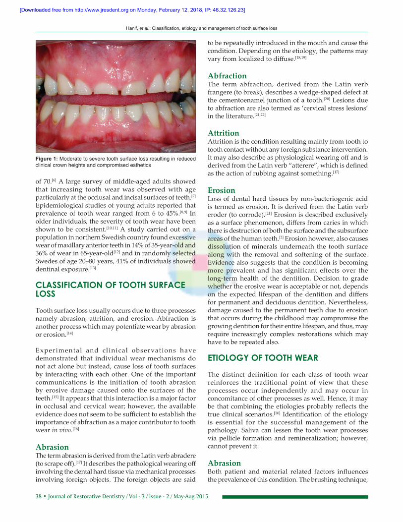
Tooth wear is a general term describing the loss of dental hard tissues from the surfaces of the teeth caused by factors other than dental caries, trauma, and developmental disorders.[1,2] Attrition, erosion, and abrasion usually cause alterations of the tooth surface and manifest as tooth wear. These processes act by distinct progressions and exhibit unique clinical characteristics [Figure 1].
Prevalence of tooth surface loss is increasing and younger patients are said to be at higher risks. The particular concern is the alarming rate of tooth wear that is now been seen in children and young adults. This was first noted in the Dental Health Survey of children in 1993, when children under the age of 5, especially those consuming
carbonated drinks, showed signs of abnormal erosion.[3] The problem is likely to continue as patients’ demands and expectations rise and healthy natural teeth are retained during the old age. For many years, erosion was a condition which invited little interest in clinical dentistry, community health services, and in dental research.[4]
Physiological wear causing vertical loss of enamel in a normal individual is approximately 0.02–0.04 mm a year[5] and wear is considered excessive when it causes esthetic concerns to the patient and causes symptoms of discomfort. Once the amount of tooth wear becomes so severe that recurrent symptoms are caused, then it is deemed ‘pathological’ tooth surface loss and becomes a challenge for a restorative dentist. The challenges faced during the clinical management of patients with tooth wear has raised considerable professional interest as its impact could be severe and may also affect an individual’s quality of life.
EPIDEMIOLOGY
The proportion of adults with severe tooth wear generally rises from approximately 3% in young people in their early 20s and to 17% in those over the age
Tooth wear is a general term describing the loss of dental hard tissues from the surfaces of the teeth. As the lifespan of individuals increase and the teeth are increasingly retained for life the incidence of non-carious tooth surface loss has also shown a rise. Little is understood about the aetiology and management of these lesions and there are several occasions where the condition is often neglected. The prevalence of tooth surface loss is difficult to establish and the reported clinical and epidemiological data are difficult to compare, due to differences in terminologies and many indices involved. The purpose of current review is to focus on the classification, aetiology and management of common non-carious conditions causing tooth surface loss.
Keywords: Tooth surface loss, tooth wear, parafunction
Access this article onlineQuick Response Code:
Website: www.jresdent.org
DOI: 10.4103/2321-4619.156643
Review Article
ABSTRACT
[Downloaded free from http://www.jresdent.org on Monday, February 12, 2018, IP: 46.32.126.23]
38 • Journal of Restorative Dentistry / Vol - 3 / Issue - 2 / May-Aug 2015
Hanif, et al.: Classification, etiology and management of tooth surface loss
to be repeatedly introduced in the mouth and cause the condition. Depending on the etiology, the patterns may vary from localized to diffuse.[18,19]
AbfractionThe term abfraction, derived from the Latin verb frangere (to break), describes a wedge‑shaped defect at the cementoenamel junction of a tooth.[20] Lesions due to abfraction are also termed as ‘cervical stress lesions’ in the literature.[21,22]
AttritionAttrition is the condition resulting mainly from tooth to tooth contact without any foreign substance intervention. It may also describe as physiological wearing off and is derived from the Latin verb “atterere”, which is defined as the action of rubbing against something.[17]
ErosionLoss of dental hard tissues by non‑bacteriogenic acid is termed as erosion. It is derived from the Latin verb eroder (to corrode).[21] Erosion is described exclusively as a surface phenomenon, differs from caries in which there is destruction of both the surface and the subsurface areas of the human teeth.[2] Erosion however, also causes dissolution of minerals underneath the tooth surface along with the removal and softening of the surface. Evidence also suggests that the condition is becoming more prevalent and has significant effects over the long‑term health of the dentition. Decision to grade whether the erosive wear is acceptable or not, depends on the expected lifespan of the dentition and differs for permanent and deciduous dentition. Nevertheless, damage caused to the permanent teeth due to erosion that occurs during the childhood may compromise the growing dentition for their entire lifespan, and thus, may require increasingly complex restorations which may have to be repeated also.
ETIOLOGY OF TOOTH WEAR
The distinct definition for each class of tooth wear reinforces the traditional point of view that these processes occur independently and may occur in concomitance of other processes as well. Hence, it may be that combining the etiologies probably reflects the true clinical scenarios.[16] Identification of the etiology is essential for the successful management of the pathology. Saliva can lessen the tooth wear processes via pellicle formation and remineralization; however, cannot prevent it.
AbrasionBoth patient and material related factors influences the prevalence of this condition. The brushing technique,
of 70.[6] A large survey of middle‑aged adults showed that increasing tooth wear was observed with age particularly at the occlusal and incisal surfaces of teeth.[7] Epidemiological studies of young adults reported that prevalence of tooth wear ranged from 6 to 45%.[8,9] In older individuals, the severity of tooth wear have been shown to be consistent.[10,11] A study carried out on a population in northern Swedish country found excessive wear of maxillary anterior teeth in 14% of 35‑year‑old and 36% of wear in 65‑year‑old[12] and in randomly selected Swedes of age 20–80 years, 41% of individuals showed dentinal exposure.[13]
CLASSIFICATION OF TOOTH SURFACE LOSS
Tooth surface loss usually occurs due to three processes namely abrasion, attrition, and erosion. Abfraction is another process which may potentiate wear by abrasion or erosion.[14]
Experimental and cl inical observations have demonstrated that individual wear mechanisms do not act alone but instead, cause loss of tooth surfaces by interacting with each other. One of the important communications is the initiation of tooth abrasion by erosive damage caused onto the surfaces of the teeth.[15] It appears that this interaction is a major factor in occlusal and cervical wear; however, the available evidence does not seem to be sufficient to establish the importance of abfraction as a major contributor to tooth wear in vivo.[16]
AbrasionThe term abrasion is derived from the Latin verb abradere (to scrape off).[17] It describes the pathological wearing off involving the dental hard tissue via mechanical processes involving foreign objects. The foreign objects are said
Figure 1: Moderate to severe tooth surface loss resulting in reduced clinical crown heights and compromised esthetics
[Downloaded free from http://www.jresdent.org on Monday, February 12, 2018, IP: 46.32.126.23]
Journal of Restorative Dentistry / Vol - 3 / Issue - 2 / May-Aug 2015 • 39
Hanif, et al.: Classification, etiology and management of tooth surface loss
brushing frequency, and the force applied while brushing are common patient‑related factors. The type of bristle material of toothbrush, stiffness of toothbrush bristles, the abrasiveness, and pH of dentifrice used are factors related to material.[17]
The most commonly cited effect of abrasion is the V‑shaped defect, which usually is ascribed to the use of an intensive horizontal brushing technique. Cervical areas are susceptible to toothbrush abrasion, particularly cuspids and first premolars, where thin buccal plates, gingival recession, and exposed root surfaces predispose cervical notching. Habits involving other intraoral objects (e.g., pipe smoking, toothpick use, and thread biting) can cause defects on the incisal and occlusal surfaces.[19] Dietary abrasion is not very prominent in modern days, as the typical western diet tends to be very soft, as opposed to primitive man’s diet which was more abrasive, and thus contributed greatly to tooth wear.
AbfractionThese lesions are usually located subgingivally, where the influence of tooth brushing abrasion is unusual; and hence, are hypothesized to be the result of eccentrically applied occlusal stresses leading to tooth flexure, rather than to be the result of abrasion alone. Weakening of the hydroxyapatite present near the cervical region of the teeth is weakened due to tensile stresses, which produces the classical wedge‑shaped defects having sharp edges near to the cementoenamel junction.[21]
AttritionAttrition mainly results from contact between opposing teeth and well‑defined wear facets are shown in the condition. The causal factors for attrition are parafunctional habits, bruxism, clenching, [23] developmental defects,[24] coarse diet, and natural teeth opposing porcelain. It is caused not only by diet or the habits, but a class III incisal relationship and lack of posterior support also lead to attrition.[25] Attrition occurs almost entirely on occlusal and incisal surfaces, although it may also effect the buccal and palatal sides of the maxillary and mandibular anterior teeth in deep vertical overlap occlusal relationships.[26]
ErosionDental erosion results when there is chronic and painless loss of dental hard tissue. The surfaces are usually etched off from the teeth surfaces due to the effect of acid and/or chelation with no involvement of bacteria. Evidence also suggests that that erosive wear also predisposes to attrition, and that the two mechanisms very often act together causing tooth surface loss.[27,28] Erosion caused due to the industrial acids has shown to be associated with severe attrition of teeth and it has also
been highlighted that severe attrition found in young individuals is mainly due to dietary erosive factors.[29]
There are intrinsic and extrinsic causes of dental erosion. Intrinsic causes are mostly of gastric reflux and include vomiting in case of anorexia, bulimia nervosa, and rumination.[30] Extrinsic causes include dietary soft drinks, citrus fruit, and food prickled with vinegar.[31] Medications, vitamin C, iron preparations, and aspirin are acidic in nature.[32] Lifestyle where people use mood improving drugs, that is, ecstasy, may also cause greater risk of erosion.[33]
TOOTH SURFACE LOSS DUE TO DENTAL CERAMICS
The application and use of tooth‑colored fixed restorations has increased with time. This has led to better research and clinical applications of the all‑ceramic crowns and bridges.[34] Due to brittleness and lower tensile strength of ceramics, the use of all ceramic crowns in high stress areas has been limited.[35]
Tooth surface loss of natural dentition if occurring, may get aggravated due to increased hardness and wear properties of opposing dental restoration as the rates of wear of antagonist natural teeth may change.[36] Excessive wear occurring at the occlusal surfaces may also lead to periodontal conditions due to abnormal loads applied over the teeth. The loss of vertical dimension of occlusion, loss of centric occlusion, and muscle fatigue may also lead to temporomandibular disorders.[37‑39] Thus, the occurrence of wear due to inherent properties of a material is also a significant factor that should be considered during the clinical application and selection of any restorative material. Seghi et al.,[40] recommended that when a material is selected for clinical application, it should be aimed that the selected material has the degree of hardness which is similar or near to that of the enamel. Dental feldspathic porcelain is being used for restoring teeth for many decades, but one of its major disadvantage is that it causes excessive wear of opposing natural dentition. Careful treatment planning and case selection is recommended during their use.[41,42] Some clinicians are reluctant to use porcelain restorations for anterior guidance. However, whilst the wear characteristics of other materials may be good, they do not satisfy patient’s esthetic demands.
Recently, high‑strength zirconia ceramic is introduced in clinical dentistry having high bending strength and very high fracture toughness.[43] Its application in clinical dentistry has seen a rise mainly because of its high values of fracture toughness, hardness, and dimensional stability.[44,45] It also has high values of frictional resistance
[Downloaded free from http://www.jresdent.org on Monday, February 12, 2018, IP: 46.32.126.23]
40 • Journal of Restorative Dentistry / Vol - 3 / Issue - 2 / May-Aug 2015
Hanif, et al.: Classification, etiology and management of tooth surface loss
in comparison to conventional feldspathic dental porcelain. High hardness exhibited by dental ceramics has shown to be associated with the greater abrasive effects overnatural teeth.[46] Since zirconia has high surface hardness, further wear was expected from it,[47] but many investigations involving zirconia showed that it causes less wear of opposing natural teeth as compared to conventional feldspathic dental porcelain.[34] Rosentritt et al.,[48] did not find any wear traces over enamel using a chewing simulator in the laboratory after using zirconia.
An investigation on the degree of wear of antagonistic teeth with zirconia using a dual‑axis chewing simulator was done in a laboratory. Using this wear test, the vertical and horizontal movements were more precisely replicated using computer software and the degrees of wearing off were more accurately compared. This was done using volume measurements rather than the height, and the environments of the mouth were more accurately simulated with the associated thermos cycling.[49] It was found that the amount of wear caused by zirconia over the opposing teeth was much lower as compared to feldspathic dental porcelain.
ManagementInitial management of tooth surface loss depends on accurate diagnosis of the condition, the identification of the etiology and frequent monitoring of the successive changes; hence to prevent further damage. Treatment planning is sometime very challenging and it is very necessary that accurate analysis of the tooth surface loss is made at an early stage and that satisfactory preventive measures are carried out. Once the risk factors are properly understood, these measures can be accurately initiated.
The interrelationship of the four modes of tooth surface loss and individual susceptibility influences the degree of tooth wear. Recognition of the multifactorial nature of the condition is the first step in its management, as failure to appreciate this may lead to inappropriate management and ultimate failure of restorative therapy. Holbrook and Arnadottir[50] stated thatif noncarious destruction of teeth is to be avoided, following must be considered:
• Recognizing and understanding that the condition exists
• Grading the severity of the condition• Likely causes are diagnosed appropriately• Monitoring the preventative measures and the
disease progress.
History and presenting complainTreatment planning for tooth wear cases is often driven by specific patient concerns. Some may complain of an esthetic impairment, such as fractured, unattractive teeth or
problems concerns due to tooth discoloration.[51] Difficulty in function could also be present, that is, inefficient mastication or accidental biting of the soft tissues. Less commonly, patients may also present due to sensitivity and pain from severely worn teeth.[4]
Dental history of the patient enables the clinician to conclude to the possible etiology. The attitude of the patient to the oral care and the motivation to keep the dentition healthy. Furthermore, an insight of the social history of a patient may disclose further understanding into the etiology of the condition. These could be details of lifestyle or occupational stress that could have led to the causative factors.[51]
Determining the severityIt is very important that the severity of tooth wear is established. It is worthwhile mentioning that the cases of tooth surface loss are subdivided into categories. These could be normal or ‘physiological’ for that person’s age, or severe or ‘pathological’ in relation to what is considered to be acceptable for an individuals of certain age groups.
In patients who exhibit only physiological wear with no esthetic or functional complains and associated symptoms of discomfort, the management strategies should be primarily limited to preventive measures and monitoring. In those patients exhibiting pathological tooth wear, there is a need of active restorative involvement along with the preventive strategies and regimes.
Grading of the severity of tooth wear has been enumerated in a number of indexes and Table 1 shows the most popular tooth wear index as stated by Smith and Knight.[16]
TREATMENT RECOMMENDATIONS
Management for the tooth surface loss follows the same protocol as it is for any other restorative procedure:
Table 1: Tooth wear index by Smith and Knight[16]
Grade Criteria0 No loss of enamel surface characteristics1 Loss of enamel surface characteristics2 Buccal, lingual, and occlusal loss of enamel, exposing
dentine for less than one‑third of the surfaceIncisal loss of enamelMinimal dentine exposure
3 Buccal, lingual, and occlusal loss of enamel, exposing dentine for more than one‑third of the surfaceIncisal loss of enamelSubstantial loss of dentine
4 Buccal, lingual, and occlusal complete loss of enamel, pulp exposure, or exposure of secondary dentineIncisal pulp exposure or exposure of secondary dentine
[Downloaded free from http://www.jresdent.org on Monday, February 12, 2018, IP: 46.32.126.23]
Journal of Restorative Dentistry / Vol - 3 / Issue - 2 / May-Aug 2015 • 41
Hanif, et al.: Classification, etiology and management of tooth surface loss
Management of acute conditionsThis usually involves adjustments of the incisal edge and sharp cusps of teeth and also, the application of a desensitizing agent or glass ionomer cements over those areas where dentine is exposed. Pulp extirpation may also be required and in cases of tooth wear which are severe, a dental extraction may be advised. In those cases where esthetics has been severely compromised, composite restorations and porcelain veneers may be provided to the patients. When underlying parafunctional tooth grinding habits exist, acute symptoms of temporomandibular joint pain may be present which will require instant consideration.
PreventionThe early management of patients with tooth wear should always be preventive, attempting to halt the disease process, and avoid any worsening. In patients who have habits of smoking and alcohol consumption, counseling should be done and their dietary habits should also be documented. A thorough dietary enquiry is often needed if it is alleged that an abnormal dietary or an eating disorder is present. A 3‑day consecutive comprehensive diet diary is recommended for the patients who are affected by severe tooth wear and have abnormal dietary habits.[52] It is often beneficial that such patients reduce the daily intake of fruits, fruit juices, carbonated drinks, or any other acidic substrates. It should also be recommended to such patients that they limit the consumption of erosive foods/beverages during their meals.
Once acid beverages have been taken, it is advised that hard cheese or dairy products are consumed as this is helpful in promoting the rehardening of enamel.[53] Chewing gum containing carbamide may help in reduction of erosive agents as they cause a rapid rise in salivary pH.[54] Habitual changes, that is, drinking of the acidic beverages using a straw and prevention of swishing beverages in the mouth, will reduce the rate of erosive tooth wear. Avoiding overzealous tooth brushing habits and the use of less abrasive toothpastes will also be beneficial.
Fluoride reduces the erosive characteristics of soft drinks, while topical applications serve protection against tooth wear after their intake. A 0.05%, alcohol‑free sodium fluoride mouth rinse can be used daily and helps to battle acidic damages and remineralizing toothpastes help to increase the hardness of the tooth surfaces which are exposed to acidic substances.[4] Patients complaining of hypersensitivity from erosive lesions may benefit from the application of a highly concentrated fluoride varnish in the dental surgery,[55] while daily use of a potassium‑containing sensitive toothpaste may also bring relief.
When dentinal hypersensitivity is a worry for individuals, the application of 0.7% fluoride solution may be applied professionally, followed by the home application of 0.4% stannous fluoride by the patient. This regime has shown to be clinically beneficial.[56] Those toothpastes which contain potassium are also considered to be suitable for dentinal sensitivity management.
Full coverage hard acrylic splints may be provided in patients when nocturnal bruxism is confirmed, that is, a Michigan splint or a Tanner appliance. However, precautions must be undertaken when providing stabilizing splints to such patients suffering from erosion, especially to the ones with gastric reflux, as there is a risk of accumulation of acidic substances within the splint which could further worsen the condition.
STABILIZATION AND DEFINITIVE RESTORATIONS
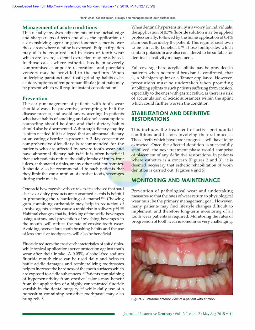
This includes the treatment of active periodontal conditions and lesions involving the oral mucosa. Those teeth which have poor prognosis will have to be extracted. Once the affected dentition is successfully stabilized, the next treatment phase would comprise of placement of any definitive restorations. In patients where esthetics is a concern [Figures 2 and 3], it is deemed necessary that esthetic rehabilitation of worn dentition is carried out [Figures 4 and 5].
MONITORING AND MAINTENANCE
Prevention of pathological wear and undertaking measures so that the rates of wear return to physiological wear must be the primary management goal. However, many patients may find lifestyle changes difficult to implement, and therefore long‑term monitoring of all tooth wear patients is required. Monitoring the rates of progression of tooth wear is sometimes very challenging.
Figure 2: Intraoral anterior view of a patient with attrition
[Downloaded free from http://www.jresdent.org on Monday, February 12, 2018, IP: 46.32.126.23]
42 • Journal of Restorative Dentistry / Vol - 3 / Issue - 2 / May-Aug 2015
Hanif, et al.: Classification, etiology and management of tooth surface loss
It is suggested that monitoring is carried out while taking the following measures:• High quality clinical photographs of the patients• Periodic study casts at the intervals of 6‑12 months
approximately• A sectional silicone index produced from the
preliminary study cast should be used as a reference guide.
It is very important that these review stages are instituted specially when passing from one treatment strategy to the other. This will also help to assess the efficacy of the completed treatment stage.
CONCLUSIONS
The prevalence of tooth surface loss is increasing. Early detection of tooth wear is of utmost important for the prevention of serious irreversible damages to an individual’s dentition. It is very important that the multifactorial nature of tooth wear and the risk factors of attrition, erosion, abrasion, and abfraction are understood. This will help us to plan the protocol of diagnosis, the strategy of the management, and a successful treatment outcome.
REFERENCES1. Litonjua LA, Andreana S, Bush PJ, Cohen RE. Tooth wear:
Attrition, erosion, and abrasion. Quintessence Int 2003;34:435‑46.2. Lussi A. Dental erosion. In: Lussi A, editor. Monographs in Oral
Science. Vol 20. Basel: Karger; 2006. p. 1‑8.3. Bishop K, Kelleher M, Briggs P, Joshi R. Wear now? An update
on the etiology of tooth wear. Quintessence Int 1997;28:305‑13.4. Mehta SB, Banerji S, Millar BJ, Suarez‑Feito JM. Current concepts
on the management of tooth wear: Part 1. Assessment, treatment planning and strategies for the prevention and the passive management of tooth wear. Br Dent J 2012;212:17‑27.
5. Lambrechts P, Braem M, Vuylsteke‑Wauters M, Vanherle G. Quantitative in vivo wear of human enamel. J Dent Res 1989;68:1752‑4.
6. Van’t Spijker A, Rodriguez JM, Kreulen CM, Bronkhorst EM, Bartlett DW, Creugers NH. Prevalence of tooth wear in adults. Int J Prosthodont 2009;22:35‑42.
7. Arens U. Oral health task force. Tooth wear. In: Arens U, editor. Oral Health, Diet and Other Factors: The Report of the British Nutrition Foundation’s Task Force. Amsterdam: Elsevier; 1999. p. 60‑2.
8. Dahl BL, Krogstad BS, Ogaatd B, Eckersberg T. Differences in functional variables, fillings and tooth wear in two groups of 19‑year old individuals. Acta Odontol Scand 1989;47:35‑40.
9. Fareed K, Johansson A, Omar R. Prevalence and severity of occlusal wear in young Saudi population. Acta Odontol Scand 1990;48:279‑85.
10. Pollman L, Berger F, Pollman B. Age and dental abrasion. Gerodontics 1987;3:94‑6.
11. Oilo G, Hatle G, Gad AL, Dahl BL. Wear of teeth in a mentally retarded population. J Oral Rehabil 1990;17:173‑7.
12. Wanman A, Wigren L. Need and demand for dental treatment a comparison between an evaluation based on an epidemiologic study of 35−50 and 65‑year olds and performed dental treatment of matched age groups. Acta Odontol Scand 1995;53:318‑24.
13. Solonen L, Heliden I, Carlsson GE. Prevalence of signs and symptoms of dysfunction in the masticatory system: An epidemiologic study in Swedish population. J Craniomandib Disord Facial 1990;4:241‑50.
14. Addy M, Shellis RP. Interaction between attrition, abrasion and erosion in tooth wear. Monogr Oral Sci 2006;20:17‑31.
15. Khan F, Young WG, Daley TJ. Dental erosion and bruxism; A tooth wear analysis from South East Queensland. Aust Dent J 1998;43:117‑27.
16. Smith BG, Knight JK. Index for measuring the wear of teeth. Br Dent J 1984;156:435‑8.
17. Imfeld T. Dental erosion. Definition, classification and links. Eur J Oral Sci 1996;104:151‑5.
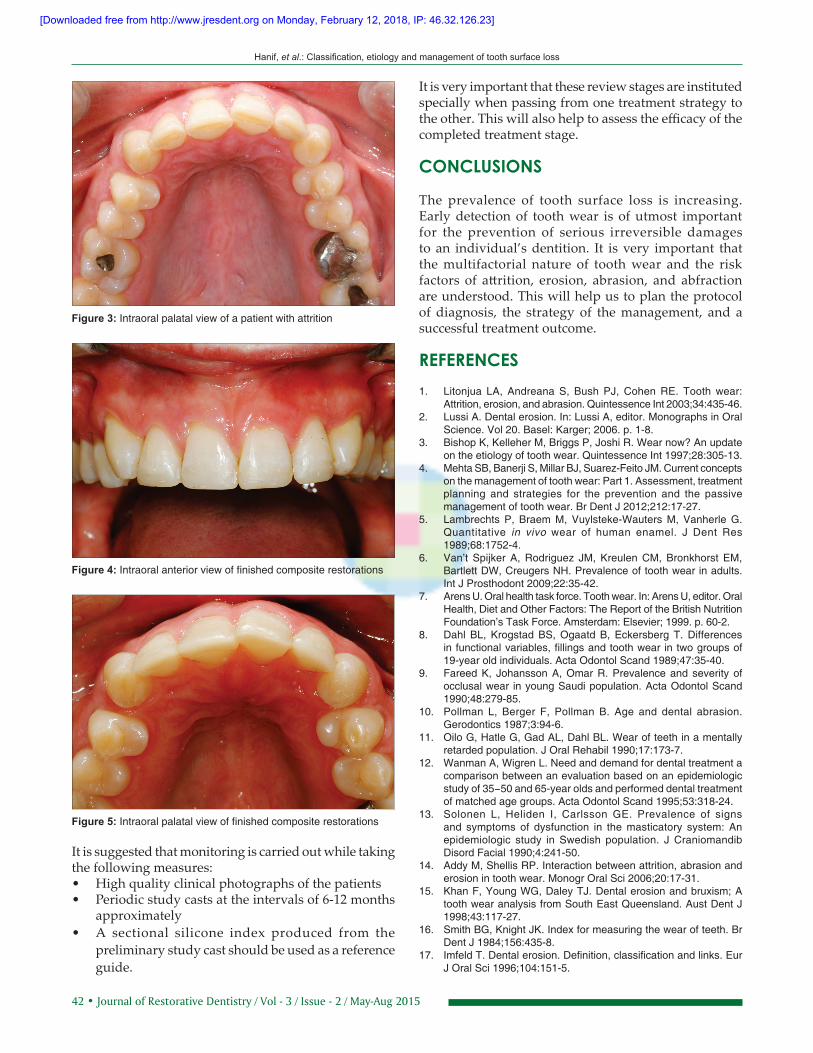
Figure 4: Intraoral anterior view of finished composite restorations
Figure 3: Intraoral palatal view of a patient with attrition
Figure 5: Intraoral palatal view of finished composite restorations
[Downloaded free from http://www.jresdent.org on Monday, February 12, 2018, IP: 46.32.126.23]
Journal of Restorative Dentistry / Vol - 3 / Issue - 2 / May-Aug 2015 • 43
Hanif, et al.: Classification, etiology and management of tooth surface loss
18. Dahl BL, Carlsson GE, Ekfeldt A. Occlusal wear of teeth and restorative materials. A review of classification, etiology, mechanisms of wear, and some aspects of restorative procedures. Acta Odontol Scand 1993;51:299‑311.
19. Haugen LK. Biological and physiological changes in the ageing dentition. Int Dent J 1992;42:339‑48.
20. Grippo JO. Abfractions: A new classification of hard tissue lesions of teeth. J Esthet Dent 1991;3:14‑9.
21. Sarode GS, Sarode SC. Abfraction: A review. J Oral Maxillofac Pathol 2013;17:222‑7.
22. Braem M, Lambrechts P, Vanherle G. Stress induced cervical lesions. J Prosthet Dent 1992;67:718‑22.
23. Anderson GC, Pintado MR, Beyer JP, DeLong R, Douglas WH. Clinical enamel wear as related to bruxism and occlusal scheme. J Dent Res 1993;72:303.
24. Licht WS, Leveton EE. Overdentures for treatment of severe attrition. J Prosthet Dent 1980;43:497‑500.
25. Chu FC, Yip HK, Newsome PR, Chow TW, Smales RJ. Restorative management of worn dentition: I. Aetiology and diagnosis. Dent Update 2002;29:162‑8.
26. Smith BG. Some facets of tooth wear. Ann R Australas Coll Dent Surg 1991;11:37‑51.
27. ten Bruggen Cate HJ. Dental erosion in industry. Br J Ind Med 1968;25:249‑66.
28. Petersen PE, Gormsen C. Oral conditions among German battery factory workers. Community Dent Oral Epidemiol 1991;19:104‑6.
29. Eccles JD, Jenkins WG. Dental erosion and diet. J Dent 1974;2:153‑9.
30. Li BU. Cyclic vomiting syndrome: Proceedings of the 1st international scientific symposium on cyclic vomiting syndrome held at St. Bartholomew’s Hospital, London, England, July 29‑30, 1994. J Pediatr Gastroenterol Nutr 1995;21.
31. Gilmoour AG, Beckett HA. The voluntary reflux phenomenon. Br Dent J 1993;175:338‑72.
32. Milosovec A, Young PJ, Lennon MA. The prevalence of tooth wear in 14 year old school children in Liverpool. Community Dent Health 1995;12:161‑6.
33. Giunta JL. Dental erosion resulting from chewable Vitamin C tablets. J Am Dent Assoc 1983;107:253‑6.
34. Jung YS, Lee JW, Choi YJ, Ahn JS, Shin SW, Huh JB. A study on the in‑vitro wear of the natural tooth structure by opposing zirconia or dental porcelain. J Adv Prosthodont 2010;2:111‑5.
35. Ardlin BI. Transformation‑toughened zirconia for dental inlays, crowns and bridges: Chemical stability and effect of low‑temperature aging on flexural strength and surface structure. Dent Mater 2002;18:590‑5.
36. Sulong MZ, Aziz RA. Wear of materials used in dentistry: A review of the literature. J Prosthet Dent 1990;63:342‑9.
37. DeLong R, Sasik C, Pintado MR, Douglas WH. The wear of enamel when opposed by ceramic systems. Dent Mater 1989;5:266‑71.
38. Mahalick JA, Knap FJ, Weiter EJ. Occusal wear in prosthodontics. J Am Dent Assoc 1971;82:154‑9.
39. Gallegos LI, Nicholls JI. In vitro two‑body wear of three veneering resins. J Prosthet Dent 1988;60:172‑8.
40. Seghi RR, Rosenstiel SF, Bauer P. Abrasion of human enamel by different dental ceramics in vitro. J Dent Res 1991;70:221‑5.
41. Coornaert J, Adriaens P, De Boever J. Long‑term clinical study of porcelain‑fused‑to‑gold restorations. J Prosthet Dent 1984;51:338‑42.
42. Cattell MJ, Clarke RL, Lynch EJ. The biaxial flexural strength and reliability of four dental ceramics–Part II. J Dent 1997;25:409‑14.
43. Scherrer SS, de Rijk WG, Belser UC. Fracture resistance of human enamel and three all‑ceramic crown systems on extracted teeth. Int J Prosthodont 1996;9:580‑5.
44. Aboushelib MN, de Jager N, Kleverlaan CJ, Feilzer AJ. Microtensile bond strength of different components of core veneered all‑ceramic restorations. Dent Mater 2005;21:984‑91.
45. Sundh A, Sjogren G. Fracture resistance of all‑ceramic zirconia bridges with differing phase stabilizers and quality of sintering. Dent Mater 2006;22:778‑84.
46. Isher RM, Moore BK, Swartz ML, Dykema RW. The effects of enamel wear on the metal‑porcelain interface. J Prosthet Dent 1983;50:627‑31.
47. Aboushelib MN, de Jager N, Kleverlaan CJ, Feilzer AJ. Effect of loading method on the fracture mechanics of two layered all‑ceramic restorative systems. Dent Mater 2007;23:952‑9.
48. Rosentritt M, Preis V, Behr M, Hahnel S, Handel G, Kolbeck C. Two‑body wear of dental porcelain and substructure oxide ceramics. Clin Oral Investig 2012;16:935‑43.
49. DeLong R, Douglas WH, Sakaguchi RL, Pintado MR. The wear of dental porcelain in an artificial mouth. Dent Mater 1986;2:214‑9.
50. Holbrook WP, Arnadóttir IB, Kay EJ. Prevention Part 3. Prevention of tooth wear. Br Dent J 2003;195:75‑81.
51. Cope GF, Cope AL. Tooth wear: Causes, diagnosis and prevention. Dent Nurs 2012;8:340‑4.
52. Watson ML, Burke FJ. Investigation and treatment of patients with teeth affected by tooth substance loss: A review. Dent Update 2000;27:175‑83.
53. Shafer W, Hine M, Levy B. A textbook of oral pathology. 7th ed. Philadelphia: WB Saunders, 1983. p. 318‑23.
54. Imfeld T, Birkhed D, Lingström P. Effects of urea in sugar free chewing gums on pH recovery in human dental plaque evaluated with three different methods. Caries Res 1998;29:172‑80.
55. Ritter AV, de L Dias W, Miguez P, Caplan DJ, Swift EJ Jr. Treating cervical dentin hypersensitivity with fluoride varnish: A randomized clinical study. J Am Dent Assoc 2006;137:1013‑20.
56. Thrash WJ, Dodds MW, Jones DL. The effects of stannous fluoride on dentinal hypersensitivity. Int Dent J 1994;1:107‑18.
How to cite this article: Hanif A, Rashid H, Nasim M. Tooth surface loss revisited: Classification, etiology, and management. J Res Dent 2015;3:37‑43.
Source of Support: Nil, Conflict of Intrest: Nil.
[Downloaded free from http://www.jresdent.org on Monday, February 12, 2018, IP: 46.32.126.23]