toll-like receptor 4 signaling leads to severe fungal ... · siliensis and constitutes the most...
TRANSCRIPT

INFECTION AND IMMUNITY, Mar. 2010, p. 1078–1088 Vol. 78, No. 30019-9567/10/$12.00 doi:10.1128/IAI.01198-09Copyright © 2010, American Society for Microbiology. All Rights Reserved.
Toll-Like Receptor 4 Signaling Leads to Severe Fungal InfectionAssociated with Enhanced Proinflammatory Immunity and
Impaired Expansion of Regulatory T Cells�
Flavio V. Loures,1 Adriana Pina,1 Maíra Felonato,1 Eliseu F. Araujo,1Katia R. M. Leite,2 and Vera L. G. Calich1*
Departamento de Imunologia, Instituto de Ciências Biomedicas, Universidade de Sao Paulo,1 and Servico de Patologia,Hospital Sírio Libanês,2 Sao Paulo, SP, Brazil
Received 22 October 2009/Returned for modification 18 November 2009/Accepted 4 December 2009
Toll-like receptors (TLRs) present in innate immune cells recognize pathogen molecular patterns andinfluence immunity to control the host-parasite interaction. The objective of this study was to characterize theinvolvement of TLR4 in the innate and adaptive immunity to Paracoccidioides brasiliensis, the most importantprimary fungal pathogen of Latin America. We compared the responses of C3H/HeJ mice, which are naturallydefective in TLR4 signaling, with those of C3H/HePas mice, which express functional receptors, after in vitroand in vivo infection with P. brasiliensis. Unexpectedly, we verified that TLR4-defective macrophages infectedin vitro with P. brasiliensis presented decreased fungal loads associated with impaired synthesis of nitric oxide,interleukin-12 (IL-12), and macrophage chemotactic protein 1 (MCP-1). After intratracheal infection with 1million yeasts, TLR4-defective mice developed reduced fungal burdens and decreased levels of pulmonarynitric oxide, proinflammatory cytokines, and antibodies. TLR4-competent mice produced elevated levels ofIL-12 and tumor necrosis factor alpha (TNF-�), besides cytokines of the Th17 pattern, indicating a proin-flammatory role for TLR4 signaling. The more severe infection of TLR4-normal mice resulted in increasedinflux of activated macrophages and T cells to the lungs and progressive control of fungal burdens butimpaired expansion of regulatory T cells (Treg cells). In contrast, TLR4-defective mice were not able toclear their diminished fungal burdens totally, a defect associated with deficient activation of T-cellimmunity and enhanced development of Treg cells. These divergent patterns of immunity, however,resulted in equivalent mortality rates, indicating that control of elevated fungal growth mediated byvigorous inflammatory reactions is as deleterious to the hosts as low fungal loads inefficiently controlledby limited inflammatory reactions.
Pathogen recognition receptors (PRRs) are a group of re-ceptors present in the membrane and cytoplasm of innateimmunity cells that recognize the presence of invading mi-crobes by interacting with conserved pathogen structures, theso called “pathogen-associated molecular patterns” (PAMPs).This initial event of innate immunity is crucial for the controlof pathogen growth and the subsequent activation of adaptiveimmunity. Toll like receptors (TLRs) constitute a major familyof pattern recognition molecules and, like other PRRs, areable to respond to different structural homologies conserved inmany microorganisms (2, 62). Activation of the TLRs is crucialfor many aspects of microbe elimination, including microbialkilling, recruitment of phagocytes to the site of infection, andactivation of dendritic cells (DCs) (52). Early TLR activationresults in the production of several inflammatory mediators,and the final balance among pro- and anti-inflammatory com-ponents regulates the type of adaptive immune response. Re-cent findings have shown that direct recognition of PAMPs byDCs is critical for priming appropriate T-cell responses, result-ing in T helper 1 (Th1), Th2, or Th17 immunity (25, 31, 33, 60).
TLR4 is the key receptor that recognizes bacterial lipopoly-saccharides (LPS), whereas TLR2 is involved in the interactionwith bacterial peptidoglycans and lipoproteins (66). As re-ported for other microorganisms, TLRs have been shown to beinvolved in host defense against different fungal pathogens. Invivo and in vitro studies have demonstrated that Cryptococcusneoformans (7, 67), Candida albicans (43, 45), and Aspergillusfumigatus (24, 41) may signal through members of the TLRfamily, mainly TLR2 and TLR4. Different components of acertain pathogen can be used to stimulate the immune system.Thus, C. albicans phospholipomannan is sensed by TLR2 (34),while O-linked mannans are recognized by TLR4 (44). Thecontribution of individual TLRs to the immune responseagainst pathogenic fungi depends on several factors, such asthe fungal morphotype, fungal species, and route of infection.Activation signals mediated by innate immunity receptors,however, are not always beneficial to the host, and TLR acti-vation can be used by pathogenic fungi to promote more-severe infections (6, 53).
Paracoccidioidomycosis (PCM) is a systemic granulomatousdisease caused by the dimorphic fungus Paracoccidioides bra-siliensis and constitutes the most prevalent deep mycosis inLatin America (28). The alveolar macrophages are the firsthost cells that interact with P. brasiliensis cells, and their acti-vation is fundamental to the control of fungal growth. Themolecular mechanisms controlling the initial steps of the in-
* Corresponding author. Mailing address: Departamento de Imuno-logia, Instituto de Ciências Biomedicas da Universidade de Sao Paulo,Av. Prof. Lineu Prestes 1730, CEP 05508-900, Sao Paulo, SP, Brazil.Phone: 55-11-30917397. Fax: 55-11-30917224. E-mail: [email protected].
� Published ahead of print on 14 December 2009.
1078
on January 1, 2020 by guesthttp://iai.asm
.org/D
ownloaded from

teraction between P. brasiliensis and phagocytes are not wellunderstood. It is known, however, that normal macrophagesare permissive to P. brasiliensis growth, while cytokine-acti-vated macrophages are able to restrain P. brasiliensis multipli-cation (12). Complement receptor 3 (CR3) and mannose re-ceptor have been shown to play important roles in the initialinteraction between P. brasiliensis cells and mouse peritonealmacrophages (14, 32, 50). Interestingly, recent work from ourlaboratory demonstrated that alveolar macrophages from sus-ceptible mice (B10.A) are easily activated by P. brasiliensisinfection and show efficient fungal killing associated with nitricoxide production, while pulmonary macrophages from resis-tant mice (A/Sn) are poorly activated and present inefficientkilling activity associated with increased levels of transforminggrowth factor � (TGF-�) (49). Despite their inefficient innateimmunity, A/Sn mice develop a balanced Th1/Th2 immuneresponse that controls fungal growth without intense tissuepathology.
In previous work, our group demonstrated the influence ofTLR2 on pulmonary PCM (15, 38). Using TLR2-normal andTLR2-deficient mice, we were able to show that the presenceof TLR2 increases the severity of in vitro and in vivo P. brasi-liensis infections. In addition, TLR2 deficiency results in in-creased Th17 immunity associated with diminished expansionof regulatory T cells (Treg cells) and increased lung pathologydue to unrestrained inflammatory reactions (38). Characteriz-ing the behavior of dendritic cells in murine PCM, Ferreira etal. observed increased expression of TLR2 by dendritic cells ofsusceptible, but not resistant, mice (26). Moreover, it has beensuggested that TLR2, TLR-4, and dectin-1 are involved in therecognition and internalization of P. brasiliensis by humanmonocytes and neutrophils, indicating important roles forthese pathogen receptors in the immune response against thefungus (10).
We decided to investigate the role of TLR4 in murine PCMby using in vitro and in vivo models of infection. Using TLR-competent C3H/HePas mice and C3H/HeJ mice, which pos-sess a missense mutation in the TLR4 gene, we were able todemonstrate that both in vitro and in vivo, TLR4 signalingincreases the severity of infection in association with increasedactivation of innate and adaptive immunity but decreased ex-pansion of Treg cells. This pattern of immunity, however, wasnot beneficial to the hosts, due to the increased lung injurymediated by inefficient control of inflammatory reactions byTreg cells.
MATERIALS AND METHODS
Fungus. P. brasiliensis Pb18, a highly virulent isolate, was used throughout thisinvestigation (36). Pb18 yeast cells were maintained by weekly subcultivation insemisolid culture medium. Washed yeast cells were adjusted to 20 � 106/ml (forin vivo infection) and 4 � 104/ml (for in vitro infection) based on hemocytometercounts. Viability was determined with Janus Green B vital dye (Merck) and wasalways higher than 85%.
Mice and i.t. infection. C3H/HeJ and C3H/HePas mice were obtained fromour Isogenic Breeding Unit (Departmento de Imunologia, Instituto de CiênciasBiomedicas, Universidade de Sao Paulo, Sao Paulo, Brazil) and were used at 8to 12 weeks of age. The C3H/HeJ strain has a point mutation in the TLR4 gene,and the C3H/HePas strain has a functional TLR4 gene. In selected experiments,C57BL/6 mice that are genetically deficient in TLR4 (TLR4 knockout [KO]) andtheir normal C57BL/6 counterparts were used. Mice were anesthetized andsubjected to intratracheal (i.t.) P. brasiliensis infection as previously described(18). Briefly, after intraperitoneal anesthesia, the animals were i.t. infected with
106 P. brasiliensis yeast cells, contained in 50 �l of phosphate-buffered saline(PBS). Mice were studied at 96 h, 2 weeks, and 11 weeks postinfection. Theexperiments were approved by the ethics committee on animal experiments ofour institution.
Phagocytic and fungicidal assays. Thioglycolate-induced peritoneal macro-phages were either isolated by adherence (2 h at 37°C under 5% CO2) toplastic-bottom tissue culture plates (1 � 106 cells/well in 24-well plates forfungicidal assays) or plated onto 13-mm-diameter round glass coverslips (1 � 106
cells/well in 24-well plates) for phagocytosis. Macrophages were washed to re-move nonadherent cells and were cultivated overnight with fresh complete me-dium (Dulbecco’s modified Eagle’s medium [DMEM; Sigma], containing 10%fetal calf serum, 100 U/ml penicillin, and 100 �g/ml streptomycin) in the pres-ence or absence of recombinant gamma interferon (IFN-�; 20 ng/ml in culturemedium; BD Pharmingen). Nonadherent cells were counted in order to evaluatethe number of remaining adherent cells used in phagocytic and killing assays. Forphagocytic assays, macrophage cultures were infected with P. brasiliensis yeasts ata macrophage/yeast ratio of 25:1. This ratio was previously determined and wasshown not to be deleterious to macrophage cultures and to be adequate forphagocytosis and killing assays (18, 49). The cells were cocultivated for 4 h at37°C under 5% CO2 to allow adhesion and ingestion of fungi. Cells were washedtwice with PBS to remove any noningested or nonadhered yeasts, and sampleswere processed for microscopy. Cells were fixed with methanol and stained withGiemsa stain (Sigma). Experimental conditions were performed in triplicate, andthe number of phagocytosed or adhered yeasts per 1,000 macrophages wasevaluated on at least three separate slides. For fungicidal assays, IFN-�-primedand unprimed macrophage cultures were infected with P. brasiliensis yeasts asdescribed above. After 48 h of culture at 37°C in a CO2 incubator, plates werecentrifuged (400 � g, 10 min, 4°C), and supernatants were stored at �70°C andwere further analyzed for the presence of nitrite and cytokines. The wells werewashed with distilled water to lyse macrophages, and suspensions were collectedin individual tubes. One hundred microliters of cell homogenates was assayed forthe presence of viable yeasts. All assays were done with five wells per conditionin more than three independent experiments.
CFU assay and histological and morphometric analyses. The numbers ofviable microorganisms in cell cultures and infected organs (lungs, liver, andspleen) from experimental and control mice were determined by counting thenumber of CFU. Animals from each group were sacrificed, and the enumerationof viable organisms was done as previously described (59). The numbers (log10)of viable P. brasiliensis organisms per gram of tissue (in vivo) or per milliliter ofcell homogenate (in vitro) are expressed as means � standard errors. For histo-logical examinations, the left lung of the infected mouse was removed, fixed in10% formalin, and embedded in paraffin. Five-micrometer-thick sections werestained with hematoxylin and eosin (H&E) for analysis of the lesions and weresilver stained for fungal evaluation. Pathological changes were analyzed based onthe size, morphology, and cell composition of granulomatous lesions, the pres-ence of fungi, and the intensity of the inflammatory infiltrates. Morphometricanalysis was performed using a Nikon DXM 1200c digital camera (magnifica-tion, �10), and Nikon NIS Elements AR 2.30 software. The areas of lesions weremeasured (in square micrometers) in 10 microscopic fields per slide for 10animals per group. Results were expressed as the mean total area of lesions foreach animal � the standard error.
Measurement of cytokines and NO. Supernatants from lung homogenates or cellcultures were separated and stored at �70°C. The levels of interleukin-2 (IL-2), IL-4,IL-5, IL-23, IL-17, IL-12, IL-10, tumor necrosis factor alpha (TNF-�), IFN-�, andthe chemokine MCP-1 (monocyte chemoattractant protein 1) were measured by acapture enzyme-linked immunosorbent assay (ELISA) with antibody pairs pur-chased from BD Pharmingen. The ELISA procedure was performed according tothe manufacturer’s protocol, and absorbances were measured with a Versa Maxmicroplate reader (Molecular Devices). NO production was quantified by the accu-mulation of nitrite in the supernatants from in vitro and in vivo protocols by astandard Griess reaction. All determinations were performed in duplicate, andresults were expressed as micromolar concentrations of NO.
Measurement of serum P. brasiliensis-specific isotypes. Levels of specific iso-types (total IgG, IgM, IgA, IgG1, IgG2a, IgG2b, and IgG3) were measured by apreviously described ELISA (18) employing a cell-free antigen prepared by usinga pool of different P. brasiliensis isolates (Pb339, Pb265, and Pb18). The averageof the optical densities obtained with sera from control mice (PBS inoculated),diluted 1:20, was considered the cutoff for each respective isotype. Optical den-sities for each dilution of experimental sera were compared to control values.The titer for each sample was expressed as the reciprocal of the highest dilutionthat presented an absorbance higher than the cutoff.
Assessment of leukocyte subpopulations in lung inflammatory exudates. After2 and 11 weeks of infection, lungs from each mouse were digested enzymat-
VOL. 78, 2010 ROLE OF TLR4 IN PULMONARY PARACOCCIDIOIDOMYCOSIS 1079
on January 1, 2020 by guesthttp://iai.asm
.org/D
ownloaded from

ically for 30 min with collagenase (1 mg/ml) and DNase (30 �g/ml) in culturemedium (Sigma). Lung cell suspensions were centrifuged in the presence of20% Percoll (Sigma) to separate leukocytes from cell debris. Total lungleukocyte numbers were assessed in the presence of trypan blue using ahemocytometer; viability was �85%. For differential leukocyte counts, sam-ples of lung cell suspensions were cytospun (Shandon Cytospin) onto glassslides and stained with the Diff-Quik blood stain (Baxter Scientific). A totalof 200 to 400 cells were counted from each sample. For flow cytofluorometry,lung leukocytes were resuspended at 106/ml in staining buffer (PBS with 0.1%NaN3 and 1% fetal calf serum). Fc receptors were blocked by unlabeledanti-CD16/32 antibodies (BD Biosciences), and cells were stained for 20 minat 4°C. Phycoerythrin (PE)-labeled monoclonal antibodies against CD40,CD86, CD11b, CD25, and CD69 and fluorescein isothiocyanate (FITC)-labeled monoclonal antibodies against IAk, CD80, CD4, and CD8 (BD Bio-sciences) were used. Treg cells were characterized by intracellular staining forFoxp3, using the Treg staining kit of BD Bioscience. Surface staining ofCD25 and intracellular FoxP3 expression were backgated on the CD4
T-cell population. Cells were fixed with 1% paraformaldehyde (Sigma) andwere stored in the dark at 4°C until analysis in a flow cytometer. The acqui-sition and analysis gates were restricted to the lymphocytes or macrophages.The expression of leukocyte markers on the cell surface and the intracellularexpression of FoxP3 in lung-infiltrating leukocytes were analyzed in aFACScalibur flow cytometer (BD Pharmingen) using FlowJo software(Tristar).
Limulus amoebocyte lysate activity assay. Solutions used for the preparationof yeast cell suspensions and the cultivation of macrophages were tested forthe presence of LPS using the Limulus amoebocyte lysate chromogenic assay(E-TOXATE; Sigma) and always showed LPS levels lower than 0.015 endo-toxin unit (EU)/ml.
Statistical analyses. Data were analyzed by Student’s t test or two-way analysisof variance depending on the number of experimental groups. P values under0.05 were considered significant.
RESULTS
TLR4 deficiency leads to less-severe fungal infection of mac-rophages, associated with decreased synthesis of NO, IL-12,and MCP-1. Before performing fungicidal studies, we askedwhether the initial interactions between P. brasiliensis yeastsand peritoneal macrophages from C3H/HeJ and C3H/HePasmice were equivalent. Macrophage cultures (1 � 106 cells/well), performed in round glass coverslips, were preactivatedwith IFN-� (20 ng/ml) or not and were infected with 4 � 104
viable yeasts (fungus/macrophage ratio, 1:25). After 4 h ofincubation, supernatants were aspirated, the monolayers weregently washed with PBS, and the cells were stained with Gi-emsa stain. An average of 1,000 macrophages were counted,and the number of ingested and/or adherent yeasts was deter-mined. As shown in Fig. 1A, TLR4-deficient macrophagespresented a lower number of associated (ingested/adhered)yeasts than normal macrophages.
Macrophages were cultivated with P. brasiliensis yeasts foran additional 48 h. Supernatants were removed and assayed forthe presence of nitric oxide and cytokines, and cell homoge-nates were plated for CFU determinations. As can be seen inFig. 1B, TLR4 signaling resulted in increased recovery of via-ble yeasts from untreated and IFN-�-primed macrophages. Inaddition, higher levels of NO were produced by IFN-�-acti-vated macrophages from TLR4-normal mice than by those ofTLR4-defective mice (Fig. 1C). P. brasiliensis infection ofunprimed macrophages did not induce NO production, whichwas detected only in IFN-�-treated macrophages.
We further asked if TLR4 expression or signaling was in-volved in the decreased fungal loads observed with TLR4-defective macrophages. Since C3H/HeJ mice have a defect inthe intracellular signaling domain but a normal extracellular
moiety, we comparatively assessed the behavior of macro-phages genetically deficient for TLR4 expression. Thus,TLR4�/� macrophages from C57BL/6 mice were infected andcompared with their TLR4/ counterparts. TLR4-deletedmacrophages showed decreased phagocytic ability (Fig. 1D),and decreased numbers of viable yeasts were recovered fromkilling assays (Fig. 1E). As with C3H/HeJ macrophages,TLR4�/� cells produced diminished levels of NO. This resultsuggested that TLR4 signaling influenced the endocytic andkilling ability of P. brasiliensis-infected macrophages.
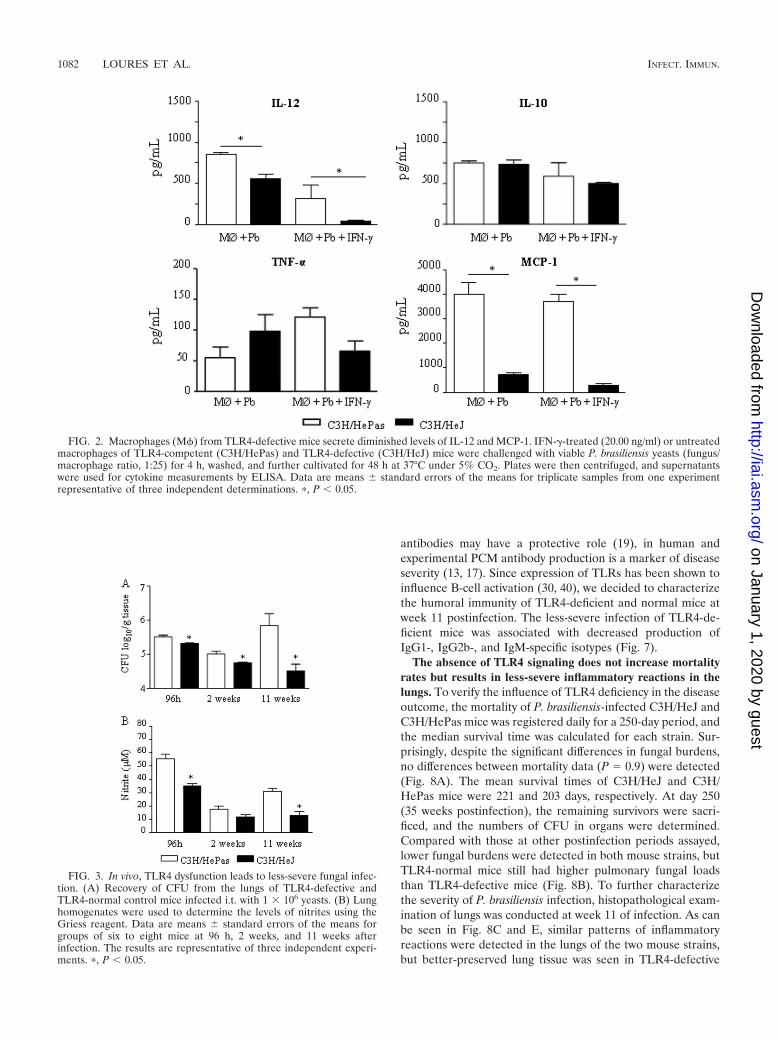
To further characterize the role of TLR4 in P. brasiliensis in-fection of C3H macrophages, culture supernatants obtained fromkilling assays were tested for the presence of some macrophage-activating cytokines (IL-12 and TNF-�), a deactivating cytokine(IL-10), and a chemokine involved in mononuclear cell chemo-taxis, MCP-1. As depicted in Fig. 2, IFN-�-treated and untreatedmacrophages from TLR4-defective mice secreted lower levels ofIL-12 and MCP-1 than those of TLR4-normal mice. IL-10 andTNF-�, however, appeared at similar levels.
In vivo, the absence of TLR4 signaling induces lower fungalloads and diminished NO production. To study the in vivo roleof TLR4, groups of C3H/HeJ and C3H/HePas mice (six toeight animals per group) were infected i.t. with 1 million P.brasiliensis yeast cells and were evaluated in the course ofinfection. Diminished fungal burdens were detected in the lungtissues of mice lacking functional TLR4 at all postinfectiontimes (96 h and 2 and 11 weeks) assayed, as can be seen in Fig.3A. In both strains, no fungal growth was observed in liver andspleen tissues (data not shown). Decreased NO levels weredetected at 96 h and at week 11 after infection, although byweek 2 similar levels were observed (Fig. 3B).
Defective TLR4 signaling determines decreased inflamma-tory reactions characterized by lower numbers of activatedmacrophages and T cells. We further analyzed the phenotypeand activation of lung inflammatory cells at weeks 2 and 11 ofP. brasiliensis infection (Fig. 4). To determine the activationprofile of pulmonary macrophages, the expression of CD11b,major histocompatibility complex (MHC) class II (IAK),CD80, CD86, and CD40 antigens was assessed by flow cytom-etry. As can be seen in Fig. 4A, all activation markers wereexpressed at lower levels by deficient macrophages, althoughsignificant differences were noticed with CD11b, the MHCclass II antigen, and CD86. To determine the lymphocyte in-flux and the activation profile of CD4 and CD8 T cells in thelungs of P. brasiliensis-infected mice, we determined the ex-pression of CD69 and CD25 by T cells freshly isolated from thelungs. The marker CD69 is a very early activation antigen (70),as well as CD25, the �-chain of the interleukin-2 receptor (56),which is rapidly upregulated on activated T cells. Comparedwith the control group, at week 2 of infection, TLR4-deficientmice presented significantly reduced recruitment of CD4 andCD8 T cells to the lungs, and the latter subpopulation alsoshowed decreased expression of CD69 (Fig. 4B). Studies atweek 11 postinfection confirmed those of week 2. TLR4-nor-mal mice presented increased numbers of CD11b, CD11c,and CD40 macrophages (Fig. 4C), besides augmented num-bers of CD4, CD8, and CD8 CD69 T lymphocytes, in theinflammatory exudates of lungs (Fig. 4D).
The limited inflammatory reaction of TLR4-deficient micewas associated with increased numbers of Treg cells. Because
1080 LOURES ET AL. INFECT. IMMUN.
on January 1, 2020 by guesthttp://iai.asm
.org/D
ownloaded from

Treg cells control the expansion of effector T cells, and becausethe number and function of these cells have been shown to beinfluenced by TLR4 activation (33), we investigated the pres-ence of CD4 CD25 FoxP3 T cells in the lung cell infiltratesof TLR4-defective and normal mice (Fig. 5). At both postin-fection periods studied, TLR4-defective mice showed in-creased numbers of CD4 CD25 FoxP3 Treg cells in theirlungs (Fig. 5).
TLR4 dysfunction leads to diminished production of proin-flammatory and Th17 cytokines. Levels of cytokines associatedwith Th1, Th2, and Th17 cells were assessed in lung homoge-nates obtained at different periods of infection. The produc-tion of type 1 (IL-12, TNF-�, and IFN-�) and type 2 (IL-4,
IL-5, and IL-10) cytokines, as well as that of the Th17-associ-ated (IL-17, IL-6, TGF-�, and IL-23) cytokines, was studied96 h, 2 weeks, and 11 weeks after infection. Mice lacking theability to signal through TLR4 showed early (96 h) deficientproduction of IL-12, TNF-�, IL-17, and IL-6 (Fig. 6A). Byweek 2, IL-17 and IL-23 appeared at lower levels in the lungsof TLR4-defective mice (Fig. 6B). This decreased productionof cytokines was confirmed at week 11, when these mice pre-sented decreased amounts of IL-12, IL-17, and TGF-� (Fig.6C). Interestingly, IL-17 and MCP-1 were constantly producedat higher levels by TLR4-normal mice (Fig. 6).
TLR4-defective mice produced lower levels of P. brasiliensis-specific antibodies. Although in some fungal infections specific
FIG. 1. TLR4 deficiency leads to less-severe fungal infection of macrophages (M) associated with decreased synthesis of NO. The phagocyticand fungicidal abilities of macrophages from mice with defective TLR4 signaling (C3H/HeJ) or defective TLR4 expression (C57BL/6 TLR4�/�)were compared with those of their TLR4-normal controls (C3H/HePas and C57BL/6 TLR4/, respectively). (A and D) For phagocytic assays,IFN-�-primed (20 ng/ml, overnight) and unprimed macrophage cultures were infected with P. brasiliensis yeasts at a macrophage/yeast ratio of 25:1.The cells were cocultivated for 4 h at 37°C under 5% CO2 to allow adhesion and ingestion of fungi. Cells were washed, fixed, and stained withGiemsa stain; an average of 1,000 macrophages were analyzed, and the number of macrophages with adhered or ingested yeasts was determined.(B and E) For fungicidal assays, IFN-�-primed and unprimed macrophages were infected with yeast cells as described for panel A. After 48 h at37°C under 5% CO2, plates were centrifuged, and supernatants were used to determine levels of nitrite and cytokines. The monolayers were washedwith distilled water to lyse macrophages, and 100 �l of cell homogenates was assayed for the presence of viable yeasts by a CFU assay. (C and F)Supernatants from fungicidal assays were used to determine the levels of nitrites using the Griess reagent. Data are means � standard errors ofthe means for quintuplicate samples from one experiment representative of three independent determinations. �, P � 0.05.
VOL. 78, 2010 ROLE OF TLR4 IN PULMONARY PARACOCCIDIOIDOMYCOSIS 1081
on January 1, 2020 by guesthttp://iai.asm
.org/D
ownloaded from
antibodies may have a protective role (19), in human andexperimental PCM antibody production is a marker of diseaseseverity (13, 17). Since expression of TLRs has been shown toinfluence B-cell activation (30, 40), we decided to characterizethe humoral immunity of TLR4-deficient and normal mice atweek 11 postinfection. The less-severe infection of TLR4-de-ficient mice was associated with decreased production ofIgG1-, IgG2b-, and IgM-specific isotypes (Fig. 7).
The absence of TLR4 signaling does not increase mortalityrates but results in less-severe inflammatory reactions in thelungs. To verify the influence of TLR4 deficiency in the diseaseoutcome, the mortality of P. brasiliensis-infected C3H/HeJ andC3H/HePas mice was registered daily for a 250-day period, andthe median survival time was calculated for each strain. Sur-prisingly, despite the significant differences in fungal burdens,no differences between mortality data (P � 0.9) were detected(Fig. 8A). The mean survival times of C3H/HeJ and C3H/HePas mice were 221 and 203 days, respectively. At day 250(35 weeks postinfection), the remaining survivors were sacri-ficed, and the numbers of CFU in organs were determined.Compared with those at other postinfection periods assayed,lower fungal burdens were detected in both mouse strains, butTLR4-normal mice still had higher pulmonary fungal loadsthan TLR4-defective mice (Fig. 8B). To further characterizethe severity of P. brasiliensis infection, histopathological exam-ination of lungs was conducted at week 11 of infection. As canbe seen in Fig. 8C and E, similar patterns of inflammatoryreactions were detected in the lungs of the two mouse strains,but better-preserved lung tissue was seen in TLR4-defective
FIG. 2. Macrophages (M) from TLR4-defective mice secrete diminished levels of IL-12 and MCP-1. IFN-�-treated (20.00 ng/ml) or untreatedmacrophages of TLR4-competent (C3H/HePas) and TLR4-defective (C3H/HeJ) mice were challenged with viable P. brasiliensis yeasts (fungus/macrophage ratio, 1:25) for 4 h, washed, and further cultivated for 48 h at 37°C under 5% CO2. Plates were then centrifuged, and supernatantswere used for cytokine measurements by ELISA. Data are means � standard errors of the means for triplicate samples from one experimentrepresentative of three independent determinations. �, P � 0.05.
FIG. 3. In vivo, TLR4 dysfunction leads to less-severe fungal infec-tion. (A) Recovery of CFU from the lungs of TLR4-defective andTLR4-normal control mice infected i.t. with 1 � 106 yeasts. (B) Lunghomogenates were used to determine the levels of nitrites using theGriess reagent. Data are means � standard errors of the means forgroups of six to eight mice at 96 h, 2 weeks, and 11 weeks afterinfection. The results are representative of three independent experi-ments. �, P � 0.05.
1082 LOURES ET AL. INFECT. IMMUN.
on January 1, 2020 by guesthttp://iai.asm
.org/D
ownloaded from

mice. The pulmonary tissue presented several confluent orisolated granulomas of various sizes containing yeast cells withpreserved morphology (Fig. 8D and F). Large aggregates ofmacrophages, rare epithelioid cells, and a poor mantle of lym-phocytes made up the granulomas, which were usually in theinterlobular septa. Plasma cells, eosinophils, and multinucle-ated cells were scarcely seen. The total areas of lesions werequantified in histological sections, and the results are shown inFig. 8G. At week 11, the areas of lesions of TLR4-normal micewere significantly larger than those presented by TLR4-defi-cient mice. Thus, the higher influx of inflammatory cells ob-served in the lungs of TLR4-normal mice was concomitantwith increased pathology of lung tissue.
DISCUSSION
The innate immune mechanisms of hosts infected with P.brasiliensis are poorly defined, but macrophages and theirpathogen recognition receptors are thought to play a crucialrole in the initial interaction of this fungus with the immunesystem (16, 26, 29, 49, 50). Despite several studies with diversefungal pathogens (16, 43), the role played by TLRs in para-coccidioidomycosis is still unclear. In a previous report wewere able to show the dual role played by TLR2 in the immu-nity to P. brasiliensis infection. TLR2 activation prevented un-
FIG. 4. Increased numbers of activated macrophages, CD4 T lymphocytes, and CD8 T lymphocytes were detected in the lungs ofTLR4-competent mice at weeks 2 and 11 of infection. Leukocyte subsets in the lung-infiltrating leukocytes (LIL) from TLR4-defective andTLR4-normal mice inoculated i.t. with 1 million P. brasiliensis yeast cells were characterized by flow cytometry. Lungs of C3H/HePas and C3H/HeJmice (six to eight mice per group) were excised, washed in PBS, minced, and digested enzymatically. At weeks 2 and 11 after infection, lung cellsuspensions were obtained and stained as described in Materials and Methods. The acquisition and analysis gates were restricted to lymphocytesor macrophages. The data are mean results from six to eight mice per group � standard errors of the means and are representative of twoindependent experiments. �, P � 0.05.
FIG. 5. TLR4-defective mice presented increased numbers of Tregcells in the lungs. FoxP3 expression by lung lymphocytes from TLR4-defective (C3H/HeJ) and normal (C3H/HePas) mice inoculated i.t.with 1 million P. brasiliensis yeast cells was determined by flow cyto-metric analysis. Lungs of six to eight mice per group were excised,washed in PBS, minced, and digested enzymatically; at 2 and 11 weeksafter infection, cell suspensions were obtained and stained as describedin Materials and Methods. The expression of leukocyte markers on thecell surface, as well as intracellular FoxP3 expression in lung-infiltrat-ing leukocytes, was analyzed by flow cytometry. Surface staining ofCD25 cells and intracellular FoxP3 expression were backgated on theCD4 T-cell population. The data are numbers of CD4 CD25
FoxP3 cells for individual mice (five or six per group) and are rep-resentative of two independent experiments.
VOL. 78, 2010 ROLE OF TLR4 IN PULMONARY PARACOCCIDIOIDOMYCOSIS 1083
on January 1, 2020 by guesthttp://iai.asm
.org/D
ownloaded from

controlled inflammatory reactions in pulmonary paracoccid-ioidomycosis associated with increased expansion of Th17 cellsand diminished function of Treg cells (38).
Initially we characterized the influence of TLR4 on thephagocytic and fungicidal abilities of macrophages. Both theabsence of TLR4 expression by TLR4�/� C57BL/6 mice anddefective TLR4 signaling (C3H/HeJ mice) resulted in deficientP. brasiliensis ingestion/adherence and lower fungal loads re-covered 48 h after cocultivation. In both deficient mousestrains, lower levels of nitric oxide (and cytokines with C3H/HeJ cells) were detected, indicating that the lower CFU countsrecovered were not due to increased activation of phagocytesand enhanced fungal killing but probably were due to de-creased endocytosis of P. brasiliensis yeasts. TLRs usually donot act as phagocytic receptors, but their engagement bypathogen components results in strong activation of inflamma-
tory responses (8, 9). There are, however, several examplesdemonstrating that cell signaling can influence endocytosis andvice versa (20, 23). Indeed, TLR4 was shown to actively par-ticipate in bacterial phagocytosis (4) and to be rapidly inter-nalized by human monocytes after in vitro interaction with P.brasiliensis yeasts or A. fumigatus conidia (10, 21). In addition,a recent paper has clearly demonstrated that TLR4 and TLR2synergize with class A scavenger receptor to mediate phagocy-tosis of Gram-negative and Gram-positive bacteria, respec-tively (3). Thus, we can suppose that TLR signaling facilitatedthe endocytosis of P. brasiliensis and further induced the acti-vation of proinflammatory pathways, which, however, were notsufficient to control the early increased fungal loads. Sinceequivalent results were obtained with macrophages lackingTLR4 expression, we believe that TLR4 signaling could haveinfluenced phagocytosis mediated by another pathogen recep-tor. Although our experiments have not identified the mainPRR involved in initial P. brasiliensis recognition (particularlydue to the number and complexity of components that com-prise fungal cell walls), we have clearly demonstrated thatTLR4 participates in the activation of innate immune cellsrequired for the initial interaction with P. brasiliensis yeasts.Our in vitro findings were validated by in vivo experiments,which demonstrated that early in infection, TLR4-normal micepresented higher fungal loads than their TLR4-defective coun-terparts and that this was accompanied by increased activationof the immune system. Additional experiments with PRR ago-nists and antagonists using TLR4-normal and -deficient cellsare needed, however, to further clarify the role of TLRs inpulmonary PCM.
Our in vivo data showed that mice expressing defectiveTLR4 developed a less-severe infection associated with lowerproduction of nitric oxide and cytokines and less migration ofinflammatory cells to the site of infection. The decreased pres-ence of activated macrophages expressing CD11b, CD86,CD40, and MHC class II molecules was concomitant withreduced synthesis of MCP-1. In addition, the diminished pres-ence of CD4 T cells and recently activated CD69 CD8 Tcells in the lungs of TLR4-defective mice demonstrates that
FIG. 6. Lung homogenates of TLR4-competent mice presented in-creased levels of proinflammatory cytokines. At 96 h, 2 weeks, and 11weeks after i.t. infection with 106 P. brasiliensis yeast cells, lungs fromTLR4-defective and TLR4-competent mice were collected and dis-rupted in 5.0 ml of PBS, and supernatants were analyzed for cytokinecontents by capture ELISAs. Data are mean cytokine levels � stan-dard errors of the means (six to eight animals per group). The resultsare representative of three independent experiments. �, P � 0.05.
FIG. 7. TLR4 deficiency determines impaired humoral immunity.Levels of P. brasiliensis-specific antibodies in the sera of TLR4-defec-tive (C3H/HeJ) and normal (C3H/HePas) mice at week 11 after i.t.infection with 1 � 106 yeast cells are shown. Sera were assayed for totalIgG, IgM, IgA, IgG1, IgG2a, IgG2b, and IgG3 by using an isotype-specific ELISA as detailed in Materials and Methods. Data are meanserum titers (log2) � standard errors (six to eight mice per group). �,P � 0.05 for comparison with controls.
1084 LOURES ET AL. INFECT. IMMUN.
on January 1, 2020 by guesthttp://iai.asm
.org/D
ownloaded from

FIG. 8. Compared with TLR4-normal mice, TLR4-defective mice present decreased fungal loads and lung pathology but equivalent survivaltimes. (A) Survival times of TLR4-defective and control mice after i.t. infection with 1 � 106 P. brasiliensis yeast cells were determined for a periodof 250 days. No significant differences were seen between the median survival times of the two mouse strains. The results are representative of twoindependent experiments. (B) By 250 days after infection, survivor mice (three to six per group) were sacrificed, and CFU counts in tissues weredetermined. No viable yeasts were recovered from livers and spleens. (C to F) Photomicrographs of pulmonary lesions of TLR4-competentC3H/HePas mice (C and D) and TLR4-defective C3H/HeJ mice (E and F) at week 11 of infection with 1 million P. brasiliensis yeasts. At thisperiod, the morphology of lesions was equivalent in the two mouse strains; fungal cells were surrounded by confluent or isolated granulomasscattered through the lung tissue. Lesions were stained with H&E (C and E) or with Grocott stain (D and F); magnification, �100. (G) Total areasof lesions in the lungs of mice (n � 10) at week 11 after infection. �, P � 0.05.
VOL. 78, 2010 ROLE OF TLR4 IN PULMONARY PARACOCCIDIOIDOMYCOSIS 1085
on January 1, 2020 by guesthttp://iai.asm
.org/D
ownloaded from

TLR4 signaling is necessary to the proper activation of adap-tive immunity to P. brasiliensis and enhanced migration ofinflammatory cells into the lungs. Consistent with these obser-vations, several reports have demonstrated that TLR4 signal-ing is needed for the activation and maturation of dendriticcells, which acquire the competent phenotype to preferentiallydifferentiate naïve T cells to the Th1 or Th17 pattern (57, 64,65). No differences in Th1 and Th2 cytokines, however, weredetected in lung homogenates. The increased production ofIL-12 and TNF-� concomitant with unaltered synthesis of Th2cytokines (IL-4, IL-5, and IL-10) indicated, however, thatTLR4 signaling promoted a cytokine milieu biased toward aproinflammatory balance. This cytokine response could haveprotected C3H/HePas mice from high fungal burdens due tothe enhanced fungicidal mechanisms of activated phagocytes.Indeed, in experimental and human PCM, cytokine-activatedphagocytes (activated mainly by IFN-�, IL-12, and TNF-�)were shown to be the most important effector cells against P.brasiliensis infection (36, 39, 46, 55). Our data on cytokineproduction showed an additional fact not previously reportedin PCM. The expression of TLR4 facilitated the expansion ofIL-17-producing cells, since IL-17 and other Th17-associatedcytokines (IL-6 and IL-23) appeared at higher levels in thelungs of TLR4-competent mice. In our previous report, wecould verify that the absence of TLR2 signaling induced en-hanced expansion of Th17 cells and that both CD4 and CD8
T cells displayed intracellular IL-17 (38). Further studies of theTLR4-deficient model will help us to characterize the pheno-type of cells involved in IL-17 production.
TLR4 ligation is important for the activation of Th1 or Th17responses (65), while TLR4-deficiency can lead to increasedexpansion of CD4 CD25 regulatory T cells (47, 48). Whenthe presence of Treg cells in the lungs of Toll-deficient andcontrol mice at weeks 2 and 11 of infection was assessed,increased numbers of CD4 CD25 FoxP3 cells were foundin the lungs of TLR4-defective mice. This finding was associ-ated with decreased fungal loads and diminished influx ofinflammatory cells to the site of infection. Since Treg cells havebeen shown to control the inductive and effector phases ofimmunity against pathogens (5), we can suppose that Treg cellscould have negatively controlled the expansion and migrationof P. brasiliensis-specific T cells to the lungs. Thus, the advan-tage of low fungal loads conferred by TLR4 deficiency ap-peared to be negatively compensated for by deficient T-cellimmunity and increased numbers of Treg cells, which appearto hamper the total clearance of fungal cells from the lungs.
At week 11 of infection, decreased levels of IL-12 weredetected, probably due to the decreased migration of macro-phages to the lungs. Interestingly, in C3H/HeJ mice, decreasedlevels of IL-17 were concomitant with diminished levels ofTGF-�, indicating that another cytokine or costimulatory sig-nal could have participated in the increased expansion of Tregcells (27).
Since the expression of TLRs has been shown to influenceB-cell activation (40), we decided to analyze the levels ofanti-P. brasiliensis isotypes in our model and observed an im-paired humoral immune response in TLR4-defective mice. Atweek 11 of infection (Fig. 7), TLR4-deficient mice producedlower levels of IgG1-, IgG2b-, and IgM-specific antibodies.This could be due to the diminished fungal loads or the de-
creased production of cytokines observed in this mouse strain.Alternatively, since almost all TLR ligands were recentlyshown to induce the expansion and differentiation of B cells(30), we can suppose that TLR4 agonists present in P. brasil-iensis yeasts could have exerted a stimulatory effect on B cellsof TLR4-normal mice, resulting in increased humoral immu-nity. Independently of the mechanisms used, this is the firstdemonstration on the stimulatory role of TLR4 in the humoralimmunity of P. brasiliensis-infected hosts.
TLR4 recognizes LPS of Gram-negative bacteria and favorsTh1immunity due to the increased ability of LPS-stimulatedDCs to produce IL-12 and TNF-� (51). In some fungal infec-tions, however, cell wall polysaccharides have been reported tofunction as TLR agonists (42, 63). To our knowledge, no stud-ies of paracoccidioidomycosis have addressed the characteriza-tion of TLR agonists. Although LPS or LPS-like componentshave not been characterized in P. brasiliensis, a few investiga-tions have described the presence of polysaccharides, lipids,and glycolipids in P. brasiliensis cell walls (35, 61). The alkali-soluble fraction of P. brasiliensis cell walls has been shown tocontain a high proportion of galactomannan (35), and it istempting to suppose that this component could play a role inTLR4 activation.
Dectin and TLR4 signaling by microbial agonists has beenreported to induce prevalent expansion of Th17 cells (1, 22, 34,37). In our model, mice that possessed functional TLR4 wereshown to have increased levels of IL-17 and other Th17-asso-ciated cytokines in their lungs. Although not investigated in thepresent work, IL-17-mediated immunity has been shown toexert deleterious or protective effects in infectious processes(54, 58). Actually, Th17 immunity can protect hosts due to itsproinflammatory and chemotactic effect on polymorphonu-clear (PMN) cells. Conversely, the enhanced oxidative metab-olism and increased synthesis of metalloproteinases can resultin tissue pathology and a detrimental effect on the hosts (11,68, 69). In our previous work on the role of TLR2 in pulmo-nary PCM, we could demonstrate the dual role of Th17 im-munity. The increased presence of inflammatory neutrophilsconferred immune protection by reducing fungal loads but alsoresulted in tissue pathology equivalent to that induced byhigher fungal burdens (38).
Mortality studies, unexpectedly, demonstrated that TLR4signaling does not influence disease outcome, since TLR4-competent and -deficient mice presented equivalent survivaltimes. In the course of the disease, both mouse strains wereable to control fungal growth and to develop granulomatousreactions. However, the higher fungal loads, the enhancedTcell immunity, and the lower expansion of Treg cells resultedin more-extensive inflammatory lesions, which exerted a dele-terious effect on the lungs of TLR4-normal mice. On the otherhand, the inefficient T-cell immunity of TLR4-deficient mice,tightly controlled by Treg cells, was not sufficient to totallyclear the diminished fungal loads of TLR4-defective mice,abolishing the initial advantage conferred by their defectivephagocytic ability. In sum, our findings indicate that high fun-gal loads accompanied by enhanced inflammatory responsesmediated by uncontrolled T-cell immunity are equivalent tolow fungal loads poorly controlled by a deficient T-cell re-sponse. Both mechanisms of immunity result in the chronicevolution of infection and equivalent mortality rates.
1086 LOURES ET AL. INFECT. IMMUN.
on January 1, 2020 by guesthttp://iai.asm
.org/D
ownloaded from

ACKNOWLEDGMENTS
This work was supported by grants from the Fundacao de Amparoa Pesquisa do Estado de Sao Paulo (Fapesp) e Conselho Nacional dePesquisas (CNPq).
We are grateful to Tania A. Costa and Paulo Albee for invaluabletechnical assistance.
REFERENCES
1. Abdollahi-Roodsaz, S., L. A. Joosten, M. I. Koenders, I. Devesa, M. F.Roelofs, T. R. Radstake, M. Heuvelmans-Jacobs, S. Akira, M. J. Nicklin, F.Ribeiro-Dias, and W. B. van den Berg. 2008. Stimulation of TLR2 and TLR4differentially skews the balance of T cells in a mouse model of arthritis.J. Clin. Invest. 118:205–216.
2. Akira, A. 2006. TLR signaling. Curr. Top. Microbiol. Immunol. 311:1–16.3. Amiel, E., A. Alonso, S. Uematsu, S. Akira, M. E. Poynter, and B. Berwin.
2009. Toll-like receptor regulation of scavenger receptor-A-mediated phago-cytosis. J. Leukoc. Biol. 85:595–605.
4. Anand, R. J., J. W. Kohler, J. A. Cavallo, J. Li, T. Dubowski, and D. J.Hackam. 2007. Toll-like receptor 4 plays a role in macrophage phagocytosisduring peritoneal sepsis. J. Pediatr. Surg. 42:927–932.
5. Belkaid, Y., and B. T. Rouse. 2005. Natural regulatory T cells in infectiousdisease. Nat. Immunol. 6:353–360.
6. Bellocchio, S., C. Montagnoli, S. Bozza, R. Gaziano, G. Rossi, S. S. Mam-bula, A. Vecchi, A. Mantovani, S. M. Levitz, and L. Romani. 2004. Thecontribution of the Toll-like/IL-1 receptor superfamily to innate and adap-tive immunity to fungal pathogens in vivo. J. Immunol. 172:3059–3069.
7. Biondo, C., A. Midiri, L. Messina, F. Tomasello, G. Garufi, M. R. Catania,M. Bombaci, C. Beninati, G. Teti, and G. Mancuso. 2005. MyD88 and TLR2,but not TLR4, are required for host defense against Cryptococcus neofor-mans. Eur. J. Immunol. 35:870–878.
8. Blander, J. M. 2007. Signalling and phagocytosis in the orchestration of hostdefence. Cell. Microbiol. 9:290–299.
9. Blander, J. M., and R. Medzhitov. 2004. Regulation of phagosome matura-tion by signals from toll-like receptors. Science 304:1014–1018.
10. Bonfim, C. V., R. L. Mamoni, M. H. Souza, and L. Blotta. 2009. TLR-2,TLR-4 and dectin-1 expression in human monocytes and neutrophils stim-ulated by Paracoccidioides brasiliensis. Med. Mycol. 7:722–733.
11. Bozza, S., T. Zelante, S. Moretti, P. Bonifazi, A. De Luca, C. D’Angelo, G.Giovannini, C. Garlanda, L. Boon, F. Bistoni, P. Puccetti, A. Mantovani, andL. Romani. 2008. Lack of Toll IL-1R8 exacerbates Th17 cell responses infungal infection. J. Immunol. 180:4022–4031.
12. Brummer, E. 1994. Interaction of Paracoccidioides brasiliensis with host de-fense cells. In M. Franco, C. S. Lacaz, A. Restrepo-Moreno, and G. DelNegro (ed.), Paracoccidioidomycosis. CRC Press, Boca Raton, FL.
13. Calich, V. L. G., and M. H. S. L. Blotta. 2005. Pulmonary paracoccidioi-domycosis, p. 201–208. In P. L. Fidel and G. B. Huffnagle (ed.), Fungalimmunology: from an organ perspective. Springer Press, New York, NY.
14. Calich, V. L. G., T. L. Kipnis, M. Mariano, C. F. Neto, and W. D. Dias daSilva. 1979. The activation of the complement system by Paracoccidioidesbrasiliensis in vitro: its opsonic effect and possible significance for an in vivomodel of infection. Clin. Immunol. Immunopathol. 12:21–30.
15. Calich, V. L., T. A. da Costa, M. Felonato, C. Arruda, S. Bernardino, F. V.Loures, L. R. Ribeiro, R. de Cassia Valente-Ferreira, and A. Pina. 2008.Innate immunity to Paracoccidioides brasiliensis infection. Mycopathologia165:223–236.
16. Calich, V. L. G., A. Pina, M. Felonato, S. Bernardino, T. A. Costa, and F. V.Loures. 2008. Toll-like receptors and fungal infections: the role of TLR2,TLR4 and MyD88 in paracoccidioidomycosis. FEMS Immunol. Med. Mi-crobiol. 53:1–7.
17. Camargo, Z. P., and L. E. Cano. 1994. Humoral immunity, p. 187–197. In M.Franco, C. S. Lacaz, A. Restrepo-Moreno, and G. Del Negro (ed.), Para-coccidioidomycosis. CRC Press, Boca Raton, FL.
18. Cano, L. E., L. M. Singer-Vermes, C. A. C. Vaz, M. Russo, and V. L. G.Calich. 1995. Pulmonary paracoccidioidomycosis in resistant and susceptiblemice: relationship among progression of infection, bronchoalveolar cell ac-tivation, cellular immune response, and specific isotype patterns. Infect.Immun. 63:1777–1783.
19. Casadevall, A., M. Feldmesser, and L. A. Pirofski. 2002. Induced humoralimmunity and vaccination against major human fungal pathogens. Curr.Opin. Microbiol. 5:386–391.
20. Cavalli, V., M. Corti, and J. Gruenberg. 2001. Endocytosis and signalingcascades: a close encounter. FEBS Lett. 498:190–196.
21. Chai, L. Y., B. J. Kullberg, A. G. Vonk, A. Warris, A. Cambi, J. P. Latge, L. A.Joosten, J. W. van der Meer, and M. G. Netea. 2009. Modulation of Toll-likereceptor 2 (TLR2) and TLR4 responses by Aspergillus fumigatus. Infect.Immun. 77:2184–2192.
22. Dennehy, K. M., J. A. Willment, D. L. Williams, and G. D. Brown. 2009.Reciprocal regulation of IL-23 and IL-12 following co-activation of Dectin-1and TLR signaling pathways. Eur. J. Immunol. 39:1379–1386.
23. Di Fiore, P. P., and P. De Camilli. 2001. Endocytosis and signaling. aninseparable partnership. Cell 106:1–4.
24. Dubourdeau, M., R. Athman, V. Balloy, M. Huerre, M. Chignard, D. J.Philpott, J. P. Latge, and O. Ibrahim-Granet. 2006. Aspergillus fumigatusinduces innate immune responses in alveolar macrophages through theMAPK pathway independently of TLR2 and TLR4. J. Immunol. 177:3994–4001.
25. Fedele, G., M. Nasso, F. Spensieri, R. Palazzo, L. Frasca, M. Watanabe, andC. M. Ausiello. 2008. Lipopolysaccharides from Bordetella pertussis and Bor-detella parapertussis differently modulate human dendritic cell functions re-sulting in divergent prevalence of Th17-polarized responses. J. Immunol.181:208–216.
26. Ferreira, K. S., K. R. Bastos, M. Russo, and S. R. Almeida. 2007. Interactionbetween Paracoccidioides brasiliensis and pulmonary dendritic cells inducesinterleukin-10 production and toll-like receptor-2 expression: possible mech-anisms of susceptibility. J. Infect. Dis. 196:1108–1115.
27. Feuerer, M., J. A. Hill, D. Mathis, and C. Benoist. 2009. Foxp3 regulatoryT cells: differentiation, specification, subphenotypes. Nat. Immunol. 10:689–695.
28. Franco, M. 1987. Host-parasite relationships in paracoccidioidomycosis.J. Med. Vet. Mycol. 25:5–18.
29. Gonzalez, A., A. Ya�ez, D. Gozalbo, and M. L. Gil. 2008. MyD88 is dispens-able for resistance to Paracoccidioides brasiliensis in a murine model ofblood-borne disseminated infection. FEMS Immunol. Med. Microbiol. 54:365–374.
30. Gururajan, M., J. Jacob, and B. Pulendran. 2007. Toll-like receptor expres-sion and responsiveness of distinct murine splenic and mucosal B-cell sub-sets. PloS One 2(9):e863.
31. Higgins, S. C., A. G. Jarnicki, E. C. Lavelle, and K. H. Mills. 2006. TLR4mediates vaccine-induced protective cellular immunity to Bordetella pertus-sis: role of IL-17-producing T cells. J. Immunol. 177:7980–7989.
32. Jimenez, M. D. P., A. Restrepo, D. Radzioch, L. E. Cano, and L. F. Garcia.2006. Importance of complement 3 and mannose receptors in phagocytosisof Paracoccidioides brasiliensis conidia by Nramp1 congenic macrophageslines. FEMS Immunol. Med. Microbiol. 47:56–66.
33. Jordan, J. M., M. E. Woods, J. Olano, and D. H. Walker. 2008. Absence ofTLR4 signaling in C3H/HeJ mice predisposes to overwhelming rickettsialinfection and decreased protective Th1 responses. Infect. Immun. 76:3717–3724.
34. Jouault, T., S. Ibata-Ombetta, O. Takeuchi, P. A. Trinel, P. Sacchetti, P.Lefebvre, S. Akira, and D. Poulain. 2003. Candida albicans phospholipoman-nan is sensed through toll-like receptors. J. Infect. Dis. 188:165–172.
35. Kanetsuna, F., L. M. Carbonell, R. E. Moreno, and J. Rodriguez. 1969. Cellwall composition of the yeast and mycelial forms of Paracoccidioides brasi-liensis. J. Bacteriol. 97:1036–1041.
36. Kashino, S. S., R. A. Fazioli, C. Cafalli-Favati, L. H. Meloni-Bruneri, C. A.Vaz, E. Burger, L. M. Singer, and V. L. G. Calich. 2000. Resistance toParacoccidioides brasiliensis infection is linked to a preferential Th1 immuneresponse, whereas susceptibility is associated with absence of IFN-gammaproduction. J. Interferon Cytokine Res. 20:89–97.
37. Leibundgut-Landmann, S., F. Osorio, G. D. Brown, and C. Reis e Sousa.2008. Stimulation of dendritic cells via the dectin-1/Syk pathway allowspriming of cytotoxic T-cell responses. Blood 112:4971–4980.
38. Loures, F. V., A. Pina, M. Felonato, and V. L. G. Calich. 2009. TLR2 is anegative regulator of Th17 cells and tissue pathology in a pulmonary modelof fungal infection. J. Immunol. 183:1279–1290.
39. Mamoni, R. L., and M. H. Blotta. 2006. Flow-cytometric analysis of cytokineproduction in human paracoccidioidomycosis. Cytokine 35:207–216.
40. Manicassamy, S., and B. Pulendran. 2009. Modulation of adaptive immunitywith Toll-like receptors. Semin. Immunol. 21:185–193.
41. Meier, A., C. J. Kirschning, T. Nikolaus, H. Wagner, J. Heesemann, and F.Ebel. 2003. Toll-like receptor (TLR) 2 and TLR4 are essential for Aspergil-lus-induced activation of murine macrophages. Cell. Microbiol. 5:561–570.
42. Monari, C., E. Pericolini, G. Bistoni, A. Casadevall, T. R. Kozel, and A.Vecchiarelli. 2005. Cryptococcus neoformans capsular glucuronoxylomannaninduces expression of Fas ligand in macrophages. J. Immunol. 174:3461–3468.
43. Netea, M. G., G. Ferwerda, C. A. van der Graaf, J. W. van der Meer, and B. J.Kullberg. 2006. Recognition of fungal pathogens by toll-like receptors. Curr.Pharm. Des. 12:4195–4201.
44. Netea, M. G., N. A. Gow, C. A. Munro, S. Bates, C. Collins, G. Ferwerda,R. P. Hobson, G. Bertram, H. B. Hughes, T. Jansen, L. Jacobs, E. T.Buurman, K. Gijzen, D. L. Williams, R. Torensma, A. McKinnon, D. M.MacCallum, F. C. Odds, J. W. Van der Meer, A. J. Brown, and B. J.Kullberg. 2006. Immune sensing of Candida albicans requires cooperativerecognition of mannans and glucans by lectin and Toll-like receptors. J. Clin.Invest. 116:1642–1650.
45. Netea, M. G., C. A. Van Der Graaf, A. G. Vonk, I. Verschueren, J. W. VanDer Meer, and B. J. Kullberg. 2002. The role of toll-like receptor (TLR) 2and TLR4 in the host defense against disseminated candidiasis. J. Infect. Dis.185:1483–1489.
46. Oliveira, S. J., R. L. Mamoni, C. C. Musatti, P. M. Papaiordanou, and M. H.
VOL. 78, 2010 ROLE OF TLR4 IN PULMONARY PARACOCCIDIOIDOMYCOSIS 1087
on January 1, 2020 by guesthttp://iai.asm
.org/D
ownloaded from

Blotta. 2002. Cytokines and lymphocyte proliferation in juvenile and adultforms of paracoccidioidomycosis: comparison with infected and non-infectedcontrols. Microbes Infect. 4:139–144.
47. Pasare, C., and R. Medzhitov. 2003. Toll pathway-dependent blockade ofCD4CD25 T cell-mediated suppression by dendritic cells. Science 299:1033–1036.
48. Pasare, C., and R. Medzhitov. 2004. Toll-dependent control mechanisms ofCD4 T cell activation. Immunity 21:733–741.
49. Pina, A., S. Bernardino, and V. L. G. Calich. 2008. Alveolar macrophagesfrom susceptible mice are more competent than those of resistant mice tocontrol initial Paracoccidioides brasiliensis infection. J. Leukoc. Biol. 83:1088–1099.
50. Popi, A. F., J. D. Lopes, and M. Mariano. 2002. gp43 from Paracoccidioidesbrasiliensis inhibits macrophage functions. An evasion mechanism of thefungus. Cell. Immunol. 218:87–94.
51. Qi, H., T. L. Denning, and L. Soong. 2003. Differential induction of inter-leukin-10 and interleukin-12 in dendritic cells by microbial toll-like receptoractivators and skewing of T-cell cytokine profiles. Infect. Immun. 71:3337–3342.
52. Reis e Sousa, C. 2004. Activation of dendritic cells: translating innate intoadaptative immunity. Curr. Opin. Immunol. 16:21–25.
53. Romani, L. 2004. Immunity to fungal infections. Nat. Rev. Immunol. 4:1–23.54. Romani, L., T. Zelante, A. De Luca, F. Fallarino, and P. Puccetti. 2008. IL-17
and therapeutic kynurenines in pathogenic inflammation to fungi. J. Immu-nol. 180:5157–5162.
55. Romano, C. C., M. J. Mendes-Giannini, A. J. Duarte, and G. Benard. 2002.IL-12 and neutralization of endogenous IL-10 revert the in vitro antigen-specific cellular immunosuppression of paracoccidioidomycosis patients. Cy-tokine 18:149–157.
56. Sakaguchi, S., N. Sakaguchi, M. Asano, M. Itoh, and M. Toda. 1995. Im-munologic self-tolerance maintained by activated T cells expressing IL-2receptor alpha-chains (CD25). Breakdown of a single mechanism of self-tolerance causes various autoimmune diseases. J. Immunol. 155:1151–1164.
57. Shen, H., B. M. Tesar, W. E. Walker, and D. R. Goldstein. 2008. Dualsignaling of MyD88 and TRIF is critical for maximal TLR4-induced den-dritic cell maturation. J. Immunol. 181:1849–1858.
58. Sieve, A. N., K. D. Meeks, S. Bodhankar, S. Lee, J. K. Kolls, J. W. Simecka,and R. E. Berg. 2009. A novel IL-17-dependent mechanism of cross protec-tion: respiratory infection with mycoplasma protects against a secondarylisteria infection. Eur. J. Immunol. 39:426–438.
59. Singer-Vermes, L. M., M. C. Ciavaglia, S. S. Kashino, E. Burguer, andV. L. G. Calich. 1992. The source of the growth-promoting factor(s) affects
the plating efficiency of Paracoccidioides brasiliensis. J. Med. Vet. Mycol.30:261–264.
60. Sporri, R., and C. Reis e Sousa. 2005. Inflammatory mediators are insuffi-cient for full dendritic cell activation and promote expansion of CD4 T cellpopulations lacking helper function. Nat. Immunol. 6:163–170.
61. Tada, H., E. Nemoto, H. Shimauchi, T. Watanabe, T. Mikami, T. Matsu-moto, N. Ohno, H. Tamura, K. Shibata, S. Akashi, K. Miyake, S. Sugawara,and H. Takada. 2002. Saccharomyces cerevisiae- and Candida albicans-de-rived mannan induced production of tumor necrosis factor alpha by humanmonocytes in a CD14- and Toll-like receptor 4-dependent manner. Micro-biol. Immunol. 46:503–512.
62. Takeda, K., T. Kaisho, and S. Akira. 2003. Toll-like receptors. Annu. Rev.Immunol. 21:335–376.
63. Toledo, M. S., E. Suzuki, A. H. Straus, and H. K. Takahashi. 1995. Glyco-lipids from Paracoccidioides brasiliensis. Isolation of a galactofuranose-con-taining glycolipid reactive with sera of patients with paracoccidioidomycosis.J. Med. Vet. Mycol. 33:247–251.
64. Weighardt, H., G. Jusek, J. Mages, R. Lang, K. Hoebe, B. Beutler, and B.Holzmann. 2004. Identification of a TLR4- and TRIF-dependent activationprogram of dendritic cells. Eur. J. Immunol. 34:558–564.
65. Wynn, T. A. 2005. T(H)-17: a giant step from T(H)1 and T(H)2. Nat.Immunol. 6:1069–1070.
66. Yang, R. B., M. R. Mark, A. Gray, A. Huang, N. H. Xie, and M. Zhang. 1998.Toll-like receptor-2 mediates lipopolysaccharide-induced cellular signaling.Nature 395:284–288.
67. Yauch, L. E., M. K. Mansour, S. Shoham, J. B. Rottman, and S. M. Levitz.2004. Involvement of CD14, toll-like receptors 2 and 4, and MyD88 in thehost response to the fungal pathogen Cryptococcus neoformans in vivo. In-fect. Immun. 72:5373–5382.
68. Zelante, T., A. De Luca, P. Bonifazi, C. Montagnoli, S. Bozza, S. Moretti,M. L. Belladonna, C. Vacca, C. Conte, P. Mosci, F. Bistoni, P. Puccetti, R. A.Kastelein, M. Kopf, and L. Romani. 2007. IL-23 and the Th17 pathwaypromote inflammation and impair antifungal immune resistance. Eur. J. Im-munol. 37:2695–2706.
69. Zelante, T., A. De Luca, C. D’Angelo, S. Moretti, and L. Romani. 2009.IL-17/Th17 in anti-fungal immunity: what’s new? Eur. J. Immunol. 39:645–648.
70. Ziegler, S. F., S. D. Levin, L. Johnson, N. G. Copeland, D. J. Gilbert, N. A.Jenkins, E. Baker, G. R. Sutherland, A. L. Feldhaus, and F. Ramsdell. 1994.The mouse CD69 gene. Structure, expression, and mapping to the NK genecomplex. J. Immunol. 152:1228–1236.
Editor: G. S. Deepe, Jr.
1088 LOURES ET AL. INFECT. IMMUN.
on January 1, 2020 by guesthttp://iai.asm
.org/D
ownloaded from