tlr4 has a tp53-dependent dual role in regulating breast cancer cell growth · tlr4 has a...
TRANSCRIPT

TLR4 has a TP53-dependent dual role in regulatingbreast cancer cell growthSvasti Haricharan1 and Powel Brown2
Department of Clinical Cancer Prevention, The University of Texas M. D. Anderson Cancer Center, Houston, TX 77030
Edited by Bert W. O’Malley, Baylor College of Medicine, Houston, TX, and approved May 12, 2015 (received for review November 6, 2014)
Breast cancer is a leading cause of cancer-related death, and it isimportant to understand pathways that drive the disease to deviseeffective therapeutic strategies. Our results show that Toll-likereceptor 4 (TLR4) drives breast cancer cell growth differentiallybased on the presence of TP53, a tumor suppressor. TP53 is muta-tionally inactivated in most types of cancer and is mutated in 30–50% of diagnosed breast tumors. We demonstrate that TLR4 activa-tion inhibits growth of TP53wild-type cells, but promotes growth ofTP53 mutant breast cancer cells by regulating proliferation. This dif-ferential effect is mediated by changes in tumor cell cytokine secre-tion. Whereas TLR4 activation in TP53 mutant breast cancer cellsincreases secretion of progrowth cytokines, TLR4 activation in TP53wild-type breast cancer cells increases type I IFN (IFN-γ) secretion,which is both necessary and sufficient for mediating TLR4-inducedgrowth inhibition. This study identifies a novel dichotomous role forTLR4 as a growth regulator and a modulator of tumor microenviron-ment in breast tumors. These results have translational relevance,demonstrating that TP53 mutant breast tumor growth can be sup-pressed by pharmacologic TLR4 inhibition, whereas TLR4 inhibitorsmay in fact promote growth of TP53wild-type tumors. Furthermore,using data generated by The Cancer Genome Atlas consortium, wedemonstrate that the effect of TP53 mutational status on TLR4 ac-tivity may extend to ovarian, colon, and lung cancers, among others,suggesting that the viability of TLR4 as a therapeutic target dependson TP53 status in many different tumor types.
TLR4 | breast cancer | TP53 | microenvironment | IFN-γ
Breast cancer has one of the highest incidence rates of cancerin women worldwide, with more than 1.5 million women di-
agnosed with the disease in 2012. Owing to its high incidence, breastcancer is also one of the leading causes of cancer-related deaths,with 40,000 women predicted to die of the disease in 2014 in the USalone. The diagnosis and treatment of breast cancer has been sig-nificantly improved by the identification of three major subtypes ofthe disease based on receptor expression: estrogen receptor (ER)-positive, human epidermal growth factor receptor 2 (HER2)-posi-tive, and triple-negative [tumors lacking ER, progesterone receptor(PR), and HER2]. Of these subtypes, ER-positive breast canceraccounts for 70–80% of all diagnosed breast tumors.ER-positive breast cancer is largely responsive to endocrine
therapy; however, intrinsic or acquired resistance occurs in one-third of cases and contributes significantly to breast cancer-associated mortality. Therefore, identifying therapeutic targetsto prevent ER-positive breast cancer mortality is a major focus ofscientific investigation. ER-positive breast tumors with a highmutation load are associated with poor patient survival, and ahigh mutation load likely affects the response to endocrinetherapy (1). Because known drivers of endocrine resistance (e.g.,PR negativity and HER2 amplification) are not enriched in thissubset, the identification of novel drivers is critical to the dis-covery of prognostic/predictive markers and generation of tar-geted therapies. In a screen for preferentially mutated genes, weidentified Toll-like receptor 4 (TLR4) as a likely driver of thispoorly surviving ER-positive subset.TLR4 is a member of the Toll-like family of proteins, which
localizes to both the cell membrane and the cytoplasm and is studied
primarily in immune cells. TLR4 is activated by a variety of ligands:DNA, RNA, and viral particles; chemotherapeutic agents; and li-popolysaccharides (LPS). TLR4 induction in immune cells canactivate numerous cancer-associated signaling cascades, includingthe MAP kinase and NFkB pathways (2, 3). These pathwaystranscriptionally activate the secretion of either proinflammatorycytokines, such as IL-6 and IL-8, or anti-inflammatory type IIFNs, including IFN-γ.TLR4 activity in tumor-recruited immune cells induces anti-
tumor immunity by modifying secreted cytokines in the tumormicroenvironment, thereby regulating T-cell maturation (4). TLR4also has been identified at the protein level in breast epithelialtumor cells (5). In contrast to its role in tumor-associated immunecells, TLR4 promotes growth (6) and chemotherapeutic resistance(7, 8) in ER-negative breast cancer cell lines, in accordance withstudies of ovarian cancer (9, 10). Based on these studies, therapiestargeting TLR4 appear to be novel viable strategies with significantpotential for treating cancer, and have in fact been proposed assuch (6–8).In this study, we demonstrate that TLR4 promotes cell growth
in TP53 mutant breast cancer, but inhibits cell growth in TP53wild-type breast cancer. Moreover, we demonstrate TP53-depen-dent differential cytokine secretion by breast cancer cells on TLR4activation, resulting in the secretion of proinflammatory cytokinesin TP53 mutant cells and the tumor antagonistic cytokine, IFN-γ,in TP53 wild-type cells. Finally, we show a similar association be-tween TLR4 and TP53 across different cancer types. Our resultsindicate that TLR4 may serve as a druggable target specifically in
Significance
This study fundamentally alters our understanding of how TLR4drives breast cancer. Although TLR4 was previously considereda tumor promoter, we demonstrate a complex, TP53-dependentrole for TLR4 in regulating tumor growth. TP53 is a tumorsuppressor commonly inactivated across cancer types. In TP53wild-type cancer cells, TLR4 activation causes secretion of IFN-γinto the microenvironment, resulting in induction of p21 andinhibition of cell growth. Conversely, TLR4 activation in TP53mutant cells promotes cancer cell growth by regulating CXCL1and CD154 secretion. In this paper, we identify a previouslyunidentified role for TLR4 in modulating tumor cell growth andmicroenvironment. The TLR4–TP53 association likely extendsacross cancer types, suggesting the need to determine the TP53status of any tumor before implementing anti-TLR4 therapy.
Author contributions: S.H. and P.B. designed research; S.H. performed research; P.B. con-tributed new reagents/analytic tools; S.H. and P.B. analyzed data; and S.H. and P.B. wrotethe paper.
The authors declare no conflict of interest.
This article is a PNAS Direct Submission.
Freely available online through the PNAS open access option.1Present address: Lester & Sue Smith Breast Center, Baylor College of Medicine, Houston,TX 77030.
2To whom correspondence should be addressed. Email: [email protected].
This article contains supporting information online at www.pnas.org/lookup/suppl/doi:10.1073/pnas.1420811112/-/DCSupplemental.
E3216–E3225 | PNAS | Published online June 10, 2015 www.pnas.org/cgi/doi/10.1073/pnas.1420811112
Dow
nloa
ded
by g
uest
on
June
24,
202
0

TP53 mutant tumors, whereas TLR4 inhibition in TP53 wild-typetumors can have adverse effects. Therefore, these data demonstratethe need to identify the TP53 mutational status of any tumor beforeadministering anti-TLR4 therapy.
ResultsTLR4 Is Frequently Mutated in ER-Positive High Mutation Load BreastTumors, and TLR4 Loss Is Associated with Poor Survival. A subset ofER-positive breast cancers have a high mutation load and areassociated with poor patient outcome (1). To ascertain the path-ways underlying this poor-outcome phenotype, we performed geneset enrichment analyses, and identified TLR4 as one of the mostfrequently mutated genes in this subset. TLR4 is mutated in ∼1%of all breast cancers, both ER-positive and ER-negative, but ismutated at >5% frequency in the poor- outcome subset of ER-positive tumors. Although TLR4 has been proposed to be a breastcancer oncogene (6–8), the TLR4mutations in breast tumors fromThe Cancer Genome Atlas (TCGA) dataset appear to disruptgene function, and manifest as either early truncating or missensemutations in protein interaction regions (Fig. 1A). Both truncatingmutations observed in breast tumors occur early in the gene andmost likely result in deletion-like or gene-silencing phenotypes;the missense mutations identified also are likely to inhibit genefunction. The first 608 amino acids of TLR4 constitute its extra-cellular domain (2); consequently, missense mutations within thisregion potentially inhibit the binding of TLR4 to its cognatepartner, myeloid differentiation protein 2 (MD2), which is es-sential for ligand recognition. Mutations in this region have been
reported to disrupt MD2–TLR4 interactions (2). The intracellularregion of TLR4 also contains clusters of missense mutations withinthe Toll/IL-1 receptor (TIR) domain (Fig. 1A), which are essentialfor the recruitment of intracellular effectors of TLR4 signaling,including myeloid differentiation primary response 88 and TIRdomain-containing adaptor protein (2, 3). This mutational profilesuggests that TLR4 gene function is disrupted in some breast tu-mors in which TLR4 may function as a tumor suppressor gene.To determine whether TLR4 gene function is dysregulated in
breast tumors, we analyzed TLR4 gene expression in two publicdatasets (Curtis and TCGA), and found that significantly lowerTLR4 gene expression in many breast tumors relative to normalbreast tissue (Fig. 1 B and C). Moreover, analysis of the cBiodatabase (Computational Biology Center, Memorial Sloan-Kettering Cancer Center) indicates that TLR4 loss through ho-mozygous/heterozygous deletion, mutation, and/or RNA-baseddisruption occurred in ∼20% of breast tumors in the TCGAdataset. These data led us to assess whether TLR4 loss is associ-ated with poor clinical outcome (both overall and disease-freesurvival) in breast cancer patients. Using the same two publicallyavailable breast cancer datasets (TCGA and Curtis), we foundthat low TLR4 gene expression did indeed associate with worseoverall survival (Curtis, Fig. 1D; TCGA, Fig. S1A) and disease-free survival (TCGA, Fig. S1B; Curtis, Fig. S1C) relative to pa-tients with high TLR4 gene expression. The association betweenTLR4 and overall survival remained significant even after multi-variate analysis (Curtis; Table S1). We then examined survivalbased on ER status, and found that the significant association
# M
utat
ions
0
5
0 200 400 600 839 aa
TLR4 mutations in breast cancer (COSMIC+cBio)
0 1000 3000 5000
TLR4high
TLR4low
Days
p<0.010 1000 3000 5000
TLR4high
TLR4low
Days
p=0.07
TP53-wildtype tumors (Curtis)TP53-mutant tumors (Curtis)
100
80
60
40
20
0Per
cent
ove
rall
surv
ival100
80
60
40
20
0Per
cent
ove
rall
surv
ival
NonsenseMissense
LRR LRR LRR TIR
0 2000 4000 6000 8000
TLR4high
TLR4low
100
80
60
40
20
0
Days
Per
cent
ove
rall
surv
ival p=0.01
All breast tumors (Curtis)
Normal tissue
Invasive Cancer
-0.5
0.5
1.5
2.5
TLR
4 ge
ne e
xpre
ssio
n
p<<0.001
-10
12
3
Normal tissue
Invasive Cancer
TLR
4 ge
ne e
xpre
ssio
n
p<<0.001
Human breast samples (Curtis) Human breast samples (TCGA)
p<<0.001ns
E TP53-mutant TP53-wildtype
Normal Cancer
-10
12
3TL
R4
gene
exp
ress
ion
Normal Cancer
0.5
1.5
2.5
3.5
A
B C
F
D
G
Fig. 1. TLR4 correlates with patient survival in a TP53-dependent manner. (A) Mutational profile of TLR4 in breast cancer modified from cBio and COSMICindicating the frequency of nonsense mutations resulting in truncation (red) and missense (blue) mutations in the context of protein interaction domains.(B and C ) Boxplots depicting decreased TLR4 gene expression in invasive breast cancer relative to normal breast tissue in two publically available datasets(Curtis, B; TCGA, C). The Student t test identified P values. (D, F, and G) Kaplan–Meier survival curves of TLR4-low (red) and TLR4-high (black) subsets(delineated based on gene expression) of the Curtis dataset for all breast cancer subtypes (D), TP53 mutant breast tumors (F ), and TP53 wild-type breasttumors (G). The log-rank test determined P values. Accompanying information is presented in Fig. S1 and Tables S1 and S2. (E ) Paired comparison of TLR4gene expression between normal tissue and associated TP53 mutant (Left) and TP53 wild-type (Right) invasive breast cancer tissue from the same patientin the TCGA dataset. The paired t test determined P values. Error bars in B and C represent SD; black lines in E indicate samples from the same patient. LRR,leucine-rich repeat; TIR, Toll/IL-1R resistance domain.
Haricharan and Brown PNAS | Published online June 10, 2015 | E3217
CELL
BIOLO
GY
PNASPL
US
Dow
nloa
ded
by g
uest
on
June
24,
202
0

between TLR4 loss and poor patient survival was restricted to ER-positive, largely TP53 wild-type, breast cancers (Fig. S1 D and E).
Clinical Relevance of TLR4 Expression Is TP53 Dependent. The fre-quency of TP53 mutagenesis varies between ER-positive and ER-negative breast cancers; ER-negative cancers are predominantlyTP53 mutant, whereas ER-positive cancers are largely TP53 wild-type. For this reason, we next investigated TLR4 gene expressionbased on TP53 mutational status. In paired samples of normalbreast and associated invasive breast cancer tissues, TLR4 ex-pression levels were significantly lower in invasive breast tumorsfrom women with TP53 wild-type tumors compared with womenwith TP53 mutant breast tumors (TCGA; Fig. 1E). Although al-most one-half of the TP53 mutant tumors analyzed demonstratedincreased TLR4 gene expression relative to normal breast tissue,<16% of TP53 wild-type tumors showed increased TLR4 ex-pression. Furthermore, >80% of the TP53 wild-type tumors hadlower TLR4 gene expression than the associated normal breasttissue. These data indicate that TP53 wild-type breast tumor cellsare more likely than TP53 mutant breast tumor cells to lose TLR4expression, and thus suggest the hypothesis that TLR4 functions asa TP53-dependent tumor suppressor.To test whether specific TP53 mutations associate with TLR4
gene expression, we next classified ER-positive breast tumors intoTLR4-low (lower than the mean value) and TLR4-high (higherthan the mean value) based on gene expression, and assessedwhether the type and site of TP53 mutations differs between thesetwo groups (Fig. S1F). We found no significant site-specific dif-ferences in TP53 mutations (R175H: P = 0.60; R273C/H: P =0.20) between the TLR4-low and TLR4-high tumors, but this maybe a reflection of the small sample size and relatively low in-cidence of repeated mutations. We also classified ER-positivetumors based on their TP53 mutations into dominant-negative,gain-of-function, and loss-of-function types (International Agencyfor Research on Cancer database) and assessed whether TLR4gene expression differed between these tumors, but found nosignificant differences.We next examined whether the effect of TLR4 expression on
breast cancer survival depended on TP53 mutational status. Wefound that, in contrast to our previous results, women with TLR4-low TP53 mutant tumors had better overall survival than womenwith TLR4-high TP53 mutant tumors (Curtis; Fig. 1F). Con-versely, consistent with our previous results across all breast tu-mors and all ER-positive tumors, women with TLR4-low TP53wild-type tumors, had significantly worse overall survival thanwomen with TLR4-high TP53 wild-type tumors (Curtis; Fig. 1G).In addition, when TP53 status was included in the multivariatesurvival analysis, the prognostic value of TLR4 no longer wasindependently significant (Curtis; Table S2), supporting thehypothesis that these two factors jointly affect patient outcome.
TLR4 Inhibits Growth in TP53 Wild-Type Breast Cancer Cells butPromotes Growth in TP53 Mutant Breast Cancer Cells. To determinewhether TLR4 is a TP53 dependent tumor suppressor, we studiedthe effect of loss of TLR4 (by both knockdown and pharmacologicinhibition) on breast cancer cell growth using a panel of TP53mutant (BT20, MDA-MB-231, MDA-MB-361, and T47D) andTP53 wild-type (MCF7 and ZR75) breast cancer cell lines. Weused pooled siRNA to knock down TLR4 expression (reducedRNA and protein expression levels confirmed in Fig. S2 A–C), andthen tested the effect of TLR4 inhibition on in vitro growth (Fig.2A). We found that TLR4 knockdown inhibited the growth ofTP53mutant breast cancer cell lines irrespective of ER status (Fig.2A). In contrast, TLR4 suppression in TP53 wild-type ER-positivebreast cancer cell lines dramatically promoted growth (Fig. 2A).These results suggest that cooperation between TLR4 and TP53 toinhibit cell growth can be lost in cells with either loss-of-function(MDA-MB-231, BT20) or dominant-negative (T47D) TP53 mu-
tations. We independently confirmed these results using TAK242,a pharmacologic inhibitor of TLR4, in both TP53 mutant (Fig. 2B)and TP53 wild-type (Fig. 2C) breast cancer cell lines. TAK242treatment inhibited TP53 mutant cell growth but stimulated TP53wild-type cell growth, as seen with knockdown of TLR4.We next attempted to activate TLR4 signaling using a TLR4
agonist, a mutant form of LPS (LPS*). We found that LPS*treatment up-regulated TLR4 RNA and protein expression (Fig.S2 D–G), and induced NFkB (Fig. S2 H and I), indicating acti-vation of TLR4 signaling. Using LPS* treatment to activateTLR4 signaling, we performed growth assays, and found thatLPS* treatment significantly promoted the growth of TP53mutantbreast cancer cells (Fig. 2D) while inhibiting the growth of TP53wild-type cells (Fig. 2E).Using three independent approaches, we determined that the
effect of LPS* on breast cancer cell growth is mediated by TLR4.First, Western blot analysis showed that LPS* treatment of MCF7cells induced NFkB only in the presence of TLR4 (Fig. S2H). In asecond analysis, preincubation with a monoclonal neutralizingantibody against TLR4 disrupted LPS*-mediated growth pro-motion of TP53 mutant cells and LPS*-induced growth inhibitionof TP53 wild-type cells (Fig. 3A). Finally, siRNA-mediated TLR4inhibition before treatment with LPS* abrogated the growth effectof LPS* on both TP53 mutant and wild-type cells (Fig. 3B). Thesedata suggest that activation of TLR4 inhibits TP53 wild-typebreast cancer cell growth while promoting the growth of TP53mutant cells.
TLR4 Inhibits Breast Cancer Cell Proliferation in a TP53-DependentManner. To understand the mechanism by which TLR4 regu-lates breast cancer cell growth, we assayed the effect of TLR4 onapoptosis and proliferation. We found no effect of TLR4 per-turbation, either by siRNA-mediated inhibition (Fig. S3A) orLPS*-induced activation (Fig. S3B) on apoptosis in TP53 wild-type cells; however, activation or suppression of TLR4 signalingdifferentially affected proliferation, depending on TP53 muta-tion status. In TP53 mutant breast cancer cells, pharmacologicTLR4 inhibition using the inhibitor TAK242 decreased phos-phohistone H3 (pH3) positivity, which is consistent with reducedmitotic activity and reduced cell growth, whereas TLR4 activa-tion using LPS* promoted pH3 positivity (Fig. 3C), which issimilarly consistent with increased growth. Conversely, pharma-cologic TLR4 inhibition with TAK242 in TP53 wild-type cellsinduced a twofold increase in pH3 positivity (concomitant withincreased cell growth), whereas TLR4 activation with LPS*resulted in a 50% decrease in pH3-positive cells (Fig. 3C), inaccordance with the observed inhibition in growth. As an in-dependent verification, cell cycle analyses also detected signifi-cant accumulation of TP53 wild-type breast cancer cells, but notTP53 mutant cells, in G0/G1 within 48 h of TLR4 activation,along with a concomitant decrease in the G2/M population (Fig.S3C). Finally, after siRNA knockdown of TLR4 in TP53 wild-type cells, LPS* treatment no longer could induce this pro-liferative block (Fig. S3D).To directly test whether TP53 is required for the growth-
inhibitory effect of TLR4, we knocked down TP53 in TP53 wild-type breast cancer cells and then treated them with LPS* (TP53inhibition confirmed in Fig. S4). Treatment of control TP53 wild-type cells with LPS* significantly inhibited growth by day 2; how-ever, after TP53 knockdown, LPS* treatment no longer inhibitedgrowth (Fig. 3D). Indeed, LPS* treatment subsequent to TP53inhibition promoted growth, similar to its effect on TP53 mutantbreast tumor cells. These data indicate that TLR4 and TP53 to-gether mediate growth, with TLR4 promoting proliferation in theabsence of TP53 activity and inhibiting growth in the presence ofwild-type TP53.
E3218 | www.pnas.org/cgi/doi/10.1073/pnas.1420811112 Haricharan and Brown
Dow
nloa
ded
by g
uest
on
June
24,
202
0

TLR4 Regulates IFN-γ Secretion by Breast Cancer Cells. Based on ourresults and previous studies on the role of TP53 in immune cells(11), we hypothesized that the dichotomous effect of TLR4 onbreast cancer cell growth is driven by TP53-dependent differentialsecretion of cytokines by tumor cells. To test this hypothesis, wefirst investigated whether the growth-inhibitory effect of TLR4is extracellular (e.g., via factors secreted by cancer cells) or in-tracellular (via direct activation of the cell cycle or proliferationpathways within the cell). We first treated TP53 wild-type (MCF7)and TP53 mutant (MDA-MB-231) breast cancer cells with con-ditioned media from LPS*-treated MDA-MB-231 cells. If TLR4induces proliferation in TP53 mutant breast cancer cells throughextracellular factors, then media from LPS*-treated TP53 mutantbreast cancer cells would be expected to promote the growth ofboth TP53 mutant and TP53 wild-type cells. However, if TLR4-
mediated cell proliferation occurs through intracellular signaling,then conditioned media from TP53mutant cells treated with LPS*should promote growth of only TP53 mutant cells, and not ofTP53 wild-type cells. Our results show that conditioned mediafrom LPS*-treated TP53 mutant MDA-MB-231 cells promotedthe growth of both TP53 mutant (MDA-MB-231) and TP53 wild-type (MCF7) cells (Fig. S5 A and B), indicating that TLR4-induced cell proliferation in TP53 mutant breast cancer cells ismediated through extracellular factors. We then repeated theexperiment using conditioned media from LPS*-treated MCF7cells. Again, we observed that conditioned media from theseLPS*-treated TP53 wild-type breast cancer cells significantlyinhibited the growth of both TP53 mutant and TP53 wild-typebreast cancer cells (Fig. S5 C and D). These data indicate thatTLR4 activation regulates the profile of factors secreted by a
02468
101214161820
Day 1 Day 3 Day 5M
DA
231
cell
n0. (
x104 ) siLuc
siTLR4
siLucsiTLR4
0
4
8
12
16
20B
T20
cell
n0. (
x103 )
Day 1 Day 4 Day 7 05
101520253035
Day 1 Day 3 Day 5
siLucsiTLR4
ZR75
cel
l no.
(x10
3 )
02468
10121416
Day 1 Day 3 Day 5
siLucsiTLR4
MC
F7 c
ell n
o. (x
103 )
Day 1 Day 4 Day 7
siLucsiTLR4
02468
10121416
T47D
ce l
l no.
(x10
3 )
02468
1012141618
Day 1 Day 4 Day 7
siLucsiTLR4
MD
A36
1 ce
ll no
. (x1
03 )
ER- TP53-mutantA
B
p<0.01
p=0.03
p=0.1
p=0.02
p<0.001
p<0.01
Gro
wth
inhi
bitio
n
MCF7
TP53-mutant TP53-wildtype
MDA231
1.5
2.02.53.0
1.0G
row
th p
rom
otio
n
p=0.02 p<0.01
TP53-mutant TP53-wildtype
p<0.01
p<0.001
VehLPS*
05
1015202530
MD
A23
1 ce
ll no
. (x1
04 )
VehLPS*
0
4
8
12
16
20
T47D
cel
l no.
(x10
5 )
6 24 48Hours
VehLPS*
0
10
20
30
40
MC
F7 c
ell n
o. (x
104 )
VehLPS*
0
1234567
ZR75
cel
l no.
(X10
4 )
6 24 48Hours
ZR751.01.52.02.53.03.5
T47D
ER+ TP53-mutant ER+ TP53-wildtype
1.00.80.6
0.20.4
0.010110010-110-210-3 10110010-110-210-3
TAK242 (μM) TAK242 (μM)
1.00.80.6
0.20.4
0.010210110010-1
TAK242 (nM)10210110010-1
TAK242 (nM)
D
C
E
Fig. 2. TLR4 inhibits growth of TP53 wild-type, and promotes growth of TP53 mutant, breast cancer cells. (A) Growth curves of TP53 mutant ER-negative(Left), TP53 mutant ER-positive (Middle), and TP53 wild-type ER-positive (Right) breast cancer cells treated with either siRNA against TLR4 (red) or controlsiRNA against luciferase (black). Validation of siRNA treatment is presented in Fig. S2 A–C. (B and C) Dose-dependence curves of TP53 mutant (B) and TP53wild-type (C) breast cancer cells treated with TAK242, a TLR4 inhibitor, at specified concentrations. Growth was determined on day 3 and day 5 posttreatmentand represented as growth relative to control (DMSO-treated) cells. (D and E) Growth curves of TP53 mutant (D) and TP53 wild-type (E) breast cancer cellstreated with a TLR4 agonist, LPS* (10 ng/mL; blue) or vehicle (black). Validation of TLR4 activation by LPS* treatment is presented in Fig. S2 D–I. The Studentt test generated the P values. Error bars represent SD.
Haricharan and Brown PNAS | Published online June 10, 2015 | E3219
CELL
BIOLO
GY
PNASPL
US
Dow
nloa
ded
by g
uest
on
June
24,
202
0
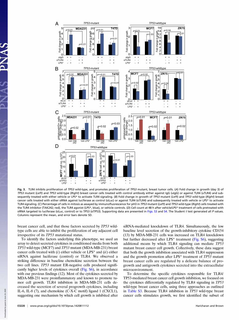
breast cancer cell, and that those factors secreted by TP53 wild-type cells are able to inhibit the proliferation of any adjacent cellirrespective of its TP53 mutational status.To identify the factors underlying this phenotype, we used an
array to detect secreted cytokines in conditioned media from bothTP53 wild-type (MCF7) and TP53mutant (MDA-MB-231) breastcancer cells treated with (i) either vehicle or LPS* and (ii) eithersiRNA against luciferase (control) or TLR4. We observed astriking difference in baseline chemokine secretion between thetwo cell lines. TP53 mutant ER-negative cells produced signifi-cantly higher levels of cytokines overall (Fig. S6), in accordancewith our previous findings (12). Most of the cytokines secreted byMDA-MB-231 were proinflammatory and known to promote tu-mor cell growth. TLR4 inhibition in MDA-MB-231 cells de-creased the secretion of several progrowth cytokines, includingIL-6, IL-8 (7), and chemokine (C-X-C motif) ligand 1 (CXCL1),suggesting one mechanism by which cell growth is inhibited after
siRNA-mediated knockdown of TLR4. Simultaneously, the lowbaseline level secretion of the growth-inhibitory cytokine CD154(13) by MDA-MB-231 cells was increased on TLR4 knockdownbut further decreased after LPS* treatment (Fig. S6), suggestingadditional means by which TLR4 signaling can mediate TP53mutant breast cancer cell growth. Collectively, these data suggestthat both the growth inhibition associated with TLR4 suppressionand the growth promotion after LPS* treatment of TP53 mutantbreast cancer cells are regulated by a delicate balance of pro-growth and antigrowth cytokines secreted into the extracellularmicroenvironment.To determine the specific cytokines responsible for TLR4/
TP53-mediated breast cancer cell growth inhibition, we focused onthe cytokines differentially regulated by TLR4 signaling in TP53wild-type breast cancer cells, using three approaches as outlinedin Table S3. Because TLR4 inhibition in TP53 wild-type breastcancer cells stimulates growth, we first identified the subset of
MC
F7 c
ell n
o. (x
103 )
TP53-wildtype
p<0.01
siLucsiTP53
LPS*
+ ++ +
++
- -- -- -
0
5
10
15
20
25p=0.03
MDA231 MCF7 ZR75T47D
p=0.02
TP53-wildtypeTP53-mutant
p=0.01
p=0.02
p<0.01
p=0.03p=0.02
pHis
tone
H3+
cells
(f old
chan
ge)
p=0.02
p=0.01
Veh LPS*VehTAK242
0.0
0.5
1.0
1.5
2.0
2.5
pHis
tone
H3+
cells
(fold
chan
ge)
0.0
0.5
1.01.5
2.0
2.5
0
4
8
12
16
20
ZR75
cel
l no.
(x10
3 )
siLucsiTP53
LPS*
+ ++ +
++
- -- -- -
p<0.01p=0.03
0
4
8
12
16
20
Fold
chan
gein
grow
th
αIgGαTLR4
LPS*
+ ++ +
++
- -- -- -
p<0.01
+ ++ +
++
- -- -- -
p=0.01
p<0.01
αIgGαTLR4
LPS*
+ ++ +
++
- -- -- -
p<0.01
+ ++ +
++
- -- -- -
p=0.04MDA231 T47D MCF7 ZR75
TP53-mutant TP53-wildtype
0
1
2
3
4
5
Fold
chan
gein
grow
th
p=0.01
0
1
2
3
Fold
chan
gein
grow
th
p=0.03
02468
10121416
Fold
chan
gein
grow
th
p<0.01
siLucsiTLR4
LPS*
+ ++ +
++
- -- -- -
p<<0.01
p<<0.01p=0.02
+ ++ +
++
- -- -- -
siLucsiTLR4
LPS*
+ ++ +
++
- -- -- -
+ ++ +
++
- -- -- -
p=0.04
MDA231 T47D MCF7 ZR75
TP53-mutant TP53-wildtype
01234567
Fold
chan
gein
grow
th p=0.02
0
1
2
3
4
5
Fold
chan
gein
grow
th
p<<0.01
0
1
2
3
4
Fold
chan
gei n
grow
th p=0.02
01234567
Fold
chan
gein
grow
th
p=0.01
Veh LPS*VehTAK242
Veh LPS*VehTAK242
Veh LPS*VehTAK242
A
B
C
D
Fig. 3. TLR4 inhibits proliferation of TP53 wild-type, and promotes proliferation of TP53 mutant, breast tumor cells. (A) Fold change in growth (day 3) ofTP53 mutant (Left) and TP53 wild-type (Right) breast cancer cells treated with control antibody either against IgG (αIgG) or against TLR4 (αTLR4) and sub-sequently treated with either vehicle or LPS* to activate TLR4 signaling. (B) Fold change in growth of TP53 mutant (Left) and TP53 wild-type (Right) breastcancer cells treated with either siRNA against luciferase as control (siLuc) or against TLR4 (siTLR4) and subsequently treated with vehicle or LPS* to activateTLR4 signaling. (C) Percentage of cells in mitosis as assayed by immunofluorescence for pH3 in TP53mutant (Left) and TP53wild-type (Right) cells treated withthe TLR4 inhibitor (TAK242; red), the TLR4 agonist (LPS*, blue), or vehicle controls. (D) Cell count at 48 h after vehicle/LPS* treatment of cells pretreated withsiRNA targeted to luciferase (siLuc, control) or to TP53 (siTP53). Supporting data are presented in Figs. S3 and S4. The Student t test generated all P values.Columns represent the mean, and error bars denote SD.
E3220 | www.pnas.org/cgi/doi/10.1073/pnas.1420811112 Haricharan and Brown
Dow
nloa
ded
by g
uest
on
June
24,
202
0

cytokines whose secretion decreased by ≤0.5-fold or increasedby ≥2-fold in TP53 wild-type (MCF7) cells treated with siRNAagainst TLR4. Similarly, we identified a second subset of cytokineswhose secretion decreased by ≤0.5-fold or increased by ≥2-fold inTP53 wild-type (MCF7) cells treated with LPS* to activate TLR4.From these two subsets, we then selected those cytokines thatchanged reciprocally in response to TLR4 inhibition and activa-tion, i.e., exhibited ≤0.5-fold decrease after TLR4 knockdown andα ≥2-fold increase with LPS* treatment or vice versa. To ensurethat we limited further analysis to only those cytokines specificallyregulated by TLR4, we excluded all cytokines whose secretion wassimilarly altered by LPS* treatment in both the presence andabsence of TLR4 in MCF7 cells. Those cytokines that remainedafter these three screens constituted our final working set of cy-tokines for further investigation.Of the cytokines that we identified through this screening pro-
cess, IFN-γ was the sole cytokine exhibiting significant reciprocalchanges in expression with TLR4 inhibition and activation, i.e., a≤0.5-fold decrease after TLR4 inhibition and a ≥2-fold increase insecretion after LPS* treatment of TP53 wild-type MCF7 cells (Fig.4A). This suggests that IFN-γ is the best candidate for mediatingthe observed growth phenotype; however, the other cytokinesidentified in our screen may well contribute to the growth phe-notype individually or in combination.IFN-γ is known to inhibit growth of breast cancer cells by in-
ducing a p21-mediated G0/G1 arrest (14), similar to the pheno-type reported here in TP53 wild-type breast cancer cells (Fig. S3 Cand D). Using ELISA, we confirmed that LPS* treatment induceda <2-fold increase in IFN-γ secretion in the extracellular micro-environment of TP53 wild-type breast cancer cells (MCF7, ZR75)
but not in that of TP53mutant breast cancer cells (MDA-MB-231,T47D) (Fig. 4B). Furthermore, in the absence of either TLR4 orTP53, LPS* treatment could not increase IFN-γ secretion by TP53wild-type breast cancer cells (Fig. 4C). To test whether IFN-γ inhuman tumors is involved in mediating TLR4-TP53–orchestratedgrowth, we assessed correlations between TLR4 and IFN-γ geneexpression in both the Curtis and TCGA datasets. In TLR4-highTP53 wild-type tumors, we found a significant increase in IFN-γgene expression relative to TLR4-low tumors, but not in TP53mutant tumors (Fig. 4 D and E).We next tested whether specific types of TP53 mutation are
associated with IFN-γ expression in ER-positive breast tumors.Using TCGA data, we examined the level of IFN-γ RNA ex-pression in ER-positive TLR4-low and TLR4-high breast tumorswith TP53 loss-of-function, gain-of-function, or dominant-nega-tive mutations (Fig. S7A). This analysis showed that in TLR4-lowtumors, IFN-γ expression is low for all TP53 mutant tumors,whereas in TLR4-high tumors, IFN-γ expression is significantlygreater in tumors with TP53 gain-of-function mutations, but notin tumors with TP53 loss-of-function or dominant-negative mu-tations. A larger sample size is needed to understand the truenature of the IFN-γ–TLR4 association in the context of thevarious classes of TP53 mutations.Next, we confirmed that TLR4 activation in TP53 wild-type
breast cancer cells preferentially induces nuclear p21, a knowndownstream effector of IFN-γ signaling and mediator of growthinhibition (Fig. 5A). LPS* treatment also increased mRNA levelsof p21 in TP53 wild-type breast cancer cells (MCF7, Fig. 5B;ZR75, Fig. S7B). To determine the coordinated roles of TP53and TLR4 in regulating p21 levels, we used siRNA knockdown
MCF7 ZR75
TP53-wildtype
TP53-wildtype
0
2
4
6
8
10
12
14
MCF7 ZR75 MDA231 T47D
IFN
-γle
vel s
(a.u
)
VehLPS*
ns
ns
p=0.04
p=0.03
0123456
IFN
-γin
con
ditio
ned
med
ia fr
om:
0123456
siTP
53 c
ells
(a.u
)
VehLPS*ns
ns
nsns
siTL
R4
cells
(a.u
)
Breast cancer cells
Rel
ativ
e IF
N-γ
gen
e ex
pres
sion
TP53-mutant
TP53-wildtype
p<0.001
Curtis dataset (ER+ tumors)
p<0.001
ns
ns
TP53-mutant
TP53-wildtype
TCGA dataset (ER+ tumors)
0.0
0.1
0.2
0.3
TLR4low TLR4high
12
34
5R
elat
ive
IFN
-γ g
ene
expr
essi
on
TLR4low TLR4high
CXCL12
CCL3CXCL11
Decreased after LPS* tx
IL-1RA
CXCL10CCL4
IL-17E
CCL444
IL-23
sTREM-1IFN-γ
Decreased after siTLR4 tx
Increased after LPS* tx IL-1A
MCP-1
PAI-1
IL-13CD54
CCL5
Increased after siTLR4 tx
A B
C D E
Fig. 4. TLR4 activation increases IFN-γ secretion from TP53 wild-type breast tumor cells, but not from TP53 mutant cells. (A) Venn diagram representingsubsets of cytokines from TP53 wild-type breast cancer cells (Fig. S6 presents results of complete array in TP53 wild-type and TP53 mutant cells) whose se-cretion changes by ≥2-fold (pink) or ≤0.5-fold (blue) in culture supernatant of cells treated with siRNA against TLR4 vs. luciferase or treated with LPS* vs.vehicle. The black arrow indicates cytokine that is reciprocally altered by siTLR4 and LPS* treatment. Fold change for each cytokine is presented in Table S3.(B and C) Changes in secreted IFN-γ from culture supernatant of TP53wild-type and TP53mutant breast cancer cells treated with LPS* (B), as well as from TP53wild-type breast cancer cells treated with siRNA against either TLR4 or TP53 and subsequently with LPS* (C) as assayed by ELISA. (D and E) Plots of meansdepicting gene expression level of IFN-γ in TLR4-high vs. TLR4-low TP53mutant and TP53wild-type breast tumors in the TCGA (D) and Curtis (E) datasets. Errorbars represent SEM. Associated data are presented in Fig. S7A. The Student t test generated all P values. Columns represent the mean, and error bars denoteSD, except where noted otherwise.
Haricharan and Brown PNAS | Published online June 10, 2015 | E3221
CELL
BIOLO
GY
PNASPL
US
Dow
nloa
ded
by g
uest
on
June
24,
202
0

against TP53 and TLR4 individually in TP53 wild-type breastcancer cells. Our results show that LPS*-induced activation ofp21 is dependent on both TP53 (MCF7: Fig. 5C; ZR75: Fig.S7B) and TLR4 (Fig. 5D). We also confirmed this effect at theprotein level (Fig. 5E). Corroborating these data, we also foundthat TLR4-high TP53 wild-type tumors have more p21, as mea-sured by gene expression (Curtis: P = 0.01; TCGA: P = 0.01),compared with TLR4-low tumors in patient tumor datasets (Fig.5 F and G). The specificity of this correlation is indicated by thelack of correlation between TLR4 and p27 gene expression in bothdatasets assayed (Fig. S7 C and D). These data indicate a role forboth TLR4 and TP53 signaling in regulating IFN-γ secretion andp21 regulation by TP53 wild-type cells.
IFN-γ Secretion by TP53 Wild-Type Breast Cancer Cells Is Both Necessaryand Sufficient to Mediate Growth Inhibition by TLR4. We next in-vestigated whether IFN-γ is both necessary and sufficient to me-diate the effect of TLR4 on TP53 wild-type breast cancer cellgrowth. To ascertain necessity, we neutralized IFN-γ in condi-tioned media from LPS*-treated TP53 wild-type cells using ananti–IFN-γ antibody, and then assessed the effect of this neu-tralized media on cell growth. We found that conditioned mediafrom LPS*-treated TP53 wild-type breast cancer cells significantlyinhibited growth by day 2, as expected, but preincubation of theconditioned media with the IFN-γ neutralization antibody ne-
gated any growth-inhibitory effect (Fig. 6 A and C). These dataindicate that TLR4 loses its growth-inhibitory effect on TP53wild-type breast cancer cells in the absence of secreted IFN-γ,even when all other secreted cytokines remain empirically un-perturbed in the microenvironment.We next pharmacologically inhibited TLR4 by treating TP53
wild-type breast cancer cells with TAK242, and then addedrecombinant IFN-γ to the culture media. Our results show thatinhibition of TLR4 in TP53 wild-type cells promotes growth byday 2; however, subsequent treatment with recombinant IFN-γblocks TAK242-induced growth promotion (Fig. 6 B and D).This result suggests that in the absence of all other secretedcytokines, the addition of IFN-γ alone is sufficient to replicatethe growth inhibition induced by TLR4 activation. These dataindicate that secreted IFN-γ is both necessary and sufficient tomediate the growth-inhibitory effect of TLR4 on TP53 wild-typebreast cancer cells.To test whether this TP53-dependent role for TLR4 in regu-
lating growth is restricted to breast cancer, we assessed for acorrelation between TP53 mutational status and TLR4 alterations(including mutations, genomic alterations, and gene expressionchanges) across multiple cancer types using publically availableTCGA datasets. This analysis identified an inverse correlationbetween the incidence of TLR4 and TP53 alterations across allcancer types included in the analysis (Fig. 7A). Cancer sites more
0.000.010.020.030.040.050.060.070.08
siLuc siTP53
p21
mR
NA
(fem
togr
ams) MCF7p=0.02
ns
VehLPS*
Veh LPS*
p<0.01
0.0
0.1
0.2
0.3
p21
mR
NA
(fgs
)
Veh LPS*
p=0.04
MCF7
p21
Vehicle LPS* MCF7
TP53-wildtype (MCF7) TP53-wildtype
TP53-wildtype TP53-wildtype
p21
GAPDH
siLucsiTP53siTLR4
LPS*
+ ++ +
+ ++ ++
- - - -- - - -- - - -- --
TP53-wildtype (MCF7)
01020304050
p21+
cells
(%)
0.000.050.100.150.200.250.300.350.400.45
p21
mR
NA
(fem
togr
ams)
siLuc siTLR4
ns
p<0.01 MCF7VehLPS*
TP53-mutant
TP53-wildtype
Rel
ativ
e p2
1 ge
ne e
xpre
ssio
n
p=0.01
ns
Curtis dataset (ER+ tumors) TCGA dataset (ER+ tumors)
1.8
1.9
2.0
2.1
TLR4low TLR4high
-0.6
-0.4
-0.2
0.0
0 .2
0.4
p=0.02
ns
TLR4low TLR4high
Rel
ativ
e p2
1 ge
ne e
xpre
ssio
n
TP53-mutant
TP53-wildtype
A B
C D E
F G
Fig. 5. LPS*-induced p21 activation requires both TLR4 and TP53. (A and B) Representative photomicrographs and accompanying quantification of nuclearlocalized p21 in TP53 wild-type cells (A), as well as quantification of RNA levels (B) 24 h after LPS* treatment. (C and D) Relative levels of p21 mRNA in TP53wild-type breast tumor cells treated with siRNA against either luciferase as control, against TP53 (C) or against TLR4 (D) and subsequently treated with eithervehicle or LPS*. Supporting data are presented in Fig. S7B. (E) Western blot confirming p21 induction after LPS* treatment in control TP53 wild-type cells butnot in cells lacking either TP53 or TLR4. (F and G) Plots of means depicting alterations in p21 gene expression changes in TLR4-low and TLR4-high tumors fromthe Curtis (F) and TCGA (G) datasets. Error bars depict SEM. Accompanying data are provided in Fig. S7 C and D. The Student t test generated all P values.Columns represent the mean, and error bars represent the SD. (Scale bar: 20 μm.)
E3222 | www.pnas.org/cgi/doi/10.1073/pnas.1420811112 Haricharan and Brown
Dow
nloa
ded
by g
uest
on
June
24,
202
0

prone to TP53 loss, such as ovarian serous, head and neck, andbladder cancers, are more likely to retain TLR4, in keeping withour hypothesis that TLR4 retention provides a growth advantageto tumor cells in the absence of TP53 function. On the otherhand, tumors that tend to maintain wild-type TP53 have ahigher incidence of TLR4 alterations, indicating that these tu-mors cells tolerate, and likely even select for, the loss of TLR4.Among lung cancers alone, lung adenocarcinomas have a higherincidence of wild-type TP53 than lung squamous carcinomas,and also exhibit a greater propensity for TLR4 loss (Fig. 7A).These observations indicate that there likely exists a complexinteraction between TLR4 and TP53 across many differenthistological tumor types.
DiscussionThe results presented herein suggest that TLR4 regulates breastcancer cell growth in a TP53-dependent manner. To our knowl-edge, this constitutes the first report of TLR4 activation of TP53wild-type breast cancer cells causing secretion of IFN-γ into theextracellular microenvironment, resulting in activation of p21 andsubsequent suppression of tumor cell proliferation (Fig. 7B).Conversely, in TP53 mutant breast cancer cells, TLR4 activationalters the balance of progrowth and antigrowth cytokines in theextracellular microenvironment, ultimately resulting in increasedproliferation and growth (Fig. 7B). These data indicate a complexrole for TLR4 in driving breast cancer in the context of TP53mutational status, and provide important insight into the effect ofTLR4 activation in tumor cells on both growth and modulation ofthe cytokine milieu in the microenvironment. This has profoundimplications for the proposed use of TLR4 as a druggable target inbreast cancer and in other cancers, and suggests that only patientswith TP53 mutant tumors can benefit from anti-TLR4 therapy.Furthermore, our results indicate that patients with TP53 wild-type tumors should not be treated with anti-TLR4 therapy, be-
cause such treatment could promote, rather than suppress, tu-mor growth. Previous reports have suggested that TLR4 has asystemic role as a tumor suppressor in mice (15), potentiallythrough the immune system. Therefore, if anti-TLR4 therapy isto be used in the clinic, it will be necessary to consider the effectson the immune system in addition to direct effects on the tumor.Future studies of TLR4 antagonists will need to assess both di-rect and systemic effects to develop these drugs for the treatmentof TP53 mutant tumors.A previous report identified TLR4 as promoting ER-negative,
TP53 mutant MDA-MB-231 breast cancer cell growth (6). Thatstudy also found that TLR4 promotes cancer cell proliferation byincreasing secretion of the proinflammatory cytokines IL-6 andIL-8 (6), supporting our results reported herein. However, thatstudy focused on a single cell line and used a candidate approachto identify IL-6 and IL-8, which might not be the principal cy-tokines mediating proliferation regulation by TLR4. Our presentstudy confirms a decrease in the secretion of both these cytokinesafter TLR4 knockdown in MDA-MB-231 cells, but also iden-tifies large decreases in the secretion of other progrowth cyto-kines, including CXCL1 (16), as well as a dramatic increase inantigrowth cytokine CD154 (13). Most importantly, the previous
TP53-wildtype (ZR75)
- ++-
- +- +
p=0.03
p=0.01
Fold
chan
gein
grow
t h
p=0.01
p=0.02
TAK242rIFN-γ
- ++-
- +- +
0123456789
Fold
chan
gein
grow
th
LPS*αIFN-γ
- ++-
- +- +
TP53-wildtype (MCF7)
p<0.01p<0.01
0.00.20.40.60.81.01.21.4
p<0.01
p<0.01
- ++-
- +- +
LPS*αIFN-γ
TP53-wildtype (MCF7)Fo
ldch
ange
ingr
owth
TP53-wildtype (ZR75)
TAK242rIFN-γ
Fold
chan
gein
grow
th
0
1
2
3
4
0
1
2
3A B
C D
Fig. 6. IFN-γ is both necessary and sufficient for mediating growth in-hibition of TP53 wild-type breast cancer cells by TLR4. (A and C) Bar graphsrepresenting the fold change in growth of TP53 wild-type breast cancercells when exposed to conditioned media from isogenic cells pretreatedwith LPS* for 48 h and incubated with antibody against either IgG or aneutralizing antibody against IFN-γ. (B and D) Bar graphs representing thefold change in growth of TP53 wild-type breast cancer cells treated se-quentially with TAK242 (10 nM) and recombinant IFN-γ. The Student t testgenerated all P values. Columns represent the mean, and error bars de-note SD.
Multiple tumor datasets (TCGA from cBio)
R² = 0.5937
0
5
10
15
20
25
15 35 55 75 95
TLR
4 al
tera
tions
(%)
TP53 alterations (%)
Skin
SarcomaBreast
Lung Adenocarcinoma
StomachColorectal
BladderHead&Neck
Lung Squamous
Ovarian serous
Working model
TLR4
IFNγ
TP53
p21IFNγ
Cyclin-CDK
p21
TLR4 ligands
TLR4
CXCL1
Cyclin-CDK
TLR4 ligands
TP53-wildtype tumor cell TP53-mutant tumor cell
IFNγR1
mutTP53
1.
2.
3.
4.
5.
6.1.
2.
3.
4.CD154
A
B
Fig. 7. IFN-γ, TLR4, and TP53 coordinately regulate growth of breasttumor cells. (A) Regression analysis indicates inverse correlation betweenalterations (mutations, genomic loss, and gene expression change) inTLR4 and TP53 (mutations, genomic loss) across multiple cancer types. R2
values were generated using a generalized linear model. (B) Workingmodel. (Left) In TP53 wild-type cells, TLR4-mediated inhibition of cellgrowth occurs through (1) TLR4 activation by ligands; (2) TLR4-mediatedinduction of IFN-γ secretion; (3) autocrine/juxtacrine activation of IFN-γgamma signaling; (4) IFN-γ/TP53 coordinated induction of p21; (5) for-mation of p21-cyclin/CDK complexes; and (6) growth inhibition. (Right) InTP53 mutant cells, TLR4-mediated up-regulation of cell growth occursthrough (1) TLR4 activation by ligands; (2) TLR4-mediated induction ofproinflammatory cytokines, including CXCL1; (3) autocrine growth pro-motion by cytokine; and (4) TLR4-mediated inhibition of anti-growthcytokines, such as CD154.
Haricharan and Brown PNAS | Published online June 10, 2015 | E3223
CELL
BIOLO
GY
PNASPL
US
Dow
nloa
ded
by g
uest
on
June
24,
202
0

study did not uncover the critical role of TP53 mutational statusin regulating the observed growth phenotype.In another previous study, Yang et al. (17) reported that
treatment of MDA-MB-231 and MCF7 cells with LPS promotedinvasion of both cell lines. Those results appear to contradictour findings, which demonstrate a growth-suppressive role forTLR4 in TP53 wild-type MCF7 cells. It is possible that TLR4signaling in TP53 wild-type cells suppresses tumor growth butalso may stimulate invasion and metastasis of the tumor cells;however, it is important to note that Yang et al. used reagentsthat can activate several TLRs in their experiments, and that inboth of the large-scale human datasets analyzed in this work,TLR4 is associated with improved survival of patients withTP53 wild-type breast cancer in a stage-independent manner.These results suggest that in TP53 wild-type tumors, highTLR4 is associated with a good outcome. Such results do notsupport the hypothesis that high TLR4 promotes metastasis.Other studies have identified TLR4 as a promoter of che-
motherapeutic resistance in MDA-MB-231 cells (7, 8). Thosestudies used HCC1806, another TP53 mutant, ER-negativebreast cancer cell line with very low baseline levels of TLR4, asa negative control. Rajput et al. (8) reported that TLR4knockdown in MDA-MB-231 cells sensitizes them to chemo-therapy, whereas overexpression of TLR4 in HCC1806 cellsrenders them resistant to chemotherapy. That group has sincepublished a follow-up study demonstrating a role for TLR4 inpromoting migration on exposure to cytotoxic therapy (8).Although both studies demonstrate an important role forTLR4 as an oncogene in breast cancer, our identification of adual role for TLR4 based on TP53 function provides impor-tant context for interpreting their results. Previous studieshave shown that in human breast cancer samples, wild-typeTP53 is associated with chemotherapeutic sensitivity (18).Other studies have reported that specific TP53 mutations orvariants are associated with chemotherapeutic resistance (19–21). In addition, a role for TP53 in resistance to cisplatintreatment has been identified in ovarian cancer (22). There-fore, our identification of the TLR4-TP53 nexus in breastcancer suggests an important avenue for further exploration ofthe mechanisms involved in resistance to cytotoxic therapies.The results presented herein validate a role for TLR4 in
tumor growth regulation independent of its immune activity,which has been a primary focus of previous TLR4-basedstudies. Our data also indicate that the TP53 mutational con-text of a breast tumor epithelial cell can significantly affect thetumor microenvironment by regulating the extracellular cyto-kine secretions of tumor cells, and have a profound effect onresponse to targeted therapy. The tumor growth and immu-nomodulatory roles of TLR4 may be integrated, because cy-tokines in the tumor microenvironment play an important rolein attracting or evading antitumor immunity (23). Therefore,the roles of TLR4 in regulating tumor growth and cytokinesecretion may act together in promoting the growth of epi-thelial TP53 mutant tumor cells and in suppressing any anti-tumor immune response.In this context, it is striking that the TP53-dependent differ-
ential role of TLR4 appears to occur in multiple cancer types inaddition to breast cancer. The global nature of this interaction issupported by the observation that an oncogenic role for TLR4in tumor cells has been identified primarily in cancer typesknown to inactivate TP53 at high frequencies, such as ovarian(24), bladder (25), and head and neck (26) cancers. Further in-vestigation is warranted in other cancer sites to define the asso-ciation between TLR4 and TP53 and its effect on tumor biologyand behavior.
Materials and MethodsCell Lines, siRNA Transfection, and Growth Assays. Cell lines were obtainedfrom American Type Culture Collection and maintained and validated asreported previously (12). TP53 mutant cell lines have the following TP53mutations: K132Q (BT-20), L194F (T47D), E166* (MDA-MB-361), and R280K(MDA-MB-231). Three TLR4 and TP53 siRNA oligonucleotides each werepurchased from Sigma-Aldrich. Transfection and validation were con-ducted as described previously (12). Growth assays were conducted intriplicate and repeated independently as described previously using Try-pan blue to identify live, viable cells and manual quantification with he-mocytometers (27).
Inhibitors, Neutralizing Antibodies, Recombinant Proteins, and Agonists.TAK242 (InvivoGen) was dissolved in DMSO and used at specified con-centrations, with concomitant amounts of DMSO as vehicle control.Recombinant IFN-γ (BD Biosciences) was dissolved in serum-free media andused at a concentration of 15 pg/mL. Neutralizing antibody to IFN-γ(eBiosciences) was used at a concentration of 2.5 μg/mL. Neutralizationwas conducted through a 2-h incubation of conditioned media witheither IFN-γ or rabbit polyclonal IgG antibody (Santa Cruz Biotechnology)at 37 °C. Neutralizing antibody to TLR4 (Imgenex) was preincubatedwith cells for 2 h at 10 ng/mL before subsequent treatment asspecified. LPS (InvivoGen) was dissolved in 1× PBS and used as describedpreviously (7).
Immunostaining and Microscopy. Immunofluorescence was performed in ac-cordance with the manufacturer’s instructions. Cells were washed in PBS,fixed in 4% PFA for 20 min at room temperature, blocked in 5% goat serumand 1% Triton X-100 in 1× PBS for 1 h at room temperature, incubated withprimary antibody in 1% goat serum and 1% Triton X-100 in 1× PBS antibodydiluent overnight at 4 °C, incubated with secondary antibody in diluent for1 h at room temperature, and then mounted with DAPI-containing mountingmedia. Primary antibodies used included pH3 (Cell Signaling Technology;1:500), p21 (Cell Signaling Technology; 1:200), NFkB (Cell Signaling Technol-ogy; 1:200), and TLR4 (Thermo Scientific; 1:200). Fluorescent images werecaptured with a Nikon microscope, quantified with ImageJ, and processedwith Adobe Photoshop software.
Apoptosis and Cell Cycle Analyses. Annexin V was used to assay apoptosis asdescribed previously (12). Cell cycle analysis was conducted in triplicate usingpropidium iodide to stain cells, followed by cell sorting with a Gallios flowcytometer (Beckman Coulter) at 24 or 48 h posttreatment with specifiedreagents. Analysis of cell cycle was conducted using FlowJo software(Tree Star).
Cytokine Array, ELISA, and Western Blot Analysis. ELISA and Western blotanalysis were conducted as described previously (12, 28). Primary antibodieswere incubated with the membrane overnight at 4 °C and included p21 (CellSignaling Technology; 1:1,000), STAT1 (Cell Signaling Technology; 1:1,000),p53 (Santa Cruz Biotechnology; 1:500), p65 (Cell Signaling Technology;1:1,000), and GAPDH (Sigma-Aldrich; 1:6,000). The cytokine array (R&D Sys-tems) was used according to the manufacturer’s instructions. Supernatantfor the ELISA and cytokine array experiments was collected 24 h posttreat-ment, and spun down for 5 min at 1,000 rpm. The supernatant was thenstored at −20 °C, as required.
Statistical Analysis. ANOVA or the Student t test was used for independentsamples with normal distribution. Where distribution was not normal(assessed using Q-Q plots with the Wilk–Shapiro test of normality), eitherthe Kruskal–Wallis or Wilcoxon rank-sum test was used. All experimentswere conducted in triplicate, and each experiment was duplicated in-dependently more than two times. Criteria for including datasets in theregression analysis were that the dataset comprised >100 samples, eachTLR4 alteration in the dataset occurred at >1% frequency, and alterationsin both TP53 and TLR4 occurred at >3% frequency. These criteria wereformulated to ensure that results from each dataset were calculablewithin the range of sensitivity of the statistical test used. Regressionanalysis was performed using R and a generalized linear model. Databasesused for human data mining were publically available resources: Onco-mine, cBio, and COSMIC. Median cutoffs were used for gene expressionstratification. All survival data were analyzed using Kaplan–Meier curvesand log-rank tests. Proportional hazards were determined using Cox re-gression. All graphs and statistical analyses were generated in eitherMicrosoft Excel or R.
E3224 | www.pnas.org/cgi/doi/10.1073/pnas.1420811112 Haricharan and Brown
Dow
nloa
ded
by g
uest
on
June
24,
202
0
ACKNOWLEDGMENTS. We thank Michelle Savage for her technical editingof the manuscript and Dr. Matthew Bainbridge for his help with statis-tical analyses and creative input. This work was supported by Cancer Cen-ter Support Grant 5 P30 CA016672-38, a Norman Brinker Award forResearch Excellence (to P.B.), a John Charles Cain Distinguished Chair
Award (to P.B.), the Mariam Rogers Fund for Breast/Women’s Cancer Re-search (P.B.), The University of Texas M. D. Anderson Cancer CenterHalliburton Fellowship in Cancer Prevention Fund (to S.H.), and Susan G.Komen Promise Grant KG081694 (to P.B.). P.B. currently serves as a Scien-tific Advisory Board member for Susan G. Komen for the Cure.
1. Haricharan S, Bainbridge MN, Scheet P, Brown PH (2014) Somatic mutation load ofestrogen receptor-positive breast tumors predicts overall survival: An analysis of ge-nome sequence data. Breast Cancer Res Treat 146(1):211–220.
2. Kim HM, et al. (2007) Crystal structure of the TLR4–MD-2 complex with bound en-dotoxin antagonist Eritoran. Cell 130(5):906–917.
3. Piao W, et al. (2013) Recruitment of TLR adapter TRIF to TLR4 signaling complex ismediated by the second helical region of TRIF TIR domain. Proc Natl Acad Sci USA110(47):19036–19041.
4. Jin B, Sun T, Yu XH, Yang YX, Yeo AE (2012) The effects of TLR activation on T-celldevelopment and differentiation. Clin Dev Immunol 2012:836485.
5. González-Reyes S, et al. (2010) Study of TLR3, TLR4 and TLR9 in breast carcinomas andtheir association with metastasis. BMC Cancer 10:665.
6. Yang H, et al. (2010) Reduced expression of Toll-like receptor 4 inhibits human breastcancer cells proliferation and inflammatory cytokines secretion. J Exp Clin Cancer Res29:92.
7. Rajput S, Volk-Draper LD, Ran S (2013) TLR4 is a novel determinant of the response topaclitaxel in breast cancer. Mol Cancer Ther 12(8):1676–1687.
8. Volk-Draper L, et al. (2014) Paclitaxel therapy promotes breast cancer metastasis in aTLR4-dependent manner. Cancer Res 74(19):5421–5434.
9. Huang JM, et al. (2014) Atractylenolide-I sensitizes human ovarian cancer cells topaclitaxel by blocking activation of TLR4/MyD88-dependent pathway. Sci Rep 4:3840.
10. Wang AC, Su QB, Wu FX, Zhang XL, Liu PS (2009) Role of TLR4 for paclitaxel che-motherapy in human epithelial ovarian cancer cells. Eur J Clin Invest 39(2):157–164.
11. Dijsselbloem N, et al. (2007) A critical role for p53 in the control of NF-kappaB-dependent gene expression in TLR4-stimulated dendritic cells exposed to Genistein.J Immunol 178(8):5048–5057.
12. Hartman ZC, et al. (2013) Growth of triple-negative breast cancer cells relies uponcoordinate autocrine expression of the proinflammatory cytokines IL-6 and IL-8.Cancer Res 73(11):3470–3480.
13. Tong AW, et al. (2001) Growth-inhibitory effects of CD40 ligand (CD154) and its en-dogenous expression in human breast cancer. Clin Cancer Res 7(3):691–703.
14. Gooch JL, Herrera RE, Yee D (2000) The role of p21 in interferon gamma-mediatedgrowth inhibition of human breast cancer cells. Cell Growth Differ 11(6):335–342.
15. Ahmed A, Wang JH, Redmond HP (2013) Silencing of TLR4 increases tumor pro-gression and lung metastasis in a murine model of breast cancer. Ann Surg Oncol20(Suppl 3):S389–S396.
16. Acharyya S, et al. (2012) A CXCL1 paracrine network links cancer chemoresistance andmetastasis. Cell 150(1):165–178.
17. Yang H, et al. (2014) Toll-like receptor 4 prompts human breast cancer cells in-vasiveness via lipopolysaccharide stimulation and is overexpressed in patients withlymph node metastasis. PLoS ONE 9(10):e109980.
18. Bertheau P, et al. (2002) Effect of mutated TP53 on response of advanced breastcancers to high-dose chemotherapy. Lancet 360(9336):852–854.
19. Bergamaschi D, et al. (2003) p53 polymorphism influences response in cancer che-motherapy via modulation of p73 dependent apoptosis. Cancer Cell 3(4):387–402.
20. Yang P, Du CW, Kwan M, Liang SX, Zhang GJ (2013) The impact of p53 in predictingclinical outcome of breast cancer patients with visceral metastasis. Sci Rep 3:2246.
21. Aas T, et al. (1996) Specific P53 mutations are associated with de novo resistance todoxorubicin in breast cancer patients. Nat Med 2(7):811–814.
22. Reles A, et al. (2001) Correlation of p53 mutations with resistance to platinum-basedchemotherapy and shortened survival in ovarian cancer. Clin Cancer Res 7(10):2984–2997.
23. Faget J, et al. (2011) Early detection of tumor cells by innate immune cells leads toT(reg) recruitment through CCL22 production by tumor cells. Cancer Res 71(19):6143–6152.
24. Wang AC, et al. (2014) TLR4 induces tumor growth and inhibits paclitaxel activity inMyD88-positive human ovarian carcinoma in vitro. Oncol Lett 7(3):871–877.
25. Wang YH, et al. (2014) Effect of TLR4 and B7-H1 on immune escape of urothelialbladder cancer and its clinical significance. Asian Pac J Cancer Prev 15(3):1321–1326.
26. Ren G, et al. (2014) Rapamycin inhibits Toll-like receptor 4-induced pro-oncogenicfunction in head and neck squamous cell carcinoma. Oncol Rep 31(6):2804–2810.
27. Chen L, et al. (2009) Inhibition of the p38 kinase suppresses the proliferation of hu-man ER-negative breast cancer cells. Cancer Res 69(23):8853–8861.
28. Li Y, et al. (2011) The rexinoid bexarotene represses cyclin D1 transcription by in-ducing the DEC2 transcriptional repressor. Breast Cancer Res Treat 128(3):667–677.
Haricharan and Brown PNAS | Published online June 10, 2015 | E3225
CELL
BIOLO
GY
PNASPL
US
Dow
nloa
ded
by g
uest
on
June
24,
202
0