title studies on myrosinase( dissertation 全文 ) issue date...
TRANSCRIPT

Title Studies on myrosinase( Dissertation_全文 )
Author(s) Ohtsuru, Masaru
Citation Kyoto University (京都大学)
Issue Date 1974-05-23
URL https://doi.org/10.14989/doctor.r2570
Right
Type Thesis or Dissertation
Textversion author
Kyoto University

STUDIES ON
ta- 7fÅq. 173 th --o
MYROSINASE
MASARU OHTSURU
1974

STUDIES ONMYROS1NASE
MASARU OHTSURU
1974

CONTENTS
INTRODUCTION ...•••}••••••}•...r...•..........
PART I. Studies on the Plant Myrosinase ......
Chapter 1. Chenical and Physical Properties .
Materials and Methods ............
Results and Discussion ...........
Chapter 2. Functional Groups ................
Materials and Methods ............
Results .-e..t.....r.....'"''''''
Discussion ...................•...
Chapter 3. Approach to the rnteraction of L-Ascorbic Acid to the Enzyme ....
Chapter 4. Binding oÅí Ascorbate to the Enzyme and the rnteraction of Ascorbat6
with the Functional Groups ......e
Materials and Methods ............
Resuits ..........................
Discussion ...............,...t-..
REFERENCES .................t ...................
1
5
5
s
8
13
13
14
18
19
21
21
22
27
29

PART Ir. Studies on the Fungous Myrosinase .••.•30
Åëhapter 1. Prodwction,•Purification and Some Preper'ties of the Extracellular Myrosinase from As er illus s dowi.30
Materials and Methods '.............30
Results ...'........................ 31
Discussion ....................•.•.34 . Chhpter 2. Effects of Various Reagents ......•35
J Results .............L..'............ 3s
Diseussion ........................37 ' ..Chapter 3. 0n the P-Glucosidase Activity .....39
Materials and Methods .............39
ReSUItS .....e..•••..-......et••... 40 Ii' "' DiSCuSSiOn ..-..e•.••....•...••e••• 42
,. .-IUiFERiENCES ..................................... 44 t'. T. 1-t , Cpa. P,'t, e.r.4• •.'Propt. gtion and Stability of the
Zntracellular Myrosinase from Ei -• - • j• ZSst er '11us ni er ...........•.•...45 ' ' ,- .`• -I{aterial- S and Methods .............45 ' ReSUItS ...t ..-..•.e....{e.•.....•. 46
Discussion .........................48

Chapter S. General Character-st-cs ............. 50
Materials and Methods .........e•.... 50
Results .......•••.}•.•.....,•}e••... 50
D-scuss-on .......t.................. 53
REFERENCES .......••••••••.•••.•••••{•.••••-.•e•.•• 55
PART III. Studies on the Bacterial Myrosinase .... S6
Chapter 1. Isolation of Bacteria ..............- S6
Materials and Methods ...............'is6
Results and Discussion ........••••.t 57
Chapter 2. Purification and General ,, fi::::.cgefEytf?.:::::::::::::;:::'.::.:, 21
66 D-scusslon .......e..i.......•.......
67REFERENCES ••.••••••..•e.•.••.•e....••••....•.•••.
68SmaY AND CONCLUSION ...........................
ACKNOWLEDGEMENT •••.••••.••.•!•...te...•.e•••.•..t 72

ABBREVrATrONS
ASAp--NPG
C. S. L.
PCMBDFPEDTAFDNBTNBS
'CFQ
NBSSDSMINT
DEPEIinan reagentKoshland
Mustard
reagent
Ex't .
L-Ascorbie Acidp-Nitrophenyl 5-glucosideCorn Steep Liquorp-MercuribenzoateDiisopropylfluorophosphateEthylenediaminetetraacetic AcidFluorodinitrobenzeneT' rinitrobenzenesulfonic AcidMonochlorotrifluoro-p-benzoquinoneN-BromosuccinimideSodiun dodecylsulfate2-Methoxy-5-nitrotroponeDiethylpyrocarbonate5-5'-Dithio-bis-(2-nitrobenzoic acid)2-Hydroxy-S-nitrobenzyl bromide
Extract of mustard seed (1.0 Kg) by80% methanol (5.0 L) was concentratedto 700 ml.

INTRODUCTION
Myrosinases [thioglucoside (glucosinolate) gluco
hydrolase, EC 3.Z.3.1] are the enzymes responsible for
the hydrolysis of mustard oil glucosides, which have been
found in plants (Cruciferae family)1,Z),fungi 3 ), bacteria4 )
and mammals S). The action of myrosinase on the mustard
oil compounds is the hydrolysis of glucose from the thio
glucoside followed by Lossen rearrangement of the aglycon
to give isothiocyanate and sulfate6 ,7).
myrosinase
Sinigrin: R- = CHZ=CH-CHZ-
The myrosinase is responsible for the development
of the flavor and pungency of many food products, such
as mustard and horseradish, by its hydrolysis of the thio
glucosides. Thus, myrosinase is a kind of the typical
flavor enzymes and would become a model of the flavor
development by enzymes. Further, the enzyme converts
progoitrin which is a thioglucoside present in high con
centration in the seeds of most Brassicaceous plants and
rapeseed meal, int~ goitrin, a potent antithyroid com
pound4 ). Therefore, it also becomes necessary to clarify
the characteristics of myrosinase in order to utilize the
rapeseed meal for the feeding of livestock.
'l'herehadbeen SOme controversies cOllcerning the"
natureott.hemyroSin3.seOn the plant myrosinase.
- 1 -

previously, Gadamer8) assigned the structure (i) for 9)mustard oil glucoside. Neuberg et al. reported theseparation of thioglucosidase and sulfatase whichhydrolyzed thioglucoside linkage and sulfuric acid esterlinkage of (r), respectively• Thereafter, it was general-ly believedlO) that the myrosinase in plants was a mixture
of the thioglucosidase and the sulfatase. Ettlinger etal.6'7), however, corrected the structure (x) to the struc-
ture (Ir) and suggested that the liberation of sulfatewould occur by the Lossen rearrangement after the cleavage 11)of the thioglucoside linkage. Nagashima et al. alsosupported the mechanism of Lossen rearrangement, andconcluded that the myrosinase was not a mixture of thetwo enzymes but a single S-thioglucosidase and the lib-eration of sulfate would occur nonenzymatically.
/S-C6HllOs /S-•C6HllOs
X O-S03- Å~X N-O-S03-
virtanen et al.i2) found an enzymatic production of
thiocyanate beside •isothiocyanate from the mustard oilglucoside in some species of the plants exhibiting myro--sina$e activity. As a result, some uncertainty occurredin the theory of Lossen rearrangement ef the glucosideeTherefore, Gaines et ai.i3'i4) carried out further inves-
tigation on the homogeneity of the myrosinase, and suc-cessfully sepavated thioglucosidase and sulfatase. Furthermore, Ettlinger et al.i5) $uggested that
there may exist two additional enzyrnes, one of which isactivated by L--ascorbic acid.
- -2.

Tsuruo et al.16
) purified the myrosinase, using a
TEAE-cellulose chromatography and showed that thioglu
cosidase and sulfatase activities were not distinguish
able. So, they concluded the myrosinase is a single ~
thioglucosidase. Later, Gaines and Howard 17 ) accepted
the theory that the myrosinase is not a mixture of two
enzymes but a single ~-thioglucosidase.
Nagashima et al. 18 ) found that the plant myrosinase
was activated strongly by L-ascorbic acid.' Schwimmer19 )
and Ettlinger et al. 1S ) investigated this phenomenon.
The later concluded that ascorbic acid behaved as a
reversibly dissociable base, closely connected with the
nucleophilic group of thioglucosidase. Kojima et al. 20 )
studied the change of the reaction product of the enzym
atic hydrolysis of mustard oil glucoside in the presence
of ascorbic acid at low pH. Tsuruo and Hata 21 ) described
that the oxidation-reduction reaction of ascorbic acid
had nothing to do with the activation reaction of myro
sinase. The presence of the effector site for ascorbic
acid was presumed by the kinetic measurements and in
stabilization of the enzyme by ascorbate on heating.
But, the above conclusions have been derived from
the investigations using the crude preparations. Thus,
the author has investigated the purification, physical
and chemical properties of the myrosinase from yellow
mustard powder, and the activation mechanism of the
myrosinase by ascorbate.
While, Reese et al. 3 ) reported that fungi
(Aspergillus sydowi etc.) produced a myrosinase (Sini
grinase). Kojima et al. 22 ) reported some of its prop
erties. Thereafter, Oginsky et al. 4 ) reported that the
myrosinase activity existed in Paracolobactrum aeroge
noides and in several other ba~terial strains.
·3-

This study was performed to intensify the under-standing on the myrosinase, which was stimulated by mybelief that microorganisms would be a more economicalsource of the enzymes required for improvement of theflavor of processed foods and the utilized resources(for example) Rapeseed meal etc.) than those fromhigher piants.
REFERENCES
1) A. Kjaer,J. Conti and I. Larsen,Acta Chem. Scand., 7, 1276 {1953). 2) Z. Nagashima and M. Uchiyama, J. Agr• Chem. Soc. Japan, 33, 881 (1959). 3) E.T, Reese, R.C. Clapp and M. Mandels, Arck. Biochem. Biophys., 75, 228 Åq1958). 4) E.L, Oginslty, A.E, Stein and M.A. Greer,
Aoc. Soc. Expt, Med., 119, 360 (1965), 5) I. Goodman, J.R. Fouts, E. Bresnick, R. Menegns and GH. Hitchings, Science, 1 130, 450 (1959). 6) M.G. Ettlinger and AJ. Lundeen, J. Am, Chem Soc., 78, 4172 C1956).
7) M.G. Eulingrr and A.J. Lundeen, ibid,, 79,
1764 C1957). 8) J. Gadamer, Arch. ptuzrm., 235, 44 (1897).
9) C. Neuberg andO.V. Scheenebeck, Biochem. Z., 265, 223 (1926); Natunvissenschaften, 21,
404 {1933).10) For e:cample; J.B. Sumner and K. Myroba'ck, "Tlie Ene)rtnes'; 1 st ecl., 1, part 1, Acadetnic
press, Neu, York, 1950.11) Z. Nagasim and M. Uehiyama, J, Agr. cae,n. Soc. Japan, 33, 1144 (19S9).
12) R. Gmelin and AJ. Vir'tanen, Acta Che,n. Scan. 13, 1474 (1959).13) RD. Gafnes and K.J. Goering, Biochem.
Biophys, Res. Comn., 2, 207 (1960).14) RD. Gaines and K.J. Goering, Arch. Bio-
che,n. Biophpts., 96, 13 (1962).15) M.G. Ettlinger, G.P.Dateo,Jr., B.W. Harriso4
T.J. Mably and C.P. Thornpson, Pbeoc. NatL Acad. Sci., 47, 187S (1961).16) I. Tsuruo, M. Yoshida and T. Hata, A,gt'. Biel.
Chem, 31, 189S (1967).17) GA. Howard and RD. Gaines, Phytochem., 7, 585 (1968),18) Z, Nagashima and M. Uchiyama, 1. Agr. (hem, Soc. Japan, 33, 980 (19S9).19) S. Schwirnmer, Acta Chem. Scand., 15, 535
(1961).20) M. Kojirna and K, Tamiya,J. VimminoL, 1O,
44 (1964).21) I. Tsuruo and T. Hata, Agr. BieL Chern., 31,
27 (1967).22) M. Kojirna and K. Tamiya, VTttamin, 28, 380
(1963).
-4-

PART !
Studies on the Plant My rosinase
Chapter 1
Chemical and Physica1 Properties
In the precedingworks;]"" Tsuruo et aLreported varieus preperties of p!ant myrosi-
nase, which is a kind of P-thjoglucosidase.Plant myrosinase is specifically activated by
L-ascorbate') and its activation mechanismhad been investigated by kinetic procedures.But, to discuss the activadon mechanism ofthe plant myrosinase by L-ascorbate, it isnecessary to obtain the detailed inforrnationabout physico•-chemical nature of the purified
enzyme. . 1 had purified the plant myrosi-nase to investigate the activation mechanism
of the enzyme by L-ascorbate. During tbecourse of purification, four proteins which
have myrosinase activity were separated. In this chapter, purification precedures and physi-
cal and chemical propenies of each enzyme are described,
MAITERIAr. AND METHODS
Materlatandehemicats, Mustardpowder(CanadaOriental Mustard Seed) was purchased from Amari
Koshin Shokuhin K.K. Sini' grin was obtained fromNalcarai ChemicaSs Ltd. and psed as the sutstrate.
Eh7ymepunfication. Theprocedureofpurificationof plant m)rresinase is shown in Fig. 1. The enzymesolution was preparcd from mpstard pewder. Theextract (1oolitcrs) from rnustard powder (`vakgÅr byO.1 M sodium pbosphate buflTer containing O.Ol M 2-
rnercaptoethanol was fractionated by amrnentumsulfate at pH 7.0. The fraction precipitated at O,4
to O.8 of arnmonium sulfate saturation was dialyzedagainst O.Ol M sodium phosphatc buffer, pH 7.0 andsubjected to DEAE-Sephadex A-50 (61) colummchrQmatography. The adsorbed pretein was elutedbatchwise with 10 liters of the same buffer containing
O.2M sodium chloride. The eluted protein was pre-cipitated at 90% saturation with arnmonium sulfateand was dialyzed against the same buffer. The dialy-
zate was applied to DEAE-Sephadex column Åq5x10cm). EIution of the protein was made with a lineargradient of O to O.2M sodiurn chtoride in the samebuffer (Fig. 2). Two separate peaks with myrosinaseactivity were eluted and are designated as F-I fraction
and F-t! fraction, respectively, F--I and F-II fractions were rechrornatographed onDEAE-Sephadex under the same condition (Fig, 2).
After concentration of each fractien by ammontum sulfate, geFfiltration on Sephadex G-200 columms (2 1, Sx1lecm) were canied out. Then, each Mtratc was respectSvely charged on CM-Sephadex column (4Å~20crn), equilibrated with O.Ol M sodium aoctate buffer, pH 5.0 and the adsorped protein was eluted by a tinear gradient of O to O.2 M sodium cbloride (Fig. 3).
Two peaks with myrositlase activity were separllted
F-IA, from each fraction (F-1, F-M and are named F-IB, F:IIA and F-llB. respectively. F-IA, F-I]B and F-IIA fractions were rechromatographed on CM- Sephadex under the same condition. F--IIB was charg-
ed on DEAE-Sephadex colunm eciuilibrated with O.O1 M
sodium phosphate butrer, pH 8.5 and eluted with a
}inear gradient ef O to 02 M sodium chloride, because F-IIB fractions could not be pur"ified by CM-Sephadex,
pH 5.0. Finally, the enzyrnes were purified by gel-
filtration on Sephadex G-L2oo column (2.5Å~90cm}
CFig. 4).
-s-

'
inyme assay. The assay mixture con!ained 2.5 pmoles of sutstrate and enzyme in a tota1 volume of 1 ml. Erizymatic activities were measured by the
liberation of gtucose!' The reactions were catrricd out under conditions regarded
to be nro order reactions.
Assay ofprotein. Protein was deterrnined by theJ mcthod of Lowry et aLi,
Determimtion ofphysical characreristics, Prior to each determination, the pTotein solutions were equi-
librated with O.OlM sodium phQsphate buffer, pH 7.0, containing.O.1M KCI by djalysis overnight at
soc. Disc electrophoresis was done with a 7.5"/. po]y-
aeryiamide ge1 coiurnn at pH 7.0. Three pl of tracking dye (O.OS"/. Bromphenol blue in water), 1 drop of glycerol, S "1 of 2-mercaptocthanol, 50 Iil
of sodium pbosphate buffer (O.1 M), and 20p] ef protein (200 psi are rnixed and of the mixture S to 10pl were applied on the gels acxordng to the pro- oedure of Osborne, and Davis.e) Electrophortsis was
pcrfermed at a constant current of 2 rrvN per ger with
the positive electrode in the lower chamber.
Isoclectric focusing was carried out by the method of
Vesterberg aod Svcnsson,tO) using LK-B colurnn(110ml) containing carrier arnpholine within a pHrange of3 Io 10. Electrophoresis was performed withthe potcntial gradient of 3SOV (O.5W} for 72hr,mai[itaining coluTnn tempcrature at 5'C by circulating
chi[1ed water. After the electrophoretic run, theampboline was fractionated to 2 mi, and tbe pH ofeach fraction was measured.
Sedimentatien patterns of the preparation werefollowed with a Hitachi model UCA-1A ultracentrifugeat 51,2oo rpm in a doubte sector ce11 or a single sector
oell. Final protein cencentration was determined bycounting the Rayleigh fringe from interferenoe pictures
taken during sedimentation. AII runs were made at20"C Analysis of photographs were made with aNikon compararneter. The ".O was deterrnined froma series of sedimentation runs at pH 7.0 and pH 5.0.
Sedimentation equilibTiurn sludies were canied out ata speed below 9,620 rpm in a threc sarnple ce11. SLopes
of 1inear plots of the logarithmic fringe displacement
against the radial distance squared were apalyzed ac-
cerding te cenventional sedimentation equilibriurnmethodii) to calculate tbe apparent weight-arangemolecrular weight.
Mustard powder
Extrected with e.1 M phosphate buffer (pH 7.0)
Ppt. with dnisSO` O,4-O.8 sain.
Dialysis
DEAE-Sephade7t pH 7.0 te,le.(p.•2IIklag,).,..,
Chrornatogrephy on DEAE-Sephadex pH 7.0 ' Elute (OnyO.2 M NaCl) 1
F-1 F-llCtlromatography en DEAE-Sephadecc pH 7.0
Gel fltration on Sephadeet G-r200
Cl lromatography on CM-Sephfdex pH s.o
FiG. 1. Purification Steps of Plant Myrosinases.
-6-

Chromatography on Sepiiadex G-200. The mele-cular weights of the enzymes were further determinedby the molecular sieve chromatography according toAndrewes.i2; Sephadex G-2oo column (1.S'x'80cm)was used with O.Ol M sodium phosphate buffer contam-ing O.1 M KCI, pH 7.0. Myoglebin, ovalbumin, serumalbutnin and r-globulin were used as the standard pre-
teins.
The elutien volurne (Ve) of each protetn was esti-mated from an elution diagram, by extrapolating both
sides of the protein peak to an apex. Dctermmationof ether get-fi1mation paTarneters were aecording to
Flodiri and Porath"3' Siegel and Monty,i4: Ackers,i5)
and Andrews.tZi
2:7; and 72 hr under N2 gas phase witb glass clistUled
HCI at 110"C. Half-cystine was determined ascysteic acid after perforrnic acid oxidation by thcmethod ef Moore.i- Tryptophan was detet'rninedspectrophotornetrically by the method ofGoodwin and
Morton.U) Hexose content was estimated by the Tiliman reac-tionie, with marmose as the standard. The mo]ecular weight was deterrninated by sodiumdodecyl sulfatepolyacrylarnide gel electrophoresis.SDS-polyacrylamide gel electropheresis was carried out
by the method of Weber and Osborn.e]
Unless otherwise stated, al1 thc purification pro-
cedures were conducted at SOC.
Chemical analysis. Amino acid analysis wasperformed with a Yartagimoto LC-5S amino acidanalyser. Duplicate samptes were hydrolyzed fer
1
-"EFsvO.5ci
6"
1
-aEtss-O.5di
6n
1ooFr"ctien
120(x20rn1)
eA
e :o.2e z
o
1.0
-aEts1-' oci
6N
60 SO Fraction
100(X 20 mP
o.4
:.
0.26 2
o
O.4
A = vO.2 - o - z
o
60 SO 1oo 120 Fraction No. (Å~20ml)FiG, 2. A; Chrornatography on DEAE-Sephadex B, C; Rechrematography on DEAE-Sephadex. ( ) Myrosinase activity, Åq ) Protein.
1.0
uEtssvpd O.5ci
n
1
Ea l.o
:-
6•
g,.,
leF:.:t.t!.,IA FrB
12e 140 1sc Froctien No. {XIOtn])
1,O
O.5
BF-ILA F-UB
h
o
.s
1.0
A"EeRvei
d
Aufi
$st
dO.5O
o 4o 80 120 160 Fraction Ne. CÅ~10ml)
FiG. 3. Chrornatography on CM-Sephadex.
A; F-I Fraction, B; F-il Fraction. ( Myrosinase activity, (- -- -) Protein.
)
-7-

:.oeu.
s;egc•"i$ •O.. 5
Tn.E
etop-i
•R
9
s .fi
Z.
g.
,-N:
'di. ," 5
-I": 'g8-
o
.F-l!A
i'tal
l •s.I3i ,1-l,
]l4
;.o
di -O.5i
se1";mcition No.
I.e 'k' ,E i- 'mv
a o N va O.5 1•k' E 8 " 4 o.
20r, x5.ni )'
.F-lgB
.ee 4e •6o '' ' so .Fr.#ction Np. (Å~ 5 ml)4.. Gel filttatiom oe Sepinade"; G-2(X).
-(-} Myrpsuiase,a.edyily.,K )?i,oui, rN
ResvLrl' AND 1 lscvssioN
il
1
1
/
1
j1d
Hemogeneity of the .enzyme. s
All of the enzyme paepmnd.ens (F-IA, B,•F-•l}IA -& F--IIB) w.er{ found to be, pllnc as
ckaermined by geg-filtrmien, Fig. 4. H, e.me-
gencity of th, e prepmtiens were alse testedby disÅëelectr.ephosesi,s ound .seEiim.Åë.ntatien .ex-
•ptrmts. A-s ,sbo, -za'n ita Fig. 5, .a single
bend w.as ,obseErTyed in the •eiectrophoresisef Ibe Tespectwe samapie. rlk}e sedjmentatienpattertas shewn .in Fjg. .6 ,alse eonfirmed :the
home.geneit.y of tire prQparati.ens.
As .sbow,B, -in fig. 7 the -relatiotaship ibe-
tween elouon veEume, Ye, .and logaiSthm -ef-u:olec..dw svei s ef '{'he pmtcins •is linear,'wtthin -the mel.ecnSar '-}whght range of 1.l:e`.y
S,tw. A pl.et nf the panitien coefiicient KD-versil, s ipgaiiim of m61ftgular weighss alsoshervs/a straight liiie. The, •mdwutaf rveighas
•Dl)tai!re.d by tare•se methed MeQ:e 'ISQ,Oee for
F--IA, -F-la & F`llA and i2S OC)e ix Y-IIBÅqMeAag cael. wt. .}, l5SiQC,)e- fi/-er iF--IA, F--IB &.
I -•-IIA .and l25.,e(]K) fo.r IF-IIB ÅqKDI;og mol. wt.).
V.alnc. s:for -s",ekes radims, a, -werc, .ca3o,ulated
,fsoEm a piet of the -parti.tton coelllcietu KD
srersus tt),,e melec, uiar radius -(Fig 8). Tbenieiec.ul.ar radjus', aÅqA'År for F-IA, B and F-IiA,
and F--llB aveie calcza1-at&cS te 1 •47 / :m.d -43
•respectiv, ely.
17tse .diff"sion -cgefiiÅëietEt, P, pf a pretei3
ima-s caiculabed •frem the fatu'liat Stokes-Hnstein diffuMon feiatieni:
D.. kT .,.ff--#"Z.-..-" ]6aga GxpaIViNhepe .a is th.e Stptk-es n;olecular radlu-s, 'T, the
,absol.ese tmpptature, ,pt. the, .syszem 'viseesk, Ye
aiadK- ,the.Be-itm. n.s,)Gms- tantirvhich- ts- equi- yal--
en, t to Rl.-ly', wh.ere R is the gas constan.t pm
-mote, •afid ?Y is ANogadr.ot/s aliEnhe.r.. Tbe
.diffusiou •eGeMcicgts, ,D, (cmasec"") .{}l' Fj--IA,
F-ll3 and F-IIA, and F-IIB -igveze caimla!ed
to be 4.28xlff and 4.67Å~IP' fespegtivei.yt the ih'ctionaf ratie (fifa) .ef .a protein rv.as
/
1
1
:
,
//
F;g.
t'
•i
g- " rifta.TÅri
;iet episSn -
Fm. 5. PQIyacrylafrtide Disc Gel-ele, cuopihoriesis Qf
?lans M,yrasinases.
.-r
-- 8-
111
il
n,l
/:
1-
i
l
:
11
:/
/
5
1
l
Ii

determined from the equation;
f/fo = a/(3vM/4:N)if3
where v' is the partial specific volume, which
is assumed to be O.741 after Svedberg andPeterson,'Oi M the molecular weight obtainedby gel filtration. The frictional ratios (flfo)
for F-IA, B, F-IIA, and F-IIB were calculated
to be 1.33 and 1.29, respectively. From theseresuJts, it rnay be considered that F-IA, B &
F-IIA shows remarkable resembrance to eachother and that F-IIB has different properttes.
Uitracentrifugal anaiysis
The molecular weight of the enzymes wereestirnated from the values obtained in threedifferent concentrations. The values for F-IA, B & F-IIA were calculated to be in a rangeof 146,ooO to 156,Ooo. Therefore, average of
molecular weight was determined to be 1 52,Ooo.
F-IIB vvas calculated to be 124,OOO.
High speed sedimentation velocity runs (see
Fig. 6) at pH 5.0 and pH 7.0 showed sym-metrical peaks. F-IA, B & F-IIA werecalÅëulated to be about 6.8 S and F-IIB, 5.8 S,
respectively, Sedimentation coefficients donot seem to be dependent on pH. These datasupport the results of molecular weight deter-
mination by gel-filtration.
Isoelectric point
The results of isoeleetric focusing areshown in Fig. 9. The peaks for activity andprotein were found in the same positions.The isoeleetric points of F-IA, B & F-IIAwere about 4.6 and that of F-IIB was about 4.8.
Chemical properties ef ihe en--.)vnes
CarboirJ'drate contents, Myrosinases con-tain some hexose which could not be removed
by either ion-exchange chromatography ordialysis. The hexose contents for F-IA, B,
F-llA St F-IIB. determined by the Til[manreaction,'"' were 15.80/., 17.89t,. 22.5% and
8.60/,i, respectively, expressed as the mannose
equivalent.
Antitio acid composition. Table I sum-marizes thc amino acid content of the enzymes.
The amino acid composition of the enzymeswere found to be alike on the whole, and that
of F-IA, B & F-IIA were strikingly similatF-IIB shewed a comparative difference toothers. It was found that the contents ofaspartic acid and histidine were lower and that
of glutamic acid, arginine and methioninewere higher than those of others. This dif-ference may be related to the fractionation
dependent on ion-exchangeability, i.e. twopeaks appeared on DEAE-Sephadex (see
Fig. 2).
SDS-polyaco,latnicte gel electrophoresis
As can be seen from Fig. IO, the relationship
between protein mobility and log molecularweight of the protein used in SDS-polyacryl-amide gel system is linear within the molecular
range of 1.10L6.10`. It should be noted thatapplying the samples individually or collect-
ively did not affect the mobitity profile.The molecular weight F-IA, F-IB & F-IIA,and F-IIB were calculated to be about 40,OOO
and 30,OOO, respectively. This suggests thatthese myrosinases consisted of at Ieast four
subunits. From the results obtained for myrostnaseisozymes frorn mustard powder (Table II) bythe methods, gel filtrations, ultracentrifugal
analysis, eleetrophoretical analysis and amino
acid anatysis, it may be confirmed that F-IA,
F-IB & F-IIA have striking resemblancesand only F-IIB is rather different. Lowerspecific activity of F-IIB niay be considered
to be resulting from the difference of the com-
position of amino acid, lower content ofcarbohydrate and the change of protein
structure with respect to rnolecular weight.
-9-

F-I A
F-I B
F--II A
F-II B
F-I A
F--I B
F-II A
F-II B
9 niiTi 45 n)in 84 rnin
6 niin .l 6 mi Tl 66 inin
FIG. 6. Sedtmcntation Pattcrns o f Plant Myrosinases.
pH 7.0 Phosphatcbuffer (O.OI M)
pH 5.0 Acetatebuffer (O.Ol M)
.10-
F
LÅí
;

120
100
s soi
60
404 s
Leg. Ol.W.)
o.s
O.6
;' O.4
6
02
o4
Log. (M.W.)
Fi6. 7. Determination of Molecular Weight of P!ant Myrosinases by Gel Filtration on Sephadex
G-2co. A ; Myogiobin, B; Ovalbumin, C; Serum albumin, D; GLobulin, E; Hemoglobin, F; Alcohol dehydrc- genase, G; Catalase, H; Urease. (e) F-IA, B & F--IIA, {") F-IIB.
:.
9itv
1,O
O.8
o
O.4
O.2
20 40 60 80 100 120 Stokes raclius, a (AÅr
FiG. 8. Plot of Partition CoeMcient Kd versus Molecular Radius.
A; Cytochrome c, B; Myoglobin, C; Hemoglo- bin, D ; Serum albumin, E; Alcohoi dehydrogenase, F; Catalase, G; Urease, H; Ferritin, I; Fibrinogen. (e) F-IA, B & F-1[A, (") F-IIB.
These facts may have a relationship to theenzymaticactivity. However,therateofactiv-ation by ascorbate being the same as in others,
it may be considered that the above facts do
12
:8=4
o
12
s="
10 20Fraction No.
30 40(X2 tn1)
FIG. 9.
A;(--)
1.0
bÅÄB o,sE
o
iO 20 Fraction
30 40No. Cx2ml)
O.6
A q fi :O.4S fi RO.2
o.6
fi " E :O.4 s d dO.2 N
lsoelectric Focusing of Plant Myrosinases.
F-IA. B & F-IIA, B; F-IIB.
Pretein, (e) Myrosinase activity, (-) pH
Log. (M.W.)5.0
FiG. 10. Dete:rnination of Molecular VVeight of Plant Myrosinases by SDS-Electrophoresis.
A; Myoglobin, B; Chymotrypsinogen, C; Aldolase. D; Serum albumin. (e) F-IA, B & F-IIA, (") F-IIB,
-11.

TABLE I. AMINo AcDCOMPOSTTION OFPLANT MYROSINASES
Amino acid F-IA F-IB F-tlA F-IIB
Lys His
ArgAspThrSer
GluProGlyAlaHalf-cys
Val
Met11eu
LeuTyrPheTrya)
GlucosamineHexese (as mannose) Åq O/,)
6.07 2.75 4.86 14.SO
5.15 5.6 8.49 5.13 4.63 3.09 l.78 3,6 1.S5 4.7S 6.51 6.434.63
3.61
5.Sl
15,8
g amine acids/1oo g protcin
5.88 2.64 4.5314.24
5.2 5,36 8 09 5,3 4.4 3.21 1.S2 3.S2 1.42 4.85 6.43 6.49 4.37 6,54.66
17.8
6.24 2.31 4.5014,83
5.24 4 97 8.87 S.76 4.4 3.17 2.33 4.28 1.94 4.99 7.4 2.72 4.72 4.74 6.2622.5
S.41 !.82 7.49 8.124.766.0
11.967.045.8
4.461.35
4.693.584.72
7.Z2.S64.401.85
3.678.6
"' Deterrnined spectrophotometrically by the rnethod of Goodwin and Morton.ii,
TABLE II. PHYSICOCHEMiCAL PRopERTIES OF PLANT MYROSINASES
F-IA F-IB F-IIA F-11B
s",v pH 7.0 (S) - pH S.O (S)Molecular weight{Sephadex G-2oo)Ve/log moSe wt, WKd/log mole wt. (g)
Sedimentatioo equilibrium (g)stokes radius (A)
Difftmsion ooeficient (cmi sec-•i)
MeCat:bohydrate content ( O/.)
tsoelecdic point
Sp. activity
+ASAt-AsAai
6.86.7
IS.8
6.96.8
150,OOO
IS5,Ooo
1S2,Ooo
474.28x10⁷ 1.33 17.8
4.61OOO
6.86.8
22,5
SNIoo -
S.85.9
125,Ooo 12S,OOO 124,OOO
434.67Å~1OT
1.29 S.6 4.8 1oo
bi ASA is L-ascorbic acid.
not affect the effected sites by ascorbate,
F-IA, B & F-IIA are quite alike but therate of activation by ascorbate are not the
same. It may be considered that the im-
perceptible differences of amino acid composi-
Uons and carbohydrate contents of the enzymesaffect the rate of activation of enzyrnes by
ascorbate.
-12-

Chapter 2
Functional Groups
Plant myrosinase is an enzyme which isspecifically activated by ascorbate~·~o.:l) However, the functional groups of the enzyme arelittle known and there are only a few papers t
• a, describing that myrosinase wasinhibited by p-mercuribenzoate.
In the previous Chapter,UI I purified theenzyme and clarified the physical and chenlicalproperties of myrosinase isoenzymes.
In this Chapter, I investigated the functionalgroup ofthe purified myrosinase by the chemical modification, using reagents discriminatingthe states of amino acids in protein anddiscussed the activation mechanism of theenzyme by ascorbate.
MATERIALS AND METHODS
Chemical reagents. Monochlorotrifluoro-p-benzo-quinone (CFQ) was purchased from Seikagaku KogyoCo., Ltd. and other chemicals were obtained from Nakarai Chemicals Ltd. All reagents were of analyticalgrade, and used without further purification.
Substrate. Sinigrin, obtained from NakaraiChemicals Ltd., was used as the substrate for myrosinase.
Enzyme preparation. The myrosinase solutoinfrom mustard powder was prepared by the methodsdescribed in the preceding Chapter, and F-IA fractionwhich was homogeneous chromatographically, Disce1ectrophoretically and ultracentrifugally, was used asthe enzyme in this experiment.
Enzyme assay. The assay mixture contained 2.5 I'
moles ofsubstrate and enzyme solution in a total volumeof I ml. Enzymatic activities were measured by theliberation of glucose, Thereactions were carried out under conditions regardedto be zero order reactions.
Assay of protein. Protein was determined by themethod of Lowry el al.71
Illhibition experimellls. Unless otherwise stated,the incubation mixtures (500 ,,1) contained myrosinase(50- 300 1'8). inhibitors (I mM) and buffer. Afterincubation for 20 min, aliquots (10- 501'1) were removed at specific times and the activities assayed.
Chemical modifications. The state of amino groupsof myrosinase was determined by CFQ.cl CFQ isstable in dioxane but is hydrolyzed gradually in water.Solutions (2-20ltl) of CFQ (100 mM) in dioxanewere added to enzyme solutions (300 "I, 260 Itg protein)and 0.2 M phosphate buffer. pH 8.0, in a total volumeof 2 ml. The mixture tJ;us obtained were incubatedat 25 c C for 15 min. During this inCUbation. the hydrolysis of CFQ and the reaction of CFQ witb theenzyme proceeded simultaneously. A blank mixturewas prepared in the same manner without enzyme solution, and the differences in absorbance at a wavelength(353 mil) between the sample mixtures and the blankmixtures were measured with a Shimazu spectrophotometer model QV-50. using LOcm cells. The moles ofamino groups whicb reacted with CFQ in myrosinasewere calculated, using d! value (21.600 M-1 em-I).
The state of -SH groups of myrosinase was determined by Ellman reagent...1 Solution (3501'1) ofEllman reagent oo-t M) were added to enzyme solution(500 pI, 1000 Ilg protein) and 0.2 M phosphate buffer.pH 8.0, in a total volume of 3.3 mL The mixturesthus obtained were incubated at 37°C. During thisincubation, the hydrolysis of the reagent and the reaction of the reagent with the enzyme proceeded inparallel. A blank mixture was prepared in the samemanner without enzyme solution. and the differencesin absorbance at a wavelength (412 m/') between the
- 13 .
sample and blank mixtures were measured at specifiedtimes. The moles of -SH groups which reacted withEllman reagent in myrosinase were calculated, usingde value (13,600 "r' em-I). In the experiment whereSDS was used, 0.2 M phosphate buffer containing 0.1 %SOS was added.
PIJolooxidarion of myrosil1ase. The enzyme (50~
300 I,g) was dissolved in [ ml ofO. J M phosphate buffer,pH 8.0, containing 0.01 % melhylene blue (MB). Thereaction mixture was placed at distance of 40 cm fromthe front lens of a 750 W projector, and illuminated al3T'C for 15 min.
Analyrical method. Amino acid analysis of photo-oxidized enzyme was carried out as follows: Thelyophilized sample of photooxidized enzyme in 6 l'
HC! was sealed in evacuated tubes and hydrolyzed at110°C for 24 hr. After the Hel had been removed bymeans of evaporation, the hydrolysate was applied10 Amberite IR-120 column (I x 5 em), in order toremove the dye (methylene blue), and was eluted with2 N ammonium hydroxide and eluate was evaporatedto expel ammonia. The preparation was subjected toamino acid analysis in a Hitachi Automatic Amino
. Acid Analyzer, Type KLA 5.
RESULTS
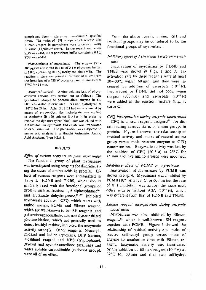
Effect of 1'arious reagents on plant myrosinaseThe functional group of plant myrosinase
was investigated using reagents for discriminating the states of amino acids in protein. Effects of various reagents were summarized inTable I. FDNB and TNBS, which shouldgenerally react with the functional groups ofprotein such as fructose I, 6-diphosphatase1-l'and glutamate dehydrogenase,!" n) inhi bitedmyrosinase activity. CFQ, which reacts withamino groups, PCMB and Ellman reagent,which are well-known to be -SH reagents, andp-diazobenzene-sulfonic acid and dye-sensitizedphotooxidation, which are generally used todetect histidyl residue, inhibited the enzymaticactivity strongly. Other reagents, N-acetylimidazol and iodine (tyrosine), DFP (serine),Koshland reagent and NBS (tryptophane),glyoxal and cyclohexanedione (arginine) andwater soluble carbodiimide (carboxyl group),were all of no effect.
- 14 -
From the above results, amino. -SH andimidazol groups may be conSidered to be thefunctional groups of myrosinase.
Inhibitory e.ffect ofFDNB alld TNBS on myro.linaseInactivation of myrosinase by FDNB and
TNBS were shown in Figs. I and 2. In·activation rate by these reagents were at most20-30j~ within 60 min, and they were in·creased by addition of ascorbate (10- 3 M).Inactivation by FDNB did not occur whensinigrin (300 mM) and ascorbate (10- 2 M)were added in the reaction mixture (Fig. I,curve C).
CFQ incorporation during enzymic inactil'ationCFQ is a new reagent, assigned %:I) for dis
criminating various states of amino groups inprotein. Figure 3 showed the relationship ofresidual activity and moles of reacted aminogroup versus mole between enzyme to CFQconcentration. Enzymatic activity was lost bythe addition of CFQ (10-3 M) at 25°C for15 min and five amino groups were modified.
Inhibitory effect of PCMB on myrosinaseInactivation of myrosinase by PCMB was
shown in Fig. 4. Myrosinase was inhibited byPCMB (I0-~ M) at 37°C for 60 min but the rateof this inhibition was almost the same eachother with or without ASA (10- 3 M), whichwas different from that of FDNB and TNBS.
Ellman reagent incorporation during enzymicinactil'ationMyrosinase was also inhibited by Ellman
reagent, u. which is well·known -SH reagenttogether with PCMB. Figure 5 showed therelationship of residual activity and moles ofreacted sulfhydryl group versus mole ofenzyme to incubation time with Ellman reagent. Enzymatic activity was inactivatedby the addition of Ellman reagent (10- 3 M) at37°C for 30 min and then two sulfhydryl

TABLE l. EffECTS OF VARIOt;S REAGE~"TS ASO METHOD
Enzymatic activity was measured by the liberation of glucose.----_.~ ----~----------------~----~
Conc.(!>!)
Time(min)
Temp.eCI
Remainingact.( ~~)
None 100Fluorodinitrobenzene 10~3 20 20 50
(FDNB)Trinitrobenzenesulfonic Acid 10-3 20 20 90
(TNBS)MonochlorotriAuoro-p·benzoq uinone 10-3 20 25 10
ICFQ)p-Mercuribenzoate 10-' 20 37 0
(PCMB)5~5'-Dithio--bis-(2-nitrobenzoicacid I 10-3 20 37 0
(Ellman reagent)p-Diazobenzenesulfonic Acid 10-3 30 25 15Photooxidation 15 37 0N-Acetylimidazole 10-3 20 37 100DiisopropyInuorophosphate 10-3 20 37 95
(DFP)2-Hydroxy-5-nitrobenzyl bromide 10-3 20 25 99
(Koshland reagent)N-Bromosuccinimide 10-3 20 37 95
(NBS)Glyoxal 10-3 20 25 100Cyclohexanedione 10-3 20 25 100Water soluble carbodiimide 10-3 20 37 100
Pr~incubalion time (min) Preincubation
o~o---~~--~~__~L-~
l g.::- 0 .::-:~v
50 ~.. u., • ...~
-.,;; ~~<; "er:: ..
er::
FIG. I. Inactivation of Plant Myrosinase by FDNB.
100 ,,1 000 ,lig) of myrosinase were pre-incubatedat 20'C with I mM FDNB in 0.2 M sodium bicarbonate buffer, pH 9.0, 10 mM (l mM") ASA and 300 mMsinigrin (both omitted in the sample of curve Bandonly sinigrin omitted in the sample of curve A*).The final volume was 0.5 m1. Aliquots (10 ItI) wereremoved at different times and assayed.
FIG. 2. Inactivation of Plant Myrosinase by TNBS.
100 I" (200 ,'g) of mYTOsinase were incubated at 2O'Cwith 1 ffiM TNBS in 0.2 M sodium bicarbonate butTer,pH 9.0, and 1 mM ASA (omitted in the sample ofcurve B), in a total volume of 0.5 ml. Aliquots (20 ,.1)were removed at different times and assayed.
- 15 .

g-o
3 ~
(iii, ,"" .., ,
2 <:: ,:.; ...° ,s ,-,
1 ~ E'0;::::
-t:r------I!.
Prclncubat ion tim e (m in I
----£--------~--&
II
II
II
I
L---~--....;:,,"':::---~~O
100
Dye-sensitized photooxidation of myrosinasePlant myrosinase was photooxidized in
phosphate buffer, pH 8.0 at 37°C in the presence of methylene blue (Fig. 7), This inactivation was protected in the presence of
FIG. 5. Ellman Reagent Incorporation during Enzyme Inactivation.
500 .III (I mg) of myrosinase were pre-incubated a37~C with I mM Ellman reagent in O.::! M sodiunphosphate buffer, pH 8.0, and in the case of denaturation with 5DS 0.2 M phosphate buffer containing 0.1 ~
SDS was used. The final volume was 3.3 ml. Aliquots (10 fll) were removed at different times anIassayed. (e) Enzymatic activity, (6) -SH modifielC...) -SH modified in the denatured enzyme by SDS
benzenesulfonic acid was shown in Fig. 6.Myrosinase was inhibited by this reagenl(10-3 M) at 25°C for 30 min, but enzymaticactivity was protected in addition of sinigrin(300 mM). Inhibition rate by this reagent wasvery strong, but this reagent is well-known asa modifying reagent for histidyl- and tyrosylresidue.n, As myrosinase was not affected byN-acetylimidazole t9l which react with tyrosylresidue, the inactivation by p-diazobenzenesulfonic acid may be considered to be dependent on the modification of histidyl residue.In order to confinn the above presumptionphotooxidation17 J of myrosinase, which is oneof the modification of histidyl residue, wasdone.
__e--.... 1'.- ....".... ',
/I
II
O~O--"---.......----I....O~-J'"""""...IOCFQ concentration (~ll
FIG. 3. CFQ Incorporation during Enzymic Inactiva-
.,,.
10or----o
Inhibitory effect of p-diazobenzenesulfonic acidon myrosinase
Inactivation of myrosinase by p-diazo-
tion.
300 !-II (260 fig) of myrosinase were incubated at 25 'Cwith 0.1 -I mM CFQ in 0.2 Msodium phosphate buffer,pH 8.0, for 15 min, in a total volume of 2.0 mJ.Aliquots (20 "I) were removed and assayed.
o~o---...-:~---~---....-'Pre-incubation lime (min)
FtG. 4. Inactivation of Plant Myrosinase by PCMB.
100 pi (200 I'g) of myrosinase were pre-incubated at37'C with I mM peMB in 0.2 M sodium phosphatebuffer, pH 7.0, and I mM ASA (omitted in the samplecurve B), in a total volume orO.5 ml. Aliquols (201'1)were removed at different times and assayed.
groups were modified. While four sulfhydrylgroups were modified when the enzyme wasdenatured by SDS (0.1 %).
- 16 .

Preincubation time [min}
In the dark, the enzyme was quile stable inthe presence of methylene blue, and in lightwithout the dye.
In general, it is known that histidine, methionine, tyrosine, tryptophane and cysteine aresensitive to photooxidation, but myrosinaseactivity was not affected by NBS and Koshland reagent. Therefore, amino acid composition of the photooxidized-enzyme wasanalyzed. Figure 8 showed the result of
Control
B
A
,:;..::::... 50"....::
DO!"----~----!~---~--'
,:" ~FIG. 6. Inactivation of Plant Myrosinase by p
Diazobenzenesulfonic Acid.
50}ll (100 /Ig) of myrosinase were incubated at 25°Cwith I mM p-diazobenzenesulfonic acid in 0.2 M sodiumphosphate buffer, pH B.O, for 30 min, and 300 filM
sinigrin (omilled in the sample of curve A), in a totalvolume of 0.5 mL Aliquots (20 1,1) were removed andassayed.
",', "
,::
.; "
..•?QCLys His NH; Arg
Sample
:~,
, ..,. ,. ..
JdH; .. ;' ,-=...~Lys His NH; Arg
A
:::~<;" 50....~"0:
c::
FIG. 7. Inactivation of Plant Myrosinase by Photooxidation.
100 /:1 (50-300 }/g) of myrosinase were illuminatedat 37°C with O.Ol % methylene blue in 0.2 Msodiumphosphate buffer, pH B.O, and 300 mM sinigrin (omittedin the sample of curve A), in a lotal volume of 0.5 mi.Aliquots (50 Ill) were removed and assayed. (0)Non-photooJl;idized enzyme
sinigrin (300 mM), and the enzyme was notphotooxidized below 25°C.
FIG. 8. Comparison of Amino Acid Compositionof the Photooxidized and Non-treated Myrosinase.
150 I'g of myrosinase were pholooxidized at 37 c Cin 0.1 M sodium phosphate buffer, pH 8.0, containing0.01 % methylene blue for 30 min. Control was notphotooxidized. Amino acid analysis was as describedin the lext.
amino acid analysis of the control and thephotooxidized enzyme. The control wasunder the same conditions, but withoutillumination. The concentrations of the usedenzymes were nearly the same. There wereno differences between the control and thephotooxidized enzymes, but only histidine.content of the latter was found to be lower.
- 17 -

DISCUSSION
The funclional groups of plant myrosinasewere investigated using reagents which discriminate the states of amino acids in proteinand by photooxidation. It was found that atleast the amino groups, sulfhydryl groups andhistidyl residues constitute the functionalgroups of plant myrosinase. FDNB andTNBS are well-known as the reagents whichare used to determine a- and s-amino groupsin protein, but they are also used to study thefunctional group of proteins. Plant myrosinase is specifically activated by I-ascorbate(10- 3 Mt but its activation mechanism isstill indistinct. Tsuruo et al. had studied onthe activation mechanism of plant myrosinase 'l
by ascorbate, and confirmed that its activationwas not based upon the oxidation and reduction by ascorbate, but was based on the changesof conformation of enzyme in addition ofascorbate. The results obtained by FONBand TNBS support the above conclusion, thatis, groups sensitive to FDNB and TNBS maybe considered not to be very reactive in thenative state of the enzyme but to become morereactive in the presence of ascorbate. Thereactive groups to FDNB and TNBS may be'Considered to be amino groups.
Myrosinase was inactivated when five aminogroups were modified by CFQ, but its activitystill remained when three amino groups weremodified. Therefore, it may be consideredthat two remaining amino groups are necessary to the activity of myrosinase. Thedifferent rate of inhibition by FDNB, TNBSand CFQ may be considered to be dependenton the shape and dimension of these reagents.
The inactivation by -SH reagents werestrong. Plant myrosinase have at least foursulfhydryl groups of which two sulfhydrylgroups COncern with the activity of myrosinase.As the rate of inhibition by PCMB was nearlythe same each other with or without ascorbatesulfhydryl groups which take part in th~
- 18 ~
enzymatic activity may be thought to be reactive in a native state in the presence o.absence of ascorbate.
Histidyl residue is also one of the functionagroups of myrosinase. However, owing tlthe interaction of the reagent used willascorbate, in the method employed, the eJfeaof ascorbate on the state of histidyl residue itprotein could not be investigated.
From the above results, amino groups, sulf.hydryl groups and histidyl residue may beconsidered to be in the active center of plantrnyrosinase, and thus the modification of anindividual group or residue by the specificreagents caused inactivation. It may be con·sidered that the above groups and residue arelocated closely to each other in the threedimensional structure of the protein. Activation by ascorbate may be assigned to the conformational changes of myrosinase caused byascorbate; Even a slight conformationalchange of myrosinase caused by ascorbatemay cause the surface exposition of the aminogroups, or the dissociation and association ofprotein, because plant myrosinase consists of atleast four subunits.m

Chapter 3
Approach to the Interaction of
L-Ascorbic Acid to the Enzyme
Nagashima ,,/ al.~l found that the plant myrosinasewas activated strongly by I-ascorbic acid (ASA).Schwimmer 20 and Ettlinger 1'/ al. 21. investigated thisphenomenon. The latter concluded that ASA behavedas a reversibly dissociable base. closely connected withthe nucleophilic group of thioglucosidase. Kojimae/ al.'l )studied the changes of the reaction product ofthe enzymatic hydrolysis of mustard oil glucoside inthe presence of ASA at low pH.
Tsuruo et al. described that the oxidationreduction reaction of ASA had nothing to do with theactivation reaction of myrosinase.
21The presence of
the effector site for ASA was presumed by the kineticmeasurements and the instabilization of the enzyme byASA on heating. I also reported~~lthat theinhibitions of plant myrosinase by f1uorodinitrobenzene(FDNB) and trinitrobenzenesulfonic acid ([NBS) werestronger in the presence of ASA.
From the above results. the author speculated thatthe activation mechanism of plant myrosinase by ASAmay be considered to be the result of slight conformational change of myrosinase or the dissociation andassociation of the enzyme.
Some experiments were carried out to elucidate theactivation mechanism of this enzyme. Enzyme reaction and assay of enzymatic activity were describedpreviously.lI Myrosinase (F-IA fraction)221 was usedas the enzyme source.
The activation was observed above 5 x 10- ~ M ASAconcentration, and the maximum activation wasaround 10- 3 M ASA (Fig. I). Activation graduallydecreased at higher concentrations. The decrease ofactivation at higher concentration can be explained byassuming that ASA acts as a competitive inhibitor ofplant myrosinase. 21
In the previous Chapter, I described thatthe plant myrosinase consisted of at least four subunits.Therefore, there is a possibility that the activation ofmyrosinase depend on the dissociation-associationmechanism of the enzyme protein by the addition ofASA. I investigated the presence of dis-sociation and association of the enzyme by gel-filtration and ultracentrifugal analysis.
2000
~~
:§u..'"E 1000~..c
W
oL:~=ltO':-":::::l(,O-->:---lL.O~-.:---JloL._,:,]--:l~.L~1~0-1Ascorbic ~cid (M)
FIG. I. Effect of Ascorbic Acid.
Enzyme reactions were carried out in the systemcontaining 2.5 pmoles of the substrate (sinigrin) in1.0 ml of water at pH 7.0 and 37·C. The myrosinaseactivity was measured as the liberation or sulfate bytitration with a recording pH-stat, Radiometer ModelSBR2fSBUlfTTTI Autotitrator. 81
Figure 2 shows the gel-filtration chromatogram ofmyrosinase on Sephadex G-150 with and without ASA(10- 3 M). The enzyme was eluted at the same positionwith or without ASA.
Sedimentation studies were carried out in a Spincomodel E ultracentrifuge.
High speed sedimentation velocity runs with orwithout ASA (10-' M) showed symmetrical peaks withthe same value or 6.8 S. (Fig. 3).
These results indicated that the enzyme was neitherdissociated nor associated by ASA. That is, theactivation of myrosinase by ASA did not result in thedissociation and asSociation of the enzyme by additionof ASA.
In the previous Chapter, 1 investigated thefunctional group of myrosinase by using reagents fordiscrimination the state of amino acid and found thatthe amino. sulfhydryl groups and the histid)il residuewere associated with the active site or myrosinase.~~)
- 19 -

Incubation Incubation Amino Enzymemi turc time (min) group activity (%)
reacted
30 14.9 90
..L 30 13.2 45
I mMA
TABLE I. UMB R OF II 0 GROUPS PER PROTEt,
MOLECULE REACTING WITH T B I THE
PR CE A 0 BSE 'CE OF AsCORBIC 10
M ro ina e \ a incubated with the reagent
(10- 3 I). nd fifteen amino group were modified
in the ab ence of , but only thirteen amino group
showed modification in the presence of A. Thisindicate that tbc functional group b ome more reacti\'e due to thc onformational change of the protein
re ult d by • and \ ould be modified by T B .Th e re ult upportcd the con lusion that the
acti ation me hani m of m ro inase by depend 00
the light conformational change of tbe protein r ultedby , and i not due to tbe di ociation and as
sociation mechani m of myrosinase.
he myro ina e \ a al 0 found to b ina tivatedtrongly in the pre ence of than without it by
B.The t:lle of ami:lo group of myrosina e was deler-
mined by T B. olulion (J ml) of T B (3.310- 3 I) wcre added to enzyme olution (200 pi, 6 pgprotcin) containing -1°1o odium bicarbonate buffer,pH 9.0, 0.5 ml of (6 10-3 I), in a 10lal olumeof 3.0 ml. The mixture thus obtained were incubated
at 20 for 30 min and the reaction \\a topped bythe addition of I ml of D (10°'0) and 1-1 I (I ).
During Ihi incubation time, the hydroly i of TNBand the reaction of T B with the ample of enz meprocecdcd in parallel. A blank mixture was preparedin Ihe samc manner without enzyme olution, and Ihedifferences in absorbance at a wa elength (340 m/')between the sample and the blank mixture wcremea ured. The moles of amino groups which reactcdwith T B in myro ina e were calculated, using J,
alue \ hich is 10,000 M- t cm- t . indicaled in TablcI, myro ina e was not inacti ated by T B (10- 3 I)for 30 min without A A, but the enzyme was rapidly
inhibitcd by T BS \ ithin 30 min in the presence of
FIG. edimentation Paltern f Plant yro ina ewilh and without . orbic id.
- 20 -
o m ~
Fraction number (X 3ml)
I . 2. el-filtr.tion on ephadex G 150 with and\ ilhout A corbic Acid.
el-filtration n erhadex G 150 column (I 90 em),cquilibrated with 0.01 I odium pho phatc buffercontaining 0.01 1 K I, pH 7.0, and I m I A (omilted in the ample f curve ), \ a performed at 5
ne ml f thc cnqme di olv d in the amc buffer( A millCd in the ample of cur e ) was ubjected10 the column and elutcd with the r pective buffcrs.
nzymatic activitie were measured by the liberationof glucose.o 01 d cribed pre iou ly.7
~0.2
8!::eQ0.,.~
0.1
.::U.."8......c
lLl

FIG. 2. Gel-filtration on Sephadex G-150 with andwithout Ascorbic Acid.
Gel-filtration on Sephadex G-150 column (I :.: 90 em),equilibrated with 0.01 M sodium phosphate buffercontaining 0.01 MKCI, pH 7.0, and I mM ASA (omitted in the sample of curve A), was performed at 5'C.One ml of the enzyme dissolved in the same buffer(ASA omitled in the sample of curve A) was subjectedto the column and eluted with the respective buffers.Enzymatic activities were measured by the liberationof glucose.GdS described previously.7!
'I.0.2
II~eci0~
t- 0.1:§".."S>...c
tz.l
0 10FrDcli,," number
30
The myrosinase was also found to be inactivatedstrongly in the presence of ASA than without it byTNBS.
The st:lte of ami:lo groups of myrosinase was determined by TNBS. Solution (I ml) of TNBS (3.3'10-' M) were added to enzyme solution (200 /d, 6BB /'gprotein) comaining 4% sodium bicarbonate buffer,pH 9.0, 0.5 ml of ASA (6 '-: /O-a M), in a total volumeof 3.0 ml. The mixture thus obtained were incubatedat 20'C for 30 min and the reaction was stopped bythe addition of 1 ml of SDS (10%) and HCl (I N).During this incubation time, the hydrolysis of TNBSand the reactions of TNBS with the sample of enzymeproceeded in parallel. A blank mixture was preparedin the same manner without enzyme solution, and thedifferences in absorbance at a wavelength (340 m,u)
between the sample and the blank mixture weremeasured. The moles of amino groups which reacledwith TNBS in myrosinase were calculated, using Jtvalue which is 10,000 M- I cm- I . As indicated in TableI, myrosinase was not inactivated by TNBS (10- 3 M)for 30 min without ASA, but the enzyme was rapidlyinhibited by TNBS within 30 min in the presence of
FIG. 3. Sedimentation Pallerns of Plant Myrosinasewith and without Ascorbic Acid.
Sample solutions contained 0.01 M sodium phosphatebuffer, pH 7.0,0.1 M KCl, I mM ASA (omitted in thesample of peak A), and 6.5 mg of myrosinase.Measurements of Ihe sedimentation coefficient wereperformed at a speed of 59,780 rpm in a double sectorcells at lO·C.
.20 -
TABLE 1. NUMBER Of AMINO GROUPS PER PROTEtNMOLECULE REACTING WITH TNBS IN THE
PRESENCE AND ABSENCE Of ASCORBIC ACID
Myrosinase was incubated with the reagent for30 min and aliquots (10/,1) were assayed for activity.Enzymatic activities were measured by the liberationof glucose, as described previously.71
Incubation Incubation Amino Enzymemixture time (min) groups activity (%)reacted
I mM TNBS 30 14.9 90I mM TNBS
-:- 30 13.2 45I mM ASA
ASA (Jo-a M). And fifteen amino groups were modifiedin the absence of ASA, but only thirteen amino groupsshowed modification in the presence of ASA. Thisindicates that the functional groups become more reactive due to the conformational change of the proteinresulted by ASA, and would be modified by TNBS.
These results supported the conclusion Ihat theactivation mechanism of myrosinase by ASA depend onthe slight conformational change of the protein resulledby ASA, and is not due to the dissociation and association mechanism of myrosinase.

Chapter 4
Binding of Ascorbate to the Enzyme
and the Interaction of Ascorbate with
the Functional Groups
In the preceding chapters, 22, 32) I purified
the enzyme and clarified the physical and chemical properties of myrosinase isoenzymes, themyrosinases from white mustard (Sinapis albaL.) and rapeseed (Brassica napus L.) have beenisolated and characterized by Janson et al.,33. 34}who showed the existence of a number of isoenzymes, also found by Henderson and McEwetf,5)and studied the functional groups of plant myrosinase using reagents which discriminate thestates of amino acids in protein and found thatamino group. -SH group and histidyl residueconstitute the active sites of the enzyme. I alsofound that the activation mechanism of myrosinase by ascorbate depend on the slight conformational change of the protein resulted by ASA,and is not due to the dissociation and associationmechanism of myrosinase. I reported that fjglucosidase activity was not activated by ASAbut was inhibited competitively at higher concentration.
In this chapter, I determined the binding ofASA ligands to myrosinase by the dialysis ratetechnique and studied spectrophotometricallythe interaction of ASA with the enzyme by theinhibitory effect by the chemical modification,using reagents discriminating the states of aminoacids, on myrosinase and fj-glucosidase activitiesin connection with the activation of the enzymeand discussed the relationship between the activecenter and the activation mechanism of theenzyme by ascorbate.
MATERIALS AND METHODS
Chemical reagents L-Ascorbie-I.C'" acid was pur·chased from New England Nuclear Corporation and2-methoxy-5-nittotroponc (MNT) was purchased fromSankyo Co., Ltd. Other chemicals were obtained fromNakarai Chemicals Ltd. All reagent were of analyticalgrade, and used without further purification.
Substrate Sinigrin, obtained from Nutritional Bio·chemicals Corporation, was used as the subsnate formyrosinase and p·nitrophcnyl tl·D-glucoside (Sigma) asthe subsnate for tl-g1ucosidasc.
Enzyme preparation The myrosinase solution fromyellow mustard powder was prepared by the methodsdescribed in the previous Chapter"), and F-IA fractionwas used as the enzyme in this experiment.
Enzyme Q5say The assay mixture contained 2.5 I'moles of substrate and enzyme solution in a total volumeof I ml. Enzymatic activities were measured by theliberation of glucose, sulfate and p-nitrophenol. I ,6)
The reactions were carried out under conditions regardedto be zero order reactions.
Assay of protein Protein was determined by themethod of Lowry et al.')
Inhibition experiments Unless otherwise staled,the incubation mixtures (5001'1) contained myrosinase(50-300I'g), inhibitors PCMB (Ia-"M), DEP (la-'M)and MNT (10") M) and buffer.Aliquots (5-201'1) were removed :it specific times andthe activities assayed.
Chemical modifications The state of amino groupsof myrosinase was determined by MNT.'") Solution(51'1) of MNT (100 mM) in N,N'.dimethylformamidewas added to enzyme solution (2001'1, 1.0 mg protein)
- 21 -

apparent that at low ASA concentrations thedialysis rate of ASA is lowered considerably bythe presence of myrasinase, indicating that alarge proportion of the total ASA is proteinbound and therefore non-dialyzable.
To evaluate the binding constant and thenumber of binding sites, the data from Fig. 2(Curve with enzyme) are treated as follows. Thesteady state concentration of isotope found inthe effluent at any given substrate concentrationis taken as a measure of the fraction of the total
Fig. 1 Diagram of the Apparatus for MeasuringSubstrate Binding by Rate of Dialysis.
The dialysis cell was adapted from the Technilab cell(Bel-Art Products) for continuous flow dialysis (1 mJsize). The discshapcd upper chambcr (19 mm in diametcr x 5 mm) was altered in two ways. It was deepen'ed to 9 mm to make the capacity about 2.5 ml anda hole (S mm in diameter) was drilled to the outside topermit additions of small volumes of substrate or otheragents to the enzyme solution (1.5 ml) during the courseof a bind ing measurement. The lower chamber (19 mmin diamcter x 10 mm) has a capacity of 2.8 ml and iscompletely fLIled with buffer solution which is pumpedthrough at a constant rate of 20 mt per hrs. The membrane, a square cut from ordinary cellophane dialysistubing (Visking Company), is clamped between theLucite blocks, which are held together by stainless steelscrews. The contents of both chambers are mixed bymeans of small magnetic stirring bars; the bar in the topchamber rests on the membrane. The inner diameterof the tubing leading from the lower chamber to thefraction collector is small, 0.015 inch, in order to mini·mize dead space. A1iquots (0.5 to 0.8 ml) from thefraction collector are diluted in scintillant (PPO 10 g,POPO 0.25 g naphthalene 100 g, dioxane 11) for liquidscintillation counting (Packard Tri·Carb 2002 Scintillation Spectrometer). The dialysis apparatus has beenused satisfactorily either in the cold room (T=So) or atroom temperature (T=2S").
and 0.2 M phosphate buffer (pH 8.5) in a total volumeof 1.5 mI. The mixture thus obtained was incubated at2S"C for 60 min. The product was passed through acolumn of Sephadex G-SO suspended in water to removeexcess reagent and by·products. Myrosinase appearedas a yeUow colored fraction. The moles of aminogroups which reacted with MNT in myrosinase werecalculated, using .:l e value (2.07 x 10' M-' cm-') at 420mJ,t.
The modifit;ltion of tryptophane residues was deter·mined by HNBB.31
) Solution (5 }.tl) of HNBB (100 mM)in acetone was added to enzyme solution (100 }.tl, SOO}.tgprotein) and 0.5 M acetate buffer (pH 5.0) in a totalvolume of 205 J,tl. The reaction mixture was incubatedat 35"C for 45 min. The product waS done by the samemethod as MNT. The moles of tryptophane residueswhich reacted with HNBB in myrosinase were calculated,using.o.e value (1.80 x 10'M"' cm-') at 410m,u (pH 10).
Photometrical measurements Measurements ofabsorption spectra and differential spectra were carriedout with a Hitachi dual wave length spectrometer 356 B.
Rate dialysis method Rate dialysis was carried outby the method of Colowick and Womack,)O) using theapparatus (Fig. 1.) consist of a dialysis with an upperchamber, containing the enzyme and labeled substrate,separated by a membrane from a lower chamber,through which buffer is pumped at a constant rate andfrom which the effluent is sampled for measurementof radioactivity.
RESULTS
Measurement of ASA binding to myro.rinaseFigure 2 illustrates the procedure used and
the results obtained in a binding measurement.The first aliquot of ligand was labeled with l4C,and suhsequant additions contained nonradioactive ligand. This procedure 38, 39) ensuresoptimal counting rates over a wide range ofligand concentrations. Note that in the absenceof diminution of free ligand concentration bybinding, dilution, or loss by dialysis, the additionof nonradioactive ligand would not alter the 14Cdialysis rate, because the effect of increasedligand concentration is exactly cancelled by thecorresponding decrease in specific activity. Thetwo curves were obtained under identical conditions except that myrosinase was omitted fromthe curve labeled "without enzyme." It is
- 22 -
from bufferreservoir
Magne'ic Stirrer
\10 froclioncollector

-,•. ~,h III •
.; .',.."
.0,
P : .
.\.': ~ Ill' ~.
1 .0--0-
'409-'; 0.".• II
?.. 'II ...... ' ~n~._..- ,. "-1111 •
,9,
/ I. o,j"'I1I· J.
1\00substrate in the upper chamber in the freely diffusible state. When excess unlabeled substrateis added, the radioactivity in the effluent is takento be that corresponding to 100% of the substrate in the free state. Thus, dividing anyobserved value by this maximum value gives thefraction of the substrate free at a given concentration. Values for the concentration of free {F)and bound {B) substrate then easily derived. Forexample, from Fig. 2, at a total ASA concentration of 2.21 x la-3 M, the fraction of ASA freeis 1100/1300 or 0.182, so that the values of Fand Bare 1.8 x l(f3 and 3.41 x l(r4 M, respectively. These values can be used to determinethe dissociation constant, Kdiss' by applying theequation
(n - B)' FKdiss = B
where n is the concentration of the total binding sites on the protein. It enzyme of knownpurity and n value is used, one measurement ofF and B at a single substrate concentration canbe used to determine Kdiss. With enzyme ofunknown purity or n value, a series of F and Bvalues at different substrate concentrations servesto evaluate both Kdiss and n. In this case, onemakes a Scatchard-type plot of B versus B/F,and determines Kdiss from the slope and n fromthe intercept, according to the linear form of theabove equation
Boon - K' B/F
Such a plot is shown in Fig. 3a. Myrosinaseappears to have 4 sites per molecule which bindASA rather strongly (Ko = 0.1 x 1(f4 M), and atleast one additional site which binds ASA lessstrongly (KO = 0.9 x 1(f4 M ). Myrosinase activity is maximumly activated with the concentration of ASA (1 mM) and then the enzyme bindsthe four ASA molecules. (Fig. 3.B).
Effect of ASA aWlIogues on myrosinase activityAs shown in Table I, the myrosinase activity
Was nOt activated by ASA analogues tested.Nagashima and Uchiyama5) showed that myrosinase was not activated by reducing reagents
Fig. 2 Measurement of Ascorbic Acid at VaryingASA Concentrations.
111 e time course of two dialysis rate measurements,without and with enzyme, is shown, but only the latteris required in a routine measurement. The specificactivity of the "C·ASA added at the beginning was 2.72mCi per mmole. After radioactivity in the effluentreached a steady state, increments of unlabeled ASAwere added to give the tota] concentrations indicatedunder the arrows, Protein concentration 11 mg per ml(=0.73 x HT' M). Buffer 0.05 M phosphate, pH 7;T = 20"C.
(glutathione, cysteine, BAL, Gallic acid) exceptASA and Ettlinger et aL 2l) reported that the
enzyme was activated by 2-0-methyl-L-ascorbatewhich didn't keep the effect of reducing power.These results showed that ascorbate was notacting on the conventional reducing agent.
Differential spectra of myrosinaseFigure 4 shows the differential spectra of
myrosinase with or without ASA. The enzymeprotein is comfornationally changed by the addi
tion of ASA.Figure 5 shows the absorption and differential
spectra of the chemical modified myrosinase byMNT. Approximately 1.5 amino residues wereappeared on the surface of the enzyme by addingASA (1(f3 M).
The spectra of HNBB modified myrosinase
- 23-

270 290 310
Fig. 3 plot of the Data Derived from the SteadyState Values in Fig. 2.
Showing Binding of " C-ASA to Plant Myrosinase lA)and Relationship of the Activation to the Binding ofASA lB).
were shown in Fig. 6. About 2.3 tryptophanylresidues were buried in the molecule when ASAwas added p(f3 M ).
These results showed the myrosinase was conformationally changed by the addition of ASA.
..I.
Inhibition by DEPInactivation of myrosinase by DEP was shown
in Fig. 8. Inactivation rates of myrosinase activities by this reagent were stronger than .B-glucosidase activities, and especially after the enzyme
Fig. 4 Differentaial Spectrum of Myrosinase With
and Without ASA.Protein concentration is 1 mg/ml in 0.05 M phosphatebuffer and 1 mM EDTA. pH 7.0. 20°C, in both compartments. Concentrations of ASA are:1) None; 2) 5 x 10"' M; 3) 1.0 x lIT' M;4) LO x 10-' M.
B
I"· ~
..... lI!", I'IC \. ,.1 "II
,,'
,;",
,:,.,,,
Inhibition by PCMBAs shown in Fig. 7, myrosinase (A, B) and
t3-g1ucosidase (C, D) activities were strongly inhibited by PCMB (1(f4 M ) at 25°C for 30 min.
Inactivation rate of myrosinase activities wereincreased by the addition of ascorbate (1{f3 M)when the activities were measured, but that ofIJ-glucosidase activities were contrary. Fromthese results, it is considered that sulfhydrylgroups are located at the active center of myrosmase.
.....1_...... ~"" _
Fig. 5 Absorption and Differential Spectra ofthe Chemical Modified Myrosinase by MNT.
Protein concentration is 0.25 mglml in 0.05 M phosphatebuffer. pH 8.5. 20°C. Concentrations of ASA are:1) None; 2) 1.0 x 10- 3 M.
- 24 -

I.,;- I..,;<i I
1 !
100 ~l (300 ~gJ of myrosinase were pre-incubated at25°C with 0.1 mM PCMB in 0.2 M potassium phosphatebuffer, pH 7.0, and 1 mM ASA (omitted on the sample
of A and C), in a total volume of 0.5 mL Aliquots (520 ~I) were removed at different times and assayed.Enzymatic reactions were carried out in the systemcontaining 2.5 ~moles of substrate, 0.2 mmole of phosphate buffer. 20 ~l of the sample in Curves a (5 ~l inCurves b). pH 7.0. and in Curves b 1 mM of ASA wasadded.
"~r.n';lb... , It>" r ... ·1 •• 011
i",-,·.n-Hi ....... " 1 ••• -llIIlrol
..,
Fig. 8 Inactivation of plant Myrosinase by DEP.
100 ~l pOG IJg) of myrosinase were pTe-incubated at25°C with 10 mM DEP in 0.2 M acetate buffer, pH 5.0,and 1 mM ASA (omitted in the sample of A and C), ina total volume of 0.5 rnl. Enzymatic reactions werecarried out as described in Fig. 7.
(,,,
-;
•
rr"'n' "boll I"'" r •••·r.", I
1'1. ~
f-r.-' ..... "hA' ,,'n I,.·
Fig. 6 Absorption and Differential Spectra of theChemical Modified Myrosinase by HNBB.
Protein concentration is 0.15 mg/ml in 0.05 M carbonatebuffer, pH 10, 20°C. Concentrations of ASA are:1) None; 2) 1.0 x 10-' M.
c),""""""""
, .',..... b.
.. -..~.~.-.-.~::-....
I'n'ln, "boll illn ll .... I .. I""
;"
~"""\\
"""", ,. "
'~.""---~-~I:!.._~....
"I ... n •••1.1 ..1 ,un 1 ...... 1 ... t
was preincubated at 25°C with 10 mM DEP and1 mM ASA, myrosinase activity was strongestlyinhibited when the enzymatic activity was measured without ASA. This showed that histidylresidues in the protein were affected by theaddition of ASA.
Inhibition by MNTInactivation of myrosinase by MNT was
Fig. 7 Inactivation of plant Myrosinase by PCMB.
- 25 -

", I" n' .. ~, .•• I 'H I .... ' "' ... I
(Sl
lOa
25bo
In'00
n170
BS
07
Rrla~lv~ Bc~ivity
l-S8corbD1,.e
llehydro- L-Ill'lc orbBt f:
0-. rabo.aflcorbate
Cluco""c-orbat.il'"
A.llc-orb)' I-p4h1l1 t.1I lc"
A.,eorbyl-stear.llt.e ll
A8(:'l,]Irbyl 2,6-di.plll.Lt-Jlt.or"
AsA an./JIlol[U("
110.3",
None
Table I Effect of the Analogues of Ascorbic Acidon Myrosinase Activity
Enzymatic activities were measured by the liberationof sulfate.
b
"
o
11'"; ......", ••••• , ....
As", An.alolt\lil"'8 w~re dillaolved in dt.et.hylfo...... ide
(10- 1 .1.
'-Glucosidase3
shown in Fig. 9. Inactivation rates of myrosinaseand i3-g1ucosidase activities by this reagent werealmost same, but in the case of B very interestingresults was obtained. Differing from otherreagents, myrosinase activity was strongly inhibited when the enzymatic activity was measuredwithout ASA, but, the activity was not affectedby this reagent when the activity was measuredwith ASA. This phenomenon was not occured
400Hyrosin...e
Fig. 9 Inactivation of Plant Myrosinase by MNT.100 j.11 (300 J.'g) of myrosinase were pre·incubared at25°C with 1 mM MNT in 0.2 M potassium phosphatebuffer, pH B.5, and 1 mM ASA (omitted in the sampleof A and C), in a total volume of 0.5 mi. Enzymaticreactions were carried out as de,cribed in Fig. 7.
110
~o004020o
o L...-;........--..1----L__..;...JU,o"
~oo~
ii~ E 2 2 SE .: --..
c.~
~ "'E "'I!"' ,,--..
"0 >~ Il
200 ~ > ~E --,j. 0" 0c E u E
~ « ,j.«~
0 "- "-; z z, I I
"" 0..
"lOCI
nil40
r~lDperat.ur{"1 CJ
20oo
10
;0
..B? .0
-!...." ~~ ..~ i ~o",j.u... ~
""u" 20..
Fig. 10 Optimum-Temperature Curves of Myrosinase and {1-glucosidase Activities of plant Myrosinase.
-_... with ASA
Reaction. were carried out at 7.0 for 15 min without ASA or for b .in with ASA •
. 26 -

in J3-glucosidase activity. This showed that aminoresidues in the protein were located at the region
which was changed by ASA.
Optimum temperature of myrosinase
Optimum temperature of myrosinase with orwithout ASA were shown in Fig. 10. Optimumtemperature of myrosinase activity was at about55°C without ASA, but that with ASA was atabout 35°C. This changing of optimum temperature suggested that the structure of myrosinaseprotein was change by the addition of ASA.Optimum temperatures of J3-glucosidase activitywere the same each other with or without ASAand they were at about 55°C which was sameto that of myrosinase without ASA. This suggest
ed J3-glucosidase activity was not accelerated byASA.
DISCUSSION
Tsuruo et al. assumed in the previous paper2)
two sites on the surface of the enzyme, namelythe substrate site for mustard oil glucoside andthe effector site for ascorbate. Furthermore,applying Pigman's model 40, 41) for tl-glucosidaseto the enzyme, two areas were distinguished 4
)
in the substrate site, one for adsorbing the glycanmoiety of the substrate and the other for adsorbing its aglycon moiety. It was also pointed outthat a possible structural alteration of the enzyme protein caused by the binding of ascorbateto the effector site exerted its effect only onthe area for aglycon moiety. Since the bindingof ascorbate to the effector site exerts no influence on p-NPG hydrolysis, the glycon moietyof p-NPG, which differs from mustard oil glucoside in aglycon moiety, is adsorbed only bythe area for glycan moiety in the substrate sitebut not by the area for aglycon moiety. Thatis, p-NPG hydrolysis is neither accelerated norits kinetics altered by the presence of ascorbate.Ettlinger et a1.22 ) described the hydrolysis of
2-4-dinitrophenyl tl-thioglucoside and desulfoglucocapparin by mustard myrosinase is not accelerated by the presence of ascorbate. Thisobservation can be similarly explained. As thesethioglucosides have different aglycons from mustard oil glucoside, they can combine with theenzyme only at the area for glycon moiety inthe substrate site, hence the hydrolyses of thetwo thioglucosides were not influenced by ascorbate.
Sinigrin competes with p-NPG at the same siteof the enzyme protein. Consequently, p-NPGhydrolysis was competitively inhibited by sinigrinand the Ki values of p-NPG hydrolysis for sinigrinare nearly equal to the Km values for sinigrinhydrolysis. 1) Moreover, the former was alteredby the absence or presence of 1(j3 M ascorbateparallelly with the alteration of the latter. Theeffects of the presence of 1(j3 M ascorbate onthe kinetic constants of the enzyme are summarized in Table II. This table also shows the
constancy of the Km value for p-NPG hydrolysisand the Ki values for salicin and glucose 4) in
sinigrin hydrolysis. It is evident from the tablethat the binding of sinigrin to the enzyme wasaffected by the presence of ascorbate but thatthe binding of klucoside or glucose remainedunaffected. These discussions concerning thesubstrate site and the effector site of the enzymeare represented as a schematic model in Fig. 11.
The experimental results obtained in thepresent chapter can be explained as follows (seeFigs. 7, 8 & 9). Since both myrosinase andklucosidase activities were strongly inhibited byPCMB with or without ASA, sulfhydryl groupsare considered to be essential to the catalyticaction of myrosinase, but the inhibition rate ofmyrosinase activity by DEP and MNT were ratherstrongly inhibited than that of tl-glucosidaseactivity, and then the enzyme which was treatedby MNT with ASA was strongly inactivated whenthe enzymatic activity was measured withoutASA, but, the enzyme was not inhibited whenthe activity was determined with ASA. Thusphenomenon didn't occure on the tl-glucosidase
- 27 -

Table 1I Effect of Ascorbate of Kinetic Constants of Myrosinasc
Hydrolysis of II-NI'GAscorb"lc
(M)
o10-3
HydrolY5is of. 5inigrin,~--------~-------""'....
l\.m v;,hw fnr l\.j v .. ltw for Ki vall"., for~ini~rin (1)1) ~Iunl~l' (M) .',1.li1";n (M)LOx 10-' 1.5 J.nx 10- 1
9.3x IO-l 1.5 I.Ox 10-1
Km valul' forp-NI'C (M)
2 X 10⁻³
2 X 10⁻³
l\.i vallie for~il1igrin (M)
2 X 10-'9x 10-'
Fig.11. SCHEMATIC MODel OF MYROSIHASE JCTION
actIvIty. :from these facts, it is considered thatat least amino and histidyl residues are situatedon the region which is altered by the addition ofASA, that is, it is close to aglycon moiety andthe locations of them are changed to acce1aratethe enzyme action by the binding of ASA to theeffector site. On the other hand, as sulfhydrylreagent inhibited strongly to myrosinase and (3glycosidase activities, sulfhydryl gt"oups areconsidered to be situated closely to glyconmoiety. These also shows that p-NPG combineswith the enzyme only at the area for glyconmoiety in the substrate site, hence the hydrolysisof p-NPG was nOt influenced MNT and DFP.The above conclusion is also supported by whichoptimum temperature of myrosinase activitieswere changed by the addition of ASA but thatof p-NPG hydrolysis were the same in spite ofpresence or absence of ASA.
That is, although the enzyme is capable ofcombining sinigrin at the substratc site, thc fitof sinigrin to the site would be imperfect in theabsence of ascorbate. This imperfect molecularfit of sinigrin would be improved by a possiblealteration in the structure 3. 4) of the enzymeby ascorbate-binding at the effector site. Andsince the alteration of the enzyme's proteinstructure caused by the binding of ascorbateexerted no effect on the area which adsorbedglycon moiety in the substrate site, and sincep-NPG, possessing a different aglycon from sinigt"in, was combined by its glycan moiety to theglycon moiety-adsorbing area, the binding stateof p-NPG would be that of an improved fitregardless of the absence or presence of ascorbate.
The inhibitory effect of ascorbate at highconcentrations is accountable by the binding ofascorbate at a different site from the specificeffector site and that of ascorbate was competitive. The Ki value (higher than the Km value forascorbate in the activation of the enzyme 2))
indicates that ascorbate-binding occurs at a different site.
- 28 -

REFERENCES
1) I. Tsuruo. M. Yoshida and T. Hata, Agr.Bioi. Chern., 31.18 (1967).
2) 1. Tsuruo and T. Hata, ibid., 31,27 (1967).3) I. Tsuruo and T. Hata, ibid., 32.479 (1968).4) I. Tsuruo and T. Hata, ibid., 32,1420...... 1432
(1968).5) Z. Nagashima and M. Uchiyama, Nippon
Nogeikagaku Kaishi, 33,980 (1959).6) I. Tsuruo and T. Hata, Agr. Bioi. Chern., 32,
1425 (1968).7) O.H. Lowry, N.J. Rosebrough o\.L. Forr and
R.T. RandalL]. Bioi. Chern., 193.265 (1951).8) K. Weber and M. Osborn, ibid., 244, 4406
(1969).9) B.J. Davis, Ann. N.Y. Acad. Sci., 121,404
(1964).10) O. Vesterberg and H. Svensson, Acta Chern.
Scand., 20, 820 (1966).11) C.H. Chervenka, A Manual of Methods for
Analytical Ultracontrifuge.12) P. Andrews, Biochern.,]., 96, 595 (1965).13) P. Flodin and J. Porath, j. Chromatog., 13,
328 (1961).14) L.M. Siegel and K.J. Monty. Biochern. Bio-
phys. Res. Cornrnun., 19, 494 (1965).15) G.K. Ackers, Biochemistry, 3,723 (1964).16) S. Moore,]. Bioi. Chern., 238,235 (1963).17) T.W. Goodwin and R.A. Morton, Biochem. j.,
40,628 (1946).18) S.P.L. Sprensen and N. Haugaard, Biochem.
Z., 260, 247 (1933); D.M. Surgenor, L.E.Strong, H.L. Taylor, R.S. Gordon, Jr. and D.M. Gibson, j. Am. Chem. Soc., 71, 1223(1949).
19) T. Svedberg and K.D. Peterson. "The Ultracentrifuge," Clarendon Press, Oxford, 1940,p.406.
20) S. Schwimmer, Acta Chern. Scand., 15, 535(1961).
21) M.G. Ettlinger, G.P. Dateo, Jr., B.W. BarrisonT.]. Mabry and c.P. Thompson, Proc. Natl.Acad. Sci., 47, 1875 (1961).
22) M. Ohtsuru and T. Hata, Agr. BioL Chern.,36,2495 (1972).
23) K. Nakaya, H. Horinishi and K. Shibata,].Biochern., 61, 337 (1967).
24) G. Ellman, Arch. Biochern. Biophys., 82, 70(1959).
25) a.M. Rosen and S.M. Rosen, Proc. NatLA cad. Sci., 55, 1156 (1966).
26) G.D. Prisco, Biochern. Biophys. Res. Commun., 26(2), 148 (1967).
27) R.B. Freedman and G.K. Radda, Biochem. I,114,611 (1969).
28) H. Fraenkel-Conrat, "Methods in Enzymology," Vol. IV, p.247, Academic Press, NewYork.
29) J.F. Riordan, W.E. Wacher and B.L. Vallee,Nature, 208, 1209 (1965); Biochemistry, 4,1758 (1965).
30) L. Weil, A.R. Buchert and J. Maher, Arch.Biochem. Biophys., 40, 245 (1952).
31) M. Kojima and K. Tamiya, j. Vitaminol.
(Japan), 10,44 (1964).32) M. Ohtsuru and T. Hata, Agr. Biol. Chem.,
37,269 (1973).33) R. Bjorkman and J.-C. Janson, Biochim.
Biophys. Acta, 276. 508 (1972). •34) B. Lonnerdal and J.-C. Janson, ibid., 315,
421 (1973).35) H.M. Henderson and T.T. McEwen, Phyto
chemistry, 11,3127 (1972).36) H. Tamaoki and Y. Murase,]. Biochem., 62,
7 (1967).37) D.E. Koshland Jr., Y.D. Karkhanis and H.G.
Latham.]. Am. Chem. Soc., 86, 1448 (1964).38) S.P. Colowick and F.C. Womack, ]. BioL
Chem., 244., 774 (1969).39) ]. Ridlington and L.G. Butler, ibid., 244,777
(1969).40) W.W. Pigman, Advances in Enzymol., 4,41
(l944).41) W.W. Pigma.n,}. Res. Natl. Bur. Stand., 27, 1
(1941).

PART II
Studies on the Fungous Myrosinase
Chapter 1
Production, Purification and Some
Properties of the Extracellular
Myrosinase from Aspergillus sydowi
Myrosinase hydrolyzes mustard oil glucosideto mustard oil (isothiocyanate), glucose andsulfate. This enzyme has been found in fungiand bacteria as well as in plants. Reese et ai. 11
prepared the enzyme from the culture brothof Aspergillus sydowi, after a screening test ofyarious fungi and bacteria, and describedsome of its characteristics. Kojima andTamiya2
' also investigated the enzyme of thesame organism. Oginsky et at.,51 after testingenzymatic activity among intestinal bacteria,used Paracorobactrum aerogenoides as the activeorganism for their experiments.
These studies were, however, made oncrude preparations and the reaction mechanism of microbial enzymes was not inves·tigated.
As for the original nature of plant myrosinase, several authors'- 71 have concluded thatthe enzyme is not a mixture of thioglucosidaseand sulfatase but a single p-thioglucosidase.However, the nature of microbial myrosinasestill remained uncertain.
In tbis Chapter, I prepared a highlypurified enzyme from Aspergillus sydowi anddescribed its characteristics. The originalnature of the fungous myrosinase is alsodiscussed in comparison with the results ofplant myrosinase.
MATERIALS AND METHODS
Strain. As~rgillus sydowi IF04284 was kindly5upplied by the In5titute for Fermentation, Osaka.
Culturol condition. The strain was precultured at29°C on a medium containing 596 malt extract, 2;'0sucrose, 0.2?6 potassium dihydrogen phosphate 0.196ammonium sulfate, 0.03% sodium nitrate, and 0.03%magnesium sulfate (as MgSO,· 7H.o), pH 6.5. Afterincubation for 3 days, mycellia were harvested andwashed two times with water. Thirty grams of thewet roycellia were inoculated to I liter of the syntbeticmedium which consisted of 0.1 % yealit e:l:tract, 0.296potassium dibydrogen phosphate, 0.196 ammoniumsulfate, 0.09?6 sodium nitrate, 0.0996 magnesiumsulfate, O.9q6 glucose and 200 ml/liter of mustardextract. Cultivation of the organism was carriedout in 2 liter flasks containing 600 ml of the mediumfor 2 weeks at 29'C under shaking on a reciprocalshaker.
Substrate. Sinigrin was prepared by the methodof Nagashima and UchiyamaBI from commercialyellow mustard (Brl1.rsica Junua) seed and used as thesubstrate of the fungous myrosinase.
En:cyme preparation. After cultivation the contentsof flasks were combined and filtered. To each literof the filtrate, 662 g of ammonium sulfate was slowlyadded with constant 3tirring. The re5ulting precipitate was collected by centrifugation and wasdialyzed against 0.01 M phosphate buffer, pH 7.0.
En::yme I1.rSGY. Enzymatic activit~ was determinedas thioglucosidase and sulfatase activities, respectively.Both reactions were carried oUt in the system containing 2.5 pmoles of the substrate and 0.1 proole ofphosphate buffer, pH 7.0, in a total volume of I ml.In the assay system for sulfatase activity, phosphatebuffer was omitted. The reactions were carried outat 3TC and pH 7.0, unless otherwise mentioned.
30 -

Thioglucosidase actlvJty and sulfatase actIvIty weremeasured by the liberation of glucose and sulfate,respectivel y. The reactions were conducted underconditions regarded to be zero order reactions.
Glucose was determined bv Sumner's dinitrosalicylic acid method,sl with a previously describedmodification. 71 The data obtained were corrected bymultiplying the factor, 1.25, to eliminate the colordevelopment caused by the substrate itself.7J
Sulfate was determined by titration with 0.01 N
sodium hydroxide, using a recording pH-stat, Radiometer t.[odel SBR2/SBUl/TTTI Autotitrater at pH 7.0under a nitrogen gas stream.
Assay oJ protein. Protein concentrations weredetermined by the method of Lowry et aJ.!o,
inhibition experiments. Testing reagents and the
enz)'me were incubated at pH 7.0 and 37 Q C for 30min before each experiment. Enzymatic reactionswere initiated by an addition of substrate.
RESULTS
Effect of pH on growthA typical culturing process of the fungus
is shown in Fig. 1. The pH value of theculture broth fell below 4 at the early stageof the cultivation, When pH of the brothwas not controlled, myrosinase activity rapidlydecreased with the fall of pH. Therefore,the broth was adjusted to alxJut pH 7 withI N sodium hydroxide. In this way enzymeproduction was accomplished smoothly.
0.02 llO
E I<:
II
'E~ • 6.~! 0.01 -
4,:,:~ 1~;;<: I
00
'05 10
Incubation time (d.)·s)
FIG. I. Culturing Process of Aspergillus s)'dowi.·
(0) pH of the broth was occasionally adjustedto neutrality with IN NaOH. (e) pH adjustmentof the broth was not carried out. (."') ~[yrosinase
production under pH adjustment and (~) underno pH adjustment of the broth. The enzymaticactivities were measured by the liberation ofglucose.
Purification of the fungous myrosinaseI) Chromatography on DEAE-cellulose. The
myrosinase solution (1100 ml, total protein3.56 g), dialyzed against 0.01 II[ phosphatebuffer, pH 7.0, was charged on a DEAEcellulose (400 m!) column which was eguili~
brated with the same buffer. The adsorbedprotein was eluted batchwise with 21iters ofthe same buffer containing 0.1 II[ sodiumchloride. The eluted protein was precipitatedby 9096 saturation with ammonium sulfateand was dialyzed against the same buffer.The dialyzate was applied to a DE.-\E-cellulosecolumn (2,-:.16 em) eguilibrated with the samebuffer. Elution of the protein was performedwith a linear gradient from 0 to 0.2 M sodiumchloride in 0.01 II[ phosphate buffer (Fig. 2).The enzyme fractions indicated by the hori~
zontal arrOw in the figure were pooled,precipitated by 90% saturation with ammonium sulfate and then dialyzed against thesame buffer.
i "1?..o-~Io.2
----- ~ 'i, -c~30 ~O
Frorlion numb.. (X 10m/)
FtG. 2. Chromatogram of Fungous Myrosinase ona DE.-\E-Cellulose Column (2 X 16 cm).
(Gl Enzymatic activity measured by glucoseli~ration, (-) Protein, (-) NaCI conC. Fractionsind\cated by the horizontal arrow were pooled.The e~perimen[al conditions are described in thetext.
2) Chromatography on DEAE-Sephadex. Thedialyzate (30ml, total protein 42 mg) wasapplied on a DE.-\E-Sephadex column (1 X 15cm) equilibrated with 0.01 M phosphate buffer,pH 7.0. Elution of the protein was performedwith a linear gradient from °to 0.2 M sodiumchloride in the same buffer, The peaks of
- 31 .

enzymatic activity and protein were elutedat nearly the same location (Fig. 3). Theenzyme fractions indicated by the horizontalarrow were pooled and dialyzed against thesame buffer. This dialyzate was used as thepurified preparation in the following experiments. Purification steps and yields of
these curves between the thioglucosidase andsulfatase activities. Optimum pH of bothactivities was at about pH 7. They werestable in the pH range from 5.5 to 8.5 andat temperature below 45 Q C.
Figure 7 shows the Lineweaver-Burk plotof the fungous myrosinase. The Km valuewas calculated to be 3.6:~ 10- 3 M.
2.0 1.0
~""E-;;.2J 0.2
--=-~
0.113Z
The chromatographic behavior of the fungousmyrosinaseSince the two activities of the enzyme,
thioglucosidase and sulfatase activities, showedno difference in pH-activity, pH-stability andtemperature-stability curves, an attempt to
separate the enzyme to thioglucosidase and
Fraction numlu'r I ~ .... 5rnll
FIG. 3. Chromatogram of Fungous Myrosinase ona DEAF~phadex Column (1 X 15 em).
(0) Enzymatic activity measured by glucoseliberation, (-) Protein, (-) NaCI conc. Fractionsindicated by the horizontal arrow were pooled.The experimental conditions are described in thetext.
the fungous myrosinase are summarized inTable I.
The enzyme was purified approximately 150fold from the culture broth and its yield was22%.
General characteristics of the fungous myrosinaseFigures 4, 5 and 6 show pH-activity, pH
stability and temperature-stability curves,respectively. There are few differences in
100
pH
FIG. 4. pH-Activity Curves of Fungous Myrosi~ase.
(0) Thioglucosidase activity,(e) Sulfatase activity.0.2111 Citrate-phosphate buffer (below pH 7.5)
and 0.2111 tris-HCI buffer (above pH 7.5) were used.
TABLE 1. SUM~ARY OF PARTIAL PURIFiCATION OF FUNGOUS :-'IYROSlNASE
Activitv Sp. activityProtein Volume YieldTreatment (pIDole; (pmoles (mg'ml) lml) (5'0),min/ml) fminjmg)
Broth 0.08 0.065 1.20 5000 100Precipitation with 0.31 0.093 3.2-1- 1100 84.40.9 sam. of (NH4hS04Adsorption on 1.36 o. i9 1.72 150 52.0DEAE-celluloseChromatography on -1-.1-1- 2.93 1.-1-0 30 31.6DEAE-eelluloseRe<:hromatograpby on 2.06 10.07 0.21 -1-1 21.6DEAE-Sepbadex
The enzymatic activities were measured by the liberation of glucose.
- 32 •

10(}~-- ~
u0.1 Z
2
~-TO.2_.... ~ ---- :;,
2.0~IIII
I :S: . 10-' 1)1\-'
FIG. 7. Lineweaver-Burlc. Plot of Fungou~ )'lyrosinasein Sinigrin Hydrolysis.
The enzymatic acti\'ilies were measured by theliberation of sulfate.
E I. 51-
10985
sinase.
The sample solution was incubated in 0.005 Mcitrate-phosphate buffer (below pH 7) and 0.005 M
tris-HCl buffer (above pH 7) for 30 min at 37°Cbefore measurement of the enzymatic activities.
(0) ThioglucosidlUe activity,(e) Sulfatase activity.
g;;,";:
L 50....>;:;a:c::
03
100
6 7pH
FIG. 5. pH-Stability Curves of the Fungous Myro-
sulfatase was made by chromatography on aDEAE-Sephadex column. The purified enzyme was chromatographed on a DEAESephadex column equilibrated with 0.01 M
phosphate buffer, pH 7.0. Elution was per-
FIG. B. Chromatogram of the Purified FungousMyrosinase on a DEAE-Sephadex Column (I X 15
cm).
(.) Thioglucosidase activity,(0) Sulfatase activity,(-) NaCI cone.
Fraction number \:.; 5ml;
formed with a linear gradient from 0 to 0.2 M
sodium chloride in the same buffer. As shownin Fig. 8, the activity peaks of thioglucosidaseand sulfatase appeared at the same positionon the chromatogram, and the ratio of thetwo activities was unity across this peak.Therefore, it was confirmed to be impossibleto distinguish the two activities chromatographically.
Temperalure 'C
() ':-20:-----:'3(}~-4-:':0----::':--===-~60:-~70
FIG. 6. Temperature-Stability Cun'es of the FungousMyrosinase.
The sample solution was incubated in 0.01 Mphosphate buffer, pH 7.0 for 15 min at each temperature before measurement of the enzymaticactivities.
(0) Thioglucosidase acth"ilY,(e) Sulfatase activity.
- .33 -

DlSCUSSIO~
A culturing medium for the production ofthe fungous myrosinase by Aspergillus sydowiwas proposed, where the enzyme could beeffectively secreted by constant maintenanceof broth in the neutral pH range.
The fungous myrosinasc was purified approximately 150 fold. Although its hydralyzing activity on the substrate was measuredby its thioglucosidase and sulfatase activities,these two activities showed good agreementin the pH-activity, pH-stability and temperature·stability curves. The chromatographicbehaviors of the two activities were not distinguishable. These findings suggest that thefungous myrosinase is not a mixture of thetwo enzymes, thioglucosidase and sulfatase.
It was inferred by Ettlinger and Lundeen~'
and by Nagashima and Uchiyama" that thethioglucoside bond of mustard oil glucosidewas cleaved by the attack of plant myrosinaseC,-thioglucosidase) resulting in the spontaneousrelease of sulfate. In accordance with theEttlinger-:\agashima opinion,u' the authorconclude that the myrosinase produced byAspergillus sydowi is-similar to plant myrasinase-not a mixture of thioglucosidase andsulfatase but a single ';-thioglucosidase. Fur·ther experimental results supporting this conclusion will be presented in later Chapters.
Optimum pH range ofthe fungous myrosinase was limited. to aboutpH 7.0 as compared with that of the plantmyrosinase7
.13
' with a range of pH 5-7. Thefungous myrosinase was less stable than thatof the plant myrosinase1
,UI in the acidic pHrange and at high temperatures (Figs. 4, 5and 6).
The Km value of the fungous myrosinasewas larger by about 20 fold than that of thenon-activated. plant myrosinase.11 The affinityof the fungous myrosinase to the substrate,sinigrin, is considerably less than that of theplant enzyme,1' so far as judged. from theKm values.
- 34 -
~H20H
HO-C-H
~o
Ascorbic acid
(one keto form)
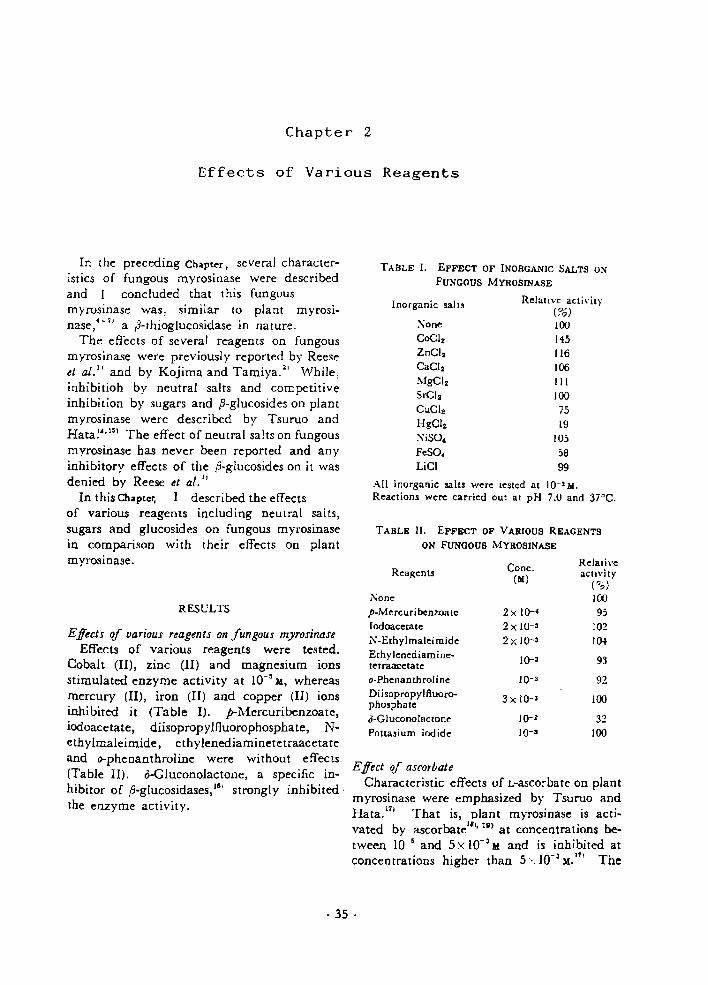
Chapter 2
Effects of Various Reagents
TABLE II. EFFECT OF VARIOUS REAGENTS
ON fuNGOUS MYROSINASE
TABLE I. EFFECT OF INORGANIC SALTS ON
FUNGOUS MYROSINASE
All inorganic salts were le~led al 10-3 M.Reactiom were carried OUI al pH 7.0 and 37"C.
Effect of ascorbateCharacteristic effects of L~ascorbateon plant
myrosinase were emphasized by Tsuruo andHata. 17
• That is, plant myrosinase is acti~
vated by ascorbatel8., IUl at concentrations be
tween 10-5 and 5 X 10-' M and is inhibited atconcentrations higher than 5'< 10-' M.
111 The
Relalive aCI;vit)'(76)100145116106III100
7519
10558
99
Inorganic salts
:'\oneCoClzZnClzCaClzMgClzSrClzCuClzHgClz
NiSO"FeSO.LiCI
Cone. RelativeReagents
(M) acti"ilY(",,)
None 100p-Mercuri benzoale 2 x 10-· 95[odoacelale 2 x 10-' 102N-Elhylmaleimide 2 x 10-' 104Elhylenediamine. 10-3 93telraacelateo-PhenaOlhroI ine 10-' 92Diisopropylfluoro- 3 x 10⁻³ 100phosphateo-Gluconolactone 10-2 32Potla~ium iodide 10-3 100
RESULTS
In the preceding Chaptcr. several characteristics of fungous myrosinase were describedand I concluded that this fungousmyrosinase was, similar to plant myrosinase:- 1
' a r3-thioglucosidase in nature.The effects of several reagents on fungous
myrosinase were previously reported by Reeseet al. ll and by Kojima and Tamiya.~' While,inhibitioh by neutral salts and competitiveinhibition by sugars and fi-glucosides on plantmyrosinase were described by Tsuruo andHata!",151 The effect of neutral salts on fungousmyrosinase has never been reported and an yinhibitory effects of the p~glucosideson it wasdenied by Reese et al. I,
In this Chapler, I described the effectsof various reagents including neutral salts,sugars and glucosides on fungous myrosinasein comparison with their effects on plantmyrosinase.
Effects of uariaus reagents on fungous myrosinaseEffects of various reagents were tested.
Cobalt (II), zinc (II) and magnesium ionsstimulated enzyme activity at 10-' J4, whereasmercury (II), iron (II) and copper (II) ionsinhibited it (Table I). p-Mercuribenzoate,iodoacetate, diisopropyJfluorophosphate, Nethylmaleimide, ethylenediaminetetraacetateand o-phenanthroline were without effects(Table II). o-Gluconolactone, a specific in~
hibitor of ,8-glucosidases,161 strongly inhibited·the enzyme activity.
- 35 .

effect of L-ascorbate on fungous myrosinase isshown in Fig. 1. The fungous myrosinase
glucose inhibited the enzyme (Table Ill). Allglucosides tested inhibited it (Table IV). In~
hibition by amygdalin was the strongest.
100 / o o o TABLE 1Il. EFFECT OF VARIOUS SUGARS
ON FUNGOUS MYROSINASE
F'lG. 1. Effect of Ascorbate on Fungous ~lyrosillase.
Reaction! were carried out at pH 7.0 and 37 C C.
All sugars were tested at 10-1 M.Reactions were carried out at pH 7.0 and 37°C.
.?O. 80
.~.60~
"~ 40,...;:a::
20
00 10-' 10-< 10-' 10- 'Ascorbate (101)
Sugars
:\oneGlucoseGalactose\-IannoseXyloseFructose~vlaltose
SucroseCellobiose
Relative activity(0,,) ,
100
4995
100
10310290
10491
Glucosides
was neither activated nor inhibited at anyconcentrations of ascorbate.
Effects of sodium chlorideFungous myrosinase was inhibited by sodium
chloride at high concentrations. The relationship between concentration of sodium chlorideand reaction rate is shown in Fig. 2.
100
S.~.-.."" 500;~
~
°0::---O=-'.'="S---:-I---I-'-.5---2---J
:-':aCI (~Ii
FIG. 2. Effect of ;';aCI on Fungous ~Iyrosinasc.
Enzymatic reactions were carried out at pH 7.0and 37~C.
Effects of sugars and glucosidesEffects of sugars and glucosides on fungous
myrosinase were te~ted. Among the sugars,
TABLE IV. EFFECT OF VARIOUS GLUCOSIDES
ON FuNGOUS MYROSINASE
Relative activity(%)
~~ 100
Salicin 7BAmygdalin 2Arbutin 73a-Methylglucoside 75p-Methylglucoside 79,3-Phenylglucoside 48p-Nittophenyl ,,-glucoside 69
p-~itrophenyl ,5-glucoside was tesled at 5 x 10⁻² M;others at 10-1 M.
Reactions were carried oul at pH 7.0 and 37°C.
-The inhibitory effects of glucose and salicinare demonstrated as Lineweaver-Burk plots inFig. 3, plotted in the absence and presenceof glucose or salicin. Evidently, the inhibi·tion by glucose and salicin are competitivein type. The Ki values for glucose and forsalicin were calculated to be 2.2:-< 10-2
M and1.4 .10- 1
M, respectively.
- 36 -

FIG. 4. Relationship of the Ionic Strength of NaClto Fungous Myrosinase Activity.
p=Ionic streDgth.Data presented in Fig. 2 are plotted.
inhibited by cobalt (II) chloride and p-mercuribenzoate. These phenomena do not agreewith the results presented here. However,results obtain~d by Kojima and Tamiya21
with an enzyme preparation from the samefungus agree ·with the present results. Perhaps, there may be two types of myrosinasesecreted by Aspergillus sydowij the one studiedby Kojima and Tamiya21 and the presentauthor and another which was studied byReese et ai. I •
Aspergillus sydowi myrosin~e was neitheractivated nor inhibited by any concentrationsof L·ascorbate (Fig. I) in spite of its characteristic effects·7
-ltl on plant myrosinase. Oginsky
et ai. 3' reported that bacterial myrosinase of
Paracolobactrum fUrogeTUJides was inhibited by5 mg/ml ascorbate. The effect of ascorbateappears to be in remarkable contrast betweenfungous and plant myrosinases.
Fungous myrosinase was inhibited by highconcentrations of sodium chloride (Fig. 2).The inhibition behavior of sodium chlorideshown in this figure resembles that of ascorbat~activated plant myrosinase;o. It also r~
sembles inhibition of fungous p-glucosidaseas studied by ]ermyn.2li In order to detectthe effect of ionic strength on this enzyme,data presented in Fig. 2 were plotted ascommon logarithms of enzymatic activitiesagainst the square roots of the ionic strengthof sodium chloride20
' (Fig. 4). A straight line
2.0
,'"/.0
u..:. 1.0.£
~
E0.3
......c'E.......,.,"0
0.2E3
"..........
DISCL'SSIOK
FIG. 3. Inhibitory Effect of Glucose and Salicin onSinigrin Hydrolysis.
Enzymatic reactions were carried out with 10-1 Mgluco-St: (0) Dr salicin (6) and without them (0)at pH 7.0 and 37°C.
Effects of various reagents on this fungousmyrosinase were investigated.. The fungousrnyrosinase was stimulated by cobalt (II), zinc(II) and magnesium ions and was inhibitedby mercury (II), iron (Il) and copper (II) ions,but metal-complexing agents (ethylenediamine·tetraacetate and o-phenanthroline) were without effects (Tables I and II). Although plantmyrosinase is an SH-enzyme, IS. fungousmyrosinase was not inhibited by p-mercuribenzoate, iodoacetate and N-ethylmaleimide.Therefore, sulfhydryl groups were probablynot required for the enzymatic activity of thisenzyme. Also, as diisopropyl1luorophospbatedid not inhibit enzymatic activity, serineresidues were probably not required for thisactivity. A fungous myrosinase prepared byReese et al. ll from a culture broth of the samefungus, Aspergillus sydowi, was reported to be
- 37 -

Mustard oil glucoside
p-Nitrophenyl ~-glucoside
S-,/RC~NO~SO
3
R- = CH =CH-CH -2 2Sinigrin:
with negative slope was obtained. Hence,the effect of sodium chloride on the enzymeis considered to be an effect of the lomcstrength of the solution:Q ,'2'
Reese et aL. i. reported that possible competitive inhibitors including several fj-glucosides had no effects on fungous myrosinase.Their report does not agree with the experimental results given in this paper. Thereason for this inconsistancy is presumablyascribable to the low concenrations of the ,5glucosides used by Reese et at.
fj-Glucosidases of almond, 23' fungousZ~' andyeast"' origins are competitively inhibited bysugars and ,a-glucosides. The conclusion thatinhibition by glucose and salicin are competitive in type (Fig. 3) suggests, similar toplant myrosinase," 1 a strong resemblance offungous myrosinase to the tl-glucosidases. Thisresemblance is also supported by the fact thatthe enzyme was inhibited by o-gluconolactone"(Table II). The conclusion presented in theprevious Chapter. that fungous myrosinase isnot a mixture of thioglucosidase and sulfatase,but a single ,a-thioglucosidase is again supportable by these results. In the present Ch"'pter,enzymatic activities were measured by liberation of sulfate, i.e., they were expressed as"sulfatase" activities. But fungous myrosinasewas competitively inhibited by glucose andsalicin. Therefore, this supports a degradation mechanism of mustard oil glucoside asproposed by Nagashima and Uchiyama,~l
where the liberation of sulfate is the resultof hydrolytic cleavage of the tbioglucosidebond of the glucoside by a single thioglucosiclase followed by spontaneous cleavage ofthe N-O bond.
The fact that fungous myrosinase was inhibited competitively by sugars and fj-gluCDsides suggests that it would hydrolyze ,a-glucoside substrate in a way similar to plantmyrosinase. 17
'
- 38 -

Chapter 3
On the ~-Glucosidase Activity
In a previous Chapter, the fungous myrosinase produced by Aspergillus sydowi wasconcluded to be a ,B-thioglucosidase as isplant myrosinase. ~-1l This conclusion was alsosupported by the fact that the enzyme wascomp:::titively inhibited by glucose and ,8glucosides.
Competitive inhibition by sugars and 13glucosides of plant myrosinase was describedby Tsuruo and Hata~~' and the possession of.a-glucosidase activity by plant myrosinasewas confirmed by these same authors 17J inspite of the belief of earlier years26I that theenzyme had a strict substrate specificity formustard oil glucoside. Resemblance of plantrnyrosinase to the ,B-glucosidases has also beenpoin ted aut.14
,17)
Previously, Reese et aI. Il reported the hydrQlysis ofp-nitrophenyl ,a-glucoside (p-NPG) and!rnitrophenyl fi..galactoside by a crude preparation of Aspergillus sydowi myrosinase. Fromthe results l91Jobtained with the plant myrosinase, purified fungous myrosinase was definitely thought to have ,a-glucosidase activity.Investigation on the ,a-glucosidase activity ofthis enzyme contributes to knowledge aboutthe relationship between myrosinases and (3glucosidases. Some results of studies in thisarea are presented bere.
MATERIALS AND METHODS
En<.ym~ pr~paration. The previously describedmethod of preparing the enzyme solution was improved as follows.
A$ptrgillu$ $ydowi IFO 428+ was cultured on theliquid medium described previously under shakingat 29'C for 2 weeks. After cultivation, the brothwas filtered and the enzyme was precipitated fromthis filtrate bv bringing ammonium sulfate concentra·tion to 90°" of saturation. The precipitate was dissolved in 0.01 M phosphate buffer, pH 7.0 and dialyzedagainst the same buffer. The dialyzate was mixedwith 60g (dry matter) of DEAE-cellulose powder inthe same buffer and adsorbed protein was eluted by0.1 M sodium chloride in 0.01 M phosphate buffer,pH 7.0. The eluted protein was precipitated by 9096of saturation of ammonium sulfate. The resultingprecipitate was dissolved in 0.01 M phosphate buffer,pH 7.0 and dialyzed against the same buffer. (Hereafter, in purification procedures, the concentration of
the enzyme solution was performed by ammonium,sulfate precipitation as described above.) The concentrate was charged on a DEAE-Sephadex column(300 ml. i.d. 4 em), which was equilibrated with thesame buffer and adsorbed protein was eluted by alinear gradient of 0-0.2 M sodium chloride. Afterconcentrating the eluate, gel-filtration on SephadexG-2oo column (450ml, i.d. 2.5cm) was carried out.The filtrate was then charged on a DEAE-Sephadexcolumn (15m!, i.d. I em). equilibrated with the samebuffer and adsorbed protein was eluted by a lineargradient of 0-0.2 M sodium chloride. After concentrating this eluate, gel-filtration on a combinedeolumn:?11 of Sephadex G-loo (120ml, i.d. 1.5em;down flow) and G-l50 (200 ml, i.d. 2.5 em; up flow)was performed. This filtrate was used throughoutthe experiment as the purified enzyme preparation.A summary of the purification procedures of the
- 39 -
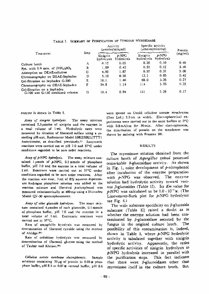
TABLE I. SUMMARY OF PURIFICATION OF FUNGOUS MYR081NASE
Activit\' Specific activi ty(pmoles/min/ml) (pmoles/min/mg) Protein
Treatment Step .... ....(mg/ml)
Sinigrin p-NPG Sinigrin p-NPGhydrolysis hydrolysis hydrolysis hydrolysis
Culture broth A 0.12 0.05 0.26 0.10 0.48
Ppt. with 0.9 sam. of (NH.)tSO. B 1.09 0.42 0.32 0.12 3.46
Adsorption on DEAwellulosc C 4.90 1.87 0.97 0.37 5.06
Chromatography On DEAE-Sephadex D 5.10 0.36 12.1 0.85 0.42
Gel-filtration on Scphadex G-200 E 18.1 1.40 68.0 5.26 0.27
Chromatography on DEAE-Scphadex F 24.8 1.14 114 5.26 0.22
Gel-filtration on a ScphadexG 19.4 0.94 III 5.26 0.17G-IOO and G-150 combined column
enzyme is shown in Table I.
Assay oj sinigrin hydrolysis. The assay mixturecontained 2.5 pmoles of sinigrin and the enzyme ina total volume of I ml. Hydrolytic rates weremeasured by titration of liberated sulfate using a recording pH-stat, Radiometer Model SBR2/SBUI/TTTIAuto-titrator, as described previously.! I Enzymaticreactions were carried out at pH 7.0 and 37°C underconditions regarded to be zero order reactions.
Assay of p-NPG h;1drol.Jsis. The assay mixture con-tained I pmole of p-NPG, 0.1 mmole of phosphatebuffer, pH 7.0 and the enzyme in a total volume ofI ml. Reactions were carried out at 37°C underconditions regarded to be zero order reactions. Afterthe reaction was over, 5 ml of 896 aqueous dipotassium hydrogen phosphate solution was added to thereaction mixture and liberated p-nitrophenol wasmeasured calorimetrically at 400 mp using a ShimadzuModel QV-50 spectrophotometer.
As.I<J) of olher glucosid, hydrolpes. The assay mix-ture contained 4 pmol"s of each glucoside, 0.1 mmoleof phosphate buffer, pH 7.0 and the enzyme in atotal volume of I ml. Enzymatic reactions werecarried out at 37"C.
Rate of amygdalin hyrolysis was measured byddermination of liberat"d cyanide using the methodof Alridge. 581
Rate of cellobiose hydrolysis was measured bydetermination of liberated glucose using the methodof Tauber and Kleiner.59 )
C,llulou (Udal' membraru electrophoresis. Samplesolutions containing 30 fig of protein in 0.05 M phosphate buffer, pH 8.5 or 0.07 M veronal buffer, pH 8.6
were spread on Oxoid cellulose acetate membranes(Oxo Ltd.) 2.2 cm in width. Electrophoretical experiments were carried out in the same buffers at O°Cwith 0.8 rnA/em for 30 min. After electrophoresis,the distribution of protein on the membrane wasshown by staining with Ponseau 3R.
RESULTS
The myrosinase solution obtained from theculture broth of Aspergillus sydowi possessedremarkable .a-glucosidase activity. As shownin Fig. 1, color development of p-nitrophenolafter incubation of the enzyme preparationwith p-NPG was observed. The enzymesolution had hydrolytic activity toward. various p-glucosides (Table II). Its Km value forp-NPG was calculated to be 1.0" 10-· M. (TheLineweaver-Burk plot for p-NPG hydrolysis:see Fig. 4.)
The wide substrate specificity on ,B-glucosidesubstrate (Table II) raised a doubt as towhether the enzyme solution had been contaminated by fi-glucosidase secreted by thefungus in the original culture broth. Thepossibility of this contamination is, indeed,shown in Table I, where p-NPG hydrolyticactivity is tabulated together with sinigrinhydrolytic activity. Apparently, the ratiosof specific activities of sinigrin hydrolysis top-NPG hydrolysis increased in parallel withthe purification steps. This fact indicatesthat there were .a-glucosidases other thanmyrosinase itself in the culture broth. But,
- 40 -
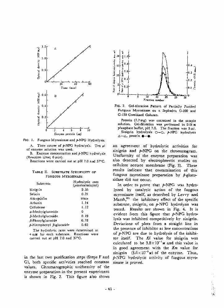
;:. 0.4
TABLE n. SUBSTRATE SPECIFICITY OF
FUNGOUS MYROSINASE
1 0 .6 B<:>c~
an agreement of hydrolytic actIVItIes forsinigrin and p-NPG on the chromatogram.Uniformity of the enzyme preparation wasalso detected by electrophoretic studies oncellulose acetate membrane (Fig. 3). Theseresults indicate that contamination of thisfungous myrosinase preparation by p-glucosidase did not occur.
In order to prove that p-NPG was hydrolyzed by catalytic action of the fungousmyrosinase itself, as described by Levvy andMarsh ,30 I the inhibitory effect of the specificsubstrate, sinigrin, on p-NPG hydrolysis wastested. Results are shown in Fig. 4. It isevident from this figure that p-NPG hydrolysis was inhibited competitively by sinigrin.Deviations of plots from a straight line inthe presence of inhibitor at low concentrationsof p-NPG are due to hydrolysis of the inhibitor itself. The Ki value for sinigrin wascalculated to be 3.8x 10⁻³
M and th.is value isin good agreement with the Km value forsinigrin (3.6x 10⁻³ M) of the enzyme. Thus,p-NPG hydrolytic activity of fungous myrosinase is proved.
.. ..•20 'i 1.0
~ 0.8• IS • 0.42 ~ ,· lO.6 i1 i 0.3
.:::i10 e" 0.4:~ 7.
0.2'"'C;
"0 0" 0.2 e0.1 ~· ~
'" '"0 00 5 30
FIG. 2. Gel-filtration Pattem of Partially PurifiedFungous Myrosinase on a Sephadex G-lOO andG-r50 Combined Column.
Protein (7.7 rug) was contained in the samplesolution. Gel-filtration was performed in 0.01 Mphosphate buffer, pH 7.0. The fraction was 5 mi.
Sinigrin hydrolysis 0-0, P-NPG hydrolysis6-6, protein e-e.
2 4 6 8 10Enz)'ll'le prolein (ug)
o
"2 0.2
.£.
'i.1.5
AE
c
~1.0
.::-~
"..."
"2 0.5
<ia
020 40 60Time (min)
Substrate Hydrolytic rate(pmoles/min/ml)
Sinigrin 9.55Salicin 0.21Amygdalin traceArbutin 1.14-Cellobiose 1.12a·Melhylglucoside 0p-Melhylglucoside 0.19,3-Phenylglucoside 0.78p-Nilrophenyl ft-glucoside 0.65
The hydrolylic rates were delermined at4 roM for each substrate. Reactions werecarried out at pH 7.0 and 37°C.
in the last two purification steps (Steps F andG), both specific activities reached constantvalues. Chromatographic uniformity of theenzyme preparation in the present experimentis shown in Fig. 2. This figure also shows
FIG. I. Fungous Myrosinase and p..NPG Hydrolysis.
A. Time course of p-NPG hydrolysis. Ten pIof enzyme solution was used.
B. Enzyme concentration and p.NPG hydrolysis(Reaction time; 6 min).
Reactions were carried out at pH 7.0 and 37°C.
- 41 -
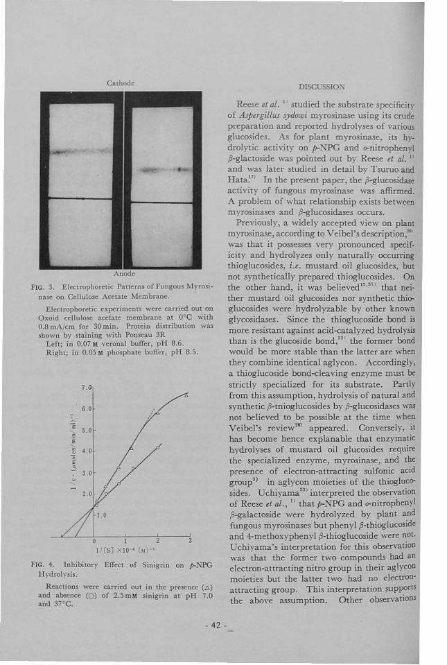
athod DISCU [0.'
FIG. 4. Inhibitory Effect of inigrin on p-NPGH ·drolysis.
Reactions were carried out in the presence (~)
and absence (0) of 2.5 mM sinigrin al pH 7.0and 37° .
FIG. 3. Electrophoretic Pallerns of Fungous lyrosinn on ellulose eetate lembrane.
£1 ctrophoretic e. periments were carried out onxoid cellulose acetale membrane at DoC with
0.8 m:\/cm for 30 min. Protein dislribulion wasshown by staining with Pons au 3R
Left; in 0.07 M verona! burTer, pH 8.6.RighI; in 0.05 M phosphate burTer pH 8.5.
R ese el al. I studied the substrat sp cificityof Aspergillus sydowi myrosinas using its crudepreparation and r ported hydrolyses of variousglucosid s. s for plant myrosinase its hydrolytic activity on p-;P and crnitrophenylp-glactosid was pointed out by R se et aI. I
and was later studied in detail by Tsuruo andHata~71 In the present paper the p-glucosidaseactivit of fungous myrosinase was affirmed.
problem of what relationship exists betweenmyrosinases aod p-glucosidases occurs.
Previously, a widely accepted iew 00 plantmyrosinase according to eibel s description,'"was that it possesses very pronounced specificity and hydrolyzes only naturally occurringthioglucosides i.e. mustard oil glucosides butnot synthetically prepared thioglucosides. Onthe other hand, it was believed".31) that neither mustard oil glucosides nor synthetic thioglucosides were hydrolyzable by other knownglycosidases. ince the tbioglucoside bond ismore resistant against acid-eatalyzed hydrolysisthan is the glucoside bond,3~1 the former bondwould be more stable than the latter are whenthey combine identical aglycon. ccordinglya thioglucoside bond-elea ing enzyme must bestrictly specialized for its substrate. Partlyfrom this assumption hydrolysis of natural andsynthetic p-tnioglucosid s by p-glucosidases wasnot belie ed to be possible at the time when
eibel's re iew"lll appeared. ConverseI ithas become hence explanable that enzymatichydrol es of mustard oil glucosides requirethe specialized enzyme myrosinase and thepre ence of electron-attracting sulfonic acidgroup3) in aglycon moieties of the thioglucosides. chiyama331 interpreted the obser ationof Reese el aI. II that p- PC and crnitrophenylp-galactoside "\ ere hydrolyzed by plant andfungous myrosinases but phenyl P-thioglucosideand 4-methoxypheo I p-thioglucoside were oot.
chiyama's interpretation for this observationwas that the former two compounds had anlectron-attracting nitro group in their aglycon
moietie but the latter two had no electronattracting group. Thi interpretation supportSthe abo e assumption. Other observations
3I 210-' (~1 -I
oI ( )
7.0
6.0
E 5.0!:E
'" 4.0..'0E
3-3.0
- 42 -

made by Reese et ai. I' and by Nagashimaand Uchiyama,31 that desulfonated mustardoil glucosides, S-ii-D-I-glucopyranosylphenylacetothiohydroxamic acid and merosinigrin,were not hydrolyzed (Nagashima) or onlyslightly hydrolyzed (Uchiyama) also lendsupport to the above assumption.
However, hydrolysis of phenyl ,B-thioglucoside by Stachybotrys atra ,8-glucosidase wasreported by jermynJ{) and hydrolysis of 6purinyl .B-tbioglucoside by almond emulsin wasreported by Goodman et ai.J~·3~' Hydrolysesof p- and o-phenyl .a-thioglucosid~s by almondemulsin were also described by Wagner andMetzner. 17' In contrast, hydrolyses of somep-thioglucosides including 6-purinyl .B-thioglucoside and 2,4-dinitrophenyl ,B-thioglucosideby plant myrosinase were reported byGoodman et al.!S· 38 I In addition, the widedistribution of the ,8-thioglucosidase whichhydrolyzes 6-purinyl ,B-thioglucoside in higheranimals was shown by Goodman et al.ss , Asfor plant and fungous myrosinases, the description of Reese et ai. II of the hydrolysesof p-NPG and o-nitrophenyl .a-galactoside wasrefered earlier in this paper. Tsuruo andHataU
17) pointed out close relationship of plantmyrosinase to the ,B-glucosidases based onexperimental results from inhibition by sugarsand fl-glucosides and the hydrolysis of p-NPG.The present paper has shown the result ofstudies on p-NPG hydrolysis by fungousmyrosinase.
All reports cited show that the distinctionsbetween myrosinases and p-glucosidases arenot so strict as previously believed, 28) andrather a close relationship exists between them.This relationship is also supported by thedata On competitive inhibition by sugars and,B-glucosides on plantUl and fungous myrosinases and, conversely, by the competitiveinhibition with phenyl fl-thioglucoside andother aromatic ,8-thioglucosides on fungous3,.s~1
and yeast~O' f3-glucosidases.In conclusion, plant and fungous myrosinases
can be rather regarded to be ,B-glucosidaseshighly specialized for hydrolyses of mustardoil glucosides, than that they are enzymeshaving strict substrate specificity for mustardeil glucosides. In an earlier study by Pigman'llon the classification of carbohydrases, J3-thioglucosidase of plant myrosinase was regardedas belonging to the J3-glucosidase family.Nagashima and Uchiyama11 also stated thatplant myrosinase is a member of the glueDsidases. These opinions are affirmed by theconclusions presented here.
- 43 -

REFERENCES
1) E.T. Reese, R.C. Clapp and M. Mandels,Arch. Biochem. Biophys., 75, 228 (1958).
2) M. Kojima and K. Tamiya, Vitamins (Tokyo),28,380 (1963); J. Vitaminol. (Tokyo), 10,44 (1964).
3) E.L. Oginsky, A.E. Stein and M.A. Greer,Proc. Soc. Expt. Med., 119,360 (1965).
4) M.G. Ettlinger and A.J. Lundeen, j. Am.Chern. Soc., 78,4172 (1956); 79,1764(1957).
5) Z. Nagashima and M. Uchiyama, NipponNogeikagaku Kaish i, 33,1144 (1959); Bull.Agr. Chern. Soc. Japan, 23, 555 (1959).
6) P. Calderon, C.S. Pederson and L.R. Mattick,J. Agr. Food Chern., 14,665 (1966).
7) I. Tsuruo, M. Yoshida and T. Hata, Agr.BioL Chem., 31, 18 (1967).
8) Z. Nagashima and M. Uchiyama, NipponNogeikagaku Kaishi, 33, 478 (1959).
9) J.B. Sumner,]. BioI. Chern., 65,393 (1925).10) a.H. Lowry, N.J. Rosebrough, A.L. Forr and
R.T. Randall, ]. BioI. Chern., 193, 265(1951).
11) R.D. Gaines and K.J. Goering, Biochem.Biophys. Res. Comn., 2,207 (1960); Arch.Biochem. Biophys., 96. 13 (1962).
12) G.A. Howard and R.n. Gaines, Phytochern.,7,585 '(1968).
13) Z. Nagashima and M. Uchiyama, NipponNogeikagaku Kaish i, 33, 484 (1959).
14) I. Tsuruo and T. Hata, Agr. Bioi. Chern., 32,1420 (1968).
15) Z. Nagashima and M. Uchiyama, NipponNogeikagaku Kaish i, 33. 478 (1959).
16) J. Conchie and G.A. Levvy, Biochem.]., 65,398 (1957).
17) 1. Tsuruo and T. Hata, Agr. BioI. Chem., 32,1425 (1968).
18) Z. Nagashima and M. Uchiyama, NipponNogeikagaku Ka ish i, 33, 980 (1959).
19) I. Tsuruo and T. Hata, Agr. BioI. Chem., 31,27 (1967).
20) I. Tsuruo and T. Hata, Agr. Biol. Chem., 32,479 (1968).
21) M.A. Jermyn, Australian]. Bioi. Sci., 8, 563(1955).
22) G.B. Kistiakowsky, P.c. Mangelsdorf, Jr.,A.J. Rosenberg and W.H.R. Shaw,]. Am.Chern. Soc., 74,5015{l952); G.B. Kistiakowsky and W.H.R. Shaw, ibid., 75,2751 (1953).
23) R. Heyworth and P.G. Walker, Biochem. J.,83,331 (1962).
24) M.A. Jermyn, Australian]. BioI. Sci., 8, 577(1955).
25) J.D. Duerksen and Halvorson,]. BioI. Chern.,233,1113 (1958).
26) S. Veibel, in "The Enzymes", 1st ed., Vol. I,
Part I, ed. by J .B. Sumner and K. Myrback,Academic Press. New York, 1950, p. 621.
27) E. Ahlgren, K.E. Eriksson and o. Vesterberg,Acta Chern. Scand., 21,937 (1967).
28) W.N. Alridge, Analyst, 69,262 (1944).29) H. Tauber and I.S. Kleiner,]. Bioi. Chem.,
99,249 (1932).30) G.A. Levvy and G.A. Marsh, Science, 119,
337 (1954).31) W.W. Pigman,]. Res. NatL Bur. Stand., 26,
197 (1941).32) C.B. Purves,]. Am. Chem. Soc., 51, 3627
(1929).33) M. Uchiyama, Nippon Nogeikagaku Kaishi,
37, 543 (1963).34) M.A. Jermyn, Australian]. BioI. Sci., 8, 577
(1955).35) I. Goodman, G.B. Elion and G.H. Hitchings,
Fe. Proc., 14,219 (1955).36) I. Goodman, J.R. Fouts and G.H. Hitchings,
ibid., 17,232 (1958).37) G. Wanger and R. Metzner, Naturwissenschaf
ten, 52, 61 (1965).38) 1. Goodman, J. R. PonL, E. Bresnick, R.
Menegas and G.H. Hitchings. Science, 130,450 (1959).
39) M.A. Jermyn, Australian J. Bio/. Sci., 11,114 (1958).
40) J.D. Duerksen and H. Halvorson, j. Bio/.Chem., 233, 1113 (1958); A.$.L. Hu, R.Epstein, H.O. Halvorson and R.M. Bock,Arch. Biochern. Biophys., 91, 210 (1960).
41) W.W. Pigman, J. Res. Nati. Bur. Stand., 30,257 (1943); Advances in Enzymol., 4, 41(1944).
- 44 -

Chapter 4
Production and Stability of the Intracellular
Myrosinase from Aspergillus niger
In previous Chapters!~3) the production,purification and various properties of myrosinase produced by Aspergillus sydowi weredescribed. This is extracellular enzyme whichis induced by a mustard extract during about2 weeks of cultivation. It is very difficult toobtain large amounts of the enzyme, becauseof the long period necessary to cultivate theorganism.
Reese et al.~) reported that the myrosinase(sinigrinase) produced by Aspergillus sydowiis an extracellular enzyme; sinigrinase wasalways found slightly in mycelial extract whenit was present in the culture filtrate, and neverfound in mycelial extract when it was absentin the culture filtrate. The nature of intracellular myrosinase is still undetermined.
In the present experiment, I surveyedorganisms that produce myrosinase in theirmycelia in hopes of obtaining a large amountof enzyme while shortening the culture periods.Enzyme activity was found in the mycelia ofAspergillus niger.
Culture conditions for the fungus andenzyme stability are described. Some characteristics of the myrosinase from Asp. nigerare also discussed in comparison with those ofthe enzyme from Asp. sydow;.
MATERIALS AND METHODS
Organisms. Various stems of fungi and bacteriawere kindly supplied by the Faculty of Agriculture ofKyoto University.
Culture conditions. Culture conditions for screen-ing were as follows. Or&anisms (80 bacteria and 20fungi) were inoculated in a medium containing 20%mustard extract, 0.1 % glucose. 0.1 % yeast extract.0.1 % potassium dihydrogen phosphate, 0.1 % ammonium sulfate and 0.05 % magnesium sulfate (as MgSO"7HzO), pH 6.5.
Fungi were cultivated for 2 weeks and bacteria for2 days at 29'C under shaking.
Aspergillus niger AKU 3302 was pre-culturedat 29'C in 100 ml of medium containing 5% malt extract, 2% sucrose, 0.2 % potassium dihydrogen phosphate. 0.1 % ammonium sulfate, 0.03 % sodium nitrate,and 0.03 % magnesium sulfate (as MgSO,· 7HzO),pH 6.5. After incubation for 2 days, mycelia wereharvested and washed twice with water. Wet myceliawere inoculated in 100 ml a f synthetic medium consisting of 0.1 % yeast extract, 0.2 %potassium dihydrogen phosphate, 0.1 % ammonium sulfate, 0.03 %sodium nitrate, 0.03 %magnesium sulfate, 0.1 %glucoseand 20 mI of mustard extract, unless otherwise stated.Cultivation was carried out in 500 ml flasks containing100 ml of the medium for 2 days at 29'C under shakingon a reciprocal shaker. Approximately 5-6 g ofmycelia (wet weight) were obtained per 100 ml of thepre-culturing medium. In all cultivating experiments,about 5 g of mycelia (wet weight) were inoculated in100 ml of induction medium.
Substrate. Sinigrin was prepared from yellowmustard seed as described previouslY,e,., and used asthe substrate of myrosinase.
Enzyme preparation. After cultivation, contentsof the flasks were filtered and washed twice with water.Approximately 5 - 6 g of wet mycelia were groundtogether with sea sand and 0.2 M phosphate buffer(pH 7.0, 15 ml), and then it was centrifuged. Thesupernatant was used in experiments as the crude
preparation.
- 45·

()~v--~--+---,I,--_L-_~:--.Jo
2
3
3
.::""
"0 3".,'08"-~ 2> .
...
o\:'0...l.'--1~0~-~2~0 --3:!-:O:----:~~O---!-::- ...... 0
~1u.sto.rd exlract I~~~}
FIG. 2. Effect of Mustard Extract Concentration ofEnzyme Production in Mycelia of A,'pergalus niger.
The fungus was pre-culturcd as described in Fig. I.After incubation for 2 days, wet mycelia (weightabout 25 g) were harvested and .... ashed twice withwater. About 5 g of the wet mycelia were inoculatedio each 100 ml of synthetic medium, in which theconcentration of mustard extract was changed.Total activities were measured by the liberation orglucose as described in the text.
FIG. I. Enz\'me Production in Mycelia of Asper1!allis niger.
The fungus was prc-cultured at 29'C On 500 mI ofmedium containing 5 ~/~ mall extract. 2 ~,~ sucrose,0.2% KH.PO., 0.1~'-;; {NH~hSO,. 0,03% NaNO, and0.03 ~~ MgSO., 7H 20, pH 6.5. After incu batioDfor 2 days, mycelia (about 25 g) were harvested andwashed twice with water, About 5 g of the wetmycelia were inoculated in each 100 ml of syntheticmedium, Total activities were measured by theliberation or glucose as described in the text.
EJlzyme assay. Enzymatic activity was determinedas p..thioglucosidase activity. The enzyme reactionwas carried out in a system containing 2.5 I' moles ofsubstrate and 0.1 f' mole of phosphate buffer, pH 7.0,in a total volwne of I ml. The reaction was carriedout at 37'C and at pH 7.0, unless otherwise staled.Thioglucosidase activity was measured by the liberation of glucose. The reaction was conducted underconditions regarded as a zero order reaction."'
Total activity. The wet mycelia were ground together with sea sand and 0.2 M phosphate buffer (pH7.0), and then it was centrifuged. The supernatantwas filled up to 50 mI with the same buffer. Totalactivity was expressed as f' moles of glucose per minuteper 50 mI unless otherwise stated.
Determination of the culture conditions
Production ofintracellular myrosinase. Therelationship between enzyme production andthe culture period is shown in Fig. I. Theenzyme was produced in considerable amounts,mostly on the first and second days.
Concentration of mustard extraCI. Therelationship between enzyme production andthe concentration of mustard extract in themedium is shown in Fig. 2. The best enzymeproduction was observed with 10 ~~ mustardextract; production gradually decreased at
the higher concentrations.
RESULTS
Suney of organisms
Results of the screening test are summarizedin Table I. Enzyme activity was determinedby using the supernatant of ground myceliawith sea sand for fungi and the soluble partafter sonication for bacteria. Strong activitywas found in the mycelia of Asp. niger AKU3302 but only slight activity was detected inAsp. sydow; IFO 4284. The same test wascarri~d .out with some others strains of Asp.
niger group (Table II); all the strains showedconsiderable activity. Asp. niger AKU 3302was used in the following experiments.
- 46 .

TABLE I GROWTH OF BACTERIA AND FUNGI ON A MUSTARD ExTRACT MEDIUM
Organisms were inoculated in a medium containing 20 % mustard extract, 0.1 % glucose, 0.1 % yeastextract, 0.1 ~~ KH 2PO., 0.1 ~/~ (NH.)zSO. and 0.D5~~ MgSO.·7H20, pH 6.5.
Fungi were cultivated for 2 weeks and bacteria for 2 days at 29'C under shaking.
Pseudomonas aeruginosa UFO 3080)Sarcina lurea (IAM 1099)Staphylococcus aureus (IFO 3061)Flavobacterium fuscus (AKU 0140)Micrococcus Iysodeikticus (lFO 3333)Alca/ygenes [aecalis (lAM 8--141-1)Proteus vulgaris (IFO 3167)Serratia maraseens (lFO 3046)Aerobaclt'r aerogenes (lFO 3320)Escherichia coli (A KU 000I)Bacterium mdaveris OFO 3731)Bacillus sliblilis (IFO 3026)Slreplocoeclls faeea/is (lFO 3181)Candida utilis (IFO 0396)Aspergillus
oryzae (AKU 3301)niger (AKU 3302)fumigarus (IFO 4040)f/avus (IFO 5839)sydowi (IFO 4284)
Penicilliumnotatum (lFO 4640)urticae (lFP 7011)
Mueor rOlizianus (!FO 5773)Phizopus ory;:ae (AKU 0021)
Growth
++-i-+++
+++
...:...++
--:-+
...:...+
Myrosinaseactivity
++
TABLE II. MYllOSrNASE PRODUCTION BY SOME OF
THE Aspergillus niger GROUP
Fungi were pre-cultured at 29"C on 100 mI ofmedium containing 5 % mall extract, 2% sucrose,0.2 % KH2PO., 0.03 % NaN03 and MgSO.· 7H20,pH 6.5, respectively. After incubation 2 days,mycelia were harvested and washed twice with water.Wet mycelia were inoculated in 100 ml of syntheticmedium.
Organism Myrosinaseactivity
Aspergillus niger IFO 4034 .L ..L
" lFO 4068 'T' -
IFO 4280
" IFO 6341
" IFO 5374 -7--;-
AKU 3302 ++
Asp. niger was pre-cultured and its myceliawere inoculated in the synthetic medium. Thegreatest amount of enzyme was produced ina medium containing O. J % glucose, 0.1 %C.S.L., 0.03 % cobalt chloride (as CoC1!·6H,O) and 10% mustard extract. Corn steepliquor was a better nitrogen source and cobaltchloride was also superior to magnesiumsulfate. Consequently, these culture condi.tions were used for the following experiments.
No enzyme was produced without the addition of inducer (mustard extract or sinigrin)in the case of Asp. sydowi. The same wastrue for Asp. niger. As shown in Table III,no enzyme was produced without mustardextract.
- 47 -

TABU III. EFFECTS OF SOME SUBSTITUTES
fOR MUSTARD EXTRACT
Aspergillus nigl'r was pre-<:ultured at 29'C on1000 ml of medium containing 5~';; malt extract, 2~';;
sucrose, 0.2% KH:P04, 0.1 % (NH4):SOh 0.03%NaNO. and MgS04·7HaO, pH 6.5. After incubation 2 days, wet mycelia (about 50 g) were harvestedand washed twice with water. About 5 g of the wetmycelia was inoculated in each 100 ml of mediumcontaining 0.1 % glucose, 0.1 % e.S. L., 0.03 % CoCI.·6H.O and substitutes for mustard extract, pH 6.5.
Effects of l'ariOliS carbon sources. Effectsof various carbon sources on enzyme production were tested (Table IV). Ascorbic acidwas more effective than glucose. From theseresults, we composed a medium containing0.1 % ascorbic acid, 0.1 % c.s.L., 0.03 %cobalt chloride and 10% of mustard extract,pH 6.5, for the induction medium.
Materials
Mustard extractMalt extractGlucoseSucroseAscorbic acidCellobioseAmygdalinTyramine
Cone.
10 %0.10.10.10.10.10.10.05
Myrosinaseactivity
++
Stability of the enzyme. Intracellular myrosinase from Asp. niger was very unstable afterextraction from the mycelia and was considerably inactivated during dialysis andprecipitation process with ammonium sulfate.Effects of various reagents to stabilize theenzyme were examined. 2-Mercaptoethanol(10- 2 M) containing ascorbic acid (10- 3 M) wasan effective stabilizer on the activity of thecrude enzyme (Table V).
TABU IV. EFFECTS OF VARIOUS CARBON SOURCES
Aspergillus niger was pre-cuJtured at 29'C on500 mJ of medium containing 5% malt extract, 2%sucrose, 0.2% KH2P04, 0.1 % (NH4hS04, 0.03 %NANO. and MgS04·7H20, pH 6.5. After incubation 2 days, mycelia (about 25 g) were harvestedand washed twice with water. About 5 g of the wetmycelia were inoculated in each 100 m1 of mediumcontaining 10% mustard extract, 0.1 %C.S.L., 0.03%CoC!:·6H10 and various carbon sources, pH 6.5.
Cone. Total activitySource ( ~.;;) (,., moles of g1ucosel
min/50ml)
Sucrose 0.1 2.0Glucose 0.1 4.5
" 0.2 4.3Ascorbic acid 0.1 8.8Sorbitol 0.1 1.6Cellobiose 0.1 1.3
DISCUSSION
To obtain enzymes from microbes, I musttake into account many conditions, of whichthe culture period is one of the most importantfactors. Generally, microbes must be cultivated for longer periods to obtain extracellularenzymes than to obtain intracellular enzymes.For myrosinaselJ from Asp. sydowi, the fungusmust be cultivated in an induction medium forat least 10 days under constant maintenanceof a neutral pH range. I searched formyrosinase in various microbial mycelia inorder to obtain it in large amounts. I alsotried to shorten the culture period. Aspergillus niger, AKU 3302 after screening 100organisms, was found to produce the enzyme.
TABLE V. EffECT Of VARIOUS REAGENTS ON STABILITY
-.... Cone.(M) Control Glucose Ascorbic acid CoO: Ascorbic acid (10- 3)
+-- 10-' 10- 3 10-3 10- 2 10-3 2-Mercaptoethanol (10- 2)Time (hr) ------~-~
0 100 100 100 100 100 100 100"h 24 72 80 91 97 45 57 9g~; Wq (80)0'":;;>3·= 48 33 34 51 65 24 12 80uo:~< (20'q (52)01
"" only 2-mercaptoethanol (10- 2 tot)
- 48 -

very powerful fungicides.!1 Since allylisothiocyanate is a product of sinigrin hydrolysis,there was a possibility that it might inhibitgrowth. Asp. sydowi probably required ahigher concentration of mustard extractbecause of the longer cultivation periodneeded. l )
It is pretty interesting that ascorbic acidwhich is a specific activator for plant myrosinaseB.OI is a good carbon source for fungousmyrosinase production.
The intracellular myrosinase was veryunstalbe, and was considerably inactivatedduring dialysis and salting out with ammoniumsulfate. Concerning microbial enzymes, weoften find stable enzymes in different strains ofthe same species. However, in the case ofAsp. niger, enzymes from all the strains testedwere unstable.
Addition of 2-mercaptoethanol, containingascorbic acid to the enzyme solution waseffective for stabilizing the enzyme. This maybe related to the results of Table IV, whichshows that ascorbic acid was an excellentcarbon source.
10 10
~
·E,
~ 8cu
"0;,
'0
"""0.e3!::E-.;"":;
°0
FIG. 3. Comparison of Enzyme Produclion byAspergillus niger and Aspergillus sydowi.
Aspergillus niger and Asp. sydowi were pre-culluredal 29'C on 500 mJ of medium containing 5% malteXlracl, 2 %sucrose, 0.2 % KHzPO., 0.1 % {NH,)zSO"0.03 %NaN03 and MgSO.· m:e, pH 6.5, respectively. After incubation for 2 days, mycelia (aboul 25 g)were harvested and washed twice with water. About5 g of the wet mycelia of Asp. niger were inoculatedin each 100 001 of medium containing 10% mustardextract, 0.1 % ascorbic acid, 0.1 % C.S.L and 0.03 %CoCh·6H!O, pH 6.5. Wet mycelia (about 5 g) ofAsp. sydow; were inoculated in each 100 ml of mediumcontaining 20% mustard extract, 0.1 % yeast extract,0.2% KH2P04 , 0.1 % (NH,)2S0,, 0.03% NaN03,0.03% MgSO.,7H30 and 0.1 % glucose, pH 6.5.Total activities were measured by the liberation ofglucose as described in the text.0, Enzymatic activity in mycelia of 4sp. !tiger; .,Enzymatic activity in mycelia of Asp. sydowi.
With Asp. niger AKU 3302, the enzyme wasproduced in considerable amounts, mostlyon the first and second days, without adjustingthe pH range. Asp, sydowi also producesmyrosinase, but it requires a long period formaximum production (Fig. 3). Some otherstrains of Asp. niger group tested, besidesAKU 3302, produced the enzyme (Table II).Asp. niger did not produce the enzyme withoutinducer (mustard extract or sinigrin) (TableIII), as with Asp. sydowi. The optimum concentration of mustard extract was 10 % forA.sp. niger and 20% for Asp, iydowf.l'
Isothiocyanates (mustard oil) appear to be
- 49 -

Chapter 5
General Characteristics
In a previous Chapter, 'I Aspergillus nigerAK U 3302, which produces myrosinase, wasfound through a screening test and its cultureconditions were det.ermined. The instabilityof this intracellular myrosinase was alsoreported.
There are several papers on the propertiesof plant myrosinase2
-tJ and on extracellular
myrosinase~·B' produced by Aspergillus sydowibut the nature of intracellular myrosinasefrom microorganisms is not yet undetermined.There is only the report by Reese et aPJ whopointed out the existence of intracellularmyrosinase in the mycelia of fungus.
In this paper, I describe the generalcharacteristics of the intracellular myrosinaseproduced by Asp. niger and compare themwith characteristics of plant and extracellularmyrosinases. The relationship of fungousand plant myrosinases to P-glucosidases isalso discussed.
MATERIALS AND METHODS
Ertzyme preparation. The grown mycelia wereharvested in a basket centrifuge, and washed twicewith water. The collected mycelia were ground withsea sand in 0.1 M sodium phosphate buffer, pH 7.8,containing 0.01 M 2-mercaptoelhano! and O.IX)! M L
ascorbic acid after which the whole was centrifuged.The supernatant was used as the crude enzyme preparation.
Subs/rate. Sinigrin was prepared from yellowmustard seed~·BJ and was used as the substrate offungous myrosinase. p-Nitrophenyl p·g1ucosidc wasof an analytical grade.
Assay of sinigrin hydrolysis. The assay mixturecontained 2.5 I'moles of sinigrin and enzyme in atotal volume of 1 mI. Enzymatic activities were determined by measuring liberated glucose and by titratingliberated sulfate at pH 6.2 and 34'C for 20 min, asdescribed previously.'.~J The reaction was conducted under conditions regarded to be those for zeroorder reactions.
Assay of p-NPG hydrolysis. The assay mixturecontained 1 pmole of p-NPG 0.1 I'mole of phosphatebuffer, pH 6.2 and the enzyme in a total volume of I ml.The reaction was carried out at 34'C for 10 min underconditions regarded to be those for zero order reactions.After the reaction was over, S ml of 8 % aqueousdipotassium hydrogen phosphate solution was addedto the reaction mixture. Liberated p-nitrophenol wasmeasured colorimetricaUy at 400 ml' using a ShimadzuModel QY-SO spectrophotometer.
Effects of various reagents. Reagents and enzymewere incubated at pH 6.2 and 34'C for 20 min beforeeach experiment. Enzymatic reactions were initiatedby the addition of substrate.
Assay of protein. Protein concentrations weredetermined by the method of Lowry el 01.10 )
RESULTS
Partial purification of the fungous myrosinaseChromatography on DEAE-Sephadex. The
crude enzyme solution (500 mI, total protein1050 mg) was dialyzed against 0.01 M phosphate butTer containing 0.01 M 2-mercaptoethanol and 0.001 M ascorbic acid, pH 7.8,and then was charged on a DEAE-Sephadexcolumn (4.5 >< 23 cm) which was eqilibratedwith the same buffer. Elution of protein wasperformed wilh a linear gradient of 0.05 to0.5 M sodium chloride in the same butTer.Myrosinase activity could not be separatedcompletely from p-glucosidase activity (Fig. 1).
- 50 -

The myrosinase in this step, however, wasalmost inactivated during dialysis and saltingout with ammonium sulfate and was protectedneither by the stabilizer (2-mercaptoethanoland ascorbic acid) of the crude preparation norby any of reagents tested. But, the enzymewas only stable during concentrating withpolyethylene glycol ~6,OOO, further purification was done by an isoelectric focusing.Fractions indicated by the horizontal arrow
0.5 0.5
0.4 0.4E E
" ".... 0:0
e 0.3 ~0 00 0.2 0"'l ':1
0.1 0.1
0 010 20
Fraction number 120 ml •• chl
FIG. ]. Chromatogram of Fungous Myrosinase ona DEAE-Sephadex Column.
(0) Myrosinase activity and (e) p-Glucosidase activity(-) NaCi concentration.
1.0 10 0.5
E<:
0,8 8 0.4 0
~
1 11:il 0.3 "0.6 6 E~ <:
::::: ;;. !:....0 0.4 0.2
~
C'>
0.2 2 ~--0.1.
---0
0 10 40 SO
Fraclion number (each 2 mil
FIG. 2. Isoelectrofocusing of the Partial PurifiedEnzyme.
The sample solution was isoelectricaIly focused on theLKB Column containing carrier ampholyte with apH range of 4 10 6.(0) Sinigrin hydrolysis, (e) p-NPG hydrolysis, (-)pH and (--) G.D. al 280 m,L.
in Fig. 2 were pooled and concentrated bypolyethylene glycol.
lsoelectric focusing. Isoelectric focusingwas carried out by the method of Vestert-.-rgand Svensson,lI) using the LKB Cl.: n ron(110 ml) containing carrier ampholyte andglycerol. The pH range of the ampholyteused was from 4 to 6. Ampholyte in thecolumn amounted to I g. The anode compartment contained glycerol (60 % v/v), towhich 0.2 m! of phosphoric acid was added.The enzyme concentrated (50 mg protein,about 20 m!) was applied to the column. Thecolumn was controlled at 4'C by circulatingchilled water. The run was performed witha maximum load of 0.5 W, implying a finalvoltage of approximately 3S0 V. After focusing for 72 hr, fractions of each 2 ml wereremoved from the column and their enzymeactivities were determined. In Fig. 2, typicalisoelectrofocusing pattern is shown. Therewere some P-glucosidase activities for p-NPGbesides myrosinase. The pI value of themyrosinase was about pH 4.8, the same asplant myrosinase. Fractions indicated bythe horizontal arrow were pooled and used inthe following experiments. A summary ofthe partial purification steps of the enzyme isshown in Table I. It also shows that fungousmyrosinase is remarkably unstable.
General characteristics of the fungous myrasinase. Figures 3, 4, 5 and 6 show the pHactivity, pH-stability, temperature-stabilityand optimum-temperature curves, respectively. Optimum pH was about pH 6.2. Theenzyme was most stable at pH 7.8 and at S"C.Optimum temperature was .at 34"C.
Effects of \'arious reagents. Copper (I),(1I), manganese (II) and cobalt (II) ions stimulated enzyme activity at 10- 3 M; whereas,mercury (II) and stannous (II) ions inhibitedit (Table If). PCMB was a strong inhibitor(Table Ill). DFP, iodoacetate and metal
- 51 -

Acti~y
&troolesfmmlfYrlJ------------Smigrin p-NPG
hYGto!ysis hydn...r1ysis
S-PCcific a9tivit·y(,umo:lf's/miafmg}~_.~-
Sinigrin p-NPGhydro!ysis hydrQlysis
Protem(mgimli
(:ru.de-~e 500 0.29'1 00.441 00.159 0.213Pialysa1.e 5lO 0.1'70 S.391 S.D4 :O.J-95ClU:'ODlatogr;tphyon :140 :0.3:16 0.254 (1.~4 0,635DEAE-Sephadel''QlnceBU:'alion,wjEb 20 .1.68 1.4.29 1>.e12 0.512PQ1yetbyleneglycoll~lecU'jc fOC1JslOg .~ ~U43 1>.164 i .916 cO.5S
2.102.f\10.4
2.5
D"~--i~-~--+--"!~~()5 6 7 W
{pH)
FiO.3. pH-AetivltyCurvcQfFtmgol,1$ Myrosinase.
Q;2MSodium p!:losphateblJffer -(pH fdJ .....9;(}) andQ.2Mscetatebuffer·(pH :S.Q).~ used..
?HFro. 4. pH-Stability Cl,mleof fl.lngous Myr()smase.
Thesamp1esoiution wasinClJbatedinQiQS MSQtiium~ba&e ,b!Jffe.r .-(pJ{ ~""'9) ,ami~te -buffer {pH~A3) .fQr 24 'hratS'C. -Reacl.iooswer.e.carried out al pH {;.2and 34'C.
.c:Onlpjex.ingage~t!>~owe(;l JiH1eeff~t.
Pianl: myrosinase~) lS 5peCificaiiy activatedhyas.corb.icacid (10··B M), while, intracelluiar
f-H;i.;. Hem-stability CurJleofFun,gous MyroSIB/lSe.
The sample soiu.tion was 1acubatOOlnO.l M ph9$JiJiate huffer,pH 7£ for 2~mm a~~!lten'l~ature
'bl::foremeasm-~en1oICfIZYlBeaptiliny.
myrosinase wasaeE:theractivated n9t'inhib~
by L~aSi-'mbate.
o-Gluconoiactooe. a speciflcllmib'itor offt--glucosi.da~,t"j inbibiUld enzyme activi...ty.
£jJec!so!$(gtJrsar..dg},uc9sUks. SJ;l$arandglucosideeff-eciS on intracellular myn:>sinase Wef¢ tested (T;.t'bie IV). Npue of thesugars tested had any eff'eet, but~~s1des
l,niribited aciivitybyaoout 20",.. 30%.
- 52 -

TABU II. EFFECT OF INORGANIC SALTS ON
FUNGOUS MYROSINASE
Reactions were carried out at pH 6.2 and 34·C.All inorganic salls were pre-incubated at 10- 3 M.
TABU III. EFFECT OF VARIOUS RE"",ENTS 01'1
FUNGOUS MYROSINASE
PCMB was pre-incubated at 6 x 10-' M; others,at 10-3 M.
Cone. (M) Relative activity (%)
DISCUSSION
General characteristics of intracellularmyrosinase produced by Aspergi//us nigerAK II 3302 were investigated. There weresome P-glucosidase activities for p-NPG in
Kinetic studies. Figure 7 shows the Line-weaver-Burk plot for intracellular myrosinase.Km value for sinigrin was calculated to be3.3 x 10- 3 M.
In order to prove that p-NPG was hydrolyzed by catalytic action of the fungous myrosinaseitself, as described by Levvy and Mash,13)the inhibitory effect of the specific substrate,sinigrin, on p-NPG hydrolysis was tested.Results are shown in Fig. 8. It is evidentfrom this figure that p-NPG hydrolysis wasinhibited competitively by sinigrin. The Kmvalue for p-NPG and Ki value for sinigrinwere calculated to be 1.5 X 10⁻⁸ M and 3.8 x10- 3 M, respectively. This Ki value is in goodagreement with the Km value for sinigrin(3.3 X 10- 3 M) of the enzyme.
100104103101107105857986
106
1009
8080739891848238
6x 10-'10-3
10- 3
10-3
10-3
10-3
10-3
10-3
10- 3
NoneGlucoseGalactoseMaltoseFructoseXyloseSalicinArbutinAmygdalinp-NPG
NonePCMBIodoacetateEDTAo-PhenanthrolineAscorbic acidKINaBrDFPo-Gluconolactone
the mycelia or Asp. niger, besides myrosinase.As described previously, the myrosinaseproduced by Asp. niger was remarkably unstable and became more so after chromatography on DEAE-Sephadex. This myrosinasewas not protected by stabilizing reagents forthe crude preparation.
Some properties of Asp. niger, Asp. sydowiand plant myrosinases were compared (TableV). Asp. niger myrosinase was less stablethan the others to heat, acid and alkali, and tochemicals. It was strongly inhibited by
Sugar or glucoside (OJ M) Relative activity (%)
TABU IV. EFFECT OF VARIOUS SUGARS AND
GLUCOSIDES ON FUNGOUS MYROSINASE
Reactions were carried out at pH 6.2 and 34·C.Enzyme activity was measured by the liberation ofsulfate, Amygdalin and p-NPG were pre-incubatedat 2 x 10- 2 M and 5 x 10⁻² M, respectively; otbers at10- 1 M.
1009599
1401321701867772345
9074
Relative activity(%)
NoneKOliCICuCICuChMaChCoChMg02
ZnCI2
Sn02HgChFe02Fe03
Inorganic salts00-3 M)
- 53 -
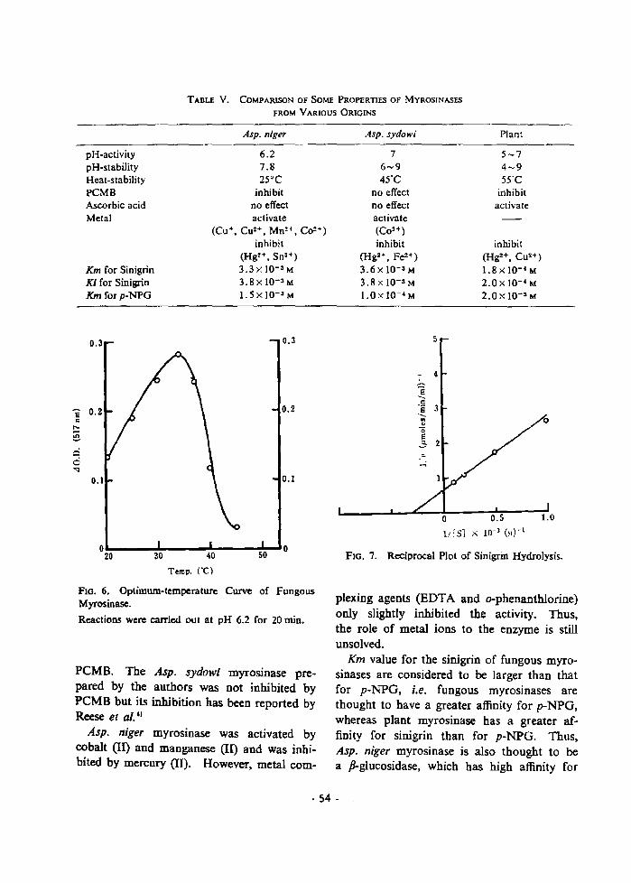
TABU V. CoMPARISON Of SOME PROPERTIES Of MYROSINASES
fROM VARIOUS ORIGINS
pH-activitypH-stabilityHeat-stabilityPCMBAscorbic acidMetal
Km for SinigrinKi for SinigrinKm for p-NPG
0.3
Asp. niger
6.270825°C
inhibitno effectactivate
(Cu+, Cu2+. MnH • Co~+)
inhibit(Hg!+. Sn3+)3.3x 10- 3 M3.8xl0-3 M
1. 5 X 10⁻³ M
0.3
Asp. sydow;
76-945'C
no effectno effectactivate(COH)
inhibit(Hg2+, FeH )
306 x 10-3 M
3.8 X 10-' M1.0x 1O-~ M
5
Plant
5-74-955°C
inhibitactivate
inhibit(HgH. CUH )
1.8xlO-·M2.0x 10-' M
2.0 x 10-' M
E 0.2c
!:::'",...,0'<l
001
oL..__~__~__~~o20
FlO. 6. Optimum-temperature Curve of FungousMyrosinase.
Reactions were carried out at pH 6.2 for 20 min.
PCMB. The Asp. sydowi myrosinase prepared by the authors was not inhibited byPCMB but its inhibition has been reported byReese el aI. II
Asp. niger myrosinase was activated bycobalt (II) and manganese (ll) and was inhibited by mercury (II). However, metal com-
_ 4
E......'g 3........"'0E
..:; 2
,...;
o 0.5 1.0
1/:5] x 1O~3 (;1)-1
FIG. 7. Reciprocal Plot of Sinigrin Hydrolysis.
plexing agents (EDTA and o-phenanthlorine)only slightly inhibited the activity. Thus,the role of metal ions to the enzyme is stillunsolved.
Km value for the sinigrin of fungous myrasinases are considered to be larger than thatfor p-NPG, i.e. fungous myrosinases arethought to have a greater affinity for p-NPG,whereas plant myrosinase has a greater affinity for sinigrin than for p-NPGo Thus,Asp. niger myrosinase is also thought to bea p-glucosidase, which has high affinity for
·54·

1.0
mustard oil glucoside. Pigman, Hl Nagashima and Uchiyama,II,l6l and Tsuruo et al.have reported that plant myrosinase is amember of the P-glucosidase. The aboveresults support this conclusion, Plant myrosinase is highly specified for mustard oil glucosides, whereas fungous myrosinases are lessspecific than the plant enzyme. Even so,fungous myrosinases have higher affinities formustard oil glucosides than for the usualP-glucosidases. This conclusion is supportedby the fact that fungous myrosinase was produced only in the presence of mustard extract.
FIG. 8. Inhibitory Effect of Sinigrin on p-NPGHydrolysis.
Reactions were carried out in the presence (e) andabsence (0) of 2.5 mM sinigrin at pH 6.2 and 34'C.
REFERENCES
J) M. Ohtsuru, I. Tsuruo and T. Hata, Aer. Bioi.ChJ:rn., 33, ]309 (1969).
2) M. Ohtsuru, I. Tsuruo and T. Hata, ibid.• 33, 131 S
(1969).3) M. Ohtsuru, I. Tsuruo and T. Hata, ibid., 33. 1320
(1969).4) E. T. Reese, R. C. Clapp and M. Mandels, ArcJr.
Biodern. Biophys., 75. 228 (1958).S) I. Tsuruo, M. Yoshida and T, Hata, Agr. Bioi.
Chern., 31, 18 (1967).6) Z. Nagasbima and M. Uchiyama. Nippon Nogei·
kagaku Kalshi, 33, 478 (1959).
7) J. B. Sumner, J. Bioi. Chern., 65, 393 (1925).
8) H. L. K1oPpini, "Chemical Constitution andAntifungal Action of Sulfur Compounds,"Drukkerij Fa Schotanus Tens, Utrecht, 1951.
9) Z. Nagashima and M. Uchiyama, Nippon Nogelkagaku Kalshi. 33, 881 (1959).
10) O. H. Lowry, N. T. Rosebrough. A. L. Farr andR. T. Randall, J. Bioi. ChJ:rn., 193, 265 (1951).
11) O. Vesterberg and H. Svensson, Acta Chern.Scant/., 20,820 (1966).
12) J. Conchie and G. A. Levvy, Biochern. J., 65, 398
(1957).13) G. A. Levvy and C. A. Mash, Sciarce. 119, 337
(1954).14) W. W. Pigman, J. Res. Natl. Bur. Stand., 26,197
(1941).15) Z. Nagashima and M. Uchiyama. Nippon Nogei
kagaku Kaishi, 33. 1144 (1959).16) Z. Nagashima and M. Uchiyama. Bull. Agr. Chern.
Soc. Japan. 23, 55S (1959).
- 55 ~

PART III
Studies on the Bacterial Myrosinase
Chapter 1
Isolation of
The fungous myrosinases were not suitable forthe perpose to obtain large amounts of the stableenzyme, as one takes 2 weeks of cultivation1 )
and the other is very unstable. 2 )
Oginsky et al}) after a screening test formyrosinase activity among intestinal bacteria,used PaTacolobactrum aeTogenoides as the activeorganism for their experiments. They havepointed out its existence and some properties onthe crude preperation of bacterial myrosinase.
In this Chapter, I surveyed microorganism thatproduces myrosinase in bacteria and identified it.And 1 studied the optimum culture conditonsfar the myrasinase production and determinedthe purification and the general characteristicsof the bacterial enzyme. The comparision ofsome properties of bacterial, fungous and plantmyrasinases, and the relationship of the myrosinases to {3-glucosidases is also discussed.
MATERlAL AND METHODS
Maln-W.s Sinigrin was obtained from NutritionalBiochemical Corpuration, Ohio, U.S. Mustard powderwas purchased from Amari Shokuhin. Ltd.• Kyoto. Allother chemieab, were obtained from commertial sources..
Bacteria
Organisms Almost all bacteria were kindly suppliedby the Faculty of Agriculture of Kyoto University.Paracolobactrum aerogenoides and Paracolobactrum coliforme were kindly supplied by the Facultry of Agricul.ture of Hokkaido University. Xl. X2 and X3 wereisolated from sinigrin solution in my Lab ..
Culture Conditions Culture conditions for screeningwere as follows.. Microorganisms were inoculated to atest tube (1.7 x 18cm). 5 m1 containing 0.1% sinigrin,0.2% KH,PO•. N~CI. 0.1% MgSO. '7aq and 0.1% NaCI(pH 7.0). These cultures were cultivated at 2B·C for48 hrs.
The growth of bacteria were measured by the absor. bance at 655 m,u. Decrease of sinigrin was measuredby the absorbance at 228 m",. The enzyme activity wasmeasured by the following method.
Identification of microorganism The identificationof isolated microorganism was carried out according tothe description in Bergey's Manual of DeterminationBacteriology, 7th edition. The medium, which was usedfor determination of physiological properties, was pur·chased from Nissin Seiyaku, Co., Ltd., Tokyo.
Enzyme preparation The cells in the culture brothwere harvested by continuous flow centrifugation andwere suspended with 0.2 M phosphate buffer, pH 7.0.The suspended cells were treated with a 19 KH~ Kaijo·Denki ultrasonic oscillator. The cells and debris wereremoved by centrifugation at 10,000 r.p.m.. The result.ant supernatant was used as the crude enzyme prepara·tion.
Enzyme assay En~ymatic activities were determinedas thioglucosidase and sulfatase activities, respectively.Both reactions were carried out in a system containing2.5 J.lmoles of sinigrin and 0.1 11mole of phosphatebuffer. pH 7.0, in a total volume of 1 m!. [n the assaysystem for sulfatase activity, phosphate buffer was
- 56 -

omitted. The reactions were carried out at 37° C andpH 7.0, unless otherwise mentioned. Thioglucosidaseactivity and sulfatase activity were measured by theliberation of glucose and sulfate, respectively. Thereactions were conducted under conditions regarded tobe zero order reactions.
Glucose was determined by Sumner's dinitrosalicylicacid method" J, with a previously described modification.') The data obtained were corrected by multiplyingby the factor, 1.25, to eliminate the color developmentca used by the substrate itself.' )
Sulfate was determined by titration with 0.01 NNaOH, using a recording pH·stat, Radiometer ModelSBR2ISBU/TITl Autotitrater at pH 7.0 under a nitro·gen gas stream.
Estimation of molecular weight The molecularweight of the enzyme Was estimated by Andrew'smethod') using a Sephadex G·l00 column equilibratedwith 0.01 M phosphate buffer containing 0.1 M KCl atpH 6.4. Bovine serum albumine (M.W. 67000), ovalbumine (M.W. 45000), chymotripsinogen (MW. 25000)and cytochrome C (M.W. 12400) were used as thestandard protein.
Calculation of sedimentation coefficientSedimentation studies were carried out in a HITACHI
model UCA-IA ultracentrifuge. All the runs wereperformed at 52,000 r.p.m. and at 20°C in a doublesector cell. The Sw10 was calculated from seriesof sedimentation runs according to the conventionalmethod." )
Assay of protein Protein concentration was deter-mined by the Lowry et al!)
Assay of (J-glucosidase activity The assay mixturecontained 1 IJmole of p.NPG, 0.1 IJmole of phosphatebuffer, pH 7.0 and the enzyme in a total volume of1 m!. The reaction was carried out at 37° C for 10 min.After the teaction was over, 5 ml of 8% KH,PO. wasadded to the reaction mixture. Liberated p-nitrophenolwas measured calorimetrically at 400 mlJ.
Table
RESULTS AND DISCUSSION
Results of screening testAs shown in Table 1, the growth was observed
in some strains, but the decrease of sinigrin wasobserved in only three strains, that is, Arthrobactrium tumefadem, Sarcina lutea and X3. Onthese three strains, the time course of myrosinaseactivity was investigated. Remarkable myrosinaseactivity was only found in the strain X3.
Screening of Bacteria
t.lI::h.il'"lo("hl .. roll (r-oO".
'nt.e • ...,.dl. "-:11
Ae,'e-b.o:ter ...r?o.c~n"l.". I'D :')2U
~ loe,-ar lUI 1111
::5.. lr.tl ..... , • ., ••,11'".1'0 _'1';'4
.. ,pol)f81.1t~jc.... no JoHAlthro.ob...c ..... r poly_rph
&1.I.p.l'"fl t"l .... li.
"c:;LllUil ."_l~r'lOI)'~lru. IIl,AI
.".IILrrl ..
n.ll-CI S....-wr.
fltl .....u .. '"IIIU ..... "" 12",~
• ubtIIJ& If0 .HJIlI;'
• ph...'I!I.CII. 110 .'~;I
tlre .. j. Ira j.i:1l
p ... i 11.1. HO )0:'0
dtculen& IrO .l:tJ:~
11 chrlll rCI_a. lAM Illl'ii4
AP"'.I"II.-u. If 0 .'.1 .. ,".,ob.rLI(I'I h .• t\Allef-llC"i"lI. I..,. fI-2tl.1
r.diobeC'"loflr I"", 1 'Ub
Mlr.. ooC'O';:-C_ Jpaodlliktieu. IFO ~~"l
tla"v. lfO ,2 .. 1
lut_. HQ 'ibj
ClulMltlo;-u. Jlo. ~J4
.-p. liIo.I.1:-3
".P- .0 •• ]1
Sl--IIph,.tru-oC'C'u,. &urru. If0 JUliO
(;.r(]"lf'l'h
DeIl;~C"....ror
:!loln1ill In.
..)""' .. In......
A(I " .. t,.~I" ••I" JI .. r-a.,"'.....
~tl .. lll Cl"'Owlh 0' A("ll'l'I'75.Ln~ ..... hl
S.nl n. .111 .nl i .C. II-a JO"'-4.
lUI"'" ,,0 1'100:1
,,0 ~ :J~
_,!C,n.I" ,," , .. ,!'>to
••••• t:l. II .. "0 In-/'l~
('Ot yo..b.~·'.·' j U~ "QUi ,.- 1'1'~
f ......, i ..."... IA' lu;li\
A,1"1 hrob.,(lr, ..,.....,...... 10" ~, Ul
y,,-d • .,,-." ''0 1:111
........II·n .. "" 1':11\1
el .."lb&I·lr. Ii. "~I ........n .... 10° I ;:ll~ I
'OIL pion
1I-a.·I ...,i ..... ••• 1'111 .... '11" 10" \7"
".I"ud_n... r"u" rio' •." .. 10" H"l
•• ~... rl".l" • ''0 11"'1
.. II l"r.1 ,'"11" II • :':t11 11..jO'lHIoIIII ,,0 1Hllo
0 .... 11. IrO 1:-,1,
.I ....ob., ... r .yA r"",·hy,"
A-IO[' .. lllIl"n..... r -III ....~ .. 1 I.
PIe-t ..U& nllC-IIr ._
$'j-""'~~001"~'"' .. r"'l"'1.I .....
r.l ..colob....1-l'P".. al"'l""O'I'I:"nold...
,·01, f01 ....
• ,• ,I , ... .. ...
- 57 •

Fig. 1 El ctron Micrograph of Strain X3.
Table 2 Morphological and Physiological Propertiesof Strain X3.
Micro copiC ob ervatlon (Fig. 1)Cell; Gram negative, Rod, motileSize of cell; 0.3 to 0.4 by 1.0 to 1.21JA ar colony; circular, conve ,entire, white and trans
lucent.Nutrient broth; turbidity, a little sediment, no ring
and gas production.
I) C.l...h.~J f1oat\.l"e
2) a.ld•• tll .elat.l.~
3) Ur....." Po.H.. I""
.) HR tpall We.at.Ple
S) vr t.~.11 float I Yilt
b) Indohtl not produ ed
7) JlO) rf'due:t lon, Po_U.I""
) 02 redu l lonl ,o.,t.I"~
Q) ",51 no productod in nut.rle"\. brot.h, but. proclu tid
on (hrlalen.en'••cdlua
Ill) Olu ona"" a.ldaUonl 'oahl'-e (brown ppl.)
II) "blonalr utJU,.Uonl lie.allve
11) 'hen lat_ninr de-.tn.tolon; le.aC.ivo
III U -.&••IUq ...-".k1, add, no co.I\11all0ft (. cb,.)
U) C"la\lnl'" atab.; no liqueht.ion (1 d.II,.JI Sl ttu,ah-l"Uaon t.rat) (er8C'nt.aUYel, aCid and ._. produced
16) Inl' • • f'dl~1 no CluoreacenCtl (Al
171 In,'. _C'dh'-I no (luore.cen e (8)
I l a- i r."C" UllU ... tloaJ '0 &Lh'p on Ch.ri.t~".~"'• ...dh,ut
Itl AAino add d radat.iOftJ
Arllnio" 'Q4l.1 &"'~ ( ") "~ct.1on ·l
LT."n. .. .~••t i Y~
Oroi hin ... 'oaiUYr
Clut•• lc acid ... ~•• tl"'e
Taxonomical properties of strain XlMorphological and physiological properties of
this strain re listed in Table 2 and production ofgas and cid from carbohydrates is shown inTable 3. From these results strain X3 was identi.fied as an Enterobacter cloacae no. 506. Thiswas used in the following experiments.
Determination of the culture conditionsProduction of myrosillase
The time course of myrosinase production anddecrease of sinigrin were studied. As shown inFig. 2, sinigrin rapidly decreased with the cultiva·tion time and the enzyme was mostly producedat 36 hrs. This is an intracellular enzyme suchas the one from Aspergillus niger2 ) and its maxi·mum production rate is also like that of Asp.niger.
,;,;
Fig.2 Production of Myrosinase and Decrease ofSinigrin.
The bacterium wa pre-cultured at 28°C on 500 ml ofmedium containing 1% meat extract, 1% yeast extrOlCt,0.1% KH, PO. and 0.1% (NH.), SO., pH 7.0. Afterincubation for 16 hr, ceUs were harvested and washedtwice with water and suspended in 20 mJ of sterilizedwater. About 1 ml of suspension was inoculated in each50 ml of synthetic medium containing 0.2% KH, PO.,0.1% NH.CI, 0.1% MgSO. -7aq, 0.1% NaCI and 0.1%sinigrin. Enzyme activity was measured by the libera·tion of glucose. Decrease of sinigrin was measured bythe absorbance at 228 mlJ.
• • Myrosinase activityo 0 Decrease of sinigrin
. 58 .

...
'00
",
o. , u... o. .c;
stance on a large scale, because it is rather onexpensive chemical. Thus the mustard ext.,which had a satisfactory effect on the myrosinaseproduction by Asp. niger2) and Asp. sydowi,l)was tested for its ability to substitute for sinigrin.When it is used alone, the mustard ext. hada poor inducing ability on the myrosinase production. However, in the medium containing 0.01%sinigrin, the effect of adding of 6% mustard ext.showed the same enzyme production as in amedium containing 0.1% sinigrin (Fig. 4).
Fig. 3 Effect of Sinigrin Concentration on EnzymeProduction.
The bacterium was pre·cultured as described in Fig. 2.After incubation for 16 hr, cells were harvested andwashed twice with water and suspended in 20 ml ofsterilized water. About 1 ml of suspension was inoculated in each 50 ml of synthetic medium containing 0.2%KH, PO., 0.1% NH. CI, 0.1 % MgSO•• 7aq, and NaCI, inwhich the concentration of sinigrin was changed.
Table 3 Production of Acid and Gas from CarbonCarbohydrate
Acid Gn Acid Ga.
GlucOIUt + + X)'lo.'C + +
C.l.ct-oae + + M.<oae +
FructosCl + + La£ino.se + +
"'annoae + + DeIt.~in
Tre ..... :ro." + + Arablnl3lte + +
St.-rICh [nulln +
Lactos't:! + + Glyc0'lcn
SucrollllO + + Glycerol +
Dul.1t.o! !lt4nnitol +
Adnitol Jnosit-ol +
Concentration of inducerBacterial myrosinase is also an induced enzyme
that is produced by coexistence with the inducersuch as sinigrin or mustard extract.
The effect of sinigrin concentration on bestenzyme production was observed with about0.3% of sinigrin (Fig. 3). However, it is notappropriate to use sinigrin as an inducing sub-
,00
10
~
~ 00
..• 0···· '0·"0 10
Fig.4 Effect of Mustard Extract Concentrationof Enzyme Production
The bacterium was pre-cultured as described in Fig. 2.After incubation for 16 hr. cells were harvested andwashed twice with water and suspended in 20 ml ofsterilized water. About 1 ml of suspension was inocu·Iated in each 50 ml of synthetic medium containing0.2% KH,PO., 0.1% NH.Cl, 0.1% MgSO"7aq, 0.1%NaCI and 0.01% sinigrin, in which the concentration ofmustard extract was changed.
Some substitutes for the inducerIt was investigated whether various glucosides
and sugars were able to substitute for sinigrin onthe myrosinase production. No enzyme wasproduced without the addition of inducer in thecases of Asp. niger2) and Asp. sydowi. 1 ) The
same was true for the bacterium. As shown inTable 4, no enzyme was produced without inducer.
- S9 -
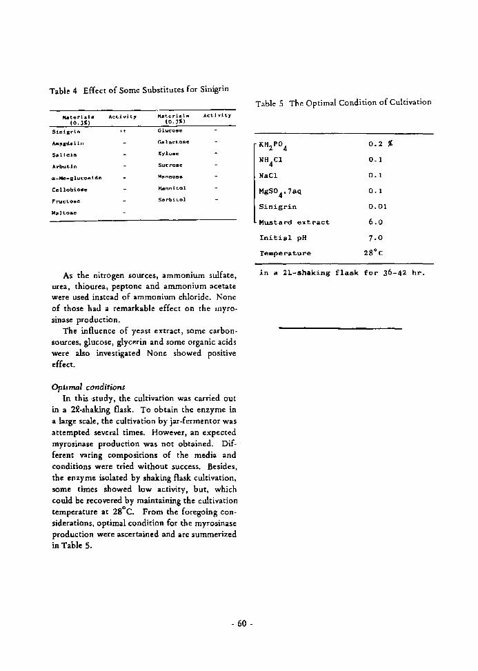
Table 4 Effect of Some Substitutes for Sinigrin
~tCl,.iall!li Acti vi ty ~ttlri.. la Act.ivity
(O.J~) (O.J~)
Sinierln .. Cluco8o
....,edd1n C.-lact.ose
Sdicin XY!Q.,e
Arbutin Sucrose
ar._Me_llucoside ~!lnn08e
C"llobiose Hannit.ol
Fructo...e Sorbitol
Malt-aBa
As the nitrogen sources, ammonium sulfate,urea, thiourea. peptone and ammonium acetatewere used instead of ammonium chloride. Noneof those had a remarkable effect on the myrosinase production.
The influence of yeast extract, SOme carbonsources, glucose, glycerin and some organic acidswere also investigated None showed positiveeffect.
Opt'7Ml conditionsIn this .study, the cultivation was carried out
in a 22-shaking flask. To obtain the enzyme ina large scale, the cultivation by jar-fermentor wasattempted several times. However, an expectedmyrosinase production was not obtained. Different varing compositions of the media andconditions were tried without success. Besides,the enzyme isolated by shaking flask cultivation,some times showed low activity. but, whichcould be recovered by maintaining the cultivationtemperature at 2S
o e. From the foregoing considerations, optimal condition for the myrosinaseproduction were ascertained and are summerizedin Table 5.
- 60 -
Table 5 The Optimal Condition of Cultivation
KH2P04
0.2 %
NH4Cl 0.1
NaCl 0.1
MgSO4' 7aq 0.1
Sinigrin 0.01
Mustard el<tract 6.0
Initial pH 7.0
Temperature 2S"C
in a 2L-shaking flask for 36-42 hr.

Chapter 2
Purification and General Characteristics
RESULTS
The procedure for the purification of bacterialmyrosinase is shown in Fig. 1. The crude preparation, precipitayed at 0.3 to 0.8 ammonium sulfatesaturation was dialyzed against 0.01 M potassiumphosphate buffer, pH 6.3 and subjected to eMSephadex CoSO column (22, 8 x 40) chromatography. The absorbed protein was eluated witha linear gradient of 0.1 to 0.3 M sodium chloridein the same buffer. The active fractions werepooled. After concentration by 0.8 satn. ofammonium sulfate, gel-filtration on SephadexG-200 column (5 x 80cm) were carried out. Theeluated active fractions were pooled and concentrated by 0.8 satn. of ammonium sulfate. Theprecipitate obtained by centrifugation was dissolved in a small volume of same buffer andsubjected to Sephadex G-100 column (2.6 x 90
B.rl .. r! ..
~su.pen.ion wi lh1 o~ 2 H p~o.ph.. t..e burr~r(pH 6.~)
Son"F.... t on
C~Lr. ru...Uen
SUPfl'f naL.an t
tPpt.. wi t.h ",-:zSO. (10.8 ..atn.
.. dJal,..l. a4aJn.t 0.01 Jot:
ph'O.phalr' burrl",.-( ptl 0.5)
ChI"" ato.raph)' On 0'I_5f!ph..d~1.
~ Er(luent (O.I-O.J M Ihellr "-250.. O. ~ .at-n.
GffJ r'JJt,.,..Lj.gn on Srph.dl!u: G-200
~ ""2S0j 1.0 .un.
G-rl riltr4ltlon on S-ep".d~.. C.-100
~"'250l '.0 '.<n.il'lel"H"o..t0l:r.ph, on Seph ..deoA G-JOO,
Fig. 1 Purification Procedure of BacterialMyrosinase.
- 61 -
'.0
II.::
1' ... 1'"" Il,..r,,-. I '~.I I~ PI"O,)'''''n................ ..-0 .
~---~ 11-(41 ,C.".d .......
Fig. 2 Rechroma togram of Bacterial Myrosinaseon Sephadex G-100.
em). Finally, the enzyme was purified by rechromatography on Sephadex G-lOO column (2.6 x90cm). As shown in Fig. 2 a chromatographycally homogenous pattern of the enzyme wasobtained. This filtrate was used throughout thefollowing experiments as the purified enzymepreparation. A summary of purification of theenzyme is shown in Table 1.
The enzyme was purified approximately to1000,fold compared to the crude preparation.
Homogeneity of the bacterial myrosinase. The analysis of the purified enzyme by ultra
centrifugation showed a single and symmetricalpattern (Fig. 3). The sedimentation coefficient,Sw20
, was caluculated to be 4.5 S from sedimentation velocity at 52,000 r.p.m..

10 ain 24 .in 36 ain 60 aiD
Fig. 3 The edimentation Pattern of the Purified Enzyme.
Estimatior1 of the molecular weightThe determination of the molecular weight
was performed by gel-flltration through SephadexG-IOO. Figure 4 shows the plots of the elutionvolumes versus the logarithms of the knownmolecular weight of standard proteins. Themolecular weight of the enzyme was estimatedto be 61,000.
General characteristics of the bacterialmyrosinase
Figures 5, 6, and 7 show pH-activity pHstability and temperature-stability curves respectively. Optimum pH was about 6.8. The enzymewas stable in the pH range from 5 to 7 and attemperature below 40°C.
Figures 8 and 9 show the Lineweaver-Burkplot of the enzyme. The Km values for sinigrinand p-NPG were calculated to be 3.7 x 1(f4 M
and 7.1 x 10⁻⁴ M respectively.
Table I ummary of Purification of Bacterial Myrosinase
Volum(ml)
A t.ivity(II- moles/min/ml)
Specific ac ivity(II- molesimin/mg)
Prot in(mg)
Crud prepara ion 2100 747.6 0.0116 41000
Chroma ography onCM-S phadex 250 410 0.59 700
OO-filtra ion onS phad G-200 0 2 0 2. 100
Gtl-fi Itra ion onS phad x G-IOO 40 162 5·9 27.6
R hroma ography onS phad G-IOO 35 175 I I . 5 15
- 62 -

·o~.--.L--~4:"'.•-:---.....----:l•.':-,-....JlUI:_ f Molend4lr- ,... ichl )
IUU
~II 0
;::
., .."->,-0"" .ut;;
';' 20
04 ..
pH
Fig. 4 Determination of Molecular Weight ofBacterial Myrosinase by Gel-Filtrationon Sephadex G-lOO.
Fig. 6 pH-Stability Curve of Bacterial Myrosinase
The sample solution was preincubated at 20°C for 24 hr.
bO5030
0'--_---l__-.J..__..J...__1:.._-J
80
Fig. 7 Heat-Stability Curve of BacterialMyrosinase.
100
.,..•'" 20
The s;p,mple solution was preincubated in 0.2 M phos.phate buffer, pH 6.8 for 30 min at each temperaturebefore measurement of enzyme activity.
100
~o
;;
:: 60
>
"'"""~
.0>.,..~ 20'"
a4 6 9
pH
The enzyme activity was me<Lsured by the liberation ofsulfate.
Fig. 5 pH-Activity Curve of Bacterial Myrosinase.
Effect of Various reagentsSulfhydryl reagents, such as peMB and iodo
acetate showed no inhibitry effect. The activity
was considerably inhihited by the chelating agent,EOTA, but not inhibited by o-phenanthlorine.6-Gluconolactone. which is a specific inhibitor
- 63 -
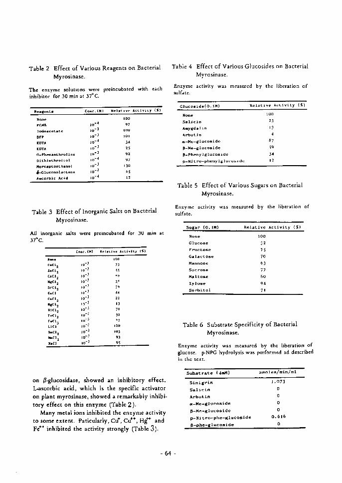
Table 2 Effect of Various Reagents on Bacterial
Myrosinase.
The enzymc selutions weTcinhibitor for 30 min at 370C.
prcincubated with each
Tabie 4 Effect efVarieus Glucosides on Bacterial
MyTosinase,
Enzyme activity was measurcd by the liberation ofsulfate.
Rel-tivf Actiyitr (S}Clucoside(e.Å}M) Relativc Activity {f)
Relsent- Conc.CM)
NanePC-(B
lodotceL-tcDFPEDTAEDTAO-Phnnanths'oli"eDitMothreitolMerc-ptoeth-nolaP.Cluconolattone
Adcotbic Acid
lot4io'3io'3
io-4io-3lnt3
to'4to-S
te-2
ie-4
too
97
tog
101
]a
:s92
97130bS
12
NeneSalicinAmygdolinAvbut]na-Me-glucosiden-He-glucosideP-Phenylglucosidcp-Nitro-phenylglvcoside
1OO
21
ls
q87
59
]4 12
Table 3 Effect ofInorganic Salts on Bacterial
Myrosinase.
Table
Enzymcsulfate.
5 Effect of Various Sugars on
Myrosinase.
actlvltY w:s rneasured by the
Bacterial
liberation of
Sugsr (O.IM) Relative Activity (X)All inorganic salts werc preincubated for SO min at370C
Conc.("} Relntivc ActSvitr (S)
"ooeCOCi2lnei2c-c12HdC12srt12cucs
c.c12ILtC12
MCI:reCl2FtCIJLiC)snCl 2)inCl zhCl
to' .1
le'.t
so- -'
lov,
10-1
io-1
io'3to-.1
lo'S
lo-:'
Le- :'
to-3
io'3to' .i
te'3
1OO
73
S5
-n
zp
79
"4
22
11
70
12-:
120
to3
93 9S
None6JuceseFructoseGalactoEeHanneseSucroseMaltoseXyleseSorbito1
1OO
32
7S
7D b3
77 so
94 71
Table 6 Substrate Specificity of Bacterial
Myrosinase.
Enzyme activity was rneasured by the liberation ofglucese. p-NPG hydrolysis was pcrferrned ad describedin thc text.
Substrate (4mM) "molesl minlml
on 6glucosidase, showed an inhibitory effect.L-ascorbic acid, which is the specific activator
on plant myrosinase, shewed a remarkably inhibi-
tory effect on this enzyme (Table2).
Many metal ions inhibited the enzyme activityto some extent, Paticularly, Cu', Cu"', Hg"` and
Fe"" inhibited the activity strongly CTable 3).
StnignnSalicinArbutincz-Me-glvcosideP-He-giucesidep-Nitro-phe-glucesideP-phe-gluceside
1,e73 o o o
oO.61b o
-64-

Fig. B Reciprocal Plot of Sinigrin Hydrolysis.
Enzyme activity was measured by the liberation ofsulfate.
. n
'-
I
1{(5J • '0-31>11- 1
Fig. 9 Reciprocal Plot of p-NPG Hydrolysis.
Effect of various glucosides and sugarsThe bacterial myrosinase has' a I3-glucosidase
activity (Fig. 2) as well as myrosinase activity,
"..'
!.,• 0
I
I/t5JI. 10-J(N)-1
Fig. 10 Inhibitory Effect of Salicin on SinigrinHydrolysis.
Enzyme activity was measured by the liberation ofsulfate.
Fig. 11 Inhibitory Effect of Sugar and Glucosideson p-NPG Hydrolysis.
like plant and fungous myrosinases. In order toclarify the relationship between the myrosinaseactivity and J3-glucosidase activity of the bacterial
- 65 -

myrosinase. effect of glucosides and sugars wastested. All glucosides tested showed an inhibi·tory effect (Table 4). Paticularly. salcin, amygdalin, arbutin and p-NPG showed stronginhibitory effect. The effect of various sugarsare shown in Table 5. All sugars tested showedinhibitory effect.
The inhibitory effect of salicin on sinigrinhydrolysis is demonstrated as Lineweaver-Burkplot in Fig. 10, plotted in the absence andpresence of salicin. The inhibition by salicin iscompetitive. The Ki value for salicin was calculated to be 6.6 x 10⁻⁴ M.
Effect of sugar ami glucosides on p-NPGhydrolysis
The effect of sugar and glucosides was studied.As shown in Fig. 1 1, glucose, sinigrin and salicinshowed competitive inhibition. The Ki valuesfor glucose, sinigrin and salicin were calculated tobe 1.2 x 10⁻⁴ M , 3 x 1(f4 M and 5.5 x 1(f4 M , res-
pectively.
DISCUSSION
Substrate specificityThe hydrolytic activity on various glucosides
was tested (Table 6 ). The bacterial myrosinasehydrolyzed p-NPG besides sinigrin.
General characteristics of the bacterial myrosinase produced by Enterobacter cloacae no. 506were investigated. Some properties of bacterial.fungous and plant myrosinases were compared(Table 7). On the effect of pH and heat, it isshown that the bacterial enzyme is rather stablelike the plant enzyme and the fungous enzymesfrom AspergiUus sydowi. However, PCMB which
Table 7 Comparison of Some Properties of Myrosinases from Various Origins
Plan~ Asp. niler Asp. sydowi Enterobacter cloaCae
pH-Activity
pH-Stabi lity
Heat Stability
PCMB
5 - 1
4 - 9
ss" C
inhibit inhibit
7
6 - 9
45u
C
no effect
6.8
5 - 7
40 ·c
no effect
Ascorbic Acid act.ivate no effect no effect inhibit
EDTA no effect no effect no effect inhibit
Molecular Weicht 120,000 -150,000
~ for Sinierin 1-8 x 10- 4•
~ for p-HPG 2.0 x 10-J.
Iti for Sinierin 2.0 x 10- 4•(p-IIPG hydrolysis )
I(i for Salicin 1.8 x 10- 1•(Siniarin hydrolysis)
J.7 x 10- 4•
1.1 x 10- 4•
).0 x 10-4•
nOne
61,000
C ...+ C ++ Hn++ -.0, u. Co
Sn.-T Kg...... f ++ H....e,
90,000 120,000
J. J x 10- 3• J.6 " 10- 3•
1.5 "10-J. \.0 " 10- 4..
3·8 "10-J • ).8 x 10-).
1.6 x 10- 1•
nOne
Inhibition
KetalActivation
- 66 -

inhibited the fungous and plant myrosinaseshowed no inhibitory effect on the bacterialmyrosinase. EDTA which showed no inhibitoryon the plant and fungous myrosinase inhibitedthe bacterial one and metal ion, Hg+~ inhibitedall the enzymes. L-Ascorbic acid which is thespecific activator on the plant enzyme inhibitedthe bacterial enzyme. Plant myrosinase possessesa rather large moleculer weight of 150,000. while,bacterial enzyme had the smaUest weight in thefoUI myrosinases.
As in the fungous and plant myrosinase, Kivalue for sinigrin in p-NPG hydrolysis is in goodagreement with the KIn value for sinigrin in sinigrin hydrolysis. This indicates that, as in fungousand plant myrosinase, the hydrolysis of p-NPGtake place on the same site on which the hydrolysis of sinigrin. While, on the Km values forsinigrin and p-NPG, the bacterial myrosinase hasa different tendency from the fungous, myrosinase, because the ratio of Km values for sinigrinto p-NPG is in the order plant<bacterial<Asp.niger<.Asp. sydowi. These facts suggest thatthe bacterial myrosinase may considered to be
a /3-glucosidase which has high affInity for mustard oil glucoside. Pigman ,9 ) Nagashima andUchiyama ,10.11) and TSUIUO and Hata12) havereported that plant myrosinase is a member ofthe /3-glucosidase. The above results supportthis conclusion. plant myrosinase is highly specified for mustard oil glucosides, whereas fungousmyrosinases are less specific than the plant enzyme, but the bacterial myrosinase is locatedbetween plant and fungous enzyme and isthought to be rather similar to the plant enzymethan the fungous enzymes. Even so, microbialmyrosinases have high affmities for mustard oilglucosides than for the usual (J-glucosidases.This conclusion is supported by the fact thatmicrobial myrosinases were produced only in thepresense of inducer such as sinigrin and mustardextract.
The differences in behaviours and characteristics of the four enzymes becomes an interestingfactor. Particularly, it is noteworthy that ascorbic acid has a very prominent effect on eachenzyme,
REFERENCES
1) M. OhtsuTu, I. Tsuruo and T. Hata, Agr. BioLChern., 33,1309 (1969).
2) M. Ohtsuru, 1. Tsuruo and T. Hata, ibid., 37,967 (1973).
3) E.L. Oginsky, A.E. Stein and M.A. Green,Proc. Soc. Expt. Med., 119,360 (1965).
4) J.B. Sumner,]. BioL Chern., 65, 393 (1925).5) I. Tsuruo, M. Yoshida and T. Hata, Agr. BioI.
Chern., 31,18 (1967).6) a.H. Lowry, N.J. Rosebrough, A.L. Forr and
R.T. Randall,J. Biol. Chern., 193,265 (1951).7) P. Andrews, Biochem. J., 96,595 (1965).
8) H.K. Schachman, In "Methods in Enzymology" edited by S. P. Colowick and H. O.Caplan, Academic Press, New York, 4, 32(1957).
9) W.W. Pigman, J. Res. Nat!. Bur. Stand., 30,257 (1943); Advances il'l Enzymology, 4,41(1944).
10) Z. Nagashima and M. Uchiyama, NipponNogei-Kagaku Kaishi, 33, 1144 (1959).
11) Z. Nagashirna and M. Uchiyama, Bull. Agr.Chern. Soc. Japan, 23, 555 (1959).
12) 1. TsuTUO and T. Hata, Agr. Bioi. Chern., 32,1425 (1968).
·67 -

SUMMARY AND CONCLUSION
PART I
In Chapter 1":Four proleins (F-IA, B, F-llA & F-llB), having myrosinase activity, were separated and
purified from mustard powder. Each enzyme was shown to be homogeneous chromatographically, u)tracentrifugally and Disc e1cctrophoretically. Molecular weights obtained bygel-filtration and sedimentation equilibrium were 153,000 (F-lA, F-I B & F-IIA) and 125,000(F-llB). Sedimentation coefficients were 6.8 S (F-IA, B & F-llA) and 5.8 S (F-IIB). Stokesradius (A), diffusion coefficient (cmz/sec) and frictional ratio (fifo) were 47, 4.28 x ]0-- and1.33 (F-IA, B & F-lll), and 43, 4.67 x 10⁻⁷ and 1.29 (F-IIB), respectively. Isoelcctric pointswere pH 4.6 (F-IA, B & F-llA) and pH 4.8 (F-llA). The enzymes were glycoprotein with9- 22 %carbohydrate. Amino acid composition of F-IA, B and F-IIA were very similar, butin case of F-IIB, glutamic acid, arginine and methionine contents were higher and asparticacid and histidine contents were lower than others. The molecular weights estimated fromSDS-polyacrylamide gel electrophoresi.s were 40,000 (F-IA, B & F~llA) and 30,000 (F-llB),respectively, and hence the enzymes are considered to have at least 4 subunits. From these reSUlts, it may be confirmed that F-IA, B & F-IIA have striking resemblances and only F-IIBis rather different.
In Chapter 2:The functional groups of the plant myrosinase was investigated using reagents which
discriminate the states of amino acids in protein. The enzymatic activity was inhibited byf1uorodinitrobenzene (FDNB), trinitrobenzenesulfonic acid ([NBS) and monochlorotriftuorop-benzoquinone (CFQ), which are specific reagents for amino group, p-mercuribenzoate(PCMB) and 5-5'-dithio-bis-(2-nitrobenzoic acid), specific for -SH group, and p-diazobenzenesulfonic acid, specific for imidazole group. The rates of inhibition by FDNB and TNBSwere accelerated by the addition of ascorbic acid (10-' 101). The enzyme was inactivated whenfive amino groups of the enzyme reacted with CFQ. The enzyme was also inactivated when two·SH groups reacted with Ellman reagent, although four -SH groups were found by Ellmanreagent in the denatured enzyme by SDS.
Plant myrosinase was found to be quite sensitive to photooxidation catalyzed by methylene blue. Amino acid analysis indicated that loss of the activity was due to degradation ofhistidyl residue of the enzyme. These results suggest that amino group, -SH group andhistidyl residue constitute the active sites of the enzyme.
In Chapters 3 and 4:The activation of myrosinase was observed above 5 x 10"s M ASA concentration, and the
activation mu:imum was around 10'" M ASA.The presence of dwociation and associ;ltion of the enzyme by ASA was investigated by
gcl-ftltration and ultra-centrifugal analysis. The enzyme was neither dissoci;lted nor associatedby ASA.
The procedure for quantitation of ligand binding to proteins by di;llysis rate measurementshas been applied to binding of L·ascorbic acid by myrosinase. Myrosinase has 4 sites permolecule which bind ASA rather strongly (Kd; 0.1 x 10'" M), and at least one additionalsite which binds ASA less suongly [Kd = 0.9 x 10-' M). Myrosinase activity is most stronglyactivated when four ASA molecules are bound to the enzyme, but it is inhibited when fiveASA molecules are bound.
- 68 -

Ascorbic acid analogues could not activate the enzyme.It was found that the enzyme protein is comformationally changed by the addition of ASA
in the photometrical analysis. Approximately, 1.5 amino residues appeared on the surface ofthe enzyme and about 2.3 tryptophanyl residues are buried in the molecule when ASA isadded (1 mM).
Myrosinase and P-glucosidase activities were strongly inhibited by p-mercurybenzoate(PCMB) (0.1 mM) with Or without ASA. Diethylpyrocarbonate (DEP) inhibited the myrosinase activity than /l-glucosidase. 2-Methoxy-5-nitrotropone (MNT) inhibited ASA treatedmyrosinase when the activity was measured without ASA, but the activity was not affectedwhen the activity was determined with ASA. This phenomenon was not prominent in /l-glucosKlase activit y.
Optimum-temperature for the myrosinase activity was at about 55~C without ASA, butwith ASA was at about 35° C. /l-Glucosidase activity was the same (55°C) with or without ASA.
A schematic model interpreting at the interaction of ASA with the enzyme and the activecenter of the enzyme is proposed.
I concluded that the activation of the plant myrosinase is occmed by the alteration of theenzyme's protein structure caused by the binding of ASA exerted no effect on the area whichadsorbed glycan moiety in the substrate site, and by neither the oxidation-reduction reactionof ASA nor the association-dissociation of the enzyme. I considered that sulfhydryl groupsare essential to the catalytic action of the plant myrosinase and amino and histidyl residues aresituated on the region which is altered by the addition of ASA, that is, it is close to aglyconmoiety and the locations of them are changed to accelarated the enzyme action by the binding of ASA to the effector site.
PART II
In Chapter 1:In order 10 obtain fungous myrosinase, ArPtrgillus sydowi IFa 4284 was cultured on a
medium containing mustard seed extract for 2 weeks. ~Iyrosinase in the broth was purifiedabout ISO fold by precipitation with ammonium sulfate and chromatography on DE.\Ecellulose and DEAE-Scphadex. Comparison of thioglucosida31: and sulfatase activities ofthe myrosinase preparation using pH-activity, pH-stability and temperature-stability (un',",revealed no differences from each other. The chromatograms of the two acti"ities onDEAE·Sephadex showed good agreement. -Consequently, Ihe myrosinase produced b,':Is/Mrgilfus sydowi was concluded to be a single f/-thioglucosidase, not a mixture of thicglucosidase and sulfatase.
In Chapter 2:
Tne effects of various reagents on Asptrgillus sydowi myrosinase were studied.The enzymatic activity was stimulated by cobalt (II), zinc (II) and magnesium ions
and inhibited by mercury (II), iron (II) and copper (II) ions. HowC\'er, mr'tal-complexingagents, SH reagents and diisopropylfluorophosphale showed no effects on enzymatic activity.In contrast to plant myrosinase, this enzyme was neither activated nor inhibited by anyconcentrations of L-ascorbate. Glucose and salicin were competitive inhibitors for theenzyme. High concentrations of sodium chloride inhibited the enzyme.
From the inhibitioQ modes of sugars and ft-g1ucosides and from Ihat of sodium chlorideagainst the enzyme, a similarity of the enzyme to .,-glucosidases was shown.
- 69-

In
ln
Chapter 3: ,,,.P"G':co,tiga,e,c..s.f`'v`\y,,2':".piEo,,e.sm.y.;esl:.az;,:ls'f.o"g6rm,ed.,"i`."g,ft1,"it.'o,p,:c,s.y.,t.B'fflyg,?T.
ExpcT{menral results indicazed a rcsemblance of fungeus myrosinase te P-glucos.idases similar
to plant myrosinasc. The retationship bctwcen fungous and plant myrosinases to thc these enzymes. P+glucesidasts are discussed frem the view of rhe substrare specificity of, The cencLusions are that distinction bctwcen plant and fungous myrosinases and the P-glucosidases arc noi as strict as previousty thought, and the myrosinases sheu!d bc con- sidered P-glucosidascs highly spccialized for the hydrolysis of mustard eii glucoside.
Chapters 4 and 5: after Screening 1oo micro-erganisms to detect inTracellular myrosinase, only Aspergitl"s
ny niger produced myrosmase. Enzyme production was induced by lhe addition of ten percent of a rnustard extiact to tbe culture medium. The enzyme was produced in considerable amounts on the first and second day ofcuStivation. L-Ascorbic acid was an excellent carbon source. The enzyme was unstable but was stabilized by coexistence with 2-mereaptoethanol (10'2 M) and ascorbic acid (10-S Fs). The enzymatic propcrties of intracellular myrosinasc produced by Aspetgittus niger AKU 3302 vvcre invesTigated. Maximum aetivity eceurred at pH 6.2. and the enzyinc was stable in
a pH range or7.6 to 8.0 al 5'C for 24 hr. Optimum temperature was abeut 34'C. Enzyrne activity was stimulated by copper (D, (Ir), mmganese qD and cebalt (II) and was inhibited
by rnercury (II) and stannous (IE) ions. However, metal complexing agcnts and DFP had Iittle effect, while PCMB was a strong inhibitor. In contrast to plant myrosinase, this enzyme
was neithcr activated nor inhibiLed by L-ascerbic acid. Glucesides and j-glucenolactone inhibited enzyme activity but sugars were ineffective, The Km value for sinigrin was 3.3 År.'
10'i M and that forp-nitTopheny] fi-glucoside was 1.5x 10'; M. The relation between rungous
myrosinases and fi-glucosidase is discussed in comparison to p;ant rnyrosinase,
PART III
In Chapters 1 and 2: Screening test for obtaining microorganisms which producc myrosinaseactwitywas carTied eut. One strain of microorganisrn showed strong abMty to produee myrosinase activity. The merphological and physiological characteristics of this strain we:e studied.
The organisrn was ldentified as an Enterobaeter cloacae no S06. Enzymc ptoduction was induced by the addition of O.Ol% sinigrin and 6% mustard extract to the culture medium. When the strain was cultivated at 28eC in a medivm containing O.Ol% sinipin, 6% musrzrd ext., e.1% KH;PO` , O.1% NH.CL O.1% NaCl and O.1% MgS04' 7iq, (pH 7.0), the highest activity wis obtained afrer 36 '- 40 hr cultivation. Myresinase in the ctll-free extract of Enterobacter cloaeae nQ. S06 was purified about 1,OOO fold by ptecipitation with arnmonium sulf:tc, chromatography en CM-Sephzdex and gel-
ftltration on SephadcxG-200 and Sephadex G-1co.Thc enzyme iMas shown to be homegeneous chromztegraphicafly and ultracentrifugally. Moltcular weight obtainedi by gel-fdtTatien was 61,OOO znd sedimentation coefficienr was 4.5 S. Maximum activity eccurred at pH 6.8, and the enzyme was stable, in a pH rznge ef S.O to 7,O bciow 400C for 24 hrs Copper Cl), (lr), mercurv CII) and ferrous Cll) iens mhibited the ;ctivity strongly. Sulthydryl reagents had litttc cffcct but EDTA was a strong inhibiton ln contrast to plant myrosinase, this enzyrne was inhibited by L-ascerbic acti. Many glucesldes and sugars inhibited the enzyme. The
-70.

relation between bacterial myrosinase and ,8-glucosidase is discussed in comparison to plantand fungous myrosina.ses. The comparison of some properties of bacterial, fungous and plantmyrosinues are discussed.
The above experimental results indicate that the striking family relation was observedbetween the myrosinases and the kJucosidases. And I concluded that the myrosinases ate akind of IJ-glucosida.ses highly speeialized for hydrolysis of mustard oll glucosides.
-11 ..

ACKNOWLEDGEMENT
The author would like to express his sincere
gratitute to Dr. Tadao Hata, Professor of Kyoto
University, for his kind guidance and encouragement
during the course of this work. The author is also
greatly indebted to Dr. Etsushiro Doi, Associate
Professor of Kyoto University, for his kind guidance
and suggestion, and to Dr. Isao Tsuruo and Mr. Naoki
Tani for their cooperation in carrying out a part of
this work. It is a great pleasure to acknowledge
the valuable advice of Dr. Rikimaru Hayashi and Mr.
Takahiko Higasa.
The author wishes to thank to Drs. Hidehiko
Tanaka and Nobuo Kato, Kyoto University, and Drs.
Isao Banno and Takezi Hasegawa, the Institute for
Fermentation, Osaka, for Identification of the
Bacteria, their helpful advice and valuable
suggestions.
The author is also indebted to Dr. Hideaki
Yamada, Professor of Kyoto University, for his many
helpful advices in this work.
The author wishes to express his sincere thanks
to stuff members of the Research Institute for Food
Science, Kyoto University.
-72-